Review

Advances in Antimicrobial Coatings for Preventing Infections of Head-Related Implantable Medical Devices

by
Irina Negut
1,
Catalina Albu
1 and
Bogdan Bita
1,2,*
1
National Institute for Lasers, Plasma and Radiation Physics, 077125 Magurele, Romania
2
Faculty of Physics, University of Bucharest, 077125 Magurele, Romania
*
Author to whom correspondence should be addressed.
Coatings 2024, 14(3), 256; https://doi.org/10.3390/coatings14030256
Submission received: 30 January 2024 / Revised: 14 February 2024 / Accepted: 18 February 2024 / Published: 21 February 2024
(This article belongs to the Special Issue Multilayer Coatings for Nanomaterials: From Synthesis to Applications)

Abstract

:
During surgery and after, pathogens can contaminate indwelling and implanted medical devices, resulting in serious infections. Microbial colonization, infection, and later biofilm formation are major complications associated with the use of implants and represent major risk factors in implant failure. Despite the fact that aseptic surgery and the use of antimicrobial medications can lower the risk of infection, systemic antibiotic use can result in a loss of efficacy, increased tissue toxicity, and the development of drug-resistant diseases. This work explores the advancements in antimicrobial coatings for head-related implantable medical devices, addressing the critical issue of infection prevention. It emphasizes the significance of these coatings in reducing biofilm formation and microbial colonization and highlights various techniques and materials used in creating effective antimicrobial surfaces. Moreover, this article presents a comprehensive overview of the current strategies and future directions in antimicrobial coating research, aiming to improve patient outcomes by preventing head-related implant-associated infections.

1. Introduction

The use of medical devices is in a continuous increase as they have become crucial elements of modern healthcare. From artificial hips to stents, heart valves and vascular grafts, implanted medical devices are currently used worldwide to improve the life quality for a high number of patients. There are also non-implanted, shorter-term-usage medical devices, such as catheters, orthopedic fixation screws, and contact lenses, which are added for the improvement of patient life quality. In this respect, the need for medical devices have given rise to an ample industry and to a broad body of research in medical devices and materials science, which include device design and clinical studies.
All implanted medical devices (IMDs), from transient, early introduced, and retrieved (e.g., urinary catheters, endotracheal tubes) to the more permanently surgically implanted ones (such as cardiac valves, embolic coils, vascular grafts, hip, knee and shoulder joints, pacemakers, coronary stents), are susceptible to device-related or implant-associated infections [1]. The infection of IMDs encompasses intricate relationships between the pathogens, the biomaterial, and the host immune response. The infection factor increases the risk of prolonged hospitalization, multiple surgical procedures at the time of implant, remote infections in other body parts, surgery interval, and tissue devitalization. It was estimated that ~2%–5% of implants are associated with infection risks in orthopedic surgery, and the infection rate for dental implants is even higher, with a value exceeding 14% [2]. Implantable and prosthetic medical devices can commonly become infected with infection-producing microorganisms, despite the best attempts to keep them sterile. Biofilms are difficult to diagnose and treat due to a lack of adequate biomarkers and can become very difficult to eliminate in the clinic given the high resistance to antibiotics.
The treatment of implant-associated infections comprises the delivery of high antibiotic doses for a prolonged time and/or replacement of the device translated into costly and risky surgeries, both of which are ineffective because of antibiotic-resistant strains and high chances of re-infection on the reimplanted device.
Biomedical researchers are nowadays focused on the development of anti-infective coatings that can reduce or eradicate infections associated with implantable biomaterials and devices as well as combat antimicrobial tolerance or resistance. Using the coating approach for controlling pathogenic strain adherence, contamination, and colonization stages leads to (i) innovative means for studying microbial virulence and biofilm formation mechanisms as well as (ii) novel tactics for designing coated surfaces that prevent and/or disrupt microbial biofilm formation [3].
In this review perspective, we shall first present the mechanisms of IMD surface bacterial colonization. We will then summarize the different surface coating techniques together with some of their advantages and disadvantages. Further, we will provide a short description of the drug-release approaches from coatings. We continue with an overview of the progress made in the coatings field over the past last years for the most common medical devices of the head. A diagram illustrating the outline of this review is depicted in Figure 1.

2. Infection on Implanted Medical Devices

In general, without a foreign body, tissue contamination by opportunistic pathogens is typically spontaneously cleared by the host’s immune defense mechanism. In contrast, in IMD infections, the biomaterial activates a local tissue response. This consists of acute and chronic inflammation, a foreign body reaction, the construction of granulation tissue and, finally, fibrous encapsulation. Bacteria can be introduced into the human body during a medical procedure by the contact with the healthcare worker, bacteria residing on the patient’s skin, etc. This contact can cause nosocomial infections in the patient, but it can also damage the IMD by triggering implant degradation and a diminution of its mechanical stability, which can further result in implant failure and revision surgery [4].
Bacterial biofilms represent an aggregate of infectious microorganisms that attach to a surface and embed themselves in a self-produced extracellular polymeric substance (EPS) or “slime” [5]. The biofilm growth, development, and dynamics can be divided into the following steps: (1) reversable attachment, (2) irreversible attachment, (3) maturation phase, and (4) dispersion [6].
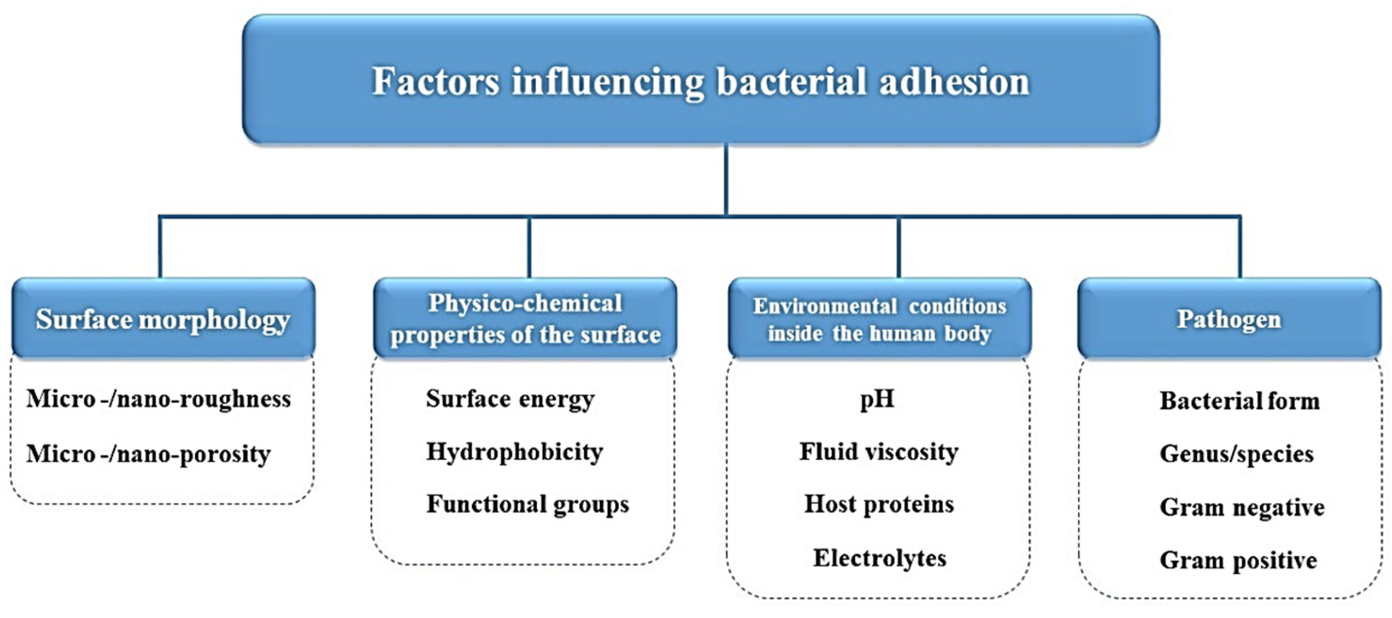
For a biofilm to form, a bacterium must find a proper surface to attach. This kind of surface is usually made from a material that has a rough/textured surface [7]. Moreover, it is known that other aspects can influence the microbial attachment, such as polarity [8], hydrophobicity [9], hydrodynamics [10], pH [11], and temperature [12]. The factors that influence bacterial adhesion on an IMD are shown in Figure 2.
Initially, free-floating bacteria adhere to a surface by means of pili, fimbriae, or flagella. In the case of an IMD, this phase coincides with the first phase of the foreign body response. When the IMD is inserted into the human body’s physiological media, the IMD’s interaction with the human fluids regulates the adsorption of various biomolecules (e.g., albumin, lipids, fibronectin) on its surface [13]. The formed thin film of biomolecules allows for weaker attractive forces (such as van der Waals, electrostatic forces, hydrophobic interactions) between the material surface and the free-floating bacteria. In this state, the infectious microorganisms are still reversible in their attachment [13]. However, with the passing of time, cells will develop stronger attractive forces with peptides from the surface of the material, resulting in an irreversible attachment ([14], p. 9).
The third stage is characterized by bacteria that form a microcolony. The self-secreted EPS, composed from proteins, DNA, and polysaccharides, shapes bacterial cell aggregation, facilitates water retention, offers nutrients, and forms a protective “fence” [15]. After the EPS construction, the microbial colonies grow in size until they form a 3D colony of ~100 μm in thickness [16].
During the last phage of biofilm formation, the dispersion, clusters of cells, and EPS disconnect and migrate to the surrounding areas and continue to populate other surfaces. During the detachment phase, microbial communities embedded into the biofilm produce saccharolytic enzymes that assist the release of the microbes. Moreover, the microbial cells upregulate the proteins expression related to flagella development to let the bacteria move to a colonization site [17]. This phase usually leads to systemic infections and even acute events [16].
The capability of bacteria to form a biofilm depends on their setting and taxonomy [15]. Both Gram-positive and Gram-negative bacteria can form biofilms on medical devices, but the most common entities are represented by Staphylococcus aureus, Pseudomonas aeruginosa, Klebsiella pneumoniae, Enterococcus faecalis, Staphylococcus epidermidis, and Escherichia coli. It was estimated that S. aureus and S. epidermidis are the major cause for prosthetic heart valve and catheter biofilm infections [18]. In addition, the majority of IMD- associated infections are caused by the staphylococcal species, being related to S. aureus and coagulase-negative staphylococci [18].
While short-term devices can be treated by cleaning or the removal of established biofilms, there is a greater worry in terms of prevention in the case of implanted devices. Usually, biofilms that form on surfaces on IMDs are resistant to the host’s defense mechanisms and are treated by antibacterial therapies and tissue debridement [18]. However, the high doses of antibiotics used for long time periods for treating biofilms have contributed to the expansion of antibiotic-resistant bacteria strains [18].
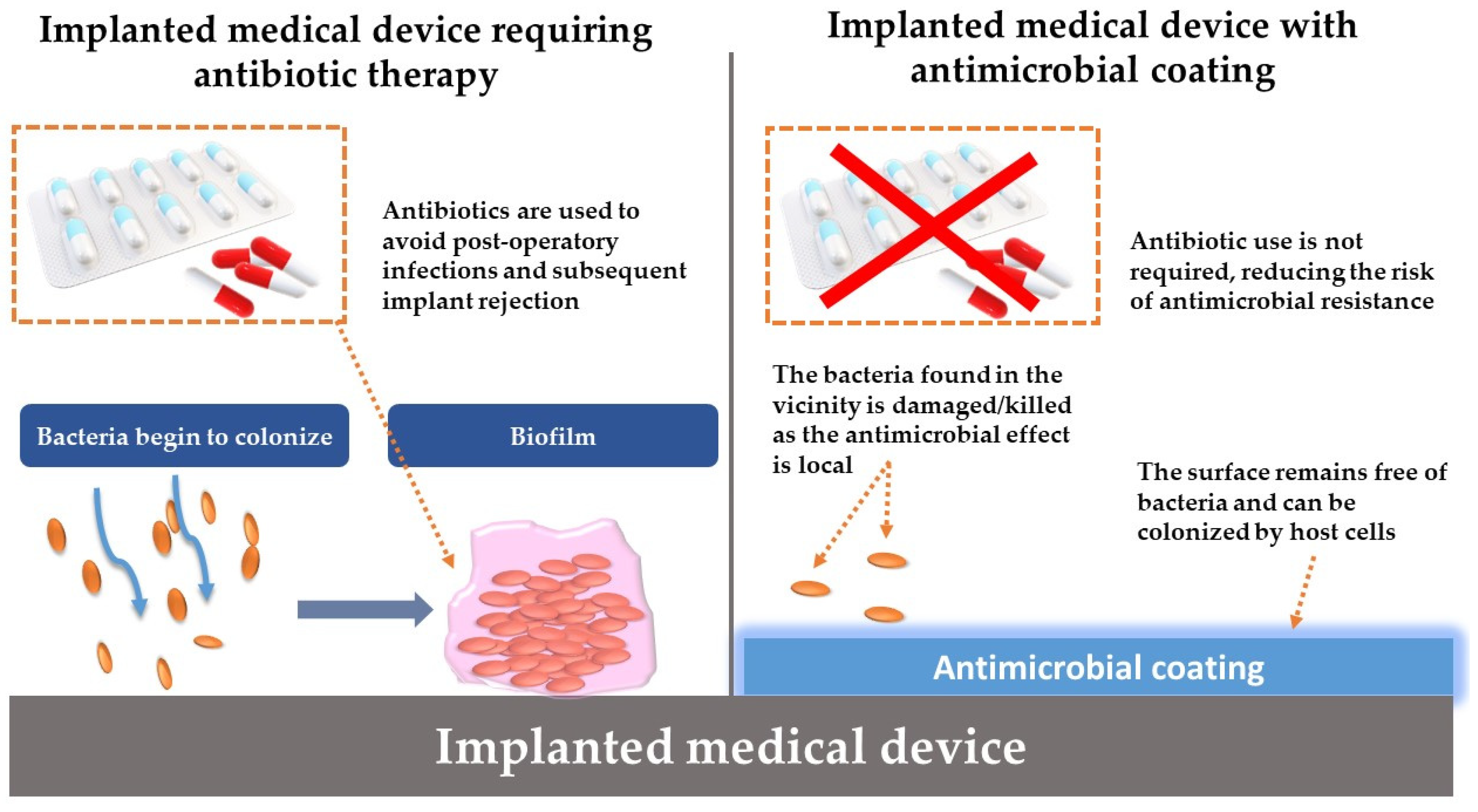
In the case of IMDs, antimicrobial coatings present several benefits over traditional antibiotics (Figure 3). A key advantage is their localized action, where the coating shields the immediate surface of the implant from microbial attack without affecting distant tissues [19]. This localized protection can enhance patient comfort by eliminating the systemic side effects and complications often associated with antibiotic use [19]. Moreover, employing antimicrobial coatings could lead to a decrease in the reliance on antibiotics, preserving them for critical therapeutic uses and potentially slowing down the development of antibiotic-resistant bacteria.

3. Techniques for the Modification of Medical Device Surfaces

Given the persistent challenge of implant-related infections, with limited advancements in reducing infection rates over several decades despite numerous tested therapies, the medical community is continuously seeking effective strategies to prevent and manage these complications. Modifying the surface of IMDs to avoid microbial attachment presents a promising route for intervention. By integrating surface alterations with antimicrobial agents, there is the potential to create coatings which not only exhibit robust biological activity but also possess enhanced antimicrobial properties [20]. Such modifications can significantly diminish microbial adherence and influence biofilm development dynamics, providing a strategic approach to managing the microbial environment around IMDs. This evolving field of research, focusing on antimicrobial surface engineering, represents a crucial frontier in devising strategies to curb biofilm-associated implant infections, highlighting the importance of innovative surface design in the fight against persistent microbial threats [21].
The control of the physical and (bio)chemical properties of an IMD surface is one of the most central issues in its design, as the interaction between a foreign body and the biological environment of the human body takes place at the interface. Once established on an implant’s surfaces, biofilms are challenging to eradicate [21,22]. Consequently, inhibiting or slowing their growth process represents an extensively used therapeutic strategy. This strategy may include altering the implant surface to avoid the initial attachment of infectious microorganisms and/or coating implants surface with antimicrobials/a matrix of gradually releasing antimicrobials to kill microorganisms on contact [22].
Multiple procedures have been used to modify material surfaces, which can be mechanical, physical, chemical, and biological, which include embedding or diffusing a novel compound onto the surface of the implant, thereby adding an extra layer that fights microbes [23]. Various antibacterial agents, such as antibiotics, peptides, enzymes, organic and inorganic compounds, and metals, have been integrated into IMD surfaces [24]. The effectiveness and safety of these agents depend on their concentration, as both their antimicrobial activity and potential toxicity to host cells are influenced by the dosage [25]. Therefore, it is crucial to finely tune the levels of these antibacterial agents to eliminate bacteria on the IMD while avoiding damage to the surrounding living tissues. In achieving this balance, coating technologies are crucial, offering a significant advancement in developing antimicrobial surfaces for implants. These methods are briefly discussed in the following section:
Mechanical functionalization includes machining, polishing, irradiation, etching, etc., which provide distinct roughness features to the surface at the meso-, micro-, and nano-scale; the roughness and the structuring can improve cell proliferation and adhesion, which result in increased medical device stability [26]. A specific example can be given with the proliferation rate of osteoblasts on titanium samples with different roughness degrees in the meso-, micro-, and nano-domains obtained by acid-etching [27].
Physical functionalization includes processes like oxidation and the passivation of metals and alloys, but it also includes any surface coating leading to a shield or activation. Multiple physical functionalization methods have been described:
(a)
The plasma spraying technique is commercially viable and extensively used as a surface coating method for the orthopedic implants [28,29]. The plasma spraying technique permits the facile control of the coating thickness, and the sample size to be coated is not limited. However, by being a linear process, it limits the uniformity of the coating on a complex shape, and the high functioning temperature can change the structure and performance of the metal substrate [28,29]. Moreover, the bonding strength is limited by the stress concentration at the coating–substrate interface.
(b)
Chemical vapor deposition (CVD): The CVD process can deposit thick coatings but necessitates a relatively high temperature. The outcomes of CVD are conformal coatings. This technique does not require solvent use, which results in obtaining a complex coating on solvent-sensitive substrates [30].
(c)
Pulsed laser deposition (PLD) and matrix-assisted pulsed laser evaporation (MAPLE): The main advantages of using laser-assisted techniques in the fabrication of thin-film coatings for implants and other medical devices is their ability to control composition and topography, even at the nanoscale [14].
(d)
Ion implantation involves the acceleration of ions in an electrical field and impacting them within a solid. Electrons are stripped from the target atoms in order to form ions, which are directed using a region with opposite charge. The energy of the ions must be selected so that they are injected into the near-surface region of the solid [14].
(e)
The sol–gel process is as a well-known and reliable coating process. The advantages of this method include composition selection, the fact that it is easy to coat complex geometries, the homogeneity of the coating layer, and the easiness of the process. Also, the range of different compositions that can be produced by the sol–gel method comprises single oxides, mixed oxides, and non-oxides, such as nitrides, borides, and chlorides. The high purity of the compounds and coatings can be maintained, as grinding and high temperatures can be avoided [31].
(f)
Deep coating involves three successive stages: dipping, withdrawing, and drying. The substrate is dipped into the solution of interest and then withdrawn at a constant speed, resulting in a good control of the coating thickness and producing no waste. The coatings produced via this technique have a low adhesion strength to the substrate and tend to crack [32].
(g)
Electrophoretic deposition is attractive due to the fact that it can be used to fabricate uniform coatings with controlled assets on complex-shaped and porous substrates at ambient temperature and without the requirement for expensive equipment [33].
Many details on above-enumerated methods along with their benefits and limitations have been described elsewhere [34,35,36].
In addition, two other main strategies are considered for surface modification: chemical and biological functionalization. The first strategy refers to the obtaining of functional biosurfaces by the chemical grafting of functional groups (by acetylation, fluorination, silanization, incorporation of sulfonate groups) or by adjustments made on the existing functional ones (by oxidation, reduction). In the second system, the modification is accomplished by the adsorption or chemical bonding of biomolecules to the surface of the medical device with the purpose of stimulating a specific cell response. These modifications are widely discussed elsewhere [37].

4. Drug Release from Coatings

Drug release from coatings on IMDs has revolutionized treatments for a range of health conditions over the last decades of innovation. Due to the presence of various implants that fall into hybrid categories, a proper classification of IMDs with the attribute of drug release is not obvious [38]. However, drug release from coatings can be regarded as “active” or “passive”.
To achieve a bacteriostatic effect, the “passive” approach primarily uses anti-adhesive coatings and bacteria-resistant nanotopographies. In this case (e.g., biodegradable and non-biodegradable coatings) the drug-release features are uncontrollable once installed clinically. However, implants are normally metallic in nature, and the elution of the drug agent must be activated by targeting external stimuli to achieve the proper drug dosage according to the patient’s needs and therapeutic effects [2]. These types of coated implants impart healing in addition to support tasks.
The “active” delivery from coatings on IMDs is usually based on the presence of a material that allows for a drug to reach the target and release the drug at the infection site [39]. A drug carrier consists of active molecules (e.g., antibiotics) either within its bulk structure or on its surface, designed so that both retain the drug activity in time and control the release rate as well. This approach reduces the side effects from the drug on uninfected regions as well as protecting the drug from degradation or solubilizing prematurely [39].
Another important aspect of the drug-release process is the drug’s capacity to be released after a stimulus is applied to the carrier, causing it to discharge its load at the appropriate location. In this respect, many stimuli-responsive polymers have been introduced to carry out this task [40]. Coatings from pH-responsive, enzymatically degradable, and soluble polymers can be used to achieve the desired controlled drug release [39]. In these polymeric systems, important properties such as receiving, transmitting, and responding quickly and efficiently to the stimulus must be accomplished. Drug elution is a process in which the drug molecules (solutes) are transferred from their original location to the matrix (biomaterials) and subsequently released in the environment in a controlled or targeted mode [41]. Nanoparticles (NPs) that have been well designed and functionalized have proven to be effective vehicles for this task [42]. In addition, the modification of numerous key parameters of an IMD, such as the nature of the polymer backbone, drug type or its concentration, implant design, and surface features, are varied to control the drug-release kinetics.
There are two sorts of stimulus triggers: endogenous and exogenous stimuli. The endogenous biological triggers include pH, redox, glucose, and enzymatic systems [43]. However, the endogenous ones assume existing variances between the infected tissue and the environment surrounding this tissue relative to normal tissue physiology. A comprehensive example of exogenous (external) stimuli are thermal, magnetic, and ultrasound effects [43]. Due to the ability to manage carrier accumulation in specific locations around the targeted tissues and the accuracy with which the load is released, the latter can be applied in more therapeutic applications [43].
The release of therapeutic drugs around an implant’s immediate environment is often controlled by diffusion or osmotic pressure and by matrix degradation [44].
Diffusion-controlled, solvent-controlled, chemically controlled (e.g., polymer degradation), and pH-sensitive are the four main processes by which a drug is released [45]. Diffusion-controlled release can be divided into two types: reservoirs and matrices. Correspondingly, chemically controlled release is accomplished by two main routes: material biodegradation and chemical cleavage of the drug moiety from a biomaterial. Conversely, solvent activation can be achieved by either osmotic effects or swelling [45].
Furthermore, drug release is typically controlled by physiochemical and biological mechanisms such as dissolution, diffusion, portioning, erosion, osmosis, swelling, targeting, and molecular interactions between the matrix and drug [46]. For a successful regulated drug release, a drug-eluting implant can demonstrate one or a combination of mechanisms models. These mechanisms either play a role in distinct stages or simultaneously during the drug-release process in targeted applications. As a result, the major mechanisms are used to classify regulated drug-releasing models relevant to drug release. The primary goal of drug-eluting models is to provide an effective, safe, and consistent supply of the drug to the target site during therapy as well as to achieve the desired therapeutic response. To this purpose, the controlled drug-release phenomenon is thought to be crucial in maintaining drug concentrations in the blood (or in a confined area in the case of IMDs) within therapeutic limits [46].
Due to recent technological advancements, an assortment of materials and composites have been explored and incorporated into the development of drug-delivery biomaterials. In this respect, many natural or synthetic materials have shown outstanding potential as controlled drug-release platforms [2]. The most common platforms for drug release include polymer coatings and ceramics [39].

Short Overview of Coating Materials Used to Deliver Anti-Infectious Compounds

The modification of IMD surfaces with coatings from polymers and antimicrobial-embedded polymers does not facilitate microbial attachment and colonization, which is advantageous in biomedical applications. Polymers serve as antimicrobial coatings and substrates for the impregnation, loading, physical adsorption, or conjugation of antimicrobial agents [47]. This attribute significantly elevates the appeal of polymer-based coatings as drug-delivery mechanisms for IMDs and enhancing IMD biocompatibility [48]. Polymers such as PLGA [49], polylactic acid (PLA) [50], chitosan [51], collagen [52], polycaprolactone (PCL) [53], polypyrrole [54], poly(3-hydroxybutyrate-co-3-hydroxyvalerate) (PHBV) [55], poly(D, l-lactide) (PDLLA) [56], etc., are extensively employed as prospective carriers for antimicrobial agents.
Ceramic materials (Table 1) are used as coatings for IMDs due to the fact that they exhibit excellent biocompatibility, minimizing adverse reactions and tissue inflammation upon implantation [57].
Ceramic coatings offer precise control over drug-release kinetics, allowing for the sustained and localized delivery of therapeutic agents to the implant site [57]. The inert nature of ceramics ensures the stability and integrity of the drug-delivery system, minimizing the degradation and potential toxicity of the loaded drugs [57]. Ceramic coatings can be tailored to accommodate various types of drugs, including antibiotics, anti-inflammatory agents, and growth factors, expanding their utility across different medical applications [57]. By delivering therapeutic agents directly to the implant site, ceramic coatings can significantly enhance treatment efficacy while reducing systemic side effects associated with conventional drug administration routes [57].
Calcium phosphates (CaPs) are the most widely used ceramic coatings owing to their similarity to bone tissue [63]. These materials are especially promising for bone regeneration, offering effective alternatives to auto- and allografts by enhancing tissue repair in critical bone defects. CaPs are particularly valued for their exceptional biocompatibility and biodegradability [58,59,60]. Many research efforts have focused on developing CaP ceramic coatings for metal substrates, aiming to mimic the natural properties of bone and enhance the longevity and stability of IMDs [58,59,60].
HA stands out among the CaPs for its significant role as the main inorganic element of bone tissue [61]. HA’s porous structure allows for the encapsulation of drugs within its matrix, facilitating controlled and sustained release. HA offers chemical versatility in loading a wide range of drugs, including antibiotics, anti-inflammatories, growth factors, and substitution with metallic ions [61]. This flexibility enables the development of multifunctional coatings capable of delivering multiple therapeutic agents simultaneously or sequentially to address complex medical conditions such as infections [61]. In addition, HA’s chemical stability helps preserve the integrity and activity of the loaded drugs during storage and release. This stability minimizes degradation and results in an efficacy of the delivered therapeutic agents over time [61]. HA coatings can be engineered to modulate drug-release kinetics according to specific therapeutic requirements. By adjusting factors such as the HA particle size, porosity, and surface modifications, researchers can fine-tune the release profile to match the desired therapeutic regimen, whether it be immediate, sustained, or pulsatile release [61].
BGs are another category of ceramic materials highly regarded for their osteoinductive and bioresorbable characteristics, making them excellent for bone tissue engineering [61]. The interest in BGs as coatings for dental implants comes from their inorganic composition and mechanical properties, which closely resemble those of natural bone [62]. Despite their potential, the limited mechanical strength of bioactive glasses restricts their application in scenarios requiring load-bearing capacity [62]. The structure of BGs is highly versatile, enabling doping with various ions and combining with different anti-infectious substances [62].

5. Coatings on Common Medical Devices of the Head

There are numerous medical devices used for head illnesses and injuries, and novel devices are being continuously introduced. The presence of these devices into the human body is usually associated with microbial colonization. In Table 2, we present the types of infections associated with specific medical devices, including dental implants, ocular prostheses, contact lenses, and sinus stents. In the following section, we will discuss the most common devices of the head, including cochlear and dental implants, contact lenses, and orbital prostheses.

5.1. Coatings on Dental Implants

Dental implants are surgical devices utilized for the restoration of lost teeth. Dental implants are an alternative to traditional prosthetic equipment in terms of reliability and patient feedback [70]. Oral cavity bacteria, similar to osteogenic cells, have a predilection for rough surfaces, creating a genuine race to colonize them [71]. In the oral cavity’s harsh circumstances (the presence of fluoride ions, lactic acid, and certain microbes), the corrosion resistance and the surface characteristics of some implants deteriorate, potentially leading to premature surface infections and eventual implant failure [72]. As a result, the coating of dental implants is needed to deliver various drugs at a controlled dose and release profile without requiring traumatic therapy. The use of biologically active substances to promote dental implant treatment at various phases (implantation, long-term use) is becoming increasingly popular [39]. A wide range of antibacterial coatings, including metal, polymer, and ceramic varieties, have been explored for their potential in dental implants [73,74,75].

5.2. Metals and Their Ions in Coatings on Dental Implants

Metals and their compounds have long been recognized for their antimicrobial properties, with elements like Ag, Zn, Ti, Co, and Au being the focus of much interest due to their distinct properties and effectiveness against microbes. Enhancing CP coatings involves substituting calcium ions with other beneficial ions like magnesium (Mg), strontium (Sr), and Zn to improve bone regeneration and the coatings’ mechanical attributes. Mg is recognized for promoting bone growth and remodeling [76]. Sr is well known for stimulating bone creation and minimizing bone loss [77], and Zn is known for increasing both the mechanical strength and antibacterial efficiency of CP coatings [78]. There is a high amount of research on metal ions in CP coatings and their impact on the coatings’ physical, mechanical, and biological properties, highlighting their role in inhibiting bacterial activity [79,80,81].
Metal ions counteract bacterial growth through three primary mechanisms: enzymatic inhibition, oxidative stress induction, and membrane disruption. Enzymatically, they block key bacterial enzymes, with Ag, Cu, and Zn known to bind to enzymes’ active sites, reducing their activity [82]. Ag ions, for instance, attach to enzymes’ thiol groups, curtailing bacterial respiration and energy production, while Cu ions hinder DNA replication and protein synthesis.
Through oxidative stress, metal ions generate reactive oxygen species (ROS) that damage bacterial cells, affecting proteins, lipids, and DNA and ultimately leading to cell death [83]. Cu ions, for example, produce hydroxyl radicals that attack bacterial cells, and Zn ions generate ROS that are lethal to bacteria [84,85].
The rupture of bacterial membranes represents another antibacterial action of metal ions, with Ag ions causing leakage of cellular contents and Cu ions damaging the membranes [86].
The antibacterial efficacy of metal ions varies across different bacterial strains. Ag ions are effective against both Gram-positive and Gram-negative bacteria, impacting cell wall formation in the former and damaging the outer membrane in the latter [87]. Cu ions disrupt DNA replication in both types of bacteria and may specifically affect the outer membrane of Gram-negative bacteria [88,89]. Zn ions, known for inhibiting DNA replication and enzyme protein synthesis, produce ROS that eradicate bacterial cells [82].
Understanding these antimicrobial mechanisms is essential for developing new coatings with effective antibacterial properties, establishing a direct link between the comprehensive knowledge of metal ions’ bactericidal actions and their application in creating advanced antibacterial coatings.
Below, we present a variety of strategies that integrate metal elements for coating dental implant surfaces with the purpose of fighting infections and aiding implant integration into the human body. The tabulated examples include nanostructured coated surfaces that slowly release NPs of pure elements (e.g., Ag, Co, or Zn) (Table 3), which were effective against the common bacteria encountered in dental implant infections [90,91,92,93,94,95,96,97,98,99].

5.3. Antibiotic-Based Coatings

Antibiotics integrated into dental implant coatings offer targeted protection against bacterial infections right at the implant site, minimizing the risk of post-surgical infections [100]. The use of broad-spectrum antibiotics in coatings can combat a wide range of oral pathogens, ensuring comprehensive protection against both Gram-positive and Gram-negative bacteria [100].
By localizing antibiotic action to the implant site, integrated coatings can significantly reduce the potential for systemic side effects compared to oral or injectable antibiotics [101]. Antibiotic coatings are effective in preventing the formation of biofilms, a leading cause of implant failure, by killing bacteria before they can settle and multiply on the implant surface [100,101]. Localized use of antibiotics (e.g., at the implant site) can help mitigate the broader issue of antibiotic resistance by reducing the unnecessary systemic use of antibiotics [100,101].
Moreover, some antibiotics in coatings not only prevent infections but may also promote bone growth and healing, enhancing the osseointegration process essential for the stability and longevity of dental implants [102].
Antibiotics integrated into implant coatings begin to act immediately after surgery, offering critical initial protection during the early healing phase when the risk of infection is highest [103]. This type of coating can provide a sustained release of antimicrobial agents over time, offering long-term protection against bacteria and enhancing the overall success rate of dental implants. The use of antibiotic coatings on dental implants is associated with improved patient outcomes, including lower infection rates, faster healing times, and higher overall satisfaction with the implant procedure [100,101].
Below, we present the most preferred antibiotics for coating dental implant surfaces with the purpose of fighting infections and aiding implant integration into the human body (Table 4).
The listed antibiotics (gentamicin, ciprofloxacin, minocycline, doxycycline, and tetracycline) are preferred for integration in coatings for dental implants due to their specific characteristics and the broad spectrum of antibacterial activity they offer, which is crucial for preventing infections associated with dental implant procedures.
Vancomycin is renowned for its effectiveness against Gram-positive bacteria, including methicillin-resistant S. aureus, which are common in implant infections [125]. This makes it an invaluable component in dental implant coatings, especially in environments with a high risk of infection by resistant bacteria. When used locally, such as in a coating for dental implants, vancomycin’s risk of contributing to antibiotic resistance is lower compared to its systemic use [125]. A novel study proposes a method to prevent bacterial infection in implants using an antibiotic delivery system consisting of vancomycin loaded into poly-L-lactic acid (PLLA) matrices [126]. A thin layer of antibiotic-containing polymer was deposited on stainless-steel surfaces using a dip-coating method. The vancomycin-containing coating exhibited antibacterial activity against S. aureus, as confirmed by an agar diffusion assay, a bacterial survival assay, and the inhibition of bacterial surface colonization without being toxic to normal cells (L929). In a recent study by Alenezi, poly(N-isopropylacrylamide) (PNIPAAm)-coated implant surfaces were loaded with vancomycin and studied against S. epidermidis [127].
The antibiotic gentamicin is known for its potent bactericidal action against a wide range of Gram-negative bacteria and some Gram-positive bacteria [128]. Gentamicin is especially preferred for its effectiveness in preventing infections caused by bacteria that are commonly found in the oral cavity and are known to cause severe infections. The scientific team led by Nichol et al. developed a coating by a sol–gel loaded with gentamicin on a Ti surface and tested it against Staphylococcus strains. Gentamicin was active against both Gram-negative and Gram-positive bacteria [129].
Ciprofloxacin is a broad-spectrum antibiotic that is effective against both Gram-positive and Gram-negative bacteria [130]. It is particularly valued for its ability to act against bacteria that are resistant to other antibiotics, making it an excellent choice for including it in implant coatings to combat potential infections from resistant strains [131]. Ciprofloxacin’s penetration ability helps in reaching and acting on bacteria that reside deep within biofilms on dental implants [132].
Minocycline, a derivative of tetracycline, is chosen for its potent antibacterial properties, especially against anaerobic bacteria and periodontal pathogens, which are significant concerns in dental implantology [133]. Its anti-inflammatory properties also contribute to its use in dental implant coatings, as it can help reduce tissue inflammation around the implant site, promoting better healing and integration of the implant [114]. In a study, dental implants were coated with minocycline-loaded niosomes prepared by a thin-film hydration method for antibacterial applications [114]. In addition, the minocycline release from the coated dental implant could be controlled for up to 7 days; the inhibition of Porphylomonas gingivalis was also noticed. An in vitro cytotoxicity study revealed that the coated implant was non-toxic to osteoblast cells [114].
Like minocycline, doxycycline is a tetracycline antibiotic with a broad spectrum of activity [134]. It is favored for its effectiveness against a variety of bacteria involved in oral infections, including periodontal pathogens [135]. Doxycycline has been shown to inhibit collagenase activity, which can be beneficial in protecting the periodontal tissue from degradation, thus supporting the healing process around dental implants [135]. A doxycycline (DOX)-treated hydroxyapatite implant surface was confirmed to diminish the progression of peri-implantitis in vivo [136]. The release of DOX could be controlled by the pH environment with a TiO2 nanotube surface coated with polylactic-co-glycolic acid (PLGA) and DOX [137].
Tetracyclines are known for their broad-spectrum antibacterial activity, including activity against bacteria that cause periodontal disease [138]. Tetracycline’s ability to bind to the bone and teeth provides a sustained antibacterial effect, which is advantageous for dental implants [138]. Additionally, tetracyclines have been reported to promote bone growth, which can be beneficial in the osseointegration of dental implants [138].

5.4. Coatings on Ocular Prostheses

Ocular prostheses (OCs) are subsequent to eye enucleation due to severe injuries, phthisis following eye inflammation, eyeball atrophy, congenital microphthalmia, leucoma, abulbia, etc. [139]. OCs consist of a dense body fabricated from biocompatible polymer and/or glass and shaped as the whole eye. Ocular prostheses placed in the remaining space after eye enucleation are often accompanied with tissue infections.
Koev et al. report on the antibacterial and antifungal properties of Ag-doped Al2O3 nanolayers deposited by a magnetron-sputtering method on a glass OC in the view of defeating infections caused by pathogenic microorganisms that follow the placement of OCs. The microbiological studies were conducted on both Gram-positive and Gram-negative bacteria to establish the antibacterial and antifungal action of the obtained nanocomposite Ag/Al2O3 coatings. The strongest action was found against P. aeruginosa—full inactivation after 2 h, E. coli—full inactivation after 5 h, and S. aureus—full inactivation after 24 h. The findings recommend the application of antibacterial and antifungal Ag/Al2O3 nanolayers to reduce eye infections when implanting OCs [139].
In the work conducted by Baino et al., Ag-containing antibacterial coatings (thickness ~50 nm) were applied by means of RF co-sputtering on two types of polymeric ocular devices: silicone scleral buckles for retinal detachment surgery and a poly(methyl methacrylate) OC for enucleated patients. The antibacterial effect of the coating was confirmed by the in vitro formation of an inhibition halo against S. aureus—one of the most common pathogens involved in ocular infections. The approach proposed in this study suggests a valuable alternative to the administration of antibiotics that may become futile towards resistant bacterial strains [140].

5.5. Coatings on Contact Lenses

Contact lenses (CLs) are commonly used for visual corrections. CLs are susceptible to microbial contaminations that lead to severe ocular infections such as contact-lens-related acute red eye, microbial keratitis, contact lens peripheral ulcer, and infiltrative keratitis [141] caused by pathogenic bacterial and fungal strains. Nearly 4.2 out of 10,000 CL wearers suffer from microbial keratitis, mainly caused by bacteria (>90%) [142]. Treating CL surfaces may be a solution to delivering antimicrobial substances and to addressing the necessity for antimicrobial effectiveness. Table 5 displays some of the recent antimicrobial strategies which have been used to change CL surfaces.

5.6. Coatings on Sinus Stents

Chronic rhinosinusitis (CRS) is a clinical syndrome characterized by mucosal inflammation of the paranasal sinuses, which results in a variety of cardinal sinonasal symptoms such as nasal congestion, nasal drainage, facial pain/pressure, and anosmia [153]. It is believed that CRS is a multifactorial condition including many endotypes and microbial phenotypes with genetic, environmental, and infectious etiologies implicated in its development [154]. The successful management of this syndrome is centered on appropriate medical therapy in conjunction with endoscopic sinus surgery for selected patients. However, there are a number of difficulties that could cause these therapy approaches to fail. The delivery of topical intranasal steroids into the sinonasal cavity is limited, even with high-volume delivery systems, and it requires patient compliance. Systemic medications, such as oral corticosteroids, reduce edema and inflammation and decrease polyp size, but the associated risks preclude long-term routine use. Even with high-volume delivery methods, the distribution of topical intranasal steroids into the sinonasal cavity is limited, and patient compliance is required. Systemic drugs, such as oral corticosteroids, reduce edema and inflammation and polyp growth, but the hazards that come with them make them inappropriate for long-term usage [155].
Drug-eluting implants (sinus stents) are a type of alternative therapy that function as a mechanical spacer while simultaneously delivering medication to the patient’s local area, regardless of patient compliance. In this respect, Lim et al. successfully coated poly-l-lactic acid (PLLA) stents with a dual layer of ciprofloxacin (hydrophilic, inner layer) and azithromycin (hydrophobic, outer layer) [156]. The ciprofloxacin nanoparticle suspension from the inner layer was confirmed by zeta potential. Both ciprofloxacin (60 µg) and azithromycin (3 mg) were uniformly coated on the surface of the stents. The stents showed sustained release patterns of ciprofloxacin/azithromycin, with 80.55 ± 11.61% of ciprofloxacin and 93.85 ± 6.9% of azithromycin released by 28 days. Furthermore, the coated stents significantly reduced P. aeruginosa biofilm mass compared with bare stents and controls (relative optical density units at 590 nm optical density: CASS, 0.037 ± 0.006; bare stent, 0.911 ± 0.015; control, 1.000 ± 0.000; p < 0.001; n = 3). In another study by Cho et al., biodegradable PLLA stents were coated with ciprofloxacin- and ivacaftor-functionalized NPs [156]. The double-layer coating (inner layer of ciprofloxacin, outer layer of ciprofloxacin + ivacaftor) was planned so that ivacaftor would be eluted during the initial insertion to increase the clearance of thick mucus. Sustained drug release was observed through 21 days without an initial burst release. Anti-biofilm formation was observed after placing the coated stent for 3 days onto a preformed 1-day P aeruginosa biofilm. The stent significantly reduced the biofilm mass compared with bare stents and controls (RO-DUs at 590 nm optical density; CISS, 0.31 ± 0.01; bare stent, 0.78 ± 0.12; control, 1.0 ± 0.00; p = 0.001; n = 3).

5.7. Coatings on Cochlear Implants

A cochlear implant (CI) represents a bionic device that is used for the functional hearing restoration of the profound and severe hearing loss of patients. CIs are composed of an external module that sits behind the ear and internal one that is implanted under the skin. The internal component can usually become infected; the most common colonizers are MRSA, P. aeruginosa, and S. pneumoniae [157].
The presence of these species has encouraged the search for new means to inhibit their growth and further biofilm development. For example, Natan et al. reported on an ultrasound-assisted deposition method for obtaining coatings from ZnO, MgF2, or ZnO-MgF2 NPs on a CI electrode [158]. The combination of ZnO and MgF2 on a single surface was found to display synergistic activity against S. pneumoniae and S. aureus biofilm development. The obtained coating was stable for at least 7 days, demonstrating that the sonochemical method was suitable for designing a strongly anchored coating onto the electrode. The potential toxicity of the ZnO-MgF2 surface was examined on primary human dermal fibroblasts isolated from the auditory canal.
In a recent study, a zwitterionic coating containing Cu, comprising anti-adhesive poly(sulfobetaine methacrylate) (PSB) and robust polydopamine (PDA), produced through a co-deposition process augmented by the addition of CuSO4/H2O2 to expedite the reaction and amplify its antibacterial capabilities, was fabricated. Through comprehensive in vitro and in vivo evaluations, the PSB/PDA(Cu) coating demonstrated exceptional biocompatibility and endowed cochlear implants (CIs) with superior anti-inflammatory and antibacterial efficacy as well as pronounced anti-biofilm formation against a spectrum of Gram-positive (S. aureus) and Gram-negative bacteria (P. aeruginosa). These outcomes suggest that the PSB/PDA(Cu) coating represents an innovative antibacterial approach, significantly enhancing the performance of CIs [159].
Polysiloxane-based materials, featuring varying levels of N-acetyl-l-cysteine (NAC) content and degrees of crosslinking, were synthesized utilizing an environmentally friendly thiolene photoaddition process initiated by 2,2-dimethoxy-2-phenylacetophenone. These newly developed materials underwent both in vitro and in vivo evaluations, targeting S. pneumoniae and employing WISTAR rats over a period of four weeks, respectively. It is noteworthy that the materials with a substantial content of crosslinked polysiloxane NAC demonstrated significant biofilm inhibition and a reduction in bacterial adhesion on their surfaces, as evidenced by both in vitro exposure to S. pneumoniae and in vivo implantation in rats. Moreover, the structural integrity of the crosslinked polysiloxane NAC remained unaltered in the presence of bacterial cells, contrasting with materials containing NAC, which exhibited signs of degradation, potentially attributable to enzymatic activity associated with NAC. Following in vivo experimentation, the CR-PNAC-18-75 sample, upon implantation in WISTAR rats for four weeks, revealed minimal bacterial presence and an absence of biofilm formation on the material surface, affirming the suitability of CR-PNAC-18-75 as a protective coating for cochlear implants [160].

6. Challenges in Coating Implantable Devices of the Head

Creating effective coatings for various IMDs poses clinical challenges that require a balance between their functional performance (such as antimicrobial properties, osseointegration, and comfort) and the physiological media in which they are placed in (like the bone, oral cavity, eye, or sinus). The advancements in nanotechnology, biomaterials science, and drug-delivery systems are pivotal in overcoming these challenges and enhancing the clinical effectiveness of coated IMDs. Ongoing research and innovation are essential to tackle the unique challenges each device presents, ensuring they are safe, effective, and successful over the long term for patient care. Furthermore, for the clinical application of coatings as drug-delivery systems, it is critical to consider various factors including the body’s reaction to foreign objects, scaling-up production, and maintaining sterility. Regulatory agencies, such as the FDA or EMA, meticulously review these factors before approving coatings for commercial use and clinical application. The development of antimicrobial-based coatings necessitates meticulous consideration. In response to the challenge of drug resistance, there has been a strategic pivot towards formulating antibiotic-releasing coatings capable of administering therapeutic dosages within a predefined temporal framework. This shift in therapeutic strategies towards the application of IMD coatings that dispense antibiotics is recognized as an efficacious method for the prevention and treatment of IMD-associated infections [161]. The optimization of antibiotic release remains a critical concern to preclude the onset of drug resistance. The controlled dissemination of antimicrobial agents offers a promising route, facilitating the localized administration of antimicrobials and thereby circumventing issues related to patient nonadherence and systemic toxicity. Moreover, the drug-release profile is contingent with the characteristics of the surrounding media and environmental variables [162].
Antibiotics that are physically immobilized on surfaces may precipitate an immediate release, potentially leading to toxic effects, whereas antibiotics that are covalently bonded ensure a gradual and sustained release over extended periods, thereby offering superior management of bacterial proliferation [163]. Nonetheless, this continuous and slow dispensation of antibiotics might foster drug resistance, and, occasionally, the quantity of antibiotics released may not achieve an effective therapeutic concentration sufficient to eradicate bacteria [164]. Implementing a responsive approach could facilitate the creation of an efficacious antibiotic-releasing coating for implants adept at controlling pathogenic organisms. Such innovative coatings are designed to discharge antibiotics in reaction to the detection of alterations within the microenvironments infected by bacteria, which serve as triggers (e.g., acidic microenvironment) [165].
Crucially, the primary obstacles in the development of antibiotic coatings encompass their cytotoxicity, enduring stability, and effectiveness [166]. Predominantly, investigations into antibiotic-infused coatings have been confined to their short-term antibacterial proficiency, devoid of a comprehensive evaluation of their biocompatibility, long-term antibacterial impact, or toxicity. The systematic review conducted by Souza et al. talks about the absence of human data up to the year 2016, which could substantiate the efficacy of antibiotic-laden implants in mitigating microbial adherence [167]. Moreover, the contemporary research landscape on antibiotic-imbued implants is characterized by significant disparities across various studies, even when identical antibiotics are utilized. This variation may be attributed to differences in study methodologies, experimental conditions, and the heterogeneity in sample sizes [167].
When talking about metallic ions embedded in coatings, some reports mention that high concentrations of Ag in coatings for dental implants could be cytotoxic towards eukaryotic cells, which would diminish the osseointegration of the implant [168].
After designing, optimizing, and in vitro testing of an IMD, one potential challenge during in vivo experiments is the foreign body response. When an IMD is introduced, it often triggers the body’s defense mechanism, leading to a foreign body reaction. This happens because the body perceives the implant as an external object, eliciting immune system cells to initiate an inflammatory and fibrotic response [169,170]. The likelihood of implant failure varies based on its surface properties, design, and overall features, with some implantable devices experiencing a failure rate of ~10% [171]. Such failures pose significant risks to the health of patients receiving these devices.
The onset of foreign body reactions to an IMD is significantly influenced by its surface properties, including its porosity, roughness, and charge [172,173,174,175]. Additionally, the size and shape of the IMD [176] are crucial in determining the body’s response. Hence, these elements should be carefully evaluated when choosing materials, manufacturing techniques, and the dimensions and form of the IMD to minimize foreign body reactions.
Sterilization is a key factor to consider when moving IMDs to clinical applications, as these devices must be sterile [177,178]. However, not all materials/coatings are compatible with dry heat or steam, as moisture or high temperatures may harm the IMD or its drug contents. Gamma/electron beam sterilization is widely used for coatings since they do not involve high temperatures [178]. Yet, gamma or electron beam radiation can alter the material properties of the IMD, such as its chemical composition, crystallinity, molecular weight, or density. The impact of radiation depends greatly on the dose and the types of materials in the IMD [178].

7. Conclusions and Future Directions

Biofilm formation on implantable and indwelling medical devices has a significant impact on surgical operations as well as on public health. It also has ramifications in terms of non-device-related human health issues. To avoid biofilm formation, good hygienic conditions and procedures are essential. Progress has been achieved in the removal and control of biofilm-associated infections as time has passed and new technologies have become available. Through the development of biomaterials and technology, antimicrobial coatings are becoming promising candidates for eradicating IMD-associated infections, as their multifactorial antimicrobial mechanisms combat microbial adherence, viability, and biofilm formation.
The current research focuses on developing coatings that not only exhibit antimicrobial properties but also promote tissue integration and healing. Incorporating bioactive molecules or growth factors into antimicrobial coatings could aid in both preventing infection and enhancing the osseointegration of implants.
While metals like Ag and Co have been extensively studied for their antimicrobial properties, there is a vast potential for exploring other materials, including organic compounds and polymers that offer antimicrobial efficacy with reduced toxicity to human cells.
By developing coatings that can respond to the presence of bacteria or to changes in the surrounding environment (e.g., pH, temperature) and release antimicrobial agents on-demand presents a promising choice. Such smart coatings could provide targeted antimicrobial action when needed, reducing the risk of developing antimicrobial resistance.
Research could focus on improving the mechanical stability and durability of coatings in physiological environments. Ensuring that coatings remain effective over the lifetime of the medical device without degrading or delaminating is crucial for its long-term success.
Considering the variability in patient responses to biomaterials, future directions could include personalized coatings based on individual patient needs or microbial flora. Tailoring coatings to target specific bacteria known to be problematic for a particular patient could enhance the efficacy of antimicrobial strategies.
As the environmental impact of medical waste becomes increasingly significant, developing antimicrobial coatings using sustainable materials and processes will be essential. Research into biodegradable, non-toxic coatings that do not contribute to pollution would be a valuable direction.
With the rise of antibiotic-resistant bacteria, it is imperative to explore coatings that utilize mechanisms of action different from traditional antibiotics. Investigating non-traditional antimicrobial strategies, such as bacteriophage-based coatings or those that interfere with bacterial communication (quorum sensing), could offer new solutions.
Leveraging advances in manufacturing technologies, such as 3D printing and nanofabrication, could allow for more precise and customizable coatings. These technologies offer the potential to create coatings with complex geometries and spatially varied compositions, opening new possibilities for antimicrobial efficacy and integration with host tissues.
Understanding the mechanisms by which antimicrobial coatings interact with microbial cells and host tissues at the molecular level is crucial. Future research should aim to elucidate and make use of these interactions, which could lead to the design of more effective and safer coatings.
Finally, conducting rigorous clinical trials and in vivo studies to assess the safety, efficacy, and durability of new antimicrobial coatings in real-world settings is essential. These studies will provide the evidence needed to translate laboratory findings into clinical practice.
By addressing these future directions, research in antimicrobial coatings can continue to advance, offering innovative solutions to prevent infections associated with medical devices and improving patient outcomes.

Author Contributions

Conceptualization, I.N. and B.B.; methodology, I.N.; software, C.A.; validation, I.N. and B.B.; formal analysis, B.B.; investigation, I.N.; resources, B.B. and I.N.; data curation, I.N.; writing—original draft preparation, C.A.; writing—review and editing, B.B.; visualization, I.N.; supervision, I.N.; project administration, I.N.; funding acquisition, I.N. All authors have read and agreed to the published version of the manuscript.

Funding

This research was funded by the Romanian Ministry of Research, Innovation, and Digitalization under Romanian National Core Program LAPLAS VII—contract No. 30N/2023 and project number PN-III-P1-1.1-PD-2021-0598 within PNCDI III.

Institutional Review Board Statement

Not applicable.

Informed Consent Statement

Not applicable.

Data Availability Statement

Not applicable.

Conflicts of Interest

The authors declare no conflicts of interest.

References

  1. Lamprou, D.A.; Scoutaris, N.; Ross, S.A.; Douroumis, D. Polymeric Coatings and Their Fabrication for Medical Devices. In Encyclopedia of Biomedical Engineering; Narayan, R., Ed.; Elsevier: Amsterdam, The Netherlands, 2018. [Google Scholar]
  2. Liu, Z.; Liu, X.; Ramakrishna, S. Surface engineering of biomaterials in orthopedic and dental implants: Strategies to improve osteointegration, bacteriostatic and bactericidal activities. Biotechnol. J. 2021, 16, 2000116. [Google Scholar] [CrossRef]
  3. Gherasim, O.; Grumezescu, A.M.; Grumezescu, V.; Iordache, F.; Vasile, B.S.; Holban, A.M. Bioactive Surfaces of Polylactide and Silver Nanoparticles for the Prevention of Microbial Contamination. Materials 2020, 13, 768. [Google Scholar] [CrossRef]
  4. Polívková, M.; Hubáček, T.; Staszek, M.; Švorčík, V.; Siegel, J. Antimicrobial Treatment of Polymeric Medical Devices by Silver Nanomaterials and Related Technology. Int. J. Mol. Sci. 2017, 18, 419. [Google Scholar] [CrossRef]
  5. Costerton, J.; Montanaro, L.; Arciola, C. Biofilm in Implant Infections: Its Production and Regulation. Int. J. Artif. Organs 2005, 28, 1062–1068. [Google Scholar] [CrossRef]
  6. Muhammad, M.H.; Idris, A.L.; Fan, X.; Guo, Y.; Yu, Y.; Jin, X.; Qiu, J.; Guan, X.; Huang, T. Beyond Risk: Bacterial Biofilms and Their Regulating Approaches. Front. Microbiol. 2020, 11, 928. [Google Scholar] [CrossRef] [PubMed]
  7. Zhang, Q.; Yu, Z.; Jin, S.; Liu, C.; Li, Y.; Guo, D.; Hu, M.; Ruan, R.; Liu, Y. Role of surface roughness in the algal short-term cell adhesion and long-term biofilm cultivation under dynamic flow condition. Algal Res. 2020, 46, 101787. [Google Scholar] [CrossRef]
  8. Manilal, A.; Sabu, K.R.; Shewangizaw, M.; Aklilu, A.; Seid, M.; Merdekios, B.; Tsegaye, B. In vitro antibacterial activity of medicinal plants against biofilm-forming methicillin-resistant Staphylococcus aureus: Efficacy of Moringa stenopetala and Rosmarinus officinalis extracts. Heliyon 2020, 6, e03303. [Google Scholar] [CrossRef] [PubMed]
  9. Subbiahdoss, G.; Reimhult, E. Biofilm formation at oil-water interfaces is not a simple function of bacterial hydrophobicity. Colloids Surf. B Biointerfaces 2020, 194, 111163. [Google Scholar] [CrossRef] [PubMed]
  10. Ionescu, A.; Brambilla, E.; Sighinolfi, M.; Mattina, R. A new urinary catheter design reduces in-vitro biofilm formation by influencing hydrodynamics. J. Hosp. Infect. 2021, 114, 153–162. [Google Scholar] [CrossRef]
  11. Eick, S. Oral Biofilms; Karger Medical and Scientific Publishers: Basel, Switzerland, 2020. [Google Scholar]
  12. Yuan, Z.; Lin, C.; He, Y.; Tao, B.; Chen, M.; Zhang, J.; Liu, P.; Cai, K. Near-Infrared Light-Triggered Nitric-Oxide-Enhanced Photodynamic Therapy and Low-Temperature Photothermal Therapy for Biofilm Elimination. ACS Nano 2020, 14, 3546–3562. [Google Scholar] [CrossRef] [PubMed]
  13. Batoni, G.; Maisetta, G.; Esin, S. Antimicrobial peptides and their interaction with biofilms of medically relevant bacteria. Biochim. Biophys. Acta 2016, 1858, 1044–1060. [Google Scholar] [CrossRef] [PubMed]
  14. Popescu, R.-C.; Fufa, O.; Apostol, A.I.; Popescu, D.; Grumezescu, A.M.; Andronescu, E. Chapter 9—Antimicrobial Thin Coatings Prepared by Laser Processing. In Nanostructures for Antimicrobial Therapy; Ficai, A., Grumezescu, A.M., Eds.; In Micro and Nano Technologies; Elsevier: Amsterdam, The Netherlands, 2017; pp. 223–236. [Google Scholar] [CrossRef]
  15. Bisht, K.; Moore, J.L.; Caprioli, R.M.; Skaar, E.P.; Wakeman, C.A. Impact of temperature-dependent phage expression on Pseudomonas aeruginosa biofilm formation. Npj Biofilms Microbiomes 2021, 7, 22. [Google Scholar] [CrossRef]
  16. Dhar, Y.; Han, Y. Current developments in biofilm treatments: Wound and implant infections. Eng. Regen. 2020, 1, 64–75. [Google Scholar] [CrossRef]
  17. Jamal, M.; Ahmad, W.; Andleeb, S.; Jalil, F.; Imran, M.; Nawaz, M.A.; Hussain, T.; Ali, M.; Rafiq, M.; Kamil, M.A. Bacterial biofilm and associated infections. J. Chin. Med. Assoc. 2018, 81, 7–11. [Google Scholar] [CrossRef]
  18. Khatoon, Z.; McTiernan, C.D.; Suuronen, E.J.; Mah, T.-F.; Alarcon, E.I. Bacterial biofilm formation on implantable devices and approaches to its treatment and prevention. Heliyon 2018, 4, e01067. [Google Scholar] [CrossRef]
  19. Fu, C.; Wang, Z.; Zhou, X.; Hu, B.; Li, C.; Yang, P. Protein-based bioactive coatings: From nanoarchitectonics to applications. Chem. Soc. Rev. 2024, 53, 1514–1551. [Google Scholar] [CrossRef]
  20. Souza, J.G.S.; Bertolini, M.M.; Costa, R.C.; Cordeiro, J.M.; Nagay, B.E.; de Almeida, A.B.; Retamal-Valdes, B.; Nociti, F.H.; Feres, M.; Rangel, E.C.; et al. Targeting Pathogenic Biofilms: Newly Developed Superhydrophobic Coating Favors a Host-Compatible Microbial Profile on the Titanium Surface. ACS Appl. Mater. Interfaces 2020, 12, 10118–10129. [Google Scholar] [CrossRef] [PubMed]
  21. Costa, R.C.; Nagay, B.E.; Bertolini, M.M.; Costa-Oliveira, B.E.; Sampaio, A.A.; Retamal-Valdes, B.; Shibli, J.A.; Feres, M.; Barão, V.A.; Souza, J.G.S. Fitting pieces into the puzzle: The impact of titanium-based dental implant surface modifications on bacterial accumulation and polymicrobial infections. Adv. Colloid Interface Sci. 2021, 298, 102551. [Google Scholar] [CrossRef]
  22. Garaicoa, J.L.; Bates, A.M.; Avila-Ortiz, G.; Brogden, K.A. Antimicrobial Prosthetic Surfaces in the Oral Cavity—A Perspective on Creative Approaches. Microorganisms 2020, 8, 1247. [Google Scholar] [CrossRef]
  23. van Oirschot, B.A.; Zhang, Y.; Alghamdi, H.S.; Cordeiro, J.M.; Nagay, B.E.; Barao, V.A.; de Avila, E.D.; Beucken, J.J.v.D. Surface Engineering for Dental Implantology: Favoring Tissue Responses Along the Implant. Tissue Eng. Part A 2022, 28, 555–572. [Google Scholar] [CrossRef]
  24. Alipal, J.; Pu’Ad, N.M.; Nayan, N.; Sahari, N.; Abdullah, H.; Idris, M.; Lee, T. An updated review on surface functionalisation of titanium and its alloys for implants applications. Mater. Today Proc. 2021, 42, 270–282. [Google Scholar] [CrossRef]
  25. Souza, J.C.M.; Sordi, M.B.; Kanazawa, M.; Ravindran, S.; Henriques, B.; Silva, F.S.; Aparicio, C.; Cooper, L.F. Nano-scale modification of titanium implant surfaces to enhance osseointegration. Acta Biomater. 2019, 94, 112–131. [Google Scholar] [CrossRef] [PubMed]
  26. Sun, X.-D.; Liu, T.-T.; Wang, Q.-Q.; Zhang, J.; Cao, M.-S. Surface Modification and Functionalities for Titanium Dental Implants. ACS Biomater. Sci. Eng. 2023, 9, 4442–4461. [Google Scholar] [CrossRef] [PubMed]
  27. Hasegawa, M.; Saruta, J.; Hirota, M.; Taniyama, T.; Sugita, Y.; Kubo, K.; Ishijima, M.; Ikeda, T.; Maeda, H.; Ogawa, T. A Newly Created Meso-, Micro-, and Nano-Scale Rough Titanium Surface Promotes Bone-Implant Integration. Int. J. Mol. Sci. 2020, 21, 783. [Google Scholar] [CrossRef]
  28. Ke, D.; Robertson, S.F.; Dernell, W.S.; Bandyopadhyay, A.; Bose, S. Effects of MgO and SiO2 on Plasma-Sprayed Hydroxyapatite Coating: An in Vivo Study in Rat Distal Femoral Defects. ACS Appl. Mater. Interfaces 2017, 9, 25731–25737. [Google Scholar] [CrossRef]
  29. Sun, L.; Berndt, C.C.; Grey, C.P. Phase, structural and microstructural investigations of plasma sprayed hydroxyapatite coatings. Mater. Sci. Eng. A 2003, 360, 70–84. [Google Scholar] [CrossRef]
  30. Martin, T.; Kooi, S.; Chang, S.; Sedransk, K.; Gleason, K. Initiated chemical vapor deposition of antimicrobial polymer coatings. Biomaterials 2006, 28, 909–915. [Google Scholar] [CrossRef]
  31. Ben-Nissan, B.; Choi, A.H. Sol-gel production of bioactive nanocoatings for medical applications. Part 1: An introduction. Nanomedicine 2006, 1, 311–319. [Google Scholar] [CrossRef]
  32. Asri, R.I.M.; Harun, W.S.W.; Hassan, M.A.; Ghani, S.A.C.; Buyong, Z. A review of hydroxyapatite-based coating techniques: Sol-gel and electrochemical depositions on biocompatible metals. J. Mech. Behav. Biomed. Mater. 2016, 57, 95–108. [Google Scholar] [CrossRef]
  33. Single-Step Electrochemical Deposition of Antimicrobial Orthopaedic Coatings Based on a Bioactive glass/chitosan/nano-silver Composite System—ScienceDirect. Available online: https://www.sciencedirect.com/science/article/abs/pii/S1742706113001244?via%3Dihub (accessed on 13 October 2021).
  34. Ratha, I.; Datta, P.; Balla, V.K.; Nandi, S.K.; Kundu, B. Effect of doping in hydroxyapatite as coating material on biomedical implants by plasma spraying method: A review. Ceram. Int. 2020, 47, 4426–4445. [Google Scholar] [CrossRef]
  35. Oukach, S.; Hamdi, H.; El Ganaoui, M.; Pateyron, B. Protective Plasma Sprayed Coating forThermo-Sensitive Substrates. MATEC Web Conf. 2020, 307, 01039. [Google Scholar] [CrossRef]
  36. Walsh, F.C.; Wang, S.; Zhou, N. The electrodeposition of composite coatings: Diversity, applications and challenges. Curr. Opin. Electrochem. 2020, 20, 8–19. [Google Scholar] [CrossRef]
  37. Tallawi, M.; Rosellini, E.; Barbani, N.; Cascone, M.G.; Rai, R.; Saint-Pierre, G.; Boccaccini, A.R. Strategies for the chemical and biological functionalization of scaffolds for cardiac tissue engineering: A review. J. R. Soc. Interface 2015, 12, 20150254. Available online: https://royalsocietypublishing.org/doi/full/10.1098/rsif.2015.0254#d3e1886 (accessed on 13 October 2021). [CrossRef]
  38. Stewart, S.A.; Domínguez-Robles, J.; Donnelly, R.F.; Larrañeta, E. Implantable Polymeric Drug Delivery Devices: Classification, Manufacture, Materials, and Clinical Applications. Polymers 2018, 10, 1379. [Google Scholar] [CrossRef]
  39. Pan, C.; Zhou, Z.; Yu, X. Coatings as the useful drug delivery system for the prevention of implant-related infections. J. Orthop. Surg. Res. 2018, 13, 220. [Google Scholar] [CrossRef]
  40. Rao, N.V.; Ko, H.; Lee, J.; Park, J.H. Recent Progress and Advances in Stimuli-Responsive Polymers for Cancer Therapy. Front. Bioeng. Biotechnol. 2018, 6, 110. [Google Scholar] [CrossRef]
  41. Belu, A.; Mahoney, C.; Wormuth, K. Chemical imaging of drug eluting coatings: Combining surface analysis and confocal Raman microscopy. J. Control. Release 2008, 126, 111–121. [Google Scholar] [CrossRef] [PubMed]
  42. Jahangirian, H.; Lemraski, E.G.; Webster, T.J.; Rafiee-Moghaddam, R.; Abdollahi, Y. A review of drug delivery systems based on nanotechnology and green chemistry: Green nanomedicine. Int. J. Nanomed. 2017, 12, 2957–2978. [Google Scholar] [CrossRef] [PubMed]
  43. Raza, A.; Rasheed, T.; Nabeel, F.; Hayat, U.; Bilal, M.; Iqbal, H.M. Endogenous and Exogenous Stimuli-Responsive Drug Delivery Systems for Programmed Site-Specific Release. Molecules 2019, 24, 1117. Available online: https://www.mdpi.com/1420-3049/24/6/1117 (accessed on 13 February 2024). [CrossRef]
  44. Gultepe, E.; Nagesha, D.; Sridhar, S.; Amiji, M. Nanoporous inorganic membranes or coatings for sustained drug delivery in implantable devices. Adv. Drug Deliv. Rev. 2010, 62, 305–315. [Google Scholar] [CrossRef]
  45. Langer, R. New Methods of Drug Delivery. Science 1990, 249, 1527–1533. [Google Scholar] [CrossRef]
  46. Bruschi, M.L. Strategies to Modify the Drug Release from Pharmaceutical Systems; Woodhead Publishing: Cambridge, UK, 2015. [Google Scholar]
  47. Shahid, A.; Aslam, B.; Muzammil, S.; Aslam, N.; Shahid, M.; Almatroudi, A.; Allemailem, K.S.; Saqalein, M.; Nisar, M.A.; Rasool, M.H.; et al. The prospects of antimicrobial coated medical implants. J. Appl. Biomater. Funct. Mater. 2021, 19, 22808000211040304. [Google Scholar] [CrossRef]
  48. Soares, Í.; Faria, J.; Marques, A.; Ribeiro, I.A.C.; Baleizão, C.; Bettencourt, A.; Ferreira, I.M.M.; Baptista, A.C. Drug Delivery from PCL/Chitosan Multilayer Coatings for Metallic Implants. ACS Omega 2022, 7, 23096–23106. [Google Scholar] [CrossRef]
  49. Kumar, S.; Singh, S.; Quadir, S.S.; Joshi, G.; Chouhan, M.; Puri, D.; Choudhary, D. Chapter 13—Polymeric (PLGA-based) nanocomposites for application in drug delivery: Current state of the art and forthcoming perspectives. In Bioresorbable Polymers and Their Composites; Verma, D., Okhawilai, M., Goh, K.L., Ramakrishna, S., Pasbakhsh, P., Sharma, M., Eds.; Woodhead Publishing Series in Biomaterials; Woodhead Publishing: Cambridge, UK, 2024; pp. 277–324. [Google Scholar] [CrossRef]
  50. Ranakoti, L.; Gangil, B.; Bhandari, P.; Singh, T.; Sharma, S.; Singh, J.; Singh, S. Promising Role of Polylactic Acid as an Ingenious Biomaterial in Scaffolds, Drug Delivery, Tissue Engineering, and Medical Implants: Research Developments, and Prospective Applications. Molecules 2023, 28, 485. [Google Scholar] [CrossRef]
  51. del Olmo, J.A.; Pérez-Álvarez, L.; Martínez, V.S.; Cid, S.B.; Ruiz-Rubio, L.; González, R.P.; Vilas-Vilela, J.L.; Alonso, J.M. Multifunctional antibacterial chitosan-based hydrogel coatings on Ti6Al4V biomaterial for biomedical implant applications. Int. J. Biol. Macromol. 2023, 231, 123328. [Google Scholar] [CrossRef]
  52. Chen, H.; Feng, R.; Xia, T.; Wen, Z.; Li, Q.; Qiu, X.; Huang, B.; Li, Y. Progress in Surface Modification of Titanium Implants by Hydrogel Coatings. Gels 2023, 9, 423. [Google Scholar] [CrossRef]
  53. Pawar, R.; Pathan, A.; Nagaraj, S.; Kapare, H.; Giram, P.; Wavhale, R. Polycaprolactone and its derivatives for drug delivery. Polym. Adv. Technol. 2023, 34, 3296–3316. [Google Scholar] [CrossRef]
  54. Borges, M.H.; Nagay, B.E.; Costa, R.C.; Souza, J.G.S.; Mathew, M.T.; Barão, V.A. Recent advances of polypyrrole conducting polymer film for biomedical application: Toward a viable platform for cell-microbial interactions. Adv. Colloid Interface Sci. 2023, 314, 102860. [Google Scholar] [CrossRef]
  55. Rodríguez-Cendal, A.I.; Gómez-Seoane, I.; de Toro-Santos, F.J.; Fuentes-Boquete, I.M.; Señarís-Rodríguez, J.; Díaz-Prado, S.M. Biomedical Applications of the Biopolymer Poly(3-hydroxybutyrate-co-3-hydroxyvalerate) (PHBV): Drug Encapsulation and Scaffold Fabrication. Int. J. Mol. Sci. 2023, 24, 11674. [Google Scholar] [CrossRef]
  56. Brzeziński, M.; Basko, M. Polylactide-Based Materials: Synthesis and Biomedical Applications. Molecules 2023, 28, 1386. [Google Scholar] [CrossRef] [PubMed]
  57. Vaiani, L.; Boccaccio, A.; Uva, A.E.; Palumbo, G.; Piccininni, A.; Guglielmi, P.; Cantore, S.; Santacroce, L.; Charitos, I.A.; Ballini, A. Ceramic Materials for Biomedical Applications: An Overview on Properties and Fabrication Processes. J. Funct. Biomater. 2023, 14, 146. [Google Scholar] [CrossRef] [PubMed]
  58. Katić, J.; Krivačić, S.; Petrović, Ž.; Mikić, D.; Marciuš, M. Titanium Implant Alloy Modified by Electrochemically Deposited Functional Bioactive Calcium Phosphate Coatings. Coatings 2023, 13, 640. [Google Scholar] [CrossRef]
  59. Farjam, P.; Luckabauer, M.; de Vries, E.; Rangel, V.; Hekman, E.; Verkerke, G.; Rouwkema, J. Bioactive calcium phosphate coatings applied to flexible poly(carbonate urethane) foils. Surf. Coat. Technol. 2023, 470, 129838. [Google Scholar] [CrossRef]
  60. Heimann, R.B. Osseoconductive and Corrosion-Inhibiting Plasma-Sprayed Calcium Phosphate Coatings for Metallic Medical Implants. Metals 2017, 7, 468. [Google Scholar] [CrossRef]
  61. Jirofti, N.; Nakhaei, M.; Ebrahimzadeh, M.H.; Moradi, A. Review on Hydroxyapatite-Based Coatings as Antibiotic Delivery System on Bone Graft Substitution for Controlling Infection in Orthopedic Surgery. J. Polym. Environ. 2023. [Google Scholar] [CrossRef]
  62. Okereke, C.O.; Onaifo, J.O.; Omorogbe, S.O.; Ogbu, A.I.; Ifijen, I.H. Bioactive Glasses for Bone Repair Application: A Review of Osteointegration and Controlled Ion Release Capabilities. In TMS 2024 153rd Annual Meeting & Exhibition Supplemental Proceedings; The Minerals, Metals & Materials Series; Springer Nature: Cham, Switzerland, 2024; pp. 311–326. [Google Scholar] [CrossRef]
  63. Jerdioui, S.; Zahraoui, K.; Elansari, L.L.; Bouammali, H.; Zelmimi, N.; Taibi, M.; Moumnassi, S.; Nandiyanto, A.B.D.; Bellaouchi, R.; Sabbahi, R.; et al. Synthesis and Characterization of Phosphocalcic Apatites with Phosphite Ions for Biomedical Applications. Moroc. J. Chem. 2023, 12, 233–248. [Google Scholar] [CrossRef]
  64. Körtvélyessy, G.; Tarjányi, T.; Baráth, Z.L.; Minarovits, J.; Tóth, Z. Bioactive coatings for dental implants: A review of alternative strategies to prevent peri-implantitis induced by anaerobic bacteria. Anaerobe 2021, 70, 102404. [Google Scholar] [CrossRef]
  65. Toribio, A.; Mart, H.; Rodr, L.; Ferrero, M.A.; Marrod, T.; Fern, I. In vitro adherence of conjunctival bacteria to different oculoplastic materials. Int. J. Ophthalmol. 2018, 11, 1895–1901. [Google Scholar] [CrossRef] [PubMed]
  66. Astley, R.A.; Mursalin, H.; Coburn, P.S.; Livingston, E.T.; Nightengale, J.W.; Bagaruka, E.; Hunt, J.J.; Callegan, M.C. Ocular Bacterial Infections: A Ten-Year Survey and Review of Causative Organisms Based on the Oklahoma Experience. Microorganisms 2023, 11, 1802. [Google Scholar] [CrossRef]
  67. Khan, S.A.; Lee, C.-S. Recent progress and strategies to develop antimicrobial contact lenses and lens cases for different types of microbial keratitis. Acta Biomater. 2020, 113, 101–118. [Google Scholar] [CrossRef]
  68. Tadj, Z.; Taleb, S. Infectious Complications of Contact Lenses: A Review of the Literature. Med. Technol. J. 2023, 5, 587–593. [Google Scholar] [CrossRef]
  69. Shariati, A.; Vesal, S.; Khoshbayan, A.; Goudarzi, P.; Darban-Sarokhalil, D.; Razavi, S.; Didehdar, M.; Chegini, Z. Novel strategies for inhibition of bacterial biofilm in chronic rhinosinusitis. J. Appl. Microbiol. 2022, 132, 2531–2546. [Google Scholar] [CrossRef]
  70. Bosshardt, D.D.; Chappuis, V.; Buser, D. Osseointegration of titanium, titanium alloy and zirconia dental implants: Current knowledge and open questions. Periodontology 2000 2017, 73, 22–40. [Google Scholar] [CrossRef] [PubMed]
  71. Asensio, G.; Vázquez-Lasa, B.; Rojo, L. Achievements in the Topographic Design of Commercial Titanium Dental Implants: Towards Anti-Peri-Implantitis Surfaces. J. Clin. Med. 2019, 8, 1982. [Google Scholar] [CrossRef] [PubMed]
  72. López-Valverde, N.; Macedo-De-Sousa, B.; López-Valverde, A.; Ramírez, J.M. Effectiveness of Antibacterial Surfaces in Osseointegration of Titanium Dental Implants: A Systematic Review. Antibiotics 2021, 10, 360. [Google Scholar] [CrossRef] [PubMed]
  73. Zheng, T.-X.; Li, W.; Gu, Y.-Y.; Zhao, D.; Qi, M.-C. Classification and research progress of implant surface antimicrobial techniques. J. Dent. Sci. 2021, 17, 1–7. [Google Scholar] [CrossRef]
  74. Camargo, S.E.A.; Mohiuddeen, A.S.; Fares, C.; Partain, J.L.; Carey, P.H.; Ren, F.; Hsu, S.-M.; Clark, A.E.; Esquivel-Upshaw, J.F. Anti-Bacterial Properties and Biocompatibility of Novel SiC Coating for Dental Ceramic. J. Funct. Biomater. 2020, 11, 33. [Google Scholar] [CrossRef]
  75. Birkett, M.; Dover, L.; Lukose, C.C.; Zia, A.W.; Tambuwala, M.M.; Serrano-Aroca, Á. Recent Advances in Metal-Based Antimicrobial Coatings for High-Touch Surfaces. Int. J. Mol. Sci. 2022, 23, 1162. [Google Scholar] [CrossRef]
  76. Mammoli, F.; Castiglioni, S.; Parenti, S.; Cappadone, C.; Farruggia, G.; Iotti, S.; Davalli, P.; Maier, J.A.; Grande, A.; Frassineti, C. Magnesium Is a Key Regulator of the Balance between Osteoclast and Osteoblast Differentiation in the Presence of Vitamin D3. Int. J. Mol. Sci. 2019, 20, 385. [Google Scholar] [CrossRef]
  77. Kołodziejska, B.; Stępień, N.; Kolmas, J. The Influence of Strontium on Bone Tissue Metabolism and Its Application in Osteoporosis Treatment. Int. J. Mol. Sci. 2021, 22, 6564. [Google Scholar] [CrossRef]
  78. Su, Y.; Wang, K.; Gao, J.; Yang, Y.; Qin, Y.-X.; Zheng, Y.; Zhu, D. Enhanced cytocompatibility and antibacterial property of zinc phosphate coating on biodegradable zinc materials. Acta Biomater. 2019, 98, 174–185. [Google Scholar] [CrossRef]
  79. Ressler, A.; Žužić, A.; Ivanišević, I.; Kamboj, N.; Ivanković, H. Ionic substituted hydroxyapatite for bone regeneration applications: A review. Open Ceram. 2021, 6, 100122. [Google Scholar] [CrossRef]
  80. Kozelskaya, A.I.; Rutkowski, S.; Frueh, J.; Gogolev, A.S.; Chistyakov, S.G.; Gnedenkov, S.V.; Sinebryukhov, S.L.; Frueh, A.; Egorkin, V.S.; Choynzonov, E.L.; et al. Surface Modification of Additively Fabricated Titanium-Based Implants by Means of Bioactive Micro-Arc Oxidation Coatings for Bone Replacement. J. Funct. Biomater. 2022, 13, 285. [Google Scholar] [CrossRef]
  81. Kozelskaya, A.I.; Rutkowski, S.; Gogolev, A.S.; Chistyakov, S.G.; Krasovsky, I.B.; A Zheravin, A.; Tverdokhlebov, S.I. Bioactive coatings on 3D printed titanium implants with a complex internal structure for bone replacement. J. Phys. Conf. Ser. 2021, 2144, 012015. [Google Scholar] [CrossRef]
  82. Frei, A.; Verderosa, A.D.; Elliott, A.G.; Zuegg, J.; Blaskovich, M.A.T. Metals to combat antimicrobial resistance. Nat. Rev. Chem. 2023, 7, 202–224. [Google Scholar] [CrossRef] [PubMed]
  83. Kessler, A.; Hedberg, J.; Blomberg, E.; Odnevall, I. Reactive Oxygen Species Formed by Metal and Metal Oxide Nanoparticles in Physiological Media—A Review of Reactions of Importance to Nanotoxicity and Proposal for Categorization. Nanomaterials 2022, 12, 1922. [Google Scholar] [CrossRef]
  84. Ameh, T.; Gibb, M.; Stevens, D.; Pradhan, S.H.; Braswell, E.; Sayes, C.M. Silver and Copper Nanoparticles Induce Oxidative Stress in Bacteria and Mammalian Cells. Nanomaterials 2022, 12, 2402. [Google Scholar] [CrossRef] [PubMed]
  85. Fasnacht, M.; Polacek, N. Oxidative Stress in Bacteria and the Central Dogma of Molecular Biology. Front. Mol. Biosci. 2021, 8, 671037. Available online: https://www.frontiersin.org/articles/10.3389/fmolb.2021.671037 (accessed on 13 February 2024). [CrossRef]
  86. Salah, I.; Parkin, I.P.; Allan, E. Copper as an antimicrobial agent: Recent advances. RSC Adv. 2021, 11, 18179–18186. [Google Scholar] [CrossRef] [PubMed]
  87. Jung, W.K.; Koo, H.C.; Kim, K.W.; Shin, S.; Kim, S.H.; Park, Y.H. Antibacterial Activity and Mechanism of Action of the Silver Ion in Staphylococcus aureus and Escherichia coli. Appl. Environ. Microbiol. 2008, 74, 2171–2178. [Google Scholar] [CrossRef] [PubMed]
  88. Nešporová, K.; Pavlík, V.; Šafránková, B.; Vágnerová, H.; Odráška, P.; Žídek, O.; Císařová, N.; Skoroplyas, S.; Kubala, L.; Velebný, V. Effects of wound dressings containing silver on skin and immune cells. Sci. Rep. 2020, 10, 15216. [Google Scholar] [CrossRef] [PubMed]
  89. Pavlík, V.; Sobotka, L.; Pejchal, J.; Čepa, M.; Nešporová, K.; Arenbergerová, M.; Mrózková, A.; Velebný, V. Silver distribution in chronic wounds and the healing dynamics of chronic wounds treated with dressings containing silver and octenidine. FASEB J. 2021, 35, e21580. [Google Scholar] [CrossRef] [PubMed]
  90. Di, H.; Qiaoxia, L.; Yujie, Z.; Jingxuan, L.; Yan, W.; Yinchun, H.; Xiaojie, L.; Song, C.; Weiyi, C. Ag nanoparticles incorporated tannic acid/nanoapatite composite coating on Ti implant surfaces for enhancement of antibacterial and antioxidant properties. Surf. Coat. Technol. 2020, 399, 126169. [Google Scholar] [CrossRef]
  91. Odatsu, T.; Kuroshima, S.; Sato, M.; Takase, K.; Valanezhad, A.; Naito, M.; Sawase, T. Antibacterial Properties of Nano-Ag Coating on Healing Abutment: An In Vitro and Clinical Study. Antibiotics 2020, 9, 347. [Google Scholar] [CrossRef] [PubMed]
  92. Wu, J.; Ueda, K.; Narushima, T. Fabrication of Ag and Ta co-doped amorphous calcium phosphate coating films by radiofrequency magnetron sputtering and their antibacterial activity. Mater. Sci. Eng. C 2019, 109, 110599. [Google Scholar] [CrossRef]
  93. Mokabber, T.; Cao, H.T.; Norouzi, N.; van Rijn, P.; Pei, Y.T. Antimicrobial Electrodeposited Silver-Containing Calcium Phosphate Coatings. ACS Appl. Mater. Interfaces 2020, 12, 5531–5541. [Google Scholar] [CrossRef] [PubMed]
  94. Thukkaram, M.; Coryn, R.; Asadian, M.; Tabaei, P.S.E.; Rigole, P.; Rajendhran, N.; Nikiforov, A.; Sukumaran, J.; Coenye, T.; Van Der Voort, P.; et al. Fabrication of Microporous Coatings on Titanium Implants with Improved Mechanical, Antibacterial, and Cell-Interactive Properties. ACS Appl. Mater. Interfaces 2020, 12, 30155–30169. [Google Scholar] [CrossRef]
  95. Guo, C.; Cui, W.; Wang, X.; Lu, X.; Zhang, L.; Li, X.; Li, W.; Zhang, W.; Chen, J. Poly-l-lysine/Sodium Alginate Coating Loading Nanosilver for Improving the Antibacterial Effect and Inducing Mineralization of Dental Implants. ACS Omega 2020, 5, 10562–10571. [Google Scholar] [CrossRef]
  96. Chu, G.; Zhang, C.; Liu, Y.; Cao, Z.; Wang, L.; Chen, Y.; Zhou, W.; Gao, G.; Wang, K.; Cui, D. A Gold Nanocluster Constructed Mixed-Metal Metal–Organic Network Film for Combating Implant-Associated Infections. ACS Nano 2020, 14, 15633–15645. [Google Scholar] [CrossRef]
  97. Karthik, R.; Thambidurai, S. Synthesis of cobalt doped ZnO/reduced graphene oxide nanorods as active material for heavy metal ions sensor and antibacterial activity. J. Alloy Compd. 2017, 715, 254–265. [Google Scholar] [CrossRef]
  98. Abdulkareem, E.H.; Memarzadeh, K.; Allaker, R.; Huang, J.; Pratten, J.; Spratt, D. Anti-biofilm activity of zinc oxide and hydroxyapatite nanoparticles as dental implant coating materials. J. Dent. 2015, 43, 1462–1469. [Google Scholar] [CrossRef] [PubMed]
  99. Goel, S.; Dubey, P.; Ray, S.; Jayaganthan, R.; Pant, A.B.; Chandra, R. Co-sputtered Antibacterial and Biocompatible Nanocomposite Titania-Zinc Oxide thin films on Si substrates for Dental Implant applications. Mater. Technol. 2018, 34, 32–42. [Google Scholar] [CrossRef]
  100. Esteves, G.M.; Esteves, J.; Resende, M.; Mendes, L.; Azevedo, A.S. Antimicrobial and Antibiofilm Coating of Dental Implants—Past and New Perspectives. Antibiotics 2022, 11, 235. [Google Scholar] [CrossRef]
  101. Zafar, M.S.; Fareed, M.A.; Riaz, S.; Latif, M.; Habib, S.R.; Khurshid, Z. Customized Therapeutic Surface Coatings for Dental Implants. Coatings 2020, 10, 568. [Google Scholar] [CrossRef]
  102. Abdulghafor, M.A.; Mahmood, M.K.; Tassery, H.; Tardivo, D.; Falguiere, A.; Lan, R. Biomimetic Coatings in Implant Dentistry: A Quick Update. J. Funct. Biomater. 2023, 15, 15. [Google Scholar] [CrossRef] [PubMed]
  103. Hickok, N.J.; Shapiro, I.M. Immobilized antibiotics to prevent orthopaedic implant infections. Adv. Drug Deliv. Rev. 2012, 64, 1165–1176. [Google Scholar] [CrossRef] [PubMed]
  104. Zhang, B.; Braun, B.M.; Skelly, J.D.; Ayers, D.C.; Song, J. Significant Suppression of Staphylococcus aureus Colonization on Intramedullary Ti6Al4V Implants Surface-Grafted with Vancomycin-Bearing Polymer Brushes. ACS Appl. Mater. Interfaces 2019, 11, 28641–28647. [Google Scholar] [CrossRef]
  105. Boot, W.; Vogely, H.; Nikkels, P.; Pouran, B.; van Rijen, M.; Ekkelenkamp, M.; Hänsch, G.; Dhert, W.; Gawlitta, D. Prophylaxis of implant-related infections by local release of vancomycin from a hydrogel in rabbits. Eur. Cells Mater. 2020, 39, 108–120. [Google Scholar] [CrossRef]
  106. Song, W.; Xiao, Y. Sequential drug delivery of vancomycin and rhBMP-2 via pore-closed PLGA microparticles embedded photo-crosslinked chitosan hydrogel for enhanced osteointegration. Int. J. Biol. Macromol. 2021, 182, 612–625. [Google Scholar] [CrossRef]
  107. Yuan, Z.; Huang, S.; Lan, S.; Xiong, H.; Tao, B.; Ding, Y.; Liu, Y.; Liu, P.; Cai, K. Surface engineering of titanium implants with enzyme-triggered antibacterial properties and enhanced osseointegration in vivo. J. Mater. Chem. B 2018, 6, 8090–8104. [Google Scholar] [CrossRef]
  108. Yang, K.; Xin, S.-S.; Qu, H.-Y.; An, G.; Wu, X.-F.; Li, S.-Q.; Gao, L.; Cui, L.-Y.; Zhi, K.-Q. Gentamicin loaded polyelectrolyte multilayers and strontium doped hydroxyapatite composite coating on Ti-6Al-4V alloy: Antibacterial ability and biocompatibility. Mater. Technol. 2021, 37, 1478–1485. [Google Scholar] [CrossRef]
  109. Wang, J.; Wu, G.; Liu, X.; Sun, G.; Li, D.; Wei, H. A decomposable silica-based antibacterial coating for percutaneous titanium implant. Int. J. Nanomed. 2017, 12, 371–379. [Google Scholar] [CrossRef]
  110. Stevanović, M.; Djošić, M.; Janković, A.; Kojić, V.; Vukašinović-Sekulić, M.; Stojanović, J.; Odović, J.; Sakač, M.C.; Yop, R.K.; Mišković-Stanković, V. Antibacterial graphene-based hydroxyapatite/chitosan coating with gentamicin for potential applications in bone tissue engineering. J. Biomed. Mater. Res. Part A 2020, 108, 2175–2189. [Google Scholar] [CrossRef]
  111. Ren, X.; van der Mei, H.C.; Ren, Y.; Busscher, H.J.; Peterson, B.W. Antimicrobial loading of nanotubular titanium surfaces favoring surface coverage by mammalian cells over bacterial colonization. Mater. Sci. Eng. C 2021, 123, 112021. [Google Scholar] [CrossRef]
  112. Geuli, O.; Metoki, N.; Zada, T.; Reches, M.; Eliaz, N.; Mandler, D. Synthesis, coating, and drug-release of hydroxyapatite nanoparticles loaded with antibiotics. J. Mater. Chem. B 2017, 5, 7819–7830. [Google Scholar] [CrossRef]
  113. Bourgat, Y.; Mikolai, C.; Stiesch, M.; Klahn, P.; Menzel, H. Enzyme-Responsive Nanoparticles and Coatings Made from Alginate/Peptide Ciprofloxacin Conjugates as Drug Release System. Antibiotics 2021, 10, 653. [Google Scholar] [CrossRef] [PubMed]
  114. Wongsuwan, N.; Dwivedi, A.; Tancharoen, S.; Nasongkla, N. Development of dental implant coating with minocycline-loaded niosome for antibacterial application. J. Drug Deliv. Sci. Technol. 2020, 56, 101555. [Google Scholar] [CrossRef]
  115. Kazek-Kęsik, A.; Nosol, A.; Płonka, J.; Śmiga-Matuszowicz, M.; Student, S.; Brzychczy-Włoch, M.; Krok-Borkowicz, M.; Pamuła, E.; Simka, W. Physico-chemical and biological evaluation of doxycycline loaded into hybrid oxide-polymer layer on Ti–Mo alloy. Bioact. Mater. 2020, 5, 553–563. [Google Scholar] [CrossRef] [PubMed]
  116. Patianna, G.; Valente, N.A.; D’Addona, A.; Andreana, S. In vitro evaluation of controlled-release 14% doxycycline gel for decontamination of machined and sandblasted acid-etched implants. J. Periodontol. 2018, 89, 325–330. [Google Scholar] [CrossRef] [PubMed]
  117. Ferreira, C.F.; Babu, J.; Hamlekhan, A.; Patel, S.; Shokuhfar, T. Efficiency of Nanotube Surface-Treated Dental Implants Loaded with Doxycycline on Growth Re-duction of Porphyromonas gingivalis. Int. J. Oral Maxillofac. Implant. 2017, 32, 322–328. Available online: https://web.a.ebscohost.com/abstract?direct=true&profile=ehost&scope=site&authtype=crawler&jrnl=08822786&asa=Y&AN=122030482&h=%2bwxO73LRG66Spcm9OUZ3f2gftntSSfddVlAa3I9MiQsAyOSVSvyn0xkTafBj5Dd9O95Kupf1tRvZAglegNnFfQ%3d%3d&crl=c&resultNs=AdminWebAuth&resultLocal=ErrCrlNotAuth&crlhashurl=login.aspx%3fdirect%3dtrue%26profile%3dehost%26scope%3dsite%26authtype%3dcrawler%26jrnl%3d08822786%26asa%3dY%26AN%3d122030482 (accessed on 13 October 2021). [CrossRef] [PubMed]
  118. Nastri, L.; De Rosa, A.; De Gregorio, V.; Grassia, V.; Donnarumma, G. A New Controlled-Release Material Containing Metronidazole and Doxycycline for the Treatment of Periodontal and Peri-Implant Diseases: Formulation and In Vitro Testing. Int. J. Dent. 2019, 2019, 9374607. [Google Scholar] [CrossRef] [PubMed]
  119. de Carvalho, L.D.; Peres, B.U.; Maezono, H.; Shen, Y.; Haapasalo, M.; Jackson, J.; Carvalho, R.M.; Manso, A.P. Doxycycline release and antibacterial activity from PMMA/PEO electrospun fiber mats. J. Appl. Oral Sci. 2019, 27, e20180663. [Google Scholar] [CrossRef] [PubMed]
  120. de Avila, E.D.; Castro, A.G.B.; Tagit, O.; Krom, B.P.; Löwik, D.; van Well, A.A.; Bannenberg, L.J.; Vergani, C.E.; van den Beucken, J.J.J.P. Anti-bacterial efficacy via drug-delivery system from layer-by-layer coating for percutaneous dental implant components. Appl. Surf. Sci. 2019, 488, 194–204. [Google Scholar] [CrossRef]
  121. Shahi, R.G.; Albuquerque, M.T.P.; Münchow, E.A.; Blanchard, S.B.; Gregory, R.L.; Bottino, M.C. Novel bioactive tetracycline-containing electrospun polymer fibers as a potential antibacterial dental implant coating. Odontology 2017, 105, 354–363. [Google Scholar] [CrossRef] [PubMed]
  122. Sun, L.; Xu, J.; Sun, Z.; Zheng, F.; Liu, C.; Wang, C.; Hu, X.; Xia, L.; Liu, Z.; Xia, R. Decreased Porphyromonas gingivalis adhesion and improved biocompatibility on tetracycline-loaded TiO2 nanotubes: An in vitro study. Int. J. Nanomed. 2018, 13, 6769–6777. [Google Scholar] [CrossRef] [PubMed]
  123. Im, S.-Y.; Kim, K.-M.; Kwon, J.-S. Antibacterial and Osteogenic Activity of Titania Nanotubes Modified with Electrospray-Deposited Tetracycline Nanoparticles. Nanomaterials 2020, 10, 1093. [Google Scholar] [CrossRef]
  124. Iwańczyk, B.; Wychowański, P.; Minkiewicz-Zochniak, A.; Strom, K.; Jarzynka, S.; Olędzka, G. Bioactive Healing Abutment as a Potential Tool for the Treatment of Peri-Implant Disease—In Vitro Study. Appl. Sci. 2020, 10, 5376. [Google Scholar] [CrossRef]
  125. Skelly, J.D.; Chen, F.; Chang, S.-Y.; Ujjwal, R.R.; Ghimire, A.; Ayers, D.C.; Song, J. Modulating On-Demand Release of Vancomycin from Implant Coatings via Chemical Modification of a Micrococcal Nuclease-Sensitive Oligonucleotide Linker. ACS Appl. Mater. Interfaces 2023, 15, 37174–37183. [Google Scholar] [CrossRef]
  126. Thamvasupong, P.; Viravaidya-Pasuwat, K. Controlled Release Mechanism of Vancomycin from Double-Layer Poly-L-Lactic Acid-Coated Implants for Prevention of Bacterial Infection. Polymers 2022, 14, 3493. [Google Scholar] [CrossRef]
  127. Alenezi, A. The Antibacterial Performance of Implant Coating Made of Vancomycin-Loaded Polymer Material: An In Vitro Study. Surfaces 2023, 6, 304–315. [Google Scholar] [CrossRef]
  128. Jackson, J.; Lo, J.; Hsu, E.; Burt, H.M.; Shademani, A.; Lange, D. The Combined Use of Gentamicin and Silver Nitrate in Bone Cement for a Synergistic and Extended Antibiotic Action against Gram-Positive and Gram-Negative Bacteria. Materials 2021, 14, 3413. [Google Scholar] [CrossRef]
  129. Nichol, T.; Callaghan, J.; Townsend, R.; Stockley, I.; Hatton, P.V.; Le Maitre, C.; Smith, T.J.; Akid, R. The antimicrobial activity and biocompatibility of a controlled gentamicin-releasing single-layer sol-gel coating on hydroxyapatite-coated titanium. Bone Jt. J. 2021, 103-B, 522–529. [Google Scholar] [CrossRef] [PubMed]
  130. Naqvi, S.A.R.; Roohi, S.; Iqbal, A.; Sherazi, T.A.; Zahoor, A.F.; Imran, M. Ciprofloxacin: From infection therapy to molecular imaging. Mol. Biol. Rep. 2018, 45, 1457–1468. [Google Scholar] [CrossRef] [PubMed]
  131. Jaworska, J.; Jelonek, K.; Jaworska-Kik, M.; Musiał-Kulik, M.; Marcinkowski, A.; Szewczenko, J.; Kajzer, W.; Pastusiak, M.; Kasperczyk, J. Development of antibacterial, ciprofloxacin-eluting biodegradable coatings on Ti6Al7Nb implants to prevent peri-implant infections. J. Biomed. Mater. Res. Part A 2020, 108, 1006–1015. [Google Scholar] [CrossRef] [PubMed]
  132. Hajinaebi, M.; Ganjali, M.; Nasab, N.A. Antibacterial Activity and Drug Release of Ciprofloxacin Loaded PVA-nHAp Nanocomposite Coating on Ti-6Al-4 V. J. Inorg. Organomet. Polym. Mater. 2022, 32, 3521–3532. [Google Scholar] [CrossRef]
  133. Yoon, S.-W.; Kim, M.-J.; Paeng, K.-W.; Yu, K.A.; Lee, C.-K.; Song, Y.W.; Cha, J.-K.; Sanz, M.; Jung, U.-W. Locally Applied Slow-Release of Minocycline Microspheres in the Treatment of Peri-Implant Mucositis: An Experimental In Vivo Study. Pharmaceutics 2020, 12, 668. [Google Scholar] [CrossRef] [PubMed]
  134. Bidell, M.R.; Lodise, T.P. Use of oral tetracyclines in the treatment of adult outpatients with skin and skin structure infections: Focus on doxycycline, minocycline, and omadacycline. Pharmacother. J. Hum. Pharmacol. Drug Ther. 2021, 41, 915–931. [Google Scholar] [CrossRef] [PubMed]
  135. Singh, S.; Khanna, D.; Kalra, S. Minocycline and Doxycycline: More Than Antibiotics. Curr. Mol. Pharmacol. 2021, 14, 1046–1065. [Google Scholar] [CrossRef]
  136. Ding, L.; Zhang, P.; Wang, X.; Kasugai, S. A doxycycline-treated hydroxyapatite implant surface attenuates the progression of peri-implantitis: A radiographic and histological study in mice. Clin. Implant. Dent. Relat. Res. 2019, 21, 154–159. [Google Scholar] [CrossRef]
  137. Alécio, A.B.W.; Ferreira, C.F.; Babu, J.; Shokuhfar, T.; Jo, S.; Magini, R.; Garcia-Godoy, F. Doxycycline Release of Dental Implants with Nanotube Surface, Coated with Poly Lactic-Co-Glycolic Acid for Extended pH-Controlled Drug Delivery. J. Oral Implant. 2019, 45, 267–273. [Google Scholar] [CrossRef]
  138. Meng, M.; Chen, Y.; Ren, H.; Zhang, Q.; Chen, S.; Zhou, X.; Zou, J. Effect of tetracyclines on pulpal and periodontal healing after tooth replantation: A systematic review of human and animal studies. BMC Oral Health 2021, 21, 289. [Google Scholar] [CrossRef]
  139. Koev, K.; Donkov, N.; Stankova, N.; Moraliiski, E.; Najdenski, H.; Nurgaliev, T.; Zaharieva, M.; Avramov, L. Application of Silver Antibacterial and Antifungal Nanolayers for Ocular Prostheses Coating. Phys. Status Solidi A 2019, 216, 1800695. [Google Scholar] [CrossRef]
  140. Baino, F.; Perero, S.; Miola, M.; Ferraris, M. Antibacterial Nanocoatings for Ocular Applicationsin. In Advances in Science and Technology; Trans Tech Publ: Beach, Switzerland, 2017; pp. 24–28. [Google Scholar]
  141. Kazemzadeh-Narbat, M.; Cheng, H.; Chabok, R.; Alvarez, M.M.; de la Fuente-Nunez, C.; Phillips, K.S.; Khademhosseini, A. Strategies for antimicrobial peptide coatings on medical devices: A review and regulatory science perspective. Crit. Rev. Biotechnol. 2020, 41, 94–120. [Google Scholar] [CrossRef]
  142. Arshad, M.; Carnt, N.; Tan, J.; Ekkeshis, I.; Stapleton, F. Water exposure and the risk of contact lens–related disease. Cornea 2019, 38, 791–797. [Google Scholar] [CrossRef]
  143. Rad, M.S.; Khameneh, B.; Sabeti, Z.; Mohajeri, S.A.; Bazzaz, B.S.F. Antibacterial activity of silver nanoparti-cle-loaded soft contact lens materials: The effect of monomer composition. Curr. Eye Res. 2016, 41, 1286–1293. [Google Scholar]
  144. Casciaro, B.; Dutta, D.; Loffredo, M.R.; Marcheggiani, S.; McDermott, A.M.; Willcox, M.D.; Mangoni, M.L. Esculentin-1a derived peptides kill Pseudomonas aeruginosa biofilm on soft contact lenses and retain antibacterial activity upon immobilization to the lens surface. Pept. Sci. 2018, 110, e23074. [Google Scholar] [CrossRef] [PubMed]
  145. Liu, X.; Chen, J.; Qu, C.; Bo, G.; Jiang, L.; Zhao, H.; Zhang, J.; Lin, Y.; Hua, Y.; Yang, P.; et al. A mussel-inspired facile method to prepare multilayer-AgNP-loaded contact lens for early treatment of bacterial and fungal keratitis. ACS Biomater. Sci. Eng. 2018, 4, 1568–1579. [Google Scholar] [CrossRef] [PubMed]
  146. Hoyo, J.; Ivanova, K.; Guaus, E.; Tzanov, T. Multifunctional ZnO NPs-chitosan-gallic acid hybrid nanocoating to overcome contact lenses associated conditions and discomfort. J. Colloid Interface Sci. 2019, 543, 114–121. [Google Scholar] [CrossRef] [PubMed]
  147. Bin Sahadan, M.Y.; Tong, W.Y.; Tan, W.N.; Leong, C.R.; Bin Misri, M.N.; Chan, M.; Cheng, S.Y.; Shaharuddin, S. Phomopsidione nanoparticles coated contact lenses reduce microbial keratitis causing pathogens. Exp. Eye Res. 2018, 178, 10–14. [Google Scholar] [CrossRef]
  148. Jadhav, R.L.; Sonwalkar, S.G.; Patil, M.V.; Shaikh, S.N.; Belhekar, S.N. Formulation and Characterization of Poly Sulfoxyamine Grafted Chitosan Coated Contact Lens. J. Pharm. Res. Int. 2020, 32, 49–57. [Google Scholar] [CrossRef]
  149. Silva, D.; de Sousa, H.C.; Gil, M.H.; Santos, L.F.; Moutinho, G.M.; Serro, A.P.; Saramago, B. Antibacterial layer-by-layer coatings to control drug release from soft contact lenses material. Int. J. Pharm. 2018, 553, 186–200. [Google Scholar] [CrossRef]
  150. Khan, S.A.; Shahid, S.; Mahmood, T.; Lee, C.-S. Contact lenses coated with hybrid multifunctional ternary nanocoatings (Phytomolecule-coated ZnO nanoparticles:Gallic Acid:Tobramycin) for the treatment of bacterial and fungal keratitis. Acta Biomater. 2021, 128, 262–276. [Google Scholar] [CrossRef] [PubMed]
  151. Liu, G.; Li, K.; Wang, H.; Ma, L.; Yu, L.; Nie, Y. Stable Fabrication of Zwitterionic Coating Based on Copper-Phenolic Networks on Contact Lens with Improved Surface Wettability and Broad-Spectrum Antimicrobial Activity. ACS Appl. Mater. Interfaces 2020, 12, 16125–16136. [Google Scholar] [CrossRef] [PubMed]
  152. Silva, D.; de Sousa, H.C.; Gil, M.H.; Santos, L.F.; Moutinho, G.M.; Salema-Oom, M.; Alvarez-Lorenzo, C.; Serro, A.P.; Saramago, B. Diclofenac sustained release from sterilised soft contact lens materials using an optimised layer-by-layer coating. Int. J. Pharm. 2020, 585, 119506. [Google Scholar] [CrossRef] [PubMed]
  153. Fokkens, W.J.; Lund, V.J.; Hopkins, C.; Hellings, P.W.; Kern, R.; Reitsma, S.; Toppila-Salmi, S.; Bernal-Sprekelsen, M.; Mullol, J.; Alobid, I.; et al. European position paper on rhinosinusitis and nasal polyps 2020. Rhinology 2020, 58, 1–464. [Google Scholar] [CrossRef]
  154. Bachert, C.; Holtappels, G. Pathophysiology of chronic rhinosinusitis, pharmaceutical therapy options. GMS Curr. Top. Otorhinolaryngol. Head Neck Surg. 2015, 14, Doc09. [Google Scholar]
  155. Orlandi, R.R.; Kingdom, T.T.; Hwang, P.H.; Smith, T.L.; Alt, J.A.; Baroody, F.M.; Batra, P.S.; Bernal-Sprekelsen, M.; Bhattacharyya, N.; Chandra, R.K.; et al. International Consensus Statement on Allergy and Rhinology: Rhinosinusitis. Int. Forum Allergy Rhinol. 2016, 6 (Suppl. S1), S22–S209. [Google Scholar] [CrossRef]
  156. Lim, D.; Skinner, D.; Mclemore, J.; Rivers, N.; Elder, J.B.; Allen, M.; Koch, C.; West, J.; Zhang, S.; Thompson, H.M.; et al. In-vitro evaluation of a ciprofloxacin and azithromycin sinus stent for Pseudomonas aeruginosa biofilms. Int. Forum Allergy Rhinol. 2020, 10, 121–127. [Google Scholar] [CrossRef]
  157. Kumar, S.; Chandra, N.; Singh, L.; Hashmi, M.Z.; Varma, A. Biofilms in Human Diseases: Treatment and Control; Springer: Berlin/Heidelberg, Germany, 2019. [Google Scholar]
  158. Natan, M.; Edin, F.; Perkas, N.; Yacobi, G.; Perelshtein, I.; Segal, E.; Homsy, A.; Laux, E.; Keppner, H.; Rask-Andersen, H.; et al. Two are Better than One: Combining ZnO and MgF2 Nanoparticles Reduces Streptococcus pneumoniae and Staphylococcus aureus Biofilm Formation on Cochlear Implants. Adv. Funct. Mater. 2016, 26, 2473–2481. [Google Scholar] [CrossRef]
  159. Chen, A.; Wang, Z.; Chen, H.; Pang, B.; Cai, H.; Chen, Z.; Ning, C.; Ma, D.; Tang, J.; Zhang, H. Zwitterion modified cochlear implants resist postoperative infection and inflammation. Mater. Today Bio 2023, 23, 100856. [Google Scholar] [CrossRef]
  160. Cozma, V.; Rosca, I.; Radulescu, L.; Martu, C.; Nastasa, V.; Varganici, C.-D.; Ursu, E.-L.; Doroftei, F.; Pinteala, M.; Racles, C. Antibacterial Polysiloxane Polymers and Coatings for Cochlear Implants. Molecules 2021, 26, 4892. [Google Scholar] [CrossRef]
  161. Brady, A.J.; Laverty, G.; Gilpin, D.F.; Kearney, P.; Tunney, M. Antibiotic susceptibility of planktonic- and biofilm-grown staphylococci isolated from implant-associated infections: Should MBEC and nature of biofilm formation replace MIC? J. Med. Microbiol. 2017, 66, 461–469. [Google Scholar] [CrossRef]
  162. Zhao, X.; Wu, H.; Guo, B.; Dong, R.; Qiu, Y.; Ma, P.X. Antibacterial anti-oxidant electroactive injectable hydrogel as self-healing wound dressing with hemostasis and adhesiveness for cutaneous wound healing. Biomaterials 2017, 122, 34–47. [Google Scholar] [CrossRef]
  163. Yang, Y.; Jiang, X.; Lai, H.; Zhang, X. Smart Bacteria-Responsive Drug Delivery Systems in Medical Implants. J. Funct. Biomater. 2022, 13, 173. [Google Scholar] [CrossRef]
  164. Uddin, T.M.; Chakraborty, A.J.; Khusro, A.; Zidan, B.R.M.; Mitra, S.; Bin Emran, T.; Dhama, K.; Ripon, K.H.; Gajdács, M.; Sahibzada, M.U.K.; et al. Antibiotic resistance in microbes: History, mechanisms, therapeutic strategies and future prospects. J. Infect. Public Health 2021, 14, 1750–1766. [Google Scholar] [CrossRef]
  165. Elashnikov, R.; Ulbrich, P.; Vokatá, B.; Pavlíčková, V.S.; Švorčík, V.; Lyutakov, O.; Rimpelová, S. Physically Switchable Antimicrobial Surfaces and Coatings: General Concept and Recent Achievements. Nanomaterials 2021, 11, 3083. [Google Scholar] [CrossRef]
  166. Rajput, V.S.; Bhinder, J. Advanced Materials for Biomedical Applications: Development and Processing; Springer Nature: Berlin/Heidelberg, Germany, 2023. [Google Scholar]
  167. Souza, J.G.S.; Bertolini, M.M.; Costa, R.C.; Nagay, B.E.; Dongari-Bagtzoglou, A.; Barão, V.A.R. Targeting implant-associated infections: Titanium surface loaded with antimicrobial. iScience 2020, 24, 102008. [Google Scholar] [CrossRef]
  168. Mas-Moruno, C.; Su, B.; Dalby, M.J. Multifunctional Coatings and Nanotopographies: Toward Cell Instructive and Antibacterial Implants. Adv. Health Mater. 2018, 8, 1801103. [Google Scholar] [CrossRef] [PubMed]
  169. Carnicer-Lombarte, A.; Chen, S.-T.; Malliaras, G.G.; Barone, D.G. Foreign Body Reaction to Implanted Biomaterials and Its Impact in Nerve Neuroprosthetics. Front. Bioeng. Biotechnol. 2021, 9, 622524. Available online: https://www.frontiersin.org/articles/10.3389/fbioe.2021.622524 (accessed on 14 February 2024). [CrossRef] [PubMed]
  170. Li, C.; Guo, C.; Fitzpatrick, V.; Ibrahim, A.; Zwierstra, M.J.; Hanna, P.; Lechtig, A.; Nazarian, A.; Lin, S.J.; Kaplan, D.L. Design of biodegradable, implantable devices towards clinical translation. Nat. Rev. Mater. 2020, 5, 61–81. [Google Scholar] [CrossRef]
  171. Dolan, E.B.; Varela, C.E.; Mendez, K.; Whyte, W.; Levey, R.E.; Robinson, S.T.; Maye, E.; O’dwyer, J.; Beatty, R.; Rothman, A.; et al. An actuatable soft reservoir modulates host foreign body response. Sci. Robot. 2019, 4, eaax7043. [Google Scholar] [CrossRef]
  172. Lamichhane, S.; Anderson, J.A.; Vierhout, T.; Remund, T.; Sun, H.; Kelly, P. Polytetrafluoroethylene topographies determine the adhesion, activation, and foreign body giant cell formation of macrophages. J. Biomed. Mater. Res. Part A 2017, 105, 2441–2450. [Google Scholar] [CrossRef]
  173. Zhang, L.; Cao, Z.; Bai, T.; Carr, L.; Ella-Menye, J.-R.; Irvin, C.; Ratner, B.D.; Jiang, S. Zwitterionic hydrogels implanted in mice resist the foreign-body reaction. Nat. Biotechnol. 2013, 31, 553–556. [Google Scholar] [CrossRef]
  174. Sussman, E.M.; Halpin, M.C.; Muster, J.; Moon, R.T.; Ratner, B.D. Porous Implants Modulate Healing and Induce Shifts in Local Macrophage Polarization in the Foreign Body Reaction. Ann. Biomed. Eng. 2014, 42, 1508–1516. [Google Scholar] [CrossRef]
  175. Madden, L.R.; Mortisen, D.J.; Sussman, E.M.; Dupras, S.K.; Fugate, J.A.; Cuy, J.L.; Hauch, K.D.; Laflamme, M.A.; Murry, C.E.; Ratner, B.D. Proangiogenic scaffolds as functional templates for cardiac tissue engineering. Proc. Natl. Acad. Sci. USA 2010, 107, 15211–15216. [Google Scholar] [CrossRef] [PubMed]
  176. Veiseh, O.; Doloff, J.C.; Ma, M.; Vegas, A.J.; Tam, H.H.; Bader, A.R.; Li, J.; Langan, E.; Wyckoff, J.; Loo, W.S.; et al. Size- and shape-dependent foreign body immune response to materials implanted in rodents and non-human primates. Nat. Mater. 2015, 14, 643–651. [Google Scholar] [CrossRef] [PubMed]
  177. A Stewart, S.; Domínguez-Robles, J.; Donnelly, R.F.; Larrañeta, E. Evaluation of sterilisation techniques for 3D-printed implantable devices. RPS Pharm. Pharmacol. Rep. 2023, 2, rqad003. [Google Scholar] [CrossRef]
  178. Tipnis, N.P.; Burgess, D.J. Sterilization of implantable polymer-based medical devices: A review. Int. J. Pharm. 2018, 544, 455–460. [Google Scholar] [CrossRef] [PubMed]
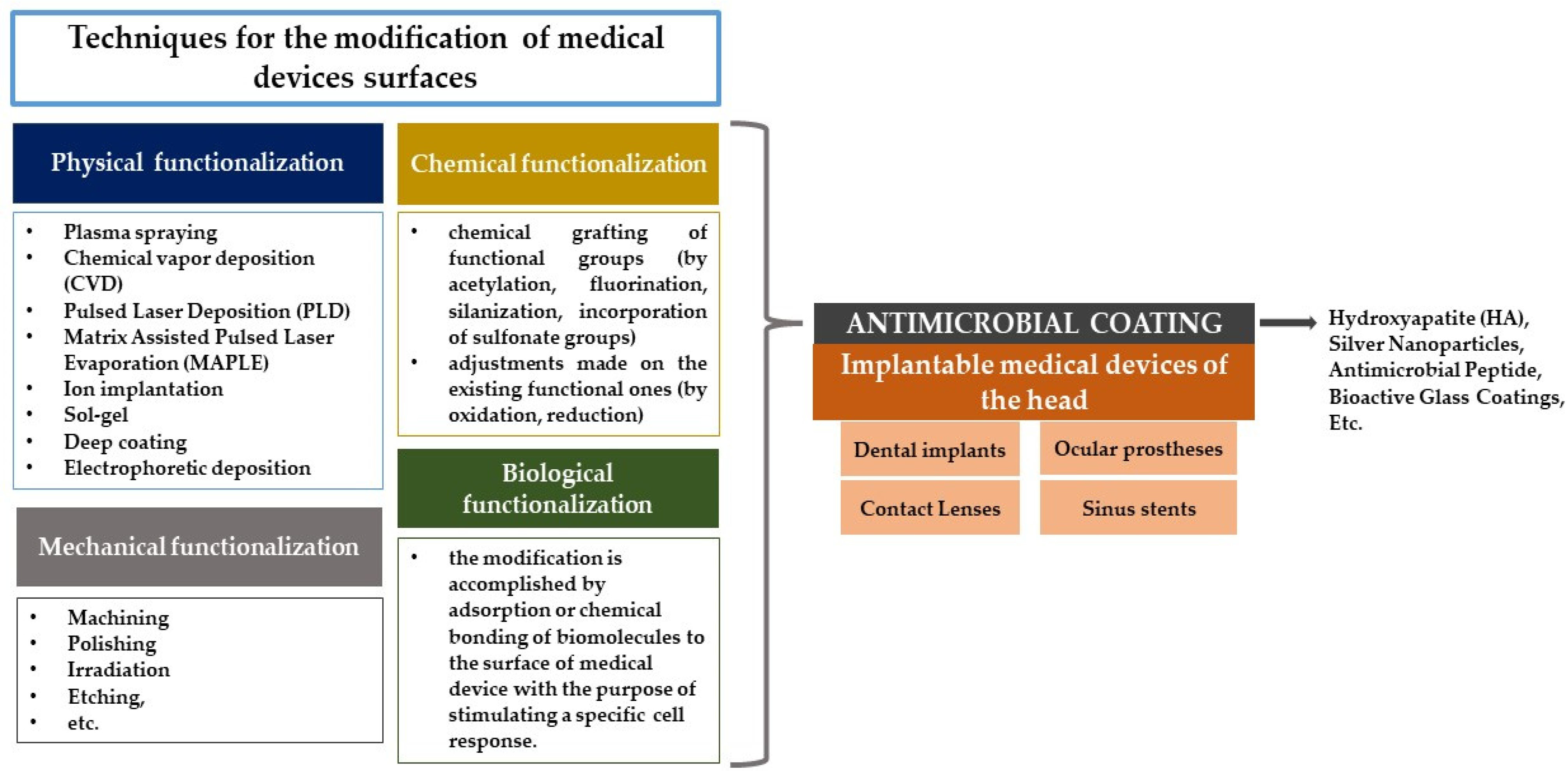
Figure 1. The diagram illustrating the outline of this review.
Coatings 14 00256 g001
Figure 2. Factors influencing bacterial adhesion.
Coatings 14 00256 g002
Figure 3. IMDs which feature antimicrobial coatings offer significant advantages over those without such coatings. IMDs lacking antimicrobial coatings are prone to bacterial colonization, where bacteria can adhere to their surfaces and develop biofilms. Typically, systemic prophylactic antibiotics are administered to combat this, which can lead to side effects for the patient and expose all bacteria to antibiotics, increasing the risk of developing antimicrobial resistance. Moreover, high doses of antibiotics are often necessary to eliminate mature biofilms. In contrast, when antimicrobial coatings are applied to the surfaces of IMDs, these surfaces are less likely to be colonized by bacteria, potentially eliminating the need for antibiotic use and thereby reducing patient side effects and bacterial exposure to antibiotics. Although the antimicrobial effects of coatings are localized, unlike systemic antibiotics, they can still exert a broader antimicrobial influence, causing nearby bacteria to either retreat or be damaged.
Coatings 14 00256 g003
Table 1. Classification of the most used ceramic materials.
MaterialAdvantage/ActivityRef.
Calcium phosphate (CP)Good osseointegration rate; corrosion resistance; cell adhesion[58,59,60]
Hydroxyapatite (HA)Cell adhesion and proliferation; enhanced osteo-conductivity and -integration[61]
Bioactive glasses (BGs)Excellent osteo-conductivity and -inductivity properties[62]
Table 2. Overview of different types of infections and responsible pathogens associated with IMDs of the head.
Medical DeviceType of InfectionCommon PathogensConcernRef.
Dental implantsPeri-implantitis,
biofilm-associated infections, mucositis
S. aureus, P. aeruginosa, E. coli, Porphyromonas gingivalisBiofilm formation leads to chronic infections, resistance to antibiotics, and challenges in achieving effective sterilization.[64]
Ocular prosthesesEndophthalmitis,
conjunctivitis
S. epidermidis, S. aureus,
Serratia marcescens
Biofilm formation on prostheses surfaces leads to persistent infections and inflammation.[65,66]
Contact lensesMicrobial keratitis,
corneal ulcers
P. aeruginosa, S. aureus,
Acanthamoeba spp., Fusarium spp.
The risk of infection increases with poor hygiene practices, overnight wear, and the use of contaminated solutions.[67,68]
Sinus stentsSinusitis,
biofilm-associated infections
S. aureus, P. aeruginosa,
S. epidermidis
Biofilms can form on stents, leading to chronic inflammation and the potential need for surgical intervention[69]
Table 3. Metal elements with antimicrobial activities used on dental implants.
ElementModified MaterialCoating MethodPathogen(s)Ref.
Silver (Ag)Ag-hydroxyapatite-tannic acidImmersionE. coli
S. aureus
[90]
Nano-AgMicrowave-assisted synthesisS. aureus[91]
Ag- and Ta-co-doped amorphous calcium phosphateRadio frequency magnetron sputteringE. coli[92]
Ag-containing calcium phosphateElectrodepositionS. aureus[93]
TiO2 coatings enriched with Ca, P, and AgPlasma electrolytic oxidationE. coli[94]
Poly-L-lysine/sodium alginate loading nano-AgPolyelectrolyte electrostatic self-assembly and reduction of Ag with dopamineS. aureus
S. mutans
[95]
Gold (Au)Au nanocluster constructed mixed-metal metal–organic networkMetal−ligand coordination-driven and solvent evaporation-induced self-assemblyE. coli[96]
Cobalt (Co)Co-doped ZnO/reduced graphene oxide nanorodsChemical co-precipitationE. coli
S. aureus
[97]
Zinc (Zn)NPs of zinc oxide (nZnO) and hydroxyapatite (nHA)Electrohydrodynamic depositionStreptococcus spp.[98]
Co-sputtered titania(Ti)-Zn-oxide nanocompositeSputteringE. coli
S. aureus
[99]
Table 4. Drugs with antimicrobial activity used for coating dental implants.
DrugTested PathogenRef.
VancomycinS. aureus[104,105,106,107]
GentamicinE. coli, S. aureus[107,108]
S. aureus[109,110]
S. aureus, S. epidermidis, P. aeruginosa[111]
P. aeruginosa[112]
CiprofloxacinS. aureus[113]
P.aeruginosa[112]
MinocyclinePorphylomonas gingivalis[114]
DoxycyclineS. aureus, S. epidermidis[115]
Streptococcus sanguinis[116], p. 14
Porphyromonas gingivalis[117]
A. actinomycetemcomitans, S. sanguinis, P. micra, E. corrodens[118]
S. mutans[119]
TetracyclinePorphyromonas gingivalis[120]
Porphyromonas gingivalis, Fusobacterium nucleatum, Prevotella intermedia, Aggregatibacter actinomycetemcomitans[121]
Porphyromonas gingivalis[122]
S. aureus[123]
S. aureus, S. epidermidis[124]
Table 5. Selection of antimicrobial strategies for antimicrobial CL surfaces.
CoatingSubstrateMethodMicroorganismRef.
AgWeflex 55 hydrogelAdsorptionP. aeruginosa, S. aureus[143,144]
Skin-derived antimicrobial peptide Esc(1–21) and its
diastereomer Esc(1–21)-1c
Soft contact lensesCovalent immobilizationP. aeruginosa[80]
Ag NPsHydrogel
(soft contact lens)
Incorporated collagen hydrogels[145]
ZnO, chitosan, and gallic acidComfilcon A (silicone-hydrogel)Sonochemical coatingS. aureus[146]
Phomopsidione NPsACUVUE® TrueEye™ (silicone hydrogel)SoakingS. marcescens, P. aeruginosa, MRSA, P. mirabilis, C. utilis[147]
Chloro sulfoxy chitosanOphthalmic lensesSoakingP. aeruginosa[148]
Moxifloxacin hydrochloride, chlorhexidine diacetate monohydrate, diclofenac sodium saltSilicone-based hydrogel
(soft contact lens)
Layer-by-layer deposition techniqueP. aeruginosa, S. aureus[149]
Gallic acid (GA) phytomolecule-coated zinc oxide NPs (ZN), phytomolecule-coated ZN + GA + tobramycinMethafilcon A (CooperVision, San Ramon, CA, USA)Sonochemical methodS. aureus, P. aeruginosa, E. coli, Aspergillus, fumigatus Fusarium solani[150]
Zwitterionic
metal–phenolic networks (MPNs) based on the coordination of
copper ions (CuII) and a poly(carboxylbetaine-co-dopamine
methacrylamide) copolymer
Aqua Moist
(Hydron Contact
Lens Co., Shanghai, China)
One-step
method due to MPN structure with enhanced adhesive
property bestowed by CuII cross-linked catechol groups
E. coli, P. aeruginosa
S. aureus,
[151]
Ionic polysaccharides (chitosan, sodium alginate, sodium hyaluronate) and genipin (crosslinker) to sustain the release of diclofenac sodium saltSilicone-based hydrogel SofLens PurevisionLayer-by-layer deposition techniqueP. aeruginosa, S. aureus[152]
Disclaimer/Publisher’s Note: The statements, opinions and data contained in all publications are solely those of the individual author(s) and contributor(s) and not of MDPI and/or the editor(s). MDPI and/or the editor(s) disclaim responsibility for any injury to people or property resulting from any ideas, methods, instructions or products referred to in the content.