Article

NbSe2 Crystals Growth by Bromine Transport

1
Institute of Solid State Physics, Bulgarian Academy of Sciences, 1784 Sofia, Bulgaria
2
Institute of Optical Materials and Technologies, Bulgarian Academy of Sciences, 1113 Sofia, Bulgaria
3
Institute of General and Inorganic Chemistry, Bulgarian Academy of Sciences, 1113 Sofia, Bulgaria
*
Author to whom correspondence should be addressed.
Coatings 2023, 13(5), 947; https://doi.org/10.3390/coatings13050947
Submission received: 13 April 2023 / Revised: 9 May 2023 / Accepted: 15 May 2023 / Published: 18 May 2023
(This article belongs to the Special Issue Application of Graphene and Two-Dimensional Materials in Thin Films)

Abstract

:
Recently, low-dimensional structures in the form of bulk crystals and nanoflakes have received considerable interest due to their 2D unique functionality and promising applications in electronics, photonics, sensing devices and photovoltaic solar cells. As a result, remarkable efforts and modifications have been made for the synthesis process of crystalline material by the vapor transport technique. Here, an alternative concept of NbSe2 crystal growth by the chemical vapor transport (CVT) technique using bromine as a vapor transport agent is presented and subsequently analyzed by X-ray diffraction (XRD), Raman and X-ray photoelectron spectroscopy (XPS) spectroscopy. X-ray powder diffraction analysis revealed hexagonal 2H-NbSe2 and 4H-NbSe2 phases, and characteristic Raman and XPS spectra typical for crystalline NbSe2 were obtained. The environmental sensitivity of the grown crystals is manifested by luminescence attributed to oxidized Nb at the samples’ surface.

1. Introduction

Two-dimensional (2D) van der Waals (vdW) materials have unique physical properties and tremendous potential for contemporary applications. The dimensionality effects exhibited by two-dimensional van der Waals materials have recently attracted extensive interest. Due to the weak vdW bonding between the layers, 2D materials can be straightforwardly exfoliated, transferred and stacked using mechanical processes, which provide a perfect platform studying magnetic, electrical, optical, mechanical and various other specific physical properties to the monolayer limit [1,2].
2H-NbSe2 is particularly actively investigated material among the transition metal dichalcogenides (TMDs). The single crystal 2H-NbSe2 is reported to be a prototypical anisotropic s-wave superconductor with zero-field critical temperature Tc = 7.2 K, along with an incommensurate charge density wave (CDW) phase at a temperature of TCDW = 33 K. The competitive relationship between the superconducting phase and CDW phase has brought about wide and sustained arguments [3]. In comparison to the bulk counterpart, monolayer NbSe2 exhibits a strongly enhanced CDW order and intrinsic Ising superconductivity [4]. Due to its specific electric properties, NbSe2 is anticipated to be a feasible Lego brick for heterojunction device applications, such as high-performance field-effect transistors or superconductor–semiconductor heterostructures for the detection of Majorana fermions [5,6]. 2H-NbSe2, 4H-NbSe2 and 3R-NbSe2 are the three most common polytypes of niobium diselenide. The first two hexagonal structures possess superconductivity below 7.2 K and 6.3 K, respectively. The third rhombohedral structure is not superconducting up to 1.2 K [7]. Therefore, it is very important to establish reliable techniques for the synthesis of corresponding high-quality crystals and thin layers. Despite there being several bottom-up and top-down methods for obtaining nanolayers, physical and chemical vapor deposition methods are not completely appropriate for synthesizing highly crystalline samples, due to the inevitable substrate- and layer-induced defects (e.g., vacancies, dislocations and grain boundaries) and the attained critical temperatures were currently far below those of the exfoliated sheets [8]. Chemical vapor transport is still the most reliable and low-priced method for growing high-quality TMD single crystals [9,10]. NbSe2 single crystals are usually grown using the iodine-assisted CVT method [11,12]. Furthermore, it was proposed [13] modified temperature-oscillating CVT method for controllable growth of NbSe2 single crystals. This kinetic process, functioning by temperature oscillation in a two-zone furnace, was used to maintain a supersaturation-dependent growth. Crystals with screw dislocations as well as homogeneous hexagon crystals were developed during the oscillation under different cooling rates. It is observed for group VI dichalcogenides that bromine is a more suitable transport additive than iodine. Chemical transport rates with iodine are low; however, transport with bromine is more effective. Moreover, the addition of bromine led to WSe2 crystals of better quality [14].
In this paper, we report on the growth conditions and characterization of NbSe2 single crystals by means of bromine vapor transport. The performed X-ray powder diffraction analysis revealed a hexagonal 2H-NbSe2 phase, afterwards characterized by Raman and XPS analysis.

2. Materials and Methods

The CVT method is a relatively quick and simple growth technique with high yield, which relies on the use of transporting agents such as I2, Br2, etc. CVT is a process of crystal growth that generates crystals via gas-phase transport. The target material is combined with a volatile transport agent in a long quartz tube/ampoule sealed under vacuum. When this tube is heated, the transport agent is vaporized. The transport agent then reacts with the target material, transforming it into a gaseous intermediate phase which can then travel along the length of the tube. Once the intermediate reaches the other, typically cooler, end of the tube, the temperature is low enough that there is no longer sufficient thermal energy to maintain the gaseous intermediate. It then decomposes into the transport agent, which remains in the gas phase, and the target material, which deposits on the tube surfaces and crystallizes [15].
NbSe2 crystals were synthesized from the elemental components in a two-step process by chemical vapor transport. First, for preliminary synthesis of polycrystalline NbSe2 material, stoichiometric amounts of Nb (purity 99.99%, Alfa Aesar, Haverhill, MA, USA) and Se (purity 99.9%, Alfa Aesar, Haverhill, MA, USA) elemental powders were sealed in a quartz ampule under vacuum and annealed in a tube furnace at 850 °C for 72 h followed by cooling at 5 °C/min rate down to 350 °C and then removed from the furnace. The reaction product was confirmed by XRD analysis to be polycrystalline NbSe2. NbSe2 single crystals were grown by chemical vapor transport CVT technique using bromine vapor transport setup (Figure 1a). For the growth of NbSe2 crystals, the grounded sintered polycrystalline NbSe2 and narrow capillary with bromine (~2 mg/cc) of ampoule volume were sealed in an evacuated (10−5 torr) quartz ampoule for compound preparation. After sealing the loaded ampoule, the NbSe2 powder and bromine were thoroughly mixed by vigorous shaking. The powder charge along with bromine were kept at one end of the ampoule. The sealed ampoule was introduced horizontally into the two-thermal-zone furnace. The furnace temperature in both the zones increased slowly to 850 °C (hottest, reaction zone) and 800 °C (cooler, growth zone). The ampoule was left at these temperatures for 168 h. After the required period of growth, the furnace was cooled down to room temperature with the rate of 50 °C/h. The ampoule was then broken, and crystals were found to have grown at the cooler end of the ampoule.
In order to completely understand the structure of NbSe2 samples, a powder X-ray diffraction (PXRD) was performed. Structure analysis by single-crystal X-ray diffraction is one of the most powerful techniques for elucidating solid-state and molecular structures. It provides common detailed information on the lattices of crystalline materials, including unit cell dimensions, bond lengths, bond angles, and details of site ordering. Powder X-ray diffraction patterns were gathered within the 2θ range from 10 to 80° with a constant step 0.02° on a Bruker D8 Advance diffractometer (Billerica, MA, USA) with tube CuKα 40 KV, 40 mA. Goniometer radius 217.5 mm, scan type coupled TwoTheta/Theta, 10–80°, step 0.02, counting time 35 s/step, primary Soller slit 4°, divergent slit 0.3°, secondary Soller slit 4°, anti-scattered slit 0.5°, LynxEye detector (Billerica, MA, USA) and detector slit 11.93 mm. Phase identification was performed with the Diffracplus EVA using the ICDD-PDF2 Database (Newtown Square, PA, USA).
The Raman spectra were collected in backscattering geometry at a HORIBA Jobin Yvon Labram HR visible spectrometer (HORIBA France SAS, Lille, France) coupled to a Peltier-cooled CCD detector. A He-Ne laser with wavelength of 632.8 nm was used for excitation with spectral resolution of ~1.5 cm−1. The laser beam was focused on a spot with approximate size of 5 µm on the sample surface using a microscope objective with magnification 50×. The laser power was attenuated to below 300 µW to prevent sample overheating. Raman line parameters were determined from fit to Voigt profiles.
The X-ray photoelectron spectroscopy (XPS) studies were performed in a VG Escalab MKII electron spectrometer (VG Scientific Limited, London, UK) using AlKα radiation with energy of 1486.6 eV under base pressure 10−8 Pa and a total instrumental resolution of 1 eV. The binding energies (BE) were determined using the C 1s line as a reference with an energy level of 285.0 eV. The accuracy of the measured BE was 0.2 eV. The photoelectron lines of the constituent elements on the surface were recorded and corrected by subtracting a Shirley-type background and quantified using the peak area and Scofield’s photoionization cross-sections. The spectra deconvolution was performed with XPSPEAK41 software (version 4.1).
The excitation source for photoluminescence (PL) spectroscopy setup was a 325 nm line of a He–Cd laser and the emissions were analyzed using a Horiba-Jobin Yvon iHR550 (1800 grooves/mm grating, HORIBA France SAS, Lille, France) spectrometer with a liquid-nitrogen-cooled CCD detector.

3. Results and Discussion

3.1. X-ray Diffraction Analysis (Crystalline Structure)

NbSe2 has a Molybdenite-like structure and crystallizes in the hexagonal P63/mmc space group. The structure is two-dimensional and consists of two NbSe2 sheets oriented in the (0, 0, 1) direction. In the structure, Nb4+ is bonded to six equivalent Se2− atoms to form distorted edge-sharing NbSe₆ pentagonal pyramids. All Nb–Se bond lengths are 2.62 Å. Se2− is bonded in a three-coordinate geometry to three equivalent Nb4+ atoms. Figure 2 shows the X-ray diffraction pattern for powdered NbSe2 which can be well indexed within the P63/mmc structure. According to the X-ray powder diffraction analysis, a hexagonal P63/mmc, 2H-NbSe2 crystalline (JCPDS card no. 01-070-5612) phase (platelets) with lattice cell parameters of a = 0.345(2) nm, b = 0.345(2) nm and c = 1.253(0) nm and a hexagonal P-6m2, 4H-NbSe2 (JCPDS card no. 01-072-1621) phase with lattice cell parameters of a = 0.344(9) nm, b = 0.344(9) nm and c = 2.539(3) were obtained. In Figure 2, some faint peaks are also detected (marked with asterisks) which match the peaks in the X-ray diffraction pattern of monoclinic [17] NbSe3 (JCPDS card no. 00-029-0950). This indicates the presence of minor inclusions of crystalline NbSe3 (needle-like shape) in the studied specimens. It should be mentioned that niobium always contains tantalum in some quantity despite the purity of the product. However, it was found [18] that Ta-doped 2H-NbSe2 sustains the original structure, yet shows an enhanced electronic property, which could be beneficial to the realization of the superconducting properties of 2H-NbSe2. Q is for quartz from the quartz ampoule used for CVT growth. Crystals of 2H- and 4H-NbSe2 were grown at 750 and 925 °C, respectively, from polycrystalline compounds, using the vapor transport technique in [19]. In 2H-NbSe2, superconducting and charge density wave (CDW) transitions were observed at Tc = 7.4 K and TcDW = 35 K, respectively. It is found that these two transitions are changed to TcDW = 42 K and TC = 6.5 K in 4H-NbSe2 [19].
Figure 3 shows an optical microscopic image of one of the grown crystal specimens that was used for further characterization. From Figure 3, the presence of naturally grown smooth surfaces is evident, whose facets contain angles of 60° and 120° indicating a crystalline hexagonal phase. Due to minor sample tilting, some of these structures are slightly smeared out, therefore several spots where such angles occur are encircled with a white line as a guide to the eye.

3.2. Raman Analysis

Various Raman-active features are expected for the normal phase of 2H-NbSe2; however, the Raman spectra of materials can be affected by many factors, including the dimensionality effect, impurities and doping. Therefore, we conducted the Raman characterization on a smooth naturally grown surface as shown in Figure 3. For the 2H-NbSe2 compound [20], the A1g and E2g Raman-active phonons and the so-called soft mode were detected [21,22,23] as depicted in Figure 4. A1g corresponds to an out-of-plane mode and its spectral line is located at ~230 cm−1. E2g is an in-plane vibration with extremely weak Raman activity upon red excitation [20] and appears at ~239 cm−1 as a faint shoulder at the high-frequency side of the A1g line. The linewidth of the A1g phonon in Figure 4 is only 5 cm−1, indicating the good quality of the obtained NbSe2 crystal. A quite similar spectrum was published for a NbSe2 crystal prepared by the flux method in Ref. [24]. For the 2H-NbSe2 structure, there is also another Raman-active mode—E1g—which is expected at ~134 cm−1 [21]. However, due to the presence of facet edges producing angles of 60° and 120° in Figure 3, it is highly probable that our Raman spectra were taken from the [001] plane of the hexagonal lattice and the E1g line is forbidden in this scattering geometry.
The broad feature centered at ~180 cm−1 (Figure 4) is assigned as “soft mode” and involves a second-order scattering processes of phonons from the periphery of the Brillouin zone, including both intralayer and interlayer vibrations [22,23]. The temperature dependence of this mode has been found to correlate with the CDW transition. It softens significantly upon cooling until reaching TCDW, below which it remains frozen [23].
The spectrum in Figure 4 contains two additional features (marked with asterisks) which are identified as originating from the minor NbSe3 inclusions in our specimen: a shoulder at the low-frequency side (~220 cm−1) of the A1g line and a feature at about 145–155 cm−1 which may also be viewed as a shoulder to the broad “soft mode” band. The shoulder at 220 cm−1 coincides with the strongest Ag Raman line of monoclinic NbSe3 at ~218 cm−1 [17], and the broad feature at 145–155 cm−1 matches the frequency range of the strongest band in the NbSe3 Raman spectrum consisting of three other Ag lines at 143, 156 and 164 cm−1 [17].
In Figure 4, the fit components corresponding to the three first-order phonon lines (A1g, E2g and Ag (NbSe3)) are plotted below the measured spectrum for clarity. In the inset, the same spectrum is shown in its full extension to above 1200 cm−1.

3.3. X-ray Photoelectron Spectroscopy (XPS)

For the XPS analysis, a small portion of the sample was pressed in a pellet and placed in the analysis chamber. XPS analysis was carried out to investigate the chemical nature and identify the elements present in the NbSe2 crystal. Figure 5a shows the XPS survey scan. The predominant peaks correspond to Nb 3d, Se 3d, C 1s and O 1s core-level spectra, respectively. As can be seen from the survey scan of NbSe2 crystals, the Se is predominant on the surface and thus the Auger SeLMM structure is quite extensive and cause spectral interferences with a wide variety of other elements. We did not detect any features typical of Br. The surface concentrations of constituent elements were calculated and are given below as O = 55.47 at.%, Se = 33.04 at.% and Nb = 9.82 at.%, respectively. The spectrum of Nb 3d appeared as a doublet corresponding to Nb 3d5/2 and Nb 3d3/2 (see Figure 5b). In the present case, this doublet has a complex structure due to the presence of Nb in two different oxidation states, as well overlapping with the SeLMM line. The deconvoluted spectrum of Nb 3d consist of a first doublet with a maximum of around 203.5 eV attributed to the formed Nb-Se bonds and a second doublet with a maximum at 207.5 eV, which corresponds to NbOx. The Se 3d spectrum shows a single peak at 55.1 eV (see Figure 5c).
We should mention here that the Se 3d peak is also a doublet but with a very small spin-orbit splitting (0.86 eV). The Se 3d was fitted with three components. The main components belong to NbSe2. The other two minor components could be ascribed to residual amorphous Se and/or trigonal Se. The obtained results are compared with data from other literature sources and show the presence of NbSe2 [25], as well as inevitable surface oxidation [26].

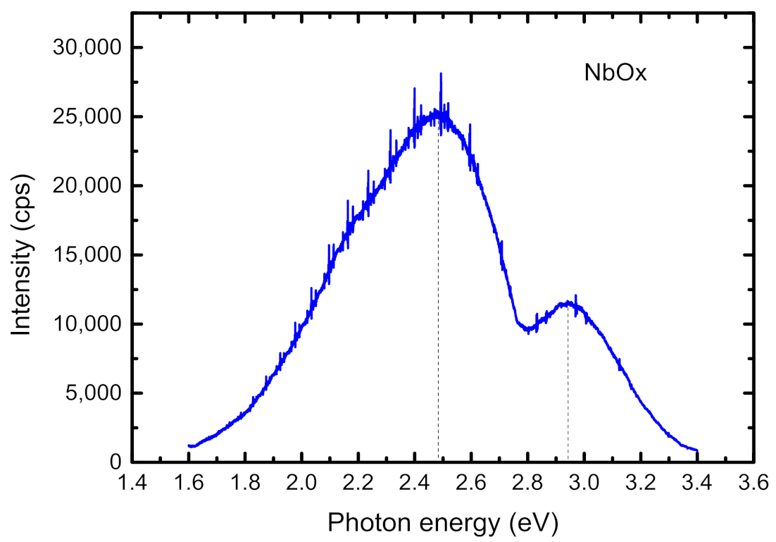
3.4. Photoluminescence

NbSe2 crystals are recognized for their sensitivity in air [27]. In atmospheric conditions, NbSe2 already readily oxidizes under very low illumination with very low intensity [28]. Figure 6 shows photoluminescence signals detected at ~2.48 eV (~500 nm) and ~2.95 eV (~420 nm), which are probably related to the (photo)oxidation of Nb to NbOx (NbO2, Nb2O5), as reported by Ref. [29]. It is observed that the photoluminescence (PL) spectra are visible for the material after 24 h of exposure to air, emphasizing the importance of protection by encapsulation, prompt measurement or the instantaneous placement of crystal samples into an oxygen-free environment as glove-box [30].

4. Conclusions

NbSe2 crystals were grown by the chemical vapor transport (CVT) technique using bromine as the vapor transport agent. The structural and chemical characterization by X-ray powder diffraction, Raman spectroscopy and X-ray photoelectron spectroscopy were performed providing evidence for hexagonal 2H and 4H-NbSe2 phases and indications of high lattice order and good quality of the obtained crystalline samples. NbSe2 crystal growth using bromine vapor transport should thus be considered as a reliable option in the development of future technologies for the synthesis of this material.

Author Contributions

Conceptualization D.D. and V.M.; methodology V.M., I.A., D.K., I.D., P.R., M.G. and D.D.; software I.A., I.D., N.M. and P.R.; formal analysis V.M., I.A., P.R., D.K., M.G. and D.D.; resources D.D.; writing—original draft preparation D.D. and V.M.; writing—review and editing V.M., P.R., I.A., D.K. and D.D., visualization I.D., P.R., I.A. and N.M.; supervision, V.M. and D.D.; funding acquisition D.D. All authors have read and agreed to the published version of the manuscript.

Funding

This research was funded by Bulgarian Science Fund under the projects KΠ-06-KOCT/13 “Synthesis and characterization of ferromagnetic 3D and 2D Van der Waals materials” and KΠ-06-H-28/8, and under the COST Action CA 17123 “Ultrafast opto-magneto-electronics for non-dissipative information technology”.

Institutional Review Board Statement

Not applicable.

Informed Consent Statement

Not applicable.

Data Availability Statement

The data presented in this study are available on reasonable request from the corresponding author.

Acknowledgments

Bulgarian Science Fund project KΠ-06-KOCT/13 “Synthesis and characterization of ferromagnetic 3D and 2D Van der Waals materials” and COST Action CA 17123 “Ultrafast opto-magneto electronics for non-dissipative information technology”. P.R., V.M. and N.M. acknowledge the financial support by the European Regional Development Fund within the Operational Programme ‘Science and Education for Smart Growth 2014–2020’ under the Project CoE ‘National Center of Mechatronics and Clean Technologies’ BG05M2OP001-1.001-0008-C01.

Conflicts of Interest

The authors declare no conflict of interest.

References

  1. Das, S.; Robinson, J.A.; Dubey, M.; Terrones, H.; Terrones, M. Beyond Graphene: Progress in Novel Two-Dimensional Materials and van der Waals Solids. Annu. Rev. Mater. Res. 2015, 45, 1–27. [Google Scholar] [CrossRef]
  2. Bian, R.; Li, C.; Liu, Q.; Cao, G.; Fu, Q.; Meng, P.; Zhou, J.; Liu, F.; Liu, Z. Recent progress in the synthesis of novel two-dimensional van der Waals materials. Natl. Sci. Rev. 2022, 9, nwab164. [Google Scholar] [CrossRef] [PubMed]
  3. Hossain, M.; Zhao, Z.; Wen, W.; Wang, X.; Wu, J.; Xie, L. Recent Advances in Two-Dimensional Materials with Charge Density Waves: Synthesis, Characterization and Applications. Crystals 2017, 7, 298. [Google Scholar] [CrossRef]
  4. Tsen, A.W.; Hunt, B.; Kim, Y.D.; Yuan, Z.J.; Jia, S.; Cava, R.J.; Hone, J.; Kim, P.; Dean, C.R.; Pasupathy, A.N. Nature of the quantum metal in a two-dimensional crystalline superconductor. Nat. Phys. 2015, 12, 208. [Google Scholar] [CrossRef]
  5. He, W.-Y.; Zhou, B.T.; He, J.J.; Yuan, N.F.Q.; Zhang, T.; Law, K.T. Magnetic field driven nodal topological superconductivity in monolayer transition metal dichalcogenides. Commun. Phys. 2018, 1, 40. [Google Scholar] [CrossRef]
  6. Kim, A.R.; Kim, Y.; Nam, J.; Chung, H.-S.; Kim, D.J.; Kwon, J.-D.; Park, S.W.; Park, J.; Choi, S.Y.; Lee, B.H.; et al. Alloyed 2D Metal–Semiconductor Atomic Layer Junctions. Nano Lett. 2016, 16, 1890–1895. [Google Scholar] [CrossRef]
  7. Andreeva, O.; Braude, I.; Mamalui, A. Selenium vacancies and their effect on the fine structure of NbSe2 quasi-two-dimensional single crystals. Phys. Met. Metallogr. 2012, 113, 888–892. [Google Scholar] [CrossRef]
  8. Dasadia, A.; Bhavsar, V. Growth, structure, electrical and optical properties of transition metal chalcogenide crystals synthesized by improved chemical vapor transport technique for semiconductor technologies. Prog. Cryst. Growth Charact. Mater. 2022, 68, 100578. [Google Scholar] [CrossRef]
  9. Brixner, L.H. Preparation and properties of the single crystalline AB2-type selenides and tellurides of niobium, tantalum, molybdenum and tungsten. J. Inorg. Nucl. Chem. 1962, 24, 257–263. [Google Scholar] [CrossRef]
  10. Kershaw, R.; Vlasse, M.; Wold, A. The preparation of and electrical properties of niobium selenide and tungsten selenide. Inorg. Chem. 1967, 6, 1599–1602. [Google Scholar] [CrossRef]
  11. Oglesby, C.S.; Bucher, E.; Kioc, C.; Hohi, H. Growth of faceted niobium diselenide. J. Cryst. Growth 1994, 137, 289–294. [Google Scholar] [CrossRef]
  12. Klemm, R.A. Pristine and intercalated transition metal dichalcogenide superconductors. Phys. C Supercond. Its Appl. 2015, 514, 86–94. [Google Scholar] [CrossRef]
  13. Li, Z.; Xi, X.; Ding, B.; Li, H.; Liu, E.; Yao, Y.; Wang, W. Thermodynamics and kinetics synergy for controlled synthesis of 2D van der Waals single-crystal NbSe2 via modified Chemical Vapor Transport. Cryst. Growth Des. 2020, 20, 706–712. [Google Scholar] [CrossRef]
  14. Schmidt, M.; Gooth, J.; Binnewies, M. Preparation and crystal growth of transition metal dichalcogenides. Z. Anorg. Allg. Chem. 2020, 646, 1183–1194. [Google Scholar] [CrossRef]
  15. Binnewies, M.; Glaum, R.; Schmidt, M.; Schmidt, P. Chemical Vapor Transport Reactions; De Gruyter: Berlin, Germany, 2012. [Google Scholar]
  16. Momma, K.; Izumi, F. VESTA 3 for three-dimensional visualization of crystal, volumetric and morphology data. J. Appl. Crystallogr. 2011, 44, 1272–1276. [Google Scholar] [CrossRef]
  17. Chen, H.; Ma, Z.; Shao, Y.; Rehman, Z.; Zhang, K.; He, Q.; Song, L. Angle-/temperature-dependence of Raman scattering in layered NbSe3 crystal. AIP Adv. 2017, 7, 095316. [Google Scholar] [CrossRef]
  18. Li, H.; Liu, S.; Chen, L.; Wu, J.; Zhang, P.; Tang, H.; Li, C.; Liu, X.; Wang, Z.; Meng, J. Atomic structures and electronic properties of Ta-doped 2H-NbSe2. RSC Adv. 2014, 4, 57541–57546. [Google Scholar] [CrossRef]
  19. Naik, I.; Rastogi, A.K. Charge density wave and superconductivity in 2H- and 4H-NbSe2: A revisit. Pramana-J. Phys. 2011, 76, 957–963. [Google Scholar] [CrossRef]
  20. Hill, H.M.; Rigosi, A.F.; Krylyuk, S.; Tian, J.; Nguyen, N.V.; Davydov, A.V.; Newell, D.V.; Hight Walker, A.R. Comprehensive optical characterization of atomically thin NbSe2. Phys. Rev. B 2018, 98, 165109. [Google Scholar] [CrossRef]
  21. Zhang, X.; Tan, Q.-H.; Wu, J.-B.; Shi, W.; Tan, P.-H. Review on the Raman spectroscopy of different types of layered materials. Nanoscale 2016, 8, 6435–6450. [Google Scholar] [CrossRef]
  22. Staley, N.E.; Wu, J.; Eklund, P.; Liu, Y.; Li, L.; Xu, Z. Electric field effect on superconductivity in atomically thin flakes of NbSe2. Phys. Rev. B 2009, 80, 184505. [Google Scholar] [CrossRef]
  23. Xi, X.; Zhao, L.; Wang, Z.; Berger, H.; Forró, L.; Shan, J.; Mak, K.F. Strongly enhanced charge-density-wave order in monolayer NbSe2. Nat. Nanotechnol. 2015, 10, 765–769. [Google Scholar] [CrossRef] [PubMed]
  24. 2Dsemiconductors USA. Available online: https://www.2dsemiconductors.com/niobium-diselenide-nbse2/ (accessed on 12 April 2023).
  25. Calavale, F.; Dreher, P.; Surdendran, A.P.; Wan, W.; Timpel, M.; Verucchi, R.; Rogero, C.; Bauch, T.; Lombardi, F.; Casanova, F.; et al. Tailoring Superconductivity in Large-Area Single-Layer NbSe2 via Self-Assembled Molecular Adlayers. Nano Lett. 2021, 21, 136–143. [Google Scholar] [CrossRef] [PubMed]
  26. Ahmad, H.M.N.; Ghosh, S.; Dutta, G.; Maddaus, A.G.; Tsavalas, J.G.; Hollen, S.; Song, E. Effects of impurities on the electrochemical characterization of liquid-phase exfoliated niobium diselenide nanosheets. J. Phys. Chem. C 2019, 123, 8671–8680. [Google Scholar] [CrossRef]
  27. El-Bana, M.S.; Wolverson, D.; Russo, S.; Balakrishnan, G.; Paul, D.M.; Bending, S.J. Superconductivity in two dimensional NbSe2 field effect transistors. Supercond. Sci. Technol. 2013, 26, 125020. [Google Scholar] [CrossRef]
  28. Holler, J.; Bauried, L.; Korn, T.; Seitz, A.; Özyigit, F.; Eichinger, M.; Schüller, C.; Watanabe, K.; Taniguchi, T.; Strunk, C.; et al. Air tightness of hBN encapsulation and its impact on Raman spectroscopy of van der Waals materials. 2D Mater. 2020, 7, 015012. [Google Scholar] [CrossRef]
  29. Huang, B.X.; Wang, K.; Church, J.S.; Li, Y.-S. Characterization of oxides on niobium by Raman and infrared spectroscopy. Electrochim. Acta 1999, 44, 2571–2577. [Google Scholar] [CrossRef]
  30. Rigosi, A.F.; Hill, H.M.; Krylyuk, S.; Nguyen, N.V.; Hight Walker, A.R.; Davydov, A.V.; Newell, D.B. Dielectric Properties of NbxW1−xSe2 Alloys. J. Res. Natl. Inst. Stand. Technol. 2019, 124, 124035. [Google Scholar] [CrossRef]
Figure 1. (a) Chemical vapor transport (CVT) method setup used for the growth of NbSe2 crystal growth and (bd) schematic crystal structure of 2H-NbSe2 along the a,b,c axis [16].
Coatings 13 00947 g001
Figure 2. X-ray diffraction patterns of NbSe2 crystals. NbSe3 peaks are marked with asterisks.
Coatings 13 00947 g002
Figure 3. Optical microscopic image of a NbSe2 crystal surface containing faceted naturally grown surfaces. Some facet edges making angles of 60° and 120° are encircled with a white line for clarity.
Coatings 13 00947 g003
Figure 4. Raman spectra of the studied NbSe2 crystal. Low-intensity components stemming from NbSe3 inclusions are marked with asterisks. Fitted peaks are plotted below the spectra for the A1g (red), E2g (blue) and Ag (NbSe3) (brown) phonons for clarity. Inset: full extension of the NbSe2 Raman spectrum.
Coatings 13 00947 g004
Figure 5. XPS survey scan (a) and spectra of Nb 3d (b) and Se 3d (c).
Coatings 13 00947 g005
Figure 6. Photoluminescence of (photo) oxidized crystal area.
Coatings 13 00947 g006
Disclaimer/Publisher’s Note: The statements, opinions and data contained in all publications are solely those of the individual author(s) and contributor(s) and not of MDPI and/or the editor(s). MDPI and/or the editor(s) disclaim responsibility for any injury to people or property resulting from any ideas, methods, instructions or products referred to in the content.

