Article

Atmospheric-Pressure Plasma Jet-Induced Graft Polymerization of Composite Hydrogel on 3D-Printed Polymer Surfaces for Biomedical Application

Department of Biomedical Engineering, Da Yeh University, Changhua 515006, Taiwan
*
Author to whom correspondence should be addressed.
Coatings 2023, 13(2), 367; https://doi.org/10.3390/coatings13020367
Submission received: 22 December 2022 / Revised: 28 January 2023 / Accepted: 2 February 2023 / Published: 6 February 2023
(This article belongs to the Special Issue Surface Coating for Biomedical Applications)

Abstract

:
Poly(lactic acid) (PLA) is currently the most widely used material in 3D printing. PLA has good mechanical properties, chemical stability, and biodegradability, but its surface is hydrophobic and cannot be effectively used. The growth metabolism of attachments, how to increase the strength of PLA with high brittleness, and 3D printing of PLA materials for the biomedical field have always been a topic of research by scientists. This experiment used fused filament fabrication (FFF) to prepare structures. First, the 3D-printed polymer surfaces were treated with an atmospheric-pressure plasma jet (APPJ) to make the surface hydrophilic and increase the number of polar functional groups on the surface. Then, UV photo-grafting polymerization of 2-hydroxyethyl methacrylate (HEMA), poly(ethylene glycol) methacrylate (PEGMA), and hydroxyapatite (HAp) was applied onto the 3D-printed polymer surfaces. The experimental results of the water contact angle for the wettability test show that APPJ-treated and UV-grafted composite hydrogels become hydrophilic to activate the 3D-printed polymer surface successfully. For the in vitro study, the effect of APPJ treatment and composite hydrogel on the viability of osteoblast-like MG63 cells was examined using the Alamar Blue cell viability assay, indicating that biocompatibility has been improved in this study. This method is expected to have potential in the application of bone scaffolds in the future.

1. Introduction

Three-dimensional printing technology, also known as additive manufacturing (AM), belongs to a rapid prototyping technology. It is an advanced manufacturing technology that gradually emerged in the 1990s and is even regarded as the third industrial revolution. Compared with the existing mechanical processing methods, various industries can manufacture products without being limited by the manufacturing method or cost and have the flexibility of the production site to produce anywhere. In addition, it can help realize the reduction of the circular economy waste generated during production and it promotes better resource utilization. According to these characteristics, 3D printing has the most significant development potential in the three major fields of aerospace, medical technology, and automobiles. Nowadays, 3D printing technologies are constantly growing, resulting in greater flexibility and customization in the designs, as well as reduced production costs [1,2]. Three-dimensional printing or additive manufacturing is the most recent revolution witnessed in the field of health care. Using a digital model to create and shape the desired product (3D structure), which will then be formed by layer-by-layer deposition of digitally controlled and operated material, the technology differs from conventional manufacturing procedures [1,2,3,4]. In recent years, there have been many developments in applying 3D printing technology in the medical field. Its clinical applications have included orthopedics, dentistry, plastic surgery, and trauma medicine. In addition, it can also customize medical treatment equipment or implanted medical materials that assist in the precise execution of surgery [3,4].
Generally, when the bone breaks, the medical implants used to fix and provide support are usually made of metal (stainless steel or titanium) or high-strength plastic poly(ether ether ketone) (PEEK). Although this type of implant has strong mechanical strength, long-term implantation will cause stress shielding and cannot be biodegraded, so the implant must be removed after fracture healing. However, surgery comes with some risks and will result in additional injury to the bones and human body [5,6,7,8]. The risks posed by the above make bones weaker and weaker. To solve these problems, 3D printing technology can be employed to use biodegradable materials implanted in the body [9,10,11]. In that case, they can be hydrolyzed and metabolized by the organism. Under the slow degradation time, the bones can slowly adapt and reinforce. Taking it out without a second operation can relieve the patient’s pain. Its degradation is about half a year to two years and the bone can also slowly adapt and recover during the degradation time [10,11,12].
Poly(lactic acid) (PLA) is a biodegradable thermoplastic polyester polymerized from renewable resources such as corn starch and sugarcane [13,14]. It has a variety of translucent colors and glossy textures, and has good thermal stability.It can withstand solvent processing temperatures of about 170–230 °C [13]. PLA is an ideal choice for preparing nanofibers in various biomedical applications because of its biodegradability, biocompatibility, and excellent strength and stiffness. It can be made into artificial scaffolds that mimic natural tissues and promote cell health [10,15,16,17]. The US Food and Drug Administration (FDA) approved their use in different biomedical applications. It is widely used in surgery orthopedics, orthodontics, traumatology, and other branches of medicine [16,17,18]. According to the literature, some scholars used 3D printing technology to form biodegradable polymer materials in the research of PLA tissue engineering scaffolds and manufactured high-porosity PLA tissue engineering scaffolds. Through tissue analysis of the scaffolds, it was found that it has cellular growth capacity and proliferation [4,10,19,20]. In terms of the characteristics of PLA during 3D printing, it is less prone to shrinkage deformation, cracking, or warping due to temperature changes [11,21,22].
Atmospheric plasma treatment refers to the operation in an environment of normal temperature and pressure, and it also has high-energy particles compared with a vacuum plasma system [23]. It can be used without a closed vacuum chamber and a vacuum pump system. Compared with a vacuum plasma system, the advantages are: (1) no need to use vacuum equipment; (2) low operating cost; (3) it can be used in a continuous process; (4) the plasma density is high and the processing speed is fast; and (5) highly flexible customization and large-area processing can further improve the production efficiency of the product [24,25,26,27]. Atmospheric plasma treatment is an easy-to-use and economical surface treatment technique. The main advantage of this technology is that the bulk properties of the material remain unchanged while enhancing surface properties and biocompatibility [28,29,30,31]. In addition, the combination of atmospheric plasma treatment and UV light graft polymerization technology can increase the adhesion of the polymer hydrogel to the surface of the substrate [31,32,33,34]. Moreover, UV light graft polymerization technology can achieve the purpose of selectively grafting and crosslinking monomers with different functional groups. Currently, UV light graft polymerization is one of the primary methods for synthesizing hydrogels in the biomedical field [35,36,37]. Due to its wide application, the UV light polymerization method has received considerable attention.
Hydrogel is an easy-to-prepare hydrophilic polymer with a three-dimensional network structure. When water penetrates the network structure to cause swelling, the formation of the network structure is mainly due to the cross-linking reaction between molecular chains [32,36,37,38]. Moreover, the properties of the hydrogel can be changed by modification, so it has excellent research potential in tissue engineering [39,40]. In recent years, people have paid attention to the preparation and new application of poly(hydroxyethyl methacrylate) (PHEMA) polymer hydrogel in various biomedical applications. PHEMA is one of the longest-studied synthetic hydrogel polymers. It is non-toxic, biocompatible, and insoluble after swelling in aqueous media [36,41,42,43]. Poly(ethylene glycol) methacrylate (PEGMA) is a hydrophilic monomer with a special binding force for water molecules [42,43,44]. Because of its non-toxicity and non-immunogenicity, PEGMA grafting can increase the adhesion and reproduction of cells, thereby improving the biocompatibility of hydrogels [42,43,44,45]. Hydroxyapatite (HAp) is a double salt of tricalcium phosphate, and calcium hydroxide is the main inorganic component of human bones and teeth. It has good biocompatibility and biological activity [37,43,46].
Bone substitute materials must have several properties, such as biocompatibility, the ability not to stimulate the body’s immune response, and the ability to degrade gradually over time. In this study, a 3D printing process PLA sample was pretreated using atmospheric plasma treatment to improve surface activation, followed by UV grafting with HEMA/PEGMA/HAp hydrogel. SEM, FTIR, swelling rate, Alamar blue assay, and analyses of wettability for surface functionalization, as well as analyses of characteristics, were performed. According to the surface modification process results, this study could reference the future development and application of 3D-printed polymer surfaces.

2. Materials and Methods

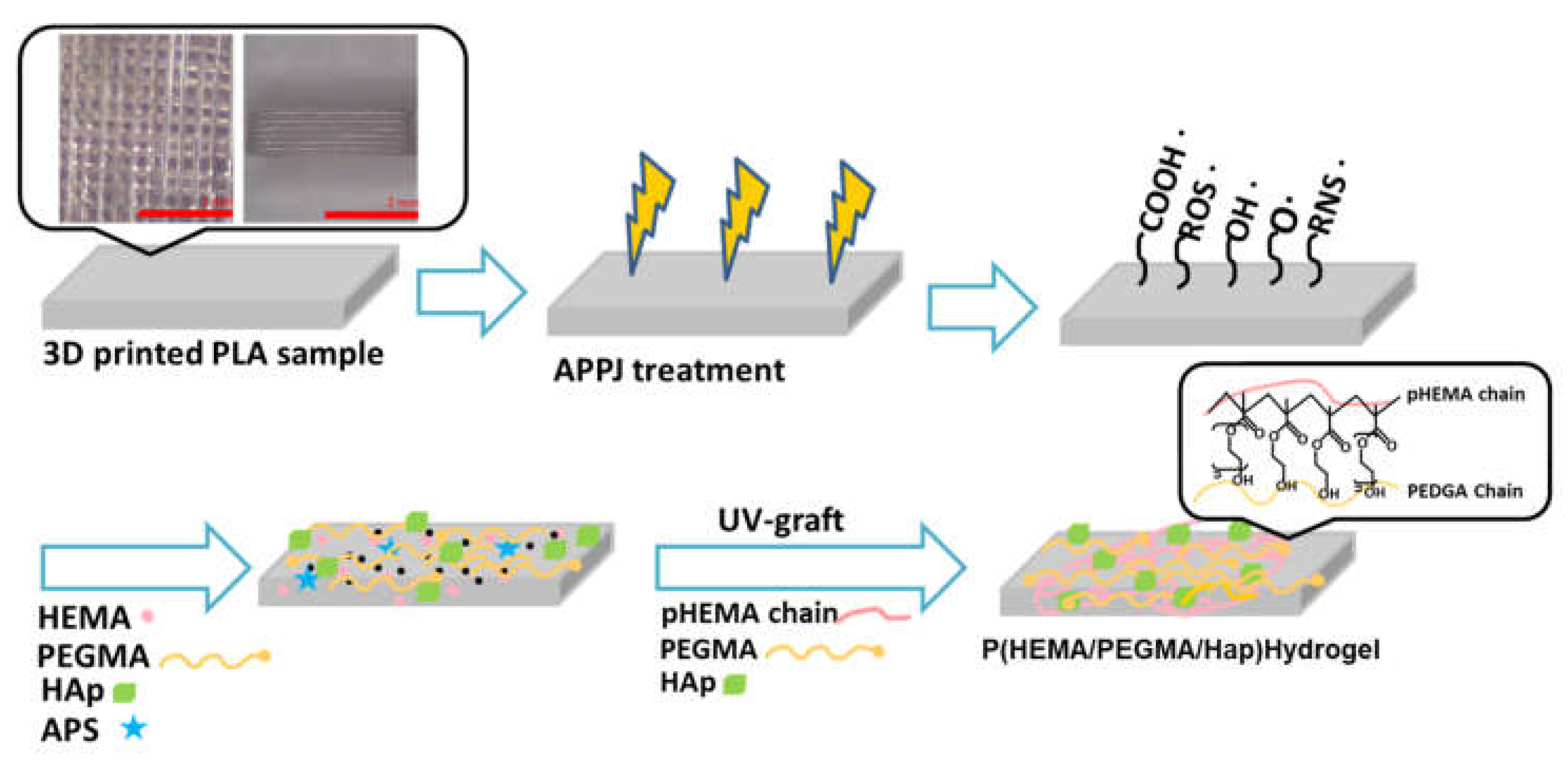
2.1. Pretreatment of Materials

PLA samples were 3D-printed using a da Vinci Jr. 1.0 A Pro (XYZPRINTING, INC., Taipei City, Taiwan) with a direct extruder, and 1.75 mm PLA filaments in white were used to fabricate specimens. It is commercially available filaments used. A PLA filament extracted from bio-compostable materials, such as corn, sugarcane, and other sugar crops was purchased from XYZPRINTING, INC., Taipei City, Taiwan. The PLA sample was printed at a suitable size of about 10 × 10 × 2 mm, cleaned in turns with alcohol and deionized water using ultrasonic cleaning for 15 min in each solution to remove contaminants, and then dried in an oven at 40 °C for overnight. All materials were handled with gloves to minimize contamination.

2.2. Atmospheric-Pressure Plasma Jet Activation Pre-Treatment

In this study, atmospheric-pressure plasma (61G20, ae Plasma 41 Co., Ltd., Taoyüan City, Taiwan) was used to treat the surface of the 3D-printed PLA samples. It is equipped with a rotating jet head (Φ = 35 mm) by an AC power system and a moving stage for area scanning. The plasma is generated using pure argon as the working gas, which is supplied at a constant flow rate of 20 slm. APPJ pretreatment was used to PLA substrates that had been three-dimensionally printed in order to create peroxide groups and activate groups on the surface. The processing power was at 600 W for a treatment time of 60 s and 90 s, respectively, and the distance between the plasma source and the surface of the samples was 10 mm.

2.3. Composite Hydrogel on 3D-Printed Polymer Surfaces by UV Light Surface Graft Polymerization

Aqueous solutions containing a mixture of 10 wt% HEMA (CH2=C(CH3)COO-CH2CH2OH, Mw = 130.15) monomer and 5 wt% PEGMA (H2C=C(CH3)CO(OCH2CH2)nOH, Mw = 360) monomer were used to soak the APPJ-treated, 3D-printed PLA sample specimen. The monomer solutions’ respective volume ratios were 1:1(H1), 1:2(H2), and 2:1(H3). The aqueous solution also contained 1 mol % ammonium persulfate (APS, (NH4)2S2O8, Mw = 228.20 g/mole) and 1 g HAp powder, as shown in Table 1. In this experiment, HAp powder was prepared using a sol-gel method with phosphoric pentoxide (P2O5) and calcium nitrate tetrahydrate (Ca(NO3)2·4H2O). All chemicals were used as received. All monomer solutions were prepared with deionized water. Graft polymerization was performed under UV light (power of 1000 W and wavelength of 365 nm) exposure for 15 min. After graft polymerization, the grafted specimens were washed with distilled water overnight to remove the homopolymer aqueous solution. Figure 1 shows the schematic illustration of the preparation of the functionalization of 3D-printed polymer surface modification.

2.3.1. Wettability (Surface Hydrophobicity/Hydrophilicity) Test

The water contact angles (WCA) on the 3D-printed PLA samples and the 3D-printed modified PLA specimens, such as plasma-deposited films, were measured by the sessile drop (0.9 μL) method with distilled water and observed by a CCD camera (Dino-Lite AM211 made by AnMo Electronics Corporation, New Taipei City, Taiwan) at room temperature. The measured water contact angle value was the average of three measurements.

2.3.2. Surface Characterization

The surface morphology of the 3D-printed PLA samples was observed using a scanning electron microscope (JSM-7800F, JEOL, Tokyo, Japan). Three-dimensional-printed PLA samples were placed on an aluminum holder and sputtering-coated into a thin layer of gold (coating for 90 s) to improve the electric conductivity. The Fourier transform infrared spectrometer (Jasco FTIR-6200, Tokyo, Japan) was used to analyze the surface functional groups after the surface modification.

2.3.3. Swelling Studies of the Treatment Composite Hydrogel on 3D-Printed Polymer Surfaces

The obtained 3D-printed PLA sample was dried in an air convection oven at 40 °C for 24 h to determine the swelling behavior of hydrogels. The dry composite hydrogel weight on a 3D-printed PLA sample (Wd) was first measured. Next, the dry composite hydrogel on the 3D-printed PLA sample was placed in solutions of deionized water and simulated body fluid (SBF) at 25 °C and 37 °C to allow the treatment 3D 3D-printed PLA sample to reach an equilibrium swelling state. Following the setting at the above temperatures for 1 min, 3 min, 5 min, 10 min, 30 min, 60 min, 120 min, 240 min, 480 min, 720 min, 1440 min, 2880 min, and 4320 min, respectively, each treatment 3D-printed PLA sample weight (Ws) was measured. All experiments were performed in triplicate. The swelling ratio (SR) of the treatment 3D-printed PLA sample was recorded during swelling at regular intervals (Equation (1)):
SR = [(Wd – Ws)/Ws] × 100%
where Wd is the weight of the 3D-printed PLA sample that underwent swelling treatment at various time intervals and Ws is the weight of the sample that underwent dry treatment.

2.3.4. In Vitro Degradation Study of the Treatment Composite Hydrogel on 3D-Printed Polymer Surfaces

The assessment of the in vitro degradation study was carried out by immersing the composite hydrogel in 3D-printed PLA samples in a solution of phosphate buffer (PBS) (pH 7.4 at 37 °C). The specimens were placed in sealable vials in 15 mL of PBS solution. Samples were removed from the buffer solution, dried at 40 °C until constant weight, and weighed at regular intervals. Samples of 3D-printed PLA were taken out of the buffer and weighed to determine weight loss. The weight loss ratio (R%) was calculated to measure the degradation. All experiments were performed in triplicate. The weight loss ratio (R%) of the treated 3D-printed PLA sample was recorded during degradation at regular intervals (Equation (2)):
R (%) = [(Wi–Wf)/Wi] × 100%
where Wi and Wf stand for the specimen weights prior to and following PBS immersion, respectively.

2.3.5. In Vitro Cell Culture of the Treatment Composite Hydrogel on 3D-Printed Polymer Surfaces

For the in vitro cell culture, the Alamar Blue cell viability assay was used to assess the MG63 cell viability. The cells were cultured on the specimens obtained using different specimen preparation conditions in an incubator at 37 ± 0.5 °C and a 5% CO2 humidified atmosphere. The optical density (OD) at a wavelength of 570 nm was measured after 1, 3, 5, and 7 culturing days using an enzyme-linked immunosorbent assay (ELISA) reader to observe the growth of MG63 cells on various specimens quantitatively. Cell viability determination is expressed as mean ± standard deviation (n = 3).

3. Results and Discussion

3.1. Wettability of Surface-Modified 3D-Printed PLA Samples

The surface wettability of the modified substrate can be measured by the contact angle of a water droplet. Unmodified 3D-printed PLA samples had a measured water contact angle of 62.2°±3.2°. The values of the water contact angle of 3D-printed PLA samples after plasma treatment are listed in Table 2. The surface of a 3D-printed PLA sample became hydrophilic after APPJ treatment at a degree of about 31° to 22°, according to the results. Thus, these results clearly show that APPJ treatment in argon can greatly enhance the hydrophilic character of the 3D-printed PLA samples, favoring the incorporation of oxygen-containing groups on their surface. The APPJ treatment caused the 3D-printed PLA sample surface to become extremely hydrophilic, and Table 3 lists the wettability of the various grafted composite hydrogels. When the APPJ treatment was exposed to oxygen in the air, the surface oxygen atoms would increase over time, allowing oxygen-containing groups to bond to the surface of the hydrocolloid, resulting in more pronounced hydrophilicity of the surface. Among them, the longer the plasma treatment time and the greater the HEMA monomer content, the more pronounced the hydrophilicity. In improving the hydrophilicity of 3D-printed PLA samples, the longer plasma treatment time was more capable than the shorter plasma treatment time.
Treatment A—3D printed PLA-p-60s; treatment B—3D printed PLA-p-90s.

3.2. FTIR Characterization of Surface-Modified 3D-Printed PLA Samples

FTIR measurements were carried out to study the differences in the modified chemical structure of the hydrogel. Figure 2 shows the 3D-printed PLA samples with UV-grafted composite at (a) APPJ treatment for 60 s and (b) APPJ treatment for 90 s. Figure 2 (spectra of pure virgin 3D-printed PLA) clearly shows that some chemical changes were observed as the PLA spool material is heated, extruded, and then cools and recrystallizes. The functional group C-O-C deformation vibration peak of PLA was found at 677 cm−1, the CH3 deformation with overlapping C-O-C stretching peak was found at 1117 cm−1, the C-COO peak was found at 997 cm−1, the -C=O ester peak was found at 1700 cm−1, at 2584–2920 cm−1 is where the CH3 stretching vibrations peak, while 3217 and 3614 cm−1 is where the OH stretching vibrations peak [47,48]. In addition, there is a change in the intensity of absorption peaks due to the incorporation of oxygen-containing polar functional groups on the surface of 3D-printed PLA samples after APPJ treatment, such as carbonyl peaks (C=O), which represents surface oxidation [27,30,49]. The asymmetrical methyl bending (CH3) peak of HEMA was discovered at 1520–1530 cm−1 after UV grafting with composite hydrogel; the ester groups were identified by a peak at C=O stretching at 1738 cm−1 and C-O stretching at 1280 cm−1; the alcoholic groups were identified by a peak at O-H bending at 1035 cm−1 and C–O stretching at 3465 cm−1; and the peak at 1642 cm−1 due to absorption indicates C=C methacrylate [37,43,44]. As for the PEGMA, its functional group ether linkage peak appears at 1115 cm−1, the OH peak at 3610–3670 cm−1, and the CH2 peak at 2855 cm−1 [43,44]. In terms of the HAp, its functional group phosphate stretching bands PO43- are located at 590 cm−1 and 1047 cm−1, the O-H peak is located at 1644 cm−1, and the CO32- peak is located at 1429 cm−1 and 1533 cm−1 [37,43,46]. All the above peaks observed in the spectrum revealed the significant groups associated with the HEMA/PEGMA/Hap chemical structures, indicating the successful attachment of thermo-sensitive hydrogels onto the 3D-printed PLA substrate. All samples showed similar FTIR spectra. The stretching of certain bonds, however, and the intensity of the peaks were different.

3.3. Surface Morphology of Surface-Modified 3D-Printed PLA Samples

In Figure 3, the surface morphology of (a) 3D-printed PLA samples, (b) APPJ-treated specimens, (c) APPJ-treated (60 s) + UV-grafted composite hydrogel specimens, and (d) APPJ-treated (90 s) + UV-grafted composite hydrogel specimens are shown. The 3D-printed PLA layers have a smooth surface (Figure 3a). Not much visual difference is seen on the 3D-printed PLA surfaces after APPJ treatment (Figure 3b). Figure 3c,d show the surface network SEM micrographs of 3D-printed PLA specimens subjected to APPJ treatment and grafting with composite hydrogels. The hydrogel covers the 3D-printed PLA surface. The SEM morphology of H1 and H3 hydrogels showed a surface of delicate pores. The SEM morphology of H2 was observed as a compact, dense, layered, and coral-like structure [37]. The presence of more PEGMA residues is the reason for the larger pores found inside the H2 network. The results also show that when the ratio of polymer monomers is different, the structure will be different.

3.4. Swelling Ratio of Surface-Modified 3D-Printed PLA Samples

The swelling ratios of the studied hydrogels after 4320 min of immersion in deionized water and SBF solution (pH 7.4) at 25 °C and 37 °C were summarized in Figure 4. The APPJ-treated and UV-grafted composite hydrogel samples reach equilibrium swelling in roughly 4320 min, according to SR% curves that measure swelling ratios. Table 4 shows the SR (%) variations of 3D-printed PLA substrate grafted with the composite hydrogel. The swelling rate will increase when the temperature rises from 25 °C to 37 °C and the hydrogel contains more HEMA monomer, whether it is deionized water or SBF solution, as indicated in the figure and table.The swelling ratio increased with an increase in the hydrophilic HEMA monomer content. Swelling behavior is highly influenced by the composition of the hydrogels. The swelling of the hydrogel may be closely related to the mesh size and, consequently, to the permeability of the hydrogels.

3.5. In Vitro Degradation of Surface-Modified 3D-Printed PLA Samples

Material degradation is a critical scientific topic in many fields. Especially in biomedical engineering, material degradation often directly affects the final effect of controlled drug release and tissue repair. In recent years, degradable polymer materials are often combined with cells to repair bone defects through the stimulation of signal factors. Table 5 shows the weight losses of APPJ-treated and UV-grafted composite hydrogel samples. The hydrolysis of the sample was studied in simulated physiological conditions in a buffer solution of pH 7.4 at 37  °C. Degradation did not occur in the initial test piece and it began to appear after seven days of immersion, but there was no noticeable weight loss in each group of samples. After immersion for 14 days, noticeable weight loss began to appear. It was observed from the experimental results that the lower degradability of the hydrogel occurs when it contains more PEGMA.

3.6. In Vitro Cytocompatibility Assay of Surface-Modified 3D-Printed PLA Samples

Alamar blue is a cell viability assay reagent that contains a cell-permeable, non-toxic, and weakly fluorescent blue indicator dye called resazurin. Alamar blue is a cell viability assay for in vitro cytocompatibility testing of 3D-printed PLA samples of APPJ-treated and UV-grafted composite hydrogel samples. Over seven days, the MG63 cells showed a time-dependent growth pattern on the samples. Compared to the untreated 3D-printed PLA surface, a statistically significant difference (p < 0.05) in proliferation was seen. In Figure 5a,b, the Alamar blue assay revealed that MG63 cell adhesion and growth on 3D-printed PLA samples with different surface treatment conditions are superior to untreated 3D-printed PLA. The cells could grow well on APPJ-treated and UV-grafted composite hydrogel, which have different surface treatment conditions layers, as evidenced by a substantial difference in cell attachment between surface modification and control from the first day of culture. Among them, the OD value of the first day after APPJ treatment is higher than that of other groups because many oxygen-containing functional groups are on the surface. As each recorded OD value, the cell proliferated on the surface is covered by composite hydrogel on samples at higher levels than the uncoated samples. On day 3, it was observed that the OD value of the APPJ-treated (90 s) and UV-grafted composite hydrogel increased obviously with culture time. The tendencies were even more apparent after seven days of incubation, except for the samples with more PEGMA content.

4. Conclusions

This study found that the hydrophilic quality of the substrate improved after APPJ treatment in the 3D-printed PLA sample. The wettability results showed this. Then, the composite hydrogel was combined by UV light surface graft polymerization on 3D-printed PLA. When the treatment time is 90 s, the UV graft of the H3 hydrogel can obtain a hydrophilic surface. The FTIR spectra composition analysis proves that the composite hydrogel could be immobilized on the 3D-printed PLA surface specimen. Besides, the cell viability of the 3D-printed PLA with APPJ-treated and UV-grafted composite hydrogel conditions became more active than that of the untreated 3D-printed PLA. It is thus uniquely able to adjust the prosthetic biomaterials of various structures with biocompatibility and meet the standards for the biomedical material to be employed in tissue engineering or biomedical applications thanks to surface modification of 3D-printed PLA samples. In conclusion, 3D-printed PLA on APPJ-treated and UV-grafted composite hydrogel samples were developed and demonstrated to be a multifunctional carrier for hydrophilic and functional surfaces. This approach holds potential for future bone scaffold applications.

Author Contributions

Methodology, S.-C.L.; Validation, J.-K.S.; Formal analysis, S.-C.L., Y.-D.W. and J.-K.S.; Investigation, S.-C.L. and Y.-D.W.; Data curation, S.-C.L., Y.-D.W. and J.-K.S.; Writing—original draft, Y.-D.W.; Writing—review & editing, S.-C.L.; Supervision, S.-C.L. All authors have read and agreed to the published version of the manuscript.

Funding

The authors would like to thank the Ministry of Science and Technology, Taiwan under Grant Number MOST 110-2813-C-212-040-E for their financial support. Special thanks are given to the National Chung Hsing University (MOST 108-2731-M-005-001) for the FESEM support.

Institutional Review Board Statement

Not applicable.

Informed Consent Statement

Not applicable.

Data Availability Statement

Not applicable.

Conflicts of Interest

The authors declare no conflict of interest.

References

  1. Saxena, K. Materials for 3D printing in Medicine. In Additive Manufacturing with Medical Applications; Banga, K.H., Ed.; CRC Press: Boca Raton, FL, USA, 2022; pp. 97–110. [Google Scholar]
  2. Horvath, J. A brief history of 3D printing. In Mastering 3D Printing; Apress: Berkeley, CA, USA, 2014; pp. 3–10. [Google Scholar]
  3. Sun, Z. 3D printing in Medical Applications. Curr. Med. Imaging Former. Curr. Med. Imaging Rev. 2021, 17, 811–813. [Google Scholar] [CrossRef]
  4. Nadagouda, M.N.; Rastogi, V.; Ginn, M. A review on 3D printing techniques for medical applications. Curr. Opin. Chem. Eng. 2020, 28, 152–157. [Google Scholar] [CrossRef]
  5. Ballarre, J.; Desimone, M.; Katunar, M.R.; Baca, M.; Orellano, J.C.; Ceré, S.M. Coated stainless steel permanent implants with bioactive surface: Bone quality as success parameter. Bone 2015, 71, 258. [Google Scholar] [CrossRef]
  6. Dick, J.C.; Bourgeault, C.A. Notch sensitivity of titanium alloy, commercially pure titanium, and stainless steel spinal implants. Spine 2001, 26, 1668–1672. [Google Scholar] [CrossRef] [PubMed]
  7. Kumar, S.S.; Chhibber, R.; Mehta, R. Evaluation of mechanical properties of peek hap bio-composite used in load bearing bone implants. Mater. Sci. Forum 2017, 909, 193–198. [Google Scholar] [CrossRef]
  8. Kumar, S.S.; Chhibber, R.; Mehta, R. Peek composite scaffold preparation for load bearing bone implants. Mater. Sci. Forum 2018, 911, 77–82. [Google Scholar] [CrossRef]
  9. Kumar, R.; Singh, R.; Farina, I. On the 3D printing of recycled ABS, PLA and hips thermoplastics for structural applications. PSU Res. Rev. 2018, 2, 115–137. [Google Scholar] [CrossRef]
  10. Tümer, E.H.; Erbil, H.Y. Extrusion-based 3D printing applications of PLA Composites: A review. Coatings 2021, 11, 390. [Google Scholar] [CrossRef]
  11. Marianna, C.; Bruna, T.; Daniel, K.; Rossana Mara, T. Structural evaluation of PLA scaffolds obtained by 3D printing via Fused Deposition Modeling (FDM) technique for applications in tissue engineering. Front. Bioeng. Biotechnol. 2016, 4, 995–997. [Google Scholar] [CrossRef]
  12. Joseph, S.S.; Aju, D. Three-dimensional reconstruction and digital printing of medical objects in purview of clinical applications. In Machine Learning and Deep Learning in Medical Data Analytics and Healthcare Applications; Taylor and Francis: Abingdon, UK, 2022; pp. 39–64. [Google Scholar]
  13. Lim, L.-T.; Cink, K.; Vanyo, T. Processing of poly(lactic acid). Poly (Lact. Acid) 2010, 189–215. [Google Scholar] [CrossRef]
  14. Anderson, J.M.; Shive, M.S. Biodegradation and biocompatibility of PLA and PLGA microspheres. Adv. Drug Deliv. Rev. 2012, 64, 72–82. [Google Scholar] [CrossRef]
  15. Herrero-Herrero, M.; Alberdi-Torres, S.; González-Fernández, M.L.; Vilariño-Feltrer, G.; Rodríguez-Hernández, J.C.; Vallés-Lluch, A.; Villar-Suárez, V. Influence of chemistry and fiber diameter of Electrospun PLA, PCL and their blend membranes, intended as cell supports, on their biological behavior. Polym. Test. 2021, 103, 107364. [Google Scholar] [CrossRef]
  16. Grémare, A.; Guduric, V.; Bareille, R.; Heroguez, V.; Latour, S.; L’heureux, N.; Fricain, J.-C.; Catros, S.; Le Nihouannen, D. Characterization of printed PLA scaffolds for Bone Tissue Engineering. J. Biomed. Mater. Res. Part A 2017, 106, 887–894. [Google Scholar] [CrossRef] [PubMed]
  17. Shah Mohammadi, M.; Bureau, M.N.; Nazhat, S.N. Polylactic acid (PLA) biomedical foams for Tissue Engineering. In Biomedical Foams for Tissue Engineering Applications; Woodhead Publishing: Sawston, UK, 2014; pp. 313–334. [Google Scholar]
  18. Sinha, S.K. Additive manufacturing (AM) of medical devices and scaffolds for tissue engineering based on 3D and 4D printing. In 3D and 4D Printing of Polymer Nanocomposite Materials; Elsevier: Amsterdam, The Netherlands, 2020; pp. 119–160. [Google Scholar]
  19. Büyük, N.İ.; Aksu, D.; Torun Köse, G. Effect of different pore sizes of 3D printed PLA-based scaffold in bone tissue engineering. Int. J. Polym. Mater. Polym. Biomater. 2022, 1–11. [Google Scholar] [CrossRef]
  20. Kaviani-Samani, S.; Bagheri-Khoulenjani, S.; Mirzadeh, H.; Dariushi, S. Biodegradable 3D printed scaffolds based on PLA for Bone Tissue Engineering. In Eco-Friendly and Smart Polymer Systems; Springer: Berlin/Heidelberg, Germany, 2020; pp. 83–86. [Google Scholar] [CrossRef]
  21. Hassanajili, S.; Karami-Pour, A.; Oryan, A.; Talaei-Khozani, T. Preparation and characterization of PLA/PCL/HA composite scaffolds using indirect 3D printing for Bone Tissue Engineering. Mater. Sci. Eng. C 2019, 104, 109960. [Google Scholar] [CrossRef]
  22. Algarni, M.; Ghazali, S. Comparative study of the sensitivity of PLA, ABS, Peek, and PETG’s mechanical properties to FDM printing process parameters. Crystals 2021, 11, 995. [Google Scholar] [CrossRef]
  23. Kuwabara, A.; Kuroda S-ichi Kubota, H. Polymer surface treatment by atmospheric pressure low temperature surface discharge plasma: Its characteristics and comparison with low pressure oxygen plasma treatment. Plasma Sci. Technol. 2007, 9, 181–189. [Google Scholar] [CrossRef]
  24. Jung, S.H.; Park, S.M.; Park, S.H.; Kim, S.D. Surface modification of fine powders by atmospheric pressure plasma in a circulating fluidized bed reactor. Ind. Eng. Chem. Res. 2004, 43, 5483–5488. [Google Scholar] [CrossRef]
  25. Chen, K.-S.; Liao, S.-C.; Tsao, S.-H.; Inagaki, N.; Wu, H.-M.; Chou, C.-Y.; Chen, W.-Y. Deposition of tetramethylsilane on the glass by plasma-enhanced chemical vapor deposition and atmospheric pressure plasma treatment. Surf. Coat. Technol. 2013, 228, S33–S36. [Google Scholar] [CrossRef]
  26. Patel, D.; Bonova, L.; Jeckell, Z.; Barlaz, D.E.; Chaudhuri, S.; Krogstad, D.V.; Ruzic, D.N. Deposition of zirconium oxide using atmospheric pressure plasma enhanced chemical vapor deposition with various precursors. Thin Solid Film. 2021, 733, 138815. [Google Scholar] [CrossRef]
  27. Bakhshzadmahmoudi, M.; Jamali, S.; Ahmadi, E. Wettability modification of polystyrene surface by cold atmospheric pressure plasma jet. Colloid Polym. Sci. 2022, 300, 103–110. [Google Scholar] [CrossRef]
  28. Gomathi, N.; Chanda, A.K.; Neogi, S. Atmospheric plasma treatment of polymers for biomedical applications. In Atmospheric Pressure Plasma Treatment of Polymers; John Wiley & Sons: Hoboken, NJ, USA, 2013; pp. 199–215. [Google Scholar]
  29. Kasza, G.; Gyulai, G.; Ábrahám, Á.; Szarka, G.; Iván, B.; Kiss, É. Amphiphilic hyperbranched polyglycerols in a new role as highly efficient multifunctional surface active stabilizers for poly(lactic/glycolic acid) nanoparticles. RSC Adv. 2017, 7, 4348–4352. [Google Scholar] [CrossRef]
  30. Turicek, J.; Ratts, N.; Kaltchev, M.; Masoud, N. Investigation of a helium tubular cold atmospheric pressure plasma source and polymer surface treatment application. Plasma Sources Sci. Technol. 2021, 30, 025005. [Google Scholar] [CrossRef]
  31. Laroussi, M. Cold plasma in medicine and Healthcare: The New Frontier in low temperature plasma applications. Front. Phys. 2020, 8, 74. [Google Scholar] [CrossRef]
  32. Kuo, Y.-L.; Chang, K.-H.; Hung, T.-S.; Chen, K.-S.; Inagaki, N. Atmospheric-pressure plasma treatment on polystyrene for the photo-induced grafting polymerization of N-isopropylacrylamide. Thin Solid Film. 2010, 518, 7568–7573. [Google Scholar] [CrossRef]
  33. Vesel, A.; Primc, G. Investigation of surface modification of polystyrene by a direct and remote atmospheric-pressure plasma jet treatment. Materials 2020, 13, 2435. [Google Scholar] [CrossRef]
  34. Zaplotnik, R.; Vesel, A. Effect of VUV radiation on surface modification of polystyrene exposed to atmospheric pressure plasma jet. Polymers 2020, 12, 1136. [Google Scholar] [CrossRef]
  35. Liu, S.-J.; Liao, S.-C. Surface modification of bamboo charcoal by O2 plasma treatment and UV-grafted thermo-sensitive AgNPs hydrogel to improve antibacterial properties in biomedical application. Nanomaterials 2021, 11, 2697. [Google Scholar] [CrossRef]
  36. Şenol, Ş.; Akyol, E. Study on the preparation and drug release property of modified PEGDA based hydrogels. J. Turk. Chem. Soc. Sect. A Chem. 2019, 6, 1–14. [Google Scholar] [CrossRef] [Green Version]
  37. Senol, S.; Akyol, E. Synthesis and characterization of hydrogels based on poly(2-hydroxyethyl methacrylate) for drug delivery under UV irradiation. J. Mater. Sci. 2018, 53, 14953–14963. [Google Scholar] [CrossRef]
  38. Chen, K.-S.; Chang, S.-J.; Feng, C.-K.; Lin, W.-L.; Liao, S.-C. Plasma deposition and UV light induced surface grafting polymerization of NIPAAM on stainless steel for enhancing corrosion resistance and its drug delivery property. Polymers 2018, 10, 1009. [Google Scholar] [CrossRef]
  39. Taaca, K.L.; Prieto, E.I.; Vasquez, M.R. Current trends in biomedical hydrogels: From traditional crosslinking to plasma-assisted synthesis. Polymers 2022, 14, 2560. [Google Scholar] [CrossRef]
  40. Augustine, R.; Alhussain, H.; Zahid, A.A.; Raza Ur Rehman, S.; Ahmed, R.; Hasan, A. Crosslinking strategies to develop hydrogels for biomedical applications. In Gels Horizons: From Science to Smart Materials; Springer: Berlin/Heidelberg, Germany, 2021; pp. 21–57. [Google Scholar]
  41. Micutz, M.; Lungu, R.M.; Circu, V.; Ilis, M.; Staicu, T. Hydrogels obtained via γ-irradiation based on poly(acrylic acid) and its copolymers with 2-hydroxyethyl methacrylate. Appl. Sci. 2020, 10, 4960. [Google Scholar] [CrossRef]
  42. Justin, G.; Guiseppi-Elie, A. Electroconductive blends of poly(HEMA-co-PEGMA-co-HMMAco-SPMA) and poly(py-co-pyBA): In Vitro biocompatibility. J. Bioact. Compat. Polym. 2009, 25, 121–140. [Google Scholar] [CrossRef]
  43. Cui, X.; Murakami, T.; Hoshino, Y.; Miura, Y. Anti-biofouling phosphorylated HEMA and PEGMA block copolymers show high affinity to hydroxyapatite. Colloids Surf. B Biointerfaces 2017, 160, 289–296. [Google Scholar] [CrossRef] [PubMed]
  44. Doğan, D.; Ulu, A.; Sel, E.; Köytepe, S.; Ateş, B. α-amylase immobilization on p(HEMA-co-PEGMA) hydrogels: Preparation, characterization, and catalytic investigation. Starch Stärke 2021, 73, 2000217. [Google Scholar] [CrossRef]
  45. Cools, P.; De Geyter, N.; Morent, R.; Uday Kumar, S.; Kumar, V.; Gopinath, P.; Jaganathan, S.K.; Deshmukh, R.R. Atmospheric pressure non-thermal plasma assisted polymerization of poly (ethylene glycol) methylether methacrylate (PEGMA) on low density polyethylene (LDPE) films for enhancement of Biocompatibility. Surf. Coat. Technol. 2017, 329, 55–67. [Google Scholar]
  46. Padmanabhan, V.P.; Kulandaivelu, R.; Santhana Panneer, D.; Vivekananthan, S.; Sagadevan, S.; Anita Lett, J. Microwave synthesis of hydroxyapatite encumbered with ascorbic acid intended for drug leaching studies. Mater. Res. Innov. 2019, 24, 171–178. [Google Scholar] [CrossRef]
  47. Thangavel, M.; Elsen Selvam, R. Review of physical, mechanical, and biological characteristics of 3D-printed bioceramic scaffolds for bone tissue engineering applications. ACS Biomater. Sci. Eng. 2022, 8, 5060–5093. [Google Scholar] [CrossRef] [PubMed]
  48. Dussault, A.; Pitaru, A.A.; Weber, M.H.; Haglund, L.; Rosenzweig, D.H.; Villemure, I. Optimizing design parameters of PLA 3D-printed scaffolds for bone defect repair. Surgeries 2022, 3, 162–174. [Google Scholar] [CrossRef]
  49. Choudhary, R.; Bulygina, I.; Lvov, V.; Zimina, A.; Zhirnov, S.; Kolesnikov, E.; Leybo, D.; Anisimova, N.; Kiselevskiy, M.; Kirsanova, M.; et al. Mechanical, structural, and biological characteristics of polylactide/wollastonite 3D printed scaffolds. Polymers 2022, 14, 3932. [Google Scholar] [CrossRef] [PubMed]
Figure 1. The schematic illustration of the preparation of the functionalization of surface modification with APPJ-treated pretreatment and then UV graft polymerization of a composite hydrogel onto the 3D-printed PLA.
Coatings 13 00367 g001
Figure 2. Fourier Transformation Infrared (FTIR) spectra of (a) APPJ-treated for 60 s and (b) APPJ-treated for 90 s.
Coatings 13 00367 g002
Figure 3. Surface morphologies of (a) 3D-printed PLA samples, (b) APPJ-treated, (c) APPJ-treated (60 s) + UV-grafted composite hydrogel 3D-printed PLA specimens, and (d) APPJ-treated (90 s) + UV-grafted composite hydrogel 3D-printed PLA specimens.
Coatings 13 00367 g003
Figure 4. The degree of hydrogel swelling in different solutions measured at 25 and 37 °C.
Coatings 13 00367 g004
Figure 5. Cytocompatibility assay of (a) APPJ-treated 60 s and (b) APPJ-treated 90 s over a period of 1 to 7 days (error bars mean ± standard deviation (n = 3)).
Coatings 13 00367 g005
Table 1. List of the feed compositions of the composite hydrogels.
HydrogelsHEMA (mL)PEGMA (mL)APS (mol%)Hap (0.1 g)
H1151510.1
H2102010.1
H3201010.1
Table 2. Wettability of APPJ-treated 3D-printed PLA samples.
Contact Angle (°)UntreatedTreatment ATreatment B
θH2O62.2° ± 3.2°31.4° ± 3.2°22.5° ± 2.7°
Table 3. Wettability of APPJ-treated and UV-grafted composite hydrogel on 3D-printed PLA samples.
Contact Angle (°)Treatment A-H1Treatment A-H2Treatmen A-H3Treatment B-H1Treatment B-H2Treatment B-H3
θH2O9.8° ± 2°13.9° ± 3.8°11.5° ± 3°10.2° ± 1.7°9.5° ± 2.3°
Table 4. SR (%) variations for two solutions evaluated at 25 and 37 °C (equilibrium swelling in approximately 4320 min).
TemperatureExperimental ParametersSolution
Deionized WaterSBF
25 °C3D-printed PLA8.61 ± 0.317.08 ± 0.49
APPJ-treated 60 s-H135.6 ± 0.2831.8 ± 0.56
APPJ-treated 60 s-H213.2 ± 0.4316.6 ± 0.36
APPJ-treated 60 s-H335.3 ± 0.2832.3 ± 0.28
APPJ-treated 90 s-H136.6 ± 0.2736.6 ± 0.46
APPJ-treated 90 s-H215.0 ± 0.3719.4 ± 0.34
APPJ-treated 90 s-H337.7 ± 0.3637.3 ± 0.35
37 °C3D-printed PLA14.1 ± 0.6715.3 ± 0.3.5
APPJ-treated 60 s-H129.9 ± 0.5634.3 ± 0.54
APPJ-treated 60 s-H217.5 ± 0.4320.9 ± 0.32
APPJ-treated 60 s-H333.5 ± 0.3736.9 ± 0.21
APPJ-treated 90 s-H132.5 ± 0.4336.6 ± 0.52
APPJ-treated 90 s-H218.4 ± 0.2621.1 ± 0.24
APPJ-treated 90 s-H334.8 ± 0.2140.7 ± 0.16
Table 5. In vitro degradation studies on 3D-printed PLA samples.
Experimental ParametersDegradation (%)
7 days14 days
APPJ-treated 60 s-H133.6 ± 1.2136.4 ± 1.78
APPJ-treated 60 s-H227.1 ± 0.7827.3 ± 0.89
APPJ-treated 60 s-H328.6 ± 0.8933.6 ± 0.76
APPJ-treated 90 s-H135.8 ± 1.0737.3 ± 1.03
APPJ-treated 90 s-H218.3 ± 1.3418.9 ± 0.65
APPJ-treated 90 s-H339.2 ± 0.9840.3 ± 0.67
Disclaimer/Publisher’s Note: The statements, opinions and data contained in all publications are solely those of the individual author(s) and contributor(s) and not of MDPI and/or the editor(s). MDPI and/or the editor(s) disclaim responsibility for any injury to people or property resulting from any ideas, methods, instructions or products referred to in the content.

