Review

Research Progress on the Application of Graphene Quantum Dots

1
Electric Power Research Institute, Yunnan Power Grid Co., Ltd., Kunming 650000, China
2
Power Dispatching Control Center, Yunnan Power Grid Co., Ltd., Kunming 650000, China
3
Kunming Anning Power Supply Bureau, Yunnan Power Grid Co., Ltd., Kunming 650000, China
4
School of Mechanical and Electrical Engineering, Kunming University of Science and Technology, Kunming 650000, China
*
Author to whom correspondence should be addressed.
Coatings 2023, 13(11), 1956; https://doi.org/10.3390/coatings13111956
Submission received: 17 September 2023 / Revised: 7 November 2023 / Accepted: 8 November 2023 / Published: 16 November 2023
(This article belongs to the Special Issue Trends and Advances in Anti-wear Materials)

Abstract

:
Graphene quantum dots (GQDs) are sets of carbon quantum dots derived from graphene or graphene oxide, and they have obvious graphene lattice properties. The number of layers in GQDs is generally no more than five layers, and the diameter size of GQDs is generally less than 10 nm. GQDs have stable photoluminescence characteristics, high specific surface areas, high conductivity levels and adjustable band gaps, and they can be used in sensing systems such as ion detection sensing, optical biosensing, electrochemical biosensing and electronic sensing. Based on the research status of GQDs in recent years and the application background of sensing systems, this review paper focuses on the synthesis strategies, sizes, chemical compositions, crystal structures, optical properties and sensor applications of GQDs.

1. Introduction

Carbon is one of the most abundant elements in the world and one of the most important material components on Earth [1]. Graphite is an isomer of carbon, including zero-dimensional fullerenes, one-dimensional carbon nanotubes, three-dimensional graphite and two-dimensional graphene (carbon material with sp2 hybrid-connected carbon atoms tightly packed into a single two-dimensional cellular lattice structure) [2,3,4], as shown in Figure 1.
Since Geim and Novoselov separated atomically thin layers of graphene from graphite in 2004 [5], graphene has received great attention due to its unique thermal, mechanical and electrical properties. And a large number of scholars have conducted in-depth and extensive research on graphene. Graphene is a zero-band gap material, and the possibility of observing its luminescence is very small, which greatly limits its application in optoelectronics [6,7]. Meanwhile, graphene exhibits an infinite exciton Bohr radius [8], and quantum confinement should work in graphene of any size, so it is possible to produce interesting phenomena not found in other semiconductors.
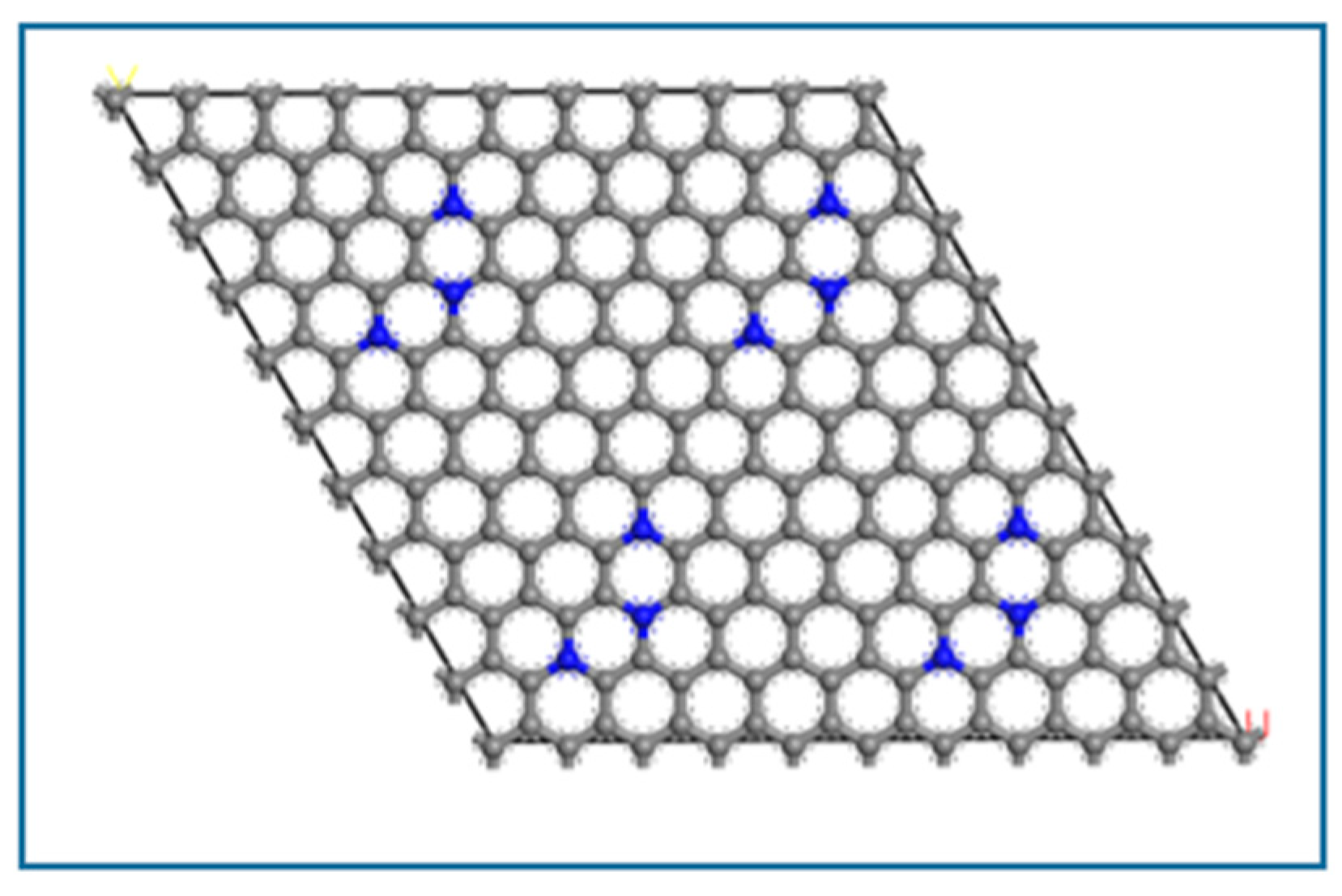
Graphene quantum dots (GQDs) are a collection of carbon quantum dots derived from graphene or graphene oxide, as shown in Figure 2, with a diameter of 2–20 nm [8,9,10]. Due to the obvious quantum constraint, edge effect and unique microstructure, GQDs exhibit many novel chemical/physical and mechanical properties, such as low cytotoxicity, excellent solubility, chemical inertia, stable photoluminescence, better surface grafting and anti-friction characteristics [4,11,12,13,14,15,16,17,18,19]. Based on the above unique structural and performances, graphene quantum dots (GQDs) have broad application prospects in functional materials, structural materials, sensors, biomedicine and so on.
It is well known that graphene quantum dots (GQDs) are a type of carbon dots (CDs), which belong to carbon-based nanomaterials [20,21,22,23]. Compared with toxic metal quantum dots, carbon dots and graphene quantum dots both show ideal low toxicity and good environmental performance. The carbon dots can be amorphous or crystalline in structure, but GQDs are typically graphene-like smaller polycyclic aromatic molecules (PAHs) produced from graphene-based raw materials or rigid synthetic chemistry, with a distinct graphene lattice. This brings about the difference in performance between graphene quantum dots and carbon dots. E.g., luminous CDs are composed of discrete, spheroidal carbon nanoparticles with a size less than 10 nm, while GQDs are single, double or fewer (3–10) layers of graphene fragments with a diameter of 10 nm or less [11,24,25,26,27]. In the early stages of research on GQDs, scholars mainly focused on the development of high-quality GQD preparation techniques and their intrinsic properties.
With the deepening of research, the basic research of GQD preparation technology and intrinsic properties has shifted to the application of GQDs in composite material, electronics, optics, energy storage, conversion devices, anti-friction components and so on [28,29]. This paper focuses on the physical and chemical properties of GQDs and their application as a key component of sensor and analysis systems, in order to expand the application of GQDs and expand the application of GQDs in the future sensing field.

2. Synthesis Strategy

The synthesis strategies for GQDs generally fall into broad categories: top–down and bottom–up processes. In the top–down process, bulk carbon materials such as graphene and carbon black are cracked and cut by chemical/electrochemical stripping, water/solvent heat treatment, microwave/ultrasonic and other methods. Although the top–down approach, with its abundant raw materials and simple operation, is ideal for mass production, non-selective chemical cutting can result in poor control over the size and morphology of the final product (GQDs). The bottom–up strategy is based on the gradual growth of small precursor molecules (ring molecules, polymers) into nanoscale GQDs through carbonization, pyrolysis and chemical vapor deposition, with a high degree of controllability and fewer defects. But poor solubility and the aggregation of the final products are major limiting factors. Therefore, careful consideration is required when selecting a synthesis method for GQDs.

2.1. Top-Down Process

2.1.1. Chemical Stripping Method

The chemical stripping method refers to the stripping of precursor carbon materials such as graphene oxide (GO), carbon nanotubes (CNTs) and carbon fibers with strong oxidants and strong acids. This is a simple, direct and inexpensive synthesis method that can be used for the mass production of high-quality GQDs. Related scholars used concentrated sulfuric acid and concentrated nitric acid mixed acid solutions to strip carbon fiber to obtain graphene quantum dots [30]. By adjusting the stirring temperature, the prepared graphene quantum dots presented a size range of 1–11 nm and a height of 0.4–2 nm and could emit blue, green and yellow fluorescence. Similarly, it has been also reported that GQDs are obtained by hydrothermal cutting carbon sheets, with a thickness of 2.4 nm [31].

2.1.2. Electrochemical Exfoliation Process

GQDs can be synthesized by the electrochemical stripping method using graphite, carbon fiber or carbon nanotubes as precursors. Kenneth Chu reported that [32] GQDs were produced via a top–down electrochemical exfoliation process, where constant current was passed through a graphite rod working electrode and a platinum plate counter electrode. Then, 0.1 M of tetrabutylammonium perchlorate was used as the nitrogen-containing supporting electrolyte dissolved in the anhydrous acetonitrile. The resulting suspension was filtered through a 0.2 μm polytetrafluoroethylene (PTFE) filter before being dialyzed for 24 h in ultrapure water using a cellulose ester dialysis bag with a molecular-weight cut-off of 1000 Da.

3. Physical and Chemical Properties

3.1. Size, Chemical Composition and Crystal Structure

3.1.1. Size

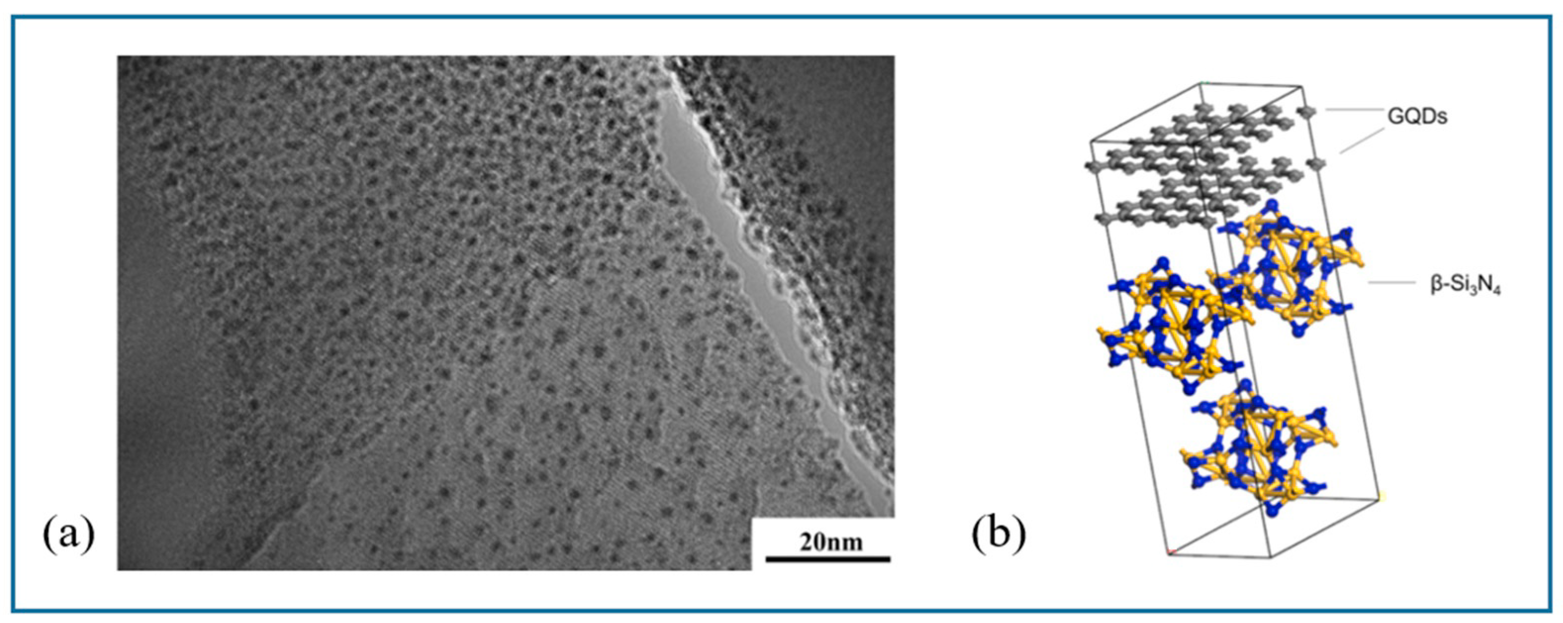
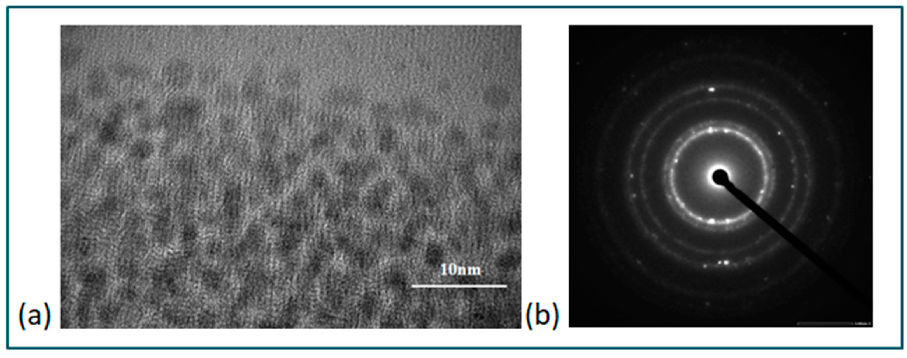
Generally, the average diameter of GQDs is mostly less than 10 nm, and the maximum diameter of GQDs reported at present is 60 nm [21,33]. Under normal circumstances, the size of GQDs is closely related to the material formula and preparation method and has little relationship with the raw material. For example, using lignin as raw material, Chen et al. [34] prepared nitrogen-doped graphene quantum dots (N-GQDs) with a size of 2.3 nm with 2% lignin in the silicon nitride matrix, while N-GQDs with a content of 3% lignin could not be prepared with the same size. With the permission of the corresponding author Chen, the relevant figures provided by the author are shown in Figure 3 and Figure 4. As another example, Pan et al. [12] used the same batch of graphene oxide (GO) as raw material and adopted different thermal reduction temperatures and hydrothermal reduction alkalinity levels to prepare graphene quantum dots with sizes of 9.6 nm and 3 nm. On the other hand, the height size of GQDs is also related to the preparation method, and the heights of GQDs prepared by the same preparation method increase with the increase in their diameter size.
However, the heights and diameters of quantum dots prepared by different preparation methods have different nonlinear relationships; namely, GQDs with large diameters may be thinner than GQDs with small diameters. For example, the diameter size of GQDs prepared by the electrochemical method can be 3 nm and 5 nm, and the corresponding height can be 1–2 nm and 3 nm, while the diameter of GQDs synthesized by K-GICs is 20 nm and the height is 0.9 nm [35]. In general, most GQDs have no more than five layers, and single GQDs have been prepared at present.

3.1.2. Chemical Composition

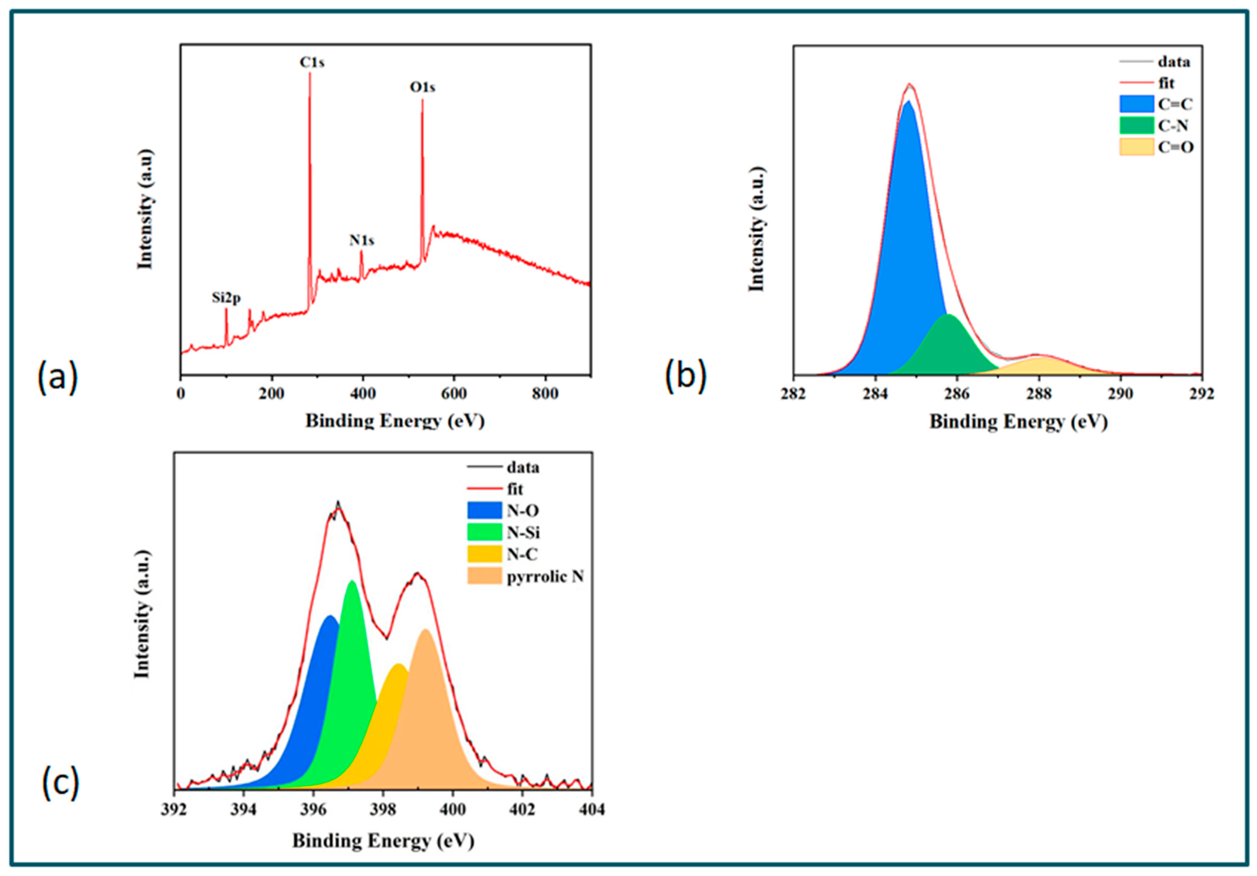
Theoretically, GQDs consist of C and H elements, and their chemical composition can be determined by X-ray photoelectron spectroscopy (XPS) and Fourier transform infrared spectroscopy (FT-IR). However, the strong attraction between GQDs and the oxidation stripping method prepared would lead to partial oxidation, and there are hydroxyl and carboxyl groups on their surfaces. In particular, scholars have also prepared nitrogen-doped graphene quantum dots (N-GQDs) and sulfur-doped graphene quantum dots (S-GQDs) using special raw materials and preparation methods. Dr. Chen from Shaanxi University of Science & Technology used XPS to further analyze N-GQDs in a silicon nitride composite. With the permission of the corresponding author, the relevant figures provided by Dr. Chen are shown in Figure 5. There are two strong peaks of C 1s, Si 2p and N 1s (Figure 5a). The C 1s peaks can be decomposed to carbon (C=C), carbon nitrite (C-N) and carbon oxide (C=O) (Figure 5b), and the N1s peak can be decomposed into peaks at 396.6 eV (corresponding to N-O), 398.8 eV (corresponding to N-C), 397.3 eV (corresponding to N-Si) and 399.5 eV (corresponding to pyrrole N) (Figure 5c). Similarly, Professor Qu Liangti from Beijing Institute of Technology and Professor Shi Gaoquan from Tsinghua University [36] prepared N-GQDs with high oxygen functional groups and electrocatalytic activity by a simple and easy electrochemical method. XPS spectra of graphene films and N-GQDs were compared and observed. The spectra of graphene films and N-GQDs both showed obvious graphite peaks (C1s, 284 eV) and oxygen-rich peaks (O 1s 532 eV), while a distinct N 1s peak was clearly observed on the spectrum of N-GQDs, but not on the spectrum of graphene films. For N-GQDs, the presence of pyridine (398.5 eV) and pyridine (401 eV) N atoms could be determined at the N1s peak, and the C-N bond (285.2 eV), C-O bond (286.6 eV), C=O key (288.3 eV) and O-C=O (289 eV) could be determined at the C1s peak. The O/C atom ratio of this oxygen-rich N-GQDs is 27%, which is basically in line with the O/C atom ratio of non-nitrogen-doped GQDs and slightly higher than the O/C atom ratio of graphene (15%). In such a setting, the atomic structure of oxygen-rich N-GQDs could be simulated based on XPS spectra and Fourier transform infrared spectroscopy (FT-IR) analysis results.

3.1.3. Crystal Structure

The crystal structure of GQDs can be determined by X-ray diffraction (XRD) and transmission electron microscopy (TEM). In general, GQDs have distinct graphene lattice properties, and the crystal structure of graphene quantum dots is related to its carbon source. GQDs prepared from corn stalk as the carbon source could be analyzed by XRD pattern [37]. The XRD results showed that there is a wide diffraction peak (related to the nanometer size of GQDs) at 2θ = 24°, which corresponds to the characteristic peak (002) of the graphite crystal surface. The XRD pattern of GQDs prepared with HBC (hexapendamum) as the carbon source observed characteristic peaks corresponding to (200), (100), (004) and (110) crystal faces [37]. The layer spacing of GQDs obtained by different carbon sources and preparation methods is also different. The layer spacing of GQDs obtained by graphene film stripping is 0.34 nm, which is close to that of graphite. The layer spacing of GQDs prepared from carbon fiber (CF) is 0.403 nm, and the larger layer spacing is related to the exfoliation and oxidation of carbon fiber [31]. The layer spacing of N-GQDs prepared by the hydrothermal treatment of graphene oxide is 0.393 nm [38], and the layer spacing of GQDs-PEG obtained by a hydrothermal reaction is 0.381 nm [26]. In general, the oxygen-containing groups widen the spacing between the graphene sheets. However, there are exceptions, and the layer spacing of highly oxidized GQDs is sometimes smaller than that of low-oxygen GQDs. The O/C of GQDs derived from carbon fiber is 23.7%, and the corresponding layer spacing is 0.403 nm [39]. The O/C of the electrochemically synthesized GQDs is 27%, and the corresponding layer spacing is 0.34 nm [36]. In any case, the crystallinity of graphene quantum dots is very high.

3.2. Optical Properties

3.2.1. Photoluminescence Properties

In terms of light absorption capacity, GQDs usually show a strong light absorption capacity in the ultraviolet region (UV), and the light absorption capacity can extend to the visible light range. Graphene materials have a strong ability to absorb light. The UV-VIS absorption spectra of graphene oxide (GO) shows peaks at ~230 nm (π-π * transition of the aromatic C = C bond) and acromial peaks at ~300 nm (n-π * transition of the C = O bond). For graphene quantum dots (GQDs), these peaks can also be observed in the UV-visible absorption spectrum, and the central wavelength of the π-π * transition peak is between 200 and 270 nm, and the central wavelength of the n-π * transition peak is greater than 260 nm. Moreover, the light absorption capacity of graphene quantum dots is related to their size and preparation method. The size of GQDs derived from CF increases from 1–4 nm to 7–11 nm, and their optical absorption peak shifts from 270 nm to 330 nm. GQDs carbonized from citric acid (~15 nm) have an absorption band of 362 nm, whereas GQDs derived from HBC only have weak acromion at 280 nm [40].
In terms of photoluminescence (PL) properties, GQDs, like CDs, can exhibit photoluminescence in different colors, such as DUV, blue, green, yellow and red [18,33,41,42,43,44,45]. The photoluminescence mechanism of GQDs may originate from their intrinsic emission and defect state emission [15]. The photoluminescence properties of graphene quantum dots are closely related to their energy gap, and the size of the band gap is directly related to the size of graphene quantum dots. For example, the blue emission of hydrothermal cut GQDs (diameter 9.6 nm) is the radiative decay of activated electrons from LUMO to HOMO. At the same time, the oxidation functional groups (such as hydroxyl, carbonyl and carboxyl groups) on the surface of GQDs cause it to form a “surface state” (i.e., the energy level between the π and π * states of C = C), leading to a series of emission traps. When a certain excitation wavelength irradiates GQDs, the surface state emission trap would dominate emissions [13]. A higher degree of surface oxidation will lead to more surface traps, which will lead to redshift emission [46]. In general, the PL of GQDs is a combination effect or competition between eigenstate emission and defect state emission, and GQDs prepared by different methods may have different photoluminescence mechanisms, which leads to the need to consider key factors such as GQDs size, excitation wavelength, pH and solvent when achieving the photoluminescence characteristics of GQDs.
In addition to strong down-conversion photoluminescence (PL) properties, some GQDs also exhibit obvious up-conversion photoluminescence properties [47]. For PEG passivated GQDs prepared by acid oxidation, the up-conversion emission peak redshifts from 390 nm to 468 nm when the excitation wavelength changes from 600 nm to 800 nm. When a low-energy photon excites an electron in the PI orbit, the PI electron is excited into a higher energy state like LUMO, and the electron then returns to a lower energy state, resulting in up-transition PL emission. Similarly, PEG-GQDs prepared by hydrothermal reactions were observed to produce up-conversion luminescence induced by such anti-Stokes transitions [26].

3.2.2. Electrochemical Luminescence Performance (ECL)

GQDs have also been observed for ECL emission properties. For example, with K2S2O8 as a coreactant, negative cycling occurs between 0 and −1.6 V, and GQDs show strong ECL emission at −1.45 V [7]. Without K2S2O8 as a coreactant, the ECL emission of GQDs cannot be observed. It is concluded that S2O82- and GQDs undergo electrochemical reduction reactions to produce SO4•− strong oxidizing free radicals and GQDs•− free radicals, and then, SO4•− strong oxidizing free radicals react with GQDs•− through electron transfer annihilation, thus obtaining excited GQDs* and achieving luminescence.
Compared with CDs, GQDs have obvious peak ELC at relative positive potential. At the same time, the maximum wavelength of the ECL spectrum of GQDs is 512 nm, which has a small redshift (12 nm) compared with the PL spectrum, indicating that the surface defects of GQDs can be ignored.
Due to their unique photoluminescence and electrochemical luminescence properties as well as their electrical properties, graphene quantum dots have been widely used in sensors as follows.

4. Applications in the Field of Sensing

GQDs have good electrical conductivity and inherent fluorescence properties, and they can be used as probes for sensors to detect various analytical targets. At present, the sensors manufactured by graphene quantum dots can be roughly divided into optical sensors, optical biosensors, electrochemical biosensors and photoelectrochemical biosensors and so on. In this paper, the applications of graphene quantum dots in ion detection sensors, fluorescent biosensors, electrochemical biosensors and electronic sensor are introduced.

4.1. Ion Detection Sensor

GQDs have excellent PL performance and are suitable for photoluminescent sensors, and their detection limit (LOD) can reach mole, picomolar or even femtomolar level, especially in ion detection, showing great application potential, as shown in Table 1. Specific applications of GQDs in ion detection are described below.
Graphene quantum dots can be used to detect silver ions. Ag+ has antibacterial and bactericidal properties, often used in industrial and medical fields; the excessive or unintentional use of silver can lead to diarrhea, skin infections, nerve or organ damage and other diseases, so the accurate detection of Ag+ has very important practical significance. Mandal et al. [48] functionalized DNA GQDs to obtain a high-performance silver ion detection system, in which the synthesized GQDs act as a fluorophore, while silver ions act as a fluorescence quencher. Therefore, GQDs can be used as a fluorescence sensing probe for the labeling-free detection of Ag+, with high sensitivity and selectivity. Similarly, Suryawanshi et al. [49] synthesized graphene quantum dots (GQDs) from biological waste and modified them to prepare aminoterminal graphene quantum dots (Am-GQDs) with higher dispersion and photoluminescence intensity. When water contains multiple metal ions (Cu2+, Co2+, Hg2+, Fe2+, Fe3+, Ni2+ and Pb2+), the fluorescence quenching of the AM-GQD sensor system, but with the introduction of L-cysteine to the Am-GQD system, the fluorescence quenching of the AM-GQD sensor system is reduced. Only Ag+ ions dissociate from Am-GQD and connect to the -SH group of L-cysteine to restore fluorescence. The method has the advantages of high sensitivity, good stability, good selectivity, simple operation, low cost and visible light detection, and it has broad application prospects in the detection of Ag+ ions in aqueous solutions.
Graphene quantum dots can also be used to detect mercury ions. Mercury ion (Hg 2+) cannot be metabolized in the human body, and accumulation in the human body will lead to heart, liver and nervous system diseases and even lead to the formation of malignant tumors. Dissolved Hg 2+ often has high chemical activity, so how to accurately detect Hg2+ content in water environments has always been the focus of attention. Sharma et al. [50] prepared sulfur and nitrogen co-doped GQDs (S, N-GQDs) by a solid-state reaction using thiourea and citric acid as precursors. When exposed to PL radiation, the prepared S, N-GQDs showed good selectivity and sensitivity to Ag+ and Hg2+ ions. The lowest detected concentrations for Ag+ and Hg2+ were as low as 12.9 and 9.14 µM, respectively. Shi et al. [51] prepared N-GQDs by solid-phase synthesis using citric acid (CA) as the carbon source and 3, 4-dihydroxy-L-phenylalanine (L-DOPA) as the nitrogen source. Studies have shown that Hg2 + and the ‘O’ atoms on the surface of N-OGQDs can be strongly chemically bonded to form a stable metal complex, and non-radiative electron transfer occurs in the d orbital of the N-OGQD excited state to Hg2+, resulting in the quenching of the photoluminescence sensor. Therefore, an efficient fluorescence sensor for detecting Hg2+ can be developed. The detection limit is as low as 8.6 nM.
Graphene quantum dots can also be used for other metal ions. Wang et al. [52] prepared GQDs using coffee grounds as the carbon source and modified them with polyethylenimide (PEI). The N in the obtained PEI-GQDs could effectively bind with Fe 3+ and Cu 2+ ions in the environment and quench PL, thus developing a detection system for Fe 3+ and Cu 2+ ions. At the same time, Wang et al. [53] also used durian as the carbon source to prepare GQDs, modified GQDs with a sulfhydryl group and then prepared photoluminescent sensors that could detect metal ions such as Hg+, Zn2+, Ag+ and Cd+. Fluorescence-enabled nanosensors based on GQDs can also be used to detect harmful lead ions (Pb2+) [54].
The sensor, which uses the selectivity of graphene quantum dots to specific metal ions to achieve the detection of target ions, is simple and low cost and can be used to detect various trace elements in the human body and small instant inspection instruments. However, the sensitivity of this type of sensor is generally poor. Therefore, how to improve the sensitivity of graphene quantum dots as ion sensors is the future development direction.

4.2. Biosensor

4.2.1. Fluorescent Biosensor

Fluorescent biosensors use biometric elements to convert the signal of the target object into optical signal output. Similar to photoluminescence (PL) sensors, biological probes (such as DNA, antibodies, enzymes, etc.) are modified on the surface of GQDs to achieve the detection of target substances by quenching or restoring the fluorescence signal inherent in GQDs [55].
Based on the PL characteristics of GQDs, Lu et al. [56] obtained a SiO2/GQDs/ssDNA-2 composite system by using nano-SiO2 as a carrier and carrying GQDs and ssDNA-2. When adenosine triphosphate (ATP) was present, SiO2/GQDs were fixed to the electrode surface to generate ECL signals; if ATP was not present, there was only a very weak ECL signal on the electrode surface, and then, a biosensor could be built to detect ATP.
Zhao et al. [57] proposed a simple and sensitive fluorescence quantitative analysis method using GQDs to detect protein kinase activity. The researchers coupled the peptide substrate to the surface of GQDs. When the substrate peptide was phosphorylated by CK2, the interaction between Zr4+ ions and phosphate groups was introduced, resulting in the fluorescence quenching of GQDs. Based on PL quenching, protein kinase activity can be easily and sensitively monitored.
Based on the physical properties of polydopamine (pDA) formation on the surface of graphene quantum dots (GQDs), Zheng et al. [58] developed a fast and simple fluorescence sensing strategy for the detection of dopamine (DA). In alkaline environment, DA self-polymerizes into pDA and combines with the surface of GQDs to form a thin film, which leads to fluorescence quenching, and then, the detection limit of DA is 8 nm.
Fluorescent biosensors based on GQDs can detect various biological calibrators by modifying biological probes, which has great application prospects, but its sensitivity needs to be further improved. In the future, how to further improve the sensitivity of graphene quantum dot fluorescent biosensors must be also a technical hotspot.

4.2.2. Electrochemical Biosensor

Electrochemical immune sensors have good medical significance and practical value. In recent years, in order to further improve the detection sensitivity, quantum dots and electroactive composite systems coated with nanoparticles have often been used to develop new electrochemical immune sensors [59]. Graphene quantum dots can also be used to build electrochemical immune sensors. Kalkal et al. [60] prepared amine-N-GQDs with uniform and small size (~3 nm) by the hydrothermal method, which has strong fluorescence, blue luminescence and long fluorescence decay life. Using biofunctionalized amine-N-GQDs as energy donors and gold nanoparticles (AuNPs) as energy receptors, the scholars constructed a fluorescent-enhanced biosensor that can be used to ultra-sensitively detect small-cell lung cancer biomarkers. Yang et al. [61] used the hydrothermal method to mount Pt-Pd metal nanoparticles on N-GQDs to form Pt-Pd/N-GQDs and immobilized Pt-Pd/N-GQDs with Au nanoparticles, that is, Pt-Pd/N-GQDs@Au was obtained. Pt-Pd/N-GQDs@Au could effectively detect carcinoembryonic antigen (CEA) and thus develop a new type of ultra-sensitive electrochemical immunosensor.
The electrochemical enzyme electrode sensor uses the enzyme as the sensitive element and the electrode to convert the undetectable substrate into the product that can be detected by the electrochemical method through the catalysis of the enzyme. In recent years, GQDs have been used as a fixing material for enzymes because of their advantages such as larger surfaces, strong conductivity, non-toxicity and easy synthesis, and GQDs, as a nanocapsuler for fixing enzymes, have shown good biological activity and stability [59]. Ju et al. [62] prepared an electrochemical biosensor N-GQDs@AuNP based on the high electrocatalytic activity of Au nanoparticles. The biosensor could be used to detect a very small amount of H2O2 associated with cervical cancer cells in human serum, and its detection limit was as low as 0.12 mM. Based on horseradish peroxidase (HRP), Muthurasu et al. [63] used the amide bond between the carboxyl group of GQDs and the amine group of the enzyme to covalently fix the enzyme on a glassy carbon electrode (GCE) modified by GQDs, so as to detect H2O2 and its content by the electrochemical method.
Electrochemical biosensors based on the functionalization of graphene quantum dots (GQDs) have been paid attention by researchers and achieved fruitful research results, but the electrochemical biosensors based on GQDs still have some problems such as unsatisfactory sensitivity, a complex preparation process and high operation requirements.

4.2.3. Electronic Sensor

Electronic sensors are generally used to detect a certain physical quantity (such as temperature, photoelectric, humidity, pressure, etc.), and can measure and control these physical quantities by converting them into electrical parameters such as voltage and current or into the on–off of a circuit [64].
Graphene quantum dots can also be used in electronic sensors because of their unique crystal structure and microstructure. Sreeprasad et al. [65] obtained GQDs by oxidation induction, edge coarsening and the cracking of synthesized graphene nanoribbons and applied them to electronic sensors of pressure and humidity. These GQDs selectively formed Coulomb blocking electroosmotic networks with a dry tunneling distance of 0.58 nm and activation energy of 3 meV at the interface of polyelectrolyte microfibers. When water caused by pressure and humidity could be transported through hygroscopic polymer microfibers (Henry constant = 0.215 torr−1), the average tunneling barrier width between GQDs was reduced by 0.36 nm (tunneling barrier = 5.11 eV), which could increase the conductivity of the device by 43 times, and the detection sensitivity of pressure and humidity was greatly improved. Hou et al. [66] developed a new fiber Bragg grating (FBG) relative humidity (RH) sensor based on polyimide (PI) doped with graphene quantum dots (GQDs). When the doping mass fraction of GQDs was 0.2%, the doping of GQDs increased the number of adsorption sites in the membrane and introduced a large number of polar oxygen-containing groups into the composite membrane. Therefore, the adsorption capacity of a PI/GQD composite membrane to water was enhanced, and the sensitivity of the humidity-sensitive membrane was significantly improved.
Graphene quantum dots can also be used for single-electron transistor (SET) charge sensors. Wang et al. [67] used electron beam or ion etching to prepare a SET charge sensor based on GQDs, where the distance of the graphene nanostructure was determined by the etching area. The SET was placed close to the GQDs, resulting in strong capacitive coupling between the two systems. Once an extra electron occupied a quantum dot, the potential in adjacent sets was altered by capacitive interactions, causing a measurable change in conductance. In this way, the charge transfer through a quantum dot could be measured using a SET charge sensor even if it is too small to be measured by conventional means.
In short, the electronic sensors based on the graphene quantum dot (GQD) electronic tunneling mechanism are still in the research and development stage, and the mechanism of electronic sensors based on GQDs needs to be further clarified, and the application scope needs to be further expanded.

5. Conclusions

As new carbon nanomaterials, GQDs have made great progress in the field of sensing systems in just a few years. With the deepening of the understanding of the size, chemical composition and crystal structure of graphene quantum dots and the continuous optimization and improvement of carbon source materials and preparation methods, GQDs have been able to carry out appropriate functional treatments and reasonable chemical modifications. More and more new materials derived from graphene quantum dots continue to emerge, and in particular, the sensing system built based on these new materials is changing with each passing day, e.g., ion detection sensors, fluorescent biosensors, electrochemical biosensors and electronic sensors. In general, the fluorescence properties, electrical properties and electrochemical properties of GQDs and their derived new materials provide a new idea and method for realizing the effective analysis and determination of metal ions, enzymes, proteins and other substances.
GQDs have shown great application potential in sensing systems and other application fields, but the related research of GQDs is still in its infancy. There are still many problems to be solved. The complexity of preparation methods, low yield quantification and photoluminescence mechanism are not clear, which should limit their application scope in traditional systems. In any case, the understanding and control of graphene quantum dots is bound to continue to strengthen. With the advancement of scientific research, the application of graphene quantum dots in the engineering field should be greatly promoted.

Funding

This research was funded by Yunnan provincial fund project (grant number No. 202104BN050011).

Data Availability Statement

Data are contained within the article.

Acknowledgments

The authors are thankful for the funding provided by Yunnan provincial fund project (grant number No. 202104BN050011).

Conflicts of Interest

X.T., F.Z., W.L., G.A. and X.X. were employed by the company Yunnan Power Grid Co., Ltd., The remaining authors declare that the research was conducted in the absence of any commercial or financial relationships that could be construed as a potential conflict of interest.

References

  1. Li, L.; Wu, G.; Yang, G.; Peng, J.; Zhu, J.J. Focusing on luminescent graphene quantum dots: Current status and future perspectives. Nanoscale 2013, 5, 4015–4039. [Google Scholar] [CrossRef] [PubMed]
  2. Lijima, S. Helical microtubules of graphic carbon. Nature 1991, 354, 56–58. [Google Scholar]
  3. Kroto, H.W.; Heath, J.R.; O’brien, S.C.; Curl, R.F.; Smalley, R.E. Buckminsterfullerene. Nature 1985, 318, 162–163. [Google Scholar] [CrossRef]
  4. Zhang, W.B.; Li, L.; Li, S.C.; Ma, J.Z.; Liu, C.; Bao, Y. Modification and application of graphene quantum dots. Acta Mater. Compos. Sin. 2022, 39, 3104–3120. [Google Scholar]
  5. Novoselov, K.S.; Geim, A.K.; Morozov, S.V.; Jiang, D.; Zhang, Y.; Dubonos, S.V.; Grigorieva, I.V.; Firsov, A.A. Electric Field Effect in Atomically Thin Carbon Films. Science 2004, 306, 666–669. [Google Scholar] [CrossRef]
  6. Li, L.S.; Yan, X. Colloidal Graphene Quantum Dots. J. Phys. Chem. Lett. 2010, 1, 2572–2576. [Google Scholar] [CrossRef]
  7. Li, L.L.; Jing, J.; Fei, R.; Wang, C.Z.; Qian, L.; Zhang, J.R.; Zhu, J.J. A Facile Microwave Avenue to Electrochemiluminescent Two-Color Graphene Quantum Dots. Adv. Funct. Mater. 2012, 22, 2971–2979. [Google Scholar] [CrossRef]
  8. Yan, X.; Cui, X.; Li, L.S. Synthesis of Large, Stable Colloidal Graphene Quantum Dots with Tunable Size. J. Am. Chem. Soc. 2010, 132, 5944–5945. [Google Scholar] [CrossRef]
  9. Yan, X.; Cui, X.; Li, B.; Li, L.S. Large, solution-processable graphene quantum dots as light absorbers for photovoltaics. Nano Lett. 2010, 10, 1869–1873. [Google Scholar] [CrossRef]
  10. Ding, Y.; Diao, Q.; Liu, D. Synthesis of graphene quantum dots and its application in gas sensing. Chin. J. Anal. Chem. 2022, 50, 495–505. [Google Scholar]
  11. Li, Y.Q.; Xiao, Y.; Tao, Q.; Yu, M.M.; Zheng, L.; Yang, S.W.; Ding, G.Q.; Dong, H.; Xie, X.M. Selective coordination and localized polarization in graphene quantum dots: Detection of fluoride anions using ultra-low-field NMR relaxometry. Chin. Chem. Lett. 2021, 32, 3921–3926. [Google Scholar] [CrossRef]
  12. Pan, D.; Zhang, J.; Li, Z.; Wu, M.H. Hydrothermal route for cutting graphene sheets into blue-luminescent graphene quantum dots. Adv. Mater. 2010, 22, 734–738. [Google Scholar] [CrossRef] [PubMed]
  13. Hu, X.R.; Zhu, W.; Zhao, M.H.; Wang, G.; Yang, S.W.; Liu, Z.D.; Zheng, L.; Guo, Q.J. Graphene quantum dots promoted the synthesis of heavily n-type graphene for near-infrared photodetectors. J. Phys. Chem. C 2020, 124, 1674–1680. [Google Scholar] [CrossRef]
  14. Tang, L.; Ji, R.; Cao, X.; Lin, Y.Y.; Jiang, H.X.; Li, X.M.; Teng, K.S.; Luk, C.K.; Zeng, S.J.; Hao, J.H.; et al. Deep ultraviolet photoluminescence of water-soluble self-passivated graphene quantum dots. ACS Nano 2012, 6, 5102–5110. [Google Scholar] [CrossRef]
  15. Lu, F.; Yang, S.W.; Song, Y.X.; Zhai, C.; Wang, Q.; Ding, G.; Kang, Z. Hydroxyl functionalized carbon dots with strong radical scavenging ability promote cell proliferation. Mater. Res. Express 2019, 6, 065030. [Google Scholar] [CrossRef]
  16. Li, X.M.; Rui, M.C.; Song, J.Z.; Shen, Z.H.; Zeng, H.B. Carbon and graphene quantumdots for optoelectronic and energy devices: A review. Adv. Funct. Mater. 2015, 25, 4929–4947. [Google Scholar] [CrossRef]
  17. Huang, H.; Yang, S.; Liu, Y.; Yang, Y.C.; Kang, Z.H. Photocatalytic polymerization from amino acid to protein by carbon dots at room temperature. ACS Appl. Bio Mater. 2019, 2, 5144–5153. [Google Scholar] [CrossRef]
  18. Dong, Y.; Chen, C.; Zheng, X.; Gao, L.L.; Cui, Z.; Yang, H.B.; Guo, C.X.; Chi, Y.W.; Chang, M. One-step and high yield simultaneous preparation of single- and multi-layer graphene quantum dots from CX-72 carbon black. J. Mater. Chem. 2012, 22, 8764–8766. [Google Scholar] [CrossRef]
  19. Gupta, V.; Chaudhary, N.; Srivastava, R.; Sharma, G.D.; Bhardwaj, R.; Chand, S. Luminscent graphene quantum dots for organic photovoltaic devices. J. Am. Chem. Soc. 2011, 133, 9960–9963. [Google Scholar] [CrossRef]
  20. He, J.L.; He, Y.L.; Chen, Y.H.; Lei, B.F.; Zhuang, J.L.; Xiao, Y.; Liang, Y.; Zheng, M.T.; Zhang, H.R.; Liu, Y.L. Solid-state carbon dots with red fluorescence and efficient construction of dual-fluorescence morphologies. Small 2017, 13, 1700075. [Google Scholar] [CrossRef]
  21. Li, M.X.; Chen, T.; Gooding, J.J.; Liu, J.Q. A review of carbon and graphene quantum dots for sensing. ACS Sens. 2019, 4, 10226–10333. [Google Scholar] [CrossRef] [PubMed]
  22. Zhang, B.L.; Li, J.H.; Lu, D.S.; Zhang, K.X.; Wang, X.J.; Ma, L. Graphene quantum dots fluorescence enhancement and pH response characteristics. Chin. Opt. 2023, 16, 523–534. [Google Scholar]
  23. Wang, B.; Lu, S. The light of carbon dots: From mechanism to applications. Matter 2022, 5, 110–149. [Google Scholar] [CrossRef]
  24. Baker, S.N.; Baker, G.A. Luminescent Carbon Nanodots: Emergent Nanolights. Angew. Chem. 2010, 49, 6726–6744. [Google Scholar] [CrossRef] [PubMed]
  25. Zhang, Z.; Zhang, J.; Chen, N.; Qu, L. Graphene quantum dots: An emerging material for energy-related applications and beyond. Energy Environ. Sci. 2012, 5, 8869–8890. [Google Scholar] [CrossRef]
  26. Shen, J.; Zhu, Y.; Yang, X. One-pot hydrothermal synthesis of graphene quantum dots surface-passivated by polyethylene glycol and their photoelectric conversion under near-infrared light. R. Soc. Chem. 2012, 36, 97–101. [Google Scholar]
  27. Xu, A.L.; Wang, G.; Li, Y.Q.; Dong, H.; Yang, S.W.; He, P.; Ding, G.Q. Carbon-based quantum dots with solid-state photoluminescent: Mechanism, implementation, and application. Small 2020, 16, 2004621. [Google Scholar] [CrossRef]
  28. Ozhukil Valappil, M.K.; Pillai, V.; Alwarappan, S. Spotlighting graphene quantum dots and beyond: Synthesis, properties and sensing applications. Appl. Mater. Today 2017, 9, 350–371. [Google Scholar] [CrossRef]
  29. Li, K.; Zhang, G.J. Research progress of carbon quantum dots and graphene quantum dots in sensing field. Lab. Med. 2022, 37, 787–792. [Google Scholar]
  30. Wang, S.J.; Lenzini, F.; Chen, D.; Tanner, P.; Han, J.S.; Thiel, D.; Lobino, M.; Li, Q. Chemically derived graphene quantum dots for high-strain sensing. J. Mater. Technol. 2023, 141, 110–115. [Google Scholar] [CrossRef]
  31. Peng, J.; Gao, W.; Gupta, B.K.; Liu, Z.; Romero-Aburto, R.; Ge, L.H.; Song, L.; Alemany, L.B.; Zhan, X.B.; Gao, G.H. Graphene quantum dots derived from carbon filers. Nano Lett. 2012, 12, 844–849. [Google Scholar] [CrossRef] [PubMed]
  32. Chu, K.; Adsetts, J.R.; He, S.J.; Zhan, Z.Y.; Yang, L.; Wong, J.M.; Love, D.A.; Ding, Z. Electrogenerated chemiluminescence and electroluminescence of N-doped graphene quantum dots fabricated from an electrochemical exfoliation process in nitrogen-containing electrolytes. Chem. A Eur. J. 2020, 26, 15892–15900. [Google Scholar] [CrossRef]
  33. Liu, R.; Wu, D.; Feng, X.; Mueelen, K. Bottom-Up Fabrication of Photoluminescent Graphene Quantum Dots with Uniform Morphology. J. Am. Chem. Soc. 2011, 133, 15221–15223. [Google Scholar] [CrossRef] [PubMed]
  34. Chen, W.; Zhao, Z.Q.; Lou, R. Study on self-derived products of nanometer lignin in silicon nitride ceramics during sintering process. Results Mater. 2021, 12, 100228. [Google Scholar] [CrossRef]
  35. Lin, L.X.; Zhang, S.W. Creating high yield water soluble luminescent graphene quantum dots via exfoliating and disintegrating carbon nanotubes and graphite flakes. Chem. Commun. 2012, 48, 10177–10179. [Google Scholar] [CrossRef]
  36. Li, Y.; Zhao, Y.; Cheng, H.; Hu, Y.; Shi, G.; Dai, L.; Qu, L. Nitrogen-Doped Graphene Quantum Dots with Oxygen-Rich Functional Groups. J. Am. Chem. Soc. 2012, 134, 15–18. [Google Scholar] [CrossRef]
  37. Teymourinia, H.; Salavati-Niasari, M.; Amiri, O.; Safardoust-Hojaghan, H. Synthesis of graphene quantum dots from corn powder and their application in reduce charge recombination and increase free charge carriers. J. Mol. Liq. 2017, 242, 447–455. [Google Scholar] [CrossRef]
  38. Yang, Y.; Wu, D.; Han, S.; Hu, P.F.; Liu, R.L. Bottom-up fabrication of photoluminescent carbon dots with uniform morphology via a soft–hard template approach. Chem. Commun. 2013, 49, 4920–4922. [Google Scholar] [CrossRef]
  39. Minati, L.; Del Piano, A. Facile Synthesis of Water-Soluble, Highly-Fluorescent Graphene Quantum Dots from Graphene Oxide Reduction for Efficient Cell Labelling. C J. Carbon Res. 2019, 5, 77. [Google Scholar] [CrossRef]
  40. Hu, C.; Liu, Y.; Yang, Y.; Cui, J.H.; Rong, J.H. One-step preparation of nitrogen-doped graphene quantum dots from oxidized debris of graphene oxide. J. Mater. Chem. B 2012, 1, 39–42. [Google Scholar] [CrossRef]
  41. Zhang, M.; Bai, L.; Shang, W.; Xie, W.J.; Ma, H.; Fu, Y.Y.; Fang, D.C.; Sun, H.; Fan, L.Z.; Han, M.; et al. Facile synthesis of water-soluble, highly fluorescent graphene quantum dots as a robust biological label for stem cells. J. Mater. Chem. 2012, 22, 7461–7467. [Google Scholar] [CrossRef]
  42. Tetsuka, H.; Asahi, R.; Nagoya, A.; Okamoto, K.; Tajima, I. Optically tunable amino-functionalized graphene quantum dots. Adv. Mater. 2012, 24, 5333–5338. [Google Scholar] [CrossRef] [PubMed]
  43. Tang, L.B.; Ji, R.B.; Li, X.M.; Bai, G.X.; Liu, C.P.; Hao, J.H.; Lin, J.Y.; Jiang, H.X. Deep ultraviolet to near-infrared emission and photoresponse in layered N-doped graphene quantum dots. ACS Nano 2014, 8, 6312–6320. [Google Scholar] [CrossRef]
  44. Yang, S.W.; Zhu, C.; Sun, J.; Peng, H.; Yuan, N.Y.; Ding, J.N.; Ding, G.Q.; Xie, X.M. Triphenylphosphine modified graphene quantum dots: Spectral modulation for full spectrum of visible light with high quantum yield. RSC Adv. 2015, 5, 33347–33350. [Google Scholar] [CrossRef]
  45. Ge, J.C.; Lan, M.H.; Zhou, B.J.; Liu, M.M.; Guo, L.; Wang, H.; Jia, Q.Y.; Niu, G.; Huang, X.; Zhou, H.Y.; et al. A graphene quantum dot photodynamic therapy agent with high singlet oxygen generation. Nat. Commun. 2014, 5, 4596. [Google Scholar] [CrossRef]
  46. Bao, L.; Zhang, Z.L.; Tian, Z.Q.; Zhang, L.; Liu, C.; Lin, Y.; Qi, B.; Pang, D.W. Electrochemical Tuning of Luminescent Carbon Nanodots: From Preparation to Luminescence Mechanism. Adv. Mater. 2011, 23, 5801–5806. [Google Scholar] [CrossRef]
  47. Shen, J.; Zhu, Y.; Chen, C.; Yang, X.L.; Li, C.Z. Facile Preparation and Upconversion Luminescence of Graphene Quantum Dots. Chem. Commun. 2011, 47, 2580–2582. [Google Scholar] [CrossRef]
  48. Mandal, D.; Khatun, S.; Gupta, A.N.; Chandra, A. DNA supported graphene quantum dots for Ag ion sensing. Nanotechnology 2019, 30, 255501. [Google Scholar] [CrossRef]
  49. Suryawanshi, A.; Biswal, M.; Mhamane, D.; Gokhale, R.; Ogale, S. Large scale synthesis of graphene quantum dots (GQDs) from waste biomass and their use as an efficient and selective photoluminescence on-off-on probe for Ag(+) ions. Nanoscale 2014, 6, 11664–11670. [Google Scholar] [CrossRef]
  50. Sharma, E.; Vashisht, D.; Vashisht, A.; Vats, V.; Singh, K. Facile synthesis of sulfur and nitrogen codoped graphene quantum dots for optical sensing of Hg and Ag ions. Chem. Phys. Lett. 2019, 730, 436–444. [Google Scholar] [CrossRef]
  51. Shi, B.; Zhang, L.; Lan, C.; Zhao, J.J.; Su, Y.B.; Zhao, S.L. One-pot green synthesis of oxygen-rich nitrogen-doped graphene quantum dots and their potential application in pH-sensitive photoluminescence and detection of mercury(II) ions. Talanta 2015, 142, 131–139. [Google Scholar] [CrossRef] [PubMed]
  52. Wang, L.; Li, W.; Wu, B.; Li, Z.; Wang, S.; Liu, Y.; Pan, D.; Wu, M. Facile synthesis of fluorescent graphene quantum dots from coffee grounds for bioimaging and sensing. Chem. Eng. J. 2016, 300, 75–82. [Google Scholar] [CrossRef]
  53. Wang, G.; Guo, Q.; Chen, D.; Liu, Z.D.; Zheng, X.H. Facile and Highly Effective Synthesis of Controllable Lattice Sulfur-Doped Graphene Quantum Dots via Hydrothermal Treatment of Durian. ACS Appl. Mater. Interfaces 2018, 10, 5750–5759. [Google Scholar] [CrossRef]
  54. Qian, Z.S.; Shan, X.Y.; Chai, L.J.; Chen, J.R.; Feng, H. A fluorescent nanosensor based on graphene quantum dots-aptamer probe and graphene oxide platform for detection of lead (II) ion. Biosens. Bioelectron. 2015, 68, 225–231. [Google Scholar] [CrossRef]
  55. Yu, J.; Yao, Z.H.; He, K.Y.; Xing, B.C.; Wang, Q.; Cheng, K.J.; Wang, L.; Xu, X.H. Nanomaterials-based optical biosensor for detection of mycotoxins in traditional Chinese medicine. Chin. J. Anal. Chem. 2023, 51, 472–483. [Google Scholar]
  56. Lu, J.; Yan, M.; Ge, L.; Wang, S.W.; Yan, J.X.; Yu, J.H. Electrochemiluminescence of blue-luminescent graphene quantum dots and its application in ultrasensitive aptasensor for adenosine triphosphate detection. Biosens. Bioelectron. 2013, 47, 271–277. [Google Scholar] [CrossRef] [PubMed]
  57. Liang, R.P.; Qiu, W.B.; Zhao, H.F.; Xiang, C.Y.; Qiu, J.D. Electrochemiluminescence resonance energy transfer between graphene quantum dots and graphene oxide for sensitive protein kinase activity and inhibitor sensing. Anal. Chim. Acta 2016, 904, 58–64. [Google Scholar] [CrossRef] [PubMed]
  58. Zheng, Z.F.; Qiu, H.Z.; Lin, X.H.; Dong, L.; Liu, A.L. A unique turn-off fluorescent strategy for sensing dopamine based on formed polydopamine (pDA) using graphene quantum dots (GQDs) as fluorescent probe. Sens. Actuators B Chem. 2015, 221, 7–14. [Google Scholar]
  59. Lei, X.Y.; Zuo, G.F.; Wang, P.; Li, Z.F. Research progress and analytical application of electrochemical biosensor based on N-GQDs. J. Funct. Mater. Devices 2020, 26, 184–190. [Google Scholar]
  60. Kalkal, A.; Pradhan, R.; Kadian, S.; Manik, G.; Packirisamy, G. Biofunctionalized graphene quantum dots based fluorescent biosensor towards efficient detection of small cell lung cancer. ACS Appl. Bio Mater. 2020, 3, 4922–4932. [Google Scholar] [CrossRef]
  61. Yang, Y.; Liu, Q.; Liu, Y.; Cui, J.J.; Liu, H.; Wang, P.; Li, Y.Y.; Chen, L.; Zhao, Z.D.; Dong, Y.H. A novel label-free electrochemical immunosensor based on functionalized nitrogen-doped graphene quantum dots for carcinoembryonic antigen detection. Biosens. Bioelectron. 2017, 90, 31–37. [Google Scholar] [CrossRef] [PubMed]
  62. Ju, J.; Chen, W. In Situ Growth of Surfactant-Free Gold Nanoparticles on Nitrogen-Doped Graphene Quantum Dots for Electrochemical Detection of Hydrogen Peroxide in Biological Environments. Anal. Chem. 2014, 87, 1903. [Google Scholar] [CrossRef] [PubMed]
  63. Muthurasu, A.; Ganesh, V. Horseradish Peroxidase Enzyme Immobilized Graphene Quantum Dots as Electrochemical Biosensors. Appl. Biochem. Biotechnol. 2014, 174, 945–959. [Google Scholar] [CrossRef] [PubMed]
  64. Lu, Y. Analysis of Electronic Sensor in Daily Life. Mod. Ind. Econ. Inf. 2022, 12, 220–221. (In Chinese) [Google Scholar]
  65. Sreeprasad, T.S.; Rodriguez, A.A.; Colston, J.; Graham, A.; Shishkin, E.; Pallem, V.; Berry, V. Electron-Tunneling Modulation in Percolating Network of Graphene Quantum Dots: Fabrication, Phenomenological Understanding, and Humidity/Pressure Sensing Applications. Nano Lett. 2013, 13, 1757–1763. [Google Scholar] [CrossRef] [PubMed]
  66. Hou, J.; Dai, J.; Zhang, F.; Yang, M. Advanced Fiber-Optic Relative Humidity Sensor Based on Graphene Quantum Dots Doped Polyimide Coating. IEEE Photonics Technol. Lett. 2022, 34, 725–728. [Google Scholar] [CrossRef]
  67. Wang, L.J.; Cao, G.; Tu, T.; Li, H.; Zhou, C.; Hao, X.J.; Su, Z.; Guo, G.C.; Jiang, H.W.; Guo, G.P. A graphene quantum dot with a single electron transistor as an integrated charge sensor. Am. Inst. Phys. 2010, 97, 183–214. [Google Scholar] [CrossRef]
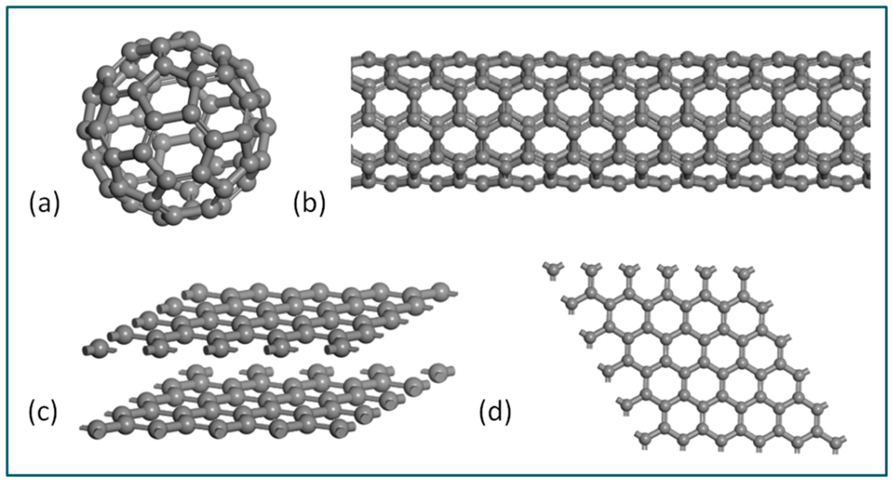
Figure 1. The crystal structure diagrams of fullerenes (a), carbon nanotubes (b), graphite (c) and graphene (d).
Coatings 13 01956 g001
Figure 2. Schematic diagram of the physical model of single-layer graphene quantum dots.
Coatings 13 01956 g002
Figure 3. TEM image (a) of N-GQDs in silicon nitride-based composites and their corresponding surface model (b).
Coatings 13 01956 g003
Figure 4. (a) Magnified TEM image and (b) the corresponding image of diffraction pattern for N-GQDs in silicon nitride-based composites.
Coatings 13 01956 g004
Figure 5. (a) XPS spectra of N-GQDs in silicon nitride composite, (b) C 1s peaks and (c) N1s peaks.
Coatings 13 01956 g005
Table 1. Summary table of graphene quantum dots used to detect metal ions.
ScholarsGQDsTypes of Metal Ions
Mandal [48]DNA GQDsAg+
Suryawanshi [49]Am-GQDsAg+
Sharma [50]S, N-GQDsAg+ and Hg2+
Shi [51]N-OGQDHg2+
Wang [52]PEI-GQDsFe 3+ and Cu 2+ ions
Wang [53]modified GQDs with sulfhydryl groupHg+, Zn2+, Ag+ and Cd+
Disclaimer/Publisher’s Note: The statements, opinions and data contained in all publications are solely those of the individual author(s) and contributor(s) and not of MDPI and/or the editor(s). MDPI and/or the editor(s) disclaim responsibility for any injury to people or property resulting from any ideas, methods, instructions or products referred to in the content.