Abstract
With the continuous application of antibacterial materials, various problems have emerged, such as expensive prices and the potential development of resistance. Cationic antibacterial agents, due to their high solubility, reactivity, and antibacterial properties, are considered as environmentally friendly and cost-effective antibacterial agents. In addition, the electrospinning technique is recognized as a versatile and high-efficiency method to produce nanofibers with multifunctional properties and adjustable structures. In this work, we prepared a series of nanofiber membranes by electrospinning technology using hexadecyl trimethyl ammonium Bromide (CTAB) and 5-Chloro-8-hydroxyquinoline (5-Cl8Q) as antibacterial agents and polylactic acid (PLA) as substrate. The antimicrobial performance of PLA/CTAB/5-Cl8Q was the highest among the prepared materials, which inhabited S. aureus and E. coli up to 99.9% and 95.9%, respectively, and the antibacterial properties were stable. In general, PLA/CTAB/5-Cl8Q has great development potential, and it can be applied to real life as a cost-effective, biodegradable and highly antibacterial material.
1. Introduction
Throughout human history, epidemics resulting from the proliferation of pathogens, such as viruses and bacteria, have consistently posed a significant threat to human health [1,2]. The emergence of antibiotic-resistant strains of bacteria, especially among Gram-negative bacteria like Escherichia coli (E. coli) and Gram-positive bacteria like Staphylococcus aureus (S. aureus), is a significant concern [3,4]. The overuse and misuse of antibiotics have accelerated the development of bacterial resistance, making it essential to develop efficient antibacterial materials [5]. Electrospinning is a versatile and widely studied technique that produces nanofibers with unique properties, such as high surface area-to-volume ratio, high porosity, and tunable features [6]. These nanofibers find applications in various fields, including tissue engineering, filtration, energy storage, and drug delivery [7]. In recent years, electrospinning has also emerged as a promising method for producing antibacterial nanofibers due to their potential in combating microbial infections [8,9].
The electrospinning technique offers numerous enticing possibilities, one of which involves the inclusion of drugs, functional fillers, or bioactive molecules within the core of electrospinning fibers. This allows for controlled and prolonged release of these substances over an extended period. Polylactic acid (PLA), a safe biopolymer, has attracted attention for its advantageous properties, such as good transparency, biocompatibility, and mechanical properties [10]. Consequently, many studies have already demonstrated the application of PLA as a direct contact material [11]. For example, Liu et al. [12] prepared the e-spun PLA/PEG/RosA/GO membrane. They identified that PEG and RosA/GO incorporated in PLA had migrated easily to the surface of the fiber. He et al. [13] developed a multifunctional AMP/S1P@PLA/gelatin wound dressing fabricated by electrospinning to inhibit bacteria growth and induce angiogenesis.
Meanwhile, PLA-based nanofiber membranes are widely used in the textile industry as antibacterial materials [14]. Silver, a natural antibiotic, is also commonly used as an antimicrobial agent [15]. Allizond et al. [16] prepared Poly acid (PLA) and PLA/poly(ethylene oxide) (PEO) antibacterial mats containing silver nanoparticles (AgNPs), which can achieve excellent antibacterial properties. Silver has been demonstrated to be effective against various strains of bacteria, including drug-resistant ones like methicillin-resistant Staphylococcus aureus (MRSA) and vancomycin-resistant Enterococcus (VRE) [17]. However, silver’s expensive price and toxicity limit its use as an antibacterial agent [18]. Due to the cheaper prices and excellent antimicrobial properties [19] of cationic antibacterial chemicals, it is considered an alternative to silver. Therefore, exploring PLA-based cationic compounds is an effective strategy to improve antimicrobial performance and reduce costs.
The degree of inhibition of bacterial growth varies with different cationic antibacterial chemicals. Lu et al. [20] prepared double-network adhesive hydrogels based on cellulose and 3,4-dihydroxyphenylalanine (DOPA)-cation copolymer, which exhibited good rapid hemostatic ability and antibacterial activity. Chiloeches et al. [21] added a biodegradable/biobased poly (PTTI) containing azoles functional groups in a proportion of 10% by weight into the electrospinning formulation. The fibers with azole salt groups exhibited excellent antibacterial activity. Batra et al. [22] fabricated hexadecyl trimethyl ammonium Bromide (CTAB) modified montmorillonite (MMT) clay (CTAB-MT) doped 3D nanowebs with good antimicrobial activity against E. coli and S. aureus bacterial strains. Nachev et al. [23] successfully prepared PLLA/5-Cl8Q by electrospinning based on a biodegradable polymer containing chemical fungicides with antifungal activity. The antifungal activity of PLLA/5-Cl8Q against P. chlamydospora and P. aleophilum was also studied, which means PLLA/5-Cl8Q is a prospective candidate for protection. Compared with silver, CTAB and 5-Cl8Q are less expensive while achieving the same effect achieved by Ag as much as possible [24,25]. CTAB and 5-Cl8Q, two cationic antimicrobial agents, are rarely used in electrospinning.
Herein, we choose PLA, which possesses inherent antimicrobial properties, as the spinning substrate. We combine it with CTAB and 5-Cl8Q, which have antimicrobial capabilities, to fabricate composite nanofibers with varying proportions and compositions. The obtained PLA/CTAB/5-Cl8Q nanofibers exhibit excellent antimicrobial properties and optimized desirable microstructure. These fibers possess good chemical stability, solvent compatibility, and permeability and display biodegradability and bactericidal performance.
2. Materials and Methods
2.1. Materials
Polylactic acid (PLA) with an average molecular weight (Mw) of 110,000 g/mol, whose polydispersity index (PDI) was 1.27, was purchased from Shanghai Macklin Biochemical Co., Ltd. (Shanghai, China) 5-Chloro-8-hydroxyquinoline (5-Cl8Q, 97%), hexadecyl trimethyl ammonium Bromide (CTAB), trichloromethane (CHCl3) and N,N-Dimethylformamide (DMF, 99.5%) were purchased from Shanghai Aladdin Bio-Chem Technology Co., Ltd. (Shanghai, China). All chemicals are Analytical Reagent and used without further purification.
2.2. Preparation of Fibers
2.2.1. Preparation of PLA Fiber Film
0.5 g PLA powder was immersed into CHCl3 solution 5.0 mL, followed by continuous stirring at room temperature for 30 min, in which particles disappeared completely. 1.0 mL of DMF was dissolved in the mixture and stirred for 60 min at room temperature. The PLA spinning precursor solution was complete. Afterwards, the PLA precursor solution was placed in a 10.0 mL plastic syringe with an 18 nozzle and sonicated for about 5 min to discharge the bubbles generated during the operation. The deposition was carried out at room temperature by setting a work distance of 10 cm, a spinning volume of 2.0 mL, 1 mL/h and 20% spinning humidity. Finally, the electrospinning film was dried for 24 h at 25 °C and named PLA Fiber.
2.2.2. Preparation of PLA/CTAB Fiber Film
The preparation of PLA/CTAB spinning precursor solution was the same as that of PLA precursor solution. The difference was that 0.1 g CTAB replaced 1.0 mL DMF. The preparation of PLA/CTAB was also similar to PLA Fiber. The deposition was carried out at room temperature by setting a work distance of 15 cm, an operating voltage of 16 kV, a spinning volume of 2.0 mL, 0.7 mL/h and 20% spinning humidity. Finally, the electrospinning film was dried for 24 h at 25 °C and named PLA/CTAB.
2.2.3. Preparation of PLA/5-Cl8Q Fiber Film
0.2 g 5-Cl8Q instead of 0.1 g CTAB, the rest of the conditions remain unchanged, and PLA/5-Cl8Q was obtained according to the preparation method of PLA/5-Cl8Q.
2.2.4. Preparation of PLA/CTAB/5-Cl8Q Fiber Film
0.2 g 5-Cl8Q was immersed into the PLA/CTAB precursor solution, prepared in advance and stirred for 180 min at room temperature. The PLA/CTAB/5-Cl8Q spinning precursor solution was complete. The electrospinning operations were carried out by setting a work distance of 15 cm, 0.5 mL/h and other conditions remain unchanged. Finally, the electrospinning film was dried for 24 h at 25 °C and named PLA/CTAB/5-Cl8Q.
2.3. Characterization
The surface topography and energy dispersive spectrometer (EDS) of fibers were examined by scanning electron microscopy (SEM). The images were acquired with an SEM-S-4800Ⅱ (JOEL, Tokyo, Japan). Before using an accelerating voltage of 15 kV, the samples needed to be coated with a platinum nanolayer. Transmission electron microscopy (TEM) images were collected using a JEM-HT770 (JOEL, Tokyo, Japan) field emission electron microscope at a 200 kV accelerating voltage. In order to analyse, the specimens were deposited on a copper grid coated with a porous carbon film. The phase of all fibers was determined by an X-ray diffractometer (D/max-2500/PC, Nippon Rigaku Corporation, Tokyo, Japan) at a scan rate of 5° min−1, ranging from 5° to 35°. For further verifying the structure of fibers, Fourier infrared spectroscopy (FTIR, Nicolet iS10, Thermo Scientific, Waltham, MA, USA) were used to collect this information. The hydrophilicities of samples were observed by the water contact angle, which required the help of an optical contact angle tester.
2.4. Antibacterial Test
The PLA Fiber, PLA/CTAB, PLA/5-Cl8Q and PLA/CTAB/5-Cl8Q were subsequently assayed to assess their antimicrobial activity against S. aureus and E. coli. Both bacteria were stored under freezing conditions and artificially cultured on agar. Four fiber film discs with a diameter of 1 cm, sterilized by UV for 30 min, were placed in two agars cultured S. aures and E. coli for 24 h at 37 °C. The antibacterial effect of the four electrospinning films was shown by the width of the inhibition circle. The wider the width, the better the effect.
The antimicrobial property of a material was indicated by the inhibiting circle, and its strength was supposed to be accurately assessed by the bacteriostatic rate. Four nanofiber films were cut into 2.5 cm2 square pieces, sterilized by UV for 30 min, inoculated 25 μL of both bacterial solutions with a CD600 value of 0.4–0.5 (approx. 106 CFUs per sample) and rested for 10 s. Located samples into tubes containing 40.0 mL of normal saline, spined vigorously, and then diluted them tenfold with normal saline. 1 mL of dilution is dissolved in 20.0 mL of agar at 50 °C and incubated at 37 °C for 24 h. The agars with bacteria were diluted in a suitable fold, in which the number of bacteria is around 30–300 colony-forming units. Without fibers, containing only bacteria in normal saline, were also tested and considered positive controls. As negative controls, normal saline was tested with the same condition and finally calculated the antibacterial rate to observe the antibacterial effect.
2.5. Statistical Analysis
All tests were performed thrice, and the mean values ± standard deviations (SD) were determined. Statistical differences were determined by one-way ANOVA analysis of variance (p < 0.05).
3. Results and Discussion
3.1. Characterization of Fiber Films
The film morphologies were investigated using scanning electron microscopy (SEM). In Figure 1a, beaded fiber structures of PLA Fiber were observed, which were attributed to the drying and clumping of droplets at the needle tip due to the low boiling point and high volatility of chloroform. To prevent needle clogging, DMF was introduced as a co-solvent, which reduced the volatilization rate of the mixture. Conversely, the higher boiling point of the precursor solution in the presence of DMF resulted in the continuous spherical generation of the outer surface during the formation of the Taylor cone. Figure 1b shows smooth-surfaced fibers of PLA/CTAB, which exhibited disorderly orientation and uneven thickness. Notably, a subnet-like structure was observed on PLA/5-Cl8Q in Figure 1c, with smooth-surfaced fibers exhibiting random orientation and uniform thickness. However, the fibers of PLA/CTAB/5-Cl8Q (Figure 1d) appeared to be disorganized again, fortunately without any beaded morphology. Moreover, it is worth mentioning that the fibrous porosity of the membrane positively correlated with its bacterial barrier properties, suggesting a stronger barrier to bacteria with higher porosity.
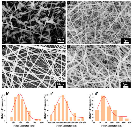
Figure 1.
SEM images of (a) PLA Fiber, (b) PLA/CTAB, (c) PLA/5-Cl8Q and (d) PLA/CTAB/5-Cl8Q. Fiber diameter distributions (b′,c′,d′) of subfigures (b,c,d), respectively.
To analyze the microstructure of each nanofiber membrane in more detail, transmission electron microscopy (TEM) was employed. As observed in the SEM result, the fibers exhibited a beaded structure (Figure 2a). Most PLA/CTAB fibers had a diameter of approximately 500 nm, with only a few fibers displaying increased thickness (Figure 2b). In contrast, the PLA/5-Cl8Q fibers exhibited a larger diameter of around 1700 nm (Figure 2c). Notably, Figure 2d displays the uneven thickness (ranging from approximately 500 nm to 1300 nm) and smooth surface of the PLA/CTAB/5-Cl8Q fibers. Changes in viscosity can affect the results of electrospinning. The SEM images of PLA/CTAB nanofibers with different concentrations are shown in Figure S1, indicating that PLA/CTAB nanofibers with 0.2 g CTAB exhibited better filament formation. Nanofibers with 0.1 g CTAB exhibited a disordered and agglomerated structure.
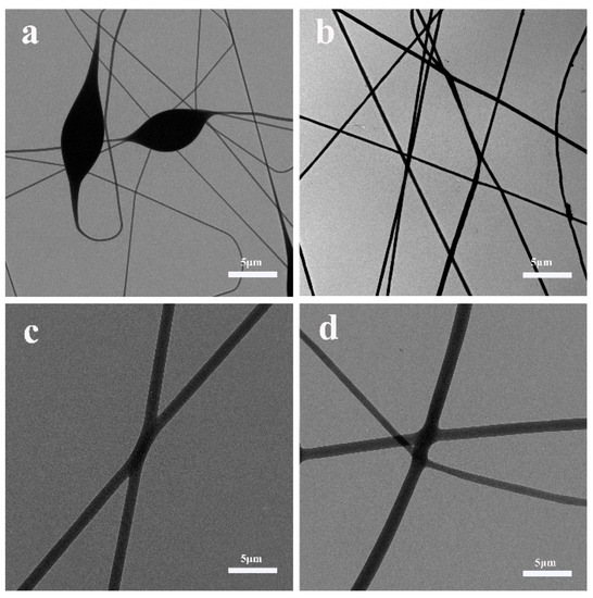
Figure 2.
TEM images of (a) PLA Fiber, (b) P LA/CTAB, (c) PLA/5-Cl8Q and (d) PLA/CTAB/5-Cl8Q.
The crystallographic structures of the products were analyzed using X-ray powder diffraction (XRD) (Figure 3a). The XRD pattern of PLA Fiber exhibited a prominent sharp diffraction peak at 15.82°, corresponding to the (110/200) plane, as well as a low-intensity diffraction peak at 18.05°, corresponding to the (203/113) plane [26]. In the XRD pattern of PLA/CTAB, observed peaks at 2θ values of 6.60, 16.90, 21.60, and 24.80 matched the diffraction peaks of CTAB. Additionally, the peak corresponding to PLA became significantly weaker, suggesting that the inclusion of CTAB disrupted the crystalline structure of PLA. As PLA and 5-Cl8Q transitioned from crystalline to amorphous states during spinning formation in the precursor solution, the XRD pattern of the PLA/5-Cl8Q sample did not exhibit the diffraction peaks of PLA and 5-Cl8Q. In comparison, the XRD pattern of PLA/CTAB/5-Cl8Q showed a few weak absorption peaks, indicating a particularly poor crystallinity in the PLA/CTAB/5-Cl8Q sample.
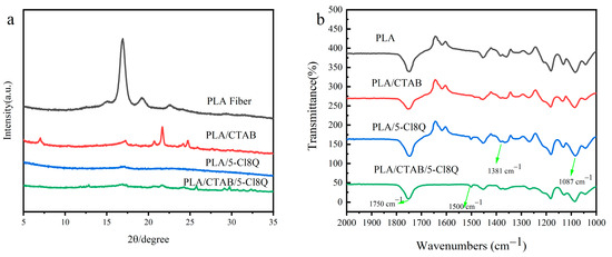
Figure 3.
(a) XRD pattern of fiber films. (b) FT-IR spectra of fiber films.
To analyze the internal structures of the samples, Fourier Transform Infrared Spectroscopy (FT-IR) was conducted (Figure 3b). The absorption peaks at 1750 cm−1, 1381 cm−1, and 1087 cm−1 are characteristic of the PLA-based polymer and were observed in the spectra of all four samples. The peak at 1750 cm−1 corresponds to the ester carbonyl C=O stretching vibration, the peak at 1381 cm−1 corresponds to the –C–C– bending vibration, and the peak at 1087 cm−1 corresponds to the lactone –C–O–C– telescopic vibration absorption. These peaks confirm the presence of PLA in all four fiber membranes, as PLA is one of the synthetic raw materials used. Additionally, a peak at 1500 cm−1, representing the quinoline ring, was observed in the infrared spectra of both PLA/5-Cl8Q and PLA/CTAB/5-Cl8Q [27]. This indicates that 5-Cl8Q is incorporated into the PLA-based polymer in these samples. It should be noted that the FT-IR spectrometer detects instability below 700 cm−1, while the vibration absorption peaks of C-Br and C-I are in the range of 700–500 cm−1. This is why the incorporation of CTAB into PLA/CTAB and PLA/CTAB/5-Cl8Q cannot be detected by FT-IR. To further support the presence of 5-Cl8Q in PLA/CTAB/5-Cl8Q, elemental mapping images (Figure 4b–e) and an Energy Dispersive X-ray Spectroscopy (EDS) spectrum (Figure 4f) were obtained. These images and spectrum confirm the presence of O, N, Cl, and Br, indicating the presence of 5-Cl8Q in PLA/CTAB/5-Cl8Q.
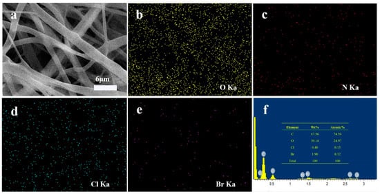
Figure 4.
(a) SEM energy-dispersive X-ray spectroscopy (SEM-EDS) image of PLA/CTAB/5-Cl8Q. Elemental mapping images of (b) O, (c) N, (d) Cl and (e) Br. (f) EDS spectrum of PLA/CTAB/5-Cl8Q.
The hydrophilicity of the fibrous membrane plays a significant role in the release of its antibacterial agent. The water contact angle of the PLA fiber was found to be 104.899° (Figure 5a), indicating that it is hydrophilic. However, the presence of beaded structures in the spinning process also contributes to its hydrophobicity to some extent. In the case of PLA/CTAB, the water contact angle was measured as 43.019° (Figure 5b). This is because CTAB, a hydrophilic polymer, constitutes a large portion of PLA/CTAB, thus contributing to its good hydrophilicity.
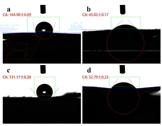
Figure 5.
Water contact angle image of (a) PLA Fiber, (b) OLA/CTAB, (c) PLA/5-Cl8Q and (d) PLA/CTAB/5-Cl8Q.
On the other hand, due to the hydrophobic nature of 5-Cl8Q, the water contact angle of PLA/5-Cl8Q, when it is incorporated, was measured as 131.174° (Figure 5c), indicating strong hydrophobicity. However, when all three components (PLA, CTAB, and 5-Cl8Q) were combined in the spinning process to form PLA/CTAB/5-Cl8Q, the resulting spun fiber exhibited a water contact angle of 52.792° (Figure 5d), indicating a certain degree of hydrophilicity. This suggests that the presence of both hydrophilic and hydrophobic components in the composite spinning process can influence the overall hydrophilicity of the fibrous membrane.
3.2. Antibacterial Analysis
To evaluate the antibacterial properties of the samples, a quantitative test using Gram-positive bacteria (S. aureus) and Gram-negative bacteria (E. coli) was conducted. The results are shown in Figure 6. PLA Fiber exhibited a weak antibacterial effect against S. aureus and E. coli. This may be due to trace amounts of weakly acidic lactic acid or oligomers in PLA. In the case of PLA/CTAB, the inhibitory zone widths were measured as 4.0 mm for S. aureus and 0.5 mm for E. coli. This indicates that PLA/CTAB has a stronger inhibitory effect on S. aureus than E. coli.
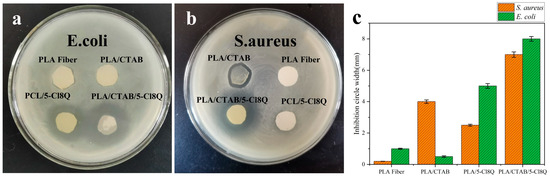
Figure 6.
Images of the qualitative inhibition circle with (a) E. coli and (b) S. aureus. (c) Inhibition circle widths of four fiber samples against S. aureus and E. coli.
On the other hand, PLA/5-Cl8Q complex fibers showed a better inhibitory effect on E. coli, with inhibitory zone widths of 2.5 mm against S. aureus and 5.0 mm against E. coli. However, when all three components (PLA, CTAB, and 5-Cl8Q) were combined in the spinning process to form PLA/CTAB/5-Cl8Q, the resulting fibers exhibited good antibacterial properties against both S. aureus and E. coli. The inhibitory zone widths were 7.0 mm against S. aureus and 8.0 mm against E. coli. This suggests that the combination of PLA, CTAB, and 5-Cl8Q enhances the overall antibacterial effect of the fibrous membrane.
In Figure 7, the antibacterial rates, and the number of viable bacteria populations in each composite nanofiber were measured after a 24-h incubation at 37 °C with both bacteria. It is evident from Figure 7c that PLA Fiber exhibits an antimicrobial rate of less than 80% against both S. aureus and E. coli. The weak antibacterial effect of PLA Fiber can be attributed to its weak acidity, and the reason for its weak antibacterial properties is that it only consists of PLA without any other antibacterial agents. Interestingly, the antibacterial activity of the various nanofiber membranes against S. aureus is generally higher than that against E. coli, which contradicts the results of the inhibition zone experiment. This can be attributed to two factors. Firstly, the hydrophilicity of the samples differs, leading to a lower penetration rate of hydrophobic samples compared to hydrophilic samples in the same bacterial solution. Secondly, during the experiment, the samples had only brief contact with the bacterial fluid, resulting in poor diffusion effects. All four spinning membranes exhibit higher antimicrobial rates against S. aureus than E. coli, which can be attributed to the structure of the bacteria. S. aureus has more negative charges on its surface, making it more attracted to positively charged cationic polymers. Among the four spinning membranes, PLA/CTAB/5-Cl8Q demonstrates the best antibacterial effect in Figure 7c. It exhibits an antibacterial rate of 95.9% against E. coli and 99.9% against S. aureus. This superior performance can be attributed to the combined effects of the number of antimicrobials and the hydrophilicity of the PLA/CTAB/5-Cl8Q composite nanofiber.
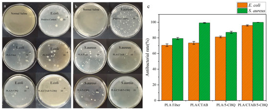
Figure 7.
Image of antimicrobial rate experimental with (a) E. coli and (b) S. aureus. (c) Images of the antibacterial rate of composite spinning films.
Figure 8a presents the antibacterial growth curve of each spun nanofiber against S. aureus. Compared to PLA Fiber, the other three spinning samples inhibited the reproduction of S. aureus. Analyzing the curve of PLA Fiber against S. aureus, it can be observed that the inhibition time is 4 h, and the inhibition effect is weak. During the initial two hours, the bacteriostatic effect of PLA/CTAB/5-Cl8Q is superior to that of PLA/CTAB and PLA/5-Cl8Q, which aligns with the results of the antibacterial rate experiments. For the fiber samples, the degree of bacterial inhibition varies with the growth cycle of the bacteria. After 12 h, the bacterial absorbance of PLA/CTAB and PLA/5-Cl8Q increases significantly, and the antimicrobial rates decrease. This can be attributed to the fact that bacteria multiply faster than the release of bacteriostatic agents. On the other hand, the absorbance of PLA/CTAB/5-Cl8Q remains consistently low, indicating that it exhibits the best antibacterial performance among these samples and maintains a stable antibacterial effect.
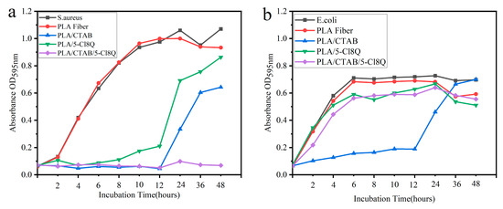
Figure 8.
Antibacterial growth curve of spinning nanofibers against (a) S. aureus and (b) E. coli.
In contrast to Figure 8a, Figure 8b depicts the inhibitory effect of each spinning sample on E. coli. Similar to S. aureus, PLA exhibits a weak inhibitory effect on E. coli. However, PLA/CTAB, PLA/5-Cl8Q, and PLA/CTAB/5-Cl8Q display much stronger antibacterial properties against E. coli. The hydrophilicity of a sample plays a role in determining its antimicrobial activity. Among the samples, PLA/CTAB, which is the most hydrophilic, exhibits the strongest antimicrobial properties. Following this, PLA/CTAB/5-Cl8Q, which has slightly lower hydrophilicity, displays a strong antibacterial effect. Lastly, PLA/5-Cl8Q, which is hydrophobic, shows the weakest antimicrobial activity. E. coli, with a lower negative charge, necessitates higher levels of cationic antibacterial agents. Furthermore, hydrophilic samples release more antimicrobial components into the bacterial fluid than hydrophobic ones. This is the reason why hydrophilic samples exhibit higher antibacterial properties. Cytotoxicity is one of the important indicators of nanofibers as medical materials, and MTT methods were employed to evaluate the biocompatibility of the prepared nanofibers. As shown in Figure S2, the PLA/CTAB/5-Cl8Q film shows the best cell viability, followed by PLA/5-Cl8Q and PLA/CTAB. PLA fiber has low cell viability due to the absence of antimicrobial agents. Wu et al. [28] prepared the polylactic acid (PLA)/tea polyphenol (TP) nanofiber membranes using coaxial electrospinning. In their work, the 6 wt% PLA/TP membrane with the best antibacterial properties had an inhibitory circle width of 1.45 mm against S. aureus and 1.20 mm for E. coli, which is lower than PLA/5-Cl8Q and PLA/CTAB/5-Cl8Q. The CA/5-Cl8Q and the CA, PEG/5-Cl8Q prepared by Spasova et al. [29] showed good antimicrobial activities whose measured zones of inhibition were 4.0 ± 0.18 and 4.5 ± 0.2 cm against S. aureus and around 4.0 ± 0.15 and 4.1 ± 0.22 cm against E. coli, respectively. Whether it is CA/5-Cl8Q or CA, PEG/5-Cl8Q, from a qualitative point of view, their antibacterial activity is not as good as PLA/CTAB/5-Cl8Q. CTAB-MT clay-doped 3D nanowebs [22] showed excellent antimicrobial activity of 97.96% and 90.48% against E. coli and S. aureus bacteria, respectively. Resistance of the PLA/CTAB/5-Cl8Q to E. coli is comparable to the CTAB-MT clay-doped 3D nanowebs. However, the Resistance of the PLA/CTAB/5-Cl8Q to S. aureus is higher than the CTAB-MT clay-doped 3D nanowebs.
4. Conclusions
In this study, a novel approach to fabricating PLA/CTAB/5-Cl8Q nanofibers with exceptional antibacterial efficacy has been successfully demonstrated. The antibacterial properties of the PLA/CTAB/5-Cl8Q composite nanofibers were superior to other materials, exhibiting an impressive inhibitory zone width of 7.0 mm against S. aureus and 8.0 mm against E. coli. The antibacterial rate of the composite nanofibers against S. aureus was determined to be 99.9%, while the antibacterial rate against E. coli reached 95.9%. These remarkable characteristics can be attributed to the inherent properties of the material itself and the physical attributes of the sample. Consequently, this research underscores the potential for developing PLA-based composite nanofibers as cost-effective, biodegradable, and effective antibacterial materials.
Supplementary Materials
The following supporting information can be downloaded at https://www.mdpi.com/article/10.3390/coatings13101686/s1, Figure S1: SEM images of (a) PLA/5-Cl8Q with 0.1 g and (b) PLA/5-Cl8Q with 0.2 g; Figure S2: Image of Cytotoxicity results.
Author Contributions
H.G. Performed the experiments and wrote the paper. L.L.: Data curation. N.L.: Data curation. L.T.: Data curation. T.Z.: Data curation. L.Z.: Data curation. T.J.: Designed the experiments, and reviewed and edited. All authors have read and agreed to the published version of the manuscript.
Funding
This study was supported by the National Natural Science Foundation of China (Nos. 22072127, 22372143), the Natural Science Foundation of Hebei Province (No. B2021203016), the Science and Technology Project of Hebei Education Department (No. ZD2022147), and the Special Project for Local Science and Technology Development Guided by the Central Government of China (Nos. 216Z1301G, 226Z1401G, 236Z1201G, 236Z1401G, 236Z1206G).
Institutional Review Board Statement
Not applicable.
Informed Consent Statement
Not applicable.
Data Availability Statement
Not applicable.
Conflicts of Interest
The authors declare no conflict of interest.
References
- Bedford, J.; Farrar, J.; Ihekweazu, C.; Kang, G.; Koopmans, M.; Nkengasong, J. A new twenty-first century science for effective epidemic response. Nature 2019, 575, 130–136. [Google Scholar] [CrossRef] [PubMed]
- Xu, X.C.; Zhang, J.; Liu, S.; Wang, C.H.; Wang, H.Y.; Fan, H.H.; Tong, Y.G.; Liu, H.Y.; Zhou, D.S. New Advances in Nanomaterial-Based Antiviral Strategies. Small Struct. 2022, 3, 2200021. [Google Scholar] [CrossRef]
- Tiri, R.N.E.; Gulbagca, F.; Aygun, A.; Cherif, A.; Sen, F. Biosynthesis of Ag–Pt bimetallic nanoparticles using propolis extract: Antibacterial effects and catalytic activity on NaBH4 hydrolysis. Environ. Res. 2022, 206, 112622. [Google Scholar] [CrossRef] [PubMed]
- Wang, S.Y.; Duan, Y.O.; Zhang, Q.Z.; Komarla, A.; Gong, H.; Gao, W.W.; Zhang, L.F. Drug Targeting via Platelet Membrane-Coated Nanoparticles. Small Struct. 2020, 1, 2000018. [Google Scholar] [CrossRef]
- Varela, M.F.; Stephen, J.; Lekshmi, M.; Ojha, M.; Wenzel, N.; Sanford, L.M.; Hernandez, A.J.; Parvathi, A.; Kumar, S.H. Bacterial Resistance to Antimicrobial Agents. Antibiotics 2021, 10, 593. [Google Scholar] [CrossRef] [PubMed]
- Ding, J.; Zhang, J.; Li, J.; Li, D.; Xiao, C.; Xiao, H.; Yang, H.; Zhuang, X.; Chen, X. Electrospun polymer biomaterials. Prog. Polym. Sci. 2019, 90, 1–34. [Google Scholar] [CrossRef]
- Zhang, C.; Li, Y.; Wang, P.; Zhang, H. Electrospinning of nanofibers: Potentials and perspectives for active food packaging. Compr. Rev. Food Sci. Food Saf. 2020, 19, 479–502. [Google Scholar] [CrossRef]
- Shi, S.; Si, Y.F.; Han, Y.T.; Wu, T.; Iqbal, M.I.; Fei, B.; Li, R.K.Y.; Hu, J.L.; Qu, J.P. Recent Progress in Protective Membranes Fabricated via Electrospinning: Advanced Materials, Biomimetic Structures, and Functional Applications. Adv. Mater. 2022, 34, 2107938. [Google Scholar] [CrossRef]
- Wang, X.M.; Sun, F.Z.; Yin, G.C.; Wang, Y.T.; Liu, B.; Dong, M.D. Tactile-Sensing Based on Flexible PVDF Nanofibers via Electrospinning: A Review. Sensors 2018, 18, 330. [Google Scholar] [CrossRef]
- Armentano, I.; Bitinis, N.; Fortunati, E.; Mattioli, S.; Rescignano, N.; Verdejo, R.; Lopez-Manchado, M.A.; Kenny, J.M. Multifunctional nanostructured PLA materials for packaging and tissue engineering. Prog. Polym. Sci. 2013, 38, 1720–1747. [Google Scholar] [CrossRef]
- Wu, J.H.; Hu, T.G.; Wang, H.; Zong, M.H.; Wu, H.; Wen, P. Electrospinning of PLA Nanofibers: Recent Advances and Its Potential Application for Food Packaging. J. Agric. Food Chem. 2022, 70, 8207–8221. [Google Scholar] [CrossRef] [PubMed]
- Liu, C.Y.; Du, G.C.; Guo, Q.Q.; Li, R.G.; Li, C.M.; He, H.W. Fabrication and Characterization of Polylactic Acid Electrospun Wound Dressing Modified with Polyethylene Glycol, Rosmarinic Acid and Graphite Oxide. Nanomaterials 2023, 13, 2000. [Google Scholar] [CrossRef] [PubMed]
- He, X.; Zhou, M.L.; Chen, X.M.; Wang, J.; Zhao, X.L.; Zhu, Y.X.; Liu, T. Development and Characterization of Multifunctional Wound Dressing with the Property of Anti-bacteria and Angiogenesis. Probiotics Antimicrob. Proteins 2022, 15, 941–954. [Google Scholar] [CrossRef] [PubMed]
- Balakrishnan, N.K.; Ostheller, M.E.; Aldeghi, N.; Schmitz, C.; Groten, R.; Seide, G. Pilot-Scale Electrospinning of PLA Using Biobased Dyes as Multifunctional Additives. Polymers 2022, 14, 2989. [Google Scholar] [CrossRef] [PubMed]
- Nedelcu, I.A.; Ficai, A.; Sonmez, M.; Ficai, D.; Oprea, O.; Andronescu, E. Silver Based Materials for Biomedical Applications. Curr. Org. Chem. 2014, 18, 173–184. [Google Scholar] [CrossRef]
- Allizond, V.; Banche, G.; Salvoni, M.; Malandrino, M.; Cecone, C.; Cuffini, A.M.; Bracco, P. Facile One-Step Electrospinning Process to Prepare AgNPs-Loaded PLA and PLA/PEO Mats with Antibacterial Activity. Polymers 2023, 15, 1470. [Google Scholar] [CrossRef]
- Tran, C.D.; Prosenc, F.; Franko, M.; Benzi, G. One-Pot Synthesis of Biocompatible Silver Nanoparticle Composites from Cellulose and Keratin: Characterization and Antimicrobial Activity. Acs Appl. Mater. Interfaces 2016, 8, 34791–34801. [Google Scholar] [CrossRef] [PubMed]
- Fijalkowski, K.; Rorat, A.; Grobelak, A.; Kacprzak, M.J. The presence of contaminations in sewage sludge—The current situation. J. Environ. Manag. 2017, 203, 1126–1136. [Google Scholar] [CrossRef]
- Zhao, S.Y.; Huang, W.J.; Wang, C.R.; Wang, Y.P.; Zhang, Y.F.; Ye, Z.P.; Zhang, J.H.; Deng, L.D.; Dong, A.J. Screening and Matching Amphiphilic Cationic Polymers for Efficient Antibiosis. Biomacromolecules 2020, 21, 5269–5281. [Google Scholar] [CrossRef]
- Lu, S.C.; Zhang, X.H.; Tang, Z.W.; Xiao, H.; Zhang, M.; Liu, K.; Chen, L.H.; Huang, L.L.; Ni, Y.H.; Wu, H. Mussel-inspired blue-light-activated cellulose-based adhesive hydrogel with fast gelation, rapid haemostasis and antibacterial property for wound healing. Chem. Eng. J. 2021, 417, 129329. [Google Scholar] [CrossRef]
- Chiloeches, A.; Fernandez-Garcia, R.; Fernandez-Garcia, M.; Mariano, A.; Bigioni, I.; d’Abusco, A.S.; Echeverria, C.; Munoz-Bonilla, A. PLA and PBAT-Based Electrospun Fibers Functionalized with Antibacterial Bio-Based Polymers. Macromol. Biosci. 2023, 23, e2200401. [Google Scholar] [CrossRef]
- Batra, R.; Purwar, R.; Kulanthaivel, S.; Mishra, P. Cetyl Trimethyl Ammonium Bromide Modified Montmorillonite-Doped Tasar Silk Fibroin/Polyvinyl Alcohol Blend 3D Nanowebs for Tissue Engineering Applications. Macromol. Mater. Eng. 2021, 306, 2100450. [Google Scholar] [CrossRef]
- Nachev, N.; Spasova, M.; Tsekova, P.; Manolova, N.; Rashkov, I.; Naydenov, M. Electrospun Polymer-Fungicide Nanocomposites for Grapevine Protection. Polymers 2021, 13, 3673. [Google Scholar] [CrossRef] [PubMed]
- Dong, W.; Green, J.; Korza, G.; Setlow, P. Killing of spores of Bacillus species by cetyltrimethylammonium bromide. J. Appl. Microbiol. 2019, 126, 1391–1401. [Google Scholar] [CrossRef] [PubMed]
- Spasova, M.; Manolova, N.; Rashkov, I.; Naydenov, M. Electrospun 5-chloro-8-hydroxyquinoline-Loaded Cellulose Acetate/Polyethylene Glycol Antifungal Membranes Against Esca. Polymers 2019, 11, 1617. [Google Scholar] [CrossRef] [PubMed]
- Ke, W.C.; Li, X.; Miao, M.Y.; Liu, B.; Zhang, X.Y.; Liu, T. Fabrication and Properties of Electrospun and Electrosprayed Polyethylene Glycol/Polylactic Acid (PEG/PLA) Films. Coatings 2021, 11, 790. [Google Scholar] [CrossRef]
- Yang, C.Y.; Chen, S.H.; Wang, J.H.; Zhu, T.H.; Xu, G.; Chen, Z.C.; Ma, X.B.; Li, W.Y. A facile electrospinning method to fabricate polylactide/graphene/MWCNTs nanofiber membrane for tissues scaffold. Appl. Surf. Sci. 2016, 362, 163–168. [Google Scholar] [CrossRef]
- Wu, J.; Liu, S.; Zhang, M.; Wu, G.; Yu, H.; Li, H.; Li, F.; Jia, L. Coaxial electrospinning preparation and antibacterial property of polylactic acid/tea polyphenol nanofiber membrane. J. Ind. Text. 2022, 51, 1778S–1792S. [Google Scholar] [CrossRef]
- Spasova, M.; Manolova, N.; Rashkov, I.; Tsekova, P.; Georgieva, A.; Toshkova, R.; Markova, N. Cellulose Acetate-Based Electrospun Materials with a Variety of Biological Potentials: Antibacterial, Antifungal and Anticancer. Polymers 2021, 13, 1631. [Google Scholar] [CrossRef]
Disclaimer/Publisher’s Note: The statements, opinions and data contained in all publications are solely those of the individual author(s) and contributor(s) and not of MDPI and/or the editor(s). MDPI and/or the editor(s) disclaim responsibility for any injury to people or property resulting from any ideas, methods, instructions or products referred to in the content. |
© 2023 by the authors. Licensee MDPI, Basel, Switzerland. This article is an open access article distributed under the terms and conditions of the Creative Commons Attribution (CC BY) license (https://creativecommons.org/licenses/by/4.0/).