Article

Theoretical Analysis of Hybrid Metal–Dielectric Nanoantennas with Plasmonic Fano Resonance for Optical Sensing

1
School of Physics and Electronic Engineering, Northeast Petroleum University, Daqing 163318, China
2
Department of Physics, Department of Materials Science and Engineering, and Department of Biomedical Engineering, City University of Hong Kong, Tat Chee Avenue, Kowloon, Hong Kong 999077, China
*
Author to whom correspondence should be addressed.
Coatings 2022, 12(9), 1248; https://doi.org/10.3390/coatings12091248
Submission received: 25 July 2022 / Revised: 20 August 2022 / Accepted: 23 August 2022 / Published: 26 August 2022

Abstract

:
A nanoantenna with Fano response is designed with plasmonic oligomers as a refractive index sensor to enhance surface-enhanced Raman scattering (SERS) in the visible light spectrum. The scattered radiation and field-enhanced interactions of the outer gallium phosphide (GaP) nanoring assembled with an inner heptamer of silver with Fano response are investigated systematically using the finite element method. The characteristics of Fano resonance are found to depend on the size, shape and nature of the materials in the hybrid nanoantenna. The confined electromagnetic field produces a single-point electromagnetic hotspot with up to 159.59 V/m. The sensitivity obtained from the wavelength shift and variation in the scattering cross-section (SCS) shows a maximum value of 550 nm/RIU. The results validate the design concept and demonstrate near-field enhancement, enabling the design of high-performance nanoantennas with enhanced optical sensing and SERS properties.

1. Introduction

Nano-optical antennas [1] can couple light waves in nano-scale devices to exceed the diffraction limit [2] of conventional optical antennas. Nanoantennas have many applications in nanophotonics due to their unique ability to confine light down to deep-subwavelength dimensions with strongly enhanced localized electric fields, resulting from localized surface plasmon resonance (LSPR) [3,4]. Because of the plasmonic enhancement, LSPR enhances the interaction between light and matter, thus, spurring the development of single-molecule detection [5,6], nanoscale light sources [7,8], surface-enhanced Raman scattering (SERS) [9,10], biosensing [11] and so on.
By adopting specific metallic nanostructures, coupling of the fundamental plasmon modes produces a phenomenon known as “Fano-like resonance”, which is caused by destructive interference between a wide continuum state and a narrow discreet state [12] and is characterized by scattering with an asymmetrical line shape [13,14]. Typically, a sharp dip is observed in the scattering spectrum for Fano interference between the broadband and narrowband optical modes [15]. Inspired by the plasmonic Fano resonant nanostructures, some coupled dielectric resonators with Fano resonance have been developed. For instance, Ye et al. discovered that adding a carbon medium to specific locations in the gold heptamer can alter the SERS response, resulting in field enhancement with Fano resonance [16]. The observation implies that the hybrid metal–dielectric composite nanostructure is a promising option for combining the field enhancement effect of the metal and accomplish low loss of the dielectric materials. Zuev et al. used a femtosecond laser to remodel gold nanoparticles in Au/Si to change the optical properties [17] and Zarrabi et al. found that the plasmonic nanoring with Fano response exhibits an enhanced near-field electric field [18]. Dutta et al. experimentally demonstrated that two disk-shape gold oligomers can provide Fano resonance in the scattering response [19] and Torres et al found that the radiated field can have a complex distribution determined by the internal geometry of the nanowire [20]. Then, researchers found numerous interesting optical phenomena can be produced significantly in the hybrid nanostructure with multiple Fano resonances. for example, Chou Chau YF et al. confirmed by using the FEM method that double-resonance peaks and third-resonance peaks at wavelengths ranging from 500 nm to 800 nm can perform high-refractive-index sensor performance [21,22]. In addition, Sung et al. confirmed that changing illumination wavelengths, angles of incidence and radius of the heptamer can considerably achieve tunability of resonance by filling with different dielectric cores [23].
Recent theoretical analysis and experimental studies on dielectric–metallic nanoantennas demonstrate that these devices exhibit Fano resonance phenomena. However, in spite of recent advances in improving near-field coupling, it is still necessary to achieve low loss and, hence, high-index dielectric nanoantennas with low dissipative loss and metal constitute an attractive platform for subwavelength optics. Among the various dielectric materials, gallium phosphide (GaP) is especially promising as it covers almost the entire visible range and shows negligible loss [24]. In addition, gallium phosphide (GaP) offers the intriguing possibilities for the development of low-loss nanophotonic antennas because the bandgap is less than 550 nm, consequently leading to a smaller loss compared to Si in the visible regime [25].
Herein, a hybrid nanostructure composed of a metal and high-refractive-index dielectric material is designed and analyzed by the finite element method. It consists of an outer GaP nanoring and an inner heptamer of silver. The near-field distributions are derived to assess coupling between the electric fields associated with the electric and toroidal modes with regard to Fano interference. To better understand the tunability of the Fano line in this system and establish the Fano resonance, the geometry parameters are optimized. Our results predict that the (E/E0)4 enhancement at the gap of the inner heptamer of silver is ~109, which is approximately 20-times larger than that of the individual heptamer of silver. This outstanding feature of the hybrid nanostructure reveals the possibility of enhanced electric near fields at Fano dips and provides insights into SERS and sensing applications.
In this paper, our proposed structure achieves optical properties that differ from previous studies in three main ways: firstly, compared to the typical scattering spectrum of dielectric nanoparticles with one peak, a hybrid metal–dielectric nanoantenna for multiple Fano resonance is investigated; secondly, the hybrid nanoantenna shows a more intense resonant electric field and theoretical simulation predicts that the (E/E0)4 enhancement is as large as 109, which is approximately 20-times larger than of the individual heptamer of silver in SERS; thirdly, the outcomes make it possible to create high-performance nanoantennas with improved optical sensing capabilities.

2. Materials and Methods

To analyze the degree of coupling between the metal and dielectric materials, the scattering properties of the hybrid nanoantenna are determined by the multipole decomposition method [26]. To illustrate the contributions of various modes, multipole decomposition including the electric dipole moment P (ED), magnetic dipole moment M (MD), toroidal dipole moment T (TD), electric quadrupole moment Qe (EQ) and magnetic quadrupole moment Qm (MQ) is considered as shown in the following [27,28]:
p = P ( r ) d r
m = i ω 2 [ r × P ( r ) ] d r
T = i ω 10 [ ( 2 r 2 P ( r ) ( r P ( r ) ) r ] d r
Q e = 3 [ r P ( r ) + P ( r ) r ] d r
Q m = 2 i ω 3 [ r × P ( r ) ] r d r
where P ( r ) is the polarization caused by scattering of the incident light wave and r′ is the position vector. The radiation power I of the various multipole moments can be derived as follows [29]:
I = 1 4 π ε 0 [ 2 ω 4 3 c 3 P 2 + 2 ω 4 3 c 3 M 2 + 4 ω 5 3 c 4 Im ( P T ) + 2 ω 6 3 C 5 T 2 + ω 6 20 c 5 Q e 2 + ω 6 20 c 5 Q m 2 ]
The overall scattering cross-section can be determined by Equation (7):
C s c a = I I i n c
where I i n c is the radiation power of the incident light wave.
To better understand the optical response of the hybrid nanoantenna, finite element method (FEM) simulation is performed on the combined heptamer of silver and GaP nanoring. All the full-wave electromagnetic calculations were performed using the commercial simulation package COMSOL 5.5 Multiphysics, which is based on the finite element method (FEM). The hybrid system was illuminated by plane wave, implemented according to the total-field scattered-field source. The incident light is propagating in z direction and the polarization is along the x direction. The spherical coordinate system is selected as the reference coordinate system type for this simulation and perfectly matched layer (PML) boundary conditions are employed in all three dimensions simulating a finite structure size. The outermost boundary was assigned a scattering boundary condition (SBC) to minimize possible reflection. The simulation region is terminated with PML and SBC to absorb the scattering energy. Depending on the element order in the model, a finer mesh is employed by hybrid nanostructures. The complete mesh consists of 74774 domain elements, 9820 boundary elements and 1025 edge elements and the step of incident wavelength from 476 nm to 1500 nm is 150. The whole nanostructure is assumed to be free standing in air and illuminated by a normally incident plane wave (k along the z direction), which is linearly polarized along the x direction. The hybrid nanoantenna schematically shown in Figure 1 consists of an outer GaP nanoring assembled with an inner heptamer of silver. The radius and height of the heptamer of silver are RAg = 25 nm and h = 40 nm, respectively, and the gap between the silver disks is 2 nm. Silver [30] is chosen as the metal because it has a small imaginary part which can greatly reduce the intrinsic loss [31]. The complex dielectric function is presented as a fitting of the Drude model for the permittivity of the silver. Figure 1b shows the nanoring with an inner radius r of 78 nm, outer radius R of 149 nm and height H of 40 nm. GaP is the dielectrics and the optical constants of silver and GaP are obtained from Palik’s handbook [32]. The arrangement of the nanoring and heptamer is displayed in Figure 1c and the distance between the nanoring and heptamer is G = 1 nm.
In addition, the fabrication of this nanoantenna can be fully realized by the existing research. The heptamer of silver can be fabricated using electron beam lithography followed by metal evaporation [33]. Then, GaP film can be RF sputtered on a SiO2 cover glass. The nanopatterning was accomplished through the use of negative resist, electron beam lithography, development and other techniques [34]. Subsequently, a hybrid nanostructure consists of an outer GaP nanoring with an inner heptamer of silver and can be accomplished according to precise control of magnetron sputtering [35].

3. Results and Discussion

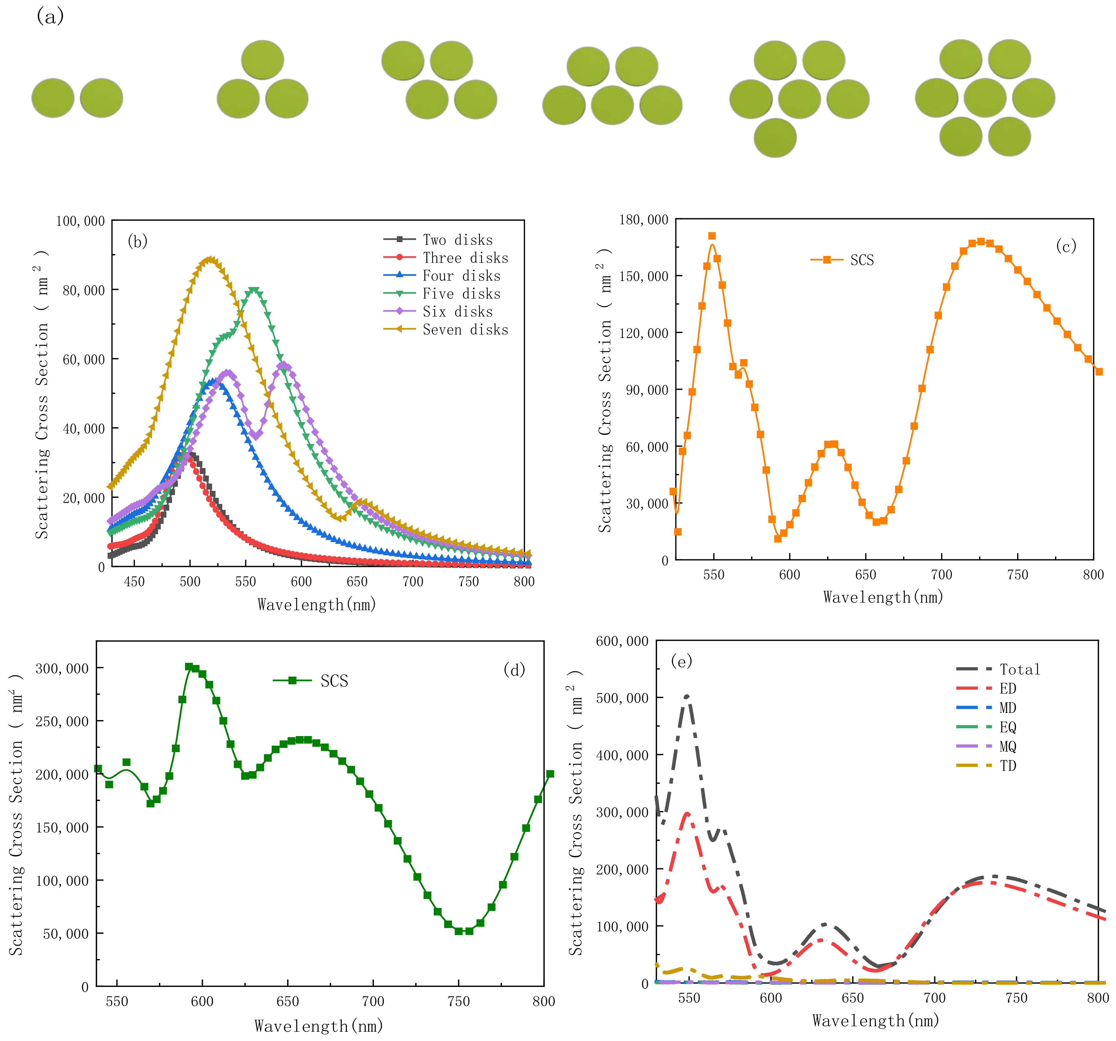
To explore the scattering characteristics of different nanoantennas, various models for the nanodisk arrangements are adopted by omitting some of the disks from the heptamer of silver model, as shown in Figure 2a. The corresponding scattering cross-section is shown in Figure 2b. The number and height of the nanoparticles in the plasma oligomers and other physical properties influence the location and intensity of Fano resonance, as shown by Daniel Dregely et al [36]. It can be observed that the Fano response can be achieved by adding more disks due to hybridization of the nanodisks. In order to produce multiple Fano resonance, seven nanodisks are implemented in the fundamental metal structure and the nanoring is introduced to the outside of the heptamer of silver. The three dips around 566 nm, 592 nm and 657 nm in the SCS spectrum stem from the combination of metal and dielectric nanoantennas, as shown in Figure 2c. The spectral lines indicate that introduction of the dielectric nanoring modifies the number and intensity of Fano resonance, thereby making the hybrid nanoantenna an excellent candidate for surface-enhanced spectroscopy. In addition, in the real situation, the proposed structure should be deposited on a substrate. The SCS of the proposed structure placed on a silicon substrate is given in Figure 2d. According to the spectral lines, it can produce two Fano dips at wavelengths ranging from 530 nm to 800 nm compared to the nanoantenna without the substrate. Thus, the nanoantenna without the substrate has better optical properties, which is used in the follow-up study. To study the reason for the formation of different dips, the scattering cross-section of the hybrid nanoantenna with a series of multipoles is shown in Figure 2e. The dips at 566 nm, 592 nm and 657 nm in the total scattering cross-section originate from the combined contributions of the electrical resonance and toroidal dipole of the hybrid nanoantenna. The EQ, MD and MQ contributions to the scattering cross-section are approximately zero and so the dips are attributed to ED and TD. Consequently, the total interference of the electric dipole and toroidal dipole moments (represented as ED and TD by the red and yellow curves) indicate partial scattering cancelation in the Fano-like spectral windows.
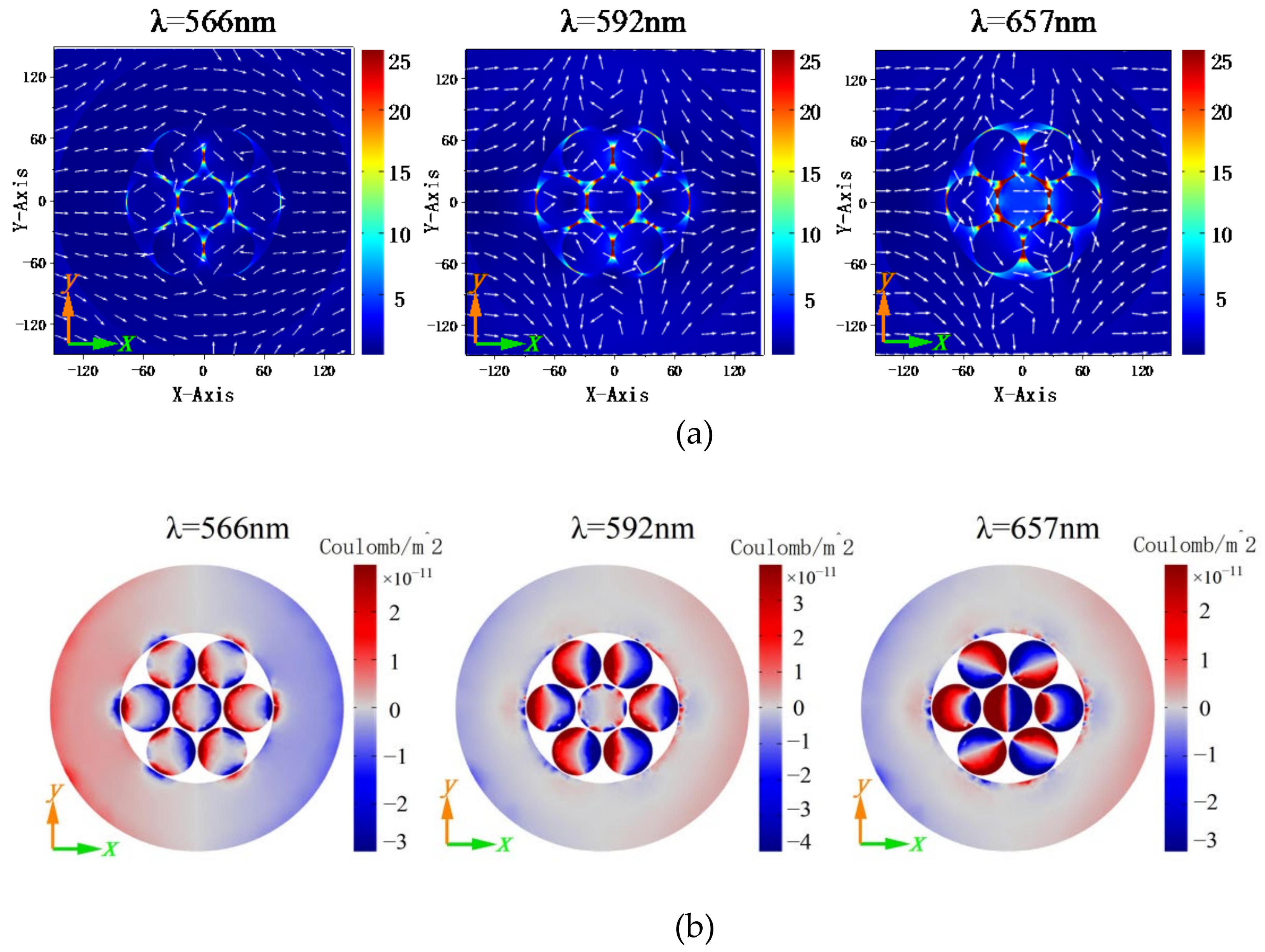
To obtain deeper physical understanding of the Fano dips excited in the hybrid nanoantenna, the near-field distribution profiles are presented in Figure 3. Figure 3a is taken form COMSOL. The region of the near field is from −149 nm to 149 nm on the x-axis and from −149 nm to 149 nm on the y-axis. The near-field distributions are determined by electric dipolar excitation and on the xoy plane and the electric field hotspot occurs naturally between the heptameric nanoantenna and outer nanoring at λ = 566 nm and λ = 592 nm. There is strong field localization and the electric field enhancement stimulates the optical response at λ = 657 nm as a result of strong coupling between the heptameric nanoantennas. The current direction is shown as the white arrow. According to the dipolar moment orientation, the Fano dips show displacement electrical current loops. Figure 3b presents the charge distributions of the nanostructure at the Fano dips. The heptameric nanoantenna interacts with the outer GaP nanoring resulting in electric dipole resonance upon x-direction polarization. The charge distributions on the xoy plane for the Fano dips are consistent with the multipole contributions to the scattering cross-section shown in Figure 2e.
To clarify how the geometry influences the scattering cross-section of the hybrid nanoantenna, a systematic investigation is performed by varying the parameters hAg, HGaP = hAg, G and rAg, as shown in Figure 4. Figure 4a shows that the different resonance peaks shift to shorter wavelengths when the height of the internal silver disks hAg is increased from 10 nm to 40 nm. In particular, SCS is the greatest when the height is 40 nm, reaching 171,000 nm2, 104,000 nm2, 61,100 nm2 and 168,000 nm2. The resonance peaks occur at λ = 549 nm, 570 nm, 629 nm and 726 nm, respectively. The scattering cross-sections are shown in Figure 4b as the inner and outer heights are varied. The resonance wavelengths of the scattering cross-sections increase as the height decreases. Meanwhile, the maximum intensity of SCS increases gradually, indicating that the optimized parameter of HGaP = hAg = 40 nm. Hence, increasing the contact area stimulates more charges on the surface of the heptamers and nanoring, consequently providing more degrees of freedom to control the scattering characteristics.
Figure 4c,d describe the spectral dependence of the scattering cross-section as a function of distance between the disks and nanoring as well as disk radius between 500 nm and 800 nm. The gap G and radius rAg of the silver disks are varied while the height of the nanoantenna HGaP is fixed. Figure 4c shows that the resonance peaks of the scattering cross-section increase as the gap is increased from 1 nm to 4 nm, in addition to a small blueshift at a wavelength of 629 nm. In this situation, widening the gap of the hybrid nanoantenna weakens coupling between the heptamer and nanoring, while improving the field enhancement between the seven disks. In contrast to the wavelength of 629 nm, the scattering intensity weakens and the resonance peak moves towards shorter wavelength as the gap is increased. Figure 4d shows that the peak positions of SCS red-shift slightly and the peak intensity increases at 549 nm and 726 nm as rAg is changed from 10 nm to 25 nm. Figure 4e depicts the effect of the GaP nanoring diameter on the scattering cross-sections. It is noticed that the resonance peak at a wavelength of 657 nm moves to a longer wavelength. The results clearly demonstrate that variations in the gap between the heptamer and nanoring, the radius and width of the nanoring all affect the overall performance at the optical wavelengths.
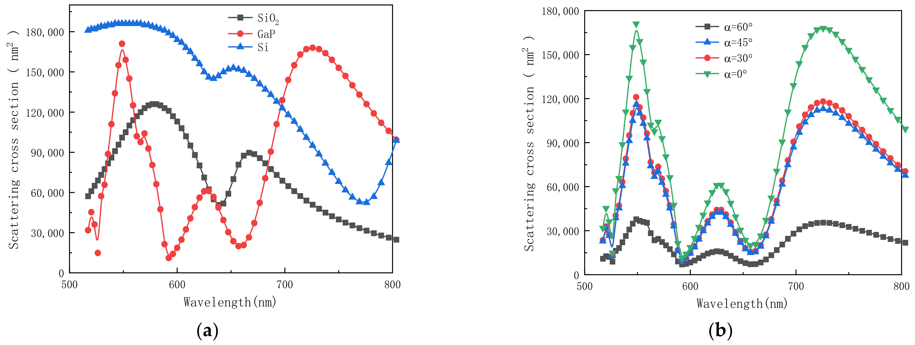
Previous studies showed that various Kerr materials can alter the performance of nanoantennas and so Kerr materials are adopted in our model, including transparent SiO2 and semiconductors of Si and GaP. Figure 5a shows the scattering spectra of the three different dielectric materials as the nanoring. Surface plasmons are collected on the surface of the seven disks and nanoring, so that different dielectrics can modify the properties of surface plasmons and scattering. Compared to SiO2 and Si, a sharper dip is observed by using GaP as the nanoring material, resulting in narrower line width and more prominent Fano phenomenon.
According to the analysis of Figure 5a, the GaP nanoring is selected for the follow-up study. The difference in the direction of incident light produces different optical properties. For example, Torres et al. achieved tuning of the directivity by means of changing the direction of the incident light [37]. In order to study the effects of polarized incident light, the scattering cross-sections of different polarized light are plotted in Figure 5b, where α is the angle between the incident light and x-axis. The position of Fano dips changes subtly and the dips occur at 566 nm, 592 nm and 657 nm, respectively, when α is equal to zero (along the x-axis direction). Compared to other directions of polarization, higher SCS can be observed for polarization along the x-axis, suggesting that coupling in the hybrid nanostructure is significantly impacted by polarization of the incident light.
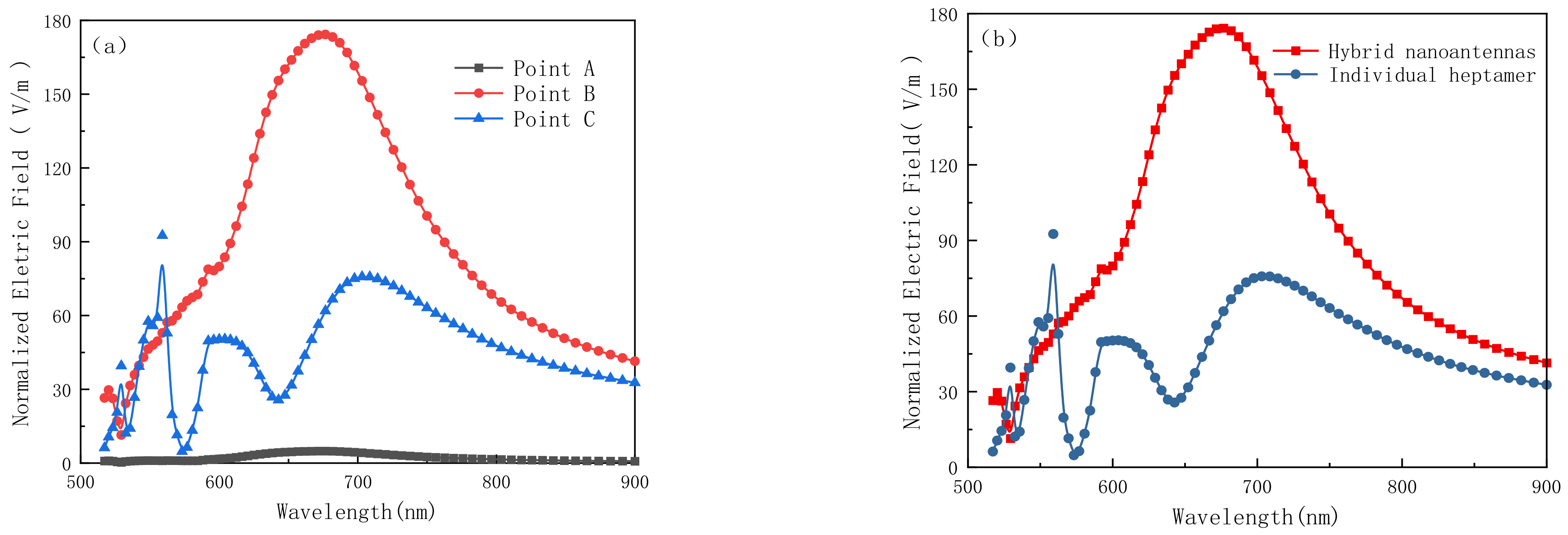
The internal geometry can determine the complex distribution of the electric field [20]. Therefore, the normalized electric fields at different points of the hybrid nanoantenna are depicted and the hybrid nanoantenna is compared to the individual heptamer by optimizing the structural parameters, which is shown in Figure 6. Figure 6a displays the relationship between the electric field enhancement factors and different points in the hybrid nanoantenna. Three points are selected on the x-axis, namely point A in the center of the heptamer at x = 0 nm, point B between the disks x = 26 nm and point C between the heptamer and nanoring at x = 77.5 nm. As shown in Figure 6a, compared to point A and C, the normalized electric field is the largest at point B where the wavelength is λ = 677 nm, which is up to 159.59 V/m. The comparison of the normalized electric fields between the hybrid nanoantenna and the individual hepatmer at point B is shown in Figure 6b. The hybrid nanoantenna shows a more intense resonant electric field (>150 folds) at point B and theoretical simulation predicts that the (E/E0)4 enhancement is as large as 109, which is approximately 20-times larger than the individual heptamer of silver in SERS.
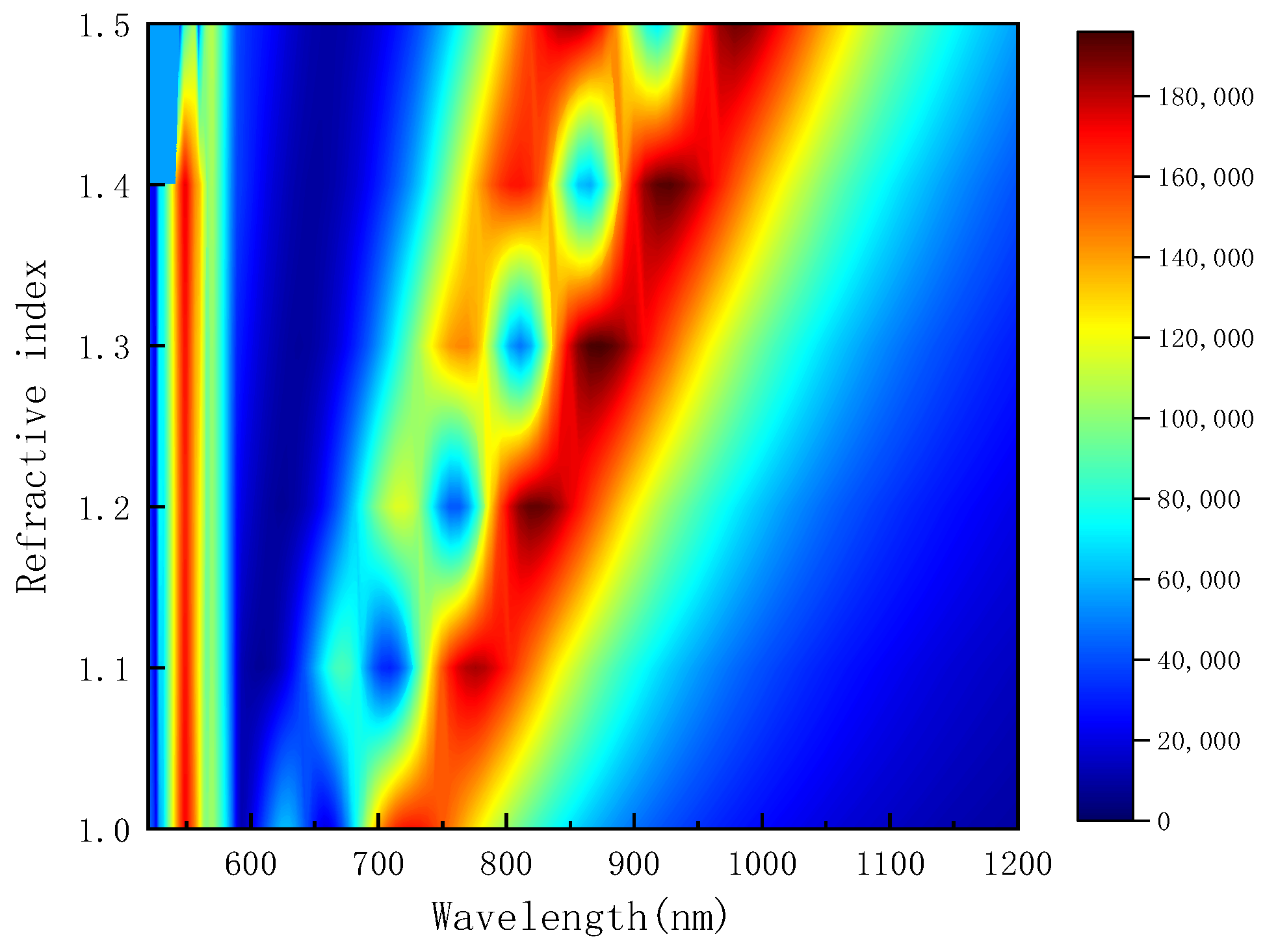
Figure 7 shows the scattering cross-sections of the hybrid nanoantenna for different refractive indexes between 1.0 and 1.5. Three evident dips occur at wavelengths of 566 nm, 592 nm and 657 nm when the refractive index is 1.0. The spectral dips move gradually in the longer wave direction at wavelengths of 592 nm and 657 nm, while the first dip at 566 nm does not change. To analyze the performance of the hybrid nanoantenna as a plasmonic sensor, specific parameters, such as the sensitivity (S) and figure of merit (FOM), are determined, as shown in the following [38,39].
S ( nm / RIU ) = Δ λ d i p Δ n
F O M ( RIU 1 ) = S F W H M
where FWHM is the full-width at half-maximum.
As shown in Table 1, the sensitivity is enhanced by increasing the refractive index. When the refractive index is 1.3, the maximum sensitivity is 550 nm/RIU at the third dip and the wavelength sensitivity is more than 1.5-times that of Fano sensors [40]. The calculated FOM values are shown in Table 2, which shows that the maximum FOM is 13 RIU −1 at the third dip for the refractive index of n = 1.1. The table shows the sensitivity (S) and figure of merit (FOM) between adjacent refractive index. The results clearly demonstrate that the refractive index affects the scattering properties and can be applied to the design of plasmonic sensors.

4. Conclusions

A nanoantenna is designed based on the embedded heptamer structure. To understand the coupling effects of the heptamer and nanoring, nanoantenna coupling from two to seven disks is analyzed to obtain the scattering cross-sections in the presence of an outer nanoring. The addition of the outer nanoring yields multi-Fano resonance characteristics that improve the near-field properties and sensitivity, which is promising for optical sensing. The effects of different structural parameters on the characteristics of Fano resonance are studied and dependence on the size, shape and nature of the materials is observed. Single-point electromagnetic hotspots are studied and the maximum intensity is found to be 159.59 V/m. The sensitivity is derived from the wavelength shift and SCS transformation, showing a maximum value of 550 nm/RIU. Our results impart valuable insights into near-field enhancement, which is vital to the design of optical sensors and SERS.

Author Contributions

Conceptualization, J.L. and D.W.; methodology, C.L.; software, D.W.; validation, J.W.; formal analysis, J.L.; investigation, J.L.; resources, L.Y.; data curation, J.L.; writing—original draft preparation, D.W.; writing—review and editing, W.L. and P.K.C.; visualization, J.L.; supervision, Q.L.; project administration, H.M.; funding acquisition, J.L. and P.K.C. All authors have read and agreed to the published version of the manuscript.

Funding

This research was funded by Natural Science Foundation Projects in Heilongjiang Province (LH2021F007), China Postdoctoral Science Foundation (2020M670881), Study Abroad returnees merit-based Aid Foundation in Heilongjiang Province (070-719900103), Northeastern University scientific research projects (2019KQ74) and Strategic Research Fund of the City University of Hong Kong (SRG) (7005505).

Institutional Review Board Statement

Not applicable.

Informed Consent Statement

Not applicable.

Data Availability Statement

Not applicable.

Conflicts of Interest

The authors declare no conflict of interest.

References

  1. Xu, Q.; Pan, C.; Chen, Y. Nano-optical antenna sensor based on localized surface plasmon resonance. In Proceedings of the 2016 IEEE International Conference on High Voltage Engineering and Application (ICHVE), Chengdu, China, 19–22 September 2016; pp. 1–4. [Google Scholar]
  2. Nikitin, A.; Remezani, M.; Rivas, J.G. Luminescent Metamaterials for Solid State Lighting. ECS J. Solid State Sci. Technol. 2015, 5, 3164–3169. [Google Scholar] [CrossRef]
  3. Liu, J.X.; He, H.; Xiao, D.; Yin, S.T.; Ji, W.; Jiang, S.Z.; Luo, D.; Wang, B.; Liu, Y.J. Recent Advances of Plasmonic Nanoparticles and Their Applications. Materials 2018, 11, 1833. [Google Scholar] [CrossRef] [PubMed]
  4. Su, K.H.; Wei, Q.H.; Zhang, X.; Mock, J.J.; Smith, D.R.; Schultz, S. Interparticle Coupling Effects on Plasmon Resonances of Nanogold Particles. Nano Lett. 2003, 3, 8. [Google Scholar] [CrossRef]
  5. Pineider, F.; Pedrueza-Villalmanzo, E.; Serri, M.; Adamu, A.M.; Smetanina, E. Plasmon-enhanced magneto-optical detection of single-molecule magnets. Mater. Horiz. 2019, 6, 1148–1155. [Google Scholar] [CrossRef]
  6. Jiang, S.; Zhang, X.Y.; Li, D.L.; Wang, T.T.; Ma, F.; Zhang, C.Y. Construction of a gold nanoparticle -based single-molecule biosensor for simple and sensitive detection of Argonaute 2 activity. J. Mater. Chem. B 2022, 10, 5594–5601. [Google Scholar] [CrossRef]
  7. Yang, A.K.; Wang, D.Q.; Wang, W.J.; Odom, T.W. Coherent Light Sources at the Nanoscale. Annu. Rev. Phys. Chem. 2017, 68, 83–99. [Google Scholar] [CrossRef] [PubMed]
  8. Sakai, K.; Kitajima, H.; Sasaki, K. Plasmonic nanostructures for shrinking structured light to access forbidden transitions. Nanophotonics 2022, 11, 11. [Google Scholar] [CrossRef]
  9. Zhang, R.X.; Du, C.L.; Sun, L.; Rong, W.X.; Li, X.; Lei, M.X.; Shi, D.N. Individual Split Au Square Nanorings for Surface-Enhanced Raman and Hyper-Raman Scattering. Plasmonics 2022, 17, 965–971. [Google Scholar] [CrossRef]
  10. Kamimura, R.; Kondo, T.; Motobayashi, K.; Lkeda, K. Surface-Enhanced Electronic Raman Scattering at Various Metal Surfaces. Phys. Status Solidi B Basic Solid State Phys. 2021, 2100589. [Google Scholar] [CrossRef]
  11. Juan-Colas, J.; Parkin, A.; Dunn, K.E.; Scullion, M.G.; Krauss, T.F.; Johnson, S.D. The electrophotonic silicon biosensor. Nat. Commun. 2016, 7, 12769. [Google Scholar] [CrossRef] [Green Version]
  12. Chau, Y.F.; Chou Chao, C.T.; Jumat, S.Z.B.H.; Kooh, M.R.R.; Thotagamuge, R.; Lim, C.M.; Chiang, H.-P. Improved Refractive Index-Sensing Performance of Multimode Fano-Resonance-Based Metal-Insulator-Metal Nanostructures. Nanomaterials 2021, 11, 2097. [Google Scholar] [CrossRef]
  13. Limonov, M.F.; Rybin, M.V.; Poddubny, A.N.; Kivshar, Y.S. Fano resonances in photonics. Nat. Photonics 2017, 11, 543–554. [Google Scholar] [CrossRef]
  14. Wan, Y.; Tan, Y.X.; Yang, Y.; Chong, H.N.; Meng, Z.Z.; Wang, J. Actively Tunable Fano Resonance in H-Like Metal-Graphene Hybrid Nanostructures. Plasmonics 2022, 17, 843–849. [Google Scholar] [CrossRef]
  15. Yang, Z.J.; Zhao, Q.; He, J. Fano interferences of electromagnetic modes in dielectric nanoblock dimers. J. Appl. Phys. 2019, 125, 063103. [Google Scholar] [CrossRef]
  16. Ye, J.; Wen, F.; Sobhani, H.; Lassiter, J.B.; Van Dorpe, P.; Nordlander, P.; Halas, N.J. Plasmonic nanoclusters: Near field properties of the Fano resonance interrogated with SERS. Nano Lett. 2012, 12, 1660–1667. [Google Scholar] [CrossRef]
  17. Zuev, D.A.; Makarov, S.V.; Milichko, V.A.; Starikov, S.A.; IMukhin, I.S.; Morozov, I.A.; Shishkin, I.I.; Krasnok, A.E.; Belov, P.A. Fabrication Of Hybrid Nanostructures Via Nanoscale Laser-Induced Reshaping For Advanced Light Manipulation. Adv. Mater. 2016, 28, 3087–3093. [Google Scholar] [CrossRef]
  18. Zarrabi, F.B.; Moghadasi, M.N. Investigated the Fano resonance in the nano ring arrangement. Optik 2017, 138, 80–86. [Google Scholar] [CrossRef]
  19. Dutta, A.; Alam, K.; Nuutinen, T.; Hulkko, E.; Karvinen, P.; Kuittinen, M.; Toppari, J.J.; Vartiainen, E.M. Influence of Fano resonance on SERS enhancement in Fano-plasmonic oligomers. Opt. Express 2019, 27, 21. [Google Scholar] [CrossRef]
  20. Torres, M.U.; Klausen, K.O.; Sitek, A.; Erlingsson, S.I.; Gudmundsson, V.; Manolescu, A. Electromagnetic field emitted by core–shell semiconductor nanowires driven by an alternating current. J. Appl. Phys. 2021, 30, 034301. [Google Scholar] [CrossRef]
  21. Chou Chau, Y.F.; Chou Chao, C.T.; Huang, H.J.; Kooh, M.R.R.; Kumara, N.T.R.N.; Lim, C.M.; Chiang, H.P. Perfect Dual-Band Absorber Based on Plasmonic Effect with the Cross-Hair/Nanorod Combination. Nanomaterials 2020, 10, 493. [Google Scholar] [CrossRef] [Green Version]
  22. Zubaidah, S.; Chou Chao, C.T.; Chou Chau, Y.F.; Mahadi, A.H.; Kooh, M.R.R.; Kumara, N.T.R.N.; Chiang, H.P. Plasmonic refractive index sensor based on the combination of rectangular and circular resonators including baffles. Chin. J. Phys. 2021, 71, 286–299. [Google Scholar] [CrossRef]
  23. Sung, M.J.; Ma, Y.F.; Chau, Y.F.; Huang, D.W. Surface plasmon resonance in a hexagonal nanostructure formed by seven core shell nanocylinders. Appl. Opt. 2010, 49, 920–926. [Google Scholar] [CrossRef] [PubMed]
  24. Fedorov, V.V.; Koval, O.Y.; Ryabov, D.R. Nanoscale Gallium Phosphide Epilayers on Sapphire for Low-Loss Visible Nanophotonics. ACS Appl. Nano Mater. 2022, 5, 8846–8858. [Google Scholar] [CrossRef]
  25. Khmelevskaia, D.; Markina, D.I.; Fedorov, V.V. Directly grown crystalline gallium phosphide on sapphire for nonlinear all-dielectric nanophotonics. Appl. Phys. Lett 2021, 118, 201101. [Google Scholar] [CrossRef]
  26. Zhou, Y.; Meng, Y.J.; Huang, W.X.; Li, K.G. Application scope of multipole method for decomposition of plas monic spectrum. Results Phys. 2022, 39, 105742. [Google Scholar] [CrossRef]
  27. Radescu, E.E.; Vaman, G. Exact calculation of the angular momentum loss, recoil force, and radiation intensity for an arbitrary source in terms of electric, magnetic, and toroid multipoles. Phys. Rev. E 2002, 65, 046609. [Google Scholar] [CrossRef]
  28. Zhang, X.; Wang, S.; Lin, Z.; Sun, H.B.; Chan, C.T. Optical force on toroidal nanostructures: Toroidal dipole versus renormalized electric dipole. Phys. Rev. A 2015, 92, 043804. [Google Scholar] [CrossRef]
  29. Evlyukhin, A.B.; Fischer, T.; Reinhardt, C.; Chichkov, B.N. Optical theorem and multipole scattering of light by arbitrarily shaped nanoparticles. Phys. Rev. B 2016, 94, 205434. [Google Scholar] [CrossRef]
  30. Ray, D.; Kiselev, A.; Martin, O.J.F. Refractive index data for 150 nm Ag measured with ellipsometry [Data set]. Zenodo 2021. [Google Scholar] [CrossRef]
  31. West, P.; Ishii, S.; Naik, G.; Emani, N.; Shalaev, V.; Boltasseva, A. Searching for better plasmonic materials. Laser Photonics Rev. 2010, 4, 795–808. [Google Scholar] [CrossRef] [Green Version]
  32. Palik, E.D. Handbook of Optical Constants of Solids; Academic Press: New York, NY, USA, 1985. [Google Scholar]
  33. Yang, M.; Chen, X.; Wang, Z.; Zhu, Y.; Pan, S.; Chen, K.; Wang, Y.; Zheng, J. Zero→Two-Dimensional Metal Nanostructures: An Overview on Methods of Preparation, Characterization, Properties, and Applications. Nanomaterials 2021, 11, 1895. [Google Scholar] [CrossRef]
  34. Hüttenhofer, L.; Tittl, A.; Kühner, L.; Cortés, E.; Maier, S.A. Anapole-Assisted Absorption Engineering in Arrays of Coupled Amorphous Gallium Phosphide Nanodisks. ACS Photonics 2021, 8, 1469–1476. [Google Scholar] [CrossRef]
  35. Zhao, X.; Wen, J.; Zhu, A.; Cheng, M.; Zhu, Q.; Zhang, X.; Wang, Y.; Zhang, Y. Manipulation and Applications of Hotspots in Nanostructured Surfaces and Thin Films. Nanomaterials 2020, 10, 1667. [Google Scholar] [CrossRef]
  36. Dregely, D.; Hentschel, M.; Giessen, H. Excitation and tuning of higher-order Fano resonances in plasmonic oligomer clusters. ACS Nano 2011, 5, 8202–8211. [Google Scholar] [CrossRef]
  37. Torres, U.M.; Sitek, A.; Manolescu, A. Anisotropic light scattering by prismatic semiconductor nanowires. Opt. Express 2019, 27, 25502–25514. [Google Scholar] [CrossRef]
  38. Liu, W.; Shi, Y.; Yi, Z.; Liu, C.; Wang, F.M.; Li, X.L.; Lv, J.W.; Yang, L.; Chu, P.K. Surface plasmon resonance chemical sensor composed of a microstructured optical fiber for the detection of an ultra-wide refractive index range and gas-liquid pollutants. Opt. Express 2021, 29, 25. [Google Scholar] [CrossRef]
  39. Melwin, G.; Senthilnathan, K. High sensitive D-shaped photonic crystal fiber sensor with Vgroove analyte channel. Optik 2020, 213, 164779. [Google Scholar] [CrossRef]
  40. Ghodsi, F.; Dashti, H.; Shokouh, J.A. Design of a multilayer nano-antenna as a hyperbolic metamaterial with Fano response for optical sensing. Opt. Quantum Electron. 2020, 52, 316. [Google Scholar] [CrossRef]
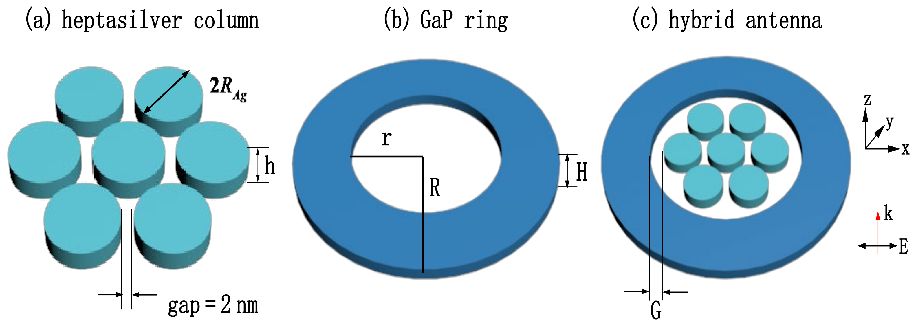
Figure 1. (a) Structural parameters and arrangement of the heptamer of silver; (b) parameters of the GaP nanoring; (c) combination of the heptamer of silver and GaP nanoring.
Coatings 12 01248 g001
Figure 2. (a) Arrangement of the silver disks; (b) comparison of the scattering cross-sections for different arrangements of the nanodisks; (c) scattering cross-section of the hybrid nanostructures; (d) scattering cross-section of the hybrid nnoantennas after adding the substrate for silicon materials; (e) multipole contributions to the scattering cross-section.
Coatings 12 01248 g002
Figure 3. (a) Normalized electric field enhancement at the Fano dips and (b) charge distributions at the Fano dips.
Coatings 12 01248 g003
Figure 4. (a) Scattering cross-sections for different internal silver disk heights; (b) scattering cross-sections for different inner and outer heights; (c) scattering cross-sections for different distances between the heptamer of silver and the GaP nanoring; (d) comparison of the scattering areas for silver disks of different radii; (e) the influence of the width of the GaP nanoring on scattering cross-section.
Coatings 12 01248 g004
Figure 5. (a) Scattering cross-sections of three different dielectric materials for the nanoring and (b) comparison of the scattering cross-sections in different directions of the incident light.
Coatings 12 01248 g005
Figure 6. (a) Normalized electric fields of the hybrid nanoantennas at points A, B and C; (b) Comparison of the normalized electric fields of the hybrid nanoantennas with individual hepatmers at point B.
Coatings 12 01248 g006
Figure 7. Effects of refractive indexes on the scattering cross-sections.
Coatings 12 01248 g007
Table 1. Sensitivity of the nanostructure for different refractive indexes.
Fano Dips First DipSecond DipThird Dip
n = 1.0
n = 1.1Δn0.10.10.1
Δλ (nm)01652
S (nm/RIU)0160520
n = 1.2Δn0.10.10.1
Δλ (nm)01747
S (nm/RIU)0170470
n = 1.3Δn0.10.10.1
Δλ (nm)01355
S (nm/RIU)0130550
n = 1.4Δn0.10.10.1
Δλ (nm)01454
S (nm/RIU)0140540
n = 1.5Δn0.10.10.1
Δλ (nm)31053
S (nm/RIU)30100530
Table 2. FOM of the nanostructure for different refractive indexes.
Fano DipsFirst DipSecond DipThird Dip
n = 1.0
n = 1.1S (nm/RIU)0160520
FWHM (nm) 742.140
FOM (RIU−1)03.813
n = 1.2S (nm/RIU)0170470
FWHM (nm) 754.854
FOM (RIU−1)03.18.7
n = 1.3S (nm/RIU)0130550
FWHM (nm) 772.250
FOM (RIU−1)01.811
n = 1.4S (nm/RIU)0140540
FWHM (nm) 882.453.5
FOM (RIU−1)01.710.1
n = 1.5S (nm/RIU)30100530
FWHM (nm) 1.710098.1
FOM (RIU−1)17.515.4
Publisher’s Note: MDPI stays neutral with regard to jurisdictional claims in published maps and institutional affiliations.