Article

Effect of Copper Nanoparticles and Ions on Epididymis and Spermatozoa Viability of Chinese Soft-Shelled Turtles Pelodiscus sinensis

1
Jiangsu Provincial Key Laboratory of Coastal Wetland Bioresources and Environmental Protection, Yancheng 224007, China
2
Jiangsu Synthetic Innovation Center for Coastal Bioagriculture, Yancheng 224007, China
3
School of Wetlands, Yancheng Teachers University, Yancheng 224007, China
4
College of Ocean and Biology Engineering, Yancheng Teachers University, Yancheng 224007, China
*
Authors to whom correspondence should be addressed.
Coatings 2022, 12(2), 110; https://doi.org/10.3390/coatings12020110
Submission received: 17 December 2021 / Revised: 12 January 2022 / Accepted: 15 January 2022 / Published: 19 January 2022

Abstract

:
Copper nanoparticles (CuNPs) have been widely used in various industrial and commercial applications, which become a potential threat to aquatic organisms. Nevertheless, their potential toxicity to the epididymis and sperm remains little known. In this study, we evaluated the effect of CuNPs and copper ions (CuSO4) on the spermatozoa viability, epididymal structure, antioxidant enzyme activity, and inflammatory cytokines in cauda epididymis of the Chinese soft-shelled turtle. Results showed that the spermatozoa viability of Chinese soft-shelled turtles decreased significantly with an increase in CuNPs or Cu ions concentrations. The epithelial cells of the epididymal duct of the Chinese soft-shelled turtles with the treatment of 5 mg kg−1 CuNPs were slightly swollen, and the connective tissue between the epididymal ducts was loose. The epithelial structure of the epididymal tube was severely damaged with an increase in Cu ion concentrations. Compared to the control, the antioxidative enzymes activities and the expression of IL-1β, TNF-α, and IL-6 mRNA in the epididymis significantly increased with the treatment of CuNPs or CuSO4. The present study revealed that Cu ions exert more harmful effect on the epididymis and spermatozoa viability of Chinese soft-shelled turtles than copper nanoparticles.

1. Introduction

Copper nanoparticles (CuNPs) are one of the most commonly used nanomaterials, which have received much attention in these years and have been widely used in fungicides, cosmetics, printers, and electronics, because of their antifungal, optical, and electrical properties [1,2]. The extensive use of CuNPs has led to direct contamination in the aquatic environment, as they can be released into aquatic ecosystems through indirect pathways, such as runoffs and leaching from industrial and agricultural sites, and also through direct pathways, with the use of antifouling paints [3,4], raising a global concern for ecological impacts of such contamination [5]. The aquatic environment is considered to be a sink for pollutants [6]. Although copper is an essential trace metal required by all living organisms, it can be toxic to organisms when it excesses the physiological requirement of organisms. The application of CuNPs has caused adverse effects on aquatic organisms, such as fish, daphnias, and algaes, due to their dynamic physicochemical processes in an aquatic environment [7,8]. The LC50 value represents a common point in the lethal physiological response to toxicity, which has been abundant in data in many crustaceans. For example, the 96 h-LC50 value for shrimps of Exopalaemon carinicauda, Echinogammars olivii, Sphaeroma serratum, and Palaemon elegans was 0.712 mg Cu/L, 0.25 mg Cu/L, 1.98 mg Cu/L, and 2.52 mg Cu/L, respectively [9,10]. In addition, for paddy field crab Paratelphusa hydrodromus and freshwater crab, Barytelphusa cunicularis, the 96 h-LC50 values recorded were 15.70 mg Cu/L and 215 mg Cu/L, respectively [11,12]. Likewise, in freshwater crayfish, Procambarus clarkia, the 96 h-LC50 value reached 162 mg Cu/L [13]. These large variations in sub-lethal effects to Cu toxicity in crustaceans appear to be species specific. There are a large number of studies on the effects of copper on fish toxicity. According to a review by Malhotra et al. [14], it can be seen that the acute and sub-chronic exposure concentrations of different fish to copper vary significantly, ranging from micrograms to approximately 250 g per liter.
The negative impacts of CuNPs on different aquatic organisms have been reported by many researchers; however, it remains difficult to conclude what is the major cause of NPs toxicity [15]. Most studies showed that the toxicity of metal nanoparticles (MNPs) to organisms was attributed to their release of soluble ions [7,16,17], while some other studies demonstrated that CuNPs could penetrate into cells or move to different organs and tissues, thus damaging the organisms [18,19]. There are also some reports indicating that the toxicity of CuNPs was caused by the combination of nanoparticles and the released copper ions [2,20]. Zhang et al. [21] illustrated that the toxicity of CuNPs to zebrafish embryonic cells was not only caused by the released Cu ions, and CuNPs and Cu ions accounted for 48.4% and 51.6% of the total Cu content, respectively.
Sperm quality analysis is considered to be a potential tool for use in the assessment of environmental quality and has been successfully applied as a biomarker to evaluate the effects of contaminants in the aquatic environment on fish reproduction [22,23]. Contaminants in aquatic environments could affect the fertilization efficiency of aquatic organisms [24], which can decrease spermatozoa motility and fertilization efficiency. Histopathological studies can be used as a dependable tool to investigate the toxic effects of environmental pollutants and to assess symptoms of damages [25]. Gürkan et al. [26] reported that 10 days of Fe2O3 NPs exposure caused significant histopathological damages to rainbow trout (Oncorhynchus mykiss). Antioxidant enzymes are known to be the first line of response upon stress, playing an important role in maintaining normal metabolism and function of the organisms [27]; they play an important role in maintaining the ROS homeostasis of organisms [28]. The classic antioxidant enzymes include superoxide dismutase (SOD), catalase (CAT), glutathione peroxidase (GPx), etc. They are very sensitive to environmental pollutants, and thus, they are usually employed as important biomarkers in numerous environmental toxicology experiments [29,30].
Previous studies suggested that contaminants could cause a potential risk to the fish species during their reproduction season when fertilization occurs [31]. Research on the effect of contamination on fish spermatozoa has mainly focused on the influence of the ionic form of metals. Few studies have performed risk assessment of toxicity of nanomaterials to male gamete motility. CuSO4, CuNPs, and CuO NPs in the aqueous environment primarily cause a reduction of spermatozoa velocity, and the effect of Cu ions on sea trout spermatozoa motility was more harmful than that of copper nanoparticles [24]. Our current understanding of the toxic of CuNPs to the sperm in reptiles is severely limited as compared to the fish. Chinese soft-shelled turtles (Pelodiscus sinensis) are one of the common species of reptiles and are widely distributed in China, which is famous for their economic and pharmacologic value. Freshwater turtles play a major role in maintaining balance in the ecosystem of any wetland. They act as scavengers by decaying dead organic matter and indicators of a healthy aquatic ecosystem. Some of them are carrion eating species that feed on aquatic weeds and reduce eutrophication. As a representative of freshwater turtles, Chinese soft-shelled turtles first appeared 120 million years ago and have an important position in the study of species evolution. Our previous results have shown that the epididymis of the Chinese soft-shelled turtle is similar to that of mammals and is divided into three parts: caput, corpus, and cauda. The cauda epididymis is the main site of sperm storage [32]. The Chinese turtles’ epididymis is unique in that sperm can be stored in its epididymis long-term with a low apoptosis rate. It can be seen that the epididymis plays an important role in the storage of sperm and the reproduction of species.
In this study, we aimed to assess the in vivo effects of CuNPs and CuSO4 on spermatozoa viability, epididymal structure, antioxidant enzyme activity, and inflammatory cytokines in cauda epididymis of the Chinese soft-shelled turtle. This study may provide preliminary information regarding the ecological impacts of CuNPs on the fertilization efficiency of aquatic organisms.

2. Materials and Methods

2.1. Chemicals and the Test Organism

CuNPs with a primary size of 40 nm were purchased from Sigma (St. Louis, MO, USA), the particle size was determined using Transmission Electron Microscopy (TEM, JEOL Ltd., Tokyo, Japan), and the pristine shape of CuNPs used in the experiment was spherical (Supplementary Materials). The stock suspension of CuNPs (100 mg L−1) was prepared by suspending the powder in MilliQ water. The suspension was sonicated (20 kHz, 100 w, 20% cycle off at 25 °C) in an ice-cold bath for at least 40 min and stirred for 30 min. Cu ions stock solution (100 mg L−1) was prepared by dissolving CuSO4 (99% purity, Sigma-Aldrich) in deionized water. The concentration of the products tested (CuNPs and CuSO4) was expressed as mg Cu L−1 in the media regardless of the dissolution and aggregation of the products.
A total of 45 adult male Chinese soft-shelled turtles (body weight 700 ± 50 g) were chosen as model species of aquatic organism, which were purchased from Yancheng, Jiangsu Province, China. The healthy individuals with basically the same size, having no damage on the body surface and showing normal activities, were selected in the experiment. Animals were treated in accordance with the Care and Use of Wild Animals in The People’s Republic of China. In the laboratory, turtles were reared in big water tanks (30 × 40 × 40-cm) containing sand and filled with freshwater during the 2021 breeding season for 7 days prior to the beginning of experiments.
Turtles were divided into five groups (Control, 5 mg kg−1, and 50 mg kg−1 CuNPs or CuSO4), and each treatment had three replicates. We calculated the volume of the required stock solution based on the body weight of the turtle; then, we set the volume to 20 mL and injected it into the abdominal cavity of the turtle. The control group was replaced with PBS (sterilized distilled water) solution. The animals were sacrificed by cervical dislocation at 24, 48, and 72 h after intraperitoneal injection. The cauda epididymis was separated according to the anatomical structure. One of each pair of cauda epididymis was used for light microscopy; the other was used to collect sperm to test its viability. Subsequently, the cauda epididymis with sperm removed was cryopreserved in liquid nitrogen for other experiments.

2.2. Light Microscopy

The samples were fixed in neutral buffered formalin, embedded in paraffin, and serial sectioned (at 5 µm). These sections were stained with Harris’ hematoxylin and eosin (HE) for light microscopic observation. Slides were assessed using an Olympus microscope (BX53, Olympus Corporation, Tokyo, Japan), camera (Olympus DP73, Olympus Corporation, Tokyo, Japan), and the AnalySIS image-analyzing system (AnalySIS).
To observe the integrity of the epididymal epithelial structure, 20 pictures of straight perpendicularly cut epithelium sections were taken at 400× magnification. In each picture, the epithelium was measured 3 times.

2.3. Epididymal Sperm Preparation and Viability Test

The epididymal sample was placed in phosphate-buffered saline and exsanguinated for 5–10 min. After transferring to fresh PBS, the connective tissue around the epididymis was removed, and the sperm in the epididymal tubules flew into PBS for 10-15 min to ensure that all sperm are obtained as much as possible. Then, we transferred the PBS buffer containing sperm to a centrifuge tube and concentrated it by centrifugation at 700× g for 10 min at 4 °C. Sperm were suspended in sterile PBS. We diluted the sperm concentration to 106 mL−1.
Sperm viability was quantified with double staining of fluorescein isothiocyanate (FITC) conjugated annexin-V and propidium iodide (PI) (Biouniquer, BU-AP0103, Biouniquer Technology, Nanjing, China). Ten thousand cells per sample were acquired with an FACS scan flow cytometer (FACScan, Becton Dickinson, Franklin Lakes, NJ, USA). Take 1 mL of the prepared sperm suspension, which was washed twice with binding buffer and processed following the manufacturer’s instructions. Cells fluorescence was analyzed with flow cytometry using the Cell Quest Pro software (Beckman Coulter, Becton Dickinson, Franklin Lakes, NJ, USA).

2.4. Biochemical Assay

The epididymal tube sample from which the sperm has been removed was homogenized with a glass homogenizer in ice-cold conditions by adding Tris-HCl buffer and a small amount of protease inhibitor. Then, homogenates were centrifuged at 9000× g for 30 min at 4 °C. The supernatant was collected for measuring oxidative stress markers. SOD, CAT, and GSH activities were determined using the kits (Nanjing Jiancheng Bioengineering Institute, Nanjing, China) and according to the manufacturer’s instructions.

2.5. Analysis of mRNA Expression Levels

Primers were designed using Primer 3 software (http://bioinfo.ut.ee/primer3/, accessed on 19 February 2021). The expression of IL-1β, TNF-α, and IL-6 mRNA in the cauda epididymis of Cu-treated soft-shelled turtles was examined using real-time PCR. Total RNA was isolated from tissue samples using the RNeasy Mini Kit Qiagen (Catalog no. 74104).
Real-time reverse transcription polymerase chain reaction (RT-PCR) was used to detect the mRNA expression levels of IL-1β, TNF-α, and IL-6. Total RNAs were extracted from epididymal epithelial cells by adding 1 mL of TRIzol Reagent (Invitrogen, Waltham, CA, USA) and reverse-transcribed to form cDNAs using the ABI7500 Real-Time PCR system (Applied Biosystems, Foster City, CA, USA) according to the manufacturer’s instructions. β-actin was used as an endogenous quantity control. The primer sequences used in the quantitative real-time PCR reactions were as shown in Table 1. The total volume of 20 μL PCR reactions was prepared by mixing 1.0 μL of cDNA sample, 10.0 μL of 2× SYBR Premix Ex Taq II (Tli RNaseH Plus, Takara Bio Inc., Shiga, Japan), 0.4 L of forward primer (10 M), 0.4 L of reverse primer (10 M), and 8.2 μL of ddH2O. The final results were presented as the ratios of the relative amount of the target gene to the control gene using the 2−ΔΔCT equation, where ΔΔCT = CT (target gene) − CT (internal control gene). The results were analyzed using the Rotor-Gene 6.0 software (Corbett Research, Mortlake, Australia). The results were comparable to those normalized to β-actin, and the statistical analysis was performed using the average relative mRNA levels from three independent samples.

2.6. Statistical Analysis

All data were expressed as means ± SE. Significant differences were analyzed by one-way ANOVA followed by Tukey’s test using SPSS (version 19.0). p < 0.05 was considered significant.

3. Results and Discussion

The widespread use of CuNPs inevitably results in their release into the aquatic environments; therefore, it is essential to evaluate their potential toxic effects on aquatic organisms. In our study, the response of the Chinese turtle to the external stimulus has gradually deteriorated, and the degree of injury was positively correlated with the increase in Cu concentration and time. At 50 mg kg−1, although the animals still survive, their physiological state has been very poor; there was almost no response to external stimuli, as the head and neck remained outside with the treatment of 50 mg kg−1 CuSO4 for 72 h. In addition, there were no changes in the weight and color of the turtles with the treatment of CuSO4 or CuNPs for 72 h.

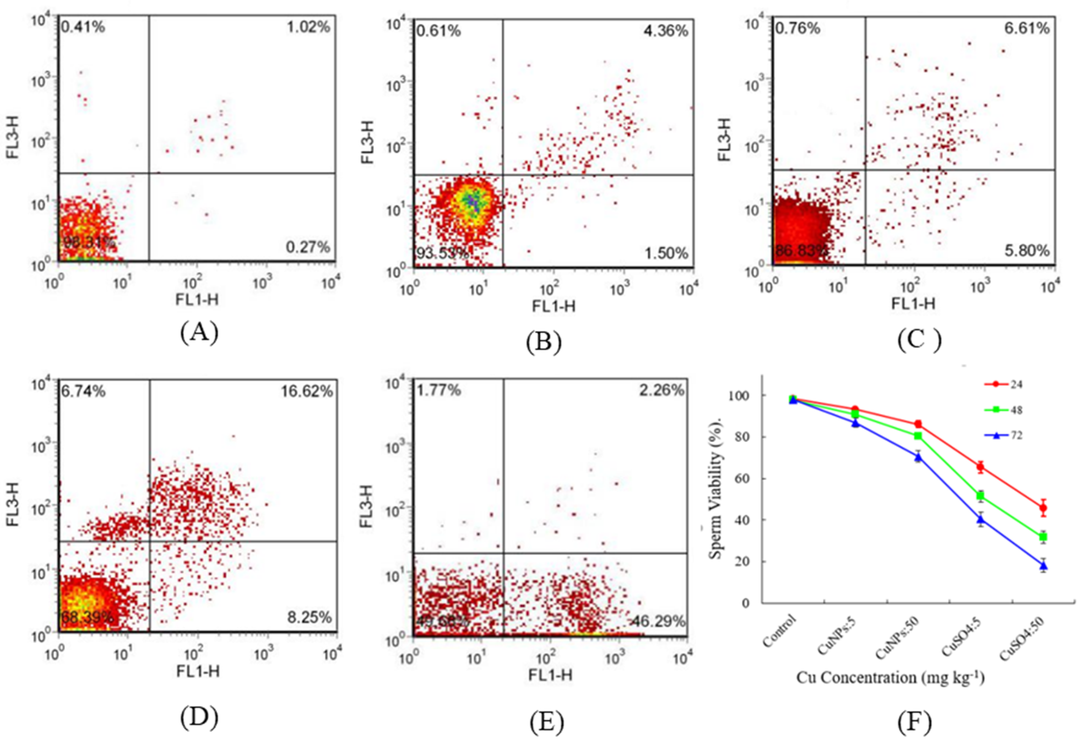
3.1. Effect of CuNPs and Cu Ions on Sperm Viability

Sperm analysis is considered to be useful for determination of the male infertility cause [33]. Some reports showed that Cu can negatively affect sperm quality parameters in rats [34] and rabbits [35] under acute exposure. In the present study, sperm viability was detected by the flow cytometry. Results showed that the treatment of CuNPs and CuSO4 resulted in a dramatic decrease in sperm viability after 24 h of injection compared with the control (Figure 1B–E). The sperm viability of Chinese soft-shelled turtles decreased significantly with a further increase in CuNPs or Cu ion concentrations (Figure 1F). Sperm viability was nearly 18.2% and 40.4% for 50 and 5 mg kg−1 CuSO4 after 72 h, respectively. Meanwhile, the viability was much higher with the treatment of 50 and 5 mg kg−1 CuNPs (Figure 1F), suggesting that Cu ions were more toxic to the sperm. The amount of soluble ions released from CuNPs is much lower than the concentration of corresponding CuSO4. The toxicity of copper ions on the organisms was much higher than that of the nanoparticles, although nanoparticles could cause toxicity to the organisms. Kowalska-Góralska et al. [24] reported that CuNPs and CuO NPs exerted less harmful effects than Cu ions on spermatozoa motility of sea trout. In line with this, Adam et al. [16] illustrated that the toxicity of CuO NPs to aquatic organisms was mainly caused by the released Cu ion.

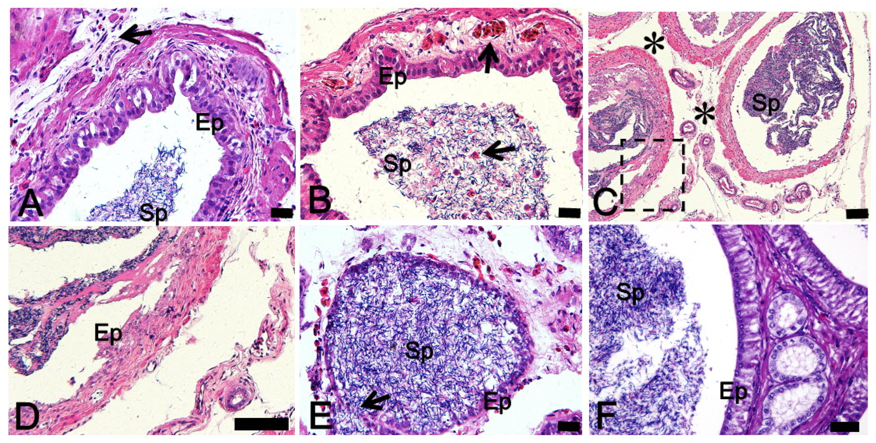
3.2. Effect of CuNPs and Cu Ions on the Histological Analysis

Compared with the control, the epithelial cells of the epididymal duct of the Chinese soft-shelled turtle with the treatment of 5 mg kg−1 CuNPs were slightly swollen, and the connective tissue between the epididymal ducts was slightly loose (Figure 2A); meanwhile, we found intravascular congestion, and a small amount of blood appeared in the lumen blood cells in the epididymis of Chinese soft-shelled turtle with the treatment of 50 mg kg−1 CuNPs; however, the overall structure of the epididymal duct is basically intact (Figure 2B). Part of the epithelium of the epididymal tube was peeled off, and the connective tissue between the epididymal tubes was very loose in the epididymis with the treatment of 5 mg kg−1 CuSO4 (Figure 2C,D). The epithelial structure of the epididymal tube was severely damaged with an increase in Cu ion concentrations (from 5 to 50 mg kg−1), cell connections were broken, and sperm in the cavity may flow to the knot tissue through the rupture (Figure 2E).
The plasma membrane is an important structure for maintaining a highly controlled intracellular environment that enables biochemical reactions and enzymes to function at optimum rates; it also plays an important role in sending and receiving cellular signals, and it takes part in the process of nutrient absorption [36]. With this in mind, the reduction in spermatic plasma membrane integrity may be directly associated with cellular damage. It is not difficult to conclude that exposure to CuNPs and Cu ions have different effects on the structure of epididymis, although so far, there are few reports on the damage of copper nanoparticles on the epididymis structure of Chinese soft-shelled turtles.

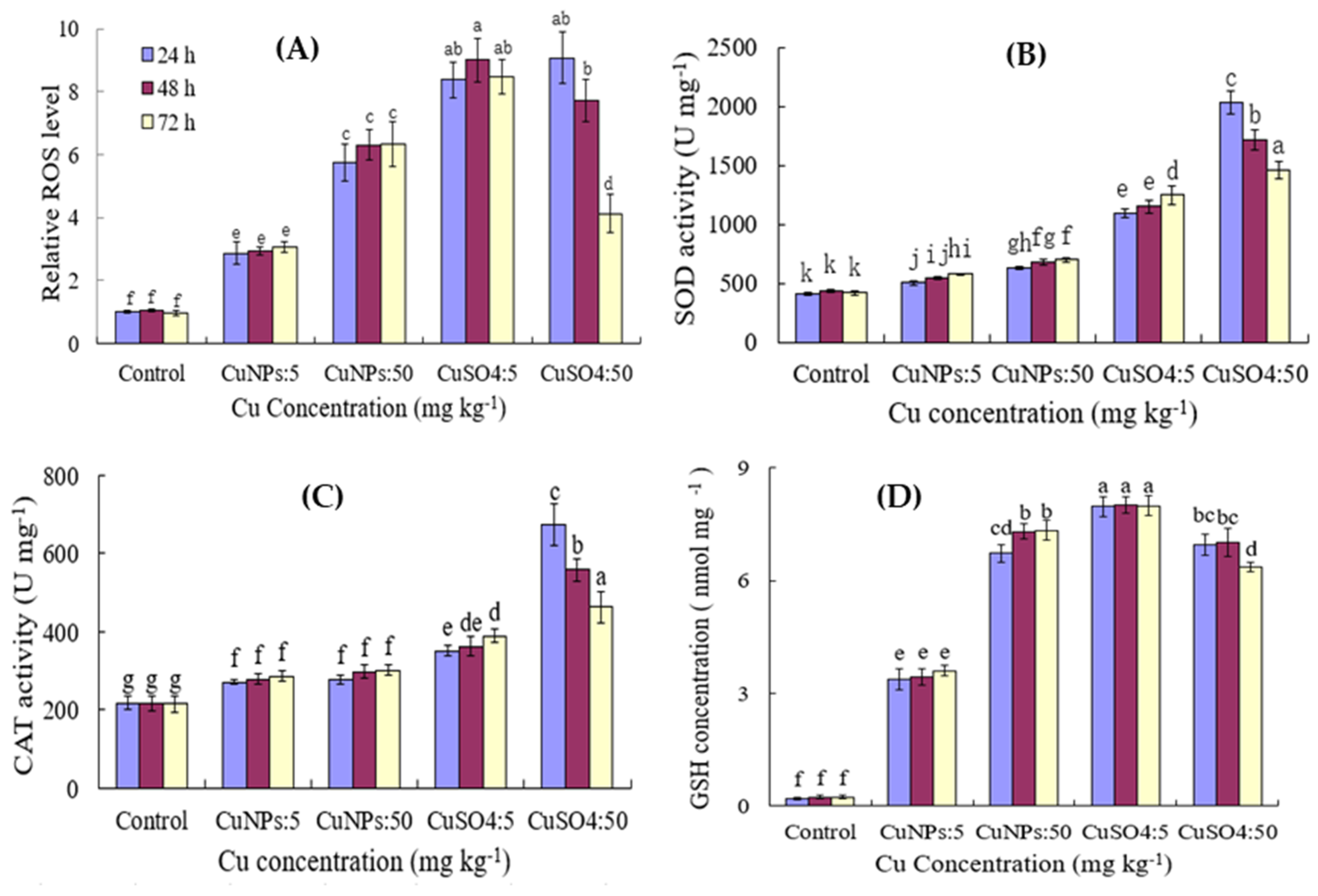
3.3. Effect of CuNPs and Cu Ions on the Antioxidant Enzyme Activity

NPs can cause oxidative damage to the aquatic tissue; they would induce the generation of reactive oxygen species (ROS) and oxidative damage. To lessen the damage, organisms have developed a detoxification system with antioxidant enzyme components, such as SOD, CAT, and GSH, which protect cells against oxidative damage and remove superoxide radicals and hydrogen peroxide [37]. In this study, the antioxidant enzymes activities were determined in the epididymis of Chinese soft-shelled turtles with the treatment of 5 and 50 mg kg−1 CuNPs and CuSO4. The results showed that the injection of CuNPs and CuSO4 resulted in a massive ROS production, and it increased with the increasing concentration of both CuNPs and CuSO4 (Figure 3A). The ROS level was not significantly affected by time, except for 50 mg kg−1 CuSO4. ROS decreased at 48 h and 72 h compared to 24 h (Figure 3A). This may be due to the epididymal epithelial necrosis rate, which increased with the treatment of CuSO4 for time. SOD, CAT, and GSH activities significantly increased with the treatment of both CuNPs and CuSO4 (Figure 3B,C), as the SOD-CAT system is the first line to defend against oxidative stress [38]. Compared to the control, SOD activity even with a low dose (5 mg kg−1) of CuNPs and CuSO4 significantly increased (p < 0.05). It increased by 37.6% and 197.0% after 72 h injection with 5 mg kg−1 CuNPs and CuSO4, respectively. There was no significant difference of antioxidant enzymes activities at different times, except for the treatment of 50 mg kg−1 CuSO4 (Figure 3). SOD, CAT, and GSH activities in the epididymis of the Chinese soft-shelled turtle with the treatment of 50 mg kg−1 CuSO4 significantly increased; however, they significantly decreased with time. This may be explained by the broken epithelial cells with the treatment of 50 mg kg−1 CuSO4 (Figure 3). The ROS levels and CAT activity decreased significantly at 48 h and 72 h. Combined with histological results, it can be seen that with time, the epididymal epithelial necrosis rate increased, resulting in a decrease in CAT activity. According to the related literature [39], ROS plays an important role in the process of removing necrotic cells. We speculate that this may be one of the important reasons for the decline in ROS levels. GSH is involved in cell protection by providing reducing capacity through the transformation of reduced to oxidized GSH and binding to metals during the detoxification process [30]. These results showed that the antioxidant capacity was significantly impaired in the epididymis due to massive ROS production, which resulted in oxidative stress. The accumulation of ROS in sperm leads to a reduction in sperm motility, viability, and enzymatic activity [40]. The accumulation of ROS to high concentrations leads to oxidative stress and subsequent lipid peroxidation (LPO), which provokes an irreversible loss of motility and inhibition of fructolysis and respiration in spermatozoa [41]. This could be an explanation of the decrease in sperm motility, viability, and enzymatic activity. In addition, sperm motility is particularly associated with mitochondria activity, as mitochondria represent the cell energy generator and concomitantly the major site of intracellular ROS formation, resulting in a disruption of electron transport. This disruption also participates in the decline of motility and amplifies LPO in the spermatozoa membrane.

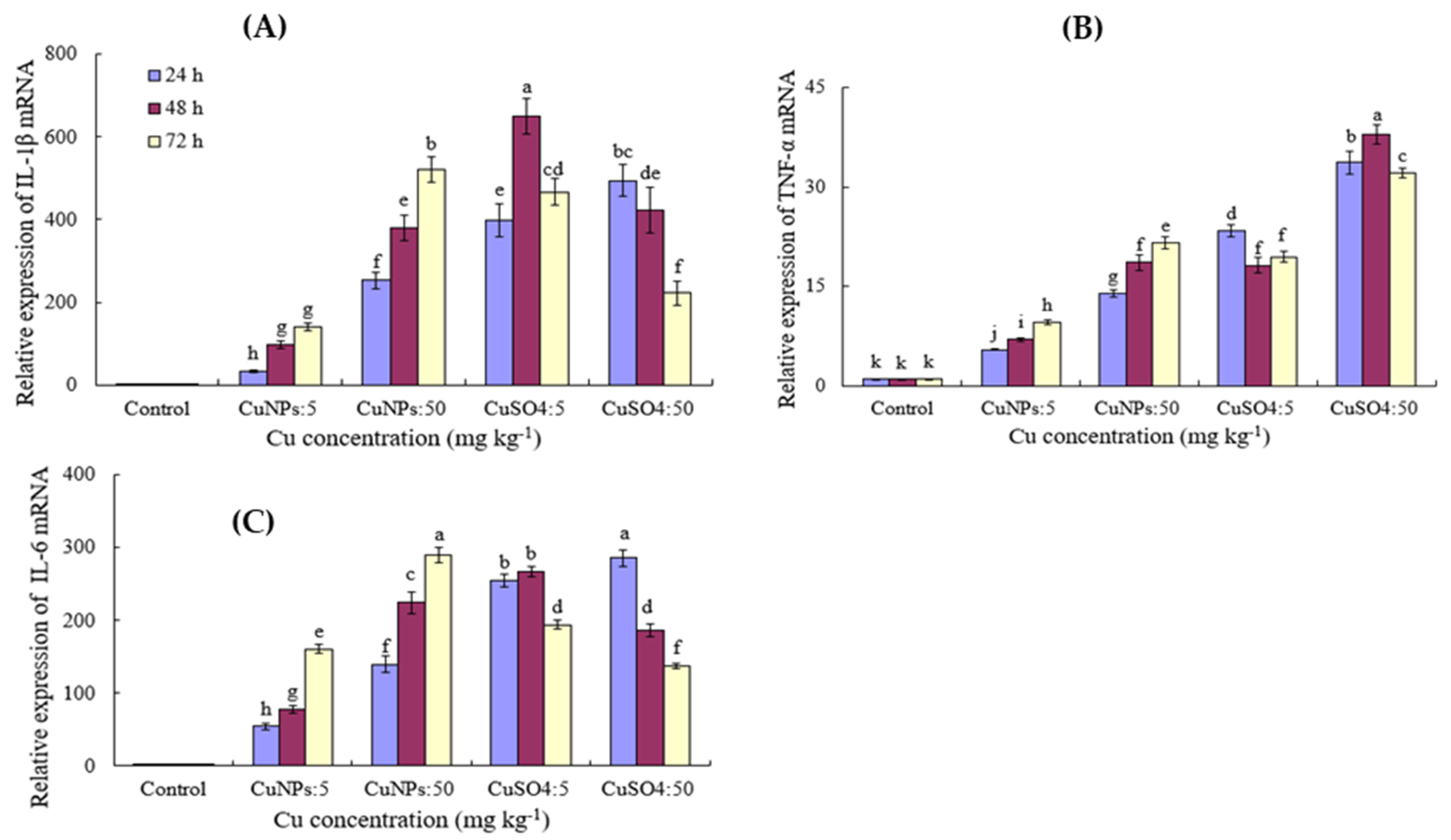
3.4. Effect of CuNPs and Cu Ions on the Relative mRNA Expression

The expression of IL-1β, TNF-α, and IL-6 mRNA in the cauda epididymis of the Chinese soft-shelled turtle treated with CuNPs and CuSO4 was detected by qPCR. The mRNA expression of the three inflammatory factors significantly increased with the treatment of CuNPs and CuSO4 (Figure 4). They increased with the increase in the CuNPs concentrations and time (Figure 4). Different from CuNPs treatment, the relative expression of IL-1β and IL-6 mRNA caused by 5 mg kg−1 CuSO4 treatment increased progressively with time, peaking at 48 h after injection. However, the injection of 50 mg kg−1 CuSO4 resulted in a significant decrease in the relative expression of IL-1β and IL-6 mRNA with time (Figure 4). IL-1β and TNFα are well-known pro-inflammatory cytokines and are generally considered to be markers to assess the inflammatory response when there is an immune stimulus in vertebrates [41]. Compelling evidence suggests that the two cytokines have similar functions to their mammalian homologues [42,43]. Our results showed that IL-1β and TNFα expression were greatly enhanced after CuNPs and CuSO4 treatment, which suggests that the inflammatory response to the epididymis of the Chinese soft-shelled turtle significantly increased after the stress of CuNPs and CuSO4. The difference of CuNPs and CuSO4 in mRNA expression of inflammatory factors may be caused by the amount of soluble ions released from CuNPs being much lower than the concentration of corresponding CuSO4. The cytokine IL-6 possesses pro-inflammatory activity, and it is mainly produced by T cells and macrophages. In reptiles, there is no report on IL-6 expression after CuNPs treatment. Our results show that CuNPs and CuSO4 treatment can also cause significant expression of the inflammatory factor IL-6 (Figure 4), but the underlying mechanism needs to be further studied.

4. Conclusions

This study showed that Cu ions exert a more harmful effect on the epididymis and spermatozoa viability of Chinese soft-shelled turtles than copper nanoparticles. Contamination of the aqueous environment with copper primarily caused a reduction in sperm viability, which may impair reproduction. The antioxidative enzymes and the relative inflammatory factors (IL-1β, TNFα, and IL-6 mRNA) expression in the epididymis of Chinese soft-shelled turtles significantly increased with the treatment of CuNPs and CuSO4. The exposure to CuSO4 caused obvious morphological damage to the epididymis of Chinese soft-shelled turtles, such as, the epithelium of the epididymal tube peeled off. These findings in this work may provide preliminary information regarding the ecological impacts of CuNPs on the fertilization efficiency of aquatic organisms.

Supplementary Materials

The following supporting information can be downloaded at: https://www.mdpi.com/article/10.3390/coatings12020110/s1, Figure S1: Representative TEM image (A) and particle size distribution (B) of CuNPs in Milli-Q water.

Author Contributions

L.Y. Conceptualization, Methodology, Writing—Original draft preparation; Y.W. Software; S.G. Data curation; Q.W. Investigation; J.C. Validation; B.T. Resources; X.B. Editing, Visualization. All authors have read and agreed to the published version of the manuscript.

Funding

This research was funded by the sponsorship of Jiangsu overseas visiting scholar program for university prominent young and middle-aged teachers and presidents and the natural science foundation of the Jiangsu higher education institutions of China (20KJA180006), innovation and entrepreneurship training program for college students in Jiangsu Province (202110324018Z), (202110324022Y), and foundation of Jiangsu Key Laboratory for Bioresources of Saline Soils (JKLBS2021007).

Institutional Review Board Statement

Not applicable.

Informed Consent Statement

Not applicable.

Data Availability Statement

Data are contained within the article or Supplementary Materials.

Conflicts of Interest

The authors declare no conflict of interest.

References

  1. Hou, J.; Wang, X.X.; Hayat, T.; Wang, X.K. Ecotoxicological effects and mechanism of CuO nanoparticles to individual organisms. Environ. Pollut. 2017, 221, 209–217. [Google Scholar] [CrossRef] [PubMed]
  2. Yang, L.; Wang, W.X. Comparative contributions of copper nanoparticles and ions to copper bioaccumulation and toxicity in barnacle larvae. Environ. Pollut. 2019, 249, 116–124. [Google Scholar] [CrossRef]
  3. Adeleye, A.S.; Oranu, E.A.; Tao, M.; Keller, A.A. Release and detection of nanosized copper from a commercial antifouling paint. Water Res. 2016, 102, 374–382. [Google Scholar] [CrossRef] [PubMed] [Green Version]
  4. Muller-Karanassos, C.; Arundel, W.; Lindeque, P.K.; Vance, T.; Turner, A.; Cole, M. Environmental concentrations of antifouling paint particles are toxic to sediment dwelling invertebrates. Environ. Pollut. 2020, 268, 115754. [Google Scholar] [CrossRef]
  5. Wu, F.; Harper, B.J.; Crandon, L.E.; Harper, S.L. Assessment of cu and CuO nanoparticle ecological responses using laboratory small-scale microcosms. Environ. Sci. Nano 2020, 7, 105–115. [Google Scholar] [CrossRef]
  6. Bashir, I.; Lone, F.A.; Bhat, R.A.; Mir, S.A.; Dar, Z.A.; Dar, S.A. Concerns and threats of contamination on aquatic ecosystems. In Bioremediation and Biotechnology; Springer: Cham, Switzerland, 2020; pp. 1–26. [Google Scholar]
  7. Zhu, Y.C.; Xu, J.H.; Lu, T.; Zhang, M.; Ke, M.J.; Fu, Z.W.; Pan, X.L.; Qian, H.F. A comparison of the effects of Cu nanoparticles and Cu sulfate on Phaeodactylum tricornutum physiology and transcription. Environ. Toxicol. Pharmacol. 2017, 56, 43–49. [Google Scholar] [CrossRef] [PubMed]
  8. Fang, R.; Gong, J.L.; Cao, W.C.; Chen, Z.P.; Huang, D.L.; Ye, J.; Cai, Z. The combined toxicity and mechanism of multi-walled carbon nanotubes and nano copper oxide toward freshwater algae: Tetradesmus obliquus. J. Environ. Sci. 2022, 112, 376–387. [Google Scholar] [CrossRef] [PubMed]
  9. Zhang, C.; Li, F.; Xiang, J. Acute effects of cadmium and copper on survival, oxygen consumption, ammonia-N excretion, and metal accumulation in juvenile Exopalaemon carinicauda. Ecotoxicol. Environ. Saf. 2014, 104, 209–214. [Google Scholar] [CrossRef]
  10. Bat, L.; Gündoğdu, A.; Sezgin, M.; Çulha, M.; Gönlügür, G.; Akbulut, M. Acute toxicity of zinc, copper and lead to three species of marine organisms from the Sinop Peninsula, Black Sea. Turk. J. Biol. 1999, 23, 537–544. [Google Scholar]
  11. Vardhanan, Y.S.; Radhakrishnan, T. Acute toxicity evaluation of copper, arsenic and HCH to paddy field crab, Paratelphusa hydrodromus (Herb). J. Environ. Biol. 2002, 23, 387–392. [Google Scholar] [PubMed]
  12. Chourpagar, A.R.; Kulkarni, G.K. Heavy metal toxicity to a freshwater crab, Barytelphusa cunicularis (westwood) from aurangabad region. Recent Res. Sci. Technol. 2011, 3, 1–5. [Google Scholar]
  13. Bini, G.; Chelazzi, G. Acclimatable cardiac and ventilatory responses to copper in the freshwater crayfish Procambarus clarkii. Comp. Biochem. Physiol. C Toxicol. Pharmacol. 2006, 144, 235–241. [Google Scholar] [CrossRef]
  14. Malhotra, N.; Ger, T.R.; Uapipatanakul, B.; Huang, J.C.; Chen, K.H.; Hsiao, C.D. Review of Copper and Copper Nanoparticle Toxicity in Fish. Nanomaterials 2020, 10, 1126. [Google Scholar] [CrossRef] [PubMed]
  15. Boyle, D.; Clark, N.J.; Handy, R.D. Toxicities of copper oxide nanomaterial and copper sulphate in early life stage zebrafish: Effects of pH and intermittent pulse exposure. Ecotoxicol. Environ. Saf. 2020, 190, 109985. [Google Scholar] [CrossRef]
  16. Adam, N.; Schmitt, C.; De Bruyn, L.; Knapen, D.; Blust, R. Aquatic acute species sensitivity distributions of ZnO and CuO nanoparticles. Sci. Total Environ. 2015, 526, 233–242. [Google Scholar] [CrossRef]
  17. Jiang, C.J.; Castellon, B.T.; Matson, C.W.; Aiken, G.R.; Hsu-Kim, H. Relative contributions of Cu oxide nanoparticles and dissolved Cu to Cu uptake kinetics of gulf killifish (Fundulus grandis) embryos. Environ. Sci. Technol. 2017, 51, 1395–1404. [Google Scholar] [CrossRef] [PubMed]
  18. Hua, J.; Vijver, M.G.; Richardson, M.K.; Ahmad, F.; Peijnenburg, W.J. Particlespecific toxic effects of differently shaped zinc oxide nanoparticles to zebrafish embryos (Danio rerio). Environ. Toxicol. Chem. 2014, 33, 2859–2868. [Google Scholar] [CrossRef] [PubMed]
  19. Wang, N.; Liu, D.; Xie, M.W.; Li, Q.B.; Liu, Q.M. Behavior and toxicity of zinc oxide nanoparticles in aquatic environment. Environ. Chem. 2016, 35, 2528–2534. [Google Scholar]
  20. Volland, M.; Hampel, M.; Katsumiti, A.; Yeste, M.P.; Gatica, J.M.; Cajaraville, M.; Blasco, J. Synthesis methods influence characteristics, behaviour and toxicity of bare CuO NPs compared to bulk CuO and ionic Cu after in vitro exposure of Ruditapes philippinarum hemocytes. Aquat. Toxicol. 2018, 199, 285–295. [Google Scholar] [CrossRef] [PubMed]
  21. Zhang, Y.J.; Ding, Z.C.; Zhao, G.; Zhang, T.; Xu, Q.H.; Cui, B.; Liu, J.X. Transcriptional responses and mechanisms of Cu nanoparticle toxicology on zebrafish embryos. J. Hazard Mater. 2018, 344, 1057–1068. [Google Scholar] [CrossRef]
  22. Lopes, F.M.; Varela Junior, A.S.; Corcini, C.D.; Da Silva, A.C.; Guazzelli, V.G.; Tavares, G.; Da Rosa, C.E. Effect of glyphosate on the sperm quality of zebrafish Danio rerio. Aquat. Toxicol. 2014, 155, 322–326. [Google Scholar] [CrossRef] [PubMed]
  23. Devaux, A.; Bony, S.; Plenet, S.; Sagnes, P.; Segura, S.; Suaire, R.; Novack, M.; Gilles, A.; Olivier, J.M. Field evidence of reproduction impairment through sperm DNA damage in the fish nase (Chondrostoma nasus) in anthropized hydrosystems. Aquat. Toxicol. 2015, 169, 113–122. [Google Scholar] [CrossRef] [PubMed]
  24. Kowalska-Góralska, M.; Dziewulsk, K.; Kulasz, M. Effect of copper nanoparticles and ions on spermatozoa motility of sea trout (Salmo trutta m. Trutta L.). Aquat. Toxicol. 2019, 211, 11–17. [Google Scholar] [CrossRef] [PubMed]
  25. Mansouri, B.; Maleki, A.; Johari, S.A.; Shahmoradi, B.; Mohammadi, E.; Davari, B. Histopathological effects of copper oxide nanoparticles on the gill and intestine of common carp (Cyprinus carpio) in the presence of titanium dioxide nanoparticles. Chem. Ecol. 2017, 33, 295–308. [Google Scholar] [CrossRef]
  26. Gürkan, M.; Gürkan, S.E.; Yılmaz, S.; Ate, M. Comparative toxicity of Alpha and Gamma iron oxide nanoparticles in Rainbow Trout: Histopathology, hematology, accumulation, and oxidative stress. Water Air Soil Pollut. 2021, 232, 37–52. [Google Scholar] [CrossRef]
  27. Kim, E.H.; Jeong, J.A.; Choi, E.K.; Jeong, T.Y. Antioxidant enzyme activity in Daphnia magna under microscopic observation and shed carapace length as an alternative growth endpoint. Sci. Total Environ. 2021, 794, 148771. [Google Scholar] [CrossRef]
  28. Chen, B.J.; Zhang, W.Y.; Niu, C.J.; Li, W.J.; Jia, H.; Storey, K.B. Antioxidant response to acute cold exposure and during recovery in juvenile Chinese soft-shelled turtles (Pelodiscus sinensis). J. Exp. Biol. 2019, 222, 197863. [Google Scholar] [CrossRef] [Green Version]
  29. Sanpradit, P.; Buapet, P.; Kongseng, S.; Peerakietkhajorn, S. Temperature and concentration of ZnO particles affect life history traits and oxidative stress in Daphnia magna. Aquat. Toxicol. 2020, 224, 105517. [Google Scholar] [CrossRef]
  30. Zhang, W.J.; Gao, J.L.; Lu, L.; Bold, T.; Li, X.; Wang, S.; Chang, Z.S.; Chen, J.; Kong, X.; Zheng, Y.X.; et al. Intracellular GSH/GST antioxidants system change as an earlier biomarker for toxicity evaluation of iron oxide nanoparticles. NanoImpact 2021, 23, 100338. [Google Scholar] [CrossRef]
  31. Zarski, D.; Cejko, B.I.; Krejszeff, S.; Palinska-Zarska, K.; Horvath, A.; Sarosiek, B.; Judycka, S.; Kowalski, R.K.; Laczynska, B.; Kucharczyk, D. The effect of osmolality on egg fertilization in common carp, Cyprinus carpio Linnaeus, 1758. J. Appl. Ichthyol. 2015, 31, 159–163. [Google Scholar] [CrossRef]
  32. Bian, X.G.; Gandahi, J.A.; Liu, Y.; Yang, P.; Liu, Y.; Zhang, L.L.; Zhang, Q.; Chen, Q.S. The ultrastructural characteristics of the spermatozoa stored in the cauda epididymidis in Chinese soft-shelled turtle Pelodiscus sinensis during the breeding season. Micron 2013, 44, 202–209. [Google Scholar] [CrossRef]
  33. Zebral, Y.D.; Anni, I.S.A.; Junior, A.S.V.; Corcini, C.D.; Silva, J.C.; Caldas, J.S.; Acosta, I.B.; Afonso, S.B.; Bianchini, A. Life-time exposure to waterborne copper IV: Sperm quality parameters are negatively affected in the killifish Poecilia vivipara. Chemosphere 2019, 236, 124332. [Google Scholar] [CrossRef] [PubMed]
  34. Sakhaee, E.; Emadi, L.; Abshenas, J.; Kheirandish, R.; Azari, O.; Amiri, E. Evaluation of epididymal sperm quality following experimentally induced copper poisoning in male rats. Andrologia 2012, 44, 110–116. [Google Scholar] [CrossRef] [PubMed]
  35. Roychoudhury, S.; Massanyi, P. In vitro copper inhibition of the rabbit spermatozoa motility. J. Environ. Sci. Health Part A 2008, 43, 651–656. [Google Scholar] [CrossRef]
  36. Zhang, Y.; Chen, X.; Gueydan, C.; Han, J. Plasma membrane changes during programmed cell deaths. Cell Res. 2018, 28, 9. [Google Scholar] [CrossRef]
  37. Yuan, J.L.; Gu, Z.M.; Zheng, Y.; Zhang, Y.Y.; Gao, J.G.; Chen, S.; Wang, Z.Z. Accumulation and detoxification dynamics of microcystin-LR and antioxidant responses in male red swamp crayfish Procambarus clarkii. Aquat. Toxicol. 2016, 177, 8–18. [Google Scholar] [CrossRef]
  38. Mahmoud, A.E.; Mahmoud, M.A.D.; Amaal, M.; Radwa, M.S. Effects of titanium dioxide nanoparticles on red swamp crayfish, Procambarus clarkii: Bioaccumulation, oxidative stress and histopathological biomarkers. Egypt. J. Aquat. Res. 2019, 45, 11–18. [Google Scholar]
  39. Bagaitkar, J.; Huang, J.; Zeng, M.Y.; Pech, N.K.; Monlish, D.A.; Perez-Zapata, L.J.; Miralda, I.; Schuettpelz, L.G.; Dinauer, M.C. NADPH oxidase activation regulates apoptotic neutrophil clearance by murine macrophages. Blood 2018, 131, 2367–2378. [Google Scholar] [CrossRef]
  40. Mostek, A.; Słowińska, M.; Judycka, S.; Karol, H.; Ciereszko, A.; Dietrich, M.A. Identification of oxidatively modified proteins due to cryopreservation of carp semen. J. Anim. Sci. 2018, 96, 1453–1465. [Google Scholar] [CrossRef] [Green Version]
  41. Varfolomeev, E.E.; Ashkenazi, A. Tumor Necrosis Factor. Cell 2004, 116, 491–497. [Google Scholar] [CrossRef] [Green Version]
  42. Reis, M.I.R.; Vale, A.D.; Pereira, P.J.B.; Azevedo, J.E.; dos Santo, N.M.S. Caspase-1 and IL-1β Processing in a Teleost Fish. PLoS ONE 2012, 7, 50450. [Google Scholar]
  43. Zhang, A.; Chen, D.; Wei, H.; Du, L.; Zhao, T.; Wang, X.; Hong, Z. Functional characterization of TNF-α in grass carp head kidney leukocytes: Induction and involvement in the regulation of NF-κB signaling. Fish Shellfish Immun. 2012, 33, 1123–1132. [Google Scholar] [CrossRef]
Figure 1. Flow cytometry analysis using Annexin V and propidium iodide (PI) staining illustrating sperm viability at 24 h. Note: (A) Control; (B) CuNPs 5 mg kg−1; (C) CuNPs 50 mg kg−1; (D) CuSO4 5 mg kg−1; (E) CuSO4 50 mg kg−1; (F) sperm viability.
Coatings 12 00110 g001
Figure 2. Photomicrograph of cauda epididymis in Chinese soft-shelled turtle with different treatment. Note: (A) Control; (A) CuNPs 5 mg kg−1; (B) CuNPs 50 mg kg−1; (C) CuSO4 5 mg kg−1; (D) Higher magnification of the boxed area in (C); (E) CuSO4 50 mg kg−1; (F) Control. The arrow in (A) shows connective tissue, the arrow in (B) shows blood cells, the asterisk in C also shows connective tissue, and the arrow in (E) shows sperm outflow. Ep, epithelium; Sp, Sperm. Scale bar: (A,B,E) = 20 μm; (C,D) = 100 μm; (F) = 50 μm.
Coatings 12 00110 g002
Figure 3. ROS and antioxidant enzyme activities with the treatment of CuNPs and CuSO4. (AD) represent the level of ROS and the activity of antioxidant enzymes SOD, CAT, and GSH, respectively. Different letters at the top of the histogram indicate significant differences between groups (p < 0.05).
Coatings 12 00110 g003
Figure 4. The relative inflammatory factors mRNA expression with the treatment of CuNPs and CuSO4. (AC) represent the relative expression levels of IL-1β, TNFα, and IL-6 mRNA, respectively. Different letters at the top of the histogram indicate significant differences between groups (p < 0.05).
Coatings 12 00110 g004
Table 1. Primers and annealing temperature for real-time PCR.
Genes Primer Sequence (5′-3′)Annealing
(°C)
Length
(bp)
GenBank
Accession Number
psIL-1β Fwd GACAATGACTTGAGCAGCA
psIL-1β Rev AGCCATGTTCAGCCTCACTT
60226JX846915
psTNF-α Fwd CAGAGAGCAGGCCCTATGAC
psTNF-α Rev TTGTTCCCCTTGAAGACCAC
60218XM_006127997
psIL-6 Fwd GGAGATGCTTGCCCAAAAT
psIL-6 Rev TGCACCAGGATTTTTCACC
60244XM_006138351
β-actin-Fwd TGTTACCCATACTGTGCCCATC β-actin-Rev
TAGCCATCTCCTGTTCAAAATCC
60212EU727174
Publisher’s Note: MDPI stays neutral with regard to jurisdictional claims in published maps and institutional affiliations.

