Article

Effective Fluorescence Detection of Hydrazine and the Photocatalytic Degradation of Rhodamine B Dye Using CdO-ZnO Nanocomposites

1
Faculty of Science and Arts, and Promising Centre for Sensors and Electronic Devices (PCSED), Department of Chemistry, Najran University, P.O. Box 1988, Najran 11001, Saudi Arabia
2
Department of Materials Science and Engineering, The Ohio State University, Columbus, OH 43210, USA
3
Department of Chemistry, Government College Solan, Solan 173212, India
4
Department of Chemistry, Himachal Pradesh University, Shimla 171005, India
5
Department of Chemistry, Jagdish Chandra DAV College, Dasuya 144205, India
6
Department of Electrical Engineering, Faculty of Engineering, Najran University, P.O. Box 1988, Najran 11001, Saudi Arabia
7
School of Semiconductor and Chemical Engineering, Jeonbuk National University, Jeonju 54896, Republic of Korea
8
Graduate School of Integrated Energy-AI, Jeonbuk National University, Jeonju 54896, Republic of Korea
*
Authors to whom correspondence should be addressed.
Adjunct Professor at the Department of Materials Science, The Ohio State University, Columbus, OH 43210, USA.
Coatings 2022, 12(12), 1959; https://doi.org/10.3390/coatings12121959
Submission received: 3 November 2022 / Revised: 8 December 2022 / Accepted: 11 December 2022 / Published: 14 December 2022
(This article belongs to the Section Thin Films)

Abstract

:
CdO-ZnO nanocomposites were synthesized using a simple solution approach, and several characterization approaches were used to examine the morphological, structural, phase, vibrational, optical, and compositional properties of these CdO-ZnO nanocomposites. The FESEM study revealed the development of aggregates ranging in size from 250 nm to 500 nm. These aggregates were composed of various CdO-ZnO nanoparticle shapes and sizes. XRD investigation revealed hexagonal wurtzite and cubic phases in ZnO and CdO, respectively. The crystal size was 28.06 nm. The band-gap energy of the produced nanocomposites was calculated using UV-Vis analysis and was determined to be 2.55 eV. The CdO-ZnO nanocomposites were employed as a promising material for the effective fluorescence detection of hydrazine and for the quicker photocatalytic degradation of Rhodamine B (RhB) dye. Within 120 min of UV light exposure, the RhB dye was 87.0% degraded in the presence of the CdO-ZnO nanocomposites and the degradation process followed zero-order and pseudo-first-order kinetics. Based on 3σ IUPAC criteria, the limit of detection for fluorescent hydrazine sensing was 28.01 µM. According to the results presented here, CdO-ZnO nanocomposites may function as both a photocatalyst for the breakdown of organic pollutants as well as an effective luminous sensor for the detection of harmful analytes.

1. Introduction

Recent years there have seen a rise in the number of organic pollutants in the environment due to the development of agriculture and the continued growth of industry. Because of commercial operations, like tanneries, petrochemicals, and pharmaceuticals, numerous organic contaminants are released into water bodies [1,2,3]. These organic effluents are generally non-biodegradable and have a tremendous influence on the environment and people’s health [4,5,6].
Among the various pollutants, hydrazine (N2H4), an oily, colorless chemical with a strong odor, is a frequently used raw material in the emulsifier, metal corrosion inhibitor, pharmaceutical, and textile industries [7,8]. It is also a common propellant used in satellite launch systems, space shuttles and rockets. However, due to its instability and toxicity, hydrazine has seriously and adversely affected people’s health [9]. In addition to causing cancer, hydrazine poisoning can damage the liver, kidneys, and lungs. The US Environmental Protection Agency (EPA) has set the hydrazine threshold value in drinking water at 10 parts per billion [10]. Therefore, it is crucial to establish appropriate methods for hydrazine monitoring to protect the environment and avoid diseases.
The cosmetics, dyeing, food processing, leather, paint manufacturing, paper and textile sectors are just a few of the industries that use dyes widely [11,12]. Due to their chemical composition and large molecular size, many industrial dyes are not biodegradable, which has led to a significant environmental problem worldwide [13,14]. Rhodamine B (RhB), with the chemical formula C28H31ClN2O3 and a molecular weight 478.5 g·mol−1, is one such dye that is used in the production of ballpoint pens, stamp pad inks, paints, dye-based lasers, dye-sensitized solar cells, carbon sheets and crackers in addition to being utilized as a textile coloring agent [15,16]. It is a neurotoxin and carcinogenic dye that irritates the skin, gastrointestinal tract, eyes and respiratory systems in both humans and animals. Long-term exposure to RhB is dangerous and can damage the thyroid and liver [17]. Many nanostructured materials, like α-Fe2O3 hollow spheres [18,19], silicon nanowires [20], ZnO nanorods [21], TiO2 nanoparticle [22], and α-MnO2/Palygorskite composites [23], have been reported as efficient photocatalysts.
The ZnO-CdO nanocomposites have been designed to improve on the benefits and minimize the drawbacks of individual oxides, i.e., ZnO and CdO. When compared to pure ZnO and CdO, the composite of CdO and ZnO has a much better charge-isolating performance [24]. Furthermore, because of the existence of structural defects, including oxygen and Zn vacancies, CdO-ZnO composites have shown excellent optical characteristics in diverse parts of the optical spectrum [25,26]. Because of their higher transmittance and lower resistance as compared to individual oxides, such composites have been used in optoelectronic devices.
CdO-ZnO nanocomposites of various morphologies and shapes, such as nanorods [27], nanofiber arrays [28], three-dimensional graded nanosphere [29], necklace-like nanofibers [30], coral-shaped [31], nanoblocks [32], hexagonal nanocones [33], cauliflower-like [34], quantum wells [35], flower-like hollow microspheres [36], and many others, have been prepared using different synthetic methods and reaction conditions. These composites have been investigated for a wide range of applications. SILAR-deposited ZnO-CdO thin-film nanocomposites have been investigated as liquefied petroleum gas sensors [37]. Well-crystalline composite CdO-ZnO hexagonal nanocones synthesized using a simple low-temperature hydrothermal process were used in the fabrication of photo-anodes and working electrodes for the designing of dye-sensitized solar cells, as well as a 4-nitroaniline chemical sensor [33]. CdO-ZnO hollow microspheres composites with a 2.6:100 (Cd:Zn) molar ratio demonstrated a roughly 16-fold higher gas sensitivity to ethanol than pure ZnO at 250 °C [36]. The improved performance was ascribed to electron transport from the ZnO conduction band to CdO, which results in the creation of the hole depletion and electron accumulation layers on the surface of ZnO and CdO respectively. This electron transfer from the ZnO conduction band to CdO is also responsible for the slower rate of photo-induced electron-hole recombination of charge carriers, which is a desirable property in photocatalytic applications [38,39]. Since both CdO and ZnO are n-type semiconductors, the formation of an n-n heterojunction between CdO and ZnO also leads to enhanced photocatalytic activity. After 180 min of irradiation, CdO/ZnO nano-fibrous materials generated through electrospinning demonstrated 98% degradation of methylene blue and 93% degradation of methyl orange dyes [40]. Under UV light irradiation, ZnO-CdO integrated with reduced graphene oxide as a photocatalyst mineralized bisphenol A, thymol blue, and ciprofloxacin by approximately 98.5, 98.38 and 99.28% within 180, 120, and 75 min, respectively [41].
The potential of ZnO nanoparticles as fluorescence sensors has also been investigated because of their photo-oxidation and photoluminescent quenching properties, whether in their pure form or modified forms [42,43]. In the current study, we have created a CdO-ZnO nanocomposite utilizing a facile solution technique, keeping in mind the aforementioned characteristics of CdO-ZnO nanocomposites. When using several characterization techniques, the as-synthesized sample was evaluated for its morphological, crystal, phase, vibrational, optical and compositional characteristics. Under exposure to UV light, the photocatalytic behavior of the CdO-ZnO nanocomposites was assessed against the degradation of RhB dye. The composites were also investigated as sensor materials for hydrazine fluorescence sensing.

2. Materials and Experimental Details

2.1. Materials

For the synthesis of the CdO-ZnO nanocomposites, zinc nitrate hexahydrate [Zn(NO3)2·6H2O] and cadmium nitrate tetrahydrate [Cd(NO3)2·4H2O] were purchased from Sigma–Aldrich, Bengaluru, India. Hydrazine of AR grade for fluorescence sensing was also procured from Sigma–Aldrich, Bengaluru, India. Rhodamine dye B dye (RhB), as the model dye, was supplied by M.P. Biomedicals, Mumbai, India. Without additional purification, all of the compounds were utilized as obtained. For all the experimental studies, the solutions were prepared with deionized (DI) water.

2.2. Synthesis of CdO-ZnO Nanocomposites

CdO-ZnO nanocomposites were synthesized utilizing a simple solution method using Zn(NO3)2·6H2O and Cd(NO3)2·4H2O salts. Equi-molar solutions of Zn(NO3)2·6H2O (100 mL) and Cd(NO3)2·4H2O (100 mL) were mixed in a beaker. A 1:1 ammonia solution was added to the mixture solution dropwise to keep the pH of the solution at 10.0. For a half-hour, the mixture was magnetically stirred. The mixed solution was refluxed at 70 °C for 5 h after stirring. The temperature of the solution was reduced to room temperature, and the resulting product was washed thoroughly with DI water and ethanol multiple times before being allowed to dry at room temperature. Finally, the obtained product was annealed for 1 h in the air at 500 °C.

2.3. Characterization Techniques for CdO-ZnO Nanocomposites

Field emission scanning electron microscopy (FESEM: JEOL-JSM-7600F, Hitachi, Tokyo, Japan) was used to characterize the morphology and surface features of the as-synthesized CdO-ZnO nanocomposites. The elemental details were analyzed using energy dispersive spectroscopy in conjunction with FESEM. X-ray diffractometer (XRD; PANalytical X’Pert PRO, Cambridge, UK) measurements with source Cu-Kα radiations, having wavelength 0.1542 nm with a scan speed of 8°/min, was used to analyze the structural, phase, and crystallinity of the nanocomposites. At room temperature, the UV-Vis spectrum of the produced CdO-ZnO nanocomposites was recorded using a Carry 100 Bio UV-Vis spectrophotometer (Agilent, Santa Clara, CA, USA) to examine their optical and band-gap characteristics. Using a Perkin Elmer LS55 fluorescence spectrophotometer (Waltham, MA, USA), the PL spectrum of the nanocomposites was acquired at room temperature.

2.4. Photocatalytic Dye Degradation

The degrading performance of the synthesized photocatalysts was investigated using the RhB dye. CdO-ZnO nanocomposite (0.05 g) was dispersed in 100 mL of 20 ppm aqueous dye solution taken in a 250 mL beaker for this process. To ensure the proper dispersion of the photocatalyst in the dye solution, ultra-sonication was carried out for 15 min. To maintain adsorption ⇌ desorption equilibrium, the solution was placed in the dark and agitated for 30 min. Before light exposure, the dye solution (3 mL) was extracted and labeled as Ao (initial dye absorbance). A 125 W mercury lamp was employed as a source of UV radiation. The degradation rate of RhB was calculated by measuring the change in absorbance at λmax = 553 nm. The degradation of the RhB dye was investigated by recording the absorbance of aliquots extracted after a regular time interval of 20 min using a UV-visible spectrophotometer. Using Equation (1), the photocatalytic degradation percentages of the CdO-ZnO nanocomposite as photocatalyst for the degradation of the RhB dye were estimated.
Percent   photodegradation = A o A A o × 100
where Ao and A are the absorbance of the aqueous RhB dye solution before and after UV light exposure to the dye solution in the presence of CdO-ZnO nanocomposite as photocatalyst.

2.5. Fluorescence-Based Hydrazine Chemical Sensor

The luminous sensor characteristics of the as-synthesized CdO-ZnO nanocomposites towards hydrazine were investigated using photoluminescent studies. Initially, a 0.01M hydrazine stock solution in a phosphate buffer solution with a pH of 7.0 was prepared. The CdO-ZnO nanocomposites were dispersed in DI water, and the photoluminescence spectrum was measured between excitations, ranging from 360–440 nm. The maxima were observed at 404 nm. Then, in phosphate buffer solution, different concentrations of hydrazine, ranging from 10–220 µM, were prepared and added to the dispersion of the nanocomposites. Before the measurements, the resulting solutions were equilibrated for 20 min. At room temperature, fluorescence was measured using a Perkin Elmer LS55 fluorescence spectrophotometer.

3. Results and Discussion

3.1. Characterization of CdO-ZnO Nanocomposites

FESEM was used to examine the microstructural and morphological features of the CdO-ZnO nanocomposites. Figure 1a–c shows the FESEM images of the CdO-ZnO nanocomposites at three different magnifications. The FESEM images (Figure 1a,b) indicate the formation of aggregates of varied dimensions (diameters ranging from 250 nm to 500 nm) as a result of annealing at 500 °C. CdO-ZnO nanoparticles of various shapes and sizes agglomerate together, as seen in the high-resolution FESEM image (Figure 1c). The EDS analysis was explored to identify the elemental composition of the as-synthesized nanocomposites. The existence of Zn, Cd, and O composites is confirmed by the EDS image (Figure 1d).
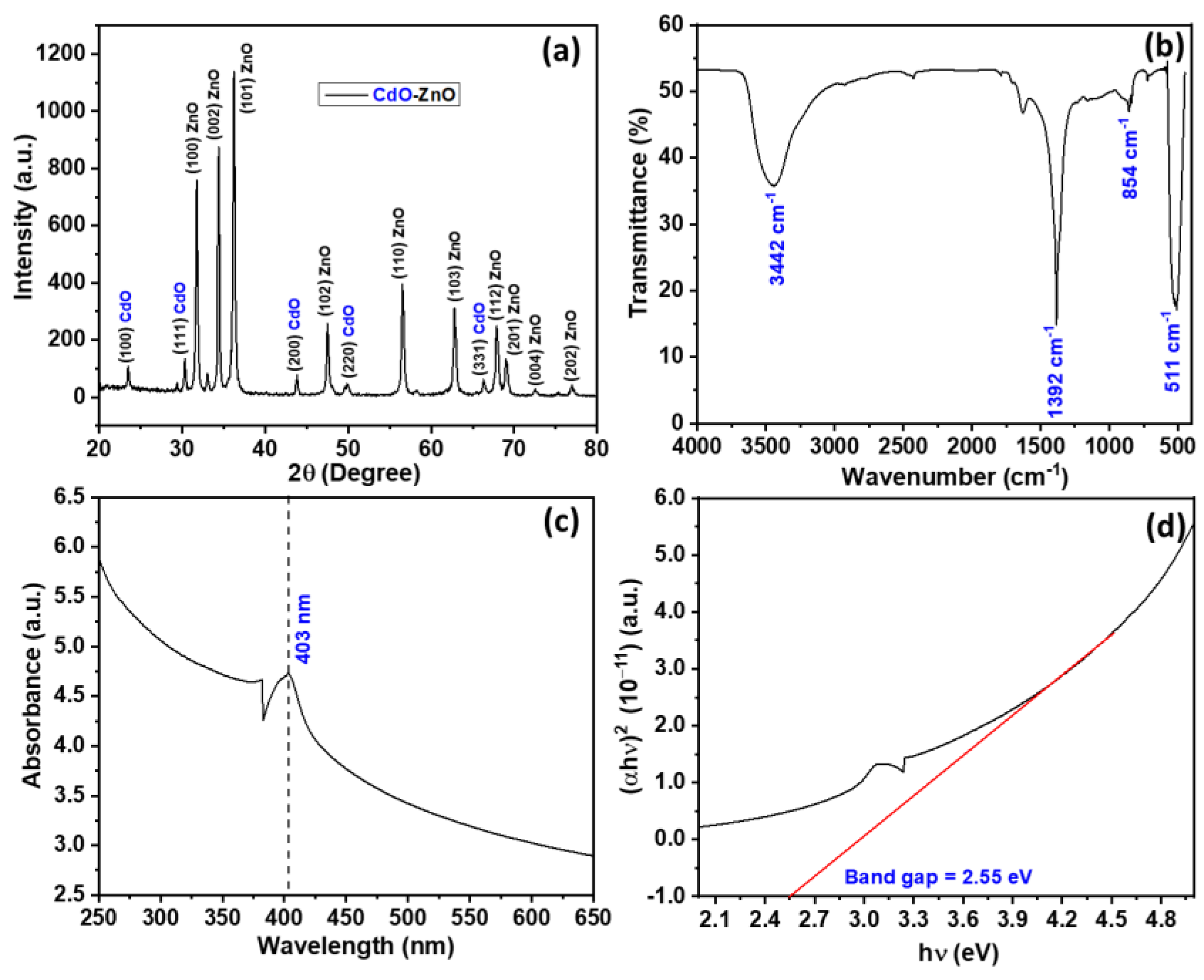
Powder XRD measurements were carried out to identify the crystallographic phase structure of the as-produced CdO-ZnO nanocomposites. Figure 2a displays these XRD patterns. Well-defined diffraction reflections at 31.75°, 34.40°, 36.25°, 47.50°, 56.55°, 62.80°, 67.90°, 69.00°, 72.55°, and 76.95°, respectively, correspond to the (1 0 0), (0 0 2), (1 0 1), (1 0 2), (1 1 0), (1 0 3), (1 1 2), (2 0 1), (0 0 4) and (2 0 2) diffraction planes of ZnO respectively. When compared to the information from the JPCDS card number 36–1451, all of the XRD peaks perfectly match the hexagonal wurtzite phase. The typical peaks for the cubic phase of CdO can be found in the XRD data at 2θ = 23.45°, 30.35°, 43.85°, 49.85°, and 66.30°, which correspond to the (1 1 0), (1 1 1), (2 0 0), (2 1 1), and (2 2 0) diffraction planes, respectively. These peaks match the JCPDS (Joint Committee for Powder Diffraction Studies (JCPDS) File No. 05-0640 and the literature very closely [44,45]. The formation of extremely crystalline CdO-ZnO nanocomposites is suggested by the strong and compacted diffraction peaks in the XRD spectrum of these materials.
Using the common Debye–Scherer equation, the crystal size (d) of the CdO-ZnO nanocomposites was also estimated (Equation (2)) [46,47,48,49].
d = 0.90   λ β   cos θ
where λ = 0.1542 nm, θ = diffraction angle, and β = the full width half maximum (FWHM). In order to investigate crystal size, the FWHM values of the seven most strong diffraction peaks were calculated. For the CdO-ZnO nanocomposites, the crystal size was 28.06 nm (Table 1).
The FTIR spectrum provides information regarding the development of metal-oxygen bonds in CdO-ZnO nanocomposites. The FTIR spectrum was investigated in the 400–4000 cm−1 region, revealing distinctive vibrational peaks at 511, 854, 1392, 1620, and 3442 cm−1 (Figure 2b). The stretching vibration of the Zn-O and Cd-O bonds is shown by the prominent wide peak at 511 cm−1 [50,51]. Another absorption peak at 1392 cm−1 could be connected to the C O 3 2 ions, which often show up in the spectrum when the FTIR samples are produced and examined in the air. The out-of-plane bending modes of these C O 3 2 ions are responsible for yet another peak at 830 cm−1 [52]. A broad band at 3442 cm−1 may be attributed to the stretching vibration of the O-H bond of physisorbed water molecules. The existence of a transient band at 1620 cm−1 can be attributed to the first overtone of the primary stretching mode of the O-H bond of physisorbed water [52].
The optical characteristics of the synthesized CdO-ZnO nanocomposites were investigated using UV-Vis absorption spectroscopy. At room temperature, the primary absorption peak for nanocomposites was seen at about 403 nm (Figure 2c). This is due to the direct transition of electrons between the valance band and conduction bands. The optical band gap (Eg) of these CdO-ZnO nanocomposites was also estimated using the absorption coefficient ‘α’ and optical band gap relationship [53,54] (Equation (3)).
( α h ν ) 2 = A   ( h ν   E g )
where A = the parameter related to the effective masses of the valence and conduction bands, h = Plank’s constant, and ν = optical frequency.
The band-gap energy of the nanocomposites was computed by extrapolating the linear section of the graph between the function (αhv)2 and photon energy (hν) and was found to be 2.55 eV (Figure 2d). This band gap is extremely small when compared to pure ZnO nanostructures (3.25–3.37 eV) [55,56,57] but higher than the CdO nanomaterials (2.2–2.5 eV) [58,59] previously reported in the literature.

3.2. Photocatalytic Degradation Applications of CdO-ZnO Nanocomposites

Based on the photodegradation of RhB dye as a pollutant substance under UV light irradiation at fixed operational parameters, i.e., a dye concentration of 20 ppm, a catalyst dose of 0.05 g, a 100 mL volume of solution, and an irradiation time of 120 min, the photocatalytic performance of the CdO-ZnO nanocomposites was investigated. In order to compare the photocatalytic effectiveness of the CdO-ZnO nanocomposites, the degradation processes were also examined under UV irradiationonly without any catalyst and also in the presence of a catalyst when the dye solution was not exposed to UV radiation.
The photocatalytic activity of the CdO-ZnO nanocomposites was investigated by measuring the primary absorption peak of RhB at 553 nm. Figure 3a depicts the UV-vis spectra of the RhB dye over the nanocomposite, and it can be seen that the strongest absorbance peak of RhB diminishes with increasing UV irradiation time. The variations inphotodegradation extent (A/Ao) and percentage photodegradation of the RhB dye as a function of exposure duration are depicted in Figure 3b,c respectively. When the dye solution underwent UV irradiation solely without any catalyst or when no UV irradiations were applied to the dye solution dispersed with a photocatalyst, no discernible degradation of the RhB dye was observed. However, the percentage degradation of the RhB dye increases significantly in the presence of CdO-ZnO nanocomposites, with about 87% degradation observed within 120 min of UV light exposure.
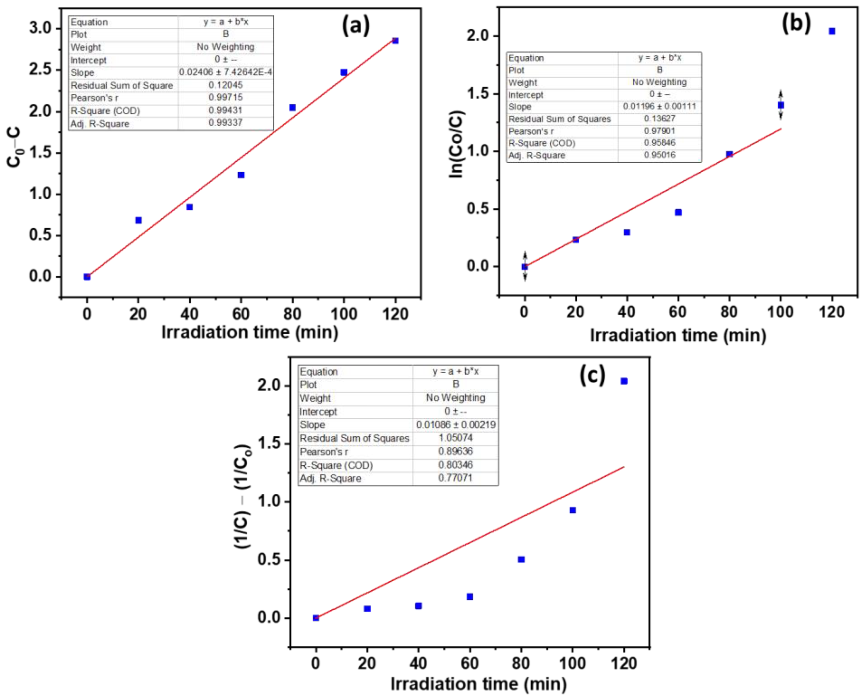
Three kinetics models: zero, pseudo-first, and pseudo-second order, were studied by using the following equations (Equations (4)–(6)) [60,61].
C 0 C =   k 0 t + 0
ln C 0 C =   k 1 t + 0
1 C 1 C 0 =   k 2 t + 0
where C0 and C are the initial dye concentration and final dye concentrations after irradiation time (t), and k0, k1, and k2are the rate constants for zero-order, pseudo-first-order and pseudo-second-order rate expressions, respectively. The equilibrium RhB concentrations were determined from the calibrated curves.
The kinetics parameters were determined by graphing the data and fitting them to a linear plot (Figure 4a–c). Table 2 lists the different kinetic parameters. In the presence of the CdO-ZnO nanocomposites, the photocatalytic degradation of the RhB dye followed zero-order and pseudo-first-order kinetics, as is evident from the coefficient of determinant (COD; R2) values.
The energy gap between the valence and conduction band, the oxidation potential of photogenerated, positively charged holes, and the effectiveness in separating photogenerated electrons and holes are all factors that affect the photocatalytic efficacy of a semiconductor photocatalyst, according to Reddy et al. [62]. When exposed to UV light, photogenerated electrons move from the ZnO valence band to the conduction band, leaving holes in the valence band. In the CdO-ZnO nanocomposite, CdO acts as a sink for the conduction band electrons of ZnO and induces a shift in the Fermi levels toward lower potentials [17,63]. This transfer further lowers the recombination of electron-hole pairs, thereby increasing photocatalytic activity. While the holes at the conduction band oxidize the hydroxyl ions, the electrons at the conduction band reduce the dissolved oxygen [64]. Additionally, the n-n heterojunction that results from the combination of n-type CdO and ZnO semiconductors speeds up the electronic transport processes. The hydroxyl radicals (OH) formed, owing to the oxidation of the adsorbed H2O or adsorbed OH ions, are the prominent strong oxidants that degrade the complex dye molecules to simpler ones (Figure 5) (Equations (7)–(17)) [11,65,66]. Table 3 compares the photocatalytic parameters for the breakdown of the RhB dye, as measured here, with the other previously reported parameters.
ZnO     hv     ZnO ( e CB ) +   ZnO ( h VB + )
CdO     hv     CdO ( e CB ) +   CdO ( h VB + )
ZnO ( e CB ) +   CdO   CdO ( e CB ) + ZnO
O 2   ( ads ) e CB O 2
O 2 +   H 2 O OH +   HO 2
2   HO 2 H 2 O 2   +   O 2  
H 2 O 2 O 2 OH + OH +   O 2  
ZnO ( h VB + ) +   H 2 O   OH + H + +   ZnO
CdO ( h VB + ) +   H 2 O   OH + H + +   CdO
OH +   Dye     Dye   degradation   products

3.3. Sensing Applications of CdO-ZnO Nanocomposites

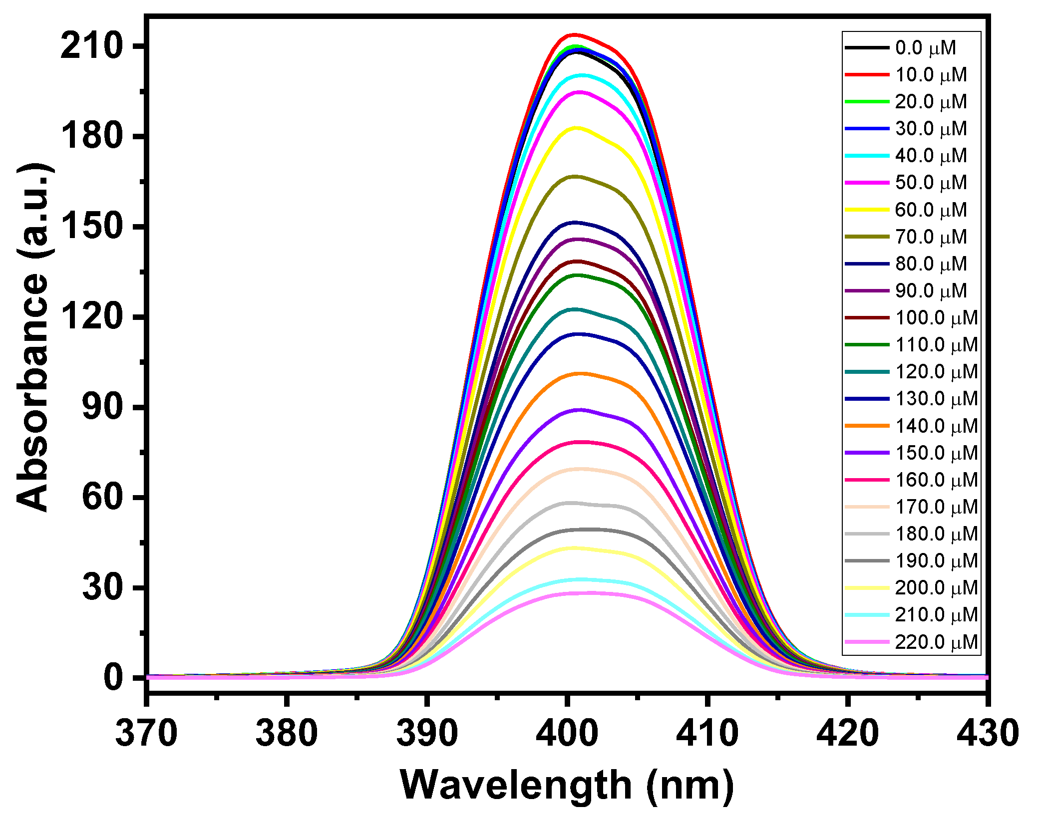
Investigations were conducted on the efficacy of the CdO-ZnO nanocomposites for hydrazine fluorescence sensing. Photoluminescence emission is greatly impacted by the presence of hydrazine in composite suspensions. With each subsequent administration of hydrazine, there is a definite indication of the quenching of fluorescence emission. As demonstrated in Figure 6, the fluorescence intensity of the CdO-ZnO nanocomposites decreased as the hydrazine concentration increased from 10 to 220 μM.
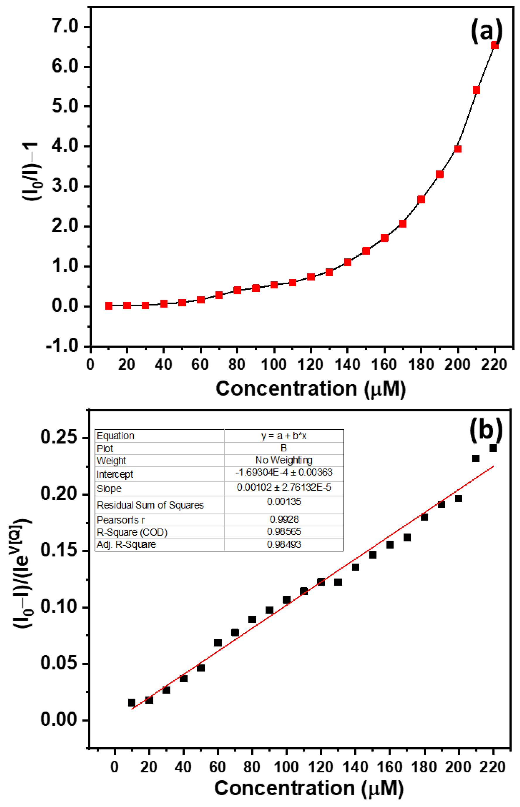
The fluorescence data were evaluated using the basic Stern–Volmer equation to better understand the quenching process of the CdO-ZnO nanocomposites by hydrazine (Equations (18) and (19)) [75,76,77].
I o I =   K sv [ Q ] + 1
I o I 1 =   K sv [ Q ]
where Ksv = Stern–Volmer quenching constant, [Q] = hydrazine (quencher) concentration, Io = fluorescence intensity in the absence of quencher, and I = fluorescence intensity after the addition of hydrazine.
Due to the nonlinear nature of the graph, however, no significant information can be drawn from the plot between (Io/I)-1 and [Q] (Figure 7a). Still, the graph exhibited linear behavior for the quenching of the nanocomposites for the hydrazine concentration, ranging between 10–130 µM. These findings show that static quenching outperforms dynamic quenching in the linear zone; however, when the concentration of the analyte hydrazine increases, the divergence from linearity corresponds to the equality of static and dynamic quenching by hydrazine as a quencher [78,79].
A modified Stern–Volmer equation was applied to better understand the quenching mechanism (Equations (20) and (21)) [80,81].
I o I =   K sv [ Q ] e V [ Q ] + 1
Equation (20) can be re-written as Equation (21)
I o I I   e V [ Q ] =   K sv [ Q ]
where V = the static quenching constant.
A linear fit of the plot I o I Ie V [ Q ] vs. [Q] exhibits a COD (R2) = 0.98565 for the CdO-ZnO nanocomposites (Figure 7b). The value of V fits 0.015 with a KSV of 1030 M−1 for the CdO-ZnO nanocomposites. It was observed that the fluorescent hydrazine sensing detecting limit was 28.01 µM. This indicates that CdO-ZnO nanocomposites can be used as an efficient luminescent sensor for the detection of hydrazine.

4. Conclusions

By using a simple solution growth technique, CdO-ZnO nanocomposites were created. A comprehensive examination verified the development of highly aggregated composites produced at high densities with outstanding crystallinity and purity. When exposed to UV rays in the presence of the as-produced CdO-ZnO nanocomposites, the RhB dyes degraded. Both zero-order and pseudo-first-order kinetics were followed in the degrading process. The inclusion of hydrazine showed a significant influence on the photoluminescence emission of the CdO-ZnO nanocomposites and its concentration further enhanced it. The detection limit for hydrazine using fluorescence sensing was found to be 28.01 µM. The findings of the current study support the notion that synthesized CdO-ZnO nanocomposites represent a promising material for photocatalytic and sensing applications.

Author Contributions

Conceptualization, A.U., R.K. (Ramesh Kumar), M.S.C., R.K. (Rajesh Kumar), A.A.I. and M.S.A.; Methodology, A.U., R.K. (Ramesh Kumar) and A.A.I.; Software, H.A.; Formal analysis, A.U., R.K. (Ramesh Kumar), M.S.C., R.K. (Rajesh Kumar), A.A.I., M.A.M.A., H.A. and M.S.A.; Investigation, R.K. (Ramesh Kumar); Resources, A.U. and M.A.M.A.; Data curation, M.S.C., M.A.M.A. and H.A.; Writing–original draft, A.U., R.K. (Ramesh Kumar), M.S.C. and R.K. (Rajesh Kumar); Writing–review & editing, A.U., R.K. (Ramesh Kumar), M.S.C., R.K., M.S.A. and R.K. (Rajesh Kumar). All authors have read and agreed to the published version of the manuscript.

Funding

The authors are thankful to the Deanship of Scientific Research at Najran University, Najran, Kingdom of Saudi Arabia for funding this work under the National Research Priority (NRP) funding program, Grant no. NU/NRP/SERC/11/1.

Institutional Review Board Statement

Not applicable.

Informed Consent Statement

Not applicable.

Data Availability Statement

Not applicable.

Acknowledgments

The authors greatly acknowledge and are thankful to the Deanship of Scientific Research at Najran University, Najran, Kingdom of Saudi Arabia for funding this work under the National Research Priority (NRP) funding program, Grant No. NU/NRP/SERC/11/1.

Conflicts of Interest

The authors declare no conflict of interest.

References

  1. Wang, Z.; Li, Y.; Cheng, Q.; Wang, X.; Wang, J.; Zhang, G. Sb-based photocatalysts for degradation of organic pollutants: A review. J. Clean. Prod. 2022, 367, 133060. [Google Scholar] [CrossRef]
  2. Abdul Mutalib, A.A.; Jaafar, N.F. ZnO photocatalysts applications in abating the organic pollutant contamination: A mini review. Total Environ. Res. Themes 2022, 3–4, 100013. [Google Scholar] [CrossRef]
  3. Feng, X.; Shang, Y.; Zhang, K.; Hong, M.; Li, J.; Xu, H.; Wang, L.; Li, Z. In situ ligand-induced Ln-MOFs based on a chromophore moiety: White light emission and turn-on detection of trace antibiotics. CrystEngComm 2022, 24, 4187–4200. [Google Scholar] [CrossRef]
  4. Tufail, M.A.; Iltaf, J.; Zaheer, T.; Tariq, L.; Amir, M.B.; Fatima, R.; Asbat, A.; Kabeer, T.; Fahad, M.; Naeem, H.; et al. Recent advances in bioremediation of heavy metals and persistent organic pollutants: A review. Sci. Total Environ. 2022, 850, 157961. [Google Scholar] [CrossRef]
  5. Kumar, R.; Singh, L. Ti3C2Tx MXene as Electrocatalyst for Designing Robust Glucose Biosensors. Adv. Mater. Technol. 2022, 1, 2200151. [Google Scholar] [CrossRef]
  6. Feng, X.; Shang, Y.; Zhang, H.; Liu, X.; Wang, X.; Chen, N.; Wang, L.; Li, Z. Multi-functional lanthanide-CPs based on tricarboxylphenyl terpyridyl ligand as ratiometric luminescent thermometer and highly sensitive ion sensor with turn on/off effect. Dalt. Trans. 2020, 49, 4741–4750. [Google Scholar] [CrossRef]
  7. Zuo, K.; Zhang, J.; Zeng, L. A smartphone-adaptable chromogenic and fluorogenic sensor for rapid visual detection of toxic hydrazine in the environment. Spectrochim. Acta Part A Mol. Biomol. Spectrosc. 2022, 283, 121765. [Google Scholar] [CrossRef]
  8. Umar, A.; Harraz, F.A.; Ibrahim, A.A.; Almas, T.; Kumar, R.; Al-Assiri, M.S.; Baskoutas, S. Iron-Doped Titanium Dioxide Nanoparticles As Potential Scaffold for Hydrazine Chemical Sensor Applications. Coatings 2020, 10, 182. [Google Scholar] [CrossRef] [Green Version]
  9. Deng, Z.; Wu, Z.; Rokni, H.; Yin, Z.; Lin, J.; Zhang, H.; Cheraghi, S. Electrochemical sensor amplified with cellulose nanofibers/Fe3O4 composite to the monitoring of hydrazine as a pollutant in water and wastewater samples. Chemosphere 2022, 309, 136568. [Google Scholar] [CrossRef]
  10. Muthusamy, S.; Yin, S.; Rajalakshmi, K.; Meng, S.; Zhu, D.; Xie, M.; Xie, J.; Lodi, R.S.; Xu, Y. Development of a quinoline-derived turn-on fluorescent probe for real time detection of hydrazine and its applications in environment and bioimaging. Dye. Pigment. 2022, 206, 110618. [Google Scholar] [CrossRef]
  11. Kumar, R.; Umar, A.; Kumar, R.; Chauhan, M.S.; Al-Hadeethi, Y. ZnO–SnO2 nanocubes for fluorescence sensing and dye degradation applications. Ceram. Int. 2021, 47, 6201–6210. [Google Scholar] [CrossRef]
  12. Kumar, R.; Kumar, G.; Umar, A. ZnO nano-mushrooms for photocatalytic degradation of methyl orange. Mater. Lett. 2013, 97, 100–103. [Google Scholar] [CrossRef]
  13. Kumar, R.; Kumar, G.; Akhtar, M.S.; Umar, A. Sonophotocatalytic degradation of methyl orange using ZnO nano-aggregates. J. Alloys Compd. 2015, 629, 167–172. [Google Scholar] [CrossRef]
  14. Lops, C.; Ancona, A.; Di Cesare, K.; Dumontel, B.; Garino, N.; Canavese, G.; Hérnandez, S.; Cauda, V. Sonophotocatalytic degradation mechanisms of Rhodamine B dye via radicals generation by micro- and nano-particles of ZnO. Appl. Catal. B Environ. 2019, 243, 629–640. [Google Scholar] [CrossRef]
  15. Amalina, F.; Abd Razak, A.S.; Krishnan, S.; Zularisam, A.W.; Nasrullah, M. A review of eco-sustainable techniques for the removal of Rhodamine B dye utilizing biomass residue adsorbents. Phys. Chem. Earth Parts A/B/C 2022, 128, 103267. [Google Scholar] [CrossRef]
  16. Sharma, J.; Sharma, S.; Bhatt, U.; Soni, V. Toxic effects of Rhodamine B on antioxidant system and photosynthesis of Hydrilla verticillata. J. Hazard. Mater. Lett. 2022, 3, 100069. [Google Scholar] [CrossRef]
  17. Kumar, R.; Umar, A.; Kumar, R.; Rana, D.; Chauhan, M.S. Low-temperature synthesis of cadmium-doped zinc oxide nanosheets for enhanced sensing and environmental remediation applications. J. Alloys Compd. 2021, 863, 158649. [Google Scholar] [CrossRef]
  18. Wang, P.; Zheng, Z.; Cheng, X.; Sui, L.; Gao, S.; Zhang, X.; Xu, Y.; Zhao, H.; Huo, L. Ionic liquid-assisted synthesis of α-Fe2O3 mesoporous nanorod arrays and their excellent trimethylamine gas-sensing properties for monitoring fish freshness. J. Mater. Chem. A 2017, 5, 19846–19856. [Google Scholar] [CrossRef]
  19. Yin, H.; Zhao, Y.; Hua, Q.; Zhang, J.; Zhang, Y.; Xu, X.; Long, Y.; Tang, J.; Wang, F. Controlled Synthesis of Hollow α-Fe2O3 Microspheres Assembled With Ionic Liquid for Enhanced Visible-Light Photocatalytic Activity. Front. Chem. 2019, 7, 58. [Google Scholar] [CrossRef]
  20. Wang, F.-Y.; Yang, Q.-D.; Xu, G.; Lei, N.-Y.; Tsang, Y.K.; Wong, N.-B.; Ho, J.C. Highly active and enhanced photocatalytic silicon nanowire arrays. Nanoscale 2011, 3, 3269–3276. [Google Scholar] [CrossRef]
  21. Waghchaure, R.H.; Adole, V.A.; Jagdale, B.S. Photocatalytic degradation of methylene blue, rhodamine B, methyl orange and Eriochrome black T dyes by modified ZnO nanocatalysts: A concise review. Inorg. Chem. Commun. 2022, 143, 109764. [Google Scholar] [CrossRef]
  22. Xu, D.; Ma, H. Degradation of rhodamine B in water by ultrasound-assisted TiO2 photocatalysis. J. Clean. Prod. 2021, 313, 127758. [Google Scholar] [CrossRef]
  23. Huang, C.; Wang, Y.; Gong, M.; Wang, W.; Mu, Y.; Hu, Z.-H. α-MnO2/Palygorskite composite as an effective catalyst for heterogeneous activation of peroxymonosulfate (PMS) for the degradation of Rhodamine B. Sep. Purif. Technol. 2020, 230, 115877. [Google Scholar] [CrossRef]
  24. Kati, N. Controlling of optical band gap of the CdO films by zinc oxide. Mater. Sci. 2019, 37, 136–141. [Google Scholar] [CrossRef] [Green Version]
  25. Mosquera, E.; del Pozo, I.; Morel, M. Structure and red shift of optical band gap in CdO–ZnO nanocomposite synthesized by the sol gel method. J. Solid State Chem. 2013, 206, 265–271. [Google Scholar] [CrossRef]
  26. Ziabari, A.A.; Ghodsi, F.E. Optoelectronic studies of sol–gel derived nanostructured CdO–ZnO composite films. J. Alloys Compd. 2011, 509, 8748–8755. [Google Scholar] [CrossRef]
  27. Saravanan, R.; Shankar, H.; Prakash, T.; Narayanan, V.; Stephen, A. ZnO/CdO composite nanorods for photocatalytic degradation of methylene blue under visible light. Mater. Chem. Phys. 2011, 125, 277–280. [Google Scholar] [CrossRef]
  28. Zheng, Z.; Gan, L.; Li, H.; Ma, Y.; Bando, Y.; Golberg, D.; Zhai, T. A Fully Transparent and Flexible Ultraviolet–Visible Photodetector Based on Controlled Electrospun ZnO-CdO Heterojunction Nanofiber Arrays. Adv. Funct. Mater. 2015, 25, 5885–5894. [Google Scholar] [CrossRef]
  29. Bai, S.; Sun, X.; Han, N.; Shu, X.; Pan, J.; Guo, H.; Liu, S.; Feng, Y.; Luo, R.; Li, D.; et al. rGO modified nanoplate-assembled ZnO/CdO junction for detection of NO2. J. Hazard. Mater. 2020, 394, 121832. [Google Scholar] [CrossRef]
  30. Naderi, H.; Hajati, S.; Ghaedi, M.; Espinos, J.P. Highly selective few-ppm NO gas-sensing based on necklace-like nanofibers of ZnO/CdO n-n type I heterojunction. Sens. Actuators B Chem. 2019, 297, 126774. [Google Scholar] [CrossRef]
  31. Cai, X.; Hu, D.; Deng, S.; Han, B.; Wang, Y.; Wu, J.; Wang, Y. Isopropanol sensing properties of coral-like ZnO–CdO composites by flash preparation via self-sustained decomposition of metal–organic complexes. Sens. Actuators B Chem. 2014, 198, 402–410. [Google Scholar] [CrossRef]
  32. Rahman, M.M.; Khan, S.B.; Marwani, H.M.; Asiri, A.M.; Alamry, K.A.; Rub, M.A.; Khan, A.; Khan, A.A.P.; Azum, N. Facile synthesis of doped ZnO-CdO nanoblocks as solid-phase adsorbent and efficient solar photo-catalyst applications. J. Ind. Eng. Chem. 2014, 20, 2278–2286. [Google Scholar] [CrossRef]
  33. Umar, A.; Akhtar, M.S.; Al-Assiri, M.S.; Al-Salami, A.E.; Kim, S.H. Composite CdO-ZnO hexagonal nanocones: Efficient materials for photovoltaic and sensing applications. Ceram. Int. 2018, 44, 5017–5024. [Google Scholar] [CrossRef]
  34. Choudhary, K.; Saini, R.; Upadhyay, G.K.; Purohit, L.P. Sustainable behavior of cauliflower like morphology of Y-doped ZnO:CdO nanocomposite thin films for CO2 gas sensing application at low operating temperature. J. Alloys Compd. 2021, 879, 160479. [Google Scholar] [CrossRef]
  35. Hu, G.; Zhang, Y. Quantum piezotronic devices based on ZnO/CdO quantum well topological insulator. Nano Energy 2020, 77, 105154. [Google Scholar] [CrossRef]
  36. Wang, T.; Kou, X.; Zhao, L.; Sun, P.; Liu, C.; Wang, Y.; Shimanoe, K.; Yamazoe, N.; Lu, G. Flower-like ZnO hollow microspheres loaded with CdO nanoparticles as high performance sensing material for gas sensors. Sens. Actuators B Chem. 2017, 250, 692–702. [Google Scholar] [CrossRef]
  37. Nwanya, A.C.; Deshmukh, P.R.; Osuji, R.U.; Maaza, M.; Lokhande, C.D.; Ezema, F.I. Synthesis, characterization and gas-sensing properties of SILAR deposited ZnO-CdO nano-composite thin film. Sens. Actuators B Chem. 2015, 206, 671–678. [Google Scholar] [CrossRef]
  38. Maafa, I.M.; Al-Enizi, A.M.; Abutaleb, A.; Zouli, N.I.; Ubaidullah, M.; Shaikh, S.F.; Al-Abdrabalnabi, M.A.; Yousef, A. One-pot preparation of CdO/ZnO core/shell nanofibers: An efficient photocatalyst. Alexandria Eng. J. 2021, 60, 1819–1826. [Google Scholar] [CrossRef]
  39. Warshagha, M.Z.A.; Muneer, M. Facile synthesis of CdO-ZnO heterojunction photocatalyst for rapid removal of organic contaminants from water using visible light. Environ. Nanotechnol. Monit. Manag. 2022, 18, 100728. [Google Scholar] [CrossRef]
  40. Saravanan, R.; Gracia, F.; Khan, M.M.; Poornima, V.; Gupta, V.K.; Narayanan, V.; Stephen, A. ZnO/CdO nanocomposites for textile effluent degradation and electrochemical detection. J. Mol. Liq. 2015, 209, 374–380. [Google Scholar] [CrossRef]
  41. Kumar, S.; Kaushik, R.D.; Purohit, L.P. ZnO-CdO nanocomposites incorporated with graphene oxide nanosheets for efficient photocatalytic degradation of bisphenol A, thymol blue and ciprofloxacin. J. Hazard. Mater. 2022, 424, 127332. [Google Scholar] [CrossRef] [PubMed]
  42. Liu, K.-K.; Shan, C.-X.; He, G.-H.; Wang, R.-Q.; Sun, Z.-P.; Liu, Q.; Dong, L.; Shen, D.-Z. Advanced encryption based on fluorescence quenching of ZnO nanoparticles. J. Mater. Chem. C 2017, 5, 7167–7173. [Google Scholar] [CrossRef]
  43. Irimpan, L.; Krishnan, B.; Deepthy, A.; Nampoori, V.P.N.; Radhakrishnan, P. Excitation wavelength dependent fluorescence behaviour of nano colloids of ZnO. J. Phys. D. Appl. Phys. 2007, 40, 5670. [Google Scholar] [CrossRef]
  44. Umm-e-Kulsoom; Gursal, S.A.; Khurshid, M.S.; Saif-ur-Rehman, M.; Minhas, A.S.; Yasin, T.; Mehboob, N.; Mehmood, M.S. Investigating the effect of adding CdO nano particles on neutron shielding efficacy of HDPE. Radiat. Phys. Chem. 2020, 177, 109145. [Google Scholar] [CrossRef]
  45. Chen, X.-X.; Chen, L.; Li, G.; Cai, L.-X.; Miao, G.-Y.; Guo, Z.; Meng, F.-L. Selectively enhanced gas-sensing performance to n-butanol based on uniform CdO-decorated porous ZnO nanobelts. Sens. Actuators B Chem. 2021, 334, 129667. [Google Scholar] [CrossRef]
  46. Al-Hadeethi, Y.; Umar, A.; Al-Heniti, S.H.; Kumar, R.; Kim, S.H.; Zhang, X.; Raffah, B.M. 2D Sn-doped ZnO ultrathin nanosheet networks for enhanced acetone gas sensing application. Ceram. Int. 2017, 43, 2418–2423. [Google Scholar] [CrossRef]
  47. Zhou, Q.; Umar, A.; Sodki, E.M.; Amine, A.; Xu, L.; Gui, Y.; Ibrahim, A.A.; Kumar, R.; Baskoutas, S. Fabrication and characterization of highly sensitive and selective sensors based on porous NiO nanodisks. Sens. Actuators B 2018, 259, 604–615. [Google Scholar] [CrossRef]
  48. Zhang, J.; Li, P.; Zhang, Z.; Wang, X.; Tang, J.; Liu, H.; Shao, Q.; Ding, T.; Umar, A.; Guo, Z. Solvent-free graphene liquids: Promising candidates for lubricants without the base oil. J. Colloid Interface Sci. 2019, 542, 159–167. [Google Scholar] [CrossRef]
  49. Umar, A.; Ammar, H.Y.; Kumar, R.; Ibrahim, A.A.; Al-Assiri, M.S. Square disks-based crossed architectures of SnO2 for ethanol gas sensing applications—An experimental and theoretical investigation. Sens. Actuators B Chem. 2020, 304, 127352. [Google Scholar] [CrossRef]
  50. Umar, A.; Kumar, S.A.; Inbanathan, S.S.R.; Modarres, M.; Kumar, R.; Algadi, H.; Ibrahim, A.A.; Wendelbo, R.; Packiaraj, R.; Alhamami, M.A.M.; et al. Enhanced sunlight-driven photocatalytic, supercapacitor and antibacterial applications based on graphene oxide and magnetite-graphene oxide nanocomposites. Ceram. Int. 2022, 48, 29349–29358. [Google Scholar] [CrossRef]
  51. Umar, A.; Ibrahim, A.A.; Kumar, R.; Algadi, H.; Albargi, H.; Alsairi, M.A.; Alhmami, M.A.M.; Zeng, W.; Ahmed, F.; Akbar, S. CdO–ZnO nanorices for enhanced and selective formaldehyde gas sensing applications. Environ. Res. 2021, 200, 111377. [Google Scholar] [CrossRef] [PubMed]
  52. Umar, A.; Ahmad, R.; Kumar, R.; Ibrahim, A.A.; Baskoutas, S. Bi2O2CO3 nanoplates: Fabrication and characterization of highly sensitive and selective cholesterol biosensor. J. Alloys Compd. 2016, 683, 433–438. [Google Scholar] [CrossRef]
  53. Barzinjy, A.A.; Azeez, H.H. Green synthesis and characterization of zinc oxide nanoparticles using Eucalyptus globulus Labill. leaf extract and zinc nitrate hexahydrate salt. SN Appl. Sci. 2020, 2, 991. [Google Scholar] [CrossRef]
  54. Ramesh, P.; Saravanan, K.; Manogar, P.; Johnson, J.; Vinoth, E.; Mayakannan, M. Green synthesis and characterization of biocompatible zinc oxide nanoparticles and evaluation of its antibacterial potential. Sens. Bio-Sensing Res. 2021, 31, 100399. [Google Scholar] [CrossRef]
  55. Umar, A.; Ibrahim, A.A.; Kumar, R.; Almas, T.; Al-Assiri, M.S.; Baskoutas, S. Nitroaniline chemi-sensor based on bitter gourd shaped ytterbium oxide (Yb2O3) doped zinc oxide (ZnO) nanostructures. Ceram. Int. 2019, 45, 13825–13831. [Google Scholar] [CrossRef]
  56. Umar, A.; Kumar, R.; Kumar, G.; Algarni, H.; Kim, S.H. Effect of annealing temperature on the properties and photocatalytic efficiencies of ZnO nanoparticles. J. Alloys Compd. 2015, 648, 46–52. [Google Scholar] [CrossRef]
  57. Ibrahim, A.A.; Kumar, R.; Umar, A.; Kim, S.H.; Bumajdad, A.; Ansari, Z.A.; Baskoutas, S. Cauliflower-shaped ZnO nanomaterials for electrochemical sensing and photocatalytic applications. Electrochim. Acta 2016, 222, 463–472. [Google Scholar] [CrossRef]
  58. Somasundaram, G.; Rajan, J.; Sangaiya, P.; Dilip, R. Hydrothermal synthesis of CdO nanoparticles for photocatalytic and antimicrobial activities. Results Mater. 2019, 4, 100044. [Google Scholar] [CrossRef]
  59. Sarker, M.A.R.; Ahn, Y.-H. Strategic insight into enhanced photocatalytic remediation of pharmaceutical contaminants using spherical CdO nanoparticles in visible light region. Chemosphere 2023, 311, 137040. [Google Scholar] [CrossRef]
  60. Chiu, Y.-H.; Chang, T.-F.M.; Chen, C.-Y.; Sone, M.; Hsu, Y.-J. Mechanistic Insights into Photodegradation of Organic Dyes Using Heterostructure Photocatalysts. Catalysts 2019, 9, 430. [Google Scholar] [CrossRef]
  61. Verma, N.; Chundawat, T.S.; Chandra, H.; Vaya, D. An efficient time reductive photocatalytic degradation of carcinogenic dyes by TiO2-GO nanocomposite. Mater. Res. Bull. 2023, 158, 112043. [Google Scholar] [CrossRef]
  62. Reddy, C.V.; Babu, B.; Shim, J. Synthesis, optical properties and efficient photocatalytic activity of CdO/ZnO hybrid nanocomposite. J. Phys. Chem. Solids 2018, 112, 20–28. [Google Scholar] [CrossRef]
  63. Subramanian, V.; Wolf, E.E.; Kamat, P. V Green Emission to Probe Photoinduced Charging Events in ZnO−Au Nanoparticles. Charge Distribution and Fermi-Level Equilibration. J. Phys. Chem. B 2003, 107, 7479–7485. [Google Scholar] [CrossRef]
  64. Sudheer Khan, S. Enhancement of visible light photocatalytic activity of CdO modified ZnO nanohybrid particles. J. Photochem. Photobiol. B Biol. 2015, 142, 1–7. [Google Scholar] [CrossRef] [PubMed]
  65. Ahmad, I.; Aslam, M.; Jabeen, U.; Zafar, M.N.; Malghani, M.N.K.; Alwadai, N.; Alshammari, F.H.; Almuslem, A.S.; Ullah, Z. ZnO and Ni-doped ZnO photocatalysts: Synthesis, characterization and improved visible light driven photocatalytic degradation of methylene blue. Inorg. Chim. Acta 2022, 543, 121167. [Google Scholar] [CrossRef]
  66. Amir, M.; Fazal, T.; Iqbal, J.; Din, A.A.; Ahmed, A.; Ali, A.; Razzaq, A.; Ali, Z.; Rehman, M.S.U.; Park, Y.-K. Integrated adsorptive and photocatalytic degradation of pharmaceutical micropollutant, ciprofloxacin employing biochar-ZnO composite photocatalysts. J. Ind. Eng. Chem. 2022, 115, 171–182. [Google Scholar] [CrossRef]
  67. Nandi, P.; Das, D. ZnO-CuxO heterostructure photocatalyst for efficient dye degradation. J. Phys. Chem. Solids 2020, 143, 109463. [Google Scholar] [CrossRef]
  68. Mukherjee, N.; Show, B.; Maji, S.K.; Madhu, U.; Bhar, S.K.; Mitra, B.C.; Khan, G.G.; Mondal, A. CuO nano-whiskers: Electrodeposition, Raman analysis, photoluminescence study and photocatalytic activity. Mater. Lett. 2011, 65, 3248–3250. [Google Scholar] [CrossRef]
  69. Pradeev Raj, K.; Sadaiyandi, K.; Kennedy, A.; Sagadevan, S. Photocatalytic and antibacterial studies of indium-doped ZnO nanoparticles synthesized by co-precipitation technique. J. Mater. Sci. Mater. Electron. 2017, 28, 19025–19037. [Google Scholar] [CrossRef]
  70. Varadavenkatesan, T.; Lyubchik, E.; Pai, S.; Pugazhendhi, A.; Vinayagam, R.; Selvaraj, R. Photocatalytic degradation of Rhodamine B by zinc oxide nanoparticles synthesized using the leaf extract of Cyanometra ramiflora. J. Photochem. Photobiol. B Biol. 2019, 199, 111621. [Google Scholar] [CrossRef]
  71. Ahmad, M.; Rehman, W.; Khan, M.M.; Qureshi, M.T.; Gul, A.; Haq, S.; Ullah, R.; Rab, A.; Menaa, F. Phytogenic fabrication of ZnO and gold decorated ZnO nanoparticles for photocatalytic degradation of Rhodamine B. J. Environ. Chem. Eng. 2021, 9, 104725. [Google Scholar] [CrossRef]
  72. Keerthana, S.P.; Yuvakkumar, R.; Ravi, G.; Pavithra, S.; Thambidurai, M.; Dang, C.; Velauthapillai, D. Pure and Ce-doped spinel CuFe2O4 photocatalysts for efficient rhodamine B degradation. Environ. Res. 2021, 200, 111528. [Google Scholar] [CrossRef] [PubMed]
  73. Tian, J.; Shao, Q.; Zhao, J.; Pan, D.; Dong, M.; Jia, C.; Ding, T.; Wu, T.; Guo, Z. Microwave solvothermal carboxymethyl chitosan templated synthesis of TiO2/ZrO2 composites toward enhanced photocatalytic degradation of Rhodamine B. J. Colloid Interface Sci. 2019, 541, 18–29. [Google Scholar] [CrossRef] [PubMed]
  74. Rajendrachari, S.; Taslimi, P.; Karaoglanli, A.C.; Uzun, O.; Alp, E.; Jayaprakash, G.K. Photocatalytic degradation of Rhodamine B (RhB) dye in waste water and enzymatic inhibition study using cauliflower shaped ZnO nanoparticles synthesized by a novel One-pot green synthesis method. Arab. J. Chem. 2021, 14, 103180. [Google Scholar] [CrossRef]
  75. Toal, S.J.; Trogler, W.C. Polymer sensors for nitroaromatic explosives detection. J. Mater. Chem. 2006, 16, 2871–2883. [Google Scholar] [CrossRef]
  76. Pritchard, G.O.; Mattinen, W.A.; Dacey, J.R. Stern–volmer plots in the photolysis of perfluoroazoethane. Int. J. Chem. Kinet. 1970, 2, 191–197. [Google Scholar] [CrossRef]
  77. Green, N.J.B.; Pimblott, S.M.; Tachiya, M. Generalizations of the Stern-Volmer relation. J. Phys. Chem. 1993, 97, 196–202. [Google Scholar] [CrossRef]
  78. Varlan, A.; Hillebrand, M. Bovine and Human Serum Albumin interactions with 3-carboxyphenoxathiin studied by fluorescence and circular dichroism spectroscopy. Molecules 2010, 15, 3905–3919. [Google Scholar] [CrossRef] [Green Version]
  79. Airinei, A.; Tigoianu, R.I.; Rusu, E.; Dorohoi, D.O. Fluorescence quenching of anthracene by nitroaromatic compounds. Dig. J. Nanomater. Biostruct. 2011, 6, 1265–1272. [Google Scholar]
  80. Hanagodimath, S.M.; Evale, B.G.; Manohara, S.R. Nonlinear fluorescence quenching of newly synthesized coumarin derivative by aniline in binary mixtures. Spectrochim. Acta-Part A Mol. Biomol. Spectrosc. 2009, 74, 943–948. [Google Scholar] [CrossRef]
  81. Xue, C.; Luo, F.T.; Liu, H. Post-polymerization functionalization approach for highly water-soluble well-defined regioregular head-to-tail glycopolythiophenes. Macromolecules 2007, 40, 6863–6870. [Google Scholar] [CrossRef]
Figure 1. (a,b) Low magnification, (c) high-magnification FESEM images, and (d) EDS spectra (and corresponding elemental composition) of the synthesized CdO-ZnO nanocomposites.
Coatings 12 01959 g001
Figure 2. (a) XRD spectrum, (b) FTIR spectrum, (c) UV-Vis spectrum, and (d) Tauc’s plot for the determination of the optical band gaps for the as-synthesized CdO-ZnO nanocomposites (red line represents fitting the linear region to evaluate the band gap at the x-axis intercept and black line is expressing the variations of (αhν)2 vs. hν.
Coatings 12 01959 g002
Figure 3. (a) UV-Visible spectra, (b) variations in decomposition (A/Ao), and (c) variations in percentage degradation of the RhB dye solution containing 0.05 g of CdO-ZnO nanocomposites as photocatalysts at different intervals.
Coatings 12 01959 g003
Figure 4. Linear fit plots for (a) zero-order, (b) pseudo-first-order (Arrows representing Data range), and (c) pseudo-second-order kinetics for RhB dye degradation.
Coatings 12 01959 g004
Figure 5. Proposed mechanism for RhB dye degradation with CdO-ZnO nanocomposites.
Coatings 12 01959 g005
Figure 6. Fluorescence quenching of the CdO-ZnO nanocomposites by hydrazine.
Coatings 12 01959 g006
Figure 7. (a) Simple and (b) modified Stern–Volmer plots for the CdO-ZnO nanocomposites.
Coatings 12 01959 g007
Table 1. Crystalline parameters for the CdO-ZnO nanocomposites.
Diffraction Planes (h k l)Diffraction Angles (°)FWHM (β)Crystal Size (nm)
(1 0 0)31.750.2549432.06
(0 0 2)34.400.2644731.12
(1 0 1)36.250.2748230.10
(1 0 2)47.500.3031228.34
(1 1 0)56.550.3374726.45
(1 0 3)62.800.3718824.77
(1 1 2)67.900.4022123.57
Table 2. Kinetic parameters for the degradation of RhB dye using CdO-ZnO nanocomposites.
Kinetic ModelRate Constant (×10−2)Half-Life Time (t1/2) R2 (COD)
Zero-orderk0 = 2.40668.25 min0.99431
Pseudo-first-orderk1 = 1.19657.94 min0.95846
Pseudo-second-orderk2 = 1.08628.04 min0.80346
Table 3. Comparison of photocatalytic degradation of RhB dye for CdO-ZnO nanocomposites, with previously reported photocatalysts.
Photocatalyst[RhB] (ppm)Catalyst Dose (g/L)Time (min)Degradation (%)Light Sourcek1
(×10−2 min−1)
Ref.
CdO-ZnO nanocomposites200.512087.0UV1.196This work
ZnO-Cu0.5O heterostructure100.0512073.5UV21.70[67]
CuO nano-whiskers1-26084.0Visible0.71[68]
In-doped ZnO nanoparticles200.512076.0UV-[69]
ZnO nanoparticles10 μM0.220098.0Solar1.7[70]
Au-ZnO nanoparticles100.318095.0UV2.47[71]
Ce-doped spinel CuFe2O4~4.252.012088.0Visible-[72]
TiO2/ZrO2 composites100.527090.5UV-[73]
Cauliflower shaped ZnO100.512075.0Solar-[74]
Publisher’s Note: MDPI stays neutral with regard to jurisdictional claims in published maps and institutional affiliations.