Review

Research Progress on Antibacterial Coatings for Preventing Implant-Related Infection in Fractures: A Literature Review

1
Department of Orthopedics, The Affiliated Changzhou Second People’s Hospital of Nanjing Medical University, Changzhou 213000, China
2
Graduate School of Dalian Medical University, Dalian 116000, China
3
Department of Orthopedics, Gonghe County Hospital of Traditional Chinese Medicine, Hainan Tibetan Autonomous Prefecture 811800, China
*
Authors to whom correspondence should be addressed.
These authors contributed equally to this work.
Coatings 2022, 12(12), 1921; https://doi.org/10.3390/coatings12121921
Submission received: 22 November 2022 / Revised: 2 December 2022 / Accepted: 4 December 2022 / Published: 8 December 2022

Abstract

:
Implant-related infection is a difficult problem in orthopaedics as it not only leads to failure in internal fixation, but also increases the financial burden and perioperative risk on patients. In the past, orthopaedic implants were designed as mechanical fixation devices simply to maintain mechanical and biological properties, not to regulate the surrounding biological microenvironment. More recently, antimicrobial biocoatings have been incorporated into orthopaedic implants to prevent and treat implant-related infections through the modulation of the local environment. This article reviews the application of orthopaedic-implant biocoating in the prevention of implant-caused infection. Although there are many candidate coatings, they are still in the preclinical testing stage, and thus additional research by biomaterials and clinicians is necessary to identify the ideal implant coatings for patients who require fracture surgery.

1. Introduction

Fracture-related infection is one of the major complications of musculoskeletal trauma surgery. The reported risk of infection in patients with closed fractures is relatively low, ranging from 0.7% to 4.2%, whereas that in patients with open fractures can be as high as 5%–30% [1]. The cost of treatment for patients with fracture-related infection is 6–7 times higher than that for patients without infection, mainly due to the need for multiple surgeries and prolonged hospital stays [2,3]. Additionally, the high infection-recurrence rates of 6%–9%, with the resulting amputation rates of 3%–5%, have brought great challenges to orthopaedists, clinical microbiologists, and infectiologists [4]. Currently, there are no relevant guidelines on the diagnosis and treatment of fracture-related infection. At present, it is believed that adequate stabilization, complete debridement, and bone grafting of infected lesions are beneficial to the treatment of the infection and to the regrowth of fractured ends. External fixation is one of the temporary or final fixation methods for bone infections and is especially suitable for severe soft-tissue injury [5]; however, it often does not provide adequate stability to the bone, especially in cases with large segmental bone defects, and the instability hampers the infection control and bone healing. Furthermore, biofilm formation on implants poses a great therapeutic challenge as it causes the development of bone infections after internal fixation [6,7].
A biofilm is a highly structured membrane-like complex composed of bacterial biomolecules, such as exopolysaccharides, matrix proteins, and extracellular DNA (eDNA), and coats surfaces colonized by bacteria. More than 80% of clinical bacterial infections are associated with biofilm formation [8], which involves bacterial adhesion, micro-colony formation, biofilm maturation, and dispersion of biofilm [9] (Figure 1). During this process, planktonic bacteria adhere to the surface via their appendage-like structures, called pilli and flagella, and through the secretion of adhesins [10]. The hydrophobicity of surfaces reduces the repulsion between microorganisms and surfaces and thus strengthens the adhesion of microorganisms [11]. Specific chemical signals initiate the proliferation of the attached microbial cells to form microcolonies [12]. Once a certain microbial cell density is reached, the quorum sensing system is activated, and the sessile bacteria secrete signalling molecules known as auto-inducers. Microbial cells communicate with each other via these signalling molecules to secrete large amounts of extracellular polymeric substances (EPSs) [13] that wrap the bacteria. Likewise, this intercellular communication enables the formation of the water-filled channels in the matrix that act as a circulatory system of the microcolonies for the delivery of important nutrients and removal of waste products [14]. Under certain conditions, microbes within a biofilm can migrate and colonize new sites [15]. For this purpose, they first produce various saccharolytic enzymes to degenerate the EPSs in their biofilm and meanwhile upregulate the proteins involved in flagellum formation. Biofilms can significantly enhance the resistance of bacteria to antibiotics and immune responses. Biofilmed microbes have been shown to tolerate 500 times more antibiotic concentration than their planktonic counterparts [16,17]. Biofilms can also impede antibiotic activity through multiple mechanisms (Table 1), including the physical barrier action of the EPSs [18], accumulation of antibiotic-degrading enzymes in the matrix [19], horizontal transfer of antibiotic-resistance genes among the microbial cells via eDNA [20], persistent or dormant cells due by the gradient of oxygen and other nutrients [21], discharge of the microbial antibiotic intake through efflux pumps on the microbial cell membrane [22], and increased mutation [23].
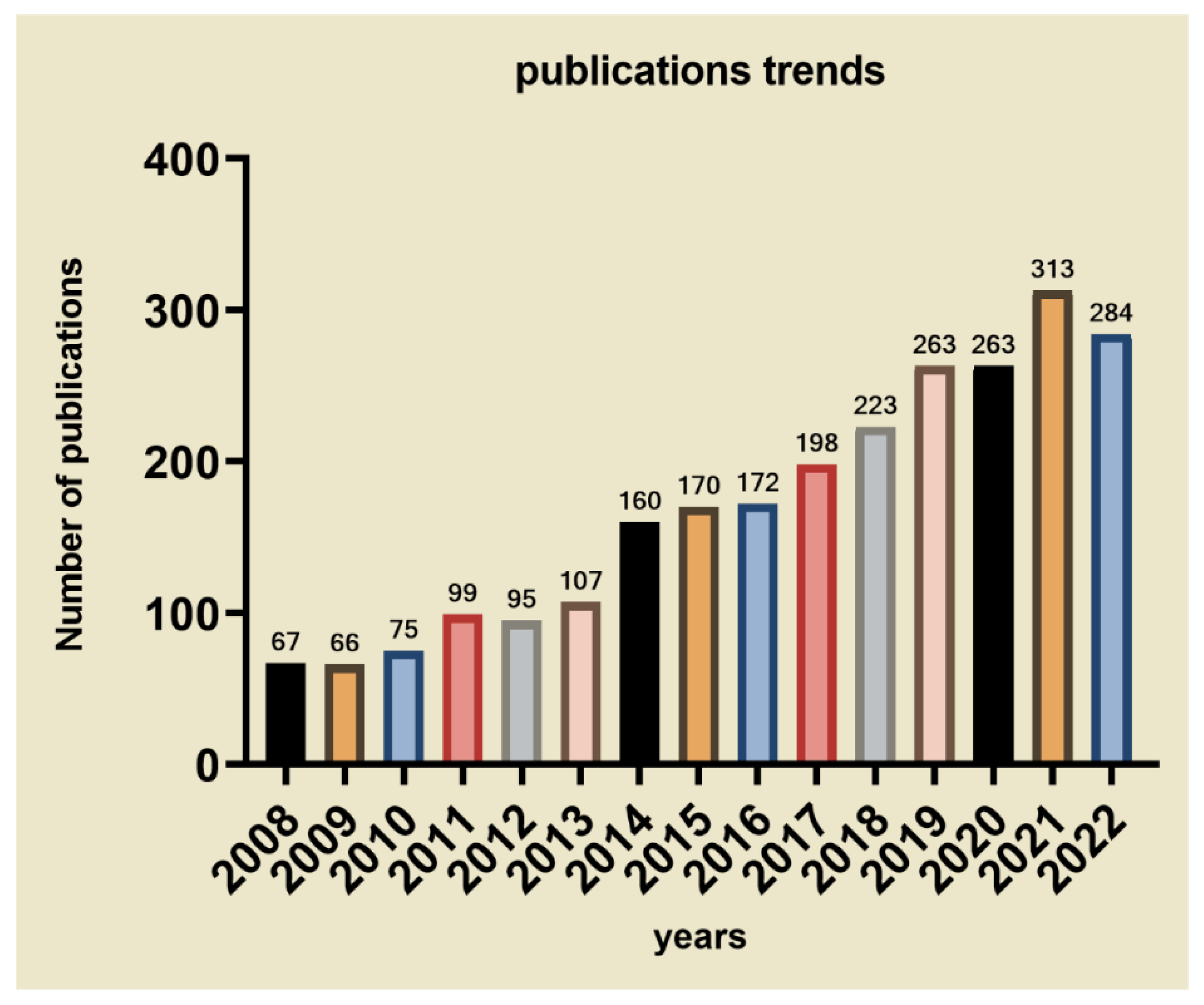
Antibacterial coating of orthopaedic implants is an effective measure against implant-associated infection. In recent years, various modes of antibacterial coatings have been studied. We counted the number of papers on the various implant-related antimicrobial surface coatings over the past decade in Figure 2. Pathogen adherence to the surfaces of orthopaedic implants involves multiple factors, including the chemical composition, surface charge, hydrophilicity, surface roughness, and surface wettability of the implant [39]. Some coatings can improve the surface properties of implants and thereby reduce bacterial attachment and biofilm formation and expansion, whereas others aim to kill the contaminating bacteria [40]. Antibacterial coating of fracture-related internal fixation devices against implant-associated infection has many significant socioeconomic benefits. Accordingly, this review summarizes the current research on the antibacterial coatings used for the surfaces of such devices.

2. Metallic Coating Materials

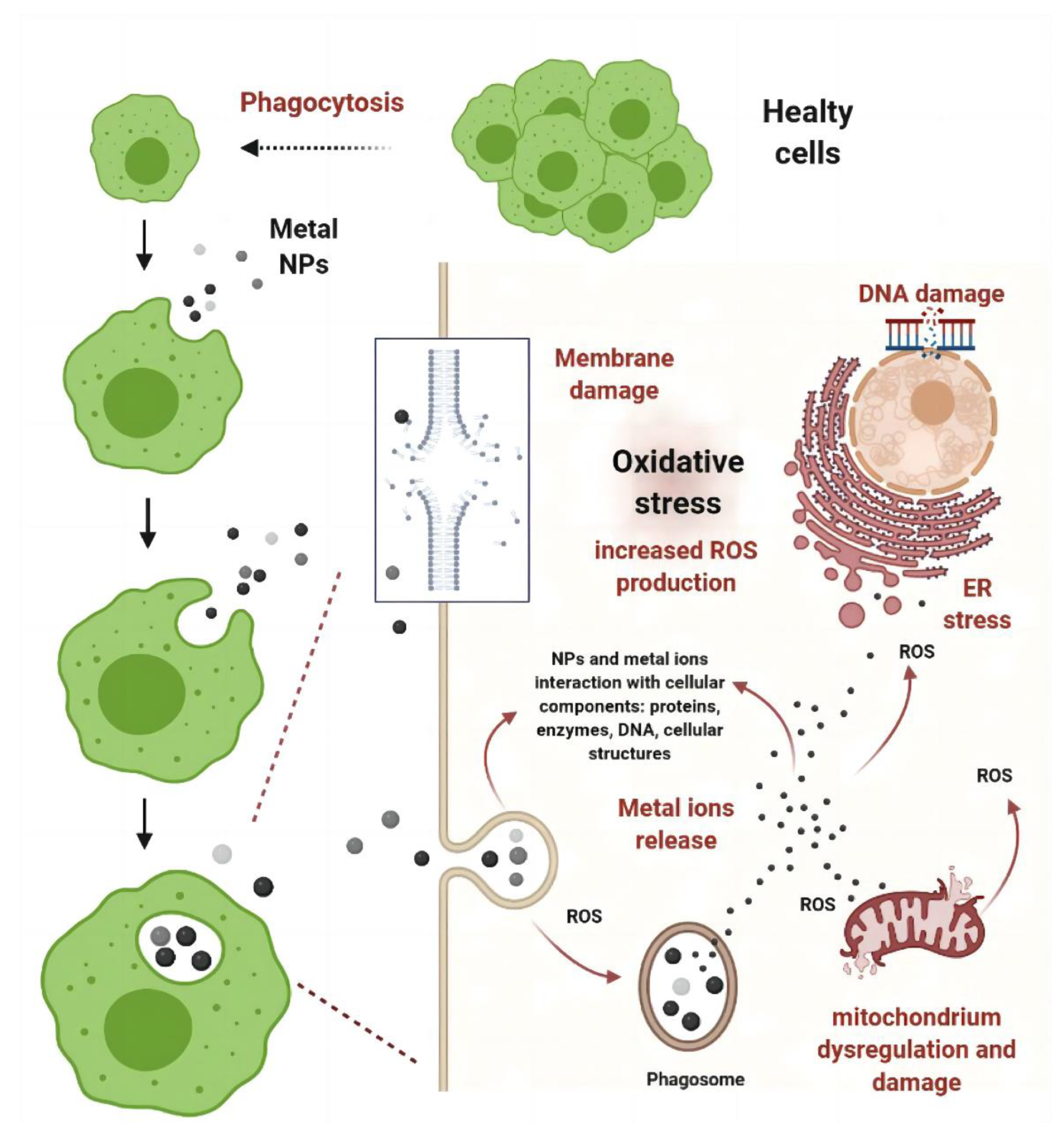
Nanoscience has recently been called “Big Science” due to the impact of the findings from this new branch of science in daily life [41]. Metal nanoparticles (NPs) are derived from metallic elements and have properties inherent to metal ions. These nanoparticles have been proven to be toxic in various biological systems [42]. The toxicity of metal NPs is mainly related to their induction of oxidative stress, leading to inflammation and major organelle dysfunction (Figure 3). It should be noted that the oxidative stress associated with metal NPs may be the result of NP-cell interactions even if the NPs do not inherently upregulate cellular reactive oxygen species (ROS). NPs can impair the respiratory chain reaction in the mitochondria and cellular ROS signalling, thereby increasing cellular oxidative stress and stimulating apoptosis [43]. Other causes of the toxicity of metal NPs include metal-ion release, cell-envelope permeation, and cell-membrane disruption [43,44]. Due to their antimicrobial properties, metal NPs, such as those made of silver (Ag), copper (Cu), zinc (Zn), selenium (Se), and titanium dioxide (TiO2), are now widely used as antimicrobial agents to coat orthopaedic implants. Table 2 reports common metal nanomaterials used in coatings.

2.1. Ag NPs

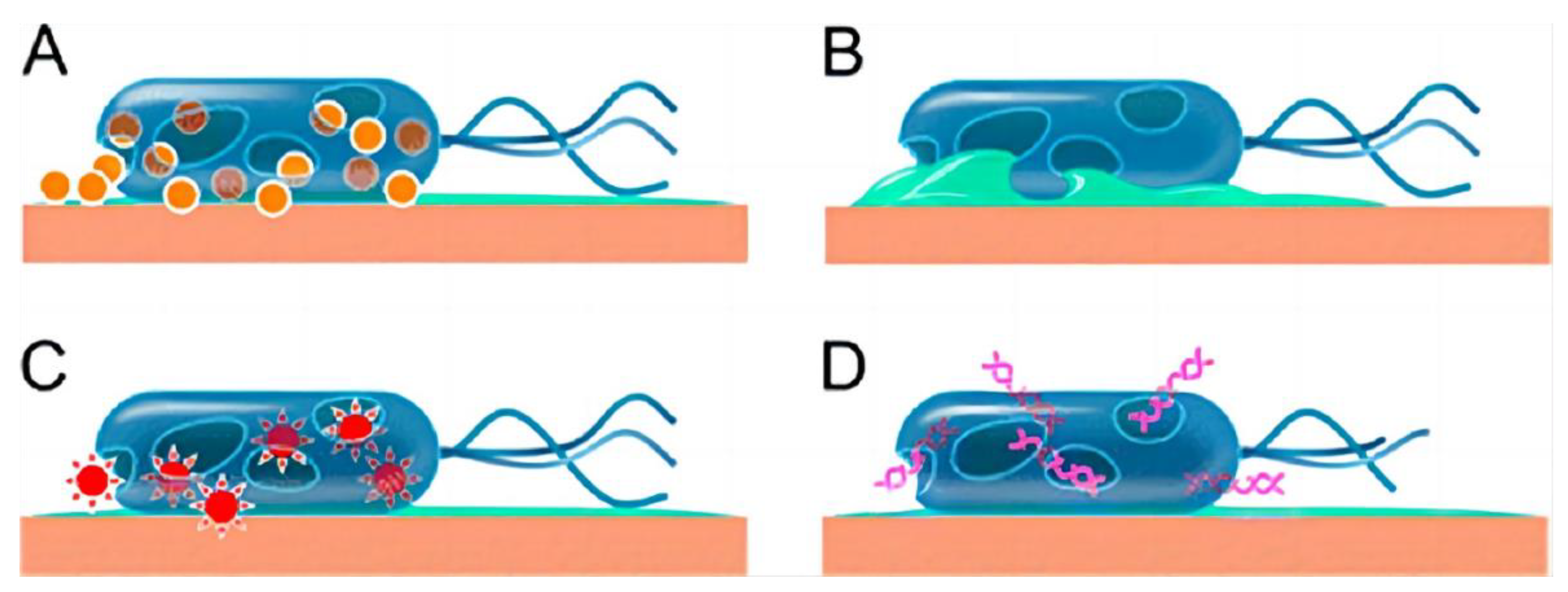
Ag is an effective antibacterial that can be used against bacterial infection in open wounds or as a coating material for various medical implants [53]. The antimicrobial activity of Ag is caused by the contact killing of germs by the released bioactive Ag ions (Figure 4). Ag coating has been shown to reduce the levels of bacterial contaminants by ≥95% when it is used to coat various products in medical settings [54]. An in vitro study on the antimicrobial effect of Ag coating of external fixation pins [55] has demonstrated that Ag-coated pins can significantly reduce the amount of biofilm-forming bacteria compared with the amounts on uncoated stainless steel or Ti implants. Shao et al. [56] have studied the effect of corrosion rate and surface energy of Ag coatings on bacterial adhesion and discovered that at low corrosion rates, the surface energy of Ag coatings suppresses bacterial adhesion. An in vivo study by Arens et al. [57] has shown that Ag-coated steel plates can significantly promote healing infection in rabbits and have good biocompatibility. Compared with uncoated Ti plate implants, Ag-coated Ti plate implants have been found to be compatible with the vasculature and the rolling and tethering behaviours of leukocytes, as indicated by the intact functional capillary density and endothelial integrity, and no sign of inflammation has been observed [45].
Additionally, the production process of Ag NPs can also affect their antibacterial activity. The Ag NPs coating surface has a more significant antibacterial effect compared with the Ag coating surface [59,60,61]. This can be due to the following reasons: on one hand, the Ag ions provided by the coating have antibacterial activity, and on the other hand, the nanostructure can provide a larger surface-to-volume ratio, enabling a more effective contact area and better exertion of the antibacterial activity [62]. A study by Huang et al. [63] has proven that the antibacterial activity and biocompatibility of Ag NPs are good, but also depend on the size of the used Ag NPs. Compared with large Ag NPs (>10 nm), small Ag NPs are more reactive and tend to form large aggregates; thus, particle characterization must be performed before toxicity assessment. In addition, although Ag ions are more toxic than the same doses of Ag NPs when the exposure is ≤48 h, Ag NPs show stronger toxicity than the ions as the exposure is prolonged. Therefore, it is unlikely that the toxicity of Ag NPs is caused solely by the dissociated Ag ions. Furthermore, comparison of size-dependent effects has indicated that Ag NPs with a diameter of 20 nm can induce cell-cycle arrest and apoptosis at the G0/G1 phase, whereas those with a diameter of 10 nm have no significant effect on the cell growth but can induce necrosis. This result may partly be related to the interaction of NPs with the cell membrane and their subsequent endocytosis [64]. Although Ag NPs have enhanced antibacterial activity, they are unstable and can easily aggregate, whereby their antibacterial activity is reduced. To improve the stability of Ag NPs in aqueous media, and thus their effectiveness as antibacterial agents, Ichimaru et al. [46] have coated triangular plate-shaped Ag NPs with one or two layers of gold (Au) atoms. These Au-coated Ag NPs have higher dispersion stability in high-salt aqueous media and higher antibacterial activity against pathogenic bacteria than pure Ag NPs. In addition, Au-coated Ag NPs have been shown to decontaminate RAW264.7 cells from bacteria, with no significant toxicity to RAW264.7 cells, and this observation highlights that NPs can be used against bacterial infection.

2.2. Cu NPs

Given the increasing antibiotic resistance in bacteria, the potential of metallic Cu as an antimicrobial has been gaining increasing attention. Contact killing is the main antimicrobial mechanism of Cu. Direct contact between bacteria and Cu can reduce the potential differences among the negatively charged domains on the bacterial cell membrane, cause membrane depolarization, and eventually lead to membrane leakage or even rupture [65]. Oxidative degradation of cellular components, including genomic and/or plasmid DNA, through ROS production may be another mechanism underlying Cu-induced cellular damage [66]. Nie et al. [48] have tested the super-hydrophilic and antimicrobial properties of CuO-coated surfaces in an in vitro experiment and shown that CuO coatings have good antibacterial activity against Escherichia coli (E. coli), methicillin-resistant Staphylococcus aureus (S. aureus) (MRSA), and vancomycin-resistant enterococci. Additionally, CuO coating has been shown to have better antibacterial properties than a Cu plate. This enhanced antimicrobial activity of CuO is probably due to the super-hydrophilic property of CuO and also because the diffusion of the bacterial suspension allows the bacteria to come into contact with Cu effectively. Therefore, increasing the effective contact area may be the research direction in the field of antibacterial coating. Fan et al. [67] have reported that Cu NPs can be used along with Ag NPs for a significantly enhanced antimicrobial effect. However, when the NPs were coated with an alloy of Ag-Cu, the antimicrobial effect was even more enhanced. In the study of Zhang et al. [49], porous TiO2 coatings doped with various amounts of Cu NPs were deposited on Ti via microarc oxidation. The results showed that the CuNP-coated samples showed excellent antibacterial activity against S. aureus. For the safety of Cu coating, in vitro and in vivo experiments on Cu release and local tissue reaction after implantation of Cu-coated Ti implants by Hoene et al. [68] have demonstrated the anti-infective ability of Cu coating. Such Cu-coated Ti implants exhibit a good antibacterial effect in vitro. Additionally, the in vivo release of Cu from these samples is measurable and only modestly increases local inflammatory responses.

2.3. ZnO NPs

Zn is an essential trace element and is involved in the catalytic activation reaction of >300 enzymes [69]. Its main function is to promote bone growth, mineralization, and formation, based on tissue-culture experiments [70]. ZnO NPs have broad antimicrobial activity against both Gram-positive and Gram-negative bacteria and have excellent biocompatibility, biological activity, and chemical stability, with low toxicity [71,72]. As in other metal NPs, various factors, including ion release, production of ROS, and direct physical contact, are considered to be the mechanisms underlying the antimicrobial effect of ZnO NPs [73]. The anti-infective, angiogenic, and osteogenic hydroxyapatite (HA)/polypyrrole/ZnO nanocomposite coating has been applied to Ti surfaces via pulsed electrochemical deposition [50]. In the study of Abdulkareem et al. [74], ZnO NPs and HA NPs were coated using electrohydrodynamic deposition onto Ti implants. The coating reduced the number of streptococci, anaerobic bacteria, and aerobic bacteria by 95%, 95%, and 90%, respectively. The study by Memarzadeh et al. [51] has indicated that substrates coated with ZnO NPs or with a mixture of 75% ZnO NPs and 25% HA NPs have significant antimicrobial activity against S. aureus. A study by Hui et al. has shown that the antibacterial efficiency of carvacrol-ZnO-palygorskite nanocomposites was superior to that of pure carvacrol, demonstrating the synergistic effect that occurs in the combined system [75]. Furthermore, ZnO-NP-based coatings can promote osteoblast proliferation and differentiation, which is beneficial in the application of orthopaedic implants [76,77].

2.4. TiO2 NPs

TiO2 is a commonly used semiconductor metal compound that can also be used as a photocatalytic antimicrobial [78]. TiO2 NPs are non-toxic to humans, highly functional, and chemically stable. They have been shown to be very effective against antibiotic-resistant bacteria [79]. The mechanism underlying the antibacterial activity of TiO2 NPs involves the degradation of biopolymers (such as proteins and polysaccharides) and the modification of the surface characteristics of objects to a hydrophilic state [80]. As long as TiO2 is exposed to ultraviolet light (UV), it is more easily oxidized. Therefore, TiO2 can induce ROS production in bacteria [81]. ROS in turn destroy the bacterial outer membrane and eventually kill the bacteria [82]. Liu et al. [83] have shown that both TiO2 and metal-doped TiO2 (Ag, Pt, Au, or Cu) have antimicrobial activity and that the bactericidal activity of TiO2 is correlated with the wettability of this compound and can be enhanced via UV irradiation. Additionally, contact time has little effect on the bactericidal effect of TiO2, indicating that the bactericidal activity of TiO2 is rapid. The results were confirmed via experiments in which some metals, such as Ag and Cu, were introduced as antibacterial materials into the composite coatings. A study by Tyllianakis et al. [84] has shown that the antibacterial effect of nanocrystalline TiO2/Ag increases with the decrease of its size. Similarly, Ivanova et al. [52] have demonstrated that nanocomposite TiO2/Cu/Ag coatings have a strong antimicrobial effect and a high potential in medical applications.

3. Organic Coating Materials

Coating orthopaedic implants with an organic antimicrobial agent, such as an antibiotic or antimicrobial peptide (AMP), is a promising approach against implant-associated infection.

3.1. Antibiotics as Coating Materials

Antibiotics are an important treatment modality for fracture-related infections and can be administered systemically as a part of routine clinical care; however, systemic administration results in low drug concentrations at the target sites. Moreover, systemic antibiotic administration can cause systemic toxicity and microbial resistance to antibiotics. Thus, the controlled release of antibiotics at target sites over an extended period is considered a superior strategy to systemic antibiotic administration [85]. One of the methods of topical antimicrobial delivery is to cover implants with a coating that can release antibiotics in the local niche. Such a coating can be prepared by soaking the implant in a solution containing the necessary antibiotics or by applying the antibiotic(s) directly onto the implant [24]. Table 3 reports the application of antibiotics in the coatings.
Gentamicin is a commonly used antibiotic in such applications because of its relatively broad antimicrobial spectrum and high thermostability [24]. Albright et al. [86] have used gentamicin-loaded chemically crosslinked layer-by-layer hydrogel coatings of polymethacrylic acid for antibacterial research. Their results show that pH-triggered antibiotic release can effectively kill S. aureus and E. coli, and gentamicin-loaded films retained their antibacterial activity against S. aureus under fluid flow in buffered conditions. Nichol et al. [87] have added gentamicin to a single-layer organic-inorganic hybrid sol-gel coating. Their in vitro experiments have revealed that the coating completely eradicates planktonic bacteria and biofilms of a panel of clinically relevant staphylococci. Additionally, their in vivo experiments have demonstrated that the coating does not interfere with bone healing.
Vancomycin belongs to the family of glycopeptide antibiotics. It has a broad antibacterial spectrum that covers most strains of bacteria, such as MRSA and methicillin-resistant S. epidermidis. Vancomycin bone cement plate has good therapeutic effects in the treatment of infected fractures, infected bone defects, fractures with osteomyelitis, infected nonunion, and various other diseases [88,89,90,91,92]. Compared with other commonly used antibiotics, such as gentamicin, vancomycin has a relatively lower toxic dose (1000 μg/mL) [93,94]; thus, coating vehicles with controlled vancomycin release are continually developed. In a sheep model of infection, vancomycin-coated steel plates have been shown to inhibit S. aureus colonization and support bone healing [95]. Given the excellent bactericidal activity of vancomycin, how to select the appropriate carriers has become the key to preparing vancomycin-containing coatings. Zarghami et al. [96] have loaded vancomycin onto chitosan and prepared chitosan/bioactive glass/vancomycin composite coatings. These composite coatings were coated with melittin via the drop-casting technique. The results show that the composite coating eliminates both the planktonic and adherent MRSA and vancomycin-resistant S. aureus. According to the report of Freischmidt et al. [97], applying a vancomycin-loaded hydroxyapatite/calcium sulphate (CaSO4) coating to the surface of implants, such as plates, intramedullary nails, and shoulder prostheses, can effectively inhibit the biofilm formation on the implants, thus preventing recurrence of bone or joint infections. Moojen et al. [98] have used a tobramycin-periapatite coating on Ti implants and thereby effectively prevented infection in rabbits. Moreover, histopathological and histomorphological scores were better in the experimental group than in the control group.
Table 3. Application of antibiotics in the coatings.
AntibioticsClassificationAntimicrobial MechanismTarget Bacterial SpeciesExample of the Application on the CoatingRef.
GentamicinaminoglycosideBlocking of ribosomal protein synthesisE. Coli
P. aeruginosa
S. aureus
Gentamicin loaded chemically crosslinked layer-by-layer hydrogel coatings of poly(methacrylic acid) yielded high bacterial killing efficiencies for S. aureus and E. coli.[86]
VancomycinglycopeptidesBlocking of cell wall synthesisMost Gram-positive bacteriaVancomycin-coated steel plates can inhibit Staphylococcus aureus colonization In a sheep infection model.[95]
tobramycinaminoglycosideBlocking of ribosomal protein synthesisE. Coli
P. aeruginosa
Tobramycin-periapatite coating on the titanium implants can effectively prevent infection in rabbits.[98]
CephalothinCephalosporinsBlocking of cell wall synthesisS.Aureus
E. Coli
Staphylococcus epidermidis
Bioactive mesoporous titanium dioxide coating loaded with Cephalothin exhibits a sufficient antibacterial effect against E. coli.[99]
minocyclineTetracyclineBlocking of ribosomal protein synthesisS.Aureus
Streptococcus pneumoniae
E. Coli
Enterococcus faecalis
Minocycline-loaded chitosan/alginate multilayer coating on titanium substrates Improved the sustainability of minocycline release and is able to kill both phytoplankton and adherent bacteria.[100]
Xia et al. [99] have investigated whether a bioactive mesoporous TiO2 coating can be used in surface drug delivery and used cephalothin as a model drug. Their results showed that the cephalothin-loaded coating exhibited a sufficient antibacterial effect against E. coli on the material surface. Lv et al. [100] have coated Ti substrates with multilayer coatings of minocycline-loaded chitosan and alginate (a broad-spectrum tetracycline-type antibiotic). This multilayer coating improved the sustainability of minocycline release and could kill planktonic and adherent bacteria.
Antibiotic carriers applied to implant coatings should be able to meet the need of slow antibiotic release and should have excellent biocompatibility. Some carriers even have inherent antibacterial properties.

3.2. AMPs as Coating Materials

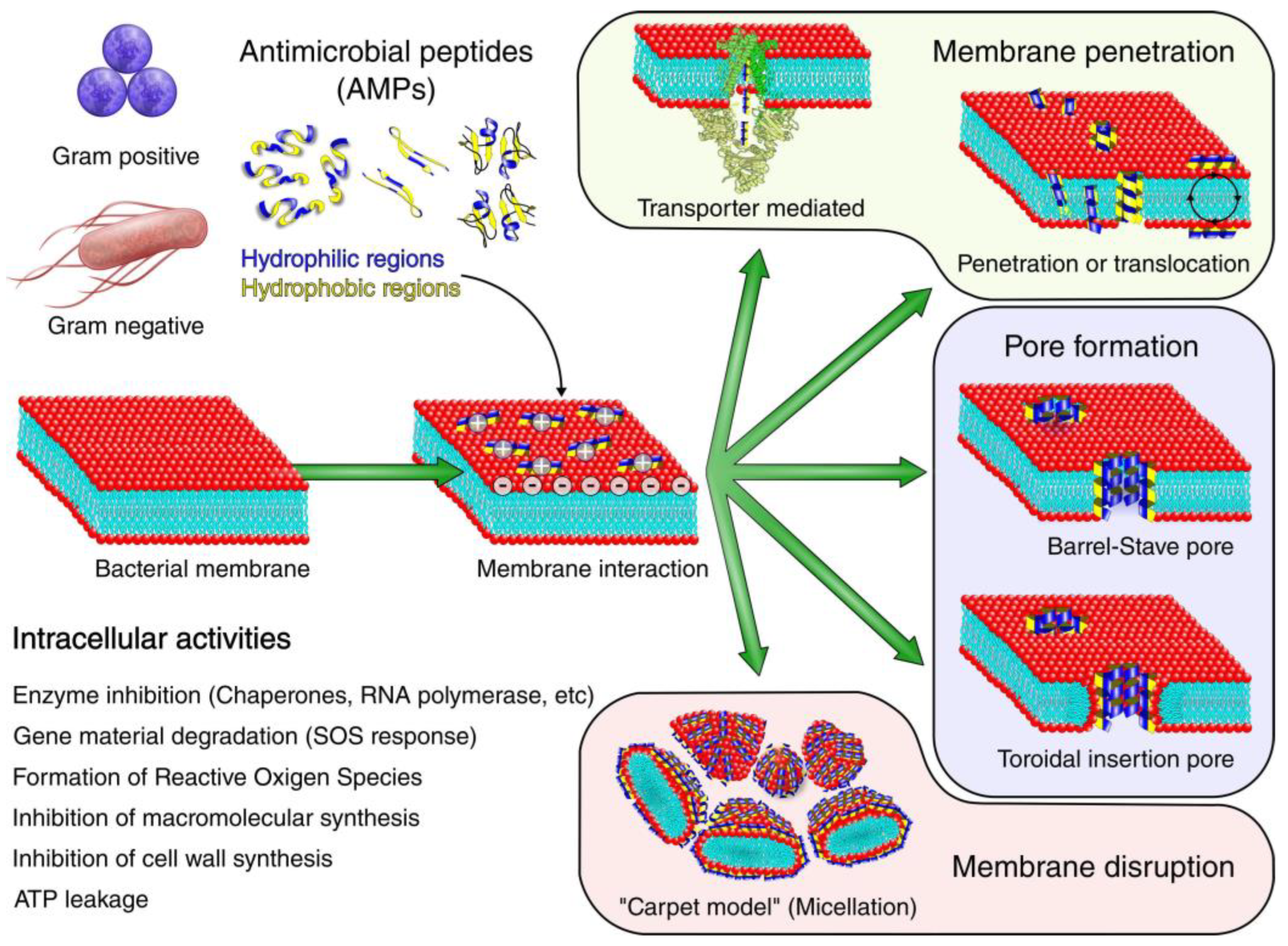
AMPs are short peptides with cationic and hydrophobic properties and are a new class of antibacterial agents that may overcome the problem of drug resistance. AMPs have broad-spectrum activity against Gram-positive and Gram-negative bacteria, fungi, and viruses [101,102]. They not only inhibit cell-wall synthesis, but also destroy the cell-wall structure [103]. Most AMPs are cationic peptides, and the consequent electrostatic attraction to microbes helps AMPs to destroy the membrane structure and enter the microbial cell [104]. Subsequently, AMPs can interfere with intracellular macromolecules and biological processes [105]. Figure 5 summarizes the antimicrobial mechanisms of AMPs. Due to their rapid and broad-spectrum antibacterial activity, numerous peptides have been proposed to be used as antimicrobials, especially against antibiotic-resistant bacteria. In recent years, various short synthetic cationic peptides have been modified from natural AMPs for improved antimicrobial activity and reduced cytotoxicity. These synthetic peptides have strong antibacterial activity against Gram-positive and Gram-negative bacteria and are biocompatible with osteoblasts [101]. Owing to their cationic nature, they can selectively interact with bacterial cells rather than mammalian cells; thus, they are highly effective against microbes and minimally toxic to host cells [106]. In vitro experiments carried out by Tian et al. [107] have shown that the corrosion rate of hydroxyapatite coatings deposited on magnesium alloy surfaces is slower than that of bare coatings. The loading capacity of the AMP in the hydroxyapatite coating was 11.16 ± 1.99 μg/cm2. The release of the AMPs in the hydroxyapatite coating could be sustained for 7 d. The AMP-loaded coating exhibited antimicrobial activity against S. aureus. After 4 d, the bacteriostatic rate was >50%, and the bacteriostatic effect lasted for 7 d.
As a generic cationic AMP, LL37 has been shown to have excellent bactericidal activity without causing bacterial resistance [109]. Studies have shown that almost all the cells in the body can release this peptide. The broad-spectrum antibacterial activity of LL37 is mainly due to the ability of this peptide to disrupt the bacterial membrane [110]. In addition to the antibacterial activity, LL37 has also been shown to be involved in immune regulation and stem-cell recruitment [111,112].
Results reported by Shen et al. [113] have shown that compared with a nanotube, a continuous nanopore structure has a stronger affinity to Ti substrates and can also significantly improve the early adhesion and osteogenic differentiation of MC3T3-E1 cells. Furthermore, these nanopore samples can potentially be loaded with the antibacterial LL37 peptide, which can be control-released for 7 d. Results from cell and animal experiments have further demonstrated that the LL37-loaded nanopore substrate has strong bactericidal and osteo-promoting effects both in vitro and in vivo. All these observations suggest that nanopore-modified materials are more suitable for clinical applications than nanotube-modified ones.
Peptide Mel4 is synthesized by removing several amino acids, including the single tryptophan, from the novel cationic melamine sequence. The peptide Mel4 is highly effective against Pseudomonas aeruginosa (P. aeruginosa), as it can disrupt the bacterial cell membrane, thereby causing the efflux of cellular contents and the lysis of the bacteria [114]. Therefore, coating Ti plates with the peptide Mel4 in the internal fixation of fractures is another promising approach against implant-related infection. An in vivo study by Zhang et al. [115] has shown that Mel4-coated Ti plates have a significant bactericidal effect on S. aureus and P. aeruginosa.

3.3. Novel Organic Coating Materials

Covalent conjugation of broad-spectrum antibacterial agents to implants is a promising approach to reducing the risk of infection. For instance, the recently discovered antibacterial agent N-alkylated 3,6-dihalocarbazole 1-(sec-butylamino)-3-(3,6-dichloro-9H-carbazol-9-yl) propan-2-ol, or simply SPI031, has a broad-spectrum. Gerits et al. [116] have covalently linked SPI031 to Ti and found that the SPI031-Ti matrix prevents the in vitro biofilm formation of S. aureus and P. aeruginosa. The biofilm formation on SPI031-Ti substrates is significantly reduced (by up to 98%) in a mouse model of infection.
Huang et al. [117] have reported the use of red phosphorus (P)/IR780/arginine-glycine-aspartic acid-cysteine (RGDC) coatings on Ti bone implants and found that due to the photothermal therapy and photodynamic therapy, in addition to the high antibacterial efficacy of RGDC, the RGDC-modified surface shows excellent performance in promoting osteogenesis in vivo.
Peeters et al. [118] have developed an anti-biofilm coating consisting of 5-(4-bromophenyl)-N-cyclopentyl-1-octyl-1H-imidazol-2-amine, namely LC0024, to coat Ti implant surfaces (LC0024-Ti). In vitro experiments have shown that on Ti implants, LC0024 reduces specifically the formation of S. aureus biofilm without affecting the number of planktonic cells on the biofilm. In other words, this compound only inhibits biofilm formation without affecting the viability of planktonic cells. The development of drug resistance is thus suppressed. Experiments in a mouse model of infection have shown that S. aureus biofilm formation is significantly reduced (by up to 96%) on LC0024 Ti substrates compared with that on uncoated Ti (control).
Importantly, the functionalization of Ti surfaces with SPI031 or LC0024 does not affect the adhesion or proliferation of human cells, and thus is not expected to interfere with osseointegration and bone repair. Taken together, these data demonstrate the clinical potential of these two compounds as antimicrobial implant coatings in reducing the incidence of implant-related infections.

4. Inorganic Coating Materials

Some non-metallic inorganic materials are also widely used in antimicrobial coatings because of their antimicrobial properties or their excellent properties as carriers for other antimicrobial coating components.

4.1. Se NPs

Se is an essential trace element, found in the range of 6–20 mg in the human body. It is found in the structure of many enzymes and plays an important role in human health [119]. Se NPs, a type of quasi-metallic NPs, have been highlighted as promising antimicrobial materials. Se NPs have efficient antimicrobial properties and can prevent the horizontal transfer of antibiotic resistance genes when used in the nanoscale [120]. Se-NP coating provides a potent anti-infective barrier against the multi-drug-resistant bacterial contamination on orthopaedic medical devices [121]. In addition, Ag-Se coating can more effectively inhibit the growth and biofilm formation of S. aureus than Ag coating. Furthermore, this Ag-Se coating has higher corrosion resistance than the Ag coatings, and its controlled release of Ag minimizes the risk of cytotoxicity [122].

4.2. Ca-P–Coated Carriers

Ca-P coatings, which have good osseointegration and osteoconductive properties due to their similarity to bone minerals, are widely used in bone-tissue engineering. Studies have shown that the antibacterial activity of vancomycin-loaded Ca-P-coated steel plates is due to the sustained drug release and the ability to inhibit the growth of S. aureus. Experiments on cultured osteoblasts have shown that Ca-P-coated plates with or without vancomycin can significantly promote the attachment of osteoblasts. Coating internal fixators with a vancomycin-loaded Ca-P coat can reduce the incidence of implant-related infections [123]. Norvancomycin-loaded biomimetic Ca3(PO4)2 coats also have excellent antibacterial activity [124].

4.3. Titanium-Iodine (Ti-I) Coatings

Iodine is known to be the main component of povidone-iodine disinfectant, which has many advantages, including a broad antibacterial spectrum, no development of drug resistance, and high effectiveness against biofilms. Due to its small size, iodine can rapidly enter microorganisms and subsequently oxidize their key biomolecules, thereby ultimately causing cell death [125]. In vitro and in vivo experiments by Shirai et al. [126] have shown that Ti-I is significantly more resistant to bacterial colonization and more cytocompatible than stainless steel or Ti. Ti-I implants have good antibacterial adhesion in vivo and can inhibit biofilm formation and growth (better than Ti or TiO2) and thus may have a high potential as novel antibacterial implants that can prevent implant-associated infection during orthopaedic surgery [127]. An investigation on the antibacterial activity of Ti-I implants by Ueoka et al. [128] has shown that the iodine level in iodine-free rats implanted with Ti-I decreases to 72% and 65% after 4 and 8 weeks, respectively. Additionally, in vitro experiments have demonstrated that Ti-I implants have stronger antimicrobial activity than Ti or TiO2 implants during the 4 and 8 week periods.

4.4. Diamond-Like Carbon (DLC) Coatings

DLC is a group of materials with a wide range of atomic bond structures and properties depending on the method used for preparing the DLC compound. DLC compounds are resistant to corrosion, have a tough structure and low friction coefficient, and also show good biocompatibility and hemocompatibility. All these properties make DLC compounds ideal for being used as coating materials in biomedical applications [129]. DLC films have anti-biofouling and antibacterial activity against S. epidermidis, S. Aureus, and P. aeruginosa in vitro [130]. The strong hydrophobicity of DLC can lead to severe membrane damage and efflux of microbial metabolites [131]. Levon et al. [132] have examined the effect of a new DLC coating on the adhesion of S. aureus in vitro and observed that DLC is more effective than some commonly used metal biomaterials, including tantalum, Ti, and chromium. Additionally, a study on the superhydrophobic surfaces of DLCs by Rahmawan et al. [133] has shown that at a micropillar spacing ratio (which is the inter-micropillar gap divided by the micropillar diameter) of <4, the static contact angle exceeds 160°, and the surface also exhibits superhydrophobicity. Superhydrophobic surfaces can minimize the contact with a liquid or particle [134]. These special surface structures can inspire research on new materials, with low adhesion rates.

5. Polymeric Coating Materials

In recent years, a wide variety of polymeric and composite bioactive materials have been developed and used in antimicrobial coatings. These materials have different functional groups, most of which function as antibiotic carriers.

5.1. Sol-Gel Membrane Carriers

Studies on Ti-alloy substrates coated with thin, resorbable sol-gel films containing a controlled-release bactericidal have shown a close correlation between the release and degradation rate of the bactericidal, suggesting that film degradation is the main mechanism of the controlled release. Additionally, by using the multilayer approach and various concentrations of vancomycin, the amount of vancomycin released has been found to be at a concentration exceeding the minimum inhibitory concentration of vancomycin against S. aureus. This finding facilitates the tailoring of the release and degradation properties of coating membranes to meet therapeutic needs by controlling the sol-gel processing parameters [135].

5.2. Poly-D,L-Lactic Acid (PDLLA) Carriers

PDLLA has many excellent properties that make it a suitable material for implant coatings. These properties include high mechanical stability, good osteoinductive potential, and excellent in vivo biocompatibility. The PDLLA coating is degraded via hydrolysis 3–6 months post-implantation, and the degradation products are metabolized in the citric acid cycle. Thus, PDLLA is safe to be used in orthopaedic internal fixation [136]. Ti alloy plates have been coated with a vancomycin-containing PDLLA coat via the solvent casting technology by using PDLLA as a carrier and used for the internal fixation of fractures. In vitro experiments have been conducted to evaluate the drug-release properties of the coated plates and the antibacterial activity of the coat against S. aureus. In vitro experiments have shown that the vancomycin-loaded plate has a sustainable drug-release ability and a bactericidal effect against S. aureus, and the antibacterial activity is maintained for at least 15 d [1]. Vancomycin-PDLLA-loaded plates are not toxic and have good biocompatibility. These findings suggest that such coated plates may be used to prevent or treat implant-associated infections [136]. A study in rabbits has shown that the release rate of norvancomycin decreases with time. On day 14 post-implantation, the concentration of norvancomycin in the tissue surrounding the plate remained above the minimum inhibitory concentration. Moreover, the implantation of norvancomycin-loaded PDLLA-coated plates can significantly reduce the probability of postoperative infection in open fracture rabbits [137].

5.3. Chitosan Carriers

Chitosan is a linear hydrophilic amino polysaccharide obtained after partial alkaline deacetylation of chitin. The chemical structures of chitin and chitosan are described in Figure 6. Chitosan is mainly extracted from crustacean shells, cephalopod endoskeleton, and fungal cell walls [138]. Chitosan is hydrophilic, biodegradable, and pH-responsive, and has inherent gel-forming properties [139]. It also has excellent antimicrobial properties. On the surface of chitosan, there are many positively charged amino groups that can interact with anions, such as glycosaminoglycans and proteoglycans, in the bacterial cell wall to alter cell permeability and promote bacterial death [140]. Furthermore, chitosan can penetrate the bacterial cell wall and nucleus to bind to DNA, suggesting that chitosan may kill bacterial cells by inhibiting DNA synthesis or transcription [141]. In addition, chitosan coating can facilitate cell attachment and growth [142]. In vivo studies have shown that chitosan coatings can form tight apposition or osseointegration of dental/craniofacial and orthopaedic implants [143]. Additionally, chitosan is ideal for use as a drug delivery carrier in antimicrobial coatings because of its hydrophilic properties, good biocompatibility, and controllable biodegradability [139]. Pishbin et al. [144] have developed a composite chitosan/bioactive glass coating loaded with gentamicin and deposited the coating on a stainless steel substrate via the electrophoretic deposition technology. The coat released 40% of its gentamicin payload within 5 d and showed improved bactericidal activity against S. aureus when compared with the control groups. Other studies have also shown that chitosan coating modified with gelatin nanospheres can be used as an antibiotic carrier, and the release of (multiple) antibiotics in the composite coating prepared via electrophoretic deposition controlled by electrical signals can regulate the antibacterial activity of metal implants, which is an important technology for the controlled-release of antibiotics and is a new strategy for the prevention of implant-related infections [145]. Some composite coatings prepared by combining chitosan with other materials have also shown good antibacterial properties. Yu et al. [146] have synthesized a multi-component lysozyme-chitosan-Ag-hydroxyapatite hybrid coating and found that it shows good synergistic antibacterial activity. Li et al. [147] have produced a chitosan/Ag composite coating on a biomedical Ni-Ti alloy by using the electrochemical deposition technology and demonstrated that the chitosan/Ag composite coating has better antibacterial activity against E. coli than Ag- or chitosan-coated Ni-Ti.

5.4. Phosphatidylcholine Carriers

Although phosphatidylcholine from natural sources has not been approved to be used as a carrier of antibiotics by the Food and Drug Administration (FDA), it is used as a component of the demineralized bone graft material matrix that has been approved by the FDA [148]. Phosphatidylcholine is a short-term, degradable drug carrier matrix that can be used to directly coat implants. Due to its complete degradability, high implant coverage, and biocompatibility, phosphatidylcholine is more advantageous than other topical delivery devices, such as polymethylmethacrylate beads or CaSO4 [149]. In vivo studies have shown that vancomycin-loaded phosphatidylcholine coatings effectively reduce bacterial biofilm formation in an orthopaedic implant-associated infection model. The local release of antibiotics inhibits the bacterial growth and biofilm formation on implants. These easy-to-apply coatings can be used during surgery to prevent implant-related infections and improve patient outcomes [150].

5.5. Polylactic-Glycolic Acid Copolymer Carriers

Electrospinning is a recently developed processing technology that utilizes electricity to produce ultrafine polymer fibres from polymer solutions [151]. Due to their high porosity, high specific surface area, good cell adhesion, and controlled-release capacity [152], electrospun fibres have been successfully employed as tissue-engineering scaffolds for the vascular system as well as the bone, nerve, and tendon tissues. These fibres have also been utilized as delivery systems for various drugs, including antibiotics. Polylactic-glycolic acid copolymers and vancomycin have been co-dissolved in trifluoroethanol, and vancomycin-coated Ti grafts have been prepared via the electrospinning technology. The release behaviour of vancomycin from the nanofibre coating has been shown to have a biphasic pattern with an onset burst on day 1, followed by a slow and controlled release over 28 d. An in vitro cytotoxicity test has shown that the vancomycin-loaded coating is not cytotoxic. Importantly, vancomycin-coated Ti implants are effective in treating implant-related infections in vivo [85].

5.6. Dendrimer Coatings

Dendrimers are a specific class of nanoscaled, hyperbranched, tree-like macromolecules, with a symmetric well-defined structure and a three-dimensional architecture. They are widely used in drug or gene delivery in nanomedicine [153,154]. Similar to AMPs, cationic antimicrobial dendrimers possess a broad spectrum of antibacterial activity due to their positive charge. As shown in Table 4, several studies have used cationic dendrimers as antimicrobial agents to generate antimicrobial coatings [155].
Khoo et al. [156] have applied polyethene glycol [81] ylated Ti-binding peptides onto Ti to obtain a bacteriophobic coating that can effectively resist protein adsorption and S. aureus adhesion. In a subsequent study, Khoo et al. [157] conjugated a Ti-binding peptide to a cysteine residue at the C-terminus of mono-, di-, or tetra-valent lysine dendrimer cores through a single PEG chain [81]. Ti-coated slides were used as model substrata. The authors assessed the effect of the coating on S. aureus adherence and biofilm formation and found that the antimicrobial properties of the coat improved with the number of Ti-binding peptide repeats. The tetravalent Ti-binding peptide—PEG dendrimer formed a serum-resistant layer and reduced S. aureus biofilm formation by 90%, compared with the uncoated Ti control beads.
Wang et al. [158] have coated Ti-based substrates with amino-terminated poly (amidoamine) (PAMAM) dendrimers modified with PEG (0%–60%). The resulting dendritic films effectively inhibited the colonization of the Gram-negative bacterium P. aeruginosa and, to a lesser extent, of the Gram-positive bacterium S. aureus. The antimicrobial activity of the film was maintained even after 30 d of storage in phosphate-buffered saline. Moreover, the dendritic membrane was less cytotoxic to human bone-derived mesenchymal stem cells and did not alter the gene expression pattern promoted by the Ca3(PO4)2 coating in osteoblasts.
Zhan et al. [159] have prepared hyaluronic acid/PAMAM dendrimer multilayers on a poly (3-hydroxybutyrate-co-4-hydroxybutyrate) substrate via a layer-by-layer self-assembly method for antimicrobial bioactivity. The coating showed anti-adhesion activity and bactericidal activity against E. coli. Both the bacterial anti-adhesion activity and bactericidal activity could be maintained even after storage in phosphate-buffered saline for up to 14 d.
In the study of Klaykruayat et al. [160], a quaternary ammonium hyperbranched dendritic PAMAM of generation 2.5, customized via post-synthesis methylation of a methyl ester terminated PAMAM-dendrimers, was employed to modify flake chitosan, and the cationic PAMAM-chitosan-dendrimers obtained were applied to a cotton fabric via a padding method. The cationic PAMAM-CTS-dendrimer film exhibited strong antimicrobial activity against S. aureus (99.99% reduction).

6. Summary

Implant-related infection in fractures is one of the most common and dreadful complications of fracture fixation surgery. It often leads to the requirement of debridement, multiple surgeries, and even removal of the fixator. At present, the internal fixation materials widely used in clinical practice are titanium or titanium alloys. However, these materials can only provide limited mechanical support and have little ability to regulate the surrounding microenvironment and prevent infection. On this basis, the global interest in antibacterial coatings is growing, and many material scientists, biomaterialists, and clinicians have carried out research that achieves valuable results. This article reviews the recent progress on antimicrobial surface coatings that are used to prevent implant-related infection in fractures, as well as research findings on the antimicrobial effects of metal, inorganic, antimicrobial drug and polymer bioactive material coatings. Biofilm formation by bacteria is a major obstacle to antibiotics and a major difficulty in treating peri-implant infections. Preventing bacteria from surface colonizing and forming a biofilm is the key to judging the antimicrobial function of the coating. Antimicrobial coating materials that prevent biofilm formation should receive more attention. We discuss the examples of combining elements to form binary and ternary composite coatings. The composite coatings composed of antibacterial materials with different functions can reduce the toxicity of a single material and improve the antibacterial properties in many aspects. For example, the combination of bactericidal materials and materials that inhibit bacterial adhesion can prevent and treat infections more efficiently, and antibiotics and their use in combination with other antimicrobial drugs can reduce antibiotic resistance.
However, these materials still have huge challenges. Although numerous in vitro and in vivo experiments have identified the antimicrobial properties of various coatings, insufficient studies have reported whether these coatings are cytotoxic, genotoxic and allergic in clinical use. In our opinion, excellent antibacterial coatings should have the following characteristics: First, it has good antibacterial properties and anti against biofilm production in in vivo and in vitro experiments. Secondly, as biomaterial coatings for human implants, excellent biocompatibility is essential. Nanotechnology holds great promise in coating preparation. The super-small size and large surface area of nanomaterials are shown to have great advantages for their biomedical applications. Researchers can manipulate the physical and chemical properties and biological characteristics of nanomaterials according to the application to prepare nanoparticle coatings with good biological compatibility. In addition, the multiple composite coating of multiple antibacterial materials can kill more effectively bacteria. Future coating studies should also be further explored in these aspects. With verifiable effectiveness and safety, fracture-related implant surface coatings will likely have broader applications.

Author Contributions

Conceptualization, X.Z. and Y.H.; writing—original draft preparation, H.W. and C.X.; writing—review and editing, H.W., C.X., Z.Y. and J.Z.; supervision, X.Z. and Y.H. All authors have read and agreed to the published version of the manuscript.

Funding

This study was supported by Changzhou Sci &Tech Program (Grant No. CJ20220120 and CJ20210104), Qinghai Province Health System Guidance Plan Project (2022-wjzdx-106), Funding from Young Talent Development Plan of Changzhou Health commission (CZQM2020059), and Changzhou High-Level Medical Talents Training Project (2022CZBJ059 and 2022CZBJ061).

Institutional Review Board Statement

Not applicable.

Informed Consent Statement

Not applicable.

Data Availability Statement

Not applicable.

Conflicts of Interest

The authors declare no conflict of interest.

References

  1. Tang, L.; Zhao, C.; Xiong, Y.; Pan, C.; Wang, A. Preparation, release profiles, and antibacterial properties of vancomycin-loaded poly(D,L-lactic) titanium alloy plates. Orthopedics 2009, 32, 324. [Google Scholar] [CrossRef]
  2. Metsemakers, W.-J.; Smeets, B.; Nijs, S.; Hoekstra, H. Infection after fracture fixation of the tibia: Analysis of healthcare utilization and related costs. Injury 2017, 48, 1204–1210. [Google Scholar] [CrossRef]
  3. Olesen, U.K.; Pedersen, N.J.; Eckardt, H.; Lykke-Meyer, L.; Bonde, C.T.; Singh, U.M.; McNally, M. The cost of infection in severe open tibial fractures treated with a free flap. Int. Orthop. 2017, 41, 1049–1055. [Google Scholar] [CrossRef] [PubMed]
  4. Bezstarosti, H.; Van Lieshout, E.M.M.; Voskamp, L.W.; Kortram, K.; Obremskey, W.; McNally, M.A.; Metsemakers, W.J.; Verhofstad, M.H.J. Insights into treatment and outcome of fracture-related infection: A systematic literature review. Arch. Orthop. Trauma Surg. 2019, 139, 61–72. [Google Scholar] [CrossRef] [Green Version]
  5. Cierny, G.; Mader, J.T.; Penninck, J.J. A clinical staging system for adult osteomyelitis. Clin. Orthop. Relat. Res. 2003, 414, 7–24. [Google Scholar] [CrossRef] [PubMed] [Green Version]
  6. Cierny, G.; Mader, J.T. Approach to adult osteomyelitis. Orthop. Rev. 1987, 16, 259–270. [Google Scholar]
  7. Stoodley, P.; Ehrlich, G.D.; Sedghizadeh, P.P.; Hall-Stoodley, L.; Baratz, M.E.; Altman, D.T.; Sotereanos, N.G.; Costerton, J.W.; Demeo, P. Orthopaedic biofilm infections. Curr. Orthop. Pract. 2011, 22, 558–563. [Google Scholar] [CrossRef]
  8. Bjarnsholt, T.; Buhlin, K.; Dufrêne, Y.F.; Gomelsky, M.; Moroni, A.; Ramstedt, M.; Rumbaugh, K.P.; Schulte, T.; Sun, L.; Åkerlund, B.; et al. Biofilm formation—What we can learn from recent developments. J. Intern. Med. 2018, 284, 332–345. [Google Scholar] [CrossRef] [PubMed]
  9. Zhou, G.; Shi, Q.S.; Huang, X.M.; Xie, X.B. The Three Bacterial Lines of Defense against Antimicrobial Agents. Int. J. Mol. Sci. 2015, 16, 21711–21733. [Google Scholar] [CrossRef] [Green Version]
  10. Kostakioti, M.; Hadjifrangiskou, M.; Hultgren, S.J. Bacterial biofilms: Development, dispersal, and therapeutic strategies in the dawn of the postantibiotic era. Cold Spring Harb. Perspect. Med. 2013, 3, a010306. [Google Scholar] [CrossRef] [Green Version]
  11. Kumar, C.G.; Anand, S.K. Significance of microbial biofilms in food industry: A review. Int. J. Food Microbiol. 1998, 42, 9–27. [Google Scholar] [CrossRef] [PubMed]
  12. Jamal, M.; Ahmad, W.; Andleeb, S.; Jalil, F.; Imran, M.; Nawaz, M.A.; Hussain, T.; Ali, M.; Rafiq, M.; Kamil, M.A. Bacterial biofilm and associated infections. J. Chin. Med. Assoc. JCMA 2018, 81, 7–11. [Google Scholar] [CrossRef] [PubMed]
  13. Froyman, W.; Landolfo, C.; De Cock, B.; Wynants, L.; Sladkevicius, P.; Testa, A.C.; Van Holsbeke, C.; Domali, E.; Fruscio, R.; Epstein, E.; et al. Risk of complications in patients with conservatively managed ovarian tumours (IOTA5): A 2-year interim analysis of a multicentre, prospective, cohort study. Lancet Oncol. 2019, 20, 448–458. [Google Scholar] [CrossRef] [PubMed]
  14. Parsek, M.R.; Singh, P.K. Bacterial biofilms: An emerging link to disease pathogenesis. Annu. Rev. Microbiol. 2003, 57, 677–701. [Google Scholar] [CrossRef]
  15. Otto, M. Staphylococcal infections: Mechanisms of biofilm maturation and detachment as critical determinants of pathogenicity. Annu. Rev. Med. 2013, 64, 175–188. [Google Scholar] [CrossRef]
  16. Anwar, H.; Dasgupta, M.K.; Costerton, J.W. Testing the susceptibility of bacteria in biofilms to antibacterial agents. Antimicrob. Agents Chemother. 1990, 34, 2043–2046. [Google Scholar] [CrossRef] [Green Version]
  17. Khoury, A.E.; Lam, K.; Ellis, B.; Costerton, J.W. Prevention and control of bacterial infections associated with medical devices. ASAIO J. 1992, 38, M174–M178. [Google Scholar] [CrossRef]
  18. Nadell, C.D.; Drescher, K.; Wingreen, N.S.; Bassler, B.L. Extracellular matrix structure governs invasion resistance in bacterial biofilms. ISME J. 2015, 9, 1700–1709. [Google Scholar] [CrossRef] [Green Version]
  19. Bagge, N.; Hentzer, M.; Andersen, J.B.; Ciofu, O.; Givskov, M.; Høiby, N. Dynamics and spatial distribution of β-lactamase expression in Pseudomonas aeruginosa biofilms. Antimicrob. Agents Chemother. 2004, 48, 1168–1174. [Google Scholar] [CrossRef] [Green Version]
  20. Hall, C.W.; Mah, T.F. Molecular mechanisms of biofilm-based antibiotic resistance and tolerance in pathogenic bacteria. FEMS Microbiol. Rev. 2017, 41, 276–301. [Google Scholar] [CrossRef] [Green Version]
  21. Germain, E.; Roghanian, M.; Gerdes, K.; Maisonneuve, E. Stochastic induction of persister cells by HipA through (p)ppGpp-mediated activation of mRNA endonucleases. Proc. Natl. Acad. Sci. USA 2015, 112, 5171–5176. [Google Scholar] [CrossRef] [PubMed]
  22. Zhang, L.; Mah, T.F. Involvement of a novel efflux system in biofilm-specific resistance to antibiotics. J. Bacteriol. 2008, 190, 4447–4452. [Google Scholar] [CrossRef] [PubMed] [Green Version]
  23. Wilton, M.; Charron-Mazenod, L.; Moore, R.; Lewenza, S. Extracellular DNA Acidifies Biofilms and Induces Aminoglycoside Resistance in Pseudomonas aeruginosa. Antimicrob. Agents Chemother. 2016, 60, 544–553. [Google Scholar] [CrossRef] [PubMed] [Green Version]
  24. Chouirfa, H.; Bouloussa, H.; Migonney, V.; Falentin-Daudré, C. Review of titanium surface modification techniques and coatings for antibacterial applications. Acta Biomater. 2019, 83, 37–54. [Google Scholar] [CrossRef]
  25. Walters, M.C., 3rd; Roe, F.; Bugnicourt, A.; Franklin, M.J.; Stewart, P.S. Contributions of antibiotic penetration, oxygen limitation, and low metabolic activity to tolerance of Pseudomonas aeruginosa biofilms to ciprofloxacin and tobramycin. Antimicrob. Agents Chemother. 2003, 47, 317–323. [Google Scholar] [CrossRef] [Green Version]
  26. Billings, N.; Millan, M.; Caldara, M.; Rusconi, R.; Tarasova, Y.; Stocker, R.; Ribbeck, K. The extracellular matrix Component Psl provides fast-acting antibiotic defense in Pseudomonas aeruginosa biofilms. PLoS Pathog. 2013, 9, e1003526. [Google Scholar] [CrossRef] [Green Version]
  27. Franklin, M.J.; Nivens, D.E.; Weadge, J.T.; Howell, P.L. Biosynthesis of the Pseudomonas aeruginosa Extracellular Polysaccharides, Alginate, Pel, and Psl. Front. Microbiol. 2011, 2, 167. [Google Scholar] [CrossRef] [Green Version]
  28. Anderl, J.N.; Franklin, M.J.; Stewart, P.S. Role of antibiotic penetration limitation in Klebsiella pneumoniae biofilm resistance to ampicillin and ciprofloxacin. Antimicrob. Agents Chemother. 2000, 44, 1818–1824. [Google Scholar] [CrossRef] [Green Version]
  29. Chiang, W.C.; Nilsson, M.; Jensen, P.; Høiby, N.; Nielsen, T.E.; Givskov, M.; Tolker-Nielsen, T. Extracellular DNA shields against aminoglycosides in Pseudomonas aeruginosa biofilms. Antimicrob. Agents Chemother. 2013, 57, 2352–2361. [Google Scholar] [CrossRef] [Green Version]
  30. Secor, P.R.; Sweere, J.M.; Michaels, L.A.; Malkovskiy, A.V.; Lazzareschi, D.; Katznelson, E.; Rajadas, J.; Birnbaum, M.E.; Arrigoni, A.; Braun, K.R.; et al. Filamentous Bacteriophage Promote Biofilm Assembly and Function. Cell Host Microbe 2015, 18, 549–559. [Google Scholar] [CrossRef] [Green Version]
  31. Borriello, G.; Werner, E.; Roe, F.; Kim, A.M.; Ehrlich, G.D.; Stewart, P.S. Oxygen limitation contributes to antibiotic tolerance of Pseudomonas aeruginosa in biofilms. Antimicrob. Agents Chemother. 2004, 48, 2659–2664. [Google Scholar] [CrossRef] [PubMed]
  32. Nguyen, D.; Joshi-Datar, A.; Lepine, F.; Bauerle, E.; Olakanmi, O.; Beer, K.; McKay, G.; Siehnel, R.; Schafhauser, J.; Wang, Y.; et al. Active starvation responses mediate antibiotic tolerance in biofilms and nutrient-limited bacteria. Science 2011, 334, 982–986. [Google Scholar] [CrossRef] [PubMed] [Green Version]
  33. Nilsson, M.; Rybtke, M.; Givskov, M.; Høiby, N.; Twetman, S.; Tolker-Nielsen, T. The dlt genes play a role in antimicrobial tolerance of Streptococcus mutans biofilms. Int. J. Antimicrob. Agents 2016, 48, 298–304. [Google Scholar] [CrossRef] [PubMed]
  34. Mah, T.F.; Pitts, B.; Pellock, B.; Walker, G.C.; Stewart, P.S.; O’Toole, G.A. A genetic basis for Pseudomonas aeruginosa biofilm antibiotic resistance. Nature 2003, 426, 306–310. [Google Scholar] [CrossRef] [PubMed]
  35. Gillis, R.J.; White, K.G.; Choi, K.H.; Wagner, V.E.; Schweizer, H.P.; Iglewski, B.H. Molecular basis of azithromycin-resistant Pseudomonas aeruginosa biofilms. Antimicrob. Agents Chemother. 2005, 49, 3858–3867. [Google Scholar] [CrossRef] [PubMed] [Green Version]
  36. Savage, V.J.; Chopra, I.; O’Neill, A.J. Staphylococcus aureus biofilms promote horizontal transfer of antibiotic resistance. Antimicrob. Agents Chemother. 2013, 57, 1968–1970. [Google Scholar] [CrossRef] [PubMed] [Green Version]
  37. Driffield, K.; Miller, K.; Bostock, J.M.; O’Neill, A.J.; Chopra, I. Increased mutability of Pseudomonas aeruginosa in biofilms. J. Antimicrob. Chemother. 2008, 61, 1053–1056. [Google Scholar] [CrossRef] [Green Version]
  38. Dalton, T.; Dowd, S.E.; Wolcott, R.D.; Sun, Y.; Watters, C.; Griswold, J.A.; Rumbaugh, K.P. An in vivo polymicrobial biofilm wound infection model to study interspecies interactions. PLoS ONE 2011, 6, e27317. [Google Scholar] [CrossRef] [Green Version]
  39. Rijnaarts, H.H.; Norde, W.; Bouwer, E.J.; Lyklema, J.; Zehnder, A.J. Bacterial Adhesion under Static and Dynamic Conditions. Appl. Environ. Microbiol. 1993, 59, 3255–3265. [Google Scholar] [CrossRef] [Green Version]
  40. Tan, G.; Xu, J.; Chirume, W.M.; Zhang, J.; Zhang, H.; Hu, X. Antibacterial and Anti-Inflammatory Coating Materials for Orthopedic Implants: A Review. Coatings 2021, 11, 1401. [Google Scholar] [CrossRef]
  41. Medici, S.; Peana, M.; Pelucelli, A.; Zoroddu, M.A. An updated overview on metal nanoparticles toxicity. Semin. Cancer Biol. 2021, 76, 17–26. [Google Scholar] [CrossRef]
  42. Sengupta, J.; Ghosh, S.; Datta, P.; Gomes, A.; Gomes, A. Physiologically important metal nanoparticles and their toxicity. J. Nanosci. Nanotechnol. 2014, 14, 990–1006. [Google Scholar] [CrossRef] [PubMed]
  43. Papp, A.; Horváth, T.; Igaz, N.; Gopisetty, M.K.; Kiricsi, M.; Berkesi, D.S.; Kozma, G.; Kónya, Z.; Wilhelm, I.; Patai, R.; et al. Presence of Titanium and Toxic Effects Observed in Rat Lungs, Kidneys, and Central Nervous System in vivo and in Cultured Astrocytes in vitro on Exposure by Titanium Dioxide Nanorods. Int. J. Nanomed. 2020, 15, 9939–9960. [Google Scholar] [CrossRef] [PubMed]
  44. Medici, S.; Peana, M.; Nurchi, V.M.; Zoroddu, M.A. Medical Uses of Silver: History, Myths, and Scientific Evidence. J. Med. Chem. 2019, 62, 5923–5943. [Google Scholar] [CrossRef]
  45. Behrendt, A.K.; Beythien, M.; Huber, J.; Zufraß, T.; Butschkau, A.; Mittlmeier, T.; Vollmar, B. New TiAg composite coating for bone prosthesis engineering shows promising microvascular compatibility in the murine dorsal skinfold chamber model. J. Mater. Sci. Mater. Med. 2015, 26, 5373. [Google Scholar] [CrossRef] [PubMed]
  46. Ichimaru, H.; Harada, A.; Yoshimoto, S.; Miyazawa, Y.; Mizoguchi, D.; Kyaw, K.; Ono, K.; Tsutsuki, H.; Sawa, T.; Niidome, T. Gold Coating of Silver Nanoplates for Enhanced Dispersion Stability and Efficient Antimicrobial Activity against Intracellular Bacteria. Langmuir ACS J. Surf. Colloids 2018, 34, 10413–10418. [Google Scholar] [CrossRef] [PubMed]
  47. Hurtuková, K.; Vašinová, T.; Kasálková, N.S.; Fajstavr, D.; Rimpelová, S.; Pavlíčková, V.S.; Švorčík, V.; Slepička, P. Antibacterial Properties of Silver Nanoclusters with Carbon Support on Flexible Polymer. Nanomaterials 2022, 12, 2658. [Google Scholar] [CrossRef]
  48. Nie, Y.; Kalapos, C.; Nie, X.; Murphy, M.; Hussein, R.; Zhang, J. Superhydrophilicity and antibacterial property of a Cu-dotted oxide coating surface. Ann. Clin. Microbiol. Antimicrob. 2010, 9, 25. [Google Scholar] [CrossRef] [Green Version]
  49. Zhang, X.; Li, J.; Wang, X.; Wang, Y.; Hang, R.; Huang, X.; Tang, B.; Chu, P.K. Effects of copper nanoparticles in porous TiO(2) coatings on bacterial resistance and cytocompatibility of osteoblasts and endothelial cells. Mater. Sci. Eng. C Mater. Biol. Appl. 2018, 82, 110–120. [Google Scholar] [CrossRef]
  50. Maimaiti, B.; Zhang, N.; Yan, L.; Luo, J.; Xie, C.; Wang, Y.; Ma, C.; Ye, T. Stable ZnO-doped hydroxyapatite nanocoating for anti-infection and osteogenic on titanium. Colloids Surf. B Biointerfaces 2020, 186, 110731. [Google Scholar] [CrossRef]
  51. Memarzadeh, K.; Sharili, A.S.; Huang, J.; Rawlinson, S.C.; Allaker, R.P. Nanoparticulate zinc oxide as a coating material for orthopedic and dental implants. J. Biomed. Mater. Res. Part A 2015, 103, 981–989. [Google Scholar] [CrossRef]
  52. Ivanova, I.A.; Pavlova, E.L.; Stoyanova, D.S.; Angelov, O.I. Antibacterial effect of TiO(2):Cu:Ag thin coatings on Pseudomonas strain measured by microbiological and ATP assays. J. Basic Microbiol. 2019, 59, 1165–1172. [Google Scholar] [CrossRef]
  53. Chopra, I. The increasing use of silver-based products as antimicrobial agents: A useful development or a cause for concern? J. Antimicrob. Chemother. 2007, 59, 587–590. [Google Scholar] [CrossRef] [PubMed] [Green Version]
  54. Taylor, L.; Phillips, P.; Hastings, R. Reduction of bacterial contamination in a healthcare environment by silver antimicrobial technology. J. Infect. Prev. 2009, 10, 6–12. [Google Scholar] [CrossRef]
  55. Furkert, F.H.; Sörensen, J.H.; Arnoldi, J.; Robioneck, B.; Steckel, H. Antimicrobial efficacy of surface-coated external fixation pins. Curr. Microbiol. 2011, 62, 1743–1751. [Google Scholar] [CrossRef]
  56. Shao, W.; Zhao, Q. Effect of corrosion rate and surface energy of silver coatings on bacterial adhesion. Colloids Surf. B Biointerfaces 2010, 76, 98–103. [Google Scholar] [CrossRef] [PubMed]
  57. Arens, D.; Zeiter, S.; Nehrbass, D.; Ranjan, N.; Paulin, T.; Alt, V. Antimicrobial silver-coating for locking plates shows uneventful osteotomy healing and good biocompatibility results of an experimental study in rabbits. Injury 2020, 51, 830–839. [Google Scholar] [CrossRef] [PubMed]
  58. Grass, G.; Rensing, C.; Solioz, M. Metallic copper as an antimicrobial surface. Appl. Environ. Microbiol. 2011, 77, 1541–1547. [Google Scholar] [CrossRef] [PubMed] [Green Version]
  59. Ekrikaya, S.; Yilmaz, E.; Celik, C.; Demirbuga, S.; Ildiz, N.; Demirbas, A.; Ocsoy, I. Investigation of ellagic acid rich-berry extracts directed silver nanoparticles synthesis and their antimicrobial properties with potential mechanisms towards Enterococcus faecalis and Candida albicans. J. Biotechnol. 2021, 341, 155–162. [Google Scholar] [CrossRef]
  60. Celik, C.; Ildiz, N.; Ocsoy, I. Building block and rapid synthesis of catecholamines-inorganic nanoflowers with their peroxidase-mimicking and antimicrobial activities. Sci. Rep. 2020, 10, 2903. [Google Scholar] [CrossRef] [Green Version]
  61. Wahab, M.A.; Luming, L.; Matin, M.A.; Karim, M.R.; Aijaz, M.O.; Alharbi, H.F.; Abdala, A.; Haque, R. Silver Micro-Nanoparticle-Based Nanoarchitectures: Synthesis Routes, Biomedical Applications, and Mechanisms of Action. Polymers 2021, 13, 2870. [Google Scholar] [CrossRef]
  62. Wahab, M.A.; Li, L.; Li, H.; Abdala, A. Silver Nanoparticle-Based Nanocomposites for Combating Infectious Pathogens: Recent Advances and Future Prospects. Nanomaterials 2021, 11, 581. [Google Scholar] [CrossRef] [PubMed]
  63. Huang, Y.H.; Chen, M.H.; Lee, B.H.; Hsieh, K.H.; Tu, Y.K.; Lin, J.J.; Chang, C.H. Evenly distributed thin-film Ag coating on stainless plate by tricomponent Ag/silicate/PU with antimicrobial and biocompatible properties. ACS Appl. Mater. Interfaces 2014, 6, 20324–20333. [Google Scholar] [CrossRef] [PubMed]
  64. Rosário, F.; Hoet, P.; Santos, C.; Oliveira, H. Death and cell cycle progression are differently conditioned by the AgNP size in osteoblast-like cells. Toxicology 2016, 368–369, 103–115. [Google Scholar] [CrossRef]
  65. Hans, M.; Erbe, A.; Mathews, S.; Chen, Y.; Solioz, M.; Mücklich, F. Role of copper oxides in contact killing of bacteria. Langmuir ACS J. Surf. Colloids 2013, 29, 16160–16166. [Google Scholar] [CrossRef]
  66. Mitra, D.; Kang, E.T.; Neoh, K.G. Antimicrobial Copper-Based Materials and Coatings: Potential Multifaceted Biomedical Applications. ACS Appl. Mater. Interfaces 2020, 12, 21159–21182. [Google Scholar] [CrossRef]
  67. Fan, X.; Yahia, L.; Sacher, E. Antimicrobial Properties of the Ag, Cu Nanoparticle System. Biology 2021, 10, 137. [Google Scholar] [CrossRef]
  68. Hoene, A.; Prinz, C.; Walschus, U.; Lucke, S.; Patrzyk, M.; Wilhelm, L.; Neumann, H.G.; Schlosser, M. In vivo evaluation of copper release and acute local tissue reactions after implantation of copper-coated titanium implants in rats. Biomed. Mater. 2013, 8, 035009. [Google Scholar] [CrossRef]
  69. Zhang, T.; Liu, J.; Fellner, M.; Zhang, C.; Sui, D.; Hu, J. Crystal structures of a ZIP zinc transporter reveal a binuclear metal center in the transport pathway. Sci. Adv. 2017, 3, e1700344. [Google Scholar] [CrossRef] [PubMed] [Green Version]
  70. Choi, S.; Liu, X.; Pan, Z. Zinc deficiency and cellular oxidative stress: Prognostic implications in cardiovascular diseases. Acta Pharmacol. Sin. 2018, 39, 1120–1132. [Google Scholar] [CrossRef] [PubMed] [Green Version]
  71. Aydin Sevinç, B.; Hanley, L. Antibacterial activity of dental composites containing zinc oxide nanoparticles. J. Biomed. Mater. Res. Part B Appl. Biomater. 2010, 94, 22–31. [Google Scholar] [CrossRef] [PubMed]
  72. Reddy, K.M.; Feris, K.; Bell, J.; Wingett, D.G.; Hanley, C.; Punnoose, A. Selective toxicity of zinc oxide nanoparticles to prokaryotic and eukaryotic systems. Appl. Phys. Lett. 2007, 90, 2139021–2139023. [Google Scholar] [CrossRef] [PubMed] [Green Version]
  73. Liu, Y.; He, L.; Mustapha, A.; Li, H.; Hu, Z.Q.; Lin, M. Antibacterial activities of zinc oxide nanoparticles against Escherichia coli O157:H7. J. Appl. Microbiol. 2009, 107, 1193–1201. [Google Scholar] [CrossRef] [PubMed]
  74. Abdulkareem, E.H.; Memarzadeh, K.; Allaker, R.P.; Huang, J.; Pratten, J.; Spratt, D. Anti-biofilm activity of zinc oxide and hydroxyapatite nanoparticles as dental implant coating materials. J. Dent. 2015, 43, 1462–1469. [Google Scholar] [CrossRef]
  75. Hui, A.; Yang, F.; Yan, R.; Kang, Y.; Wang, A. Palygorskite-Based Organic-Inorganic Hybrid Nanocomposite for Enhanced Antibacterial Activities. Nanomaterials 2021, 11, 3230. [Google Scholar] [CrossRef]
  76. Colon, G.; Ward, B.C.; Webster, T.J. Increased osteoblast and decreased Staphylococcus epidermidis functions on nanophase ZnO and TiO2. J. Biomed. Mater. Res. Part A 2006, 78, 595–604. [Google Scholar] [CrossRef]
  77. Suh, K.S.; Lee, Y.S.; Seo, S.H.; Kim, Y.S.; Choi, E.M. Effect of zinc oxide nanoparticles on the function of MC3T3-E1 osteoblastic cells. Biol. Trace Elem. Res. 2013, 155, 287–294. [Google Scholar] [CrossRef]
  78. Irshad, M.A.; Nawaz, R.; Rehman, M.Z.U.; Adrees, M.; Rizwan, M.; Ali, S.; Ahmad, S.; Tasleem, S. Synthesis, characterization and advanced sustainable applications of titanium dioxide nanoparticles: A review. Ecotoxicol. Environ. Saf. 2021, 212, 111978. [Google Scholar] [CrossRef]
  79. Kaseem, M.; Hamad, K.; Ur Rehman, Z. Review of Recent Advances in Polylactic Acid/TiO(2) Composites. Material 2019, 12, 3659. [Google Scholar] [CrossRef] [Green Version]
  80. Younis, A.B.; Haddad, Y.; Kosaristanova, L.; Smerkova, K. Titanium dioxide nanoparticles: Recent progress in antimicrobial applications. Wiley Interdiscip. Rev. Nanomed. Nanobiotechnology 2022, e1860. [Google Scholar] [CrossRef]
  81. Luttrell, T.; Halpegamage, S.; Tao, J.; Kramer, A.; Sutter, E.; Batzill, M. Why is anatase a better photocatalyst than rutile?--Model studies on epitaxial TiO2 films. Sci. Rep. 2014, 4, 4043. [Google Scholar] [CrossRef] [PubMed]
  82. Nosaka, Y.; Nosaka, A.Y. Generation and Detection of Reactive Oxygen Species in Photocatalysis. Chem. Rev. 2017, 117, 11302–11336. [Google Scholar] [CrossRef] [PubMed]
  83. Liu, L.F.; Barford, J.; Yeung, K.L.; Si, G. Non-UV based germicidal activity of metal-doped TiO2 coating on solid surfaces. J. Environ. Sci. (China) 2007, 19, 745–750. [Google Scholar] [CrossRef]
  84. Tyllianakis, M.; Dalas, E.; Christofidou, M.; Kallitsis, J.K.; Chrissanthopoulos, A.; Koutsoukos, P.G.; Bartzavali, C.; Gourdoupi, N.; Papadimitriou, K.; Oikonomou, E.K.; et al. Novel composites materials from functionalized polymers and silver coated titanium oxide capable for calcium phosphate induction, control of orthopedic biofilm infections: An “in vitro” study. J. Mater. Sci. Mater. Med. 2010, 21, 2201–2211. [Google Scholar] [CrossRef]
  85. Zhang, L.; Yan, J.; Yin, Z.; Tang, C.; Guo, Y.; Li, D.; Wei, B.; Xu, Y.; Gu, Q.; Wang, L. Electrospun vancomycin-loaded coating on titanium implants for the prevention of implant-associated infections. Int. J. Nanomed. 2014, 9, 3027–3036. [Google Scholar] [CrossRef] [Green Version]
  86. Albright, V.; Zhuk, I.; Wang, Y.; Selin, V.; van de Belt-Gritter, B.; Busscher, H.J.; van der Mei, H.C.; Sukhishvili, S.A. Self-defensive antibiotic-loaded layer-by-layer coatings: Imaging of localized bacterial acidification and pH-triggering of antibiotic release. Acta Biomater. 2017, 61, 66–74. [Google Scholar] [CrossRef] [PubMed]
  87. Nichol, T.; Callaghan, J.; Townsend, R.; Stockley, I.; Hatton, P.V.; Le Maitre, C.; Smith, T.J.; Akid, R. The antimicrobial activity and biocompatibility of a controlled gentamicin-releasing single-layer sol-gel coating on hydroxyapatite-coated titanium. Bone Jt. J. 2021, 103-b, 522–529. [Google Scholar] [CrossRef]
  88. Yu, X.; Wu, H.; Li, J.; Xie, Z. Antibiotic cement-coated locking plate as a temporary internal fixator for femoral osteomyelitis defects. Int. Orthop. 2017, 41, 1851–1857. [Google Scholar] [CrossRef] [PubMed]
  89. Wu, H.; Yu, S.; Fu, J.; Sun, D.; Wang, S.; Xie, Z.; Wang, Y. Investigating clinical characteristics and prognostic factors in patients with chronic osteomyelitis of humerus. Burn. Trauma 2019, 7, 34. [Google Scholar] [CrossRef] [PubMed] [Green Version]
  90. Jia, C.; Wang, X.; Yu, S.; Wu, H.; Shen, J.; Huang, Q.; Xie, Z. An antibiotic cement-coated locking plate as a temporary fixation for treatment of infected bone defects: A new method of stabilization. J. Orthop. Surg. Res. 2020, 15, 44. [Google Scholar] [CrossRef] [Green Version]
  91. Wang, G.; Luo, W.; Zhou, Y.; Zhu, Z.; Zhao, Z.; Liu, S.; Li, J.; Feng, X.; Zheng, Y.; Liang, J.; et al. Custom-Made Antibiotic Cement-Coated Nail for the Treatment of Infected Bone Defect. BioMed Res. Int. 2021, 2021, 6693906. [Google Scholar] [CrossRef]
  92. Keller, D.M.; Pizzo, R.A.; Patel, J.N.; Viola, A.; Yoon, R.S.; Liporace, F.A. Use of antibiotic-cement coated locking plates in the setting of periprosthetic infection and infected nonunion. Injury 2022, 53, 2567–2572. [Google Scholar] [CrossRef] [PubMed]
  93. Rathbone, C.R.; Cross, J.D.; Brown, K.V.; Murray, C.K.; Wenke, J.C. Effect of various concentrations of antibiotics on osteogenic cell viability and activity. J. Orthop. Res. Off. Publ. Orthop. Res. Soc. 2011, 29, 1070–1074. [Google Scholar] [CrossRef] [PubMed]
  94. Edin, M.L.; Miclau, T.; Lester, G.E.; Lindsey, R.W.; Dahners, L.E. Effect of cefazolin and vancomycin on osteoblasts in vitro. Clin. Orthop. Relat. Res. 1996, 333, 245–251. [Google Scholar] [CrossRef]
  95. Stewart, S.; Barr, S.; Engiles, J.; Hickok, N.J.; Shapiro, I.M.; Richardson, D.W.; Parvizi, J.; Schaer, T.P. Vancomycin-modified implant surface inhibits biofilm formation and supports bone-healing in an infected osteotomy model in sheep: A proof-of-concept study. J. Bone Jt. Surg. Am. Vol. 2012, 94, 1406–1415. [Google Scholar] [CrossRef] [PubMed] [Green Version]
  96. Zarghami, V.; Ghorbani, M.; Bagheri, K.P.; Shokrgozar, M.A. Prevention the formation of biofilm on orthopedic implants by melittin thin layer on chitosan/bioactive glass/vancomycin coatings. J. Mater. Sci. Mater. Med. 2021, 32, 75. [Google Scholar] [CrossRef]
  97. Freischmidt, H.; Armbruster, J.; Reiter, G.; Grützner, P.A.; Helbig, L.; Guehring, T. Individualized Techniques of Implant Coating with an Antibiotic-Loaded, Hydroxyapatite/Calcium Sulphate Bone Graft Substitute. Ther. Clin. Risk Manag. 2020, 16, 689–694. [Google Scholar] [CrossRef]
  98. Moojen, D.J.; Vogely, H.C.; Fleer, A.; Nikkels, P.G.; Higham, P.A.; Verbout, A.J.; Castelein, R.M.; Dhert, W.J. Prophylaxis of infection and effects on osseointegration using a tobramycin-periapatite coating on titanium implants--an experimental study in the rabbit. J. Orthop. Res. Off. Publ. Orthop. Res. Soc. 2009, 27, 710–716. [Google Scholar] [CrossRef]
  99. Xia, W.; Grandfield, K.; Hoess, A.; Ballo, A.; Cai, Y.; Engqvist, H. Mesoporous titanium dioxide coating for metallic implants. J. Biomed. Mater. Res. Part B Appl. Biomater. 2012, 100, 82–93. [Google Scholar] [CrossRef]
  100. Lv, H.; Chen, Z.; Yang, X.; Cen, L.; Zhang, X.; Gao, P. Layer-by-layer self-assembly of minocycline-loaded chitosan/alginate multilayer on titanium substrates to inhibit biofilm formation. J. Dent. 2014, 42, 1464–1472. [Google Scholar] [CrossRef]
  101. Kazemzadeh-Narbat, M.; Kindrachuk, J.; Duan, K.; Jenssen, H.; Hancock, R.E.W.; Wang, R. Antimicrobial peptides on calcium phosphate-coated titanium for the prevention of implant-associated infections. Biomaterials 2010, 31, 9519–9526. [Google Scholar] [CrossRef] [PubMed]
  102. Brogden, N.K.; Brogden, K.A. Will new generations of modified antimicrobial peptides improve their potential as pharmaceuticals? Int. J. Antimicrob. Agents 2011, 38, 217–225. [Google Scholar] [CrossRef] [PubMed] [Green Version]
  103. Omardien, S.; Brul, S.; Zaat, S.A. Antimicrobial Activity of Cationic Antimicrobial Peptides against Gram-Positives: Current Progress Made in Understanding the Mode of Action and the Response of Bacteria. Front. Cell Dev. Biol. 2016, 4, 111. [Google Scholar] [CrossRef] [PubMed] [Green Version]
  104. Savini, F.; Loffredo, M.R.; Troiano, C.; Bobone, S.; Malanovic, N.; Eichmann, T.O.; Caprio, L.; Canale, V.C.; Park, Y.; Mangoni, M.L.; et al. Binding of an antimicrobial peptide to bacterial cells: Interaction with different species, strains and cellular components. Biochim. Et Biophys. Acta. Biomembr. 2020, 1862, 183291. [Google Scholar] [CrossRef] [PubMed]
  105. Graf, M.; Wilson, D.N. Intracellular Antimicrobial Peptides Targeting the Protein Synthesis Machinery. Adv. Exp. Med. Biol. 2019, 1117, 73–89. [Google Scholar] [CrossRef]
  106. Kazemzadeh-Narbat, M.; Noordin, S.; Masri, B.A.; Garbuz, D.S.; Duncan, C.P.; Hancock, R.E.; Wang, R. Drug release and bone growth studies of antimicrobial peptide-loaded calcium phosphate coating on titanium. J. Biomed. Mater. Res. Part B Appl. Biomater. 2012, 100, 1344–1352. [Google Scholar] [CrossRef]
  107. Tian, J.; Shen, S.; Zhou, C.; Dang, X.; Jiao, Y.; Li, L.; Ding, S.; Li, H. Investigation of the antimicrobial activity and biocompatibility of magnesium alloy coated with HA and antimicrobial peptide. J. Mater. Sci. Mater. Med. 2015, 26, 66. [Google Scholar] [CrossRef]
  108. Ageitos, J.M.; Sánchez-Pérez, A.; Calo-Mata, P.; Villa, T.G. Antimicrobial peptides (AMPs): Ancient compounds that represent novel weapons in the fight against bacteria. Biochem. Pharmacol. 2017, 133, 117–138. [Google Scholar] [CrossRef]
  109. Alves, D.; Pereira, M.O. Mini-review: Antimicrobial peptides and enzymes as promising candidates to functionalize biomaterial surfaces. Biofouling 2014, 30, 483–499. [Google Scholar] [CrossRef]
  110. Xhindoli, D.; Pacor, S.; Benincasa, M.; Scocchi, M.; Gennaro, R.; Tossi, A. The human cathelicidin LL-37--A pore-forming antibacterial peptide and host-cell modulator. Biochim. Biophys. Acta 2016, 1858, 546–566. [Google Scholar] [CrossRef]
  111. Coffelt, S.B.; Marini, F.C.; Watson, K.; Zwezdaryk, K.J.; Dembinski, J.L.; LaMarca, H.L.; Tomchuck, S.L.; Zu Bentrup, K.H.; Danka, E.S.; Henkle, S.L.; et al. The pro-inflammatory peptide LL-37 promotes ovarian tumor progression through recruitment of multipotent mesenchymal stromal cells. Proc. Natl. Acad. Sci. USA 2009, 106, 3806–3811. [Google Scholar] [CrossRef]
  112. He, Y.; Mu, C.; Shen, X.; Yuan, Z.; Liu, J.; Chen, W.; Lin, C.; Tao, B.; Liu, B.; Cai, K. Peptide LL-37 coating on micro-structured titanium implants to facilitate bone formation in vivo via mesenchymal stem cell recruitment. Acta Biomater. 2018, 80, 412–424. [Google Scholar] [CrossRef] [PubMed]
  113. Shen, X.; Al-Baadani, M.A.; He, H.; Cai, L.; Wu, Z.; Yao, L.; Wu, X.; Wu, S.; Chen, M.; Zhang, H.; et al. Antibacterial and osteogenesis performances of LL37-loaded titania nanopores in vitro and in vivo. Int. J. Nanomed. 2019, 14, 3043–3054. [Google Scholar] [CrossRef] [PubMed] [Green Version]
  114. Yasir, M.; Dutta, D.; Willcox, M.D.P. Comparative mode of action of the antimicrobial peptide melimine and its derivative Mel4 against Pseudomonas aeruginosa. Sci. Rep. 2019, 9, 7063. [Google Scholar] [CrossRef] [Green Version]
  115. Zhang, S.; Zhou, X.; Liu, T.; Huang, Y.; Li, J. The effects of Peptide Mel4-coated titanium plates on infection rabbits after internal fixation of open fractures. Arch. Orthop. Trauma. Surg. 2022, 142, 729–734. [Google Scholar] [CrossRef] [PubMed]
  116. Gerits, E.; Kucharíková, S.; Van Dijck, P.; Erdtmann, M.; Krona, A.; Lövenklev, M.; Fröhlich, M.; Dovgan, B.; Impellizzeri, F.; Braem, A.; et al. Antibacterial activity of a new broad-spectrum antibiotic covalently bound to titanium surfaces. J. Orthop. Res. Off. Publ. Orthop. Res. Soc. 2016, 34, 2191–2198. [Google Scholar] [CrossRef] [PubMed]
  117. Huang, B.; Tan, L.; Liu, X.; Li, J.; Wu, S. A facile fabrication of novel stuff with antibacterial property and osteogenic promotion utilizing red phosphorus and near-infrared light. Bioact. Mater. 2019, 4, 17–21. [Google Scholar] [CrossRef] [PubMed]
  118. Peeters, E.; Hooyberghs, G.; Robijns, S.; De Weerdt, A.; Kucharíková, S.; Tournu, H.; Braem, A.; Čeh, K.; Majdič, G.; Španič, T.; et al. An antibiofilm coating of 5-aryl-2-aminoimidazole covalently attached to a titanium surface. J. Biomed. Mater. Res. Part B Appl. Biomater. 2019, 107, 1908–1919. [Google Scholar] [CrossRef]
  119. Minich, W.B. Selenium Metabolism and Biosynthesis of Selenoproteins in the Human Body. Biochem. Mosc. 2022, 87, S168–S177. [Google Scholar] [CrossRef]
  120. Truong, L.B.; Medina-Cruz, D.; Mostafavi, E.; Rabiee, N. Selenium Nanomaterials to Combat Antimicrobial Resistance. Molecules 2021, 26, 3611. [Google Scholar] [CrossRef]
  121. Tran, P.A.; O’Brien-Simpson, N.; Palmer, J.A.; Bock, N.; Reynolds, E.C.; Webster, T.J.; Deva, A.; Morrison, W.A.; O’Connor, A.J. Selenium nanoparticles as anti-infective implant coatings for trauma orthopedics against methicillin-resistant Staphylococcus aureus and epidermidis: In vitro and in vivo assessment. Int. J. Nanomed. 2019, 14, 4613–4624. [Google Scholar] [CrossRef] [PubMed]
  122. Liang, X.; Zhang, S.; Gadd, G.M.; McGrath, J.; Rooney, D.W.; Zhao, Q. Fungal-derived selenium nanoparticles and their potential applications in electroless silver coatings for preventing pin-tract infections. Regen. Biomater. 2022, 9, rbac013. [Google Scholar] [CrossRef] [PubMed]
  123. Fei, J.; Liu, G.D.; Pan, C.J.; Chen, J.Y.; Zhou, Y.G.; Xiao, S.H.; Wang, Y.; Yu, H.J. Preparation, release profiles and antibacterial properties of vancomycin-loaded Ca-P coating titanium alloy plate. J. Mater. Sci. Mater. Med. 2011, 22, 989–995. [Google Scholar] [CrossRef]
  124. Pan, C.J.; Dong, Y.X.; Zhang, Y.Y.; Nie, Y.D.; Zhao, C.H.; Wang, Y.L. Enhancing the antibacterial activity of biomimetic HA coatings by incorporation of norvancomycin. J. Orthop. Sci. Off. J. Jpn. Orthop. Assoc. 2011, 16, 105–113. [Google Scholar] [CrossRef] [PubMed]
  125. Lepelletier, D.; Maillard, J.Y.; Pozzetto, B.; Simon, A. Povidone Iodine: Properties, Mechanisms of Action, and Role in Infection Control and Staphylococcus aureus Decolonization. Antimicrob. Agents Chemother. 2020, 64, e00682-20. [Google Scholar] [CrossRef]
  126. Shirai, T.; Shimizu, T.; Ohtani, K.; Zen, Y.; Takaya, M.; Tsuchiya, H. Antibacterial iodine-supported titanium implants. Acta Biomater. 2011, 7, 1928–1933. [Google Scholar] [CrossRef] [Green Version]
  127. Inoue, D.; Kabata, T.; Ohtani, K.; Kajino, Y.; Shirai, T.; Tsuchiya, H. Inhibition of biofilm formation on iodine-supported titanium implants. Int. Orthop. 2017, 41, 1093–1099. [Google Scholar] [CrossRef]
  128. Ueoka, K.; Kabata, T.; Tokoro, M.; Kajino, Y.; Inoue, D.; Takagi, T.; Ohmori, T.; Yoshitani, J.; Ueno, T.; Yamamuro, Y.; et al. Antibacterial Activity in Iodine-coated Implants Under Conditions of Iodine Loss: Study in a Rat Model Plus In Vitro Analysis. Clin. Orthop. Relat. Res. 2021, 479, 1613–1623. [Google Scholar] [CrossRef]
  129. Roy, R.K.; Lee, K.R. Biomedical applications of diamond-like carbon coatings: A review. J. Biomed. Mater. Res. Part B Appl. Biomater. 2007, 83, 72–84. [Google Scholar] [CrossRef]
  130. Liu, C.; Zhao, Q.; Liu, Y.; Wang, S.; Abel, E.W. Reduction of bacterial adhesion on modified DLC coatings. Colloids Surf. B Biointerfaces 2008, 61, 182–187. [Google Scholar] [CrossRef]
  131. Marciano, F.R.; Bonetti, L.F.; Santos, L.V.; Da-Silva, N.S.; Corat, E.J.; Trava-Airoldi, V.J. Antibacterial activity of DLC and Ag–DLC films produced by PECVD technique. Diam. Relat. Mater. 2009, 18, 1010–1014. [Google Scholar] [CrossRef]
  132. Levon, J.; Myllymaa, K.; Kouri, V.P.; Rautemaa, R.; Kinnari, T.; Myllymaa, S.; Konttinen, Y.T.; Lappalainen, R. Patterned macroarray plates in comparison of bacterial adhesion inhibition of tantalum, titanium, and chromium compared with diamond-like carbon. J. Biomed. Mater. Res. Part A 2010, 92, 1606–1613. [Google Scholar] [CrossRef] [PubMed]
  133. Rahmawan, Y.; Moon, M.W.; Kim, K.S.; Lee, K.R.; Suh, K.Y. Wrinkled, dual-scale structures of diamond-like carbon (DLC) for superhydrophobicity. Langmuir 2010, 26, 484–491. [Google Scholar] [CrossRef] [PubMed]
  134. Helbig, R.; Nickerl, J.; Neinhuis, C.; Werner, C. Smart skin patterns protect springtails. PLoS ONE 2011, 6, e25105. [Google Scholar] [CrossRef] [PubMed] [Green Version]
  135. Radin, S.; Ducheyne, P. Controlled release of vancomycin from thin sol-gel films on titanium alloy fracture plate material. Biomaterials 2007, 28, 1721–1729. [Google Scholar] [CrossRef]
  136. Tang, L.; Zhao, C.; Xiong, Y.; Wang, A. Preparation, antibacterial properties and biocompatibility studies on vancomycin-poly(D,L)-lactic loaded plates. Int. Orthop. 2010, 34, 755–759. [Google Scholar] [CrossRef] [Green Version]
  137. Fei, J.; Yu, H.J.; Pan, C.J.; Zhao, C.H.; Zhou, Y.G.; Wang, Y. Efficacy of a norvancomycin-loaded, PDLLA-coated plate in preventing early infection of rabbit tibia fracture. Orthopedics 2010, 33, 310. [Google Scholar] [CrossRef]
  138. Kou, S.G.; Peters, L.M.; Mucalo, M.R. Chitosan: A review of sources and preparation methods. Int. J. Biol. Macromol. 2021, 169, 85–94. [Google Scholar] [CrossRef]
  139. Muxika, A.; Etxabide, A.; Uranga, J.; Guerrero, P.; de la Caba, K. Chitosan as a bioactive polymer: Processing, properties and applications. Int. J. Biol. Macromol. 2017, 105, 1358–1368. [Google Scholar] [CrossRef]
  140. Afroz, M.M.; Kashem, M.N.H.; Piash, K.; Islam, N. Saccharomyces Cerevisiae as an Untapped Source of Fungal Chitosan for Antimicrobial Action. Appl. Biochem. Biotechnol. 2021, 193, 3765–3786. [Google Scholar] [CrossRef]
  141. Tan, H.; Ma, R.; Lin, C.; Liu, Z.; Tang, T. Quaternized chitosan as an antimicrobial agent: Antimicrobial activity, mechanism of action and biomedical applications in orthopedics. Int. J. Mol. Sci. 2013, 14, 1854–1869. [Google Scholar] [CrossRef] [PubMed]
  142. Ressler, A. Chitosan-Based Biomaterials for Bone Tissue Engineering Applications: A Short Review. Polymers 2022, 14, 3430. [Google Scholar] [CrossRef] [PubMed]
  143. Bumgardner, J.D.; Chesnutt, B.M.; Yuan, Y.; Yang, Y.; Appleford, M.; Oh, S.; McLaughlin, R.; Elder, S.H.; Ong, J.L. The integration of chitosan-coated titanium in bone: An in vivo study in rabbits. Implant. Dent. 2007, 16, 66–79. [Google Scholar] [CrossRef] [PubMed]
  144. Pishbin, F.; Mouriño, V.; Flor, S.; Kreppel, S.; Salih, V.; Ryan, M.P.; Boccaccini, A.R. Electrophoretic deposition of gentamicin-loaded bioactive glass/chitosan composite coatings for orthopaedic implants. ACS Appl. Mater. Interfaces 2014, 6, 8796–8806. [Google Scholar] [CrossRef]
  145. Song, J.; Chen, Q.; Zhang, Y.; Diba, M.; Kolwijck, E.; Shao, J.; Jansen, J.A.; Yang, F.; Boccaccini, A.R.; Leeuwenburgh, S.C. Electrophoretic Deposition of Chitosan Coatings Modified with Gelatin Nanospheres To Tune the Release of Antibiotics. ACS Appl. Mater. Interfaces 2016, 8, 13785–13792. [Google Scholar] [CrossRef]
  146. Yu, W.-Z.; Zhang, Y.; Liu, X.; Xiang, Y.; Li, Z.; Wu, S. Synergistic antibacterial activity of multi components in lysozyme/chitosan/silver/hydroxyapatite hybrid coating. Mater. Des. 2018, 139, 351–362. [Google Scholar] [CrossRef]
  147. Li, P.; Zhang, X.; Xu, R.; Wang, W.; Liu, X.; Yeung, K.W.K.; Chu, P.K. Electrochemically deposited chitosan/Ag complex coatings on biomedical NiTi alloy for antibacterial application. Surf. Coat. Technol. 2013, 232, 370–375. [Google Scholar] [CrossRef]
  148. Han, B.; Tang, B.; Nimni, M.E. Combined effects of phosphatidylcholine and demineralized bone matrix on bone induction. Connect Tissue Res. 2003, 44, 160–166. [Google Scholar] [CrossRef]
  149. Jennings, J.A.; Carpenter, D.P.; Troxel, K.S.; Beenken, K.E.; Smeltzer, M.S.; Courtney, H.S.; Haggard, W.O. Novel Antibiotic-loaded Point-of-care Implant Coating Inhibits Biofilm. Clin. Orthop. Relat. Res. 2015, 473, 2270–2282. [Google Scholar] [CrossRef] [Green Version]
  150. Jennings, J.A.; Beenken, K.E.; Skinner, R.A.; Meeker, D.G.; Smeltzer, M.S.; Haggard, W.O.; Troxel, K.S. Antibiotic-loaded phosphatidylcholine inhibits staphylococcal bone infection. World J. Orthop. 2016, 7, 467–474. [Google Scholar] [CrossRef]
  151. Ji, W.; Sun, Y.; Yang, F.; van den Beucken, J.J.J.P.; Fan, M.; Chen, Z.; Jansen, J.A. Bioactive electrospun scaffolds delivering growth factors and genes for tissue engineering applications. Pharm. Res. 2011, 28, 1259–1272. [Google Scholar] [CrossRef] [PubMed]
  152. Choi, J.S.; Leong, K.W.; Yoo, H.S. In vivo wound healing of diabetic ulcers using electrospun nanofibers immobilized with human epidermal growth factor (EGF). Biomaterials 2008, 29, 587–596. [Google Scholar] [CrossRef] [PubMed]
  153. Alfei, S.; Marengo, B.; Domenicotti, C. Polyester-Based Dendrimer Nanoparticles Combined with Etoposide Have an Improved Cytotoxic and Pro-Oxidant Effect on Human Neuroblastoma Cells. Antioxidants 2020, 9, 50. [Google Scholar] [CrossRef] [PubMed] [Green Version]
  154. Alfei, S.; Catena, S.; Turrini, F. Biodegradable and biocompatible spherical dendrimer nanoparticles with a gallic acid shell and a double-acting strong antioxidant activity as potential device to fight diseases from “oxidative stress”. Drug Deliv. Transl. Res. 2020, 10, 259–270. [Google Scholar] [CrossRef] [PubMed]
  155. Alfei, S.; Schito, A.M. From Nanobiotechnology, Positively Charged Biomimetic Dendrimers as Novel Antibacterial Agents: A Review. Nanomaterials 2020, 10, 2022. [Google Scholar] [CrossRef]
  156. Khoo, X.; Hamilton, P.; O’Toole, G.A.; Snyder, B.D.; Kenan, D.J.; Grinstaff, M.W. Directed assembly of PEGylated-peptide coatings for infection-resistant titanium metal. J. Am. Chem. Soc. 2009, 131, 10992–10997. [Google Scholar] [CrossRef]
  157. Khoo, X.; O’Toole, G.A.; Nair, S.A.; Snyder, B.D.; Kenan, D.J.; Grinstaff, M.W. Staphylococcus aureus resistance on titanium coated with multivalent PEGylated-peptides. Biomaterials 2010, 31, 9285–9292. [Google Scholar] [CrossRef] [Green Version]
  158. Wang, L.; Erasquin, U.J.; Zhao, M.; Ren, L.; Zhang, M.Y.; Cheng, G.J.; Wang, Y.; Cai, C. Stability, antimicrobial activity, and cytotoxicity of poly(amidoamine) dendrimers on titanium substrates. ACS Appl. Mater. Interfaces 2011, 3, 2885–2894. [Google Scholar] [CrossRef]
  159. Zhan, J.; Wang, L.; Liu, S.; Chen, J.; Ren, L.; Wang, Y. Antimicrobial Hyaluronic Acid/Poly(amidoamine) Dendrimer Multilayer on Poly(3-hydroxybutyrate-co-4-hydroxybutyrate) Prepared by a Layer-by-Layer Self-Assembly Method. ACS Appl. Mater. Interfaces 2015, 7, 13876–13881. [Google Scholar] [CrossRef]
  160. Klaykruayat, B.; Siralertmukul, K.; Srikulkit, K. Chemical modification of chitosan with cationic hyperbranched dendritic polyamidoamine and its antimicrobial activity on cotton fabric. Carbohydr. Polym. 2010, 80, 197–207. [Google Scholar] [CrossRef]
Figure 1. Diagrammatic sketch of the biofilm formation process on the titanium surface. 1. Bacterial adhesion. 2. Formation of microcolonies. 3. Maturation of the biofilm. 4. Biofilms produce an exopolysaccharidic matrix that protect them from the host immune response and antibiotics. Reprinted with permission from [24] 2019 Acta Materialia Inc.
Coatings 12 01921 g001
Figure 2. Timeline of papers on various implant-related antimicrobial surface coatings included on web of science in the last decade.
Coatings 12 01921 g002
Figure 3. Oxidative stress and inflammation from phagocytosed metallic NPs leading to cell death. Reprinted with permission from ref [41]. 2021 Elsevier Ltd.
Coatings 12 01921 g003
Figure 4. Diagrammatic sketch of contact killing. (A) Silver ions are released from the silver containing materials and cause cell damage. (B) The cell membrane ruptures because of silver and other stress phenomena, leading to loss of membrane potential and cytoplasmic content. (C) Silver ions induce the generation of reactive oxygen species, which cause further cell damage. (D) Genomic and plasmid DNA becomes degraded. Reprinted with permission from ref [58] 2011, American Society for Microbiology.
Coatings 12 01921 g004
Figure 5. Diagrammatic sketch of the antimicrobial mechanisms of the antimicrobial peptides. Reprinted with permission from ref [108] 2017 Elsevier.
Coatings 12 01921 g005
Figure 6. Diagrammatic sketch of the chemical structure of chitin and CS. Reprinted with permission from ref [139] 2017 Elsevier B.V.
Coatings 12 01921 g006
Table 1. Mechanisms of biofilm-based antibiotic resistance and tolerance in pathogenic bacteria.
MechanismsExamplesRef.
Biofilm matrixAntimicrobial penetrationBiofilms of P. aeruginosa reduce the diffusion of tobramycin.[25]
PolysaccharidesPsl(a exopolysaccharide produced by Pseudomonas aeruginosa) appears to play a role in resistance to colistin, polymyxin B, tobramycin and ciprofloxacin at early stages of biofilm development.[26]
Biofilms lacking Pel(a component of the biofilm glycocalyx in P. aeruginosa) were found to be more susceptible to tobramycin and gentamicin compared to wild-type biofilms.[27]
antibiotic-degrading enzymesK. pneumoniae biofilms produce β-lactamase that was found to effectively degrade ampicillin. [28]
Extracellular DNADNA added to P. aeruginosa biofilms from exogenous sources can become incorporated into the biofilm matrix, resulting in an increased level of resistance by 3-fold for tobramycin and by 2-fold for gentamicin.[29]
BacteriophagesThe presence of Pf phage allows P. aeruginosa to form liquid crystal biofilms with a higher tolerance to tobramycin.[30]
Nutritional limitation and stress responsesPhysiological heterogeneity, hypoxia and reduced growth rateThe gradient of oxygen in P. aeruginosa biofilms can hypoxia the deep layers of the biofilm. Cell had decreased metabolic activity in the hypoxic zone, and this slow growth rate conferred tolerance to antibiotics.[31]
Amino acid starvation and the stringent responseActive starvation responses mediate antibiotic tolerance in biofilms and nutrient-limited P. aeruginosa.[32]
cell-wall-modifying enzymesThe dltABCD operon was a positive hit in a screen for biofilm-specific gentamicin tolerance genes in Streptococcus mutans, the increased negative charge of the ΔdltA strain promotes uptake of gentamicin.[33]
glycosyltransferasesThe ndvB locus was identified as a P. aeruginosa biofilm-specific antibiotic resistance gene, ΔndvB biofilms were 16-fold more susceptible to tobramycin and 8-fold more susceptible to both gentamicin and ciprofloxacin than wild-type biofilms.[34]
efflux pumpsP. aeruginosa biofilm resistance to azithromycin was dependent on the presence of either the MexAB-OprM or the MexCD-OprJ pumps.[35]
genetic diversityHorizontal gene transferIn Staphylococcus aureus, the conjugal transfer frequency of a multidrug resistance plasmid was on the order of 10,000 times greater in biofilms than planktonic cultures.[36]
Mutation frequencyThe mutation frequency for selection of ciprofloxacin resistant mutants was approximately 2-log higher in P. aeruginosa biofilm cells than in planktonic cells.[37]
multispecies interactionsIn an in vivo polymicrobial wound model, P. aeruginosa growing in a monospecies biofilm was 2-fold more susceptible to gentamicin treatment than P. aeruginosa in a polymicrobial biofilm with S. aureus, Enterococcus faecalis and Finegoldia magna.[38]
Table 2. Common metal nanomaterials used in coatings.
Metal NanomaterialsAntimicrobial MechanismCharacteristicExample of the Application on the CoatingRef.
AgNPsContact Killing
ROS release and Oxidative stress
Efficient contact killing capability causes cell membrane perforation and leakage of intracellular compounds.Silver-plated titanium plate implants showed better biocompatibility than pure titanium plates with metal implants, and successfully cured the rabbit infection.
Triplate silver nanoparticle coatings with gold atoms have high antibacterial activity against bacteria.
Silver nanoclusters enhanced the antimicrobial properties of the surface of flexible polymer.
[45,46,47]
CuNPsContact Killing
ROS release and Oxidative stress
Compared to AgNPs, CuNPs have weaker antimicrobial properties and require higher concentrations to inhibit microorganisms.Copper plate and copper dot oxide coatings had good antibacterial activity against E. coli and MRSA.
Porous titanium dioxide coating of titanium doped with CuNPs deposited showed good antibacterial activity against S. aureus.
[48,49]
ZnO NPsContact Killing
ROS release and Oxidative stress
Extensive antimicrobial spectrum
good biocompatibility
chemical stability
low toxicity.
Zoxide and HA nanoparticle composite coatings reduced the number of Streptococcus, Anaerobes, and aerobic bacteria by 95%, 95%, and 90%, respectively.
100% ZnO NPs and 75% ZnONPs/25% HANPs composite-coated substrates have significant antimicrobial activity against Staphylococcus aureus.
[50,51]
TiO2 NPsThe generation of ROS and the degradation of biopolymers
Change the surface features to a hydrophilic state
Photo-induced antimicrobial properties by ultraviolet (UV) light.
Non-toxic to humans
good chemical stability to biomaterials.
Nanocomposite TiO2:Cu:Ag coatings have a strong antimicrobial effect.[52]
Table 4. Dendrimers (Ds) in antimicrobial surface coatings.
Apellation of CoatingsClassification of DendrimersAction SubstrateTarget Bacterial SpeciesCharacteristicRef.
Titanium-binding
peptides/multivalent PEGylated-lysine
dendrimer cores
Peptide-based DsTi-coated slidesS. aureusAntimicrobial properties of the coated improved with the number of TBP repeats[156,157]
Poly(amidoamine)
dendrimers
Cationic Dstitanium
substrates
P.aeruginosa, S. aureusHigher bactericidal activity against Gram-negative bacteria than against Gram-positive bacteria
durable bacteriophobic abilities
good stability and biocompatibility
[158]
Hyaluronic
Acid/Poly(amidoamine) Dendrimer Multilayer
Cationic DsPoly(3-hydroxybutyrate-co-4-hydroxybutyrate)E.coliDurable anti-adhesive activity and bactericidal activity
good biocompatibility
[159]
Cationic
PAMAM-CTS-Ds
Cationic Dscotton fabricS. aureusAntibacterial activity comparable to natural CS films[160]
Publisher’s Note: MDPI stays neutral with regard to jurisdictional claims in published maps and institutional affiliations.