Article

Rheological Properties of Film-Forming Dispersions of Selected Biopolymers Used for Packaging Films or Food Coating

Department of Food Engineering and Process Management, Institute of Food Sciences, Warsaw University of Life Sciences (WULS-SGGW), 02-776 Warszawa, Poland
*
Author to whom correspondence should be addressed.
Coatings 2022, 12(11), 1704; https://doi.org/10.3390/coatings12111704
Submission received: 15 October 2022 / Revised: 4 November 2022 / Accepted: 7 November 2022 / Published: 9 November 2022

Abstract

:
Film-forming solutions based on four types of biopolymers were prepared and their rheological properties were determined. High methylated apple pectin and sodium alginate were used at the concentrations 1%, 1.5%, and 2%, whereas soy protein isolate and pork gelatin were obtained at 4%, 8%, and 12%. The parameters determining the production of the appropriate type of packaging film or edible coating are the setting time on the substrate, regardless of its type, and the gelation temperature, which were analyzed in the study by determination of flow curves and rheological parameters of prepared dispersions. The Newtonian model was used to describe the flow curves of the gelatin and sodium alginate solutions, while the Ostwald–de Waele model was used to describe the flow curves of the soy protein isolate and high methylated apple pectin solutions. The apparent viscosity of all solutions increased with increasing biopolymer concentrations, from 0.0042 to 0.0061 Pa·s and from 0.0187 to 0.0884 Pa·s for high-methylated apple pectin and sodium alginate, respectively; whereas, for a protein-based solution the viscosity increase was from 0.024 to 0.100 Pa·s and from 0.0018 to 0.0056 Pa·s for soy and gelatin, respectively. Modulus of elasticity curves appeared only at the highest concentrations, which means that the formation of the continuous structure of film or coating occurs by different mechanisms depending on the biopolymer type and its amount dispersed in aqueous solutions.

1. Introduction

Films and coatings differ in the most important tasks that are placed on them. Films are mainly used to reduce or control the transfer of, e.g., gases or water vapor, to or from the product [1]. In contrast, coatings are more often used to improve the physical integrity of the product to be coated to enhance the appearance of the product [2,3]. Films and coatings belong to the group of edible packaging [4,5,6]. They are defined as a thin layer of material that can either be eaten with the product or dissolved during the preliminary operations performed, but do not require prior disposal [7]. The layer can be a barrier to external factors (e.g., oxygen, water) and constitute a barrier inside the product [8]. Edible films are obtained by creating a thin layer from a solution of natural polymers [5,9,10]. They have good mechanical properties and consistency [11], which allows the film to be made separately from the product and then applied to the product. In contrast, coatings are formed directly on the coated product [12,13]. Edible packaging made of polymers (polysaccharides, starch, pectin, proteins) are natural ingredients, so they can be consumed by humans and animals without risk to health [4]. The inclusion of bioactive substances leads to determination of active packaging as a novel approach aimed at developing packaging materials that also carry active agents to improve food quality and safety [14]. Crosslinking agents such as formaldehyde or transglutaminase poly (acrylic acid)/montmorillonite nanocomposites have also been used to improve the film forming and barrier properties [15]. One of the parameters determining the production of the appropriate type of film or coating, which may constitute a type of edible packaging, is the setting time on the substrate, regardless of its type, which results in higher storage and production costs for both packaging in the form of films and coatings, as well as packaged and coated products [9,16,17,18,19]. The decisive parameter in the case of edible coatings is temperature. Too high a coating temperature or too long a setting time of the coating may cause a loss of nutritional value of food and attractiveness for the consumer [20]. The study of the gelation process of film-forming solutions used in food is related to the growing interest in modern food packaging. Edible packaging—films and coatings—belong to this group of packaging. They can not only protect the product from deterioration but also make the product more attractive to consumers by adding flavor and aromatic or coloring additives. An example may be edible films with fruit and vegetable purees that can serve as healthy snacks, edible oven bags, and wraps for sushi instead of pancakes, tortillas, or lavash [21]. At the same time, these types of natural biopolymers with the addition of nano components are part of the sustainable development strategy of the packaging industry. These eco-friendly materials also alleviate the environmental concerns associated with plastic-related pollution due to their excellent biodegradability, renewability, bioavailability, and non-toxicity [22,23]. Due to the unfavorable changes that may occur in the coated product during the coating process, one of the most important parameters in the application of edible films and coatings is the gel-forming temperature and the rheological properties of film-forming substances [24,25]. The techniques for determining the gel temperature can be divided into three categories: rheological, thermal, and visual. The rheological method is based on determining the point at which the sol turns into a gel (gel point), and thus the temperature at which the sol to gel is converted. The gel point is determined from the viscosity modulus (G″) and the elastic modulus (G′). The gelation temperature is the temperature at which the viscosity modulus G″ has the same value as the elastic modulus G′ [25,26,27,28,29,30]. The thermokinetic method is based on the examination of solutions using a differential scanning calorimeter to detect the exothermic reaction signal and its description using a kinetic model [29]. The visual method is the least accurate method for determining the gelation temperature. It consists in storing polymer solutions at different temperatures and visually assessing the temperature at which the solution gels [27]. Due to the complexity of interactions that may occur during coating, both in film-forming solutions and coated food, an attempt was made to determine rheological parameters for selected biopolymer solutions [21,31]. The experiments aimed to investigate the rheological properties and the gelling process of selected solutions of protein and polysaccharide hydrocolloids. The possibilities of determining the flow curves and rheological parameters for the tested film-forming solutions based on gelatin, soy protein isolate, sodium alginate, and highly methylated film-forming solutions of apple pectin were investigated.

2. Materials and Methods

2.1. Materials

The research material consisted of solutions prepared with the use of protein and polysaccharide hydrocolloids (Table 1): pork gelatin BLOOM 180 (Agnex, Białystok, Poland), soy protein isolate SUPRO 670 (DuPont Poland Sp.z o.o., Warsaw, Poland), sodium alginate (Sigma-Aldrich, Warsaw, Poland), and apple high methylated pectin (Naturex, Warsaw, Poland).

2.2. Preparation of Film-Forming Solutions

Distilled water in an amount of 300 cm3 was heated in a beaker to 80 °C on a heating plate with a magnetic stirrer (IKA RCT basic, Staufen, Germany)). The tested film-forming substances were weighed on an analytical balance (RADWAG PS 600/C/2, RADWAG, Radom, Poland): proteins—respectively 20 g, 40 g, and 80 g for concentrations of 4%, 8%, and 12% (w/v), polysaccharides—respectively 5 g, 7.5 g, and 10 g for concentrations of 1%, 1.5%, and 2% (w/v). After reaching the temperature of 80 °C, appropriate weighed amounts of the substance were added to distilled water at the temperature of 80 °C and stirred for 30 min. After adding the soy protein isolate, sodium alginate, and high methylated apple pectin, the solutions were further mixed using a blender (BOSCH Mixxo Quattro, Gerlingen, Germany) to facilitate the dissolution of the added substances. After 30 min of mixing, the solutions were cooled to 50 °C and glycerol was added in the amount of 50% (v/w) based on the weight of the hydrocolloid used, followed by 20 min of stirring at 50 °C. Then, the solutions were cooled to 20 °C.

2.3. Determination of Flow Curves of Film-Forming Solutions

The test was carried out on the MARS 40 rheometer by HAAKE (Vreden, Germany). The solutions were tested at the temperature of 50 °C in the system of coaxial cylinders with the shear rate increasing linearly to 100 s−1. The apparent solution viscosity was determined for a shear rate of 50 s−1.

2.4. Examination of the Gelling Process

The measurements were carried out with the use of a MARS 40 rheometer by HAAKE (Vreden, Germany) in the plate-plate measurement system (type T60). An amount of 2.8 mL of solution was placed on the rheometer plate with a syringe. It was then heated under controlled stress and oscillation at 1 Hz for 5 min until the initial test temperature was reached. Then, the solutions were cooled linearly at the rate of 1 K/min, 3 K/min, and 5 K/min. The initial and final cooling temperatures of the solutions are provided in Table 1. The final temperature was held for 5 min. The Rheowin Job Manager (HAAKE, Vreden, Germany) program was used to record and process the obtained results. The gelation temperature was read from the graph based on the elastic modulus G′ and the viscosity modulus G″ as the point of intersection of the G′ and G″ modulus curves. Measurements for a given solution and cooling rate were performed in triplicate.

2.5. Calculation Methods

The following rheological models were used for the mathematical description of the flow curves of the investigated film-forming solutions [32]:
Newton’s model:
τ = η · γ ˙  
Ostwald–de Waele model:
τ = k ( γ ˙ ) n
where: τ—shear stress (Pa), η—dynamic viscosity coefficient (Pa·s), γ ˙ —shear speed (s1), k—consistency coefficient (Pa·sn), and n—the flow index.
The optimal model for describing the flow curves of the tested solutions was selected based on the value of the determination coefficient R2. The apparent viscosity was also determined at a shear rate of 50 s1.

2.6. Statistical Analysis

Statistical analysis was performed with the use of the Statistica 13.1 program. Spearman’s R correlation with the significance level α = 0.05 was used to determine the correlation between the hydrocolloid concentration. Additionally, based on the gelation temperature of the solutions, homogeneous groups were determined using Tukey’s test (α = 0.05).

3. Results

3.1. Flow Curves of Tested Film-Forming Solutions

The flow curves of the gelatin solutions are straight lines from the origin of the coordinate system, so these solutions are Newtonian liquids (Figure 1A).

3.1.1. Gelatin Solutions

The value of the dynamic viscosity coefficient, determined using the Equation (1), is higher than the higher the gelatin concentration in the tested solution and it varies from 0.0024 ± 0.0001 Pa · s for a concentration of 4% to 0.0100 ± 0.0011 Pa s for a concentration of 12% (Table 2). Statistical analysis showed a strong positive correlation of 0.964901 between the concentration of the gelatin solution and the apparent viscosity of the tested gelatin solutions (Table 2). Duan et al. [33] studied gelatin solutions from calf bones and catfish skin at concentrations of 1.4%. They obtained results confirming the tendency that the higher the concentration, the higher the solution viscosity. Both the gelatin solution from catfish skin and calf bones had a viscosity of up to 2 mPa·s. Marcotte et al. [34] tested gelatin solutions with concentrations of 2%, 3%, and 4% at 20 °C, 40 °C, 60 °C, and 80 °C. Based on the R2 parameter, it was shown that gelatin exhibits the characteristics of the Newtonian model. For a gelatin solution with a concentration of 4%, the dynamic viscosity coefficient was at the level of 0.0060 Pa·s, while for the same gelatin solution with a concentration of 4%, tested at 80 °C, the dynamic viscosity coefficient was 0.0050 Pa·s. Both solutions showed a correlation to the Newtonian model at the level of R2 = 0.96.
The results of the dynamic viscosity coefficient obtained by Marcotte et al. [34] are analogous to those obtained in this study, however, the flow curves show a smaller correlation to the Newtonian model. The dynamic viscosity of the gelatin solution with a concentration of 4% was 26 mPa·s, while for the gelatin solution with a concentration of 8%, the dynamic viscosity coefficient was 58 mPa·s. The results obtained by Wang et al. [35], who tested gelatin solutions with 4% and 8% concentrations at 40 °C, are analogous to those obtained in this study.

3.1.2. Soy Protein Isolate Solutions

Figure 1B shows the flow curves of the tested solutions of soy protein isolate with concentrations of 4%, 8%, and 12%. The course of the flow curves of the soy protein isolate solutions indicates that they are shear-thinning liquids with no yield limit. Based on the R2 parameter, they were described using the power model (Equation (2)). The rheological parameters and the value of the apparent viscosity coefficient at the shear rate of 50 s−1 are presented in Table 3.
The flow curves of the tested solutions of soy protein isolate show that with the increase in the concentration of soy protein isolate, the deviation from the Newtonian behavior of the solution during flow decreases, which is also evidenced by the increase in the value of the n melt flow index from 0.457 for an isolate concentration of 4% to 0.874 for a concentration of 12%. The apparent viscosity of soy protein isolate solutions with concentrations of 4%, 8%, and 12% is 0.0018, 0.0026, and 0.0056 Pa·s, respectively. Statistical analysis showed a strong positive correlation of 0.986394 between the concentration of the soy protein isolate solution and the apparent viscosity of the soy protein isolate solution (Table 3). Liu et al. [36] tested solutions of 8-molar urea and 16%, 18%, 20%, and 22% of soy protein isolate at concentrations of 16%, 18%, 20%, and 22% with a 1% addition of sodium sulfite based on the weight of the hydrocolloid. The flow curve of the 18% soy protein isolate solution was determined at 50 °C, while the flow curves of the 16%, 20%, and 22% solutions were determined at 25 °C. The remaining rheological parameters were determined at temperatures ranging from 25 to 70 °C. All solutions tested by the authors showed the nature of shear-thinned liquids, similar to the solutions of soybean isolate with and without the addition of pumpkin puree with concentrations of 4%, 8%, and 12% discussed in this paper. However, the obtained shear stress of an 18% soy protein isolate solution at 50 °C at a shear rate of 100 s−1 is approximately 100 times greater than that of 12%. The apparent viscosity of the solutions obtained was, respectively, 0.2816 Pa·s, 0.8608 Pa·s, 2.3811 Pa·s, and 6.6886 Pa·s for soy protein isolate solutions with concentrations of 16%, 18%, 20%, and 22%. Such differences are due, inter alia, to the fact that they tested solutions with higher concentrations. Another reason may be the way of preparing the solutions—the solutions were not prepared from distilled water, but from urea and sodium sulfite solutions, and without the addition of glycerin.

3.1.3. Sodium Alginate Solutions

Figure 1C shows the flow curves of the tested sodium alginate solutions with concentrations of 1%, 1.5%, and 2%. The flow curves of sodium alginate solutions are straight lines originating from the origin of the coordinate system. This means that sodium alginate solutions are Newtonian liquids. The value of the dynamic viscosity coefficient, determined with the use of Equation (1), increases with increasing the concentration from 0.0187 ± 0.0001 Pa·s at a concentration of 1% to 0.0884 ± 0.0002 Pa·s at a concentration of 2%. Statistical analysis showed a strong positive correlation of 0.960769 between the concentration of sodium alginate solution and the dynamic viscosity of sodium alginate solutions (Table 2). Comaposada et al. [37] tested solutions of six types of sodium alginate with concentrations of 2%, 3%, and 4% at 12 °C. For a sodium alginate solution with a concentration of 2%, they obtained, depending on the supplier, viscosity values from 391 mPa s to 6050 mPa·s. The differences in viscosity between the different types of sodium alginate used were explained by the possible differences in molecular weight. The difference between the results obtained by these authors and the results obtained in this study probably results from the examination of the viscosity of sodium alginate solutions at different temperatures. Additionally, the authors confirmed the tendency that the higher the concentration of the solution, the higher the viscosity of the solution. Cevoli et al. [38] tested sodium alginate solutions with concentrations of 1%, 1.5%, and 2% at 25 °C. Based on the R2 parameter, the Ostwald–de Waele power model was chosen to describe the flow curves. The values of the k coefficient of consistency for the sodium alginate solution with concentrations of 1%, 1.5%, and 2% were 0.66 Pa·sn, 2.51 Pa·sn, and 7.08 Pa·sn, respectively, while the n flow index was 0.71, 0.62, and 0.56, respectively. The power model was chosen based on the R2 parameter, which ranged from 0.986 for a 2% solution to 0.997 for a 1% solution, while in this study a much higher correlation was with Newton’s model.

3.1.4. High Methylated Apple Pectin Solution

Figure 1D shows the flow curves of solutions with concentrations of 1%, 1.5%, and 2% of high methylated apple pectin. The flow curves of a high methylated apple pectin solution are nonlinear curves through the center of the coordinate system, which means that they are shear-thinning liquids. Based on the R2 parameter (Table 2), the Ostwald–de Waele model (Equation (2)) was chosen to describe the flow curves. The higher the concentration of the high methylated apple pectin solution, the higher the shear stress of the tested solutions, but in the case of a 1% high methylated apple pectin solution, the shear stress at a shear rate of up to 20 s−1 is higher than the shear stress of a 1.5% highly methylated apple pectin solution. This is due to the nature of the curves—the flow curve of a 1.5% high methylated apple pectin solution has a more flattened shape than that of a 1% solution. As in the case of all tested solutions, the apparent viscosity of high-methylated apple pectin solutions increases with increasing the concentration of high-methylated apple pectin solutions from 0.0042 ± 0.0001 Pa·s for a 1% solution to 0.0061 ± 0.0001 Pa·s for a 2% solution. Statistical analysis showed a strong positive correlation of 0.948683 between the concentration of high methylated apple pectin solution and the dynamic viscosity of high methylated apple pectin solutions (Table 3). Huang et al. [39] studied the viscosity of pectin extracted from sugar beet pulp. They prepared a solution with a concentration of 5% (w/v) stabilized with hydrochloric acid to pH = 1.2. The apparent viscosity was determined at 50 °C. Depending on the type of pectin drying, after extraction (with hot air or vacuum) the results were 0.959 Pa·s and 0.947 Pa·s. The apparent viscosity of the solutions was determined based on the Herschel–Bulkley model. The differences in the results between the study by Huang et al. [30] and the present study result from the differences in concentration and possibly from a different origin of pectin and the addition of hydrochloric acid to the pectin solution. Marcotte et al. [34] tested pectin solutions with concentrations of 1%, 3%, and 5% at 20 °C, 40 °C, and 60 °C. Based on the R2 parameter, which was 0.99, they used a power model. The k parameter of a pectin solution with a concentration of 1% at 40 °C was 0.027 Pa·sn; for a pectin solution with a 3% concentration it was 0.41 Pa·sn; and for a pectin solution with a 5% concentration, the parameter k was 2.19 Pa·sn. The n parameter of the 1% pectin solution was 0.85; for the 3% pectin solution it was 0.86; and for the 5% pectin solution it was 0.76. Both the values of the k and n parameters show a similar tendency to those obtained in this study—the values of the k parameter are higher with increasing solution concentration. The parameter n in the middle concentration value increases, and in the highest concentration, it has the lowest value.

3.2. Influence of Hydrocolloid Concentration and Cooling Rate on the Gelling

3.2.1. Gelatin Solutions

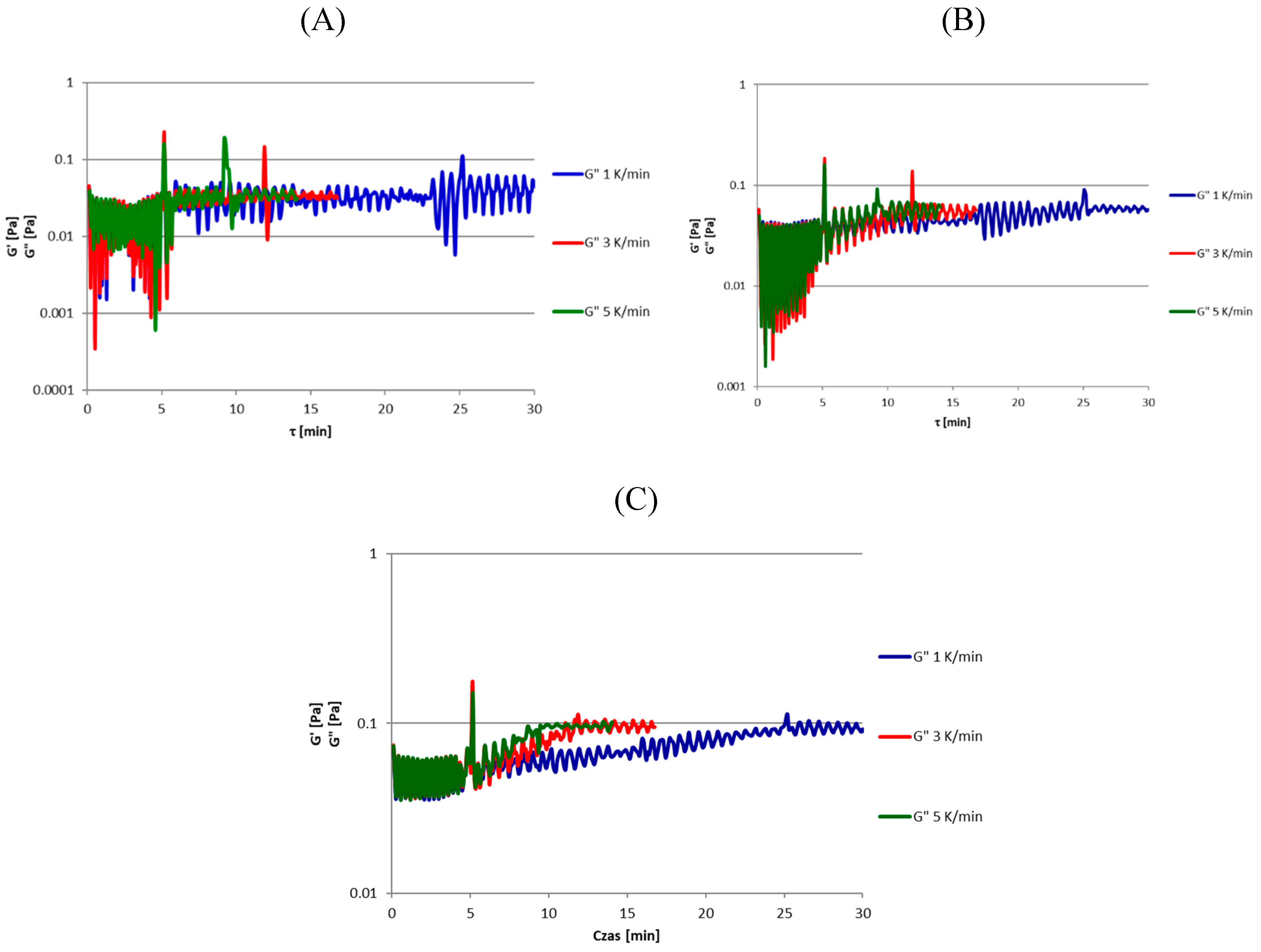
The course of the curves during the experiment for both the cooling rates of 1 K/min, 3 K/min, and 5 K/min modules G′ and G″ of the 12% gelatin solution (Figure 2C) is the most stable—there are no visible fluctuations in the measurement values on the graph, contrary to the graphs for gelatin solutions with concentrations of 4% and 8% (Figure 2A,B).
The curves of the course of the gelatinization process for a gelatin solution with a concentration of 4% are characterized by initial fluctuations in the measurement values, which can be explained by the low apparent viscosity of the solution—0.0024 Pa·s. Fluctuations in the measurement values during the test stabilize with the increase in the viscosity of the solution, i.e., with the decrease in temperature and the appearance of the elastic modulus G′. Gelatin solutions with concentrations of 8% and 12%, which have a higher apparent viscosity, are more stable at the beginning of the experiment. The differences in the viscosity values between the solutions also result in differences in the initial values of the viscosity modulus G″; the gelatin solution with a concentration of 4% has the lowest value of the viscosity modulus, both at the beginning and the end of the experiment, while the values of viscosity modulus G″ for gelatin solutions with concentrations of 8% and 12% reach similar values throughout the gelling process. The different course of the G″ viscosity modulus curves of the two solutions is due to the different initial temperatures of the process; therefore, the 12% solution viscosity modulus curves have a more elongated shape. For the cooling rate of 1 K/min and 5 K/min in the course of the variability of the modulus of elasticity G′ and viscosity G″ of the gelatin solution with a concentration of 8%, there is a decrease in the value of these parameters after exceeding the gel formation temperature of the solution, which may result from the uneven course of the gelation process on the plate and “pouring out” of the solution from between the solidified fragments of the gel. Such a phenomenon does not occur during the experiment at the cooling rate of 3 K/min, which may indicate that the given cooling rate is optimal for the conducted experiment. For gelatin solutions with concentrations of 8% and 12%, there is a relationship between the cooling rate and the values of modulus of elasticity G′ and viscosity G″, although their values are of the same order of magnitude. The highest values of G′ and G″ modules are at the cooling rate of 5 K/min and the lowest are at 3 K/min. There is no such relationship in the gelation plot for a 4% gelatin solution, due to the similar size of the G′ and G″ modules—especially at cooling rates of 1 K/min and 5 K/min. In the sixth minute of the test, both G′ and G″ moduli are nearly identical. Table 3 shows the gelatinization temperatures of the gelatin solutions. For each of the concentrations of gelatin solutions tested in the study, the highest gelation temperature was found at the lowest cooling rate (1 K/min)—21.69 °C, 25.57 °C, and 25.36 °C, respectively, for solutions with concentrations of 4%, 8%, and 12%. This is probably because the solutions were heated for the longest time which could cause some water in the sample to evaporate. This can be confirmed by the lowest gelation temperature obtained at a cooling rate of 5 K/min in the case of 8% and 12% gelatin solutions. The lowest gel formation temperatures were obtained for a gelatin solution with a concentration of 4% and the highest for a solution with a concentration of 8%. The reason that higher gelation temperatures were obtained for the 8% gelatin solution than for the 12% solution may be due to the different starting temperatures of the process. The multivariate analysis of variance with the significance level α = 0.05 showed a statistically significant influence of the cooling rate and gelatin concentration on the gelatinization temperature of the solution. An interaction was also demonstrated between the cooling rate and the hydrocolloid concentration. Based on Tuckey’s test (α = 0.05), six homogeneous groups were distinguished (Table 3). Tosh and Marangoni [27], while examining the gelatinization of gelatin solutions, obtained temperatures of 22.4 °C, 24.5 °C, and 28.4 °C, respectively, for solutions with concentrations of 2.5%, 5%, and 10% of gelatin. The cooling rate at which the experiment was carried out was 0.2 °C/min. They also showed a relationship between the concentration of the gelatin solution and the gelatinization temperature—the higher the concentration of the solution, the higher the temperature of the sol–gel transformation. The results obtained by Tosh and Marangoni [27] at a cooling rate of 0.2 °C/min compared with the results of testing gelatin solutions at a cooling rate of 1 K/min, 3 K/min, and 5 K/min confirm the conclusion about the influence of the cooling rate on the gel formation temperature. Anvari and Chung [40] investigated a complex of 2% fish gelatin solution with gum arabic (1:1), kept for 24 h at 10 °C (A) and 30 °C (B). This study also showed a correlation between the cooling rate and the gelation temperature. For solution A, the results were 3.4 °C, 3.5 °C, 3.9 °C, and 4.7 °C for cooling rates of 0.033 °C/min, 0.025 °C/min, 0.016 °C/min, and 0.012 °C/min. Solution B resulted in 3.3 °C/min, 3.8 °C/min, and 4.7 °C/min, respectively, at rates of 0.025 °C/min, 0.016 °C/min, and 0.012 °C/min. In variant A, gelation was not obtained at the cooling rate of 0.05 °C/min, while in variant B it was at 0.033 °C/min and 0.05 °C/min. This was explained by the fact that at higher cooling rates it is impossible to properly cross-link the structure and the formation of hydrogen bonds enabling the sol-to-gel transition; however, testing the gelatin solution at a cooling rate of 100 times does not confirm the thesis presented by Anvari and Chung [40]. A more likely explanation for the non-gel formation is that the concentration of the gelatin solutions is too low. Van Otterloo and Cruden [41] studied the gelatinization of gelatin solutions (260 Bloom) with concentrations ranging from 1 to 10%. The test was carried out by cooling the gelatin solutions from 50 to 5 °C at a rate of 1.5 K/min. The gel initiation temperature was determined as the point of intersection of the viscosity modulus G″ and the modulus of elasticity G′ and the gel finalization temperature, i.e., the temperature at which the difference in value between the viscosity modulus G″ and the modulus of elasticity G′ does not change despite the temperature decrease. The values of the gelation temperature (gel initiation temperature) that they obtained for the solutions with concentrations of 4%, 8%, and 10% were 26.2 °C, 33.6 °C, and 37.2 °C, respectively—values significantly higher than those obtained in this study. The authors also showed that as the concentration of the gelatin solution increases, the gelatinization temperature of the solution increases, while in the case of finalizing the gelation, no correlation was found between the concentration of the solution and the gelatinization temperature of the solution.

3.2.2. Soy Protein Isolate Solutions

Figure 3 shows the variability of the modulus of elasticity G′ and the modulus of viscosity G″ as a function of time during the examination of the gelation process of soy protein isolate solutions with concentrations of 4%, 8%, and 12% at different cooling rates used in the work.
The soy protein isolate solution did not form a gel in any of the tested concentrations—no elastic modulus G′ was found during the experiment. Comparing the course of the gelation curves for soy protein isolate solutions with concentrations of 4%, 8%, and 12%, it can be seen that as the concentration of the solution increases, the course of the curves is more stabilized and the increase in the viscosity modulus G″ is clearly visible, especially for a solution with a concentration of 12% (Figure 3C). The course of the gelation curves of the 4% soy protein isolate solution (Figure 3A) is practically a straight line with large deviations in the measurement values. The diagram shows no differences in the course of the gelation curves for different cooling rates of the solutions—all of them are practically straight lines. Figure 3B shows the course of the gelling curves of an 8% soy protein isolate solution—it shows that the greatest fluctuations in measurements occur during the initial heating of the solution at a constant temperature. When testing all types of solutions at all cooling rates, characteristic peaks are visible when the test conditions change—at the start and end of the cooling process, it may be a measurement error of the device itself. All G″ viscosity modulus curves have a similar value range. After 5 min of constant temperature heating, the module values are 0.01–0.1 Pa. However, the lower the concentration of the soy protein isolate solution, the less stable the measurements are—possibly because the lower the concentration of the solution, the lower the viscosity of the solution. The lack of stabilization of the values of the tested parameters during the measurements at all tested concentrations of soy protein isolate solutions may be because the soy protein isolate does not form a homogeneous mixture, but a suspension. Even though the solution does not form a gel under the experimental conditions, Galus et al. [42] obtained edible films from the soy protein isolate solution prepared according to the methodology of this work, which indicates a different mechanism of coating formation by the soy protein isolate solution. Liu et al. [36] tested solutions of soy protein isolate with concentrations of 16%, 18%, 20%, and 22% in solutions with 8 molar urea solution and the addition of 1% sodium sulfite based on the weight of the hydrocolloid. The authors obtained a gel from a 22% concentration of soy protein isolate, while solutions with lower concentrations did not gel. The obtained gel was physical and was thermoreversible. The mechanism that occurs in forming the coatings from the soy protein isolate solution at pH = 10 may be coacervation, but it is also likely that when the coatings are dried in the climate chamber, some of the water evaporates, thereby increasing the concentration allowing gel formation to occur. Yuan et al. [43] tested the complex of a 2% soy protein isolate solution with a 2% chitosan solution mixed in various amounts (0.067, 0.125, and 0.2 g of soy protein isolate solution per gram of chitosan solution) using acetic acid or sodium acetate buffer. The tests were carried out at a temperature of 25 ° C. In a study by Yuan et al. [43], no gelation of soy protein isolate solutions was observed either. All types of mixtures tested reached the values of the viscosity modulus G″ higher than those obtained in this work, there was also a modulus G′ with higher values than the viscosity modulus G″. Caillard et al. [44] investigated the gel formation time of soy protein isolate solutions at 25 °C. Soy protein isolate solutions were prepared by mixing with HPLC-grade water at room temperature using a magnetic stirrer. The solution was then adjusted to pH 8 with 1 M NaOH solution and heated at 105 °C for 30 min in a 1:1 water ethylene glycol bath. After returning to room temperature, the pH was adjusted to 8 again. Glutaraldehyde, to a final concentration of 16 mM, 32 mM, or 64 mM, or glyceraldehyde, in such an amount as glyceraldehyde in the solution, was added to 6.5% and 9.5% soy protein isolate solutions. One Mole NaCl solution was added to half of the soy protein isolate solutions with the addition of glyceraldehyde to a final concentration of 120 mM solution. The time at which gel formation occurs was measured to the point where the graph of the elastic modulus G′ intersects the graph of the viscosity modulus G″. During the test, both the viscosity modulus G″ and the modulus of elasticity G′ appear, and the curves have a stable course compared with the graphs obtained in this test. For soy protein isolate solutions with the addition of glutaraldehyde (regardless of the concentration of both soy protein isolate, glutaraldehyde, and NaCl), it was less than 2 min, while for soy protein isolate solutions with both 6.5% and 9.5% concentrations with the addition of aldehyde glycerol was 110 ± 14 min and no statistically significant difference was found depending on the concentration of soy protein isolate and glyceraldehyde. The results obtained by Caillard et al. [44] indicate that the type of aldehyde added to the soy protein isolate had the most important influence.

3.2.3. High Methylated Apple Pectin Solution

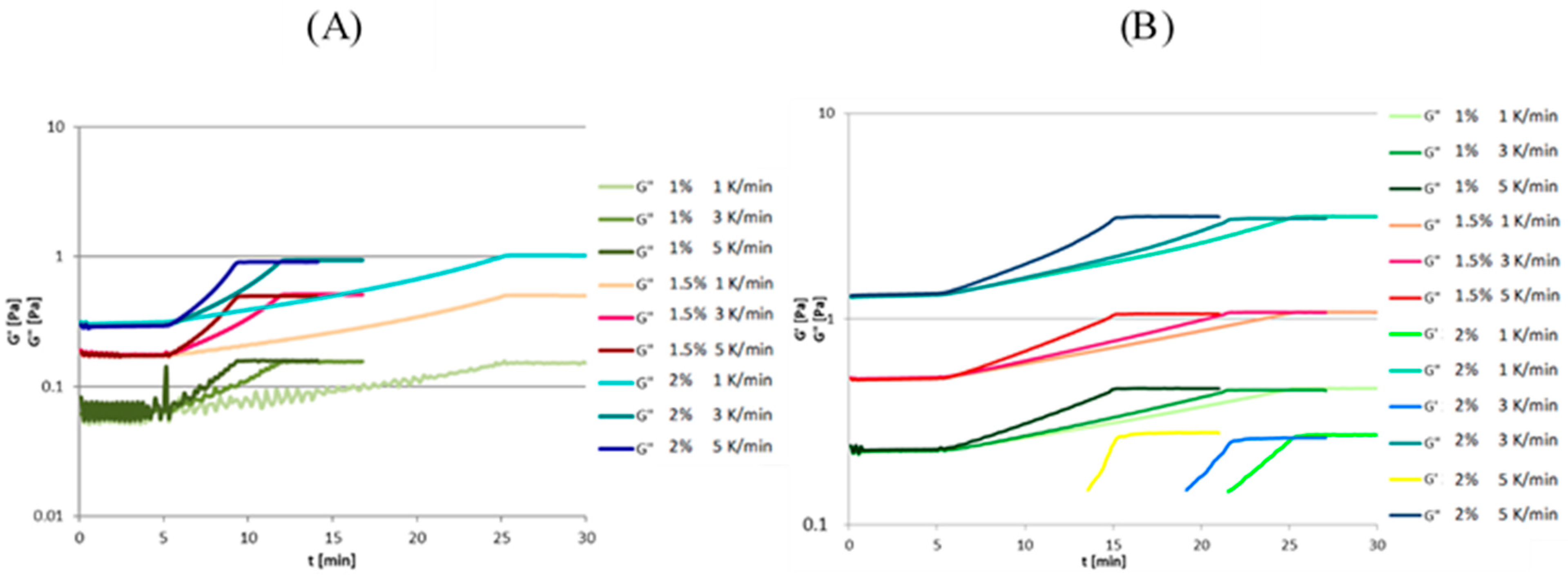
Figure 4A shows the variability of the modulus of elasticity G′ and the modulus of viscosity G″ as a function of time during the examination of the gelation process of high methylated apple pectin solutions with concentrations of 1%–1.5% at different cooling rates (1 K/min, 3 K/min, and 5 K/min).
None of the high-methylated apple pectin solutions formed a gel—the solutions do not have the characteristics of viscoelastic fluids, and thus they do not have the elastic modulus G′. The higher the concentration of the high-methylated apple pectin solution, the faster the solution becomes stable upon initial heating to a constant temperature. Fluctuations in the values of the measured parameters during the gelling process of the 1% high-methylated apple pectin solution did not stabilize. This can be explained by the low apparent viscosity of a 1% high-methylated apple pectin solution, which is 0.0042 ± 0.0001 Pa·s at 50 °C (Table 2); therefore, the measurements stabilize faster with increasing pectin concentration. Likewise, the higher the concentration of the solution, the higher the values of the viscosity modulus G″. The values of the modulus of viscosity G″ and elasticity G′ increase with increasing concentration of the pectin solution, but the values are of the same order of magnitude. The initial and final values of the viscosity modulus G″ between the individual cooling rates (within the solutions of the same concentration) do not differ. Additionally, regardless of the concentration of the solution, the curves tested at the same cooling rates take the same shape. The lack of gelling of highly-methylated pectin solutions results from the conditions that must be met for the high-methylated pectin to form gels. High methylated pectin solution should have a sugar concentration above 55% and a pH below 3.5 to form gels [45]. Kastner et al. [46] investigated the effect of the cooling rate of solutions of non-standardized highly methylated lemon pectin at a concentration of 0.27% with sucrose (60%) on the formation of the gel structure. High-methylated lemon pectin solutions were cooled from 105 °C to 20 °C at 5 cooling rates—0.25 K/min, 0.50 K/min, 0.75 K/min, 1.00 K/min, and 2.00 K/min. The authors showed a slight increase in the gelation temperature with a decrease in the cooling rate, however, the difference in the gelation temperature between the individual cooling rates is not statistically significant. High methylated citrus pectin solutions with the addition of sucrose meet the conditions that must exist for the gel solutions. Additionally, it is worth noting that the authors did not show a statistically significant effect on the cooling rate of the solutions, which was demonstrated in this study for gelatin solutions.

3.2.4. Sodium Alginate Solution

Figure 4B shows the variability of the modulus of elasticity G′ and the modulus of viscosity G″ as a function of time during the examination of the gelation process of sodium alginate solutions with concentrations of 1%, 1.5%, and 2% at different cooling rates (1 K/min, 3 K/min, and 5 K/min). The diagram shows that as the concentration increases, the values of the viscosity modulus G″ increase. Both the initial and final values of the viscosity modulus G″ at the tested cooling rates are almost identical. The same trend is apparent for all sodium alginate solutions, regardless of concentration, which illustrates that the cooling rate does not affect the values of the viscosity modulus G″. The gelation curves of sodium alginate solutions with the concentration of 1%, 1.5%, and 2% show the linear increase in the value of the viscosity modulus G″ during the cooling of the solutions, regardless of the cooling rate of the sodium alginate solutions and their concentration. In the case of a 1% sodium alginate solution at the beginning of the test, fluctuations in the measurements of the values of the parameters tested are visible, which stabilize in the first minute of heating at a constant temperature. When a sodium alginate solution with a concentration of 2% was tested, the elastic modulus G′ appeared and regardless of the cooling rate of the sodium alginate solution, it had the same minimum value (approx. 0.15 Pa) and maximum (approx. 0.28 Pa). Despite the appearance of the elastic modulus G′ when testing a 2% sodium alginate solution, it would not be possible to intersect the curve of the elastic modulus G′ and the viscosity modulus G″ because the negative temperature would cause the solution to freeze. The lack of gelling of sodium alginate solutions results from the conditions under which sodium alginate gels—in the presence of calcium ions in the solution or at a pH below 4 [45]. Even though sodium alginate does not gel under the tested conditions, it forms edible films [47]. Goudoulas and Germann [48] studied the gelling process of mixtures of sodium alginate and gelatin solutions. First, gelatin solutions with concentrations of 5% and 10% with the addition of sodium azide (0.1%) were prepared, and then sodium alginate solutions with concentrations of 2%, 5%, and 10% were prepared. Gelatin and sodium alginate solutions were mixed to obtain solutions: gelatin with a concentration of 2.5% and 5% without the addition of sodium alginate, gelatin (5%) and sodium alginate (1%, 2.5%, and 5%), and gelatin (2.5%) and alginate (1 or 2.5%). The solutions were cooled from 45 to 5 °C at a cooling rate of 1 °C/min. It has been shown that the higher the sodium alginate concentration, the higher the gel formation temperature. In the case of all types of solutions, it was possible to determine the gelatinization temperatures of the solutions, but the gels would not be formed if there was no gelatin in the tested solutions. Yang et al. [49] studied low and medium-viscosity sodium alginate solutions with a concentration of 1% with and without the addition of 4 mmol/mL CaCl2. To monitor the gelling process, the solutions were tested at 100 °C and observed under a microscope. The time after which the solutions gelled was determined based on the intersection of the change curves of the elastic modulus G′ and the viscosity modulus G″. Both the solutions with and without the addition of CaCl2 showed gelling properties. The difference between the solutions with and without the addition of CaCl2 was the different times needed to obtain a gel. Both low and medium-viscosity solutions of sodium alginate with the addition of CaCl2 gelled in the second minute of the test. In contrast, sodium alginate solutions were without the addition of CaCl2 in the fourth minute of the experiment, regardless of whether the sodium alginate had a low or medium viscosity. Perhaps the reason why the sodium alginate gelled under the given conditions was due to the high temperature at which the test was carried out.

4. Conclusions

The value of the analyzed rheological parameters is influenced by both the concentration of the hydrocolloid in the solution and its type. The apparent viscosity of all solutions increases with increasing concentration. Only in the case of gelatin solutions was it possible to determine the gel formation temperature. It was shown that the cooling rate and the concentration of the solution had a statistically significant effect on the gelation temperature. As the concentration of the solution increases and the cooling rate increases, the gelation temperature decreases. Although the viscosity values increase with increasing concentration, the lowest values of the G″ viscosity modulus were obtained at a sodium alginate solution concentration of 1.5%. Even though solutions with different concentrations of soy protein isolate (4%–12%) and sodium alginate and highly methylated pectin (1%–1.5%) were used in the experiment, similar tendencies of the solutions can be noticed when testing these solutions. The modulus of elasticity curves only appears at the highest concentrations. This means that the formation of coatings occurs by different mechanisms in the case of soy protein isolate solutions as well as sodium alginate and high methylated pectin.

Author Contributions

Conceptualization, M.J. and I.S.; methodology, M.J. and I.S.; software, M.J.; validation, M.J., I.S., A.C. and S.G.; formal analysis, M.J. and I.S.; investigation, M.J. and I.S.; resources, M.J.; data curation, M.J.; writing—original draft preparation, M.J.; writing—review and editing, M.J., I.S., A.C. and S.G.; visualization, M.J.; supervision, M.J. and S.G.; project administration, M.J.; funding acquisition, M.J. All authors have read and agreed to the published version of the manuscript.

Funding

The work was co-financed by a statutory activity subsidy from the Polish Ministry of Science and Higher Education for the Faculty of Food Sciences of Warsaw University of Life Sciences.

Institutional Review Board Statement

Not applicable.

Informed Consent Statement

Not applicable.

Data Availability Statement

Not applicable.

Acknowledgments

The authors would like to thank Julia Jońska for her help in conducting the research.

Conflicts of Interest

The authors declare no conflict of interest.

References

  1. Asgher, M.; Qamar, S.A.; Bilal, M.; Iqbal, H.M.N. Bio-based active food packaging materials: Sustainable alternative to conventional petrochemical-based packaging materials. Food Res. Int. 2020, 137, 109625. [Google Scholar] [CrossRef] [PubMed]
  2. Panahirad, S.; Dadpour, M.; Peighambardoust, S.H.; Soltanzadeh, M.; Gullón, B.; Alirezalu, K.; Lorenzo, J.M. Applications of carboxymethyl cellulose- and pectin-based active edible coatings in preservation of fruits and vegetables: A review. Trends Food Sci. Technol. 2021, 110, 663–673. [Google Scholar] [CrossRef]
  3. Kowalska, H.; Marzec, A.; Domian, E.; Kowalska, J.; Ciurzynska, A.; Galus, S. Edible coatings as osmotic dehydration pretreatment in nutrient-enhanced fruit or vegetable snacks development: A review. Compr. Rev. Food Sci. Food Saf. 2021, 20, 5641–5674. [Google Scholar] [CrossRef] [PubMed]
  4. Mohamed, S.A.A.; El-Sakhawy, M.; El-Sakhawy, M.A. Polysaccharides, Protein and Lipid -Based Natural Edible Films in Food Packaging: A Review. Carbohydr. Polym. 2020, 238, 116178. [Google Scholar] [CrossRef]
  5. Rangaraj, V.M.; Rambabu, K.; Banat, F.; Mittal, V. Natural antioxidants-based edible active food packaging: An overview of current advancements. Food Biosci. 2021, 43, 101251. [Google Scholar] [CrossRef]
  6. Trajkovska Petkoska, A.; Daniloski, D.; D’Cunha, N.M.; Naumovski, N.; Broach, A.T. Edible packaging: Sustainable solutions and novel trends in food packaging. Food Res. Int. 2021, 140, 109981. [Google Scholar] [CrossRef] [PubMed]
  7. Galus, S.; Lenart, A. Effect of protein concentration on kinetics of water vapour adsorption by coatings prepared on the basis of whey protein isolate. Food Sci. Technol. Qual. 2011, 4, 66–73. [Google Scholar] [CrossRef]
  8. Mikus, M.; Galus, S. Powlekanie żywności–materiały, metody i zastosowanie w przemyśle spożywczym. Zywnosc Nauka Technol. Jakosc/Food Sci. Technol. Qual. 2020, 125, 5–24. [Google Scholar] [CrossRef]
  9. Jeya Jeevahan, J.; Chandrasekaran, M.; Venkatesan, S.P.; Sriram, V.; Britto Joseph, G.; Mageshwaran, G.; Durairaj, R.B. Scaling up difficulties and commercial aspects of edible films for food packaging: A review. Trends Food Sci. Technol. 2020, 100, 210–222. [Google Scholar] [CrossRef]
  10. Galus, S.; Mikus, M.; Ciurzyńska, A.; Domian, E.; Kowalska, J.; Marzec, A.; Kowalska, H. The Effect of Whey Protein-Based Edible Coatings Incorporated with Lemon and Lemongrass Essential Oils on the Quality Attributes of Fresh-Cut Pears during Storage. Coatings 2021, 11, 745. [Google Scholar] [CrossRef]
  11. Galus, S.; Lenart, A. Optical, mechanical, and moisture sorption properties of whey protein edible films. J. Food Process Eng. 2019, 42, e13245. [Google Scholar] [CrossRef]
  12. Maringgal, B.; Hashim, N.; Tawakkal, I.S.M.A.; Mohamed, M.T.M. Recent advance in edible coating and its effect on fresh/fresh-cut fruits quality. Trends Food Sci. Technol. 2020, 96, 253–267. [Google Scholar] [CrossRef]
  13. Liu, C.; Huang, J.; Zheng, X.; Liu, S.; Lu, K.; Tang, K.; Liu, J. Heat sealable soluble soybean polysaccharide/gelatin blend edible films for food packaging applications. Food Packag. Shelf Life 2020, 24, 100485. [Google Scholar] [CrossRef]
  14. Garcia-Henao, C.E.; Valderrama-Sanchez, V.; Arboleda-Murillo, J.A.; Pinzon, M.I.; Sanchez, L.T.; Villa, C.C. Bioactive food coating: A review. Packag. Technol. Sci. 2022. [Google Scholar] [CrossRef]
  15. Lim, J.W.; Lim, W.S.; Lee, M.H.; Park, H.J. Barrier and structural properties of polyethylene terephthalate film coated with poly(acrylic acid)/montmorillonite nanocomposites. Packag. Technol. Sci. 2021, 34, 141–150. [Google Scholar] [CrossRef]
  16. Mikus, M.; Galus, S.; Ciurzynska, A.; Janowicz, M. Development and Characterization of Novel Composite Films Based on Soy Protein Isolate and Oilseed Flours. Molecules 2021, 26, 3738. [Google Scholar] [CrossRef] [PubMed]
  17. Diaz-Montes, E.; Castro-Munoz, R. Edible Films and Coatings as Food-Quality Preservers: An Overview. Foods 2021, 10, 249. [Google Scholar] [CrossRef]
  18. Chen, H.; Wang, J.; Cheng, Y.; Wang, C.; Liu, H.; Bian, H.; Pan, Y.; Sun, J.; Han, W. Application of Protein-Based Films and Coatings for Food Packaging: A Review. Polymers 2019, 11, 2039. [Google Scholar] [CrossRef] [Green Version]
  19. Sun, X.; Wang, J.; Dong, M.; Zhang, H.; Li, L.; Wang, L. Food spoilage, bioactive food fresh-keeping films and functional edible coatings: Research status, existing problems and development trend. Trends Food Sci. Technol. 2022, 119, 122–132. [Google Scholar] [CrossRef]
  20. Zambrano-Zaragoza, M.L.; Quintanar-Guerrero, D. Novel Techniques for Extrusion, Agglomeration, Encapsulation, Gelation, and Coating of Foods. In Encyclopedia of Food Security and Sustainability; Ferranti, P., Berry, E.M., Anderson, J.R., Eds.; Elsevier: Oxford, UK, 2019; pp. 379–392. [Google Scholar]
  21. Kadzińska, J.; Janowicz, M.; Kalisz, S.; Bryś, J.; Lenart, A. An overview of fruit and vegetable edible packaging materials. Packag. Technol. Sci. 2019, 32, 483–495. [Google Scholar] [CrossRef]
  22. Khalid, M.Y.; Arif, Z.U. Novel biopolymer-based sustainable composites for food packaging applications: A narrative review. Food Packag. Shelf Life 2022, 33, 100892. [Google Scholar] [CrossRef]
  23. Pinto, L.; Bonifacio, M.A.; De Giglio, E.; Santovito, E.; Cometa, S.; Bevilacqua, A.; Baruzzi, F. Biopolymer hybrid materials: Development, characterization, and food packaging applications. Food Packag. Shelf Life 2021, 28, 100676. [Google Scholar] [CrossRef]
  24. Suhag, R.; Kumar, N.; Petkoska, A.T.; Upadhyay, A. Film formation and deposition methods of edible coating on food products: A review. Food Res. Int. 2020, 136, 109582. [Google Scholar] [CrossRef] [PubMed]
  25. Taktak, W.; Hamdi, M.; Chentir, I.; Boughriba, S.; Ben Azaza, Y.; Li, S.; Nasri, M.; Karra-Chaâbouni, M.; Nasri, R. Development of emulsion gelatin gels for food application: Physicochemical, rheological, structural and thermal characterization. Int. J. Biol. Macromol. 2021, 182, 1–10. [Google Scholar] [CrossRef]
  26. Winter, H.H. Can the gel point of a cross-linking polymer be detected by the G′–G″ crossover? Polym. Eng. Sci. 1987, 27, 1698–1702. [Google Scholar] [CrossRef]
  27. Tosh, S.M.; Marangoni, A.G. Determination of the maximum gelation temperature in gelatin gels. Appl. Phys. Lett. 2004, 84, 4242–4244. [Google Scholar] [CrossRef]
  28. Saha, D.; Bhattacharya, S. Hydrocolloids as thickening and gelling agents in food: A critical review. J. Food Sci. Technol. 2010, 47, 587–597. [Google Scholar] [CrossRef] [Green Version]
  29. Harkous, A.; Colomines, G.; Leroy, E.; Mousseau, P.; Deterre, R. The kinetic behavior of Liquid Silicone Rubber: A comparison between thermal and rheological approaches based on gel point determination. React. Funct. Polym. 2016, 101, 20–27. [Google Scholar] [CrossRef]
  30. Huang, T.; Tu, Z.-C.; Wang, H.; Shangguan, X.; Zhang, L.; Zhang, N.-H.; Bansal, N. Pectin and enzyme complex modified fish scales gelatin: Rheological behavior, gel properties and nanostructure. Carbohydr. Polym. 2017, 156, 294–302. [Google Scholar] [CrossRef] [Green Version]
  31. Wang, Y.; Wang, J.; Sun, Q.; Xu, X.; Li, M.; Xie, F. Hydroxypropyl methylcellulose hydrocolloid systems: Effect of hydroxypropy group content on the phase structure, rheological properties and film characteristics. Food Chem. 2022, 379, 132075. [Google Scholar] [CrossRef]
  32. Pałacha, Z.; Sitkiewicz, I. Physical Properties of Food; WNT: Warsaw, Poland, 2010; p. 340. [Google Scholar]
  33. Duan, R.; Zhang, J.; Liu, L.; Cui, W.; Regenstein, J.M. The functional properties and application of gelatin derived from the skin of channel catfish (Ictalurus punctatus). Food Chem. 2018, 239, 464–469. [Google Scholar] [CrossRef] [PubMed]
  34. Marcotte, M.; Taherian Hoshahili, A.R.; Ramaswamy, H.S. Rheological properties of selected hydrocolloids as a function of concentration and temperature. Food Res. Int. 2001, 34, 695–703. [Google Scholar] [CrossRef]
  35. Wang, L.Z.; Liu, L.; Holmes, J.; Kerry, J.F.; Kerry, J.P. Assessment of film-forming potential and properties of protein and polysaccharide-based biopolymer films. Int. J. Food Sci. Technol. 2007, 42, 1128–1138. [Google Scholar] [CrossRef]
  36. Liu, P.; Xu, H.; Zhao, Y.; Yang, Y. Rheological properties of soy protein isolate solution for fibers and films. Food Hydrocoll. 2017, 64, 149–156. [Google Scholar] [CrossRef] [Green Version]
  37. Comaposada, J.; Gou, P.; Marcos, B.; Arnau, J. Physical properties of sodium alginate solutions and edible wet calcium alginate coatings. LWT-Food Sci. Technol. 2015, 64, 212–219. [Google Scholar] [CrossRef]
  38. Cevoli, C.; Balestra, F.; Ragni, L.; Fabbri, A. Rheological characterisation of selected food hydrocolloids by traditional and simplified techniques. Food Hydrocoll. 2013, 33, 142–150. [Google Scholar] [CrossRef]
  39. Huang, X.; Li, D.; Wang, L.-J. Characterization of pectin extracted from sugar beet pulp under different drying conditions. J. Food Eng. 2017, 211, 1–6. [Google Scholar] [CrossRef]
  40. Anvari, M.; Chung, D. Effect of cooling–heating rate on sol-gel transformation of fish gelatin–gum arabic complex coacervate phase. Int. J. Biol. Macromol. 2016, 91, 450–456. [Google Scholar] [CrossRef]
  41. van Otterloo, J.; Cruden, A.R. Rheology of pig skin gelatine: Defining the elastic domain and its thermal and mechanical properties for geological analogue experiment applications. Tectonophysics 2016, 683, 86–97. [Google Scholar] [CrossRef]
  42. Galus, S.; Lenart, A.; Voilley, A.; Debeaufort, F. Effect of oxidized potato starch on the physicochemical properties of soy protein isolate-based edible films. Food Technol. Biotechnol. 2013, 51, 403–409. [Google Scholar]
  43. Yuan, Y.; Kong, Z.-Y.; Sun, Y.-E.; Zeng, Q.-Z.; Yang, X.-Q. Complex coacervation of soy protein with chitosan: Constructing antioxidant microcapsule for algal oil delivery. LWT 2017, 75, 171–179. [Google Scholar] [CrossRef]
  44. Caillard, R.; Remondetto, G.E.; Subirade, M. Rheological investigation of soy protein hydrogels induced by Maillard-type reaction. Food Hydrocoll. 2010, 24, 81–87. [Google Scholar] [CrossRef]
  45. Rutkowski, A.G.S.; Dabrowski, K. Compendium of Food Additives; Hortimex: Konin, Poland, 2003; pp. 362–363. [Google Scholar]
  46. Kastner, H.; Kern, K.; Wilde, R.; Berthold, A.; Einhorn-Stoll, U.; Drusch, S. Structure formation in sugar containing pectin gels–Influence of tartaric acid content (pH) and cooling rate on the gelation of high-methoxylated pectin. Food Chem. 2014, 144, 44–49. [Google Scholar] [CrossRef] [PubMed] [Green Version]
  47. Liu, S.; Li, Y.; Li, L. Enhanced stability and mechanical strength of sodium alginate composite films. Carbohydr. Polym. 2017, 160, 62–70. [Google Scholar] [CrossRef]
  48. Goudoulas, T.B.; Germann, N. Phase transition kinetics and rheology of gelatin-alginate mixtures. Food Hydrocoll. 2017, 66, 49–60. [Google Scholar] [CrossRef]
  49. Yang, Y.; Campanella, O.H.; Hamaker, B.R.; Zhang, G.; Gu, Z. Rheological investigation of alginate chain interactions induced by concentrating calcium cations. Food Hydrocoll. 2013, 30, 26–32. [Google Scholar] [CrossRef]
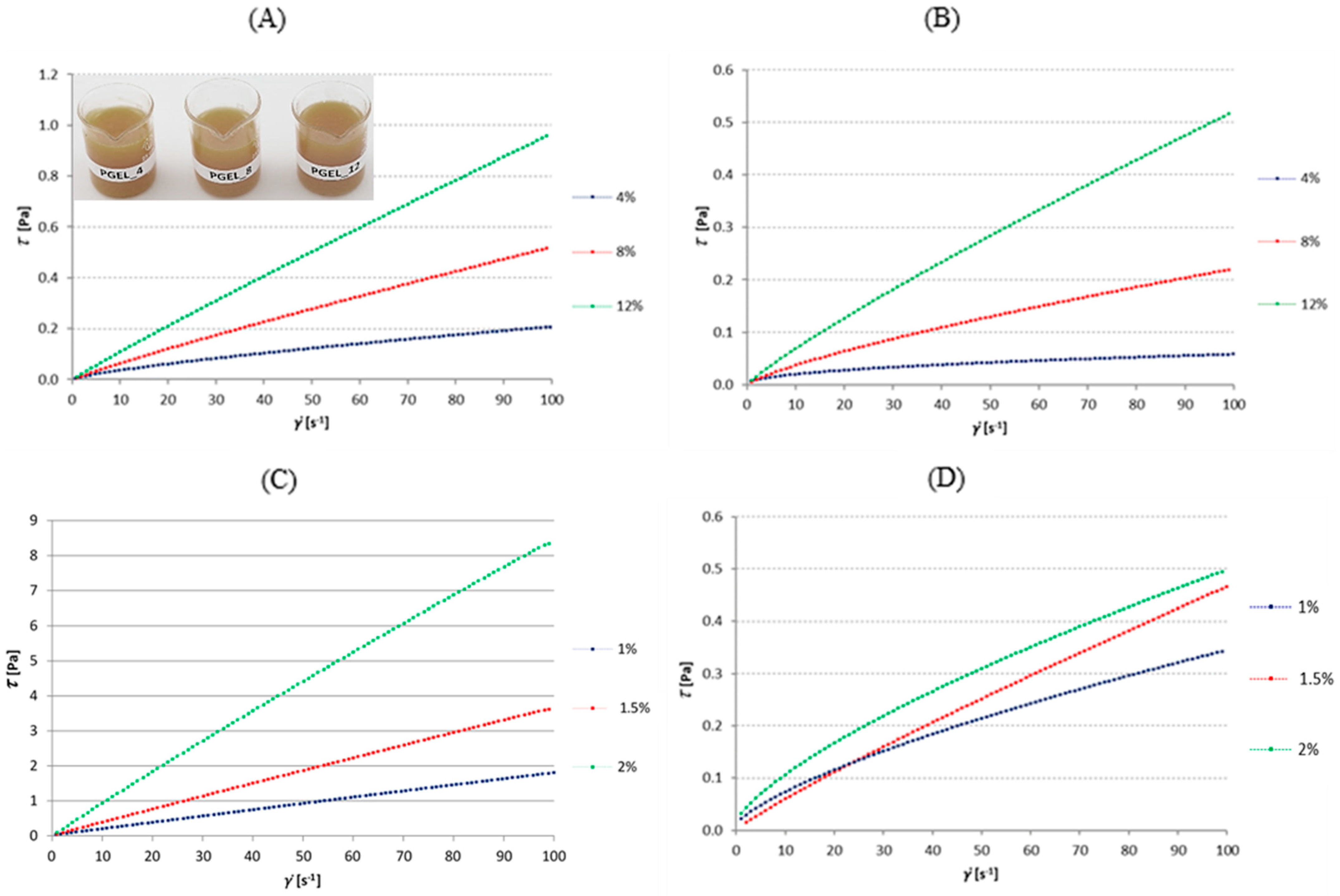
Figure 1. Flow curves of tested hydrocolloids solutions of various concentrations: (A)—gelatin, (B)—soy protein isolate, (C)—sodium alginate, and (D)—high-methylated apple pectin. The figure also includes a photo of the tested gelatin solutions (PGEL_4, 8, 12).
Coatings 12 01704 g001
Figure 2. Gelatinization curves of a gelatin solution with different concentrations and cooling rates. Gelatin concentration: (A)—4%, (B)—8%, and (C)—12%.
Coatings 12 01704 g002
Figure 3. Gelatinization curves of a soy protein isolate solution with different concentrations and cooling rates. Soy protein isolate concentration: (A)—4%, (B)—8%, and (C)—12%.
Coatings 12 01704 g003
Figure 4. Gelatinization curves of high-methylated apple pectin (A) and sodium alginate (B) with different concentrations and cooling rates.
Coatings 12 01704 g004
Table 1. Raw material composition and temperature of the subjects of film-forming solutions.
Type of HydrocolloidConcentration (%)Glycerol (%)Initial Temperature (°C)Final Temperature (°C)
Gelatin4502210
8503010
12504010
Soy protein isolate450200
850200
1250200
Sodium alginate150200
1.550200
250200
High-methylated apple pectin150250
1.550250
250250
Table 2. Rheological parameters and results of correlation analysis of tested solutions of selected hydrocolloids.
Gelatin
Concentration (%)knR2Apparent Viscosity (Pa·s)
4 0.9990.0024 ± 0.0001
8 0.9990.0055 ± 0.0005
12 0.9930.0100 ± 0.0011
The correlation coefficient between the concentration of gelatin solutions and the apparent viscosity of the subject solutions
0.964901
Soy Protein Isolate
Concentration (%)knR2Apparent Viscosity (Pa·s)
40.0071290.64960.98320.0018 ± 0.0000
80.0064490.76610.99150.0026 ± 0.0000
120.0092330.87370.99850.0056 ± 0.0000
The correlation coefficient between the concentration of soy protein isolate solutions and the apparent viscosity of the subject solutions
0.986394
Sodium Alginate
Concentration (%)knR2Apparent Viscosity (Pa·s)
1 0.9990.0187 ± 0.0001
1.5 0.9990.0374 ± 0.0002
2 0.9990.0884 ± 0.0002
The correlation coefficient between the concentration of sodium alginate solutions and the apparent viscosity of the subject solutions
0.960769
High-Methylated Apple Pectin
Concentration (%)knR2Apparent Viscosity (Pa·s)
10.007760.6970.9970.0042 ± 0.0001
1.50.013850.88910.0050 ± 0.0000
20.020060.6960.9970.0061 ± 0.0001
The correlation coefficient between the concentration of high-methylated apple pectin solutions and the apparent viscosity of the subject solutions
0.948683
Table 3. Gelation temperature of gelatin at a cooling rate of 1 K/min, 3 K/min, and 5 K/min and interaction coefficients between the cooling rate and the hydrocolloid concentration.
Concentration (%)Cooling Rate (K/min)Gelation Temperature (°C)
4121.69 ± 0.18 a
319.74 ± 0.30 e
520.74 ± 0.12 f
8125.57 ± 0.23 d
323.21 ± 0.31 c
522.40 ± 0.14 b
12125.36 ± 0.05 d
323.00 ± 0.26 c
522.26 ± 0.02 ab
Statistical analysis of the effect of concentration and cooling rate on the gelatinization temperature of gelatin solutions
SSDegrees of FreedomMSFp-Value
Cooling rate (K/min)32.32216.16395.70.0·10−6
Concentration (%)51.01225.51624.50.0·10−6
Cooling rate * Concentration5.3841.3532.90.0·10−6
* Mean values with standard deviations in brackets. Different superscript letters (a–f) within the same columns indicate significant differences between the samples (p < 0.05).
Publisher’s Note: MDPI stays neutral with regard to jurisdictional claims in published maps and institutional affiliations.