Abstract
Three-dimensional laser scanning technology can be used to quickly, efficiently, and accurately obtain spatial three-dimensional information of cultural relics without contacting the target during the scanning process. The results of this study showed that the extraction of human bones from the Shenna ruins via the auxiliary application of three-dimensional scanning technology reduced human intervention and destruction on the site compared with the traditional archaeological human bone packaging and extraction work method. When combined with the application of three-dimensional scanning technology, the original data information extracted on the spot were more comprehensive and accurate. Additionally, the technology provided us with important scientific data which can be used to discuss the phylogenetic composition of the ancient Qiang people in the settlement village, as well as a new applications of ideas for three-dimensional laser scanning technology usage in the field extraction of cultural relics. However, a follow-up study is needed to improve the comparisons of its applications, providing a conventional auxiliary means for cultural relic extraction and a technical means for cultural relic protection evaluation.
1. Introduction
Bone cultural relics, an important type of cultural relic unearthed in archaeological excavations, have been valuable in the disciplines of archeology, physical human osteology, and paleopathology [1,2,3]. The historical information that they provide is used to reveal and explore the behavior, production activities, and production relations of ancient humans, which increases the accuracy of reconstructions of the system, politics, economy, and life at that time. The Shenna ruins (Figure 1) cover a total area of about 100,000 square meters and are located in the north of Xiaoqiao Village, Chengbei District, Xining City, Qinghai Province. In 2006, the State Council announced them as the sixth batch of national key cultural relic protection units. Many types of artifacts have been unearthed at the site. Among them, the spine, chest, and pelvis bones of two humans were unearthed. However, they were seriously decayed and their preservation status was poor, so protective restoration measures needed to be taken as soon as possible [4]. In addition, the reconstruction of the conservation hall requires the temporary backfilling of the archaeological site. On this basis, information about the rescue extraction and the protection of the original site of the human bone cultural relics are required.
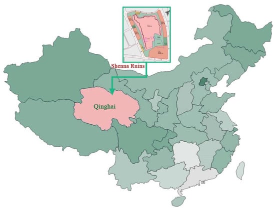
Figure 1.
Location map of Shenna ruins, China.
Studies on the digital techniques [5] of paleontological measurements and records are increasingly emerging. As the most commonly used surface imaging technique, photogrammetry [6] has been developed and innovated upon since the early 19th century and is still considered to be comparable to manual measurements in terms of accuracy. Methods used in recent years, which include a variety of digital technologies, combined with the application of archaeological data preservation, provide effective references. Some scholars have used close-range photogrammetry [7] and high-precision ground control survey technology [8] to record the scene of dinosaur footprint data, and some scholars have conducted studies with portable devices, which enable the use of rapid clinical computed tomography (CT) [9] and provide archaeological survey results using 3D scanning technologies [10]. With the advancement of digital technology, the study of paleontology and the measurement and recording of various relics is becoming increasingly rich.
Three-dimensional laser scanning technology [11], also called real-scene reproduction technology, caused a technological revolution in the fields of surveying and mapping following global positioning system (GPS) technology. It broke through the traditional single-point measurement method and has the unique advantages of high efficiency and high precision. Three-dimensional laser scanning technology can provide three-dimensional point cloud data of the scanned object surface, and so it can be used to obtain high-precision and high-resolution 3D digital models of cultural relics [12]. As such, in this study, we used 3D technology to assist with the site extraction of the human bones unearthed from the Shenna site and to help protect the original site.
The digital information obtained by 3D technology can be used to establish archaeological archives of cultural relics; it can also be used to combine the traditional biometric techniques to conduct biological archaeological research on human bones [13,14,15,16]. More importantly, with accurate digital point cloud data, these digital information data can be used to establish the unearthed location coordinates of cultural relics, which is convenient for the restoration and display of the original site at later times, and the real location of the cultural relics can be restored and displayed at the excavation site. Additionally, the quantitative comparison of the point cloud data, excluding the comparison of the current picture, is supplemented with the original comparison data before restoration, facilitating a detailed comparison of bone cultural relics before and after restoration and refined restoration guidance [17,18,19].
2. Equipment and Methods
2.1. Equipment
For this research, we used a 3D laser scanner (model: HandySCAN700, accuracy 0.03 mm, the Intel Core I7, 3820QM, 16G or higher RAM and an NVIDIA Quadro K2000M graphics card. Software VXelements.), tripods, full-frame digital cameras, lighting assistance systems, trimble realworks, Windows operating system, etc.
2.2. Methods
As shown in Figure 2, we used 3D scan technology to assist with site surveys of the cultural relics to obtain more digitized cultural relic information, which we used to ensure site safety, obtain refined information collection, and extract the human bone cultural relics unearthed at the Shenna ruins.
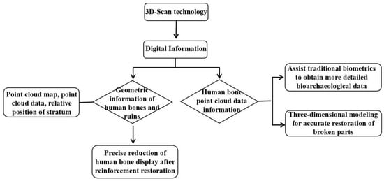
Figure 2.
Introduction to the application of 3D scan technology.
2.3. Collection and Recording of Current Information
In field operations, data collection parameters are easily affected by the excavation zone, unearthed location situation, light, and collection speed. So, the operation generally needs to be designed according to the actual situation. The 3D laser scanner that we selected for this research was a Trimble TX8, with a 1,000,000 dots/s scanning speed, a 360° × 317° field angle, and a 2 mm 120 m range measurement accuracy, which could provide fast and accurate spatial data for the experimental subjects.
2.3.1. Point Cloud Data Acquisition
Data acquisition is the basis of data postprocessing, and the acquisition of high-quality point cloud data, as required, can reduce the data processing workload and increase the accuracy of the reconstruction of the 3D model of cultural relics.
(1) Deploy the target
As shown in Figure 3, at two different scanning sites, at least 3 common targets must be present. To ensure the accuracy of the point cloud during splicing, the targets cannot be in a straight line.
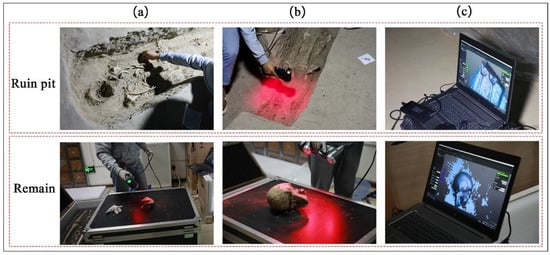
Figure 3.
The 3D scan process of human bone ((a) deploying the target; (b) point cloud data acquisition; (c) point cloud data processing).
(2) When scanning the same target, each public target should be kept in sight with multiple measuring stations as much as possible to reduce the number of target point movements
(3) Coordinate system selection
Since the coordinate system of the point cloud data obtained in each scan is different, splicing errors occur when the point cloud data are merged, which affect the overall point cloud data accuracy. Therefore, the reference coordinate system must be selected before scanning.
In addition, since three-dimensional laser scanning technology uses the photoelectric distance measurement principle, environmental humidity has an impact on the accuracy of the point cloud data obtained by scanning in field operations. Therefore, a time period where the environment and temperature and humidity are stable should be selected for data collection.
2.3.2. Point Cloud Data Processing
After using 3D scanning to obtain the point cloud data of the measured object, data post-processing is required to obtain a three-dimensional model of the measured object, which includes point cloud data aligning and denoising, three-dimensional model reconstruction, and data comparison.
Aligning and denoising of the point cloud data belong to the data preprocessing stage. It provides accurate and reliable point cloud models for subsequent three-dimensional model reconstruction and texture mapping, speeds up and reduces the difficulty of three-dimensional model reconstruction, and increases the accuracy of the three-dimensional model after reconstruction.
The specific process of the entire data processing work is as follows:
(1) Point cloud alignment
In actual scanning operations, unearthed human bones are located in the burial pits of ruins, and obtaining complete spatial information with only one measuring station is often impossible. Therefore, multimeasuring station scanning must be performed. Through point cloud splicing, the point cloud data of different measuring stations are spliced into complete entity point cloud data before they can be applied to the reconstruction of a human bone model.
(2) Point cloud denoising
Point cloud denoising involves removing redundant data that do not belong to the scanned object from the initial data. In the spliced three-dimensional point cloud data, the acquired point cloud data and image data are preprocessed, the point cloud data unrelated to the measured object are removed, and the fault and error points in the initial point cloud are eliminated. This results in the point cloud data being more streamlined and effective, saves computer storage space, and increases the processing speed of the subsequent data.
(3) Three-dimensional model reconstruction
The point cloud data model scanned by a three-dimensional laser scanner are composed of discrete points in space. The scanning interval of the scanner results in gaps between these discrete points, which do not constitute the real object surface. Therefore, the denoised point cloud data are encapsulated through some special algorithm models to obtain the topological surface reconstruction of the mesh.
(4) Point cloud data processing and comparison
The digital three-dimensional model of human bone cultural relics constructed with three-dimensional laser scanning technology has high accuracy [20], and it can accurately reflect the preservation status of the cultural relics. Through the 3D comparison function of Geomagic Studio software, a colored 3D model reflecting the error between the two models is generated, which can provide the maximum, average, and standard deviation of the two models. It can compare the point cloud model of human bone cultural relics with the point cloud data model obtained in multiple time phases, and can obtain accurate size parameters, geometric topological relationship, external texture, and other current information about the human bone cultural relics through the constructed three-dimensional model, thereby providing quantitative point cloud data information for subsequent human bone restoration and comparison after restoration.
2.4. Preservation Investigation and Status Assessment
(1) Composition and ingredients of bones
In this study, we used SEM-EDS, XRD, and IR [21] to detect the microscopic morphology, structure, composition, and functional groups of the bones of the Shenna ruins. From the test results, we could learn if the remains had been contaminated or preserved [11]. Additionally, we classified the unearthed remains according to the results. We used 3D scan technology to collect digital information about the extractable remains, and we restored and protected the cultural bone relics that were severely deteriorated by using in situ reinforcement; additionally, we did not extract the relics to prevent further damage.
(2) Existing environment of the ruins
Regarding the existing environment of the remains, we mainly measured soil acidity and XRD to detect soil composition [22] to determine if it helped preserve the cultural bone relics. In addition, in the disease investigation process, we found traces of insects and microorganisms on the burial pit walls, and past studies have shown that these factors have an adverse effect on the preservation of the remains.
(3) Preservation status assessment
According to the survey results (as shown in Figure 4), the main problems affecting the remains of the Shenna ruins were decay, fracture, biological pests, etc. The spine, pelvis, and ribs tend to be more decayed and powdered due to their innate structural features, and could hardly be touched. The problems with the limbs and bones were relatively mild. After creating a comprehensive record of the human bone diseases at the Shenna ruins, we extracted and evaluated some of the scattered bones, such as the skulls and limbs, which accounted for about 60% of the remains, and we reinforced the remaining bones and embedded soil in situ.
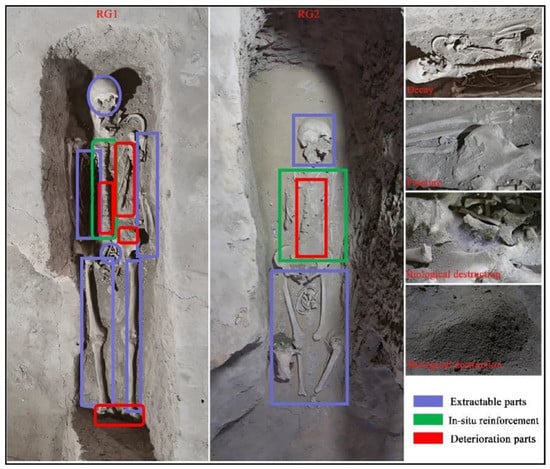
Figure 4.
Extracted parts of human bone.
After we cleaned up, as shown in Figure 5, we first performed a pre-reinforcement of the bone samples that could be extracted, and then we used a disposable needle tube to absorb an appropriate amount of TEOS ethanol solution to infiltrate and reinforce the bone samples. After the mechanical strength had considerably increased, we reinforced the self-made CB material via secondary penetration to consolidate the mechanical strength of the remains. The on-site human bone extraction process included the following steps:
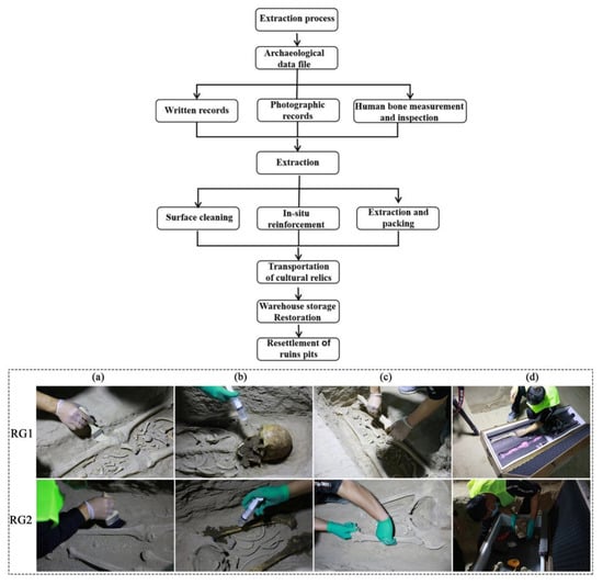
Figure 5.
Human bone extraction process ((a) surface cleaning; (b) in situ reinforcement; (c) extraction; (d) packing).
2.5. Extraction Process
(1) Collect, sort, and archive the existing archaeological data.
(2) Assign a registration number for each human bone component; fill in detailed written records including storage location, age, texture, cultural relic characteristics, disease status, etc.; and describe in the methods and materials used in the restoration process on the restoration card.
(3) Photographic record: Take photographic records from different angles of the cultural relics before extraction, accurately and completely record the appearance and detailed characteristics of the human bones, reflect the original state of the cultural relics from multiple angles, and run through the photographic records during and after the restoration.
(4) Information collection and recording before extraction: take a picture of the entire human bone pit before extraction and then record the arrangement of the human bone artifacts, the integrity of the human bone skeleton structure and the finger marks, and the original samples of each part of the human bone.
(5) Collect digital three-dimensional scanning information (as shown in Figure 5).
(6) Measure the human bones on site: according to the anthropometric method and the measurement method recorded in the anthropometric manual, use the pelvis to measure the humerus, ulna, radius, femur, tibia, and fibula. The left and right sides are separately measured, and the measurement unit is millimeters. After the data are collected, use computer software for mathematical statistical analysis.
(7) Protect the human bones during the extraction and in situ. Judge and mark the extractable human bones based on the on-site survey and the apparent morphology of the preserved human bones, and perform in situ protection treatment for the decayed skeleton parts that are difficult to separate from the site body. Before cleaning the surface of the human bone cultural relics, select test blocks for protection and reinforcement tests. Under the premise of the experiment, choose the dosage ratio of the reinforcement reagent and number of reinforcements to ensure that the cleaning and extraction process will not damage the cultural relics and ruins.
① Use a soft brush to clean up the floating soil and debris on the surface of the human bone cultural relics; if some of the stains on the surface of the human bones are difficult to remove, dip cotton swabs into deionized water and ethanol reagents and roll the swab on the surface of the human bone cultural relics to clean up the stains. Note: The operation process should be moderate in strength to prevent scratches on the surface of the cultural relics. In the extraction process, use a sponge to support the human bone skeleton, properly package the human bone with a soft plastic cloth, and then wrap and fix it with tape paper; afterward, number, photograph, record, and place it in a special collection box. ② Carefully clean up the part of the damaged human bone skeleton that is protected by the original site and the crispy powder and use it to pre-reinforce the human bone parts that are on the site with the CB materials. Absorb the reinforcement reagent with a syringe and inject it evenly onto the surface of the skeleton to strengthen the reinforcement, and quickly absorb the excess reinforcement reagent with absorbent paper. After the reinforcement reagent is dry, spray again. At the same time, use the CB materials to reinforce the earthen ruins where the human bones were located twice. For the spine and ribs that are weak and rotted, use the reinforcements two to three times.
(8) Transport and storage: After the human bones and cultural relics extracted on site are transported and placed in the cultural relics storage room, place them according to the record number and drawings. The storage conditions need to meet the storage environment of the bone cultural relics warehouse: temperature: 14–24 °C; relative humidity: 50%–60%; illumination: 50 Lux; no ultraviolet rays; clean atmosphere; no harmful gases; and dust-, insect-, and mildew-proof.
(9) Restoration and resettlement of the ruins pit: Restore the restored and reinforced human bones and reset them to their original location according to the information collected and recorded before extraction and the three-dimensional scanning digital information file. Reset the placement order strictly in accordance with the order at the time of the on-site extraction and the digitized mark position. After resetting, perform three-dimensional digital scanning again to obtain the point cloud data after resetting. Through a computer data stacking comparison, finely adjust the resetting placement position to ensure the position is as close as possible to the original appearance of the unearthed human bones.
3. Results and Discussion
3.1. Human Bone Microstructure and Composition
The pH value was between 7.8 and 8.2, which indicates that the environment in which the human bones were buried was alkaline, which is unfavorable for the preservation of the organic components in the bones, which directly leads to protein hydrolysis and a decline in the connection capacity between the inorganic structure and organic components, which in turn leads to the occurrence of diseases such as embrittlement and powdering, as was found in the bone cultural relics. The test results showed that the inorganic mineral composition in the soil was CaCO3 and SiO2; the longer bones are buried underground, the more minerals will gradually replace the organic components in the bone and fill the gaps and micropores, resulting in bone mineralization and a loss in the value of the archaeological information of the deep excavation. However, through XRD tests before and after obtaining the human bone ashes at the Shenna site, we found that although the remains had many diseases and the inorganic structure was destroyed, their mineral composition had not changed, so they were not contaminated, meaning further research can still be carried out. The SEM-EDS results (as is shown in Figure 6) also confirmed from a microscopic perspective that the remains were affected by various factors that caused the porosity and mechanical strength of the remains to be reduced, meaning that we have a more comprehensive and detailed understanding of the preservation status of the remains.
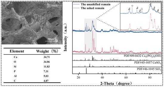
Figure 6.
Microstructure and composition of human bone.
Combining the results of the preliminary site survey of the cultural relics and the storage environment investigation, we found that the remains could be classified according to the preservation conditions they underwent, and that the design of on-site extraction or in situ protection methods could be adopted on a regular basis.
3.2. Extracted Parts of Human Bone
As shown in Figure 7, compared with the relatively flat data obtained by traditional photography and conventional biological measurements, 3D scan technology accurately uses geometric information to form three-dimensional models of the bone cultural relics and their surrounding environment. After extraction, they can be efficiently and accurately restored in situ, and the digital information obtained will have an indispensable position in the new technical analyses and applications that will be generated in the future. In addition, the method has advantages when bonding and filling bone fragments. The retention of the spatial three-dimensional information of cultural relics provides an important data basis and reference for the future protection of cultural relics.
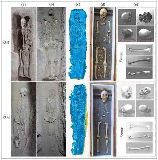
Figure 7.
Extracted human bone ((a) the original; (b) after extraction; (c) three-dimensional point cloud data of site pit after extraction; (d) extracted parts; (e) three-dimensional digital photograph after extraction).
Aiming to fulfill the protection objectives of the extraction of human bones at Shenna ruins and the later restoration and display of the original site, we used 3D scanning technology to obtain three-dimensional point cloud data on cultural relics. During the scanning process, we continuously scanned the human bone remains in all directions to obtain dense three-dimensional scatter points of the measured objects, and we directly obtained the point cloud data on the surface of the target object. After the obtained point cloud data were modeled by a computer, we obtained complete and accurate current geometric information about the human bone remains in the site. We preprocessed the point cloud data, which we then extracted or restored in multiple time phases. We compared the collected data to capture the point cloud data differences to precisely control the in situ resetting. Through the three-dimensional modeling of the point cloud data, and the analysis of the deformation of the multitemporal data point cloud and the amount of point cloud data after the superposition, we ensured the accuracy of the restoration before and after the restoration.
4. Conclusions
Three-dimensional laser scanning technology, which can also be called “real scene replication technology”, can be used to quickly and efficiently collect data. The technology is highly automated and highly precise. This technology can scan any visible target from a covered surface and quickly and accurately obtain the spatial three-dimensional information of cultural relics, including three-dimensional coordinate data and digital photos, without the need for human contact with the target during the scanning process. In this study, we applied the minimal intervention principle by practicing site protection with this technology. Compared with the traditional archaeological human bone packaging and extraction work methods, the auxiliary application of three-dimensional scanning technology to the extraction of human bones from the Shenna ruins reduced human intervention and destruction on the site. When combined with the application of three-dimensional scanning technology, the original data information extracted on the spot were more comprehensive and accurate, which provided us with important scientific data to discuss the phylogenetic composition of the ancient Qiang people in the settlement village. However, a follow-up study is needed to improve the comparisons of its applications, providing a conventional auxiliary means for cultural relic extraction and a technical means for cultural relic protection evaluation.
Author Contributions
J.L. conceived the research, designed the research methodology; performed the experiments, data acquisition, and processing; and drafted the manuscript. K.L. and F.Z. collected the point cloud data. X.F. and J.Y. performed the measurements and data acquisition. X.C. and Y.L. reviewed and corrected the manuscript. B.M., J.W., and J.C. designed the research methodology, conducted the data analysis, and revised the manuscript. Y.L., J.W., and J.C. contributed equally to this work. All authors have read and agreed to the published version of the manuscript.
Funding
The authors gratefully acknowledge the National Natural Science Foundation of China (No. 22102094 and 41601232), the Fundamental Research Funds for the Central Universities (GK 202103061, GK 202103058, and GK 202205024), and the Key Research and Development Program of Shaanxi Province, China (No. 2021SF-457) for their financial support.
Institutional Review Board Statement
Not applicable.
Informed Consent Statement
Not applicable.
Data Availability Statement
The datasets used and/or analysis results obtained for the current study are available from the corresponding author on request.
Conflicts of Interest
The authors declare that they have no conflicts of interest related to this work. We declare that we do not have any commercial or associative interest that represents a conflict of interest in connection with this work submitted.
Abbreviations
3D scan: Three-dimensional laser scanning technology; RG: human bone; SEM-EDS: scanning electron microscopy–energy dispersive spectrometer; XRD: X-ray diffractometry; TEOS: tetraethyl orthosilicate; CB: Ethyl Silicate/Oxalic Acid/Barium Sediment; GPS: global positioning system.
References
- Liu, Y.; Hu, Q.; Zhang, K.; Yang, F.W.; Yang, L.; Wang, L.Q. In-situ growth of calcium sulfate dihydrate as a consolidating material for the archaeological bones. Mater. Lett. 2021, 282, 128713. [Google Scholar] [CrossRef]
- Rougé-Maillart, C.; Vielle, B.; Jousset, N.; Chappard, D.; Telmon, N.; Cunha, E. Development of a method to estimate skeletal age at death in adults using the acetabulum and the auricular surface on a portuguese population. Forensic Sci. Int. 2009, 188, 91–95. [Google Scholar] [CrossRef]
- Wu, X.Z.; Ma, J.B.; Yuan, D.S. Chongqing southern song dynasty yashu site unearthed bone artifacts and its buried background. Quat. Sci. 2016, 36, 312–321. [Google Scholar] [CrossRef]
- Guarino, F.M.; Angelini, F.; Vollono, C.; Orefice, C. Bone preservation in human remains from the terme del sarno at pompeii using light microscopy and scanning electron microscopy. J. Archaeol. Sci. 2006, 33, 513–520. [Google Scholar] [CrossRef]
- Díez Díaz, V.; Mallison, H.; Asbach, P.; Schwarz, D.; Blanco, A. Comparing surface digitization techniques in palaeontology using visual perceptual metrics and distance computations between 3D meshes. Palaeontology 2021, 64, 179–202. [Google Scholar] [CrossRef]
- Marín-Buzón, C.; Pérez-Romero, A.; López-Castro, J.L.; Ben Jerbania, I.; Manzano-Agugliaro, F. Photogrammetry as a New Scientific Tool in Archaeology: Worldwide Research Trends. Sustainability 2021, 13, 5319. [Google Scholar] [CrossRef]
- Lauria, G.; Sineo, L.; Ficarra, S. A detailed method for creating digital 3D models of human crania: An example of close-range photogrammetry based on the use of Structure-from-Motion (SfM) in virtual anthropology. Archaeol. Anthropol. Sci. 2022, 14, 42–54. [Google Scholar] [CrossRef]
- Breithaupt, B.H.; Matthews, N.A.; Noble, T.A. An Integrated Approach to Three-Dimensional Data Collection at Dinosaur Tracksites in the Rocky Mountain West. Ichnos 2004, 11, 11–26. [Google Scholar] [CrossRef]
- Hamm, C.A.; Mallison, H.; Hampe, O.; Schwarz, D.; Mews, J.; Blobel, J.; Issever, A.S.; Asbach, P. Efficiency, workflow and image quality of clinical Computed Tomography scanning compared to photogrammetry on the example of a Tyrannosaurus rex skull from the Maastrichtian of Montana, U.S.A. J. Paleontol. Tech. 2018, 21, 1–13. [Google Scholar]
- Marcy, A.E.; Fruciano, C.; Phillips, M.J.; Mardon, K.; Weisbecker, V. Low resolution scans can provide a sufficiently accurate, cost- and time-effective alternative to high resolution scans for 3D shape analyses. PeerJ 2018, 6, e5032. [Google Scholar] [CrossRef] [PubMed]
- Song, L.M.; Li, D.P.; Chang, Y.L.; Xi, J.T.; Guo, Q.H. Steering knuckle diameter measurement based on optical 3d scanning. Optoelectron. Lett. 2014, 10, 473–476. [Google Scholar] [CrossRef]
- Zhou, M.Q.; Li, C.; Xie, G.D.; Ren, P. Extraction method of archaeological line graph based on 3dmodel. Trans. Beijing Insitute Technol. 2018, 38, 286–292. [Google Scholar] [CrossRef]
- Vanezis, P.; Blowes, R.W.; Linney, A.D.; Tan, A.C.; Richards, R.; Neave, R. Application of 3-d computer graphics for facial reconstruction and comparison with sculpting techniques. Forensic Sci. Int. 1989, 42, 69–84. [Google Scholar] [CrossRef]
- Rusinek, H.; Noz, M.E., Jr.; Maguire, G.Q.; Kalvin, A.; Haddad, B.; Dean, D.; Cutting, C. Quantitative and qualitative comparison of volumetric and surface rendering techniques. IEEE Trans. Nucl. Sci. 1991, 38, 659–662. [Google Scholar] [CrossRef]
- Yang, J.L.; Cao, J.; Yang, H.M.; Li, Y.H.; Wang, J.L. Digitally assisted preservation and restoration of a fragmented mural in a tang tomb. Sens. Imaging 2021, 22, 14–32. [Google Scholar] [CrossRef]
- Yang, L.; Sheng, Y.H.; Pei, A.P.; Wu, Y. 3d spatial morphological analysis of mound tombs based on lidar data. Ann. GIS 2020, 27, 315–325. [Google Scholar] [CrossRef]
- Kontopoulos, I.; Nystrom, P.; White, L. Experimental taphonomy: Post-mortem microstructural modifications in sus scrofa domesticus bone. Forensic. Sci. Int. 2016, 266, 320–328. [Google Scholar] [CrossRef] [PubMed]
- Tang, H.; Geng, G.H.; Zhou, M.Q. Application of digital processing in relic image restoration design. Sens. Imaging 2019, 21, 6. [Google Scholar] [CrossRef]
- Cao, J.F.; Yan, M.M.; Jia, Y.M.; Tian, X.D.; Zhang, Z.B. Application of a modified inception-v3 model in the dynasty-based classification of ancient murals. EURASIP J. Adv. Signal Process. 2021, 2021, 49. [Google Scholar] [CrossRef]
- Moropoulou, A.; Labropoulos, K.C.; Delegou, E.T.; Karoglou, M.; Bakolas, A. Non-destructive techniques as a tool for the protection of built cultural heritage. Constr. Build. Mater. 2013, 48, 1222–1239. [Google Scholar] [CrossRef]
- Edwards, H.G.M.; Carter, E.A.; Kelloway, S.J.; Privat, K.L.; Harrison, T.M. Raman spectroscopic and elemental analysis of bone from a prehistoric ancestor: Mrs ples from the sterkfontein cave. J. Raman Spectrosc. 2021, 52, 2272–2281. [Google Scholar] [CrossRef]
- Wu, K.N.; Wang, W.J.; Zha, L.S.; Ju, B.; Feng, L.W.; Chen, Z.; Yu, X. Review of paleosol and palaeoenxironment in ancient culture sites. Acta Pedol. Sin. 2014, 51, 1169–1182. [Google Scholar] [CrossRef]
Publisher’s Note: MDPI stays neutral with regard to jurisdictional claims in published maps and institutional affiliations. |
© 2022 by the authors. Licensee MDPI, Basel, Switzerland. This article is an open access article distributed under the terms and conditions of the Creative Commons Attribution (CC BY) license (https://creativecommons.org/licenses/by/4.0/).