Assembly and Transport Properties of Nanoscale Biopolyelectrolyte Multilayers


Abstract
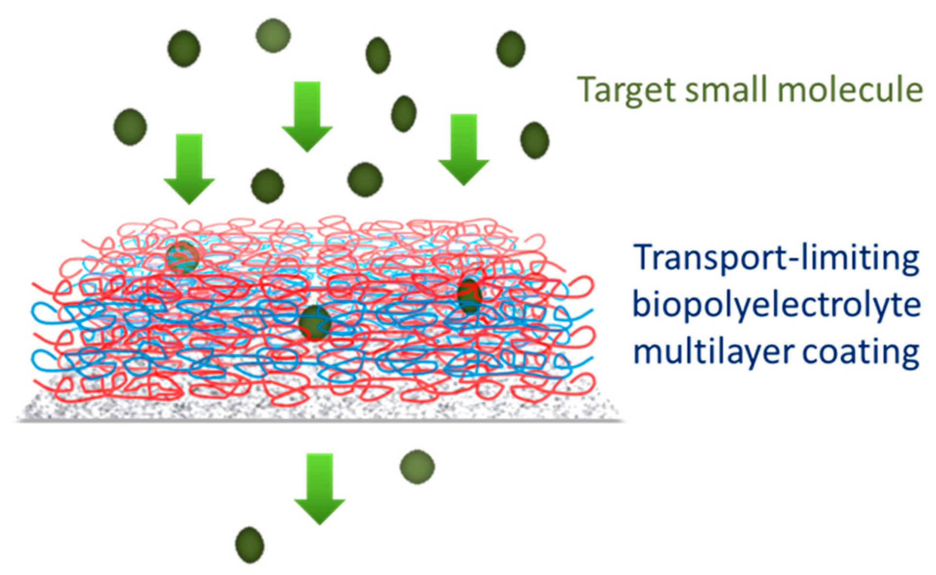
:1. Introduction
2. Materials and Methods
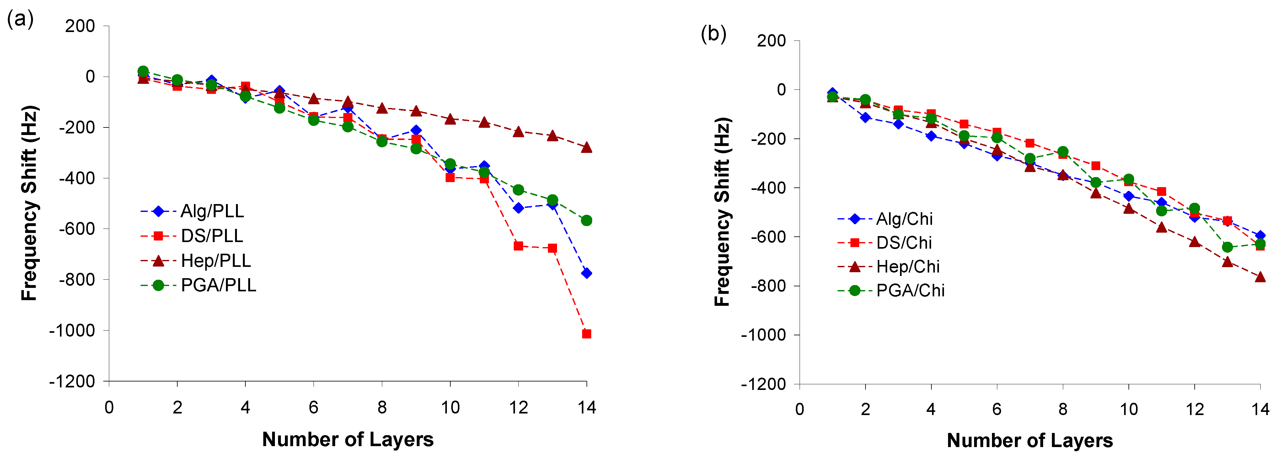
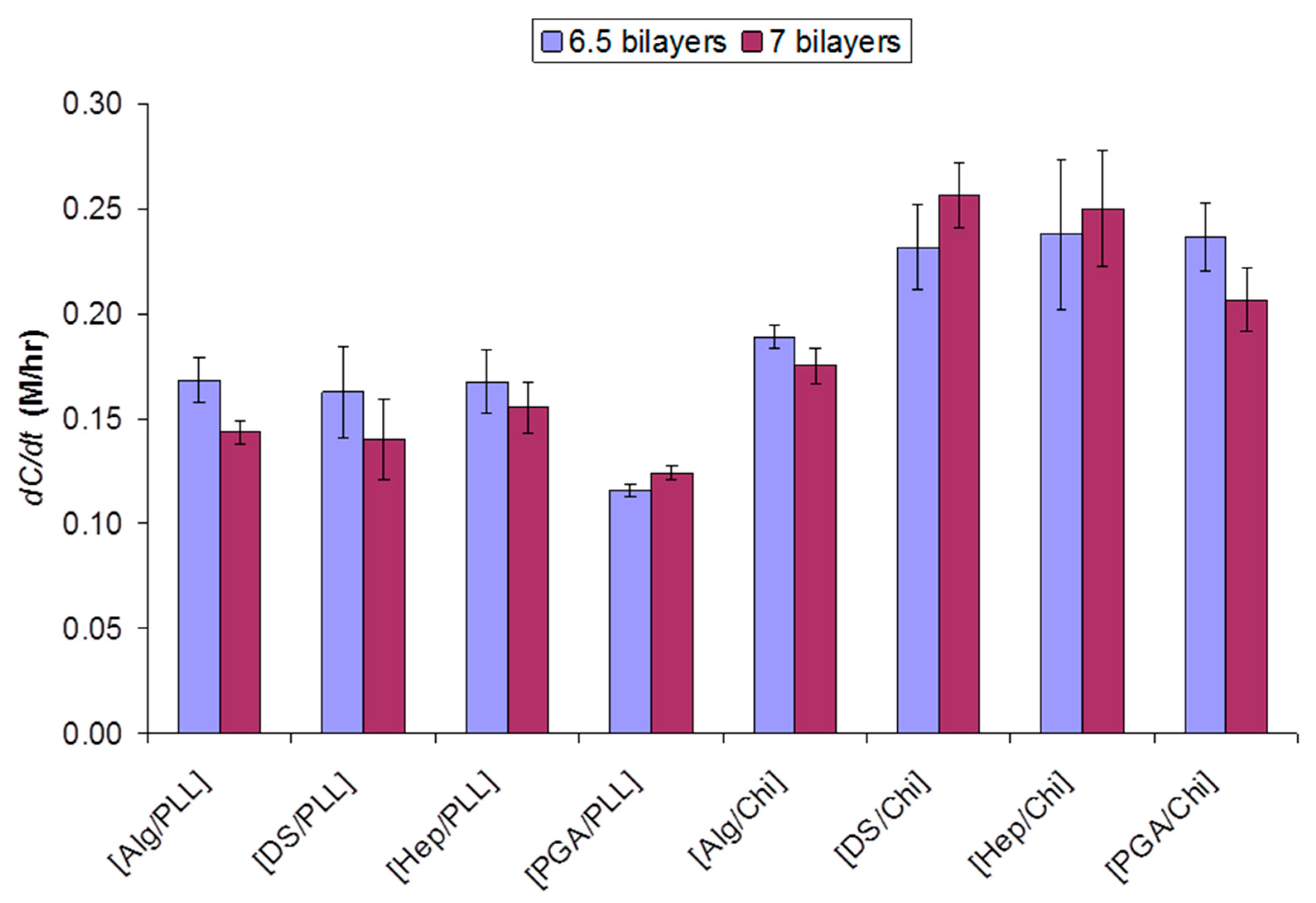
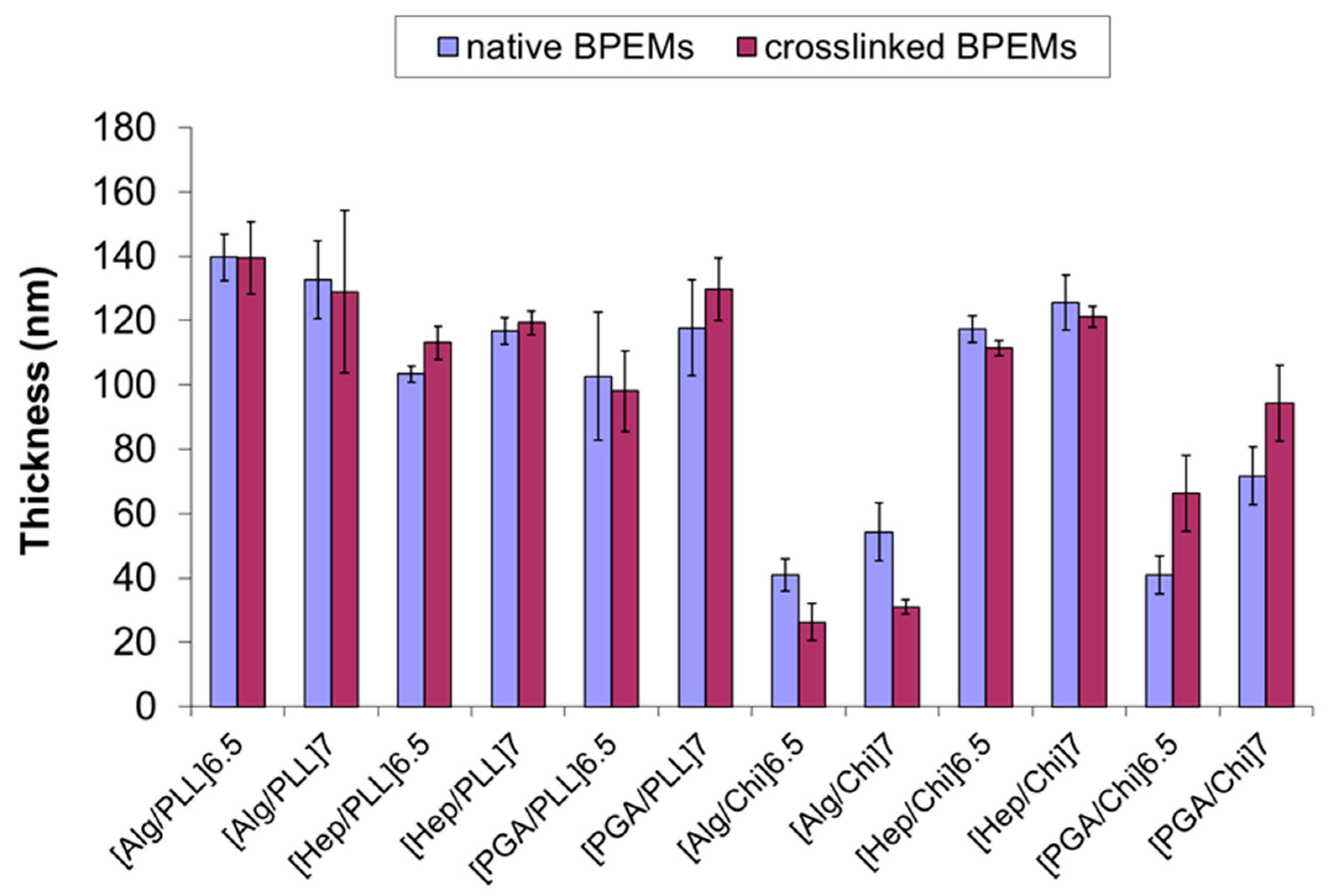
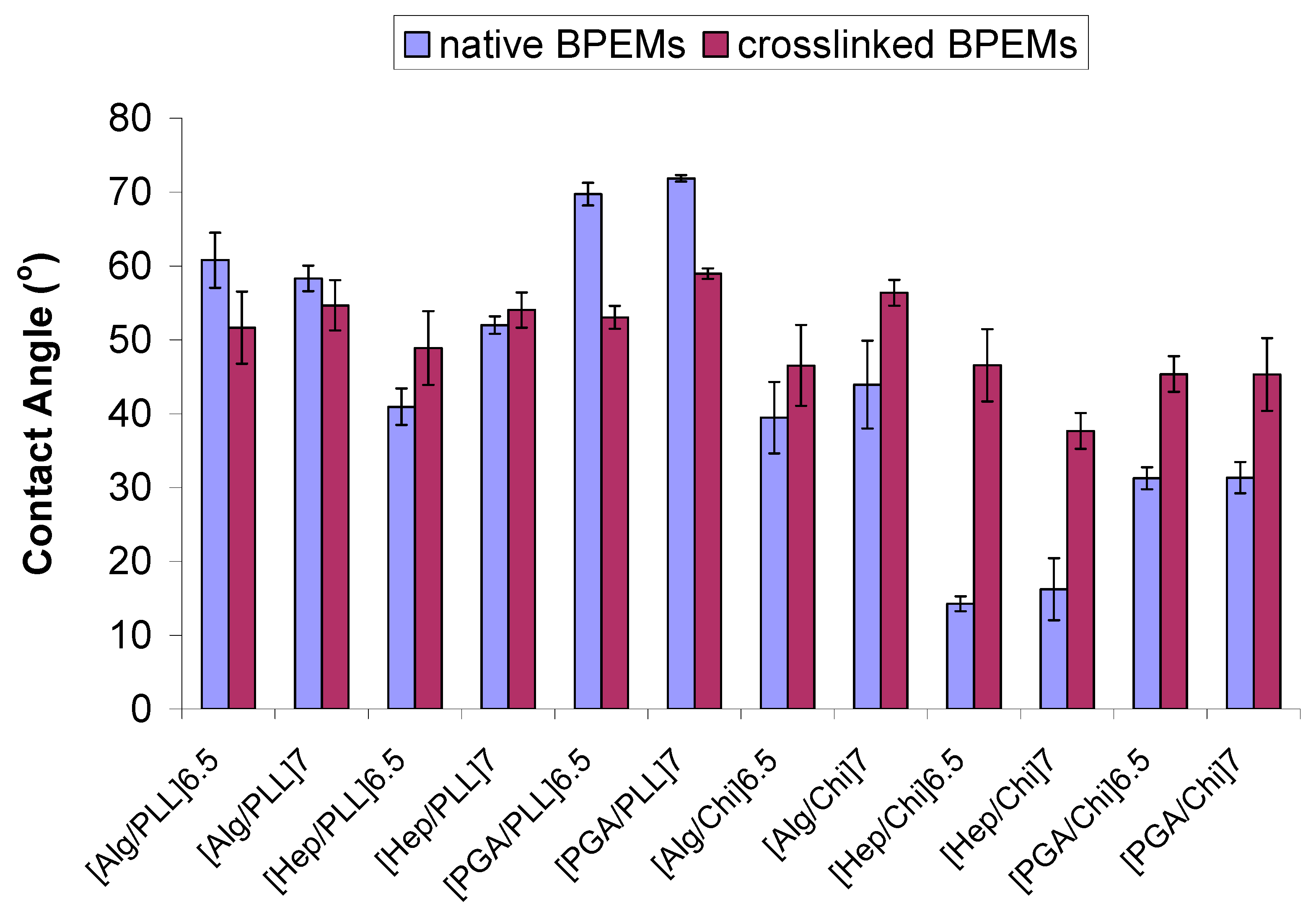
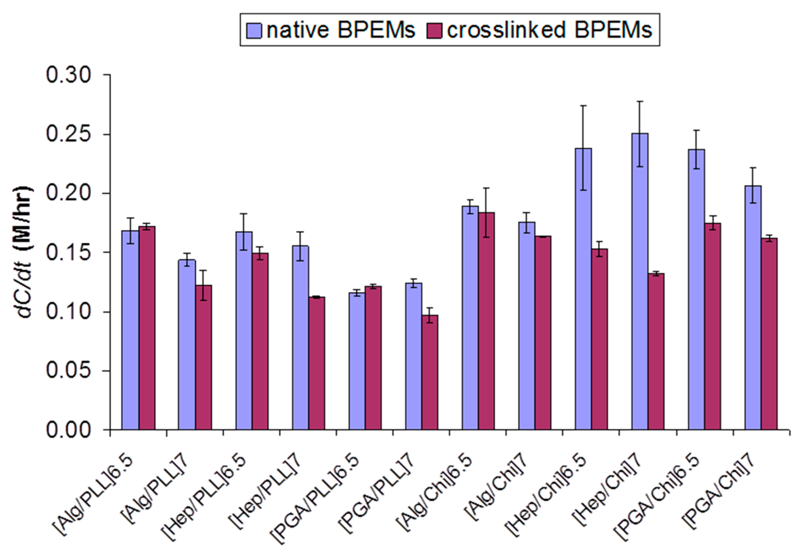
3. Results and Discussion
4. Conclusions
Supplementary Materials
Author Contributions
Funding
Institutional Review Board Statement
Informed Consent Statement
Data Availability Statement
Acknowledgments
Conflicts of Interest
References
- Gough, D.A.; Lucisano, J.Y.; Tse, P.H.S. Two-Dimensional Enzyme Electrode Sensor for Glucose. Anal. Chem. 1985, 57, 2351–2357. [Google Scholar] [CrossRef]
- Ladam, G.; Schaaf, P.; Decher, G.; Voegel, J.-C.; Cuisinier, F.J.G. Protein adsorption onto auto-assembled polyelectrolyte films. Biomol. Eng. 2002, 19, 273–280. [Google Scholar] [CrossRef]
- Anderson, J.M. Biomaterials Science, 1st ed.; Ratner, B., Hoffman, A., Schoen, F., Lemons, J., Eds.; Academic Press: Cambridge, UK, 1996; pp. 165–173. [Google Scholar]
- Decher, G.; Lvov, Y.; Schmitt, J. Proof of multilayer structural organization in self-assembled polycation-polyanion molecular films. Thin Solid Film. 1994, 244, 772–777. [Google Scholar] [CrossRef]
- Decher, G. Fuzzy Nanoassemblies: Toward Layered Polymeric Multicomposites. Science 1997, 277, 1232–1237. [Google Scholar] [CrossRef]
- Decher, G.; Schlenoff, J.B. (Eds.) Multilayer Thin Films: Sequential Assembly of Nanocomposite Materials, 1st ed.; Wiley-VCH: Weinheim, Germany, 2003; p. 543. [Google Scholar]
- Lvov, Y.M.; Sukhorukov, G.B. Protein architecture: Assembly of ordered films by means of alternated adsorption of oppositely charged macromolecules. Membr. Cell Biol. 1997, 11, 277–303. [Google Scholar]
- Sano, M.; Lvov, Y.; Kunitake, T. Formation of Ultrathin Polymer Layers on Solid Substrates by Means of Polymerization-Induced Epitaxy and Alternate Adsorption. Annu. Rev. Mater. Sci. 1996, 26, 153–187. [Google Scholar] [CrossRef]
- Lvov, Y.; Ariga, K.; Onda, M.; Ichinose, I.; Kunitake, T. Alternate Assembly of Ordered Multilayers of SiO2 and Other Nanoparticles and Polyions. Langmuir 1997, 13, 6195–6203. [Google Scholar] [CrossRef]
- Lvov, Y.; Ariga, K.; Ichinose, I.; Kunitake, T. Assembly of Multicomponent Protein Films by Means of Electrostatic Layer-by-Layer Adsorption. J. Am. Chem. Soc. 1995, 117, 6117–6123. [Google Scholar] [CrossRef]
- Keller, S.W.; Kim, H.-N.; Mallouk, T.E. Layer-by-Layer Assembly of Intercalation Compounds and Heterostructures on Surfaces: Toward Molecular “Beaker” Epitaxy. J. Am. Chem. Soc. 1994, 116, 8817–8818. [Google Scholar] [CrossRef]
- Kotov, N.A.; Dekany, I.; Fendler, J.H. Layer-by-Layer Self-Assembly of Polyelectrolyte-Semiconductor Nanoparticle Composite Films. J. Phys. Chem. 1995, 99, 13065–13069. [Google Scholar] [CrossRef]
- Schüler, C.; Caruso, F. Preparation of enzyme multilayers on colloids for biocatalysis. Macromol. Rapid Commun. 2000, 21, 750–753. [Google Scholar] [CrossRef]
- Anzai, J.-i.; Takeshita, H.; Kobayashi, Y.; Osa, T.; Hoshi, T. Layer-by-Layer Construction of Enzyme Multilayers on an Electrode for the Preparation of Glucose and Lactate Sensors: Elimination of Ascorbate Interference by Means of an Ascorbate Oxidase Multilayer. Anal. Chem. 1998, 70, 811–817. [Google Scholar] [CrossRef] [PubMed]
- Hoshi, T.; Saiki, H.; Kuwazawa, S.; Tsuchiya, C.; Chen, Q.; Anzai, J.i. Selective Permeation of Hydrogen Peroxide through Polyelectrolyte Multilayer Films and Its Use for Amperometric Biosensors. Anal. Chem. 2001, 73, 5310–5315. [Google Scholar] [CrossRef] [PubMed]
- Lvov, Y.; Antipov, A.A.; Mamedov, A.; Möhwald, H.; Sukhorukov, G.B. Urease Encapsulation in Nanoorganized Microshells. Nano Lett. 2001, 1, 125–128. [Google Scholar] [CrossRef]
- Boura, C.; Menu, P.; Payan, E.; Picart, C.; Voegel, J.C.; Muller, S.; Stoltz, J.F. Endothelial cells grown on thin polyelectrolyte mutlilayered films: An evaluation of a new versatile surface modification. Biomaterials 2003, 24, 3521–3530. [Google Scholar] [CrossRef]
- Fu, J.; Ji, J.; Yuan, W.; Shen, J. Construction of anti-adhesive and antibacterial multilayer films via layer-by-layer assembly of heparin and chitosan. Biomaterials 2005, 26, 6684–6692. [Google Scholar] [CrossRef]
- Thompson, M.T.; Berg, M.C.; Tobias, I.S.; Rubner, M.F.; Van Vliet, K.J. Tuning compliance of nanoscale polyelectrolyte multilayers to modulate cell adhesion. Biomaterials 2005, 26, 6836–6845. [Google Scholar] [CrossRef]
- Buck, M.E.; Breitbach, A.S.; Belgrade, S.K.; Blackwell, H.E.; Lynn, D.M. Chemical modification of reactive multilayered films fabricated from poly(2-alkenyl azlactone)s: Design of surfaces that prevent or promote mammalian cell adhesion and bacterial biofilm growth. Biomacromolecules 2009, 10, 1564–1574. [Google Scholar] [CrossRef] [PubMed] [Green Version]
- Boudou, T.; Crouzier, T.; Nicolas, C.; Ren, K.; Picart, C. Polyelectrolyte Multilayer Nanofilms Used as Thin Materials for Cell Mechano-Sensitivity Studies. Macromol. Biosci. 2011, 11, 77–89. [Google Scholar] [CrossRef] [PubMed]
- Rodrigues, S.N.; Gonçalves, I.C.; Martins, M.C.L.; Barbosa, M.A.; Ratner, B.D. Fibrinogen adsorption, platelet adhesion and activation on mixed hydroxyl-/methyl-terminated self-assembled monolayers. Biomaterials 2006, 27, 5357–5367. [Google Scholar] [CrossRef] [PubMed]
- Chen, H.; Hu, X.; Zhang, Y.; Li, D.; Wu, Z.; Zhang, T. Effect of chain density and conformation on protein adsorption at PEG-grafted polyurethane surfaces. Colloids Surf. B Biointerfaces 2008, 61, 237–243. [Google Scholar] [CrossRef] [PubMed]
- Park, J.; McShane, M.J. DualFunction Nanofilm Coatings with Diffusion Control and Protein Resistance. ACS Appl. Mater. Interfaces 2010, 2, 991–997. [Google Scholar] [CrossRef] [PubMed]
- Tang, L.; Thevenot, P.; Hu, W. Surface Chemistry Influences Implant Biocompatibility. Curr. Top. Med. Chem. 2008, 8, 270–280. [Google Scholar] [CrossRef]
- Boulmedais, F.; Frisch, B.; Etienne, O.; Lavalle, P.; Picart, C.; Ogier, J.; Voegel, J.C.; Schaaf, P.; Egles, C. Polyelectrolyte multilayer films with pegylated polypeptides as a new type of anti-microbial protection for biomaterials. Biomaterials 2004, 25, 2003–2011. [Google Scholar] [CrossRef] [PubMed] [Green Version]
- Stein, E.W.; Singh, S.; McShane, M.J. Microscale Enzymatic Optical Biosensors Using Mass Transport Limiting Nanofilms. 2. Response Modulation by Varying Analyte Transport Properties. Anal. Chem. 2008, 80, 1408–1417. [Google Scholar] [CrossRef] [PubMed]
- Stein, E.W.; Grant, P.S.; Zhu, H.; McShane, M.J. Microscale Enzymatic Optical Biosensors Using Mass Transport Limiting Nanofilms. 1. Fabrication and Characterization Using Glucose as a Model Analyte. Anal. Chem. 2007, 79, 1339–1348. [Google Scholar] [CrossRef] [Green Version]
- Gerritsen, M.; Jansen, J.A.; Lutterman, J.A. Performance of subcutaneously implanted glucose sensors for continuous monitoring. Neth. J. Med. 1999, 54, 167–179. [Google Scholar] [CrossRef]
- Koppolu, B.; Rahimi, M.; Nattama, S.; Wadajkar, A.; Nguyen, K.T. Development of multiple-layer polymeric particles for targeted and controlled drug delivery. Nanomedicine 2010, 6, 355–361. [Google Scholar] [CrossRef] [PubMed] [Green Version]
- Wang, C.; Ye, W.; Zheng, Y.; Liu, X.; Tong, Z. Fabrication of drug-loaded biodegradable microcapsules for controlled release by combination of solvent evaporation and layer-by-layer self-assembly. Int. J. Pharm. 2007, 338, 165–173. [Google Scholar] [CrossRef]
- Boudou, T.; Crouzier, T.; Ren, K.; Blin, G.; Picart, C. Multiple Functionalities of Polyelectrolyte Multilayer Films: New Biomedical Applications. Adv. Mater. 2010, 22, 441–467. [Google Scholar] [CrossRef]
- Liu, X.; Bruening, M.L. Size-Selective Transport of Uncharged Solutes through Multilayer Polyelectrolyte Membranes. Chem. Mater. 2004, 16, 351–357. [Google Scholar] [CrossRef]
- Johansson, J.A.; Halthur, T.; Herranen, M.; Soderberg, L.; Elofsson, U.; Hilborn, J. Build-up of collagen and hyaluronic acid polyelectrolyte multilayers. Biomacromolecules 2005, 6, 1353–1359. [Google Scholar] [CrossRef]
- Etienne, O.; Schneider, A.; Taddei, C.; Richert, L.; Schaaf, P.; Voegel, J.-C.; Egles, C.; Picart, C. Degradability of Polysaccharides Multilayer Films in the Oral Environment: An In Vitro and In Vivo Study. Biomacromolecules 2005, 6, 726–733. [Google Scholar] [CrossRef]
- Collin, D.; Lavalle, P.; Garza, J.M.; Voegel, J.-C.; Schaaf, P.; Martinoty, P. Mechanical properties of cross-linked hyaluronic acid/poly-(L-lysine) multilayer films. Macromolecules 2004, 37, 10195–10198. [Google Scholar] [CrossRef]
- Sauerbrey, G. Verwendung von Schwingquarzen zur Wägung dünner Schichten und zur Mikrowägung. Z. Für Phys. A Hadron. Nucl. 1959, 155, 206–222. [Google Scholar] [CrossRef]
- Richert, L.; Boulmedais, F.; Lavalle, P.; Mutterer, J.; Ferreux, E.; Decher, G.; Schaaf, P.; Voegel, J.-C.; Picart, C. Improvement of Stability and Cell Adhesion Properties of Polyelectrolyte Multilayer Films by Chemical Cross-Linking. Biomacromolecules 2004, 5, 284–294. [Google Scholar] [CrossRef] [PubMed] [Green Version]
- Raabo, E.; Terkildsen, T.C. On the Enzymatic Determination of Blood Glucose. Scand. J. Clin. Lab. Investig. 1960, 12, 402–407. [Google Scholar] [CrossRef]
- Elbert, D.L.; Herbert, C.B.; Hubbell, J.A. Thin Polymer Layers Formed by Polyelectrolyte Multilayer Techniques on Biological Surfaces. Langmuir 1999, 15, 5355–5362. [Google Scholar] [CrossRef]
- Lavalle, P.; Gergely, C.; Cuisinier, F.J.G.; Decher, G.; Schaaf, P.; Voegel, J.C.; Picart, C. Comparison of the Structure of Polyelectrolyte Multilayer Films Exhibiting a Linear and an Exponential Growth Regime: An In Situ Atomic Force Microscopy Study. Macromolecules 2002, 35, 4458–4465. [Google Scholar] [CrossRef]
- Dai, J.; Jensen, A.W.; Mohanty, D.K.; Erndt, J.; Bruening, M.L. Controlling the Permeability of Multilayered Polyelectrolyte Films through Derivatization, Cross-Linking, and Hydrolysis. Langmuir 2001, 17, 931–937. [Google Scholar] [CrossRef]
- Liu, H.; Faucher, K.M.; Sun, X.-L.; Feng, J.; Johnson, T.L.; Orban, J.M.; Apkarian, R.P.; Dluhy, R.A.; Chaikof, E.L. A Membrane-Mimetic Barrier for Cell Encapsulation. Langmuir 2002, 18, 1332–1339. [Google Scholar] [CrossRef]
- Maurstad, G.; Mørch, Y.A.; Bausch, A.R.; Stokke, B.T. Polyelectrolyte layer interpenetration and swelling of alginate-chitosan multilayers studied by dual wavelength reflection interference contrast microscopy. Carbohydr. Polym. 2008, 71, 672–681. [Google Scholar] [CrossRef]
- Alves, N.M.; Picart, C.; Mano, J.F. Self Assembling and Crosslinking of Polyelectrolyte Multilayer Films of Chitosan and Alginate Studied by QCM and IR Spectroscopy. Macromol. Biosci. 2009, 9, 776–785. [Google Scholar] [CrossRef] [PubMed]
- Glinel, K.; Prevot, M.; Krustev, R.; Sukhorukov, G.B.; Jonas, A.M.; Mohwald, H. Control of the water permeability of polyelectrolyte multilayers by deposition of charged paraffin particles. Langmuir 2004, 20, 4898–4902. [Google Scholar] [CrossRef] [PubMed]
- Schneider, A.; Vodouhê, C.; Richert, L.; Francius, G.; Le Guen, E.; Schaaf, P.; Voegel, J.-C.; Frisch, B.; Picart, C. Multifunctional Polyelectrolyte Multilayer Films: Combining Mechanical Resistance, Biodegradability, and Bioactivity. Biomacromolecules 2006, 8, 139–145. [Google Scholar] [CrossRef] [Green Version]
- Boulmedais, F.; Bozonnet, M.; Schwinte, P.; Voegel, J.C.; Schaaf, P. Multilayered polypeptide films: Secondary structures and effect of various stresses. Langmuir 2003, 19, 9873–9882. [Google Scholar] [CrossRef]
- Pilbat, A.M.; Ball, V.; Schaaf, P.; Voegel, J.C.; Szalontai, B. Partial poly(glutamic acid) <-> poly(aspartic acid) exchange in layer-by-layer polyelectrolyte films. Structural alterations in the three-component architectures. Langmuir 2006, 22, 5753–5759. [Google Scholar] [CrossRef]
- Boudou, T.; Crouzier, T.; Auzély-Velty, R.; Glinel, K.; Picart, C. Internal Composition versus the Mechanical Properties of Polyelectrolyte Multilayer Films: The Influence of Chemical Cross-Linking. Langmuir 2009, 25, 13809–13819. [Google Scholar] [CrossRef]


| Composition | CA (°) | L (nm) | ||
|---|---|---|---|---|
| 6.5 Bilayers | 7 Bilayers | 6.5 Bilayers | 7 Bilayers | |
| [Alg/Chi] | 39 (±4.8) | 44 (±5.9) | 41 (±5.0) | 54 (±9.1) |
| [DS/Chi] | 13 (±1.4) | 18 (±1.6) | 78 (±11.6) | 101 (±5.7) |
| [Hep/Chi] | 14 (±1.0) | 16 (±4.2) | 117 (±4.2) | 126 (±8.6) |
| [PGA/Chi] | 31 (±1.5) | 31 (±2.1) | 41 (±5.8) | 72 (±9.1) |
| [Alg/PLL] | 61 (±3.7) | 58 (±1.7) | 140 (±7.2) | 133 (±12.1) |
| [DS/PLL] | 8 (±1.4) | 17 (±1.6) | 134 (±4.9) | 135 (±4.0) |
| [Hep/PLL] | 41 (±2.5) | 52 (±1.2) | 103 (±2.6) | 117 (±4.1) |
| [PGA/PLL] | 70 (±1.9) | 72 (±0.4) | 103 (±19.9) | 118 (±14.9) |
| Composition | D (×10−10 cm2/s) | |||
|---|---|---|---|---|
| Native BioPEMs | Crosslinked BioPEMs | |||
| 6.5 Bilayers | 7 Bilayers | 6.5 Bilayers | 7 Bilayers | |
| [Alg/Chi] | 8.2 (±1.0) | 10.0 (±2.9) | 4.4 (±0.3) | 5.4 (±0.1) |
| [DS/Chi] | 19.1 (±1.7) | 27.4 (±1.6) | n/a | n/a |
| [Hep/Chi] | 29.4 (±3.7) | 33.5 (±8.6) | 17.7 (±0.4) | 17.1 (±0.7) |
| [PGA/Chi] | 10.3 (±3.0) | 15.7 (±4.4) | 10.6 (±3.9) | 16.2 (±3.2) |
| [Alg/PLL] | 24.8 (±1.5) | 20.1 (±2.2) | 24.9 (±4.5) | 16.5 (±5.7) |
| [DS/PLL] | 23.0 (±3.7) | 19.9 (±3.1) | n/a | n/a |
| [Hep/PLL] | 18.3 (±1.4) | 19.1 (±0.9) | 17.9 (±2.1) | 14.1 (±0.6) |
| [PGA/PLL] | 12.5 (±0.4) | 15.2 (±0.1) | 12.2 (±1.5) | 12.7 (±1.1) |
Publisher’s Note: MDPI stays neutral with regard to jurisdictional claims in published maps and institutional affiliations. |
© 2021 by the authors. Licensee MDPI, Basel, Switzerland. This article is an open access article distributed under the terms and conditions of the Creative Commons Attribution (CC BY) license (https://creativecommons.org/licenses/by/4.0/).
Share and Cite
Park, J.; McShane, M.J. Assembly and Transport Properties of Nanoscale Biopolyelectrolyte Multilayers. Coatings 2021, 11, 1024. https://doi.org/10.3390/coatings11091024
Park J, McShane MJ. Assembly and Transport Properties of Nanoscale Biopolyelectrolyte Multilayers. Coatings. 2021; 11(9):1024. https://doi.org/10.3390/coatings11091024
Chicago/Turabian StylePark, Jaebum, and Michael J. McShane. 2021. "Assembly and Transport Properties of Nanoscale Biopolyelectrolyte Multilayers" Coatings 11, no. 9: 1024. https://doi.org/10.3390/coatings11091024
APA StylePark, J., & McShane, M. J. (2021). Assembly and Transport Properties of Nanoscale Biopolyelectrolyte Multilayers. Coatings, 11(9), 1024. https://doi.org/10.3390/coatings11091024