Article

Chemical Solution Deposition of La-Substituted BiFe0.5Sc0.5O3 Perovskite Thin Films on Different Substrates

1
Institute of Chemistry, Vilnius University, Naugarduko 24, LT-03225 Vilnius, Lithuania
2
Laboratory of Spectroelectrochemistry, Department of Organic Chemistry, Center for Physical Sciences and Technology, Sauletekio av. 3, LT-10257 Vilnius, Lithuania
3
Department of Materials and Ceramic Engineering/CICECO, University of Aveiro, 3810-193 Aveiro, Portugal
*
Author to whom correspondence should be addressed.
Coatings 2021, 11(3), 307; https://doi.org/10.3390/coatings11030307
Submission received: 15 February 2021 / Revised: 3 March 2021 / Accepted: 4 March 2021 / Published: 8 March 2021

Abstract

:
In the present work, polycrystalline Bi0.67La0.33Fe0.5Sc0.5O3 thin films were synthesized using a simple and cost-effective chemical solution deposition process employing the spin coating technique. In order to check the feasibility of the fabrication of thin films on various types of substrates, the films were deposited on Pt-coated silicon, silicon, sapphire, corundum, fused silica and glass. Based on the results of thermogravimetric analysis of precursor and thermal stability study, it was determined that the optimal annealing temperature for the formation of perovskite structure is 600 °C. It was observed that the relative intensity of the pseudocubic peaks (001)p and (011)p in the XRD patterns is influenced by the nature of substrates, suggesting that the formed crystallites have some preferred orientation. Roughness of the films was determined to be dependent on the nature of the substrate.

1. Introduction

In recent decades multiferroic materials have attracted huge scientific and technological interest due to their unique physical properties. The coexistence of intimately coupled ferroic orders makes it possible to influence the state of the material by applying external electrical or magnetic fields [1]. A number of families of oxide materials with magnetic, ferroelectric, and ferroelastic properties and their combinations are composed of three-dimensional chains of metal–oxygen octahedra [2]. The perovskite family is probably the most famous in this respect [3].
Bismuth ferrite (BFO, BiFeO3) is one of the most known multiferroic materials with a perovskite crystal structure [4]. This perovskite demonstrates outstanding physical properties such as large remnant polarization (∼100 µC/cm2), high Curie temperature (TC ∼ 1100 K) and high Néel temperature (TN ∼643 K) [5,6]. Its direct band gap (Eg) of around 2.2–2.8 eV allows for efficient light absorption in the visible spectral range [7]. One of the approaches commonly employed for the tuning of magnetic, electrical, structural and optical properties of BFO considers a partial substitution of Bi3+ and/or Fe3+ ions by isovalent or aliovalent ions [8,9,10]. The remarkable BFO derivative, which is in fact A and B co-substituted BFO, is the Bi1−xLaxFe0.5Sc0.5O3 perovskite system [11,12]. In the bulk form of this material, the room temperature compositional crossover from the antipolar phase, the incommensurate modulation of displacements of Bi/La and oxygen of which is described by the Imma(00γ)s00 superspace group [13] to the nonpolar Pnma phase [14], occurs in the narrow range between x = 0.33 and 0.34 with no phase coexistence [15]. This very narrow compositional range is of a great interest, since it corresponds to a solid solution with the maximal lattice-magnetic coupling effect expected, since TCTN [15]. It is known that the perovskites phases of the Bi1−xLaxFe0.5Sc0.5O3 system (x < 0.80) can be prepared in forms of ceramics only using high-pressure synthesis [13,14,15], and they are metastable at ambient pressure. It should be noticed that while Bi0.67La0.33Fe0.5Sc0.5O3 and Bi0.66La0.34Fe0.5Sc0.5O3 in bulk form have been studied [11,12,15], to the best of our knowledge there are no reports on the synthesis and investigation of physical properties of their thin films.
Fabrication of multiferroic materials in the form of 2D nanostructures is of particular technological importance for the development and miniaturization of electronic devices [16,17]. Moreover, the aspects such as preferred orientation or surface strain can be considered as an additional powerful tool, allowing the physical properties of the materials to be tuned [18,19]. For the fabrication of multicomponent oxide thin films, various deposition approaches, including vacuum-based and non-vacuum based techniques, can be successfully employed [20,21,22,23,24]. It is well known that for some advanced deposition methods, such as metal organic chemical solution deposition, the precise stoichiometric control in multicomponent systems is a very challenging task because of different volatility of precursors, different deposition rates, etc. [25,26]. The methods based on physical vapor deposition also suffer from the deviation from desired product stoichiometry [27]. In this light, chemical solution deposition can be considered as a reasonable choice for production of mixed metal oxide films. This method is remarkable due to its low cost, simplicity, good mixing of starting materials at the molecular level and ease of processing for large-area wafers. Excellent stoichiometry control of the end product is easily achieved by the use of appropriate amounts of starting materials. All these features allow homogeneous and single-phase products to be obtained [28,29,30].
This study is the first step towards the systematic investigation of physical properties of Bi1−xLaxFe0.5Sc0.5O3 thin films. The main objective of this work was to develop a simple and reliable procedure for the deposition of Bi0.67La0.33Fe0.5Sc0.5O3 2D nanostructures. Additionally, this work was aimed at checking the feasibility of preparation of the Bi0.67La0.33Fe0.5Sc0.5O3 thin films on different kinds of substrates (monocrystalline, polycrystalline, amorphous, transparent and conductive) for further characterization of physical properties. For this reason, soda lime glass, polycrystalline Al2O3, fused silica, monocrystalline silicon wafer, sapphire and Pt-coated silicon (Pt/TiO2/SiO2/Si) substrates were selected. The influence of the substrate on structural properties and morphology of deposited thin films was investigated.

2. Materials and Methods

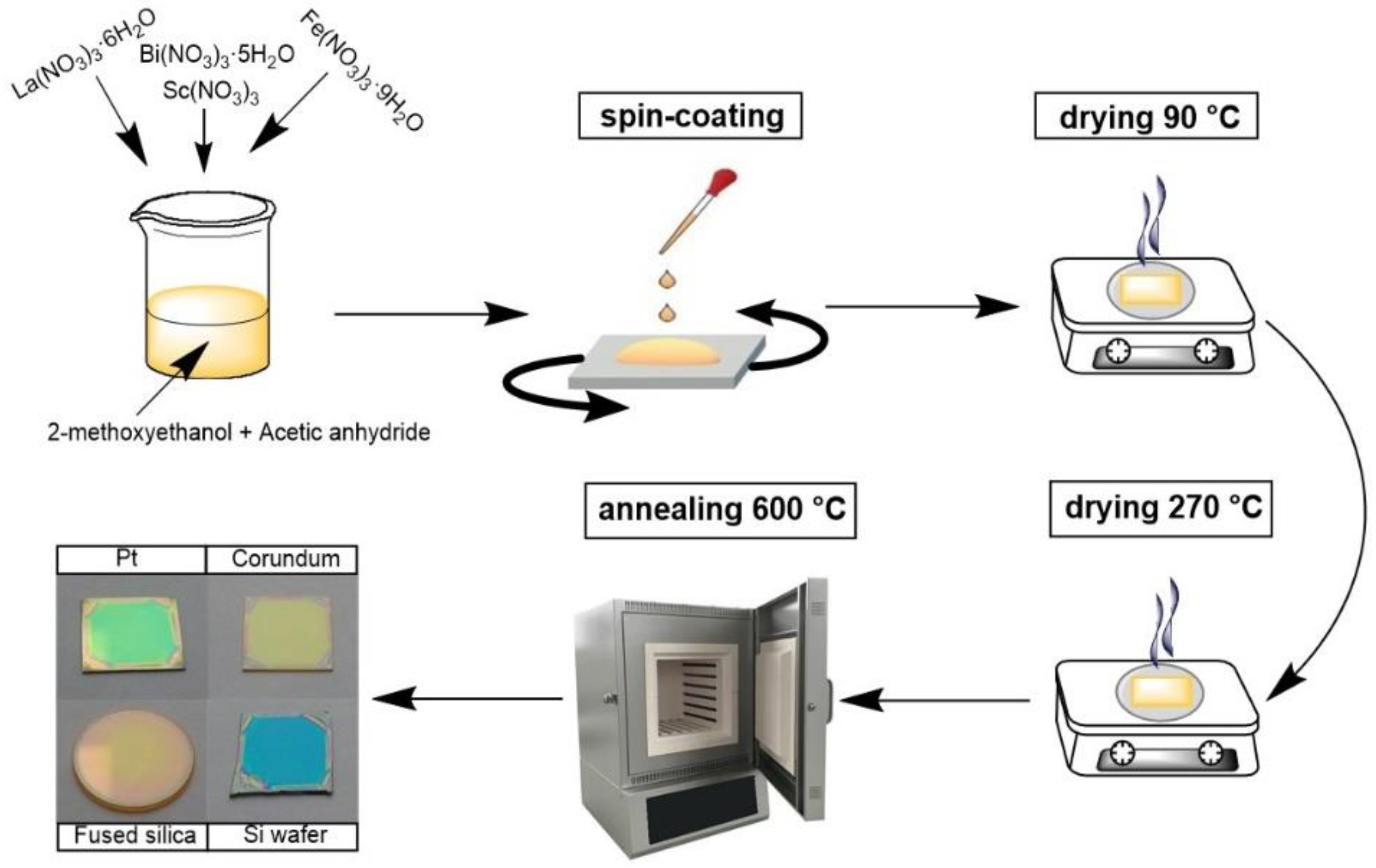
2.1. Preparation of Precursor Solution

The synthesis of precursor solution was performed on the basis of a previously reported procedure [31]. An appropriate amount of scandium oxide (Sc2O3, abcr, 99.9%) was dissolved in boiling concentrated nitric acid and evaporated to dryness. The obtained scandium salt was further dissolved in a certain volume of 2-methoxyethanol (C3H8O2, Sigma-Aldrich, Darmstadt, Germany, 99.8%). Metal nitrate hydrates La(NO3)3∙6H2O (Roth, Karlsruhe, Germany, 99.99%), Bi(NO3)3∙5H2O (Roth, 98%) and Fe(NO3)3∙9H2O (Sigma-Aldrich, 98%) were added to the above solution according to the stoichiometry of the final product (Bi0.67La0.33Fe0.5Sc0.5O3). A certain amount of acetic anhydride ((CH3CO)2O, Sigma-Aldrich, ≥99%) was then added to the prepared mixture; the volume ratio of 2-methoxyethanol and acetic anhydride was 3:1. The total concentration of metal ions in the obtained clear precursor solution was 0.25 M. Thermogravimetric analysis and elemental analysis of metal nitrates by means of inductively coupled plasma optical emission spectrometry (ICP-OES) were performed prior synthesis in order to estimate the actual metal content. Hereafter in the text, the chemical formula (Bi0.67La0.33Fe0.5Sc0.5O3) will be abbreviated as BLFSO.

2.2. Deposition of Thin Films

Multilayered thin films were prepared on Pt-coated silicon (Pt/TiO2/SiO2/Si, University Wafer, South Boston, MA, United States), silicon (CrysTec, Berlin, Germany), R-cut sapphire (CrysTec), polycrystalline corundum (MTI, Richmond, CA, United States), fused silica (Altechna, Vilnius, Lithuania) and glass (Roth) substrates by the spin coating technique using a SCS P6700 spin coater. Prior to deposition, the substrates were cleaned with a concentrated sulfuric acid and hydrogen peroxide (30%) mixture followed by washing with deionized water and isopropanol. Several drops of precursor solution were placed onto substrates and spin coated for 30 s at 3000 rpm. The as-deposited wet films were dried on a hot plate in a two-step manner: 5 min at 90 °C followed by 5 min at 270 °C. The complete cycle was repeated a certain number of times prior to a final annealing in a muffle furnace at different temperatures for 2 h (the heating rate was 1 °C/min). All films were deposited using the same precursor solution on the same day. An aliquot of precursor solution was evaporated and annealed under identical conditions in order to obtain BLFSO powders for comparison. A schematic representation of the deposition procedure is shown in Figure 1.

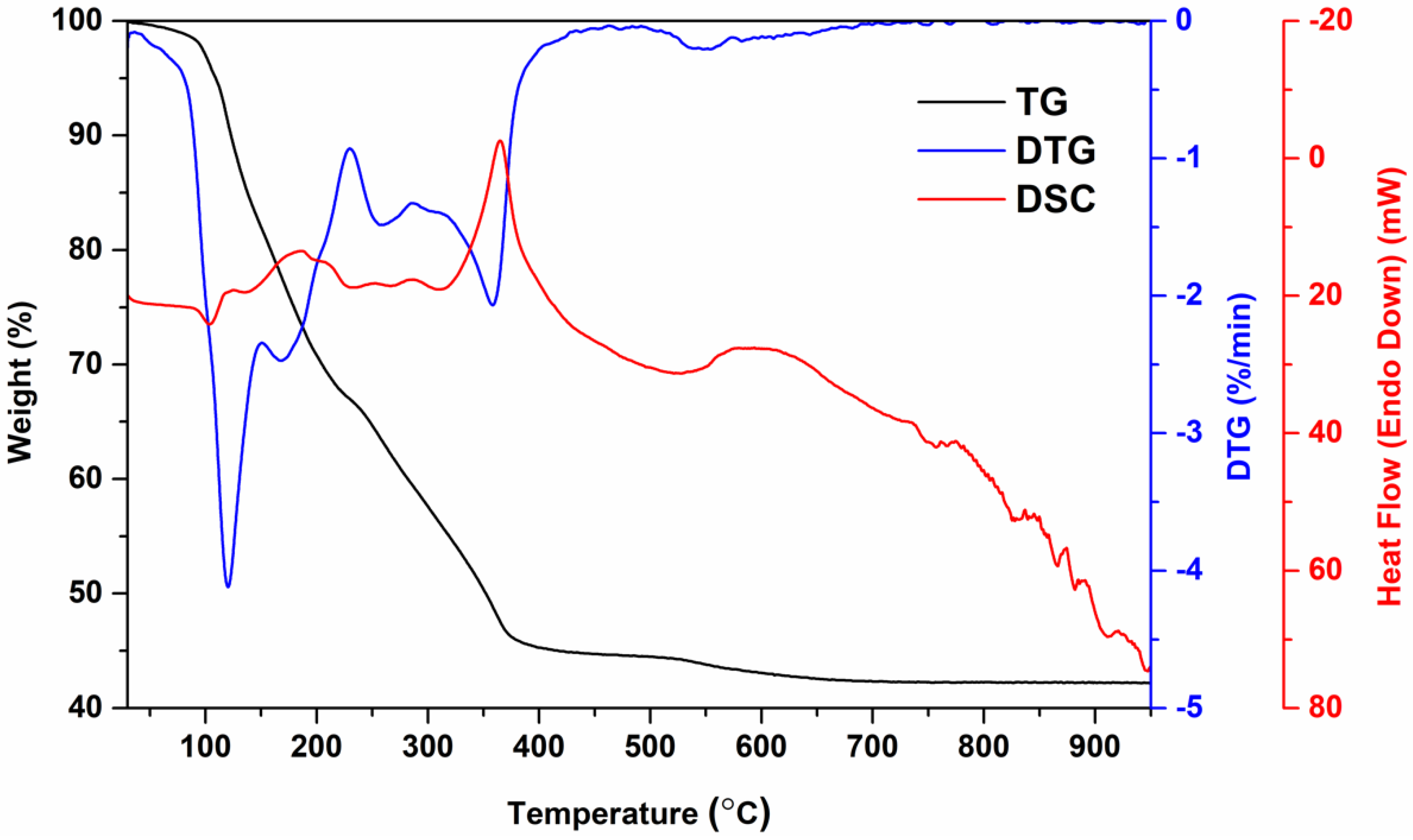
2.3. Characterization

The thermal decomposition of dried precursor solution was analyzed by thermogravimetry and differential scanning calorimetry (TG–DSC) using a Perkin Elmer STA 6000 simultaneous thermal analyzer. A dried sample of about 5–10 mg was heated from 25 to 900 °C at a heating rate of 5 °C/min in dry flowing air (20 mL/min). The crystal structure of the obtained samples was investigated by X-ray diffraction (XRD) using a PANalytical X’Pert powder diffractometer (Theta-Omega goniometer, Malvern Panalytical, Malvern, UK, Ni-filtered Cu Kα radiation, PIXcel1D detector, Malvern Panalytical, Malvern, UK, tube settings 45 kV, 40 mA). The deposited films were measured in grazing incidence X-ray diffraction (GIXRD) mode using a multi-purpose sample stage. The XRD patterns were collected with a grazing angle of 2° in the range from 20 to 80 degrees with a step of 0.02° and a counting time of about 7 s per step. Prior to each measurement, the substrate position was aligned against the direct beam by means of the omega angle correction. The XRD measurements of the powder samples were performed using a spinner sample stage (Bragg-Brentano geometry, a 2θ angular range of 20°–80°, step 0.02°, 0.5 s per step, a sample rotation of 4 s−1). The obtained XRD data were treated using the PANalytical HighScore 5.0 package. The morphology of the films was characterized using a Hitachi SU-70 scanning electron microscope (SEM) (Hitachi, Tokyo, Japan). For the surface characterization of the deposited films, a BioScope Catalyst atomic force microscope (AFM) from Bruker (BioscopeII/Catalyst, Karlsruhe, Germany) was used. The peak force tapping mode was applied using a ScanAsyst-Air silicon probe (nominal tip spring constant 0.4 N/m, scan rate 0.5 Hz). Two types of images were recorded: 20 µm × 20 µm and 5 µm × 5 µm.

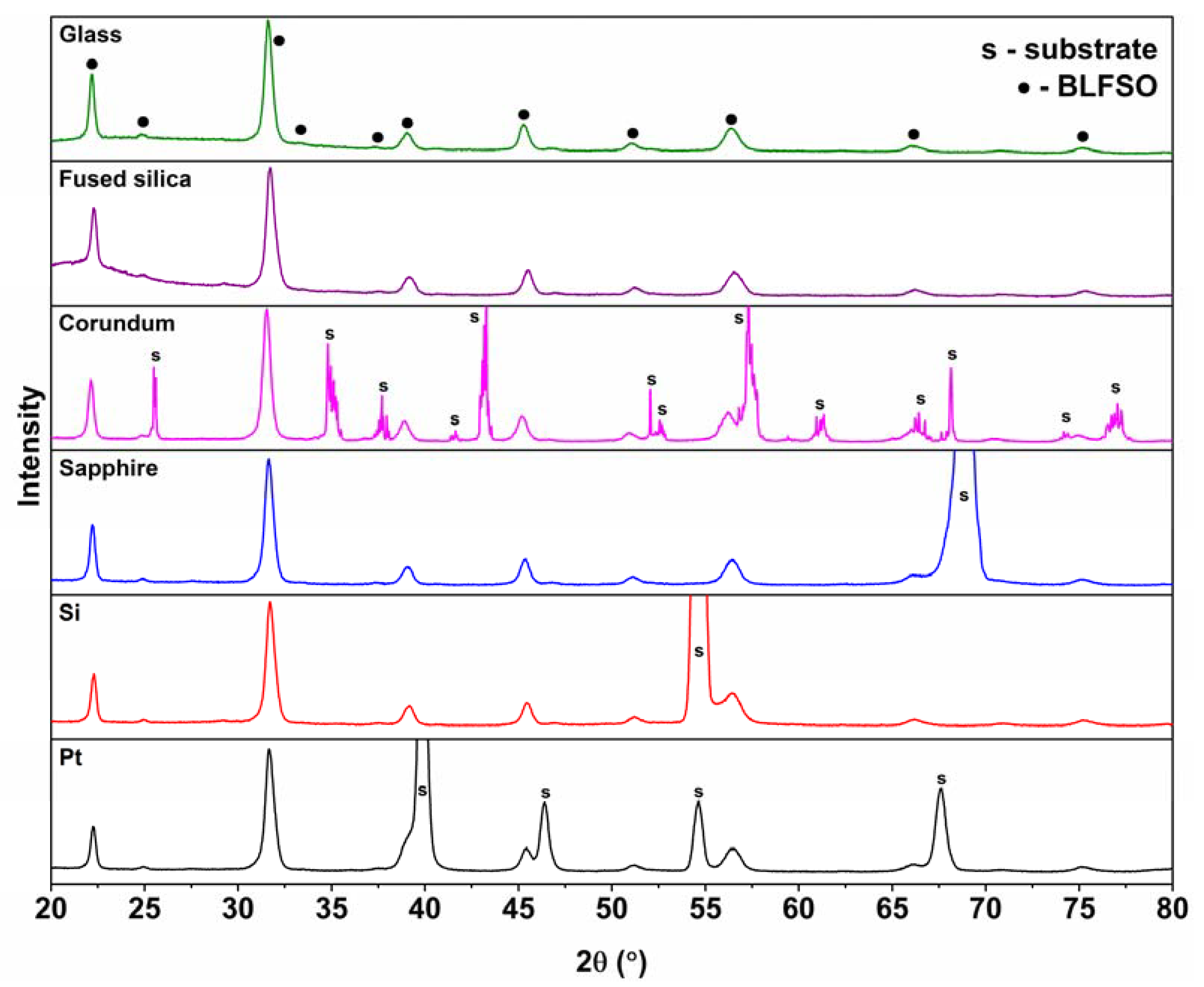
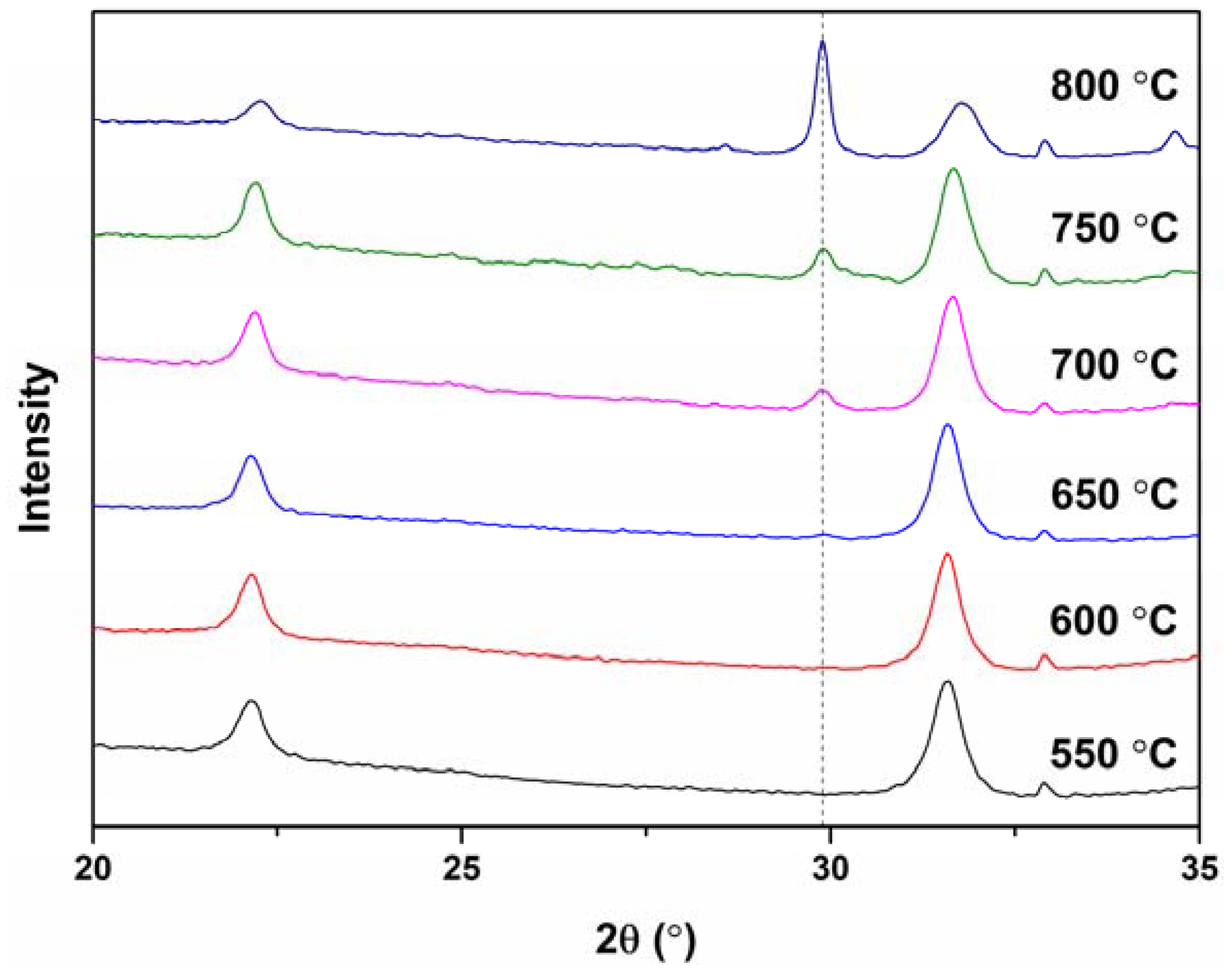
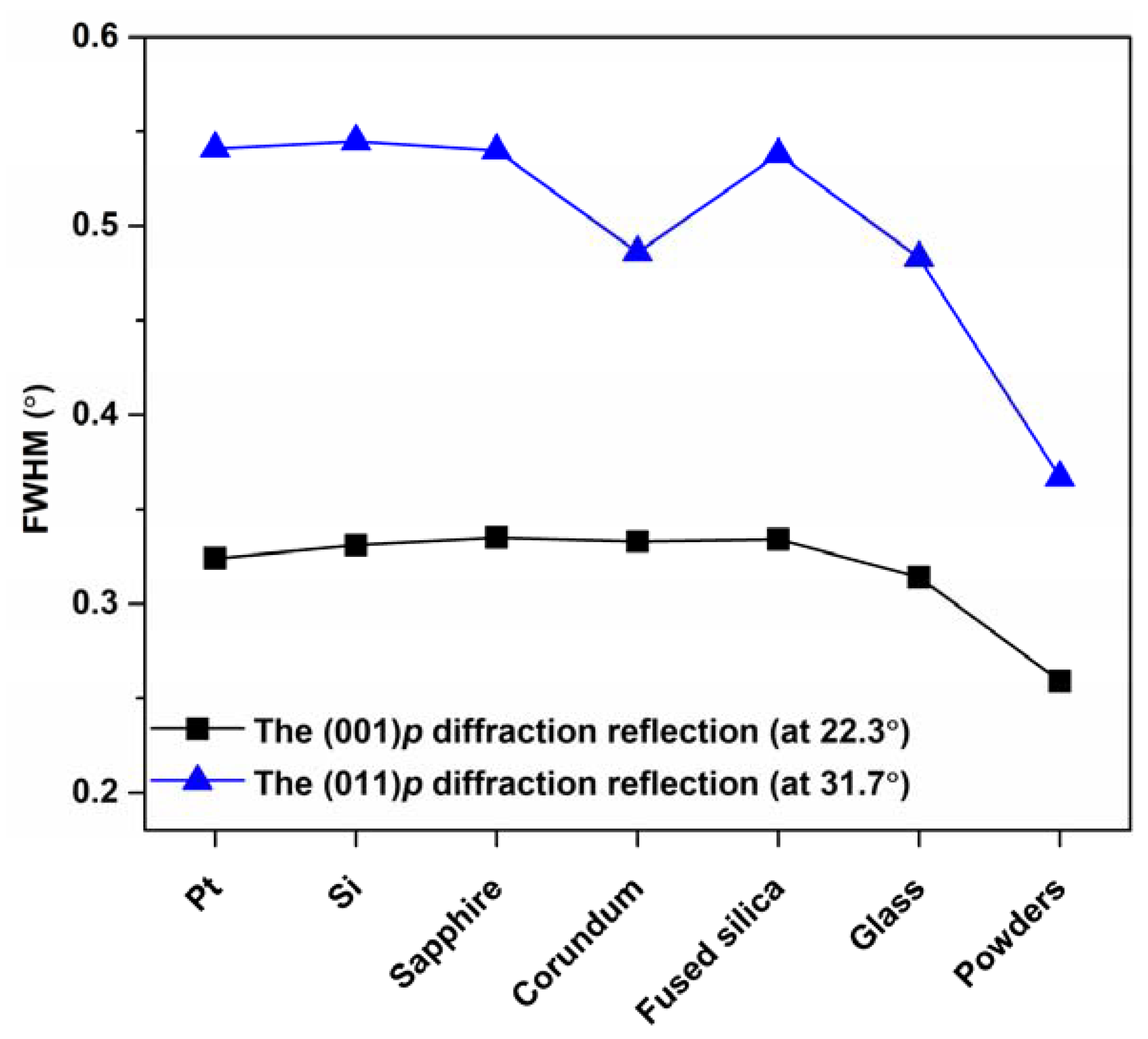
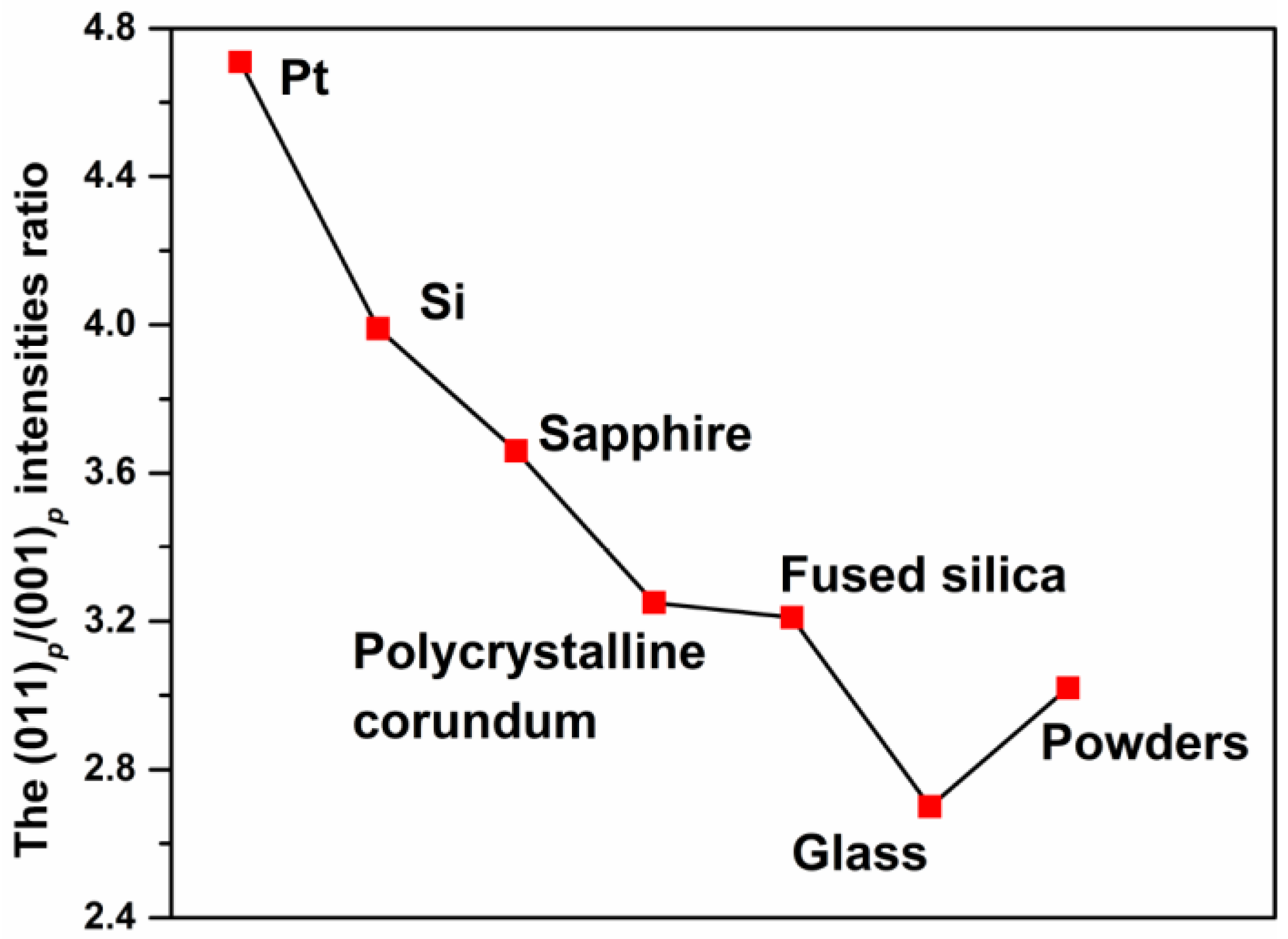
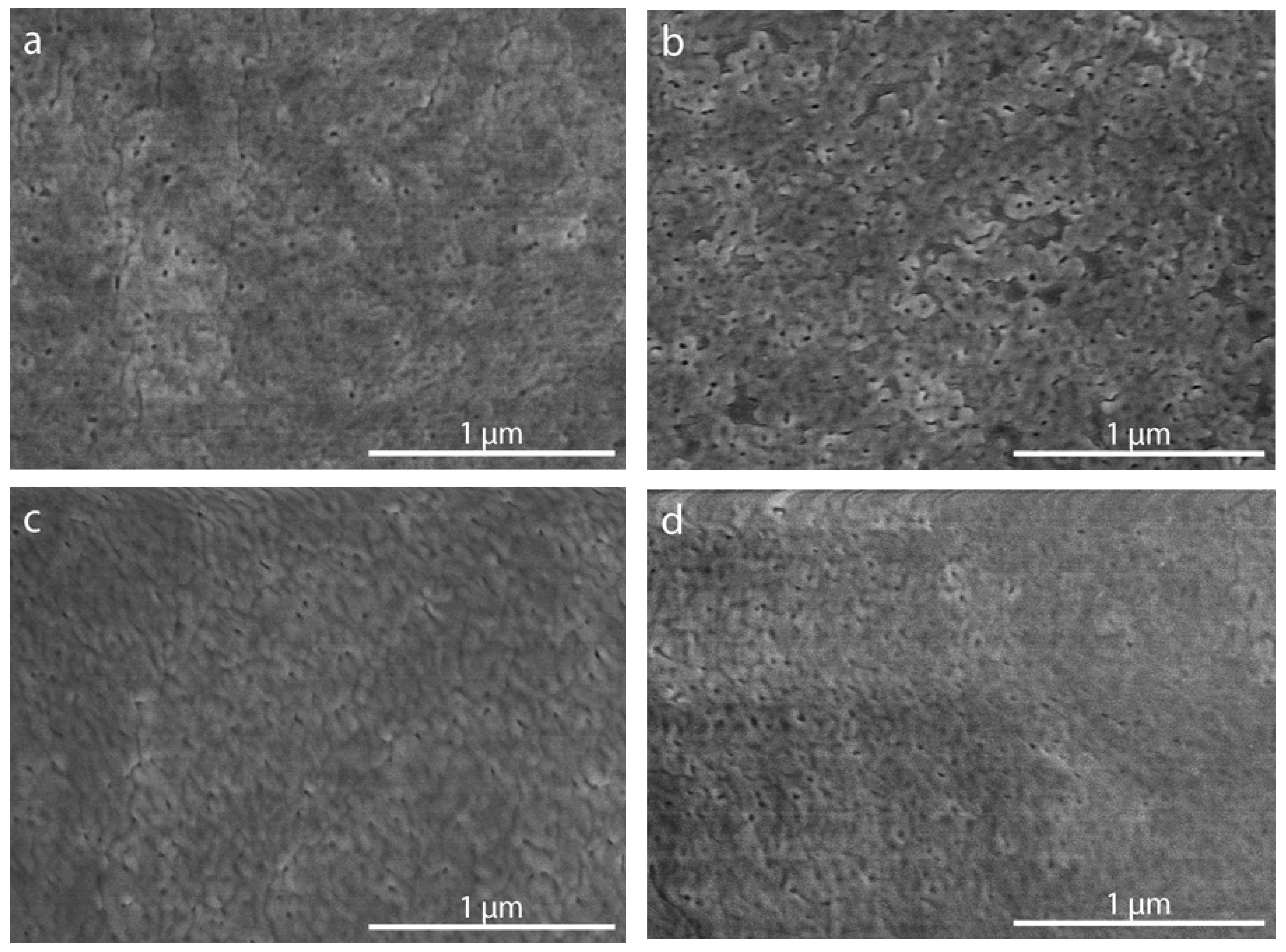
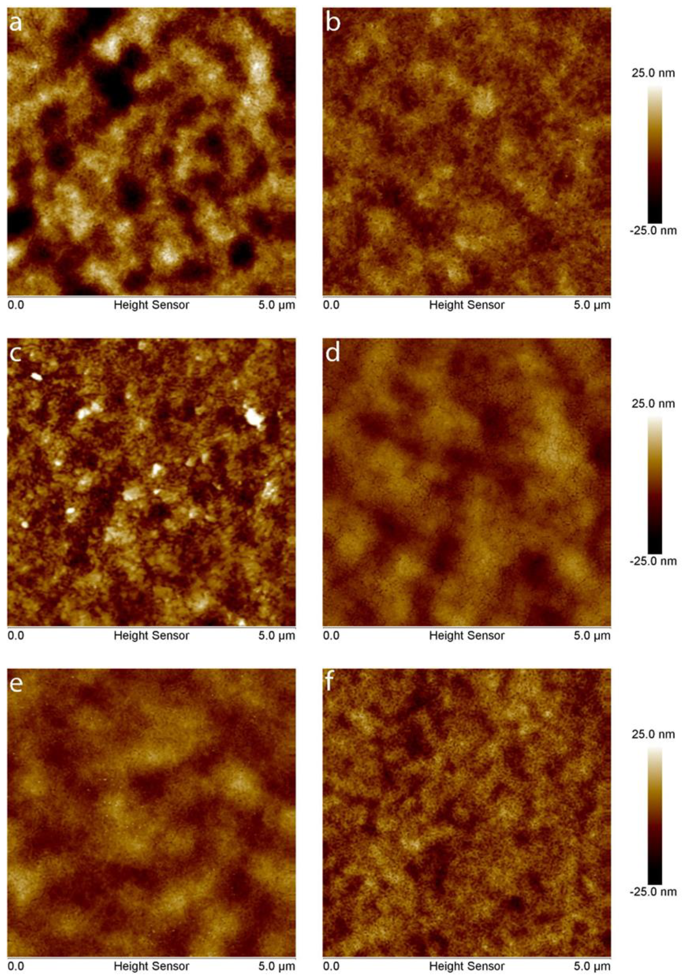
3. Results and Discussion

The thermal decomposition behavior of dried BLFSO precursor solution was investigated by simultaneous TG–DSC measurements (Figure 2) in order to determine the annealing temperature range suitable for the formation of a perovskite structure. Three main steps in the thermal degradation process can be seen. Insignificant weight loss accompanied by a weak endothermic signal can be noticed at low temperatures around 100 °C. This process could be ascribed to the removal of residual water and organic solvent. Further considerable weight loss corresponds to the decomposition of nitrates and organic species. The constant mass of the residual was observed at temperatures above 600 °C; therefore, this temperature was chosen for the final thermal treatment of the deposited films. Moreover, a higher annealing temperature would lead to the melting of glass substrates, whereas for correct comparison of the films, annealing temperature should be the same for all substrates. Based on a TG curve, the total weight loss was calculated to be 57%.
The phase crystallinity and purity of all synthesized samples were characterized by means of XRD analysis. Figure 3 represents the XRD patterns of the BLFSO coatings deposited on different substrates and annealed at 600 °C for 2 h. It is evident that the crystalline phase was formed after annealing regardless of the nature of the substrate. Intense and well-resolved diffraction peaks corresponding to the pseudocubic perovskite structure [15] could be seen in all the XRD patterns. In the case of Pt/TiO2/SiO2/Si, silicon, sapphire and corundum, the diffraction reflections arising from the substrates were also seen. The broad diffraction peaks indicate the nanocrystalline nature of the films, which was expected after crystallization at relatively low temperatures. The obtained results suggest that proposed deposition procedure is suitable for the fabrication of perovskite thin films on monocrystalline, polycrystalline, amorphous, transparent and conductive substrate, which provides broad possibilities and flexibility in terms of the further investigation of the physical properties of this material.
It is well known that high annealing temperatures may lead to the formation of dense, well-crystallized films. However, the limitations caused by the metastable nature of bulk BLFSO do not suggest high-temperature processing. For this reason, some of the synthesized films were additionally annealed at different temperatures in order to determine the limiting annealing conditions. The XRD patterns of BLFSO films deposited on Pt/TiO2/SiO2/Si substrate and annealed at 550–800 °C are depicted in Figure 4.
XRD data were collected in the 20–35 2θ range, which contained the most intense peaks and where impurity phases such as Bi2O3, Bi2Fe4O9 or Bi25FeO39 are usually detected [32,33]. It could be seen that there were no changes in the XRD patterns when the temperature was increased up to 600 °C. After annealing at 650 °C, a low intensity diffraction peak at ca. 30° appeared. The intensity of this peak gradually increased with an increase of annealing temperature. At the same time, the intensity of the most intense peak of the perovskite phase (at around 31.6°) decreased, and the peak shifted towards a higher 2θ region. One more peak, located at around 34.7°, arose after annealing at temperatures above 700 °C. The observed annealing-induced changes indicated decomposition of the perovskite phase and suggested that 600 °C was the most suitable annealing temperature. In order to reveal possible influence of the substrates on crystal structure, strain and/or texture of the deposited films, the full width at half maximum (FWHM) values for the two most intense diffraction reflections (001)p and (011)p (in pseudocubic perovskite lattice) located at ca. 22.3° and 31.7° were estimated and compared with the respective values for the powder obtained by calcination of the precursor solution at 600 °C (Figure 5).
Although in the vicinity of the compositional crossover from the Imma(00γ)s00 phase to the non-polar Pnma phase, the symmetry of the primitive perovskite cell of the Bi1−xLaxFe0.5Sc0.5O3 perovskites is near cubic [15], a transition between these phases could be detected by variation in the FWHM value of the most characteristic diffractions reflections. As seen from Figure 5, for each particular substrate, FWHM of the (001)p reflection was regularly smaller than that of the (011)p reflection, suggesting that all the obtained films were of the same symmetry.
The (011)p to (001)p intensities ratios for different substrates are shown in Figure 6. It is seen that intensity ratios were clearly dependent on the nature of the substrates. This value was estimated for the Pt substrate to be about 4.7. The lowest values were obtained for the amorphous substrates; for instance, it was only about 2.6 for the film on glass substrate. For the powder prepared from the precursor solution, this value was about 3.0.
The microstructure of the BLFSO films was examined by SEM. The most representative plane views of the films deposited on Pt/TiO2/SiO2/Si, silicon, sapphire and fused silica followed by annealing at 600 °C are given in Figure 7. The morphology of the films fabricated on all types of substrates was very similar. The obtained films were uniform but not absolutely dense: some pores could be observed. Thickness of the films was determined by SEM analysis of the cross-section. It was found that single-layered film obtained by one deposition cycle was about 50 nm thick. Moreover, the thickness of the films was independent of the type of substrate.
Figure 8 demonstrates 2D AFM images of the BLFSO films deposited on different substrates. The obtained images are in good agreement with the obtained SEM micrographs and confirm the presence of nanodimensional pores. The roughness of the films and non-coated substrates was estimated by calculation of the root mean square of height values (RMS), which are summarized in Table 1. The RMS values were calculated for the relatively large (20 µm × 20 µm) and the relatively small (5 µm × 5 µm) areas; the obtained results were in good agreement. It is seen that the roughness of the film was higher than that of uncoated substrate with an exception for mechanically polished corundum, which possessed a relatively rough initial surface. The RMS values varied in the range from 3.1 to 7.3 nm, and the highest roughness was observed for the Pt/TiO2/SiO2/Si substrate and the lowest for the fused silica one.

4. Conclusions

Polycrystalline Bi0.67La0.33Fe0.5Sc0.5O3 thin films were successfully fabricated by chemical solution deposition using a spin coating technique. Multi-layered films with perovskite structure were prepared on Pt/TiO2/SiO2/Si, silicon, sapphire, corundum, fused silica and glass substrates after annealing at 600 °C. Processing at higher temperatures resulted in the decomposition of the perovskite phase. The films were found to be of the same crystal symmetry regardless of the substrate used. At the same time, the relative intensity of the pseudocubic peaks (001)p and (011)p in XRD patterns was rather different for the films on different substrates, suggesting that the perovskite crystallites have some preferred orientation. The roughness of the films varied in the range from 3.1 to 7.3; the highest roughness was observed for the Pt/TiO2/SiO2/Si substrate and the lowest for the fused silica substrate.

Author Contributions

Conceptualization, A.N.S. and A.Z.; methodology, A.Z.; formal analysis, D.G., D.K., L.M., A.N.S. and A.Z.; investigation, D.G., D.K., L.M., A.N.S. and A.Z.; resources, A.K.; writing—original draft preparation, D.G.; writing—review and editing, A.K.; visualization, D.G.; supervision, A.K. All authors have read and agreed to the published version of the manuscript.

Funding

This work was supported by a Research grant BUNACOMP (No. S-MIP-19-9) from the Research Council of Lithuania (LMTLT). The research performed in the University of Aveiro was developed within the scope of the project CICECO-Aveiro Institute of Materials, UIDB/50011/2020 and UIDP/50011/2020, financed by national funds through the FCT/MEC and when appropriate co-financed by FEDER under the PT2020 Partnership Agreement.

Institutional Review Board Statement

Not applicable.

Informed Consent Statement

Not applicable.

Data Availability Statement

Data is contained within the article or supplementary material.

Conflicts of Interest

The authors declare no conflict of interest.

References

  1. Vopson, M.M. Fundamentals of multiferroic materials and their possible applications. Crit. Rev. Solid State Mater. Sci. 2015, 40, 223–250. [Google Scholar] [CrossRef] [Green Version]
  2. Lu, C.; Wu, M.; Lin, L.; Liu, J.-M. Single-phase multiferroics: New materials, phenomena, and physics. Natl. Sci. Rev. 2019, 6, 653–668. [Google Scholar] [CrossRef] [Green Version]
  3. Liu, H.; Yang, X. A brief review on perovskite multiferroics. Ferroelectrics 2017, 507, 69–85. [Google Scholar] [CrossRef]
  4. Catalan, G.; Scott, J.F. Physics and applications of bismuth ferrite. Adv. Mater. 2009, 21, 2463–2485. [Google Scholar] [CrossRef]
  5. Molak, A.; Mahato, D.K.; Szeremeta, A.Z. Synthesis and characterization of electrical features of bismuth manganite and bismuth ferrite: Effects of doping in cationic and anionic sublattice: Materials for applications. Prog. Cryst. Growth Charact. Mater. 2018, 64, 1–22. [Google Scholar] [CrossRef]
  6. Chen, L.; Cheng, Z.; Xu, W.; Meng, X.; Yuan, G.; Liu, J.; Liu, Z. Electrical and mechanical switching of ferroelectric polarization in the 70 nm BiFeO3 film. Sci. Rep. 2016, 6, 19092. [Google Scholar] [CrossRef] [PubMed] [Green Version]
  7. Ceballos-Sanchez, O.; Sanchez-Martinez, A.; Flores-Ruiz, F.J.; Huerta-Flores, A.M.; Torres-Martínez, L.M.; Ruelas, R.; García-Guaderrama, M. Study of BiFeO3 thin film obtained by a simple chemical method for the heterojunction-type solar cell design. J. Alloys Compd. 2020, 832, 154923. [Google Scholar] [CrossRef]
  8. Catalano, M.R.; Spedalotto, G.; Condorelli, G.G.; Malandrino, G. MOCVD growth of perovskite multiferroic BiFeO3 films: The effect of doping at the A and/or B sites on the structural, morphological and ferroelectric properties. Adv. Mater. Interfaces 2017, 4, 1601025. [Google Scholar] [CrossRef]
  9. McLeod, J.A.; Pchelkina, Z.V.; Finkelstein, L.D.; Kurmaev, E.Z.; Wilks, R.G.; Moewes, A.; Solovyev, I.V.; Belik, A.A.; Takayama-Muromachi, E. Electronic structure of BiMO3 multiferroics and related oxides. Phys. Rev. B 2010, 81, 144103. [Google Scholar] [CrossRef] [Green Version]
  10. Arnold, D.C. Composition-driven structural phase transitions in rare-earth-doped BiFeO3 ceramics: A review. IEEE Trans. Ultrason. Ferroelectr. Freq. Control 2015, 62, 62–82. [Google Scholar] [CrossRef]
  11. Fertman, E.L.; Fedorchenko, A.V.; Khalyavin, D.D.; Salak, A.N.; Baran, A.; Desnenko, V.A.; Kotlyar, O.V.; Cizmar, E.; Feher, A.; Syrkin, E.S.; et al. Multiferroic Bi0.65La0.35Fe0.5Sc0.5O3 perovskite: Magnetic and thermodynamic properties. J. Magn. Magn. Mater. 2017, 429, 177–181. [Google Scholar] [CrossRef] [Green Version]
  12. Fedorchenko, A.V.; Fertman, E.L.; Desnenko, V.A.; Kotlyar, O.V.; Cizmar, E.; Shvartsman, V.V.; Lupascu, D.C.; Salamon, S.; Wende, H.; Salak, A.N.; et al. Magnetic properties of the Bi0.65La0.35Fe0.5Sc0.5O3 perovskite. Acta Phys. Pol. A 2017, 131, 1069–1071. [Google Scholar] [CrossRef]
  13. Khalyavin, D.D.; Salak, A.N.; Lopes, A.B.; Olekhnovich, N.M.; Pushkarev, A.V.; Radyush, Y.V.; Fertman, E.L.; Desnenko, V.A.; Fedorchenko, A.V.; Manuel, P.; et al. Magnetic structure of an incommensurate phase of La-doped BiFe0.5Sc0.5O3: Role of antisymmetric exchange interactions. Phys. Rev. B 2015, 92, 224428. [Google Scholar] [CrossRef]
  14. Khalyavin, D.D.; Salak, A.N.; Manuel, P.; Olekhnovich, N.M.; Pushkarev, A.V.; Radysh, Y.V.; Fedorchenko, A.V.; Fertman, E.L.; Desnenko, V.A.; Ferreira, M.G.S. Antisymmetric exchange in La-substituted BiFe0.5Sc0.5O3 system: Symmetry adapted distortion modes approach. Z. Krist. Cryst. Mater. 2015, 230, 767–774. [Google Scholar] [CrossRef] [Green Version]
  15. Salak, A.N.; Khalyavin, D.D.; Zamaraite, I.; Stanulis, A.; Kareiva, A.; Shilin, A.D.; Rubanik, V.V.; Radyush, Y.V.; Pushkarev, A.V.; Olekhnovich, N.M.; et al. Metastable perovskite Bi1−xLaxFe0.5Sc0.5O3 phases in the range of the compositional crossover. Phase Transit. 2017, 90, 831–839. [Google Scholar] [CrossRef]
  16. Lu, C.; Hu, W.; Tian, Y.; Wu, T. Multiferroic oxide thin films and heterostructures. Appl. Phys. Rev. 2015, 2, 021304. [Google Scholar] [CrossRef] [Green Version]
  17. Martin, L.W.; Chu, Y.H.; Ramesh, R. Advances in the growth and characterization of magnetic, ferroelectric, and multiferroic oxide thin films. Mater. Sci. Eng. R Rep. 2010, 68, 89–133. [Google Scholar] [CrossRef] [Green Version]
  18. Yan, F.; Zhu, T.J.; Lai, M.O.; Lu, L. Enhanced multiferroic properties and domain structure of La-doped BiFeO3 thin films. Scr. Mater. 2010, 63, 780–783. [Google Scholar] [CrossRef]
  19. Chen, X.; Hu, G.; Yan, J.; Wang, X.; Yang, C.; Wu, W. Enhanced multiferroic properties of (1 1 0)-oriented BiFeO3 film deposited on Bi3.5Nd0.5Ti3O12-buffered indium tin oxide/Si substrate. J. Phys. D Appl. Phys. 2008, 41, 225402. [Google Scholar] [CrossRef]
  20. Zhang, Q.; Sando, D.; Nagarajan, V. Chemical route derived bismuth ferrite thin films and nanomaterials. J. Mater. Chem. C 2016, 4, 4092–4124. [Google Scholar] [CrossRef]
  21. Deng, X.; Huang, J.; Sun, Y.; Liu, K.; Gao, R.; Cai, W.; Fu, C. Effect of processing parameters on the structural, electrical and magnetic properties of BFO thin film synthesized via RF magnetron sputtering. J. Alloys Compd. 2016, 684, 510–515. [Google Scholar] [CrossRef]
  22. Deng, X.; Zeng, Z.; Xu, R.; Qin, X.; Li, X.; Wang, Y.; Gao, R.; Wang, Z.; Chen, G.; Cai, W.; et al. Effect of annealing atmosphere on structural and multiferroic properties of BiFeO3 thin film prepared by RF magnetron sputtering. J. Mater. Sci. Mater. Electron. 2019, 30, 16502–16509. [Google Scholar] [CrossRef]
  23. Hu, C.-W.; Yen, C.-M.; Feng, Y.-C.; Chen, L.-H.; Liao, B.-Z.; Chen, S.-C.; Liao, M.-H. Multi-ferroic properties on BiFeO3/BaTiO3 multi-layer thin-film structures with the strong magneto-electric effect for the application of magneto-electric devices. Coatings 2021, 11, 66. [Google Scholar] [CrossRef]
  24. Abramov, A.; Alikin, D.; Sobol, A.; Myakishev, D.; Slabov, V.; Trusov, L.; Safina, V.; Turygin, A.; Vasiliev, A.; Shur, V.; et al. Chemical solution deposition of BiFeO3 films with layer-by-layer control of the coverage and composition. Coatings 2020, 10, 438. [Google Scholar] [CrossRef]
  25. Dubourdieu, C.; Kang, S.B.; Li, Y.Q.; Kulesha, G.; Gallois, B. Solid single-source metal organic chemical vapor deposition of yttria-stabilized zirconia. Thin Solid Film. 1999, 339, 165–173. [Google Scholar] [CrossRef]
  26. Murauskas, T.; Kubilius, V.; Saltyte, Z.; Plausinaitiene, V. Metalorganic chemical vapor deposition and investigation of nonstoichiometry of undoped BaSnO3 and La-doped BaSnO3 thin films. Thin Solid Film. 2019, 692, 137575. [Google Scholar] [CrossRef]
  27. Jiang, J.; Shen, W.; Hertz, J.L. Fabrication of epitaxial zirconia and ceria thin films with arbitrary dopant and host atom composition. Thin Solid Film. 2012, 522, 66–70. [Google Scholar] [CrossRef]
  28. Zarkov, A.; Stanulis, A.; Mikoliunaite, L.; Katelnikovas, A.; Jasulaitiene, V.; Ramanauskas, R.; Tautkus, S.; Kareiva, A. Chemical solution deposition of pure and Gd-doped ceria thin films: Structural, morphological and optical properties. Ceram. Int. 2017, 43, 4280–4287. [Google Scholar] [CrossRef]
  29. Jonauske, V.; Stanionyte, S.; Chen, S.-W.; Zarkov, A.; Juskenas, R.; Selskis, A.; Matijosius, T.; Yang, T.C.K.; Ishikawa, K.; Ramanauskas, R.; et al. Characterization of sol-gel derived calcium hydroxyapatite coatings fabricated on patterned rough stainless steel surface. Coatings 2019, 9, 334. [Google Scholar] [CrossRef] [Green Version]
  30. Gul, E.; Stanulis, A.; Barushka, Y.; Garskaite, E.; Ramanauskas, R.; Morkan, A.U.; Kareiva, A. The influence of thermal processing on microstructure of sol–gel-derived SrSnO3 thin films. J. Mater. Sci. 2017, 52, 12624–12634. [Google Scholar] [CrossRef]
  31. Karoblis, D.; Griesiute, D.; Mazeika, K.; Baltrunas, D.; Karpinsky, D.V.; Lukowiak, A.; Gluchowski, P.; Raudonis, R.; Katelnikovas, A.; Zarkov, A.; et al. A facile synthesis and characterization of highly crystalline submicro-sized BiFeO3. Materials 2020, 13, 3035. [Google Scholar] [CrossRef] [PubMed]
  32. Carranza-Celis, D.; Cardona-Rodríguez, A.; Narváez, J.; Moscoso-Londono, O.; Muraca, D.; Knobel, M.; Ornelas-Soto, N.; Reiber, A.; Ramírez, J.G. Control of multiferroic properties in BiFeO3 nanoparticles. Sci. Rep. 2019, 9, 3182. [Google Scholar] [CrossRef] [PubMed]
  33. Selbach, S.M.; Einarsrud, M.-A.; Grande, T. On the thermodynamic stability of BiFeO3. Chem. Mater. 2009, 21, 169–173. [Google Scholar] [CrossRef]
Figure 1. Schematic representation of the synthesis of BLFSO thin films and optical photographs of the films deposited on Pt/TiO2/SiO2/Si, corundum, silicon and fused silica substrates.
Coatings 11 00307 g001
Figure 2. TG–DTG–DSC curves of dried Bi–La–Fe–Sc–O precursor solution.
Coatings 11 00307 g002
Figure 3. XRD patterns of the BLFSO films processed at 600 °C on different substrates.
Coatings 11 00307 g003
Figure 4. XRD patterns of the BLFSO films annealed at different temperatures (Pt/TiO2/SiO2/Si substrate).
Coatings 11 00307 g004
Figure 5. FWHM of the (001)p and (011)p reflections for the films fabricated on different substrates.
Coatings 11 00307 g005
Figure 6. Ratio of the intensities of the (011)p and (001)p reflections in XRD patterns of the BLFSO films fabricated on different substrates. The point corresponding to the respective value for the powder sample is shown for comparison.
Coatings 11 00307 g006
Figure 7. Plane view SEM images of the BLFSO films deposited on Pt/TiO2/SiO2/Si (a), silicon (b), sapphire (c) and fused silica (d).
Coatings 11 00307 g007
Figure 8. AFM images of the BLFSO films processed at 600 °C on Pt/TiO2/SiO2/Si (a), silicon (b), sapphire (c), corundum (d), fused silica (e) and glass (f).
Coatings 11 00307 g008
Table 1. Roughness of selected substrates and films calculated as RMS.
SubstrateRoughness of Substrate, nmRoughness of Film on the Respective Substrate, nm
Pt/TiO2/SiO2/Si, Si/SiO24.2 ± 0.17.3 ± 0.8
Silicon0.6 ± 0.33.8 ± 0.1
Sapphire1.2 ± 0.25.8 ± 0.9
Corundum9.2 ± 24.8 ± 0.2
Fused silica0.8 ± 0.13.1 ± 0.5
Glass1.1 ± 0.14.2 ± 0.3
Publisher’s Note: MDPI stays neutral with regard to jurisdictional claims in published maps and institutional affiliations.

