Article

Detection of Virulence Genes and Biofilm Forming Capacity of Diarrheagenic E. coli Isolated from Different Water Sources

1
Department of Biochemistry, Government College University Faisalabad, Faisalabad 38000, Pakistan
2
Department of Biological Sciences, International Islamic University, H-10 Campus, Islamabad 44000, Pakistan
3
Department of Botany, Government College University Faisalabad, Faisalabad 38000, Pakistan
4
Department of Chemical Engineering, School of Mining, Metallurgy and Chemical Engineering, University of Johannesburg, P. O. Box 17011, Doornfontein 2028, South Africa
5
Department of Biological and Chemical Engineering, Aarhus University, Nørrebrogade 44, 8000 Aarhus, Denmark
6
Department of Food Sciences & Nutrition, College of Food & Agriculture Sciences, King Saud University, Riyadh 11451, Saudi Arabia
7
Department of Environmental Sciences and Engineering, Government College University, Allama Iqbal Road, Faisalabad 38000, Pakistan
8
Department of Biological Sciences and Technology, China Medical University, Taichung 40402, Taiwan
*
Authors to whom correspondence should be addressed.
Coatings 2021, 11(12), 1544; https://doi.org/10.3390/coatings11121544
Submission received: 17 November 2021 / Revised: 2 December 2021 / Accepted: 8 December 2021 / Published: 16 December 2021

Abstract

:
Diarrheagenic Escherichia coli (DEC) are associated with frequent incidences of waterborne infections and pose health risk to individuals who contact water for domestic or recreational uses. Detection of DEC pathotypes in drinking water can be used as an indicator of fecal contamination. This study aimed to investigate the occurrence of DEC pathotypes and their capacity to form biofilms in drinking water samples collected from different water sources. In this study, PCR analysis was used to determine the occurrence of four clinically significant virulence genes of diarrheagenic E. coli, eaeA (Enteropathogenic E. coli), stx1, stx2 (Enterohemorrhagic E. coli) and sth (Enterotoxigenic E. coli), in drinking water samples (n = 35) by using specific primers and conditions. PCR amplicons were visualized by using agarose gel electrophoresis. A total of 12/35 (34%) samples were detected as positive for at least one of the four DEC virulence genes and 11/12 (91%) E. coli isolates harbored virulence gene while 1/12 (8%) E. coli isolates harbored none. The eaeA and sth genes were the most detected genes (75%), while stx1 and stx2 genes were least detected genes (66%). Biofilm assay confirmed that ETEC pathotypes can cause damage in enteric walls by attaching and effacing to persist diarrheal conditions. This study indicated that drinking water of different sources is contaminated with potential DEC pathotypes and it can be a source of diarrheal diseases. The amplification of four virulence genes associated with DEC pathotypes (EPEC, EHEC and ETEC) in drinking water demonstrates that potentially virulent DEC pathotypes are distributed in water sources and may be a cause of health concern. There is, therefore, an urgent need to monitor DEC pathotypes in drinking water.

1. Introduction

In order to ensure good health, the availability of safe and good quality water is very essential [1]. Waterborne infections are major cause of high morbidity and mortality rate and have become a public health problem [2]. In developing countries, ground water is the sole elementary source of drinking water supply, which may contain several pathogens, viruses, bacteria and protozoa, causing 2.5 million deaths from enteric diarrheal diseases annually [3,4]. Diarrheal and other gastrointestinal illnesses are common infections among infants and children in developing countries [5]. Improper sanitation and hygiene cause comparatively greater health risk [6,7]. Escherichia coli are one of inhabitants of gut flora and most commonly used indicator for fecal contamination in different drinking water distribution systems [8]. E. coli can survive in drinking water for 4 to 12 weeks, depending upon various environmental factors. According to its biological significance, E. coli live safely in the intestine as harmless commensal [9] and pathogenic strains of bacteria cause intestinal and extra-intestinal infections both in healthy and immunocompromised individuals [2,10].
The accessory genome expresses pathogenic traits that are developed by horizontal gene transfer. The virulence genes are located on the accessory genome: a kind of genome plasticity formed by gain and loss of genetic information [1,11]. On the basis of virulence determinants (acquired), specific combinations and horizontal gene transfer, diarrheagenic E. coli pathotypes are classified into different pathotypes. These pathotypes are enteropathogenic E. coli (EPEC), enteroinvasive E. coli (EIEC), enterohemorrhagic E. coli (EHEC), enteroaggregative E. coli (EAEC), diffusely adherent E. coli (DAEC) and enterotoxigenic E. coli (ETEC) [11,12]. These pathotypes define different clinical features, phenotypic traits, epidemiological evidences and specific virulence factors of DEC pathotypes. These pathotypes vary for their clinical, epidemiological and pathogenic significance [2] (Table 1).
DEC pathotypes form colonies on the epithelia of intestines by either inhabiting and invading the intestinal cells. They are specialized to form adhesions on the epithelia and known for their attaching and effacing ability to form lesions [9]. They possess resistance against immune system as the colonies target and respond in a similar manner. The virulence factors inject themselves in the cells by mimicking the cell ligands and disrupt plasticity of cytoskeleton [13], thereby reducing endocytic trafficking; they make themselves resistant to phagocytosis. The host cells respond and defend by inducing inflammatory reactions. It changes the intestinal fluid balance to excrete unwanted and harmful bacteria that cause diarrhea [14,15]. The extent of infection depends on the interaction between the cell and bacteria and also on the defense system [16,17,18].
DEC pathotypes have developed important structures for attaching and effacing the cells such as bundle forming pili (bfp), EspA filaments and EAF shiga-like toxins and intimin [19,20]. The bfp encoded by a ~80 kb adherence factor forms bacterial micro-colonies. Shiga-like toxin and intimin are encoded by stx and eaeA genes, respectively. The eaeA gene is an E. coli outer membrane protein. Intimin is known for its intimate adhesions. EHEC are shiga toxin producers posessing two main classes: stx1 and stx2; they are further divided into many subtypes [20,21]. STEC detection methods mostly depend on serotype 0157H7. The EPEC secreted proteins are translocated through a type III secretion system [16,22]. It forms a pore inside the bacterial membrane. EspA filaments are parts of type III secretion system [23] and directly related to the epithelial cells due to its interaction in a manner functioning as adhesions [17,24]. EAF is important for its role in pathogenicity, biofilm production and colonization. EspA forms a filament-like translocation tube outside the bacteria [25], while EspB and EspD are incorporated inside the membrane of host cells [26]. It allows the movement of other proteins through the cell membrane, e.g., Tir an effector protein [23].
The presence of DEC pathotypes forming biofilms can cause a serious threat because it can cause difficulties in medical treatments [27,28]. DEC pathotypes, especially ETEC, either make biofilms or attach to already existing biofilms. Biofilms are bacterial surface groups surrounded by extracellular matrix [29,30]. It provides bacteria with shelter, prevents its desiccation, make it resistant to antibiotics and protects it from bacteriophages. Biofilms are rapidly developed on both abiotic and biotic surfaces that are in direct contact with water [18,27]. Bacterial biofilm production is highly complex and regulated. They have evolved a complex system of DNA, exopolysaccharides and proteins to protect themselves from environmental stress [31,32]. In developing countries, very few studies have been performed with respect to reporting the virulence genes of DEC isolated from drinking water. Therefore, this study aimed to detect virulence-associated genes of DEC pathotypes associated with human pathology and to assess their ability to form biofilms to determine the risk of drinking contaminated water.

2. Material and Method

2.1. Water Samples Collection

Drinking water samples were collected from different water sources: ten samples were collected from tank water, five samples were collected from industries, five samples were infused water, three samples were collected from standing water areas, five samples were collected from wells and seven samples were collected from tap labeled as 1–35, respectively. A total of 35 drinking water samples were collected for this study. An amount of 200 mL of drinking water sample was collected from each site in sterilized glass containers having lids according to the American Public Health Association 2001. These samples were transported to laboratory in a cooler and processed in 4 h.

2.2. Isolation and Biochemical Identification of E. coli

The collected samples were cultured on MacConkey agar (Oxoid) plates by swabbing sterilized cotton swab and incubated at 37 °C for 24 h. These plates were observed to detect grown bacterial colonies. Only suspected colonies were considered for Gram’s reaction. Gram-negative bacillus colonies were detected. Each suspected sample was subjected to further biochemical testing. Triple Sugar Iron agar (TSI) test was used for biochemical characterization. The autoclaved TSI agar was allowed to set in a slanted position in sterilized test tubes. The slants were inoculated by picking a colony from the MacConkey agar plates and kept in an incubator at 37 °C overnight.

2.3. Extraction of DNA

The boiling centrifugation method was used to extract DNA from all isolated E. coli colonies as described by [33], with slight modifications. Isolated E. coli colonies were subcultured on nutrient agar at 37 °C. Isolated E. coli colonies were picked from a nutrient agar plate in a sterile Eppendorf tube and washed with 1 mL sterile normal saline (NS). It was centrifuged at 10,000 rev/min for 2 min. The pellet was recovered and re-suspended in 100 μL distilled water. Then, the tubes were boiled at 85 °C for 10 min. The lysate was centrifuged at 13,000 rev/min for 5 min. The supernatant containing template DNA stock was stored at 20 °C. The presence and concentration of extracted template DNA were measured by using agarose gel electrophoresis in the sample, and it was compared with a specific DNA marker of known concentration.

2.4. Polymerase Chain Reaction (PCR)

PCR (Thermal cycle, London, UK) was performed for the detection of target genes stx1 and stx2 for EHEC, sth for ETEC and eaeA for EPEC by using specific primers (Table 2). The reagents used for each reaction in volumes of 25 μL were as follows: 7 μL syringe water, 2.5 μL 100 mM KCl, 3 μL 3 mM MgCl2, 1 μL 400 μM dNTPs, 2.5 μL 20 mM tris-HCl, 0.5 μL forward primer, 0.5 μL reverse primer, 2.5 μL 0.1% gelatin, 5.0 μL DNA (Template DNA) and 0.5 μL 2.5 units Taq Polymerase. The PCR mixture was subjected to an initial denaturation step at 95 °C for 5 min, followed by 35 cycles of denaturation at 95 °C for 45 s, primer annealing at 57 °C for 45 s, extension at 72 °C for 45 s and the final extension at 72 °C for 10 min.

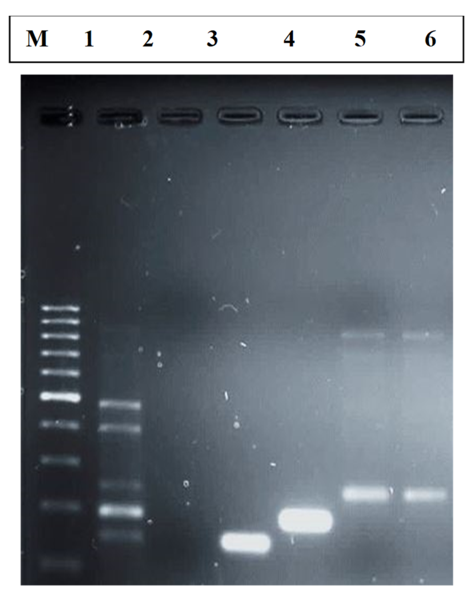
2.5. Visualization of PCR Products

Agarose (2%) was used to make agarose gel. The casting tray was flooded by 10× TBE buffer near the gel surface, and 5 μL PCR product of each sample along with DNA dye was loaded into each well. An amount of 3 μL of DNA ladder was injected in the first well of casting tray. Gel electrophoresis apparatus was then connected to a power supply of 100 V/cm for 30 min. The gel was removed from the holder and visualized by a UV illuminator.

2.6. Biofilm Assay

The PCR positive samples were further assayed to check biofilm production qualitatively. In order to assay biofilm formation, 3 mL of autoclaved Luria Bertani broth was taken in a test tube and incubated overnight at 37 °C at a 130 rpm shaker. An amount of 3 mL of LB was poured in each test tube, and three sets were made and they were autoclaved. An amount of 100 µL of culture was inoculated into each test tube of triplicate sets. These three sets of test tubes were incubated at 37 °C for different time intervals, i.e., one set for 24 h, the second set for 48 h and the third set for 72 h. After 24 h, cultured media were discarded from test tubes and stained with 0.5% crystal violet dye for 5 min. Each test tube was washed with distilled water. Ninety-five percent ethanol (4 mL) was used to dissolve the ring formed in test tubes after staining them with 0.5% crystal violet dye and optical density was measured at 570 nm of all the three sets of test tubes incubated at 37 °C for 24, 48 and 72 h [30,37].

2.7. Statistical Analysis

Statistical analysis for the prevalence of virulence genes of EPEC, ETEC and EHEC in drinking water samples was performed using Microsoft Excel spreadsheet and the result was presented as a percentage or graph.

3. Results and Discussion

3.1. Distribution of Isolated Organisms

All collected water samples (n = 35) were screened for the presence of DEC bacteria. Twelve out of thirty-five (34%) water samples were screened as positive for the presence of DAE; thus, 12 E. coli bacteria samples were isolated from collected drinking water. The distribution of water samples was as follows: 30% (3/10) were from tank water, 20% (1/5) were from industrial water, 20% (1/5) were from infused water, 66% (2/3) were from standing water, 20% (1/5) were from well water and 57% (4/7) were from tap water (Table 3).

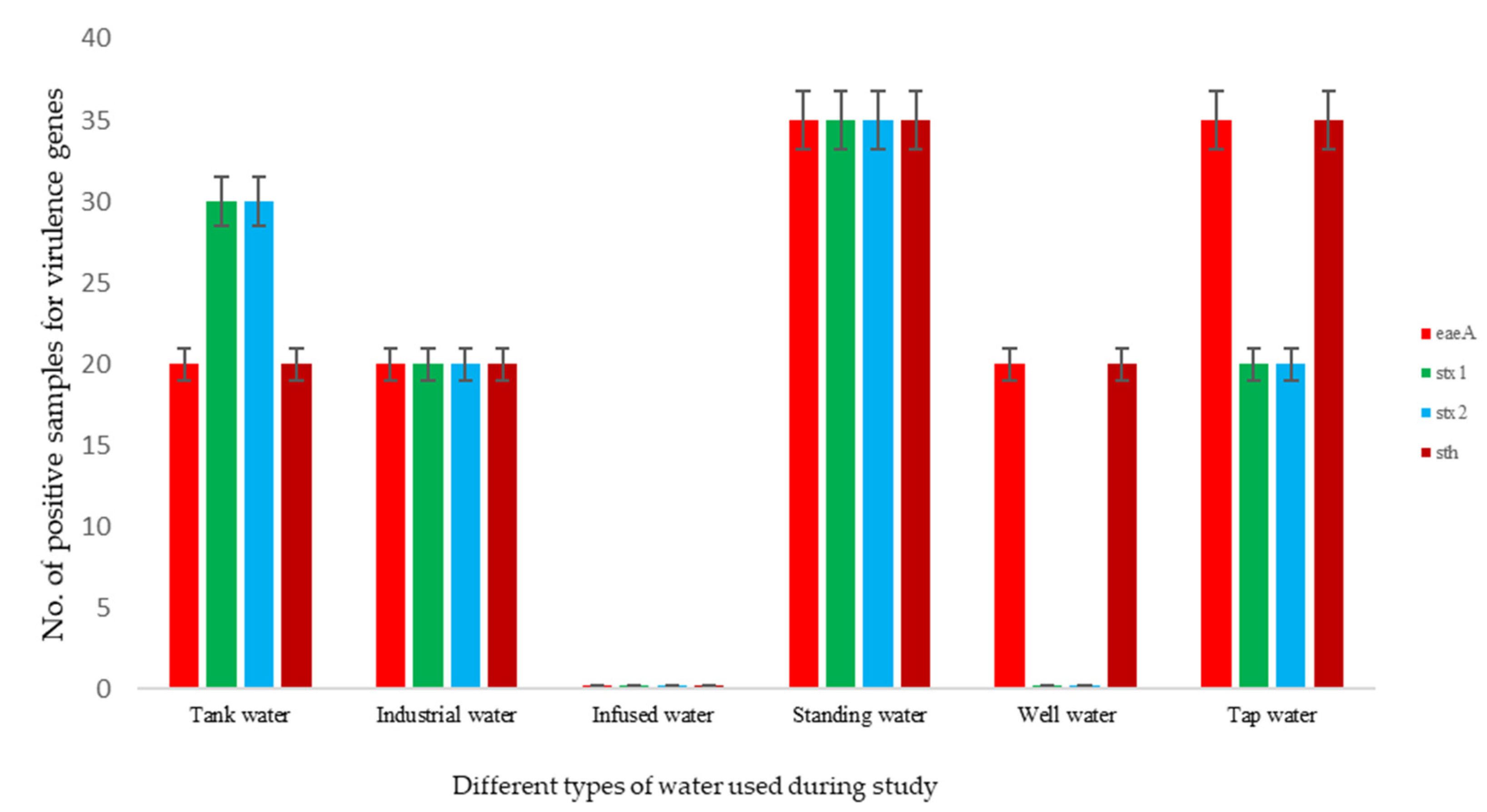
3.2. Prevalence of Virulence Genes from E. coli Isolates
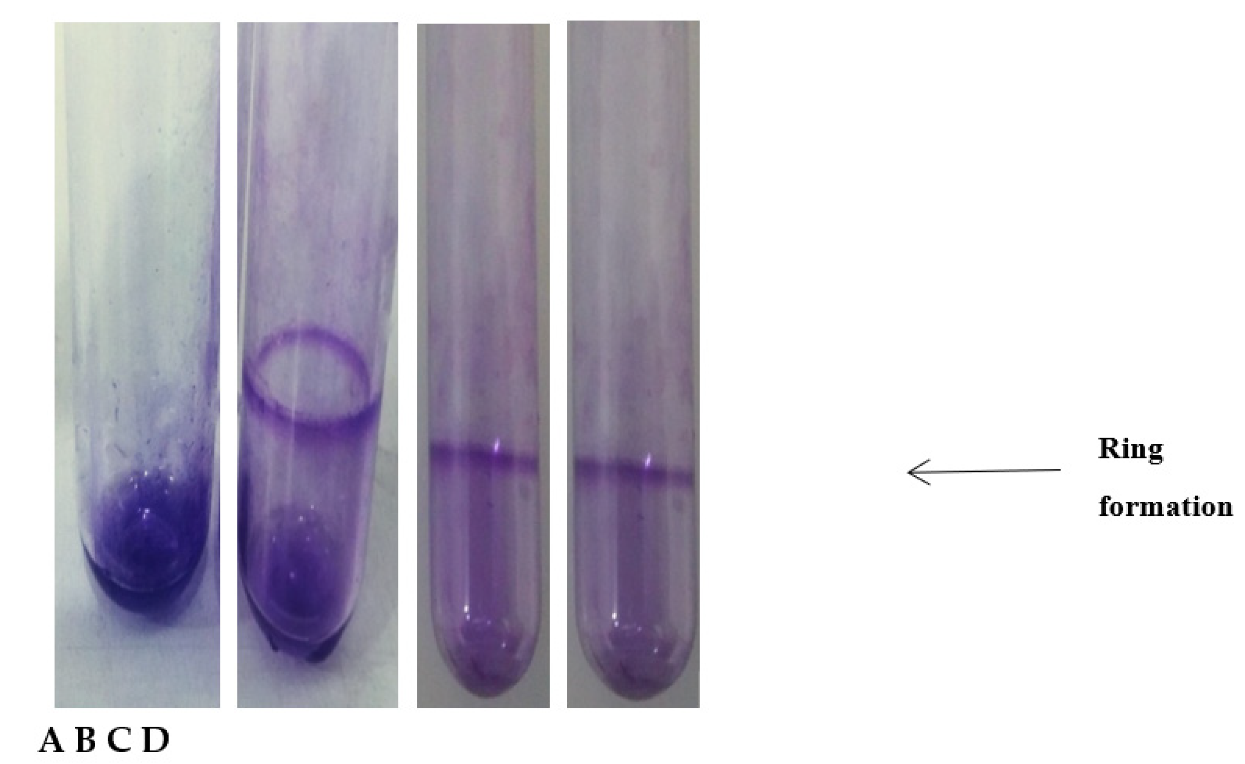
Amongst 35 collected water samples, twelve confirmed E. coli isolates were evaluated for various virulence genes eaeA, stx1, stx2 and sth. Ninety-one percent (11/12) of the E. coli isolates harbored virulence gene while 8% (1/12) E. coli isolates harbored none. The most frequent virulence factor genes were eaeA and sth, each of which was detected in 75% of E. coli isolates. In contrast, four virulence genes, including eaeA, stx1, stx2 and sth, were each observed in 41% of E. coli isolates (Table 3). In the current study, the frequencies of virulence genes were as follows: eaeA (75%), stx1 (66%), stx2 (66%) and sth (75%). These readings showed that the prevalence rate of virulence genes eaeA and sth was very high among DEC pathotypes isolated from drinking water. Therefore, the E. coli strains isolated from drinking water carried the virulence-associated eaeA and sth genes more frequently than compared to stx1 and stx2 (Figure 1). In addition, the results of the PCR for the identification of eaeA, stx1, stx2 and sth genes are shown in Figure 2.

3.3. Biofilm Assay

A biofilm assay was used to check the ability of biofilm production of EPEC, EHEC and ETEC pathotypes. The PCR positive samples were further assayed to check biofilm production. All PCR positive samples were incubated in LB media for different time intervals at 37 °C. Set A of test tubes containing only LB media was taken as control. Three sets of test tubes were prepared; B was incubated for 24 h, C was incubated for 48 h and D was incubated for 72 h. Zero-point five percent crystal violet dye was used to stain all the test tubes. After staining, the test tubes were washed with distilled water in order to remove excessive dye. The most prominent biofilm ring was observed in the test tube incubated for 24 h possessing the ETEC pathotype. These strains developed adhesion to the walls of test tubes and formed a ring. The ring formation around the walls of test tubes confirmed biofilm production by ETEC pathotype (Figure 3).
All the samples that showed positive results in the qualitative biofilm assay were further processed in a quantitative biofilm assay. The ring was dissolved by 95% ethanol, and optical density (OD) was measured in spectrophotometer at 570 nm (Table 4).
The OD 0.2 of sample 1 reported that these strains can strongly adhere to biotic and abiotic surfaces. Sample 2 producing an OD of 0.15 showed that these strains are also involved in biofilm production with moderate adhesion to the surfaces. Sample 3 with OD 0.1 showed that the strains were involved in weak adhesion to the surfaces with biofilm production as shown by [37]. In current research, virulence genes were detected in DEC isolates to detect the presence of virulent E. coli strains in drinking water. Other studies have also reported the presence E. coli strains as fecal indicator bacteria originating from defective hygiene and poor sanitation and animal fecal contact in freshwater places in developing countries [2,17,38]. The presence of ETEC in drinking water is indicative of a high risk of contamination. In general, these results found that the risk of contamination may increase over time, and it is high time to follow appropriate preventive measures.

4. Conclusions

Water sources harbor different pathotypes of DEC, which can possibly be suspended by natural or synthetic events. In the present study, four virulence genes (eaeA, stx1, stx2 and sth) were investigated pertaining to three DEC pathotypes (EPEC, EHEC and ETEC), which signify a potential health concern for individual drinking water from these sources. Approximately 34% of analyzed water samples were found positive for at least one of the four virulence genes. The most frequent pathotypes were EPEC and ETEC as eaeA and sth genes were detected in 75% of pathotypes. The frequencies of pathotypes were as follows: EPEC (75%), EHEC (66%) and ETEC (75%). These findings demonstrated that the rate of prevalence of EPEC and ETEC is very high among DEC pathotypes isolated from drinking water sources. The biofilm assay demonstrated that ETEC pathotypes form adhesions on the walls of test tubes. All ETEC positive samples showed different strength of adhesion from weak to strong. These findings suggest that these four virulence genes are responsible for waterborne infections. This study detected the presence of E. coli pathotypes carrying virulence genes isolated from different drinking water samples. Moreover, the biofilm forming capacity of ETEC pathotypes revealed that attaching and effacing ETEC to enteric walls can result in persistent infection. It can cause serious difficulties in medical treatments. In this manner, this study highlights the significance of sanitation and good hygiene in developing countries. Therefore, the detection of virulence genes of E. coli from drinking water directs us to the need to study its origin, reservoir and transmission pathway to create a better preventive and controlling plans. As for public health concern, this data will prove to be a better source for estimating the risk factors related to gastrointestinal infections, and the data will provide a better understanding about public health complications caused by E. coli pathotypes carrying virulence genes. In this study, a set of four virulence genes has been tested and linked to assign the definite pathotypes as a source of human diarrhea. Additional screening of other virulence genes along with serotyping and other assays may provide data on the pathogenicity of DEC isolates.

Author Contributions

Conceptualization, S.T. (Sobia Tabassum) and S.A. (Sadia Aslam); methodology, S.T. (Sadaf Tariq).; software, S.A. (Shafaqat Ali); writing—original draft preparation, S.T. (Sadaf Tariq); writing—review and editing, M.S.; visualization, W.H.A.-Q.; supervision, M.S. All authors have read and agreed to the published version of the manuscript.

Funding

This work was funded by the Researchers Supporting Project Number (RSP-2021/293) King Saud University, Riyadh, Saudi Arabia.

Institutional Review Board Statement

Not applicable.

Informed Consent Statement

Not applicable.

Data Availability Statement

Not applicable.

Conflicts of Interest

The authors declare no conflict of interest.

References

  1. Ahmad, A.; Jini, D.; Aravind, M.; Parvathiraja, C.; Ali, R.; Kiyani, M.Z.; Alothman, A. A novel study on synthesis of egg shell based activated carbon for degradation of methylene blue via photocatalysis. Arab. J. Chem. 2020, 13, 8717–8722. [Google Scholar] [CrossRef]
  2. Aravind, M.; Ahmad, A.; Ahmad, I.; Amalanathan, M.; Naseem, K.; Mary, S.M.M.; Zuber, M. Critical green routing synthesis of silver NPs using jasmine flower extract for biological activities and photocatalytical degradation of methylene blue. J. Environ. Chem. Eng. 2021, 9, 104877. [Google Scholar] [CrossRef]
  3. Pervaiz, M.; Ahmad, I.; Yousaf, M.; Kirn, S.; Munawar, A.; Saeed, Z.; Rashid, A. Synthesis, spectral and antimicrobial studies of amino acid derivative Schiff base metal (Co, Mn, Cu, and Cd) complexes. Spectrochim. Acta Part A Mol. Biomol. Spectrosc. 2019, 206, 642–649. [Google Scholar] [CrossRef] [PubMed]
  4. Hussain, S.; Khan, A.J.; Arshad, M.; Javed, M.S.; Ahmad, A.; Shah, S.S.A.; Qiao, G. Charge storage in binder-free 2D-hexagonal CoMoO4 nanosheets as a redox active material for pseudocapacitors. Ceram. Int. 2021, 47, 8659–8667. [Google Scholar] [CrossRef]
  5. Zhan, M.; Hussain, S.; AlGarni, T.S.; Shah, S.; Liu, J.; Zhang, X.; Liu, G. Facet controlled polyhedral ZIF-8 MOF nanostructures for excellent NO2 gas-sensing applications. Mater. Res. Bull. 2021, 136, 111133. [Google Scholar] [CrossRef]
  6. Kashif, M.; Ngaini, Z.; Harry, A.V.; Vekariya, R.L.; Ahmad, A.; Zuo, Z.; Alarifi, A. An experimental and DFT study on novel dyes incorporated with natural dyes on titanium dioxide (TiO2) towards solar cell application. Appl. Phys. A 2020, 126, 1–13. [Google Scholar] [CrossRef]
  7. Saleem, M.; Irfan, M.; Tabassum, S.; Albaqami, M.D.; Javed, M.S.; Hussain, S.; Zuber, M. Experimental and theoretical study of highly porous lignocellulose assisted metal oxide photoelectrodes for dye-sensitized solar cells. Arab. J. Chem. 2021, 14, 102937. [Google Scholar] [CrossRef]
  8. Kashif, M.; Jaafar, E.; Bhadja, P.; Low, F.W.; Sahari, S.K.; Hussain, S.; Al-Tamrah, S.A. Effect of potassium permanganate on morphological, structural and electro-optical properties of graphene oxide thin films. Arab. J. Chem. 2021, 14, 102953. [Google Scholar] [CrossRef]
  9. Zhang, X.Z.; Xu, P.H.; Liu, G.W.; Ahmad, A.; Chen, X.H.; Zhu, Y.L.; Qiao, G.J. Synthesis, characterization and wettability of Cu-Sn alloy on the Si-implanted 6H-SiC. Coatings 2020, 10, 906. [Google Scholar] [CrossRef]
  10. Fallah, Z.; Zare, E.N.; Ghomi, M.; Ahmadijokani, F.; Amini, M.; Tajbakhsh, M.; Varma, R. Toxicity and remediation of pharmaceuticals and pesticides using metal oxides and carbon nanomaterials. Chemosphere 2021, 275, 130055. [Google Scholar] [CrossRef]
  11. Bibi, S.; Ahmad, A.; Anjum, M.A.R.; Haleem, A.; Siddiq, M.; Shah, S.S.; Al Kahtani, A. Photocatalytic degradation of malachite green and methylene blue over reduced graphene oxide (rGO) based metal oxides (rGO-Fe3O4/TiO2) nanocomposite under UV-visible light irradiation. J. Environ. Chem. Eng. 2021, 9, 105580. [Google Scholar] [CrossRef]
  12. Ahmad, I.; Jamal, M.A.; Iftikhar, M.; Ahmad, A.; Hussain, S.; Asghar, H.; Khan, S. Lanthanum-zinc binary oxide nanocomposite with promising heterogeneous catalysis performance for the active conversion of 4-nitrophenol into 4-aminophenol. Coatings 2021, 11, 537. [Google Scholar] [CrossRef]
  13. Javed, M.S.; Khan, A.J.; Ahmad, A.; Siyal, S.H.; Akram, S.; Zhao, G.; Alfakeer, M. Design and fabrication of bimetallic oxide nanonest-like structure/carbon cloth composite electrode for supercapacitors. Ceram. Int. 2021, 47, 30747–30755. [Google Scholar] [CrossRef]
  14. Javed, M.S.; Najim, T.; Hussain, I.; Batool, S.; Idrees, M.; Mehmood, A.; Shah, S.S.A. 2D V2O5 ultrathin nanoflakes as a binder-free electrode material for high-performance pseudocapacitor. Ceram. Int. 2021, 47, 25152–25157. [Google Scholar] [CrossRef]
  15. Beena, V.; Rayar, S.L.; Ajitha, S.; Ahmad, A.; Albaqami, M.D.; Alsabar, F.A.A.; Sillanpää, M. Synthesis and characterization of Sr-doped ZnSe nanoparticles for catalytic and biological activities. Water 2021, 13, 2189. [Google Scholar] [CrossRef]
  16. Siyal, S.H.; Javed, M.S.; Ahmad, A.; Sajjad, M.; Batool, S.; Khan, A.J.; Najam, T. Free-standing 3D Co3O4@ NF micro-flowers composed of porous ultra-long nanowires as an advanced cathode material for supercapacitor. Curr. Appl. Phys. 2021, 31, 221–227. [Google Scholar] [CrossRef]
  17. Syah, R.; Ahmad, A.; Davarpanah, A.; Elveny, M.; Ramdan, D.; Albaqami, M.D.; Ouladsmane, M. Incorporation of Bi2O3 residuals with metallic bi as high performance electrocatalyst toward hydrogen evolution reaction. Catalysts 2021, 11, 1099. [Google Scholar] [CrossRef]
  18. Abbas, Q.; Javed, M.S.; Ahmad, A.; Siyal, S.H.; Asim, I.; Luque, R.; Tighezza, A.M. ZnO nano-flowers assembled on carbon fiber textile for high-performance supercapacitor’s electrode. Coatings 2021, 11, 1337. [Google Scholar] [CrossRef]
  19. Bibi, S.; Khan, A.; Khan, S.; Ahmad, A.; Sakhawat Shah, S.; Siddiq, M.; Al-Kahtani, A.A. Synthesis of Cr doped LiMnPO4 cathode materials and investigation of their dielectric properties. Int. J. Energy Res. 2021. [Google Scholar] [CrossRef]
  20. Raees, A.; Jamal, M.A.; Ahmad, A.; Ahmad, I.; Saeed, M.; Habila, M.A.; Alomar, T.S. Synthesis and characterization of Ceria incorporated Nickel oxide nanocomposite for promising degradation of methylene blue via photocatalysis. Int. J. Environ. Sci. Technol. 2021, 1–8. [Google Scholar] [CrossRef]
  21. Beena, V.; Rayar, S.L.; Ajitha, S.; Ahmad, A.; Iftikhar, F.J.; Abualnaja, K.M.; Ali, S. Photocatalytic dye degradation and biological activities of Cu-doped ZnSe nanoparticles and their insights. Water 2021, 13, 2561. [Google Scholar] [CrossRef]
  22. Mahmud, Z.H.; Kabir, M.H.; Ali, S.; Moniruzzaman, M.; Imran, K.M.; Nafiz, T.N.; Islam, M.S.; Hussain, A.; Hakim, S.A.I.; Worth, M.; et al. Extended-spectrum beta-lactamase-producing Escherichia coli in drinking water samples from a forcibly displaced, densely populated community setting in Bangladesh. Front. Public Health 2020, 8, 228. [Google Scholar] [CrossRef] [PubMed]
  23. Ochien, G.; Atieno, L. Prevalence of Enterotoxigenic Escherichia coli among Children under Five Years in Siaya County, Western Kenya. Master’s Thesis, Maseno University, Kisumu, Kenya, 2021. [Google Scholar]
  24. Hassan, A.; Ojo, B.; Abdulrahman, A. Escherichia coli as a global pathogen. Achiev. J. Sci. Res. 2021, 3, 239–260. [Google Scholar]
  25. Marquezini, M.G.; da Costa, L.H.; Bromberg, R. Occurrence of the seven most common serotypes of Shiga toxin-producing Escherichia coli in beef cuts produced in meat-processing plants in the state of São Paulo, Brazil. J. Food Prot. 2021. [Google Scholar] [CrossRef] [PubMed]
  26. Fonseca, T.G.; Motta, E.A.; Mass, A.P.; Fongaro, G.; Ramos, F.M.; Machado, M.S.; Bocchese, D.C.; Viancelli, A.; Michelon, W. Toxicity and enterobacteriaceae profile in water in different hydrological events: A case from south Brazil. Water Air Soil Pollut. 2021, 232, 1–12. [Google Scholar] [CrossRef]
  27. Bel, J.S.; Khaper, N.; Kurissery, S.; Leung, K.T. A novel comparison of virulence genes, biofilm-forming capacity, antibiotic resistance, and level of reactive oxygen species of sediment, sewage, and O157 E. coli. Water Air Soil Pollut. 2021, 232, 1–19. [Google Scholar] [CrossRef]
  28. Angulo-Zamudio, U.A.; Gutiérrez-Jiménez, J.; Monroy-Higuera, L.; Flores-Villaseñor, H.; Leon-Sicairos, N.; Velazquez-Roman, J.; Vidal, J.E.; Tapia-Pastrana, G.; Canizalez-Roman, A. Non-diarrheagenic and diarrheagenic E. coli carrying supplementary virulence genes (SVG) are associated with diarrhea in children from Mexico. Microb. Pathog. 2021, 157, 104994. [Google Scholar] [CrossRef]
  29. Ravi, M.; Ngeleka, M.; Kim, S.-H.; Gyles, C.; Berthiaume, F.; Mourez, M.; Middleton, D.; Simko, E. Contribution of AIDA-I to the pathogenicity of a porcine diarrheagenic Escherichia coli and to intestinal colonization through biofilm formation in pigs. Vet. Microbiol. 2007, 120, 308–319. [Google Scholar] [CrossRef]
  30. Corzo-Ariyama, H.A.; García-Heredia, A.; Heredia, N.; García, S.; León, J.; Jaykus, L.; Solís-Soto, L. Phylogroups, pathotypes, biofilm formation and antimicrobial resistance of Escherichia coli isolates in farms and packing facilities of tomato, jalapeño pepper and cantaloupe from Northern Mexico. Int. J. Food Microbiol. 2019, 290, 96–104. [Google Scholar] [CrossRef]
  31. Safadi, R.A.; Abu-Ali, G.S.; Sloup, R.E.; Rudrik, J.T.; Waters, C.M.; Eaton, K.A.; Manning, S.D. Correlation between in vivo biofilm formation and virulence gene expression in Escherichia coli O104: H4. PLoS ONE 2012, 7, e41628. [Google Scholar] [CrossRef] [PubMed]
  32. Sukkua, K.; Rattanachuay, P.; Sukhumungoon, P. Ex vivo adherence to murine ileal, biofilm formation ability and presence of adherence-associated of human and animal diarrheagenic Escherichia coli. Southeast Asian J. Trop. Med. Public Health 2016, 47, 40. [Google Scholar]
  33. Al-Gallas, N.; Annabi, T.A.; Bahri, O.; Boudabous, A. Isolation and characterization of shiga toxin-producing Escherichia coli from meat and dairy products. Food Microbiol. 2002, 19, 389–398. [Google Scholar] [CrossRef]
  34. Chandran, A.; Mazumder, A. Occurrence of diarrheagenic virulence genes and genetic diversity in Escherichia coli isolates from fecal material of various avian hosts in British Columbia, Canada. Appl. Environ. Microbiol. 2014, 80, 1933–1940. [Google Scholar] [CrossRef] [PubMed] [Green Version]
  35. Huang, S.-W.; Hsu, B.-M.; Su, Y.-J.; Ji, D.-D.; Lin, W.-C.; Chen, J.-L.; Shih, F.-C.; Kao, P.-M.; Chiu, Y.-C. Occurrence of diarrheagenic Escherichia coli genes in raw water of water treatment plants. Environ. Sci. Pollut. Res. 2012, 19, 2776–2783. [Google Scholar] [CrossRef]
  36. Chandran, A.; Mazumder, A. Prevalence of diarrhea-associated virulence genes and genetic diversity in Escherichia coli isolates from fecal material of various animal hosts. Appl. Environ. Microbiol. 2013, 79, 7371–7380. [Google Scholar] [CrossRef] [Green Version]
  37. Wakimoto, N.; Nishi, J.; Sheikh, J.; Nataro, J.P.; Sarantuya, J.; Iwashita, M.; Manago, K.; Tokuda, K.; Yoshinaga, M.; Kawano, Y. Quantitative biofilm assay using a microtiter plate to screen for enteroaggregative Escherichia coli. Am. J. Trop. Med. Hyg. 2004, 71, 687–690. [Google Scholar] [CrossRef] [PubMed]
  38. Moglad, E.H.; Jalil Adam, O.A.E.; Alnosh, M.M.; Altayb, H.N. Retracted: Detection of virulence genes of diarrheagenic Escherichia coli strains from drinking water in Khartoum State. J. Water Health 2020. [Google Scholar] [CrossRef]
Figure 1. Comparative distribution of virulence genes by individual E. coli isolates.
Coatings 11 01544 g001
Figure 2. Result of PCR Assay for detection of virulence gene. M: DNA size ladder 1000 bp; number 1: positive control; 2: negative control; 3: eaeA; 4: sth; 5: stx1; 6: stx2.
Coatings 11 01544 g002
Figure 3. Biofilm assay. (A): Control; (B): E. coli culture inoculated for 24 h; (C): E. coli culture inoculated for 48 h; (D): E. coli inoculated culture for 72 h.
Coatings 11 01544 g003
Table 1. Clinical manifestation, intestinal pathology and epidemiological features of epidemics caused by five basic pathotypes of diarrheagenic E. coli.
PathotypeClinical ManifestationPathologySusceptible Population
ETECWatery diarrheaNot any definite changeChildren in developing areas, travelers to risky areas
EIECBacillary diarrheaDisrupted mucosa with inflammation of large intestineCommon in developing countries, affect all ages
EPECGastroentritisAttaching and effacing lesionsInfants and travelers to risky areas
EHECBloody diarrheaAttaching and effacing lesions in intestine followed by necrosisInhabitants of industrialized areas
EAECPersistent diarrheaInflammatory responses in intestine, cytotoxicity of enterocytesChildren in developing areas and travelers to those areas
Table 2. Primers used in study.
PathotypeTarget GenePrimer Sequence
(5′–3′)
Size of Product (kb)References
EHECstx1F:ACTTCTCGACTGCAAAGACGTATG
R:ACAAATTATCCCCTGAGCCACTATC
132[34]
EHECstx2F:TTCCGGAATGCAAATCAG TC
R:CGATACTCCGGAAGCACATTG
264[35]
EPECeaeAF:CCGATTCCTCTGGTGACGA
R:CCACGGTTTATCAAACTGATAACG
105[36]
ETECsthF:TTCACCTTTCCCTCAGGATG
R:CTATTCATGCTTTCAGGACCA
120[36]
Table 3. Virulence genes of E. coli isolates from different water sources.
SampleeaeA (EPEC)stx1 (EHEC)stx2 (EHEC)sth (ETEC)
1----
2----
3----
4+++-
5-+++
6----
7----
8++++
9----
10----
11----
12++++
13----
14----
15----
16----
17----
18----
19----
20----
21++++
22++++
23----
24----
25----
26+--+
27----
28----
29++++
30-++-
31+--+
32----
33----
34----
35+--+
Table 4. Incidence of biofilm formation in each group classified by absorbance.
SamplesOD570Strength of Adhesion
10.2Strongly adherent
20.15Moderately adherent
30.1Weakly adherent
Publisher’s Note: MDPI stays neutral with regard to jurisdictional claims in published maps and institutional affiliations.

