Review

Surface Modifications of Biodegradable Metallic Foams for Medical Applications

by
Renáta Oriňaková
*,
Radka Gorejová
,
Zuzana Orságová Králová
and
Andrej Oriňak
Department of Physical Chemistry, Faculty of Science, P.J. Šafárik University in Košice, Moyzesova 11, 041 54 Košice, Slovakia
*
Author to whom correspondence should be addressed.
Coatings 2020, 10(9), 819; https://doi.org/10.3390/coatings10090819
Submission received: 24 July 2020 / Revised: 17 August 2020 / Accepted: 20 August 2020 / Published: 24 August 2020
(This article belongs to the Special Issue Polymer Coatings for Biomaterials)

Abstract

:
Significant progress was achieved presently in the development of metallic foam-like materials improved by biocompatible coatings. Material properties of the iron, magnesium, zinc, and their alloys are promising for their uses in medical applications, especially for orthopedic and bone tissue purposes. Current processing technologies and a variety of modifications of the surface and composition facilitate the design of adjusted medical devices with desirable mechanical, morphological, and functional properties. This article reviews the recent progress in the design of advanced degradable metallic biomaterials perfected by different coatings: polymer, inorganic ceramic, and metallic. Appropriate coating of metallic foams could improve the biocompatibility, osteogenesis, and bone tissue-bonding properties. In this paper, a comprehensive review of different coating types used for the enhancement of one or several properties of biodegradable porous implants is given. An outline of the conventional preparation methods of metallic foams and a brief overview of different alloys for medical applications are also provided. In addition, current challenges and future research directions of processing and surface modifications of biodegradable metallic foams for medical applications are suggested.

1. Introduction

Over the past couple of decades, advancing technology has seriously affected the human population. The evolution of technological facilities and the application of modern healing methods and procedures have significantly extended and increased the quality of human life compared to the earlier periods [1,2,3]. This is associated with the development of biomaterials as well.
Biomaterials are commonly used as medical devices to repair damaged body tissue or replace it completely. These appliances interact with body fluids, tissues, and cells directly [3,4,5,6,7]; therefore, they should meet several requirements to be suitable candidates for the medical implant. Biocompatibility, non-toxicity, biofunctionality, and mechanical properties similar to those in substituted tissue [1,2,3,4,8,9,10,11] are of great importance for implant design. Additional conditions may arise depending on the specific application case [2]. The implants can be fabricated from metallic, ceramic, polymer, or composite materials [3,4,7,11,12,13].
Medical implants (e.g., joint prostheses, internal fixations, coronary stents) help efficiently overcome the variety of health-related problems [1,2,3,4,7]. They can be generally grouped into two categories, long-term or bio-inert and short-term or resorbable, based on the application period of the implant within the body and their mutual interaction. The main limitation of bio-inert materials is that they may lead to prolonged physical irritation, long-term migration of the implant, thrombus formation, interferences with standard imaging equipment induced by the magnetic field, restriction posed on the development of new tissue within young patients, and chronic inflammatory reactions [2,5,7,11,14,15,16,17]. Whereas the long-term implants are designed to stay continually in the body as foreign objects, the short-term medical implants are needed for a definite period to support the healing of the injured tissue, and after this time have to be completely removed. This is usually carried out with a second surgery [2,5,10,14].
Recently, innovative degradable biomaterials have been proposed as a new kind of highly bioactive materials which enhance the desirable interactions between the biomaterials and physiological implantation sites [10]. These devices can provide temporary support during tissue recovery for a certain time given by the application area, and after a complete healing process, they degrade and are efficiently excreted by the body [2,4,5,6,8,10,12,18,19,20,21]. The biodegradable implants can facilitate and foster treatment, adapt to growth in young patients, eliminate additional surgery processes, and reduce costs. Biodegradable systems provide large advantages in specific applications, including pediatric, orthopedic, and cardiovascular surgery [5,6,9,10,11,18,22].
The utilization of metallic materials for medical applications has a long history [23]. Metallic materials exhibit significantly better mechanical properties in comparison with polymers or ceramics. A high tensile strength, ductility, and toughness of metals make them suitable for load-bearing implantable devices without leading to large deformations and permanent dimensional changes [3,5,11,15,17,22,23,24]. Absorbable metals have recently gained great attention in material engineering and medical communities, with a focus on iron, magnesium, zinc, and their alloys [1,2,6,8,9,12,14,16,19,20,21,23,25,26,27].
A majority of the available research studies in the field of absorbable medical implant materials have been accomplished using magnesium-based materials because they possess good biocompatibility and mechanical properties in the range of human bones [5,7,12,13,15,17,18,21,22,25,28,29]. Moreover, they exhibit excellent osteoconductivity and bone growth stimulation [5,10,30,31]. However, the high corrosion rate of Mg in the human body environment and the release of hydrogen gas during degradation are the major drawbacks that restrict its application for bone tissue as a load-bearing biodegradable material [10,16,21,26]. Still, magnesium-based materials appear to be applicable to the production of small temporary implants [12].
Fe-based materials are under development as an alternative to Mg alloys because of their optimal biocompatibility, low price, and outstanding mechanical properties. Thereby, the iron-based materials are suitable for higher load-bearing biodegradable implants [9,12,14,23,25]. Moreover, iron and iron-based alloys do not release hydrogen gas during biodegradation in body fluids [14,16,26]. However, the corrosion rate of iron in the body is excessively slow, which can impose issues similar to those of permanent implants [2,12,17,21,22]. The other disadvantage is the ferromagnetic behavior of pure iron, which causes problems during magnetic resonance imaging [12,16,32]. Furthermore, the higher Young’s modulus of Fe-based materials as compared to the human bone can lead to an imbalance between the implant and the bone’s [15,20,21,27,30,31] mechanical properties. The fabrication of porous materials and/or development of Fe-based alloy systems and deposition of coatings might bring solutions to this problem [13,21,22,32,33,34].
The corrosion rate of Zn and its alloys is appropriate for biodegradable applications, but the high density and lower mechanical strength are the major flaws of these materials [12,17,21,26,33].
Thus, in metallic biodegradable biomaterials research, great effort is devoted to finding the optimum level of corrosion and mechanical properties under in vivo conditions. Ideally, the degradation rate of the absorbable materials should be appropriate to allow the implant to maintain its mechanical integrity during the necessary healing period, and at the same time to enable the human body to eliminate any toxic effects that could be associated with the degradation by-products [6,8,16,17,19]. Furthermore, in the in vivo environment, the mechanical mismatch between implant and bone triggers the resorption of surrounding bone tissue and leads to the failure of the implant. This phenomenon is known as stress shielding [2,15,17,31,35]. Porous materials have been developed to overcome the stiffness inconsistency between the bone and the implant material and to achieve a suitable balance between mechanical support and degradation rate of the implant during the tissue healing process. Also, porous biomaterials provide a suitable structure for the accommodation and growth of cells, rapid vascular invasion, tissue ingrowth, and exchange of nutrients and metabolites [20,21,32,33,36,37].
The purpose of this paper is to review the recent advances in the surface and composition modification techniques of biodegradable metallic foams for medical applications, to emphasize the most important results, and to provide a potential outline for further development and perspectives. The work consists of three main sections covering production methods of absorbable metallic foam biomaterials, modification of the composition, and modification of the surface of degradable foams by different coatings. Polymer, inorganic ceramic, metallic, and composite coating materials are reviewed.

2. Medical Applications of Metallic Foams

Besides the other industrial purposes of metallic foams [38], a significant portion of research work is done in the field of medical applications. Porous structures are used in the tissue’s engineering as permanent or temporary devices that are intended to replace the damaged tissue to achieve its original functionality [39,40,41,42] (Figure 1).
The development of a porous scaffold with appropriate biodegradability and mechanical properties matching these of the healing tissue is of great importance nowadays. This process requires an interdisciplinary approach with cooperation between engineers and natural science researchers. Metallic foams are used in a variety of load-bearing and dental applications, from which the bone scaffolds are the most widespread [6,44,45,46]. Their interconnected porous structure plays an important role in nutrient and waste transportation. Biofunctionalization and control of the metallic surface are also crucial for cell proliferation and differentiation. The implant surface-body interface is therefore of huge importance because of the primary biomolecule adsorption process, cell interactions, and the formation of calcium phosphates layer [44].
Actual research is targeting not only at developing a biocompatible hard-tissue scaffold with ideal mechanical properties, which is a standard, but also at the fabrication of smart biomaterials with improved functionality [47]. Future challenges are therefore oriented at the development of the new generation of smart scaffolds with possible drug delivery potential [48].

3. Fabrication Methods of Metallic Foams for Biomedical Applications

Although the common medical fields for the utilization of porous materials involve cardiovascular surgery, orthopedics, and tissue engineering, the term “porous metals” is only associated with orthopedic applications [33]. Generally, the porous metallic materials with the open-cell structure or at least partially open porosity are of interest in orthopedics, since they are desired for tissue ingrowth, vascularization, and cell growth.
Porous materials can be made with an open or closed pore type. Besides, the porous structure can be partially closed and partially open. Porous materials with a large volume of porosity are commonly termed foams or cellular solids [4,29,37]. Several methods have been developed for the fabrication of porous biomaterials, including plasma spraying, sintering of metal powders, injection of gases or the addition of gas-releasing blowing agents to metal melt, utilization of space holders, 3D-printing, pressure less microwave sintering, etc. [4,15,29,34,49,50,51]. The preparation route significantly influences the material properties.
Different fabrication methods have been used to produce porous metallic materials for biomedical applications [11,30,33,52,53]. In addition to conventional production processes such as casting, foaming, sintering, novel production techniques such as additive manufacturing, 3D-printing, or laser sintering can be used to produce biomedical implants with complicated shapes and structures [5,12,20,23,25]. Moreover, several combinations of powder metallurgy concepts with modern fabrication technologies to produce porous structures have been studied recently [2,5,52]. Different fabrications routes of metallic foams and their outcomes are listed and depicted in Figure 2. The infiltration casting method is routinely used to fabricate porous Mg-based and Zn-based structures [25,54,55,56]. Foaming with blowing agents and the Gasar process are very common manufacturing techniques for porous Mg and Mg alloys [5,29,30,51,57]. Also, spark plasma sintering was successfully used to produce Mg porous materials via the space holder method using sodium chloride particles and crystalline carbamide powder [58] and Zn porous materials using a low compacting pressure [59]. However, the casting and foaming of iron are very difficult due to its high melting temperature. In consequence, the powder metallurgy is frequently applied to produce Fe-based foams by means of a space holder procedure using the space holders or by a replication technique utilizing the polyurethane sponge templates [10,12,13,22,26,45,60]. On the contrary, the manufacturing of Zn-based porous materials using the space holder method is rare due to the low melting point of zinc and its susceptibility to reacting with ammonia formed from ammonium bicarbonate that is widely used as the low-temperature porogen material [59]. The main advantages of conventional methods are that they provide simple and low-cost devices as compared to additive manufacturing techniques. Among them, the powder metallurgy technique possesses some benefits, such as the possibility to fabricate very pure materials with open-cell porosity, tailored pore size and distribution, and complex configurations in nearly final shapes, thus reducing costs by eliminating the intermediate treatment [2,12,23,45,61]. The main limitations of conventional fabrication methods are random pore architecture and slow fabrication time as compared to additive manufacturing [20].
The novel sophisticated fabrication methods such as laser additive manufacturing and 3D-printing are increasingly adopted recently to produce interconnected porous Mg-based and Fe-based scaffolds with intricate structures and shapes [2,14,23,50,52,55,63,64]. A powder bed fusion method [65,66] and laser powder bed fusion method [67] have been shown to be capable of fabricating the functionally graded uniform porous Zn structures.
The major advantages of additive manufacturing techniques are the reduced fabrication time, high precision, and accuracy [2,23]. However, the safety issues associated with the flammability of magnesium hamper the application of laser additive manufacturing of Mg and its alloys [64]. Also, the usability of additive manufacturing techniques to produce porous Zn materials is problematic because of its low melting and boiling temperatures [59]. Other drawbacks of novel fabrication methods are high machine and processing prices and the limited number of materials that can be used in these technologies [20]. Recently, 3D-printing is the most actively explored method from the set of available additive manufacturing technologies thanks to the serious reduction of machine prices over the last years [2].
In addition, the combination of traditional techniques, in particular casting and powder metallurgy concepts, with additive manufacturing techniques have gained increasing attention nowadays due to the possibility to achieve the rapid production of porous scaffolds with complex structure, tailored geometries, and precisely controlled topological parameters [2,20,34,44,55]. The major advantage of these hybrid strategies is flexibility in the fabrication of porous structures with various porosities, predefined cell shapes, and geometries with numerous combinations [2,11]. The main advantages and disadvantages of common fabrication methods of metallic foams for biomedical applications are summarized in Table 1.

4. Biodegradable Metallic Foams

In recent times, a great number of new materials based on metals have been evolved for use in medical practices. The ability of metals to degrade in the physiological environment could be utilized in the preparation and use of biodegradable materials. Metallic foams with controlled porosity and composition should serve as a scaffolding material that can undergo corrosion in vivo. Biodegradation in the human body is a complex process determined by various factors from which metal composition is one of the great importance. Biodegradable foams made from magnesium, iron, zinc, and their alloys are the most extensively studied groups at this time. The mechanical performance of the metallic foams, in contrary to the polymers, predisposes these materials to exploitation in the load-bearing applications. Porous structures provide an alternative to the traditional dense implants suffering from several problems connected to their hardness and suppressed possibility of vascularization [43]. High strength and ductility metallic foams, in comparison to the polymers, could be employed in bone treatment processes.

4.1. Iron and Fe-Based Biodegradable Foams

Iron is an essential biogenic element participating in numerous metabolic processes in the human body. Recently, scientists have also dealt with its use in the field of biomedicine. Motivated by its satisfactory mechanical properties and corrosion rates, comparable to the time of bone regeneration, investigation of the iron-based degradable foams is focused mainly on the orthopedical applications. Relative density, porosity, and microstructure all influence the resultant mechanical performance of the material, which can be affected by alloying. The elastic modulus of pure iron (211 GPa) is higher compared to that of magnesium (41 GPa) and its alloys (44 GPa) and 316 L stainless steel (190 GPa) [60]. It was shown that the flexural elastic modulus, hardness, strain, and the ultimate tensile strength of the Fe foams prepared using NH4HCO3 as a space-holder are dependent on the total porosity and preparation conditions (Figure 3) [16,45]. The study of topologically ordered porous structures (TOPs) also shows a relationship between the flexural modulus of elasticity/strength and strut size [20]. It is important to note that the elastic modulus of the pure Fe depends on the material itself, while the stiffness of the metallic foam is highly dependent on the cell structure [27].
If we take into account that biocorrosion is ongoing in the environment of the human body fluids, a study of mechanical properties in two different phases seems reasonable. However, the mechanical behavior of iron foams with different cell sizes investigated under wet and dry conditions did not show a significant difference. It was proved that mechanical properties of foam-like materials are dependent on the different geometry of the studied sample and other properties, e.g., pore size, cell size, density, strut thickness, edge length or wall thickness, and their ratios [27]. Deterioration of the material in the simulated body fluid greatly affects its resulting mechanical properties. The compression test carried out after static corrosion showed a decrease in the elastic modulus, yield, and compression strengths (Figure 4) [27]. However, the complex understanding of the iron foam failure mechanism still needs to be stated.
Degradation behavior of the iron-based cellular material can be evaluated on the electrochemical basis or by carrying out immersion tests. Two factors influencing material corrosion rates to the greatest extent are the material itself and its composition, and surface microstructure and homogeneity. The most considerable question associated with iron-based biocorrodible materials is their insufficient corrosion rate that needs to be improved in a controllable manner. The series of iron-based metallic foams prepared by the replication method from polyurethane foam have been studied, where the addition of alloying elements or other additives caused the change in corrosion rates [10,13,46,49,68,69] confirmed by both static and dynamic degradation tests. The process of long-term biodegradation in a medium resulted in the production of corrosion products which was associated with the change in pH level and ion concentration in solution (Figure 5) [50]. Both of the parameters mentioned could affect the resultant biocompatibility of the material.
The toxic effects of iron and iron-based biodegradable materials are widely studied to state their cytocompatibility. Preliminary in vitro tests showed uncertain biocompatibility of the studied Fe foams [69,70,71], and therefore approaching an experimental setup simulating in vivo conditions still needs to happen. Coating preparation and treatment, which will be described later in this article, can lead to biocompatibility improvement.

4.2. Magnesium and Mg-Based Biodegradable Foams

The favorable biocompatibility and mechanical properties of magnesium and magnesium-based alloys have recently attracted lots of interest in their use as a promising biomaterial [30,60,72]. Rapid corrosion in the simulated body fluids and related hydrogen evolution (Figure 6) [39] still limits Mg from further use. To compensate for these drawbacks, different alloying elements are used to slow down the degradation rate of pure Mg. Porous magnesium foams prepared by powder metallurgy were tailored and investigated, showing appropriate mechanical properties and changeable in vitro degradation rates increasing with the increased porosity [73]. The higher magnesium content in Al-Mg alloys prepared by Song et al. led to the elevated corrosion rates since the pore structures degenerated with increasing Mg content [74]. This phenomenon was assigned to a decrease in viscosity of the melt surface resulting from the formation of magnesium oxides which may agglomerate locally with other oxides.
Other open-cell AZ31 alloy foams were obtained by infiltration casting using the salt mold to make human-like designed cellular material [54]. Zhang et al. prepared Mg-Zn-Ca alloy with high uniform porosity and studied its degradation, mechanical properties, and biocompatibility [55]. Degradation testing indicated rapid corrosion in initial states which was previously obtained in Reference [73] with early-stage pH increase and its subsequent settlement. Besides the major application of magnesium foams in bone-healing, they could be also utilized as drug release agents [48].

4.3. Zinc and Zn-Based Biodegradable Foams

Zinc is considered to be the youngest member of the biodegradable metals family. It has attracted some attention mainly due to its favorable biodegradation in a physiological environment and significant role in the human biological system. Its lacking mechanical properties may be a limiting factor for its further use in the biomedical field. As in the case of magnesium, it is possible to solve these deficiencies via alloy formation. This approach has been utilized in the preparation of one of the first Zn-Mg biodegradable alloys by Wang [75]. Up to date, several studies are dealing with porous biodegradable zinc-based alloys. Hou et al. studied porous zinc and Zn-3 wt.% Cu alloy prepared using NaCl as a placeholder [56] and found that both the compressive properties and degradation rate of Zn scaffolds were enhanced by the addition of Cu. Porous zinc fabricated using spark plasma sintering was introduced as a potential orthopedic implant by Čapek et al. [59]. The influence of the initial powder size on the resultant properties was studied, and it was found that it had significantly affected pore size and shape. Another method established in the fabrication process of these materials, which is beneficial because of its ability to easily design pore topology, is additive manufacturing [76]. In the studies carried out by Li et al. [65,66] the great potential of the 3D-printed scaffolds was presented, and it seems to be a promising method for future fabrication options for orthopedical scaffolds. Some of the zinc and zinc-based foams [56,65] which were discussed above are depicted in Figure 7 for comparison.

5. Surface Modification of Metallic Foams

The interface between the biomaterial surface and the human body environment represents a complex and considerable system. Primary biological interactions are mediated and affected by the molecules localized on the material surface, and the corrosive medium also interacts with the system on this area of implants foremost. Accordingly, surface chemistry can notably influence the overall implant behavior during the healing period [77]. Enhancement of all of the material properties can be achieved by the modification of metallic foams surface when the appropriate coating composition and fabrication method is chosen [78].

5.1. Methods of Surface Modification of Metallic Foams

5.1.1. Electrophoretic Deposition

One of the effective coating methods is electrophoretic deposition (EPD) [79]. This material processing technique involves applying charged particles in a stable colloidal suspension onto a conductive substrate where they act as one of two oppositely charged electrodes in an EPD cell (Figure 8). By applying a potential, the suspended coating particles are polarized and oriented toward the substrate, thereby forming a uniform coating. The raw EPD coating originates from the powder conformation and the formation of a bond between the particles and the resultant coating layer is formed after a further systemic step; for example, sintering [80,81].
The EPD method is a simple method suitable for the preparation of complex coatings with the possibility of modifying the resulting thickness and overall coating morphology. Using the universal and cost-effective EPD method, it is possible to prepare biocompatible, stable, and corrosion-resistant coatings [77]. Various types of material can be deposited by EPD, including oxides, polymer, metals, glasses, or composite materials [83,84,85].
This method was successfully used for surface modification of metallic foam structures for biomedical applications. The aim of this modification was to coat implant devices for effective tissue engineering, promoting osteogenesis, adhesion, migration, differentiation, and proliferation [86]. In vitro and in vivo studies have confirmed that EPD coatings are suitable for orthopedic applications, where the coating radically contributes to the improved fixation of implants [87].
Wen et al. designed iron foam material coated by calcium phosphate/chitosan composite layer. The coatings were electrophoretically deposited from 40% nano-hydroxyapatite (nHA)/ethanol suspension mixed with 60% nHA/chitosan-acetic acid aqueous solution. The conversion process was provided in a phosphate buffer solution (PBS) and was followed by an in vitro immersion test. This test showed that iron foam oxidation, while the matrix decreased, did not influence the overall bioactivity of an implant. The mechanical properties of prepared iron foam-calcium phosphate/chitosan samples were similar to those of human bone [6].

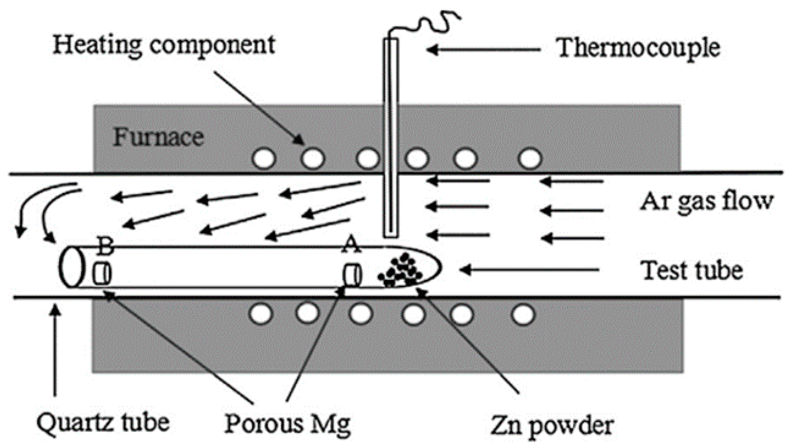
5.1.2. Thermal Evaporation Technique

The physical thermal evaporation deposition technique (PVD) includes a variety of vacuum deposition methods intended for thin films and coatings production [88]. PVD is a powerful process when the material is converted from the condensed phase to the steam phase and then back to the thin layer condensation phase. The main principle of the process lies in removing atoms and clusters of atoms or molecules from the vapor stream in a metal crucible that contains a certain volume of the target material by heating the crucible, either by flowing current through it or using a heating filament. The target material is heated via Joule effect to a convenient temperature when there is appropriate vapor pressure during the PVD [89]. The great advantage of PVD is its simple and low-cost instrumentation and effortless experimental process [90].
In the study of Wang et al., a PVD method using a classic tubular resistance furnace (TRF) was used for the synthesis of a Zn coating of Mg foams (Figure 9). A long quartz test tube was used in the TRF system to create various reaction conditions and a gaseous environment with super-saturated Zn vapor [91].
It was declared, that the compressive yield strength and elastic modulus of Zn coated Mg foams are almost twice that of uncoated Mg foam, so the PVD method provides an effective approach to enhance the mechanical properties of Mg biodegradable foam [91].

5.1.3. Dip-Coating Method

The dip-coating method (also known as impregnation or saturation) is a simple and commonly used deposition method for the production of uniform and thin coating layers on substrates with various morphology, such as irregularly shaped objects and foam-like structures. During this process, the selected substrate is dipped into a coating bath with low viscosity [92]. Generally, dip coating is a five-step process (Figure 10), consisting of:
  • Immersion: The substrate is dipped into the coating bath at a constant speed.
  • Startup: After immersion, the substrate remains in the bath for a selected time, and then it is ready to be pulled out.
  • Deposition: The deposition of the thin coating layer starts while the substrate is pulling out. The resulting thickness of the coating directly depends on the speed pulling the substrate from the coating bath. A slower pull speed causes the thinner coating of the film.
  • Drainage: Excess fluid is drained in this step.
  • Evaporation: The final step involves the evaporation of fluid from the substrate surface and the creation of the final thin coating. Volatile solvents are evaporated earlier in step 3 [93].
Via the application of the dip-coating method, we can obtain sol-gel coatings [94]. The sol-gel approach offers many advantages, including the possibility to coat large and curved substrates. The preparation of composites, such as the organic-inorganic coating, is a great benefit of this method [95].
The dip-coating method was used for the coating of iron foams in the study of Haverová et al. After the sintering of iron samples, the iron foams were immersed in a solution containing 5, 10, and 15 wt.% of polyethylene glycol (PEG), and the polymer coating layer was formed by a subsequent sol-gel process [22]. The same dip-coating method protocol was used by this research group for the deposition of PEG (Figure 11) and polyethyleneimine (PEI) onto the iron foam samples [9,49,96,97].
The iron scaffolds were coated with polymer polylactic acid (PLA) and composite polylactic acid/hydroxyapatite (PLA/HA) coatings by Hrubovčáková et al. PLA granules were dissolved in chloroform and stirred to solve 5 wt.%. To produce a PLA/HA solution, PLA pellets and HA powder in ratio 1:1 were dissolved in chloroform. The samples were coated in five cycles using a dip coating machine [98].
This coating method was also chosen by the research group of Julmi et al. A coating of PLA on magnesium spongious implants prepared by 3D printing was applied by dip coating [99].

5.1.4. Vacuum Infiltration

In vacuum infiltration, the polymer is poured over the fiber and placed in a vacuum chamber. Under the vacuum, air will be pulled out of the fiber, causing the polymer to be pulled in. The vibration of the vacuum chamber may be used to assist in the removal of air bubbles [100].
This specific method was used by Yusop et al. Porous pure iron was vacuum-infiltrated by poly(lactic-co-glycolic acid) (PLGA) to form fully dense PLGA-infiltrated porous iron. The properties of vacuum infiltrated PLGA samples were compared to partially dense PLGA samples prepared using the dip-coating method. Results showed that the compressive strength and toughness of the iron foam with PLGA coated by vacuum infiltration were higher compared to dip-coated PLGA foam samples. A strong interfacial interaction was observed between the PLGA layer and the iron surface [101].

5.1.5. Conversion Coating Method

Conversion coatings are widely used in metal substrates for their ability to improve corrosion resistance and promote organic coating adhesion. The application process includes a cleaning of the metal, removing impurities, organic contamination, surface oxides, and surface defects. The conversion coating method is an alternative to anodizing for some metals [102].
This coating method consists of immersing a metallic substrate into a chemical medium, with or without the electric current application. The coating on the substrate surface is formed by a chemical reaction between the metallic material and the electrolyte solution. It is important that the coating itself becomes an integral part of the device, and thus no mechanical interface is formed between the coating and the metal matrix of the substrate [103].
Su et al. obtained the CaP coated iron foam samples via the conversion method. An optimum CaP conversion coating parameter on pure iron was first designed by taking three Ca/P ratio levels. To obtain the CaP-coated samples, the pH of the phosphating bath was adjusted to 2.8–3.0 by adding NaOH solution, and iron foam samples were coated subsequently. Two different AgNO3 concentrations (0.2 and 0.5 mmol.dm−3) were used to incorporate Ag into the CaP coating by co-deposition and conversion coating [35].

5.1.6. Micro-Arc Oxidation

Micro-arc oxidation (MAO), also known as plasma electrolytic oxidation (PEO), is a novel and attractive plasma assisted surface engineering process for magnesium, aluminium, and titanium alloys. Due to its great corrosion resistance and environmentally safe coating process, MAO technology is an appropriate alternative to anodizing techniques [104]. In an electrolytic bath with high electric energy, the surface of a selected alloy can be converted into a dense and hard ceramic oxide coating [105]. Due to the formation of a strong, adherent coating and minimal changes to the substrate properties, MAO coatings improve overall mechanical properties along with providing thermal and electrical barriers. The MAO technique parameters, such as current density, voltage, cathode metal, conductivity, and pH electrolyte, as well as some substrate factors, such as microstructure and chemical composition, strongly influence the final coating properties [104].
Ruá et al. investigated Mg porous samples coated by the MAO method using phosphate/calcium electrolyte. The MAO process was performed in the two-electrode potentiostatic mode. A porous morphology of homogenous calcium phosphate coating was obtained. The possibility of modification and controlling the degradability and bioabsorbability of the MAO coated Mg foam scaffolds was confirmed [106].

5.2. Coatings

The use of implant devices requires strict demands, such as proper medical functionality and excellent biocompatibility. It is not possible to omit any of them. The first requirement represents the therapeutic, regenerative, and supporting function of the implant [107]. Biocompatibility is required due to the direct contact of the implant with the internal environment of the human body and the need for acceptance of the implant by the immune system [108]. The human body’s response to a foreign object can lead to very serious harms such as biological membrane damage, thrombogenesis, local inflammation formation, and even total tissue damage, mostly at the interface of the bone and the implant device [109]. Increasing the biocompatibility of foam-like materials can be achieved by applying coatings of various types: polymer, inorganic ceramic, and composite.

5.2.1. Polymer Coatings

Polyethylene Glycol

One of the most versatile polymers suitable for the coating of metallic biodegradable foam-like materials is polyethylene glycol. PEG is a biocompatible, non-toxic polymer that improves cell adhesion and acts as a surface protector. Several studies have shown that PEG coatings suppress platelet adhesion and reduce thrombus formation, tissue damage, and minimize cytotoxic effects. PEG forms two-phase systems with other polymers and does not harm proteins, membranes, and cells, although it interacts with them directly [110].
The chemical structure of PEG is based on its inert molecular conformation in aqueous solution. This polymer is composed of uncharged hydrophilic groups and shows very high surface mobility. An attractive advantage of PEG is its similar molecular structure with water. Thus, PEG is soluble in water as well as in other polar compounds. PEG also binds via strong hydrogen bonds with oxygen in ethers and hydrogen in water [111].
Oriňaková et al. and Haverová et al. discovered that the polymer coating improved the degradation rate and biocompatibility of iron foams. The iron foams were produced by the replication method and were coated with the PEG layer using the dip-coating method subsequently. They proved that the stiffness and quasi-elastic gradient for PEG-coated foams increased with increasing PEG content. An acceleration of the corrosion rate due to the higher solubility of corrosive iron products in the local acidic solution caused by the oxidative degradation of the PEG layer was observed. The PEG coating was also evaluated for its overall biocompatibility, with improved hemocompatibility and cytocompatibility. The corrosion rate and mechanical properties of the iron porous samples were successfully adjusted with the PEG layer using References [9,22].

Polyethyleneimine

Polyethyleneimine (PEI) is an organic cationic polymer that contains repeating units composed of an amine group and a CH2CH2 spacer. Two forms of PEI, linear, and branched PEI, exist. PEI is well known for its usage in many biological applications [108]. For the first time, Boussif et al. applied the PEI vector for gene and oligonucleotide transfection in cell culture and in vivo [112]. Although it can be utilized as a drug carrier or in tumor imaging, PEI deserves attention also in coating research [113]. By coating metal foams with an implant function, PEI forms a phase between the implant and the internal environment of the body. Due to high charge density, PEI produces a positively charged complex with nucleic acids, which provides efficient transfection and protection against nuclease-mediated degradation in cells. Due to a large number of protonable amino nitrogen, PEI coatings buffer the pH. PEI is a biocompatible polymer that helps to improve the overall acceptance of coated equipment by a living organism. In addition to this, it is possible to modulate the PEI molecular structure with various polymers, create layers, load it with drugs, and use it for specific treatment processes.
Iron foams with open-cell porosity with PEI coating were reported by Gorejová et al. The effect of the deposited PEI layer on the properties of the iron-based foam was observed. The deposition of the PEI layer on the samples’ surface led to the changes in final morphology. A significant decrease in the surface area was observed after the application of the coating (from 1.19 mg m−2 for pure iron to 0.04 mg m−2 for Fe-PEI (15 wt.%)). Desirable enhancement of the corrosion rate was mediated through the polymer’s cracking; hence the corrosion medium penetration was also possible in polymer-coated samples. A schematic process of the corrosion degradation process is shown in Figure 12. Based on the results reported in this study, it is possible to modify the degradation behavior of iron foam devices by varying the concentration of the PEI coating [97]. This was also confirmed by Yao et al., who declared that not only the thickness but also the concentration of the coated layer influenced the resulting cytotoxicity [114].
The effect of PEI coating and bovine serum albumin (BSA) interaction on the corrosion resistance of iron biodegradable foams was investigated by Oriňaková et al. Adsorption of BSA, as a general human transport protein in blood serum, onto Fe-PEI was more rapid in comparison to adsorption onto bare Fe due to the electrostatic nature of BSA adsorption to polymeric layer. The corrosion behavior of bare and coated samples was electrochemically tested in Hanks’ solution. A change of the biodegradation mechanism was observed in the presence of PEI and/or BSA. It was found that the presence of BSA resulted in a surface-induced complexation [96].

Polylactic Acid

Polylactic acid is a biodegradable, biocompatible, and thermoplastic polyester, mainly derived from corn starch. The monomer unit of PLA, the lactic acid (LA), is naturally produced from the bacterial fermentation of corn, sugarcane, potatoes, and other biomass. The use of PLA can replace the use of petroleum-made polymers, while its natural origin does not reduce its mechanical properties or good processability. Its advantages include excellent biocompatibility, low production costs, and the ability to reduce the degradation process. The properties of PLA result from the ratio of D and L isomers, differing in melting point, crystallinity, and brittleness [115]. The crystallinity of PLA can be completely diminished after the incorporation of 15% mezo-lactide or d-lactide in poly (l-lactide). The copolymerization of L and D mezoforms generates an amorphous structure in the resulting polymer [116].
In the study of Julmi et al., PLA coatings were applied onto the MgF2 sponge-like structures. The deposition process was performed using a simple dip-coating method involving dipping the implants into a PLA solution. The resulted coating was disproportionately distributed inside the samples, without clogging the pores (Figure 13). The outer surface of the sponge-like sample was covered by PLS coating more homogeneously. Research showed that the PLA coating improves corrosion resistance and mechanical properties [99].
In the research of Hrubovčáková et al., porous iron foams with PLA and PLA/HA coatings were studied. Iron foams with an open interconnected structure, similar to the structure of cancellous bone, have been successfully manufactured via the replication method. The pore size was determined between 300 and 800 μm, and the porosity was over 90%. PLA and PLA/HA coatings were assessed concerning the emerging changes of iron foams properties. Experiments have shown that both selected coatings improved the mechanical properties of metal foams. Further, electrochemical and static immersion tests exhibited that coated iron foams were degraded faster than uncoated ones. The highest corrosion rates were determined for PLA coated iron samples since the hydrolysis of PLA accelerated degradation. The presence of HA in composite coatings slowed it down, contrarily. Biocompatibility assays were performed on pre-osteoblastic cells, showing higher cell viability on PLA and PLA/HA-coated samples as compared to uncoated foams [98].
A new method for accelerating iron degradation via the incorporation of poly (lactic and glycolic acid) (PLGA) was introduced by Yusop et al. Coating of the iron foams was performed using the vacuum infiltration (PIPI) and dip-coating methods (PCPI). Mechanical properties such as compressive strength were comparable to the properties of cancellous bone for both types of samples. Electrochemical corrosion testing showed a strong interaction between the PLGA and the iron foam surface which led to faster degradation of the metal foams (Figure 14). The rate of corrosion degradation was the fastest for the PIPI samples. The degradation of PCPI and PIPI was induced by the hydrolysis of PLGA, which produced soluble monomers consisting of carboxylic acid groups. At the same time, hydrolysis, in addition to reducing oxygen, induces the evolution of hydrogen, accelerating the degradation of the material via two electron-consuming cathodic reactions. The cytocompatibility test of iron foams with PLGA coating performed using fibroblasts showed good cell viability during the early and most active degradation period [101].

Chitosan

Chitosan is a biopolymer widely used in medical applications. Chitosan is a cationic polysaccharide produced by the deacetylation of chitin that naturally occurs in the exoskeleton of crustaceans and is obtained from an alkalizing process at high temperature [6,7]. This biodegradable and biocompatible polymer is used in many different forms such as gels, films, membranes, etc., in a large number of applications. Chitosan is often applied in tissue engineering. It degrades rapidly, is nontoxic, and underlies chemical and enzymatic modifications. Thanks to it having a similar structure to the glycosaminoglycans of the bone extracellular matrix, chitosan stimulates cell adhesion, proliferation, and osteoinduction [117,118].

5.2.2. Inorganic Ceramic Coatings

Calcium Phosphate

Calcium phosphate-based bioceramics, tricalcium phosphate or hydroxyapatite (HA), show high similarity to bone minerals via their mineral compositions. Generally, natural bone is a nanostructured composite material composed of 70% nanostructured inorganic HA crystals and 22% organic collagen fibers Type I [119]. Calcium phosphate’s osteoconductivity is based on the ability to generate molecular interactions with surrounding tissues, leading to the formation of a surface layer of apatite. There are several different forms of calcium phosphate with different properties like degradation rates. For instance, β-tricalcium phosphate degrades more rapidly compared to hydroxyapatite (HA) [120]. Coating with calcium phosphate implants has been shown to arouse osteoid formation on the surface. The resorption of calcium phosphate biomaterials occurs after the activation of osteoclastic and multinucleated cells [121]. Another application of calcium phosphate includes the carrier function of antibiotics, which binds with bioceramics via their entire structures [122].
Calcium phosphate coatings were reported in the study of Rúa et al. Biocompatible coating was prepared using the MAO process onto magnesium biodegradable foams with potential biomedical function [106]. SEM images of uncoated and MAO-coated Mg foam are shown in Figure 15.
The resulting bioceramic MAO coating consisted of calcium phosphate compounds, the main elements of the bone structure, and it was observed that the values of the Young’s modulus of the prepared samples were suitable for bone tissue applications [106].
In the study of Oriňaková et al., a thin and discontinuous HA and MnHA coating was electrochemically deposited onto the sintered iron-based foams to improve their biocompatibility without decreasing the degradation rate. The chemical composition of HA ceramic coating was investigated using X-Ray diffraction (XRD) analysis. Energy-dispersive X-ray spectroscopy (EDX) analysis confirms the specific Ca/P molar ratio in the bioceramic coating. The degradation rate set by an electrochemical and immersion corrosion test increased in this order: Fe/HA, Fe/MnHA, uncoated Fe [70].
The use of bisphosphonates (FeBiP) and anti-catabolic drugs, such as strontium (FeSr) ranelate, for implant coating was analyzed by Ray et al. Open-cell iron foam structures were coated to enhance bone defect reparation, as well as to increase the biocompatibility and stability of the iron implant devices. Strontium ranelate and bisphosphonates are the treatment alternatives for osteoporosis; while strontium enhances osteoblastogenesis and decreases osteoclastogenesis, bisphosphonates inhibit osteoclast resorption and improve bone strength. In vivo testing and ToF-SIMS analysis revealed overlapping of Ca signals with Fe for both FeSr and FeBiP, indicating tissue in-growth into the scaffolds. Iron foams with strontium and bisphosphonate coating are very promising implant bone grafts for fracture defect reparation in osteopenic bone [26].

5.2.3. Composite Coatings

Composite material coatings, such as biopolymers combined with calcium phosphate, fulfill several functions promoting osteoblast adhesion, migration, differentiation, and proliferation. Composite materials have immense potential in bone repair and regeneration processes. The electrophoretic deposition (EPD) approach followed by a conversion process has been employed to deposit hydroxyapatite nanoparticles/chitosan as a composite coating with a pure HA phase by Wen et al. The coatings were deposited from 40% nano-hydroxyapatite (nHA)/ethanol suspension mixed with 60% nHA/chitosan-acetic acid aqueous solution (Figure 16). The in vitro immersion test proved that the application of the iron-based foams had no significant influence on the biocompatibility of potential implant devices [6].
The research of Su et al. evaluated the improvement of the surface bioactivity of the iron biodegradable foams by preparing a calcium phosphate (CaP) conversion coating. Silver (Ag), known for its antibacterial property, was incorporated onto the CaP coating via co-deposition (Ag/CaP-c) and post-treatment (Ag/CaP-p), subsequently. The prepared coatings enhanced the mineralization ability and mechanical integrity of the iron foam (Figure 17). Electrochemical and immersion tests were performed. The high resistance and capacitance of the Ag/CaP coatings decreased the degradation rate and protects the iron foam from mechanical properties loss. The Ag loading enhanced CaP coating features and brings a new cost-effective and functional approach to increasing the degradation behavior and antibacterial potential of CaP layers deposited on bioabsorbable iron foam scaffolds [35].

5.2.4. Metal Coatings

Wang et al. studied magnesium foams with Zn coating prepared by the PVD method. Two types of coating growth mechanisms were proposed to explain the resulting phenomenon. Competition exists between thermal diffusion under high temperature and the deposition model under low temperatures. The resulting metal zinc coating was dense and homogeneous. The coated samples were tested for mechanical and corrosion properties (Figure 18). The results showed that Zn coatings brought an evident improvement in compressive strength and revealed the possibility of biodegradation modification. It was confirmed that the diffusion layer accelerated the corrosion of the Mg foam due to the galvanic effect, although the Zn-based coating possesses great anti-corrosion properties and has high potential as bone implantation material [91].

5.3. Influence of Coatings on Biocompatibility and Corrosion Properties of Metallic Foams

Selected coatings were prepared using many different production methods. What unites each coating method and the resulting coating is the effort to increase and improve the biocompatibility of the metallic porous material to be used as implant equipment. Biocompatibility is a material’s ability to fulfill the required tasks without causing any local or systemic negative responses in the recipient environment. In general, biocompatibility is a very dynamic process because of constant changes in material properties and host medium response in time due to the degradation processes, diseases, and overall aging. By appropriately choosing a biocompatible coating, it is possible to avoid the biological rejection of the implant. An unsuitable selected implant coating leads to the activation of immune cells and initiates inflammatory processes [123,124].
The most important advantages and disadvantages of biocompatible coatings of porous metallic materials are listed in Table 2.
Besides the impact on biocompatibility, coatings significantly influence the corrosion properties of scaffold material. Table 3 summarizes degradation properties (represented by corrosion rates in mm y−1) of various coated scaffolds and compares them to these of uncoated metals.
It can be clearly seen that coatings change the corrosion rate of material and thereby influence the time during which the scaffold can support the damaged tissue. During the degradation of some polymeric coatings (PLGA) [101], an acidic environment is created which contributes to the dissolution of the passivation layer of corrosion products. Via this dissolution, a new layer of pure metal is exposed which results in further corrosion. Interfacial interaction between the hydrophilic polymer layer (PEG) and the iron surface leads to the enhanced oxidation rate of iron [22]. In another case (PEI coating) [97], isolated islands of pure iron can serve as an entrance gate for electrolyte penetration. On the contrary, a bioceramic layer of HAP [70] lowered the degradation rate of pure iron but had a positive influence on its biocompatibility. We can see from these studies that polymeric coatings may play an important role when enhancing of corrosion rate is necessary. Bioceramic or inorganic coatings, on the other hand, lower the corrosion speed but can greatly influence the biological performance of the material.

5.4. Promising Biomaterials

Biodegradable metallic foams have been extensively studied for bone substitute applications, especially for load-bearing implants and bone tissue engineering scaffolds, since the porous structures enable the tailoring of mechanical properties and degradation behavior and provide space for bone ingrowth and vascularization. To date, many different biomaterials based on Mg, Fe, and Zn were developed; however, only a few of them meet requirements indispensable for clinical applications.
Degradable implants made of Fe-based porous materials are not in clinical practice yet, but performed studies brought promising results regarding successful future clinical applications [2]. For example, open-cell iron foams with a PEG layer and PLGA layers exhibited improved biocompatibility and desired adjustment of the corrosion rate and mechanical properties [9,22,101] and can be considered for clinical studies. Moreover, iron foams with a bisphosphonate and strontium coating represent very promising materials for implant bone grafts for fracture defect reparation in osteopenic bone [26].
Further promising biomaterials are Mg-based foams with calcium phosphate coatings prepared using the MAO process [106] and Mg foams with dense and homogeneous Zn coating prepared using the PVD method [91] which improved the compressive strength while allowing the decrease in corrosion rate required for Mg-based material. These resorbable biomaterials have high potential as bone implantation materials.

6. Future Research Directions

The development of advanced degradable metallic biomaterials with open-cell porous structures is a hot area of research. One of the current challenges on the way to practical applications is the finding of an efficient technique for the manufacturing of interconnected porous structures with desired structures and properties at a large scale in a cost-effective manner. This is associated with the elucidation of the influence of pores structure, size, shape and distribution on the resultant mechanical and degradation properties and the biomedical performance of fabricated materials. Therefore, systematic research needs to be considered in the future.
Control of the degradation rate and mode is still an actual problem that needs to be solved. To implement the control, the structure and composition of the materials should be configured in accordance with the particular needs of the specific applications.
Another challenge is the enhancement of the bioactivity of fabricated biomaterials. Coating with different organic or inorganic films may also contribute to the controlling of the degradation rate and significantly influence the mechanical properties, surface chemistry, and topography, and thus biocompatibility, osteogenesis, and bone tissue-bonding properties of the implant through the adjustment of the scaffold-body interface. Moreover, the understanding of time-dependent changes in mechanical properties and biocompatibility during the degradation represents the other challenge.
Future research directions in this field are focused on the development of smart biomaterials with added value in the form of special functionality, such as the new generation scaffolds with drug delivery potential. A combination of manufacturing techniques, multilayer structures, and composite coatings may be the advisable solutions to control the properties of biomaterials and to promote their applicability.
Another interesting research direction could be the incorporation of drugs carrying biodegradable polymers or ceramics into the structure or onto the surface of degradable metallic biomaterials.
Also, the development of new tools applicable to the real-time prediction of in vivo degradation and mechanical behavior, such as numerical modeling, will support the wider use of resorbable biomaterials and enhance the development of new materials.

7. Conclusions

Biodegradable metallic foams are conventionally made of iron, magnesium, and zinc because of their appropriate biological properties and degradative abilities. The major field of their application falls under bone scaffold development. Appropriate mechanical, corrosion, and biological properties designed for an actual healed tissue are a matter of course.
There are several approaches commonly used to develop optimal biomaterials with favorable mechanical and degradation properties for orthopedic implant applications. Firstly, a variety of manufacturing methods allows us significantly affect the properties of porous biomaterials through the control of the structure, porosity, pore shape, size and distribution. Furthermore, a wide range of surface modifications and composition changes foster the design of tuned biomaterials with desirable mechanical, morphological, and functional properties for biomedical applications.
Future research efforts should be directed towards the invention of complex multifunctional smart biomaterials.

Author Contributions

Conceptualization, R.O., R.G., Z.O.K. and A.O.; writing—original draft preparation, R.O., R.G., Z.O.K. and A.O.; writing—review and editing, R.O.; visualization, R.G. and Z.O.K.; supervision, R.O. and A.O.; project administration, R.O.; funding acquisition, R.O. All authors have read and agreed to the published version of the manuscript.

Funding

This research received no external funding.

Acknowledgments

This work was supported by the Slovak Research and Development Agency (projects APVV-16-0029), Scientific Grant Agency of the Ministry of Education, Science, Research and Sport of the Slovak Republic (projects VEGA 1/0074/17) and Internal Research Grant System of Faculty of Science of Pavol Jozef Šafárik University in Košice (VVGS- pf-2019-1049).

Conflicts of Interest

The authors declare no conflict of interest.

References

  1. Prakasam, M.; Locs, J.; Salma-Ancane, K.; Loca, D.; Largeteau, A.; Berzina-Cimdina, L. Biodegradable Materials and Metallic Implants—A Review. J. Funct. Biomater. 2017, 8, 44. [Google Scholar] [CrossRef] [Green Version]
  2. Zivic, F.; Grujovic, N.; Pellicer, E.; Sort, J.; Mirovic, S.; Adamovic, D.; Vulovic, M. Biodegradable Metals as Biomaterialsfor Clinical Practice: Iron-Based Materials. In Biomaterials in Clinical Practice; Springer: Cham, Switzerland, 2017; p. 225. ISBN 978-3-319-68025-5. [Google Scholar]
  3. Radenkovic, G.; Petkovic, D. Metallic Biomaterials. In Biomaterials in Clinical Practice; Springer: Cham, Switzerland, 2017; p. 183. ISBN 978-3-319-68025-5. [Google Scholar]
  4. Adamovic, D.; Ristic, B.; Zivic, F. Review of Existing Biomaterials—Method of Material Selection for Specific Applications in Orthopedics. In Biomaterials in Clinical Practice; Springer: Cham, Switzerland, 2017; p. 47. ISBN 978-3-319-68025-5. [Google Scholar]
  5. Munir, K.; Wen, C.; Li, Y. Production methods and characterization of porous Mg and Mg alloys for biomedical applications. In Metallic Foam Bone: Processing, Modification and Characterization and Properties; Elsevier: Amsterdam, The Netherlands, 2017; pp. 25–82. ISBN 978-0-08-101290-1. [Google Scholar]
  6. Wen, Z.; Zhang, L.; Chen, C.; Liu, Y.; Wu, C.; Dai, C. A construction of novel iron-foam-based calcium phosphate/chitosan coating biodegradable scaffold material. Mater. Sci. Eng. C 2013, 33, 1022–1031. [Google Scholar] [CrossRef] [PubMed]
  7. Radha, R.; Sreekanth, D. Insight of magnesium alloys and composites for orthopedic implant applications—A review. J. Magnes. Alloy 2017, 5, 286–312. [Google Scholar] [CrossRef]
  8. Hermawan, H. Updates on the research and development of absorbable metals for biomedical applications. Prog. Biomater. 2018, 7, 93–110. [Google Scholar] [CrossRef] [Green Version]
  9. Oriňaková, R.; Gorejová, R.; Orságová, Z.; Haverová, L.; Oriňak, A.; Maskaľová, I.; Kupková, M.; Džupon, M.; Baláž, M.; Hrubovčáková, M.; et al. Evaluation of Mechanical Properties and Hemocompatibility of Open Cell Iron Foams with Polyethylene Glycol Coating. Appl. Surf. Sci. 2019, 505, 144634. [Google Scholar] [CrossRef]
  10. Oriňak, A.; Oriňaková, R.; Orságová Králová, Z.; Morovská Turoňová, A.; Kupková, M.; Hrubovčáková, M.; Radoňak, J.; Džunda, R.; Turoňová Morovská, A.; Kupková, M.; et al. Sintered metallic foams for biodegradable bone replacement materials. J. Porous Mater. 2014, 21, 131–140. [Google Scholar] [CrossRef]
  11. Gorejová, R.; Haverová, L.; Oriňaková, R.; Oriňak, A.; Oriňak, M. Recent advancements in Fe-based biodegradable materials for bone repair. J. Mater. Sci. 2019, 54, 1913–1947. [Google Scholar] [CrossRef]
  12. Čapek, J.; Vojtěch, D.; Oborná, A. Microstructural and mechanical properties of biodegradable iron foam prepared by powder metallurgy. Mater. Des. 2015, 83, 468–482. [Google Scholar] [CrossRef]
  13. Oriňáková, R.; Oriňák, A.; Bučková, L.M.; Giretová, M.; Medveckỳ, L.; Labbanczová, E.; Kupková, M.; Hrubovčáková, M.; Koval, K. Iron based degradable foam structures for potential orthopedic applications. Int. J. Electrochem. Sci. 2013, 8, 12451–12465. [Google Scholar]
  14. Li, Y.; Zhou, J.; Pavanram, P.; Leeflang, M.A.; Fockaert, L.I.; Pouran, B.; Tümer, N.; Schröder, K.U.; Mol, J.M.C.; Weinans, H.; et al. Additively manufactured biodegradable porous magnesium. Acta Biomater. 2018, 67, 378–392. [Google Scholar] [CrossRef]
  15. Agarwal, S.; Curtin, J.; Duffy, B.; Jaiswal, S. Biodegradable magnesium alloys for orthopaedic applications: A review on corrosion, biocompatibility and surface modifications. Mater. Sci. Eng. C 2016, 68, 948–963. [Google Scholar] [CrossRef] [PubMed] [Green Version]
  16. Zhang, Q.; Cao, P. Degradable porous Fe-35wt.%Mn produced via powder sintering from NH4HCO3 porogen. Mater. Chem. Phys. 2015, 163, 394–401. [Google Scholar] [CrossRef]
  17. Heiden, M.; Walker, E.; Stanciu, L. Magnesium, Iron and Zinc Alloys, the Trifecta of Bioresorbable Orthopaedic and Vascular Implantation—A Review. Biotechnol. Biomater. 2005, 5, 1000178. [Google Scholar]
  18. Meagher, P.; O’Cearbhaill, E.D.; Byrne, J.H.; Browne, D.J. Bulk Metallic Glasses for Implantable Medical Devices and Surgical Tools. Adv. Matter. 2016, 28, 5755–5762. [Google Scholar] [CrossRef] [PubMed] [Green Version]
  19. Ulum, M.F.; Caesarendra, W.; Alavi, R.; Hermawan, H. In-Vivo Corrosion Characterization and Assessment. Coatings 2019, 9, 282. [Google Scholar] [CrossRef] [Green Version]
  20. Sharma, P.; Pandey, P.M.M. Morphological and mechanical characterization of topologically ordered open cell porous iron foam fabricated using 3D printing and pressureless microwave sintering. Mater. Des. 2018, 160, 442–454. [Google Scholar] [CrossRef]
  21. Sharma, P.; Pandey, P.M. Corrosion behaviour of the porous iron scaffold in simulated body fluid for biodegradable implant application. Mater. Sci. Eng. C 2019, 99, 838–852. [Google Scholar] [CrossRef]
  22. Haverová, L.; Oriňaková, R.; Oriňak, A.; Gorejová, R.; Baláž, M.; Vanýsek, P.; Kupková, M.; Hrubovčáková, M.; Mudroň, P.; Radoňák, J.; et al. An in vitro corrosion study of open cell Iron structures with PEG coating for bone replacement applications. Metals 2018, 8, 499. [Google Scholar] [CrossRef] [Green Version]
  23. He, J.; He, F.-L.; Li, D.-W.; Liu, Y.-L.; Liu, Y.-Y.; Ye, Y.-J.; Yin, D.-C. Advances in Fe-based biodegradable metallic materials. RSC Adv. 2016, 6, 112819–112838. [Google Scholar] [CrossRef]
  24. Lefevre, L.-P.; Banhart, J.; Dunand, D.C. Porous Metals and Metallic Foams: Current Status and Recent Developments. Adv. Eng. Mater. 2008, 10, 775–787. [Google Scholar] [CrossRef] [Green Version]
  25. Lietaert, K.; Weber, L.; Van Humbeeck, J.; Mortensen, A.; Luyten, J.; Schrooten, J. Open cellular magnesium alloys for biodegradable orthopaedic implants. J. Magnes. Alloy 2013, 1, 303–311. [Google Scholar] [CrossRef] [Green Version]
  26. Ray, S.; Thormann, U.; Eichelroth, M.; Budak, M.; Biehl, C.; Rupp, M.; Sommer, U.; El Khassawna, T.; Alagboso, F.I.; Kampschulte, M.; et al. Strontium and bisphosphonate coated iron foam scaffolds for osteoporotic fracture defect healing. Biomaterials 2018, 157, 1–16. [Google Scholar] [CrossRef] [PubMed]
  27. Alavi, R.; Trenggono, A.; Champagne, S.; Hermawan, H. Investigation on Mechanical Behavior of Biodegradable Iron Foams under Different Compression Test Conditions. Metals 2017, 7, 202. [Google Scholar] [CrossRef]
  28. Pellicer, E.; Lorenzetti, M.; Fornell, J.; Baró, M.D.; Novak, S.; Sort, J. Progress Beyond the State-of-the-Art in the Field of Metallic Materials for Bioimplant Applications. In Biomaterials in Clinical Practice; Springer: Cham, Switzerland, 2017; p. 25. ISBN 978-3-319-68025-5. [Google Scholar]
  29. Erryani, A.; Pramuji, F.; Annur, D.; Kartika, I.; Amal, M.I. Microstructures and Mechanical Study of Mg Alloy Foam Based on Mg-Zn-Ca-CaCO3 System. OP Conf. Ser. Mater. Sci. 2017, 202, 012028. [Google Scholar] [CrossRef]
  30. Malladi, L.; Mahapatro, A.; Gomes, A.S.; Steffin, A. Fabrication of magnesium-based metallic scaffolds for bone tissue engineering. Mater. Technol. 2018, 33, 173–182. [Google Scholar] [CrossRef]
  31. Ryan, G.; Pandit, A.; Apatsidis, D.P. Fabrication methods of porous metals for use in orthopaedic applications. Biomaterials 2006, 27, 2651–2670. [Google Scholar] [CrossRef]
  32. Čapek, J.; Msallamová, Š.; Jablonská, E.; Lipov, J.; Vojtěch, D. A novel high-strength and highly corrosive biodegradable Fe-Pd alloy: Structural, mechanical and in vitro corrosion and cytotoxicity study. Mater. Sci. Eng. C 2017, 9, 550–562. [Google Scholar] [CrossRef]
  33. Lietaert, K.; Wauthle, R.; Schrooten, J. Porous Metals in Orthopedics. In Biomaterials in Clinical Practice; Springer: Cham, Switzerland, 2017; p. 281. ISBN 978-3-319-68025-5. [Google Scholar]
  34. Sharma, P.; Pandey, P.M. A novel manufacturing route for the fabrication of topologically-ordered open-cell porous iron scaffold. Mater. Lett. 2017, 222, 160–163. [Google Scholar] [CrossRef]
  35. Su, Y.; Champagne, S.; Trenggono, A.; Tolouei, R.; Mantovani, D.; Hermawan, H. Development and characterization of silver containing calcium phosphate coatings on pure iron foam intended for bone scaffold applications. Mater. Des. 2018, 148, 124–134. [Google Scholar] [CrossRef] [Green Version]
  36. Liao, Y.; Qiu, G.; Yang, Y.; Lv, X.; Bai, C. Preparation and compressive properties of magnesium foam. Xiyou Jinshu Cailiao Yu Gongcheng/Rare Met. Mater. Eng. 2016, 45, 2498–2502. [Google Scholar]
  37. Chen, Q.Z. Foaming technology of tissue engineering scaffolds—A review. Eng. Technol. 2011, 3, 34–47. [Google Scholar] [CrossRef]
  38. Trinidad, J.; Marco, I.; Arruebarrena, G.; Wendt, J.; Letzig, D.; Sáenz De Argandoña, E.; Goodall, R. Processing of magnesium porous structures by infiltration casting for biomedical applications. Adv. Eng. Mater. 2014, 16, 241–247. [Google Scholar] [CrossRef]
  39. Polo-Corrales, L.; Latorre-Esteves, M.; Ramirez-Vick, J.E. Scaffold Design for Bone Regeneration. J. Nanosci. Nanotechnol. 2014, 14, 15–56. [Google Scholar] [CrossRef] [PubMed] [Green Version]
  40. Alvarez, K.; Nakajima, H. Metallic scaffolds for bone regeneration. Materials 2009, 2, 790–832. [Google Scholar] [CrossRef]
  41. Salerno, A.; Netti, P.A. Introduction to Biomedical Foams. In Biomedical Foams for Tissue Engineering Applications; Woodhead Publishing: Sawston/Cambridge, UK, 2014; ISBN 9780857096968. [Google Scholar]
  42. Guarino, V.; Ambrosio, L. 2—Properties of biomedical foams for tissue engineering applications. In Biomedical Foams for Tissue Engineering Applications; Woodhead Publishing: Sawston/Cambridge, UK, 2014; pp. 40–70. ISBN 9780857096968. [Google Scholar]
  43. Servier Medical Art to Illustrate your Publications and Powerpoint Presentations. Available online: https://smart.servier.com/ (accessed on 10 July 2020).
  44. Staiger, M.P.; Pietak, A.M.; Huadmai, J.; Dias, G. Magnesium and its alloys as orthopedic biomaterials: A review. Biomaterials 2006, 27, 1728–1734. [Google Scholar] [CrossRef]
  45. Čapek, J.; Vojtěch, D. Microstructural and mechanical characteristics of porous iron prepared by powder metallurgy. Mater. Sci. Eng. C 2014, 43, 494–501. [Google Scholar] [CrossRef]
  46. Oriňaková, R.; Oriňak, A.; Markušová, L.; Labbanczová, E.; Kupková, M.; Hrubovčaková, M.; Fedorková, A. Biodegradable Open Cell Iron Foams for Potential Skeletal Application. Powder Metall. Prog. 2012, 12, 219–223. [Google Scholar]
  47. Zhang, K.; Wang, S.; Zhou, C.; Cheng, L.; Gao, X.; Xie, X.; Sun, J.; Wang, H.; Weir, M.D.; Reynolds, M.A.; et al. Advanced smart biomaterials and constructs for hard tissue engineering and regeneration. Bone Res. 2018, 6, 1–15. [Google Scholar] [CrossRef]
  48. Aghion, E.; Yered, T.; Perez, Y.; Gueta, Y. The prospects of carrying and releasing drugs via biodegradable magnesium foam. Adv. Eng. Mater. 2010, 12, 374–379. [Google Scholar] [CrossRef]
  49. Oriňaková, R.; Gorejová, R.; Macko, J.; Oriňak, A.; Kupková, M.; Hrubovčáková, M.; Ševc, J.; Smith, R.M. Evaluation of in vitro biocompatibility of open cell iron structures with PEG coating. Appl. Surf. Sci. 2019, 475, 515–518. [Google Scholar] [CrossRef]
  50. Li, Y.; Jahr, H.; Lietaert, K.; Pavanram, P.; Yilmaz, A.; Leeflang, M.; Pouran, B.; Gonzalez-Garcia, Y.; Weinans, H.; Mol, J.M.C.; et al. Additively manufactured biodegradable porous iron. Acta Biomater. 2018, 77, 380–393. [Google Scholar] [CrossRef]
  51. Banhart, J. Manufacturing Routes for Metallic Foams. JOM 2000, 52, 22–27. [Google Scholar] [CrossRef]
  52. Chou, D.T.; Wells, D.; Hong, D.; Lee, B.; Kuhn, H.; Kumta, P.N. Novel processing of iron-manganese alloy-based biomaterials by inkjet 3-D printing. Acta Biomater. 2013, 9, 8593–8603. [Google Scholar] [CrossRef]
  53. Quadbeck, P.; Stephani, G.; Kümmel, K.; Adler, J.; Standke, G. Synthesis and Properties of Open-Celled Metal Foams. Mater. Sci. Forum 2007, 534–536, 1005–1008. [Google Scholar] [CrossRef]
  54. Posada, V.M.; Orozco, C.; Ramirez Patino, J.F.; Fernandez-Morales, P. Human Bone Inspired Design of an Mg Alloy-Based Foam. Mater. Sci. Forum 2018, 933, 291–296. [Google Scholar] [CrossRef]
  55. Zhang, Y.Q.; Li, Y.; Liu, H.; Bai, J.; Bao, N.R.; Zhang, Y.; He, P.; Zhao, J.N.; Tao, L.; Xue, F.; et al. Mechanical and Biological Properties of a Biodegradable Mg-Zn-Ca Porous Alloy. Orthop. Surg. 2018, 10, 160–168. [Google Scholar] [CrossRef] [Green Version]
  56. Hou, Y.; Jia, G.; Yue, R.; Chen, C.; Pei, J.; Zhang, H.; Huang, H.; Xiong, M.; Yuan, G. Synthesis of biodegradable Zn-based scaffolds using NaCl templates: Relationship between porosity, compressive properties and degradation behavior. Mater. Charact. 2018, 137, 162–169. [Google Scholar] [CrossRef]
  57. Zhou, C.; Liu, Y.; Zhang, H.; Chen, X.; Li, Y. Compressive and Corrosion Properties of Lotus-Type Porous Mg-Mn Alloys Fabricated by Unidirectional Solidification. Metall. Mater. Trans. A Phys. Metall. Mater. Sci. 2020, 51, 3238–3247. [Google Scholar] [CrossRef]
  58. Kang, M.H.; Lee, H.; Jang, T.S.; Seong, Y.J.; Kim, H.E.; Koh, Y.H.; Song, J.; Jung, H. Do Biomimetic porous Mg with tunable mechanical properties and biodegradation rates for bone regeneration. Acta Biomater. 2019, 84, 453–467. [Google Scholar] [CrossRef]
  59. Čapek, J.; Jablonská, E.; Lipov, J.; Kubatík, T.F.; Vojtěch, D. Preparation and characterization of porous zinc prepared by spark plasma sintering as a material for biodegradable scaffolds. Mater. Chem. Phys. 2018, 203, 249–258. [Google Scholar] [CrossRef]
  60. Yusop, A.H.; Bakir, A.A.; Shaharom, N.A.; Abdul Kadir, M.R.; Hermawan, H. Porous Biodegradable Metals for Hard Tissue Scaffolds: A Review. Int. J. Biomater. 2012, 2012, 1–10. [Google Scholar] [CrossRef] [Green Version]
  61. Popescu, I.N.; Vidu, R.; Bratu, V. Porous Metallic Biomaterials Processing (Review) Part 1: Compaction, Sintering Behavior, Properties and Medical Applications. Sci. Bull. Valahia Univ.-Mater. Mech. 2017, 15, 28–40. [Google Scholar] [CrossRef] [Green Version]
  62. Wen, C.E.; Yamada, Y.; Shimojima, K.; Chino, Y.; Hosokawa, H.; Mabuchi, M. Compressibility of porous magnesium foam: Dependency on porosity and pore size. Mater. Lett. 2004, 58, 357–360. [Google Scholar] [CrossRef]
  63. Li, Y.; Jahr, H.; Pavanram, P.; Bobbert, F.S.L.; Puggi, U.; Zhang, X.Y.; Pouran, B.; Leeflang, M.A.; Weinans, H.; Zhou, J.; et al. Additively manufactured functionally graded biodegradable porous iron. Acta Biomater. 2019, 96, 646–661. [Google Scholar] [CrossRef]
  64. Yang, Y.; He, C.; Dianyu, E.; Yang, W.; Qi, F.; Xie, D.; Shen, L.; Peng, S.; Shuai, C. Mg bone implant: Features, developments and perspectives. Mater. Des. 2020, 185, 108259. [Google Scholar] [CrossRef]
  65. Li, Y.; Pavanram, P.; Zhou, J.; Lietaert, K.; Bobbert, F.S.L.; Kubo, Y.; Leeflang, M.A.; Jahr, H.; Zadpoor, A.A. Additively manufactured functionally graded biodegradable porous zinc. Biomater. Sci. 2020, 96, 646–661. [Google Scholar] [CrossRef] [Green Version]
  66. Li, Y.; Li, W.; Bobbert, F.S.L.; Lietaert, K.; Dong, J.H.; Leeflang, M.A.; Zhou, J.; Zadpoor, A.A. Corrosion fatigue behavior of additively manufactured biodegradable porous zinc. Acta Biomater. 2020, 106, 439–449. [Google Scholar] [CrossRef]
  67. Qin, Y.; Wen, P.; Voshage, M.; Chen, Y.; Schückler, P.G.; Jauer, L.; Xia, D.; Guo, H.; Zheng, Y.; Schleifenbaum, J.H. Additive manufacturing of biodegradable Zn-xWE43 porous scaffolds: Formation quality, microstructure and mechanical properties. Mater. Des. 2019, 181, 107973. [Google Scholar] [CrossRef]
  68. Markušová-Bučková, L.; Oriňaková, R.; Oriňak, A.; Gorejová, R.; Kupková, M.; Hrubovčáková, M.; Baláž, M.; Kováľ, K. Static corrosion test of porous iron material with polymer coating. Powder Metall. Prog. 2016, 16, 99–106. [Google Scholar] [CrossRef] [Green Version]
  69. Oriňaková, R.; Oriňak, A.; Giretová, M.; Medvecký, L.; Kupková, M.; Hrubovčáková, M.; Maskal’ová, I.; Macko, J.; Kal’avský, F. A study of cytocompatibility and degradation of iron-based biodegradable materials. J. Biomater. Appl. 2016, 30, 1060–1070. [Google Scholar] [CrossRef]
  70. Oriňaková, R.; Oriňak, A.; Kupková, M.; Hrubovčáková, M.; Markušová-Bučková, L.; Giretová, M.; Medvecký, L.; Dobročka, E.; Petruš, O.; Kaľavský, F. In vitro degradation and cytotoxicity evaluation of iron biomaterials with hydroxyapatite film. Int. J. Electrochem. Sci. 2015, 10, 8158–8174. [Google Scholar]
  71. Wegener, B.; Sievers, B.; Utzschneider, S.; Müller, P.; Jansson, V.; Rößler, S.; Nies, B.; Stephani, G.; Kieback, B.; Quadbeck, P. Microstructure, cytotoxicity and corrosion of powder-metallurgical iron alloys for biodegradable bone replacement materials. Mater. Sci. Eng. B Solid-State Mater. Adv. Technol. 2011, 176, 1789–1796. [Google Scholar] [CrossRef]
  72. Vahidgolpayegani, A.; Wen, C.; Hodgson, P.; Li, Y. Production Methods and Characterization of Porous Mg and Mg Alloys for Biomedical Applications; Woodhead Publishing: Sawston/Cambridge, UK, 2016; ISBN 9780081012901. [Google Scholar]
  73. Zhuang, H.; Han, Y.; Feng, A. Preparation, mechanical properties and in vitro biodegradation of porous magnesium scaffolds. Mater. Sci. Eng. C 2008, 28, 1462–1466. [Google Scholar] [CrossRef]
  74. Song, Y.H.; Park, S.H.; Kim, S.Y.; Seo, C.H.; Hur, B.Y. A Study on Al-Mg Alloy Foams by Melt Foaming Method. Solid State Phenom. 2009, 124–126, 1841–1844. [Google Scholar]
  75. Wang, X.; Lu, H.M.; Li, X.L.; Li, L.; Zheng, Y.F. Effect of cooling rate and composition on microstructures and properties of Zn-Mg alloys. Trans. Nonferrous Met. Soc. China 2007, 17, 122–125. [Google Scholar]
  76. Zadpoor, A.A. Additively manufactured porous metallic biomaterials. J. Mater. Chem. B 2019, 7, 4088–4117. [Google Scholar] [CrossRef] [Green Version]
  77. Guglielmotti, M.B.; Olmedo, D.G.; Cabrini, R.L. Research on implants and osseointegration. Periodontol 2000 2019, 79, 178–189. [Google Scholar] [CrossRef]
  78. Saleh, M.M.; Saleh, M.M.; Touny, A.H.; Al-Omair, M.A. Biodegradable/biocompatible coated metal implants for orthopedic applications. Biomed. Mater. Eng. 2016, 27, 87–99. [Google Scholar] [CrossRef] [Green Version]
  79. Van der Biest, O.O.; Vandeperre, L. Electrophoretic Deposition of Materials. Annu. Rev. Mater. Sci. 1999, 29, 327–352. [Google Scholar] [CrossRef]
  80. Amrollahi, P.; Krasinski, J.S.; Vaidyanathan, R.; Tayebi, L.; Vashaee, D.; Advanced, H.; Advanced, H.; State, O. Electrophoretic deposition (EPD): Fundamentals and applications from nano- to micro- scale structures. In Handbook of Nanoelectrochemistry; Springer: Cham, Switzerland, 2015; pp. 1–27. ISBN 9783319152073. [Google Scholar]
  81. Besra, L.; Liu, M. A review on fundamentals and applications of electrophoretic deposition (EPD). Prog. Mater. Sci. 2007, 52, 1–61. [Google Scholar] [CrossRef]
  82. Avcu, E.; Baştan, F.E.; Abdullah, H.Z.; Rehman, M.A.U.; Avcu, Y.Y.; Boccaccini, A.R. Electrophoretic deposition of chitosan-based composite coatings for biomedical applications: A review. Prog. Mater. Sci. 2019, 103, 69–108. [Google Scholar] [CrossRef]
  83. Gunputh, U.; Le, H. Composite coatings for implants and tissue engineering scaffolds. Biomed. Compos. 2017, 2017, 111–138. [Google Scholar]
  84. Mesquita-Guimarães, J.; Henriques, B.H.; Silva, F.S. Bioactive Glass Coatings, 2nd ed.; Elsevier Ltd.: Amsterdam, The Netherlands, 2018; ISBN 9780081009369. [Google Scholar]
  85. Ding, D. Processing, Properties and Applications of Ceramic Matrix Composites, SiCf/SiC: An Overview; Woodhead Publishing: Sawston/Cambridge, UK, 2014; Volume 2, ISBN 9780081021675. [Google Scholar]
  86. Amini, A.R.; Laurencin, C.; Nukavarapu, S.P. Bone Tissue Engineering: Recent Advances and Challenges. Crit. Rev. Biomed. Eng. 2012, 40, 363–408. [Google Scholar] [CrossRef] [PubMed] [Green Version]
  87. Pishbin, F.; Simchi, A.; Ryan, M.P.; Boccaccini, A.R. Electrophoretic deposition of chitosan/45S5 Bioglass® composite coatings for orthopaedic applications. Surf. Coat. Technol. 2011, 205, 5260–5268. [Google Scholar] [CrossRef]
  88. Koskinen, J. Cathodic-Arc and Thermal-Evaporation Deposition; Elsevier: Amsterdam, The Netherlands, 2014; Volume 4, ISBN 9780080965338. [Google Scholar]
  89. Antunes, R.A.; de Oliveira, M.C.L. Effect of Surface Treatments on the Fatigue Life of Magnesium and Its Alloys for Biomedical Applications; Elsevier Ltd.: Amsterdam, The Netherlands, 2015; Volume 1, ISBN 9781782420828. [Google Scholar]
  90. Kang, C.W.; Fang, F.Z. State of the art of bioimplants manufacturing: Part II. Adv. Manuf. 2018, 6, 137–154. [Google Scholar] [CrossRef] [Green Version]
  91. Wang, X.; Wang, X.; Wang, D.; Zhao, M.; Han, F. A novel approach to fabricate Zn coating on Mg foam through a modified thermal evaporation technique. J. Mater. Sci. Technol. 2018, 34, 1558–1563. [Google Scholar] [CrossRef]
  92. Neacşu, I.A.; Nicoară, A.I.; Vasile, O.R.; Vasile, B.Ş. Inorganic micro- and nanostructured implants for tissue engineering. In Nanobiomaterials in Hard Tissue Engineering: Applications of Nanobiomaterials; William Andrew Publishing: Norwich, NY, USA, 2016; pp. 271–295. ISBN 9780323428620. [Google Scholar]
  93. Kakaei, K.; Esrafili, M.D.; Ehsani, A. Graphene and Anticorrosive Properties; Elsevier: Amsterdam, The Netherlands, 2019; Volume 27, ISBN 9780128145234. [Google Scholar]
  94. Brinker, C.J.; Hurd, A.J. Fundamentals of sol-gel dip-coating. J. Phys. III 1994, 4, 1231–1242. [Google Scholar] [CrossRef]
  95. Faustini, M.; Louis, B.; Albouy, P.A.; Kuemmel, M.; Grosso, D. Preparation of sol-gel films by dip-coating in extreme conditions. J. Phys. Chem. C 2010, 114, 7637–7645. [Google Scholar] [CrossRef]
  96. Oriňaková, R.; Gorejová, R.; Králová, Z.O.; Oriňak, A.; Shepa, I.; Hovancová, J.; Kovalčíková, A.; Bujňáková, Z.L.; Király, N.; Kaňuchová, M.; et al. Influence of albumin interaction on corrosion resistance of sintered iron biomaterials with polyethyleneimine coating. Appl. Surf. Sci. 2020, 509, 145379. [Google Scholar] [CrossRef]
  97. Gorejová, R.; Oriňaková, R.; Králová, Z.O.; Baláž, M.; Kupková, M.; Hrubovčáková, M.; Haverová, L.; Džupon, M.; Oriňak, A.; Kal’avskỳ, F.; et al. In vitro corrosion behavior of biodegradable iron foams with polymeric coating. Materials 2020, 13, 184. [Google Scholar] [CrossRef] [Green Version]
  98. Hrubovčáková, M.; Kupková, M.; Džupon, M.; Giretová, M.; Medvecký, L.; Džunda, R. Biodegradable polylactic acid and polylactic acid/hydroxyapatite coated iron foams for bone replacement materials. Int. J. Electrochem. Sci. 2017, 12, 11122–11136. [Google Scholar] [CrossRef]
  99. Julmi, S.; Krüger, A.K.; Waselau, A.C.; Meyer-Lindenberg, A.; Wriggers, P.; Klose, C.; Maier, H.J. Processing and coating of open-pored absorbable magnesium-based bone implants. Mater. Sci. Eng. C 2019, 98, 1073–1086. [Google Scholar] [CrossRef] [PubMed]
  100. Tiwari, A.; Gerhardt, R.A.; Szutkowska, M. Advanced Ceramic Materials; John Wiley and Sons: Hoboken, NJ, USA, 2016. [Google Scholar]
  101. Yusop, A.H.M.; Daud, N.M.; Nur, H.; Kadir, M.R.A.; Hermawan, H.; Hakim, A.; Yusop, M.; Daud, N.M.; Nur, H.; Rafiq, M.; et al. Controlling the degradation kinetics of porous iron by poly(lactic-co-glycolic acid) infiltration for use as temporary medical implants. Sci. Rep. 2015, 5, 11194. [Google Scholar] [CrossRef] [PubMed]
  102. Hughes, A.E. Conversion Coatings; Elsevier: Amsterdam, The Netherlands, 2018; ISBN 9780128098943. [Google Scholar]
  103. Pfeifer, M. Manufacturing Process Considerations. In Mater. Enabled Des; Butterworth-Heinemann: Oxford, UK, 2009; pp. 115–160. ISBN 9780750682879. [Google Scholar]
  104. Simchen, F.; Sieber, M.; Kopp, A.; Lampke, T. Introduction to Plasma Electrolytic Oxidation—An Overview of the Process and Applications. Coatings 2020, 10, 628. [Google Scholar] [CrossRef]
  105. Ly, X.N.; Yang, S. Influence of current mode on microstructure and corrosion behavior of micro-arc oxidation (MAO) biodegradable Mg-Zn-Ca alloy in Hank’s solution. Surf. Coat. Technol. 2019, 358, 331–339. [Google Scholar] [CrossRef]
  106. Rúa, J.M.; Zuleta, A.A.; Ramírez, J.; Fernández-Morales, P. Micro-arc oxidation coating on porous magnesium foam and its potential biomedical applications. Surf. Coat. Technol. 2019, 360, 213–221. [Google Scholar] [CrossRef]
  107. Bhattacharya, P.; Neogi, S. Techniques for Deposition of Coatings with Enhanced Adhesion to Bio-Implants. In Adhes. Pharm. Biomed. Dent. Fields; John Wiley and Sons: Hoboken, NJ, USA, 2017; pp. 235–255. ISBN 9781119323501. [Google Scholar]
  108. Pacelli, S.; Manoharana, V.; Desalvo, A.; Lomis, N.; Jodhaa, K.S.; Prakash, S.; Paul, A. Tailoring biomaterial surface properties to modulate host-implant interactions: Implication in cardiovascular and bone therapy. J. Mater. Chem. B Mater. Biol. Med. 2016, 4, 1586–1599. [Google Scholar] [CrossRef] [PubMed] [Green Version]
  109. Hou, N.Y.; Perinpanayagam, H.; Mozumder, M.S.; Zhu, J. Novel development of biocompatible coatings for bone implants. Coatings 2015, 5, 737–757. [Google Scholar] [CrossRef] [Green Version]
  110. Alcantar, N.A.; Aydil, E.S.; Israelachvili, J.N. Polyethylene glycol-coated biocompatible surfaces. J. Biomed. Mater. Res. 2000, 51, 343–351. [Google Scholar] [CrossRef]
  111. Zarrintaj, P.; Saeb, M.R.; Jafari, S.H.; Mozafari, M. Application of Compatibilized Polymer Blends in Biomedical Fields; Elsevier Inc.: Amsterdam, The Netherlands, 2019; ISBN 9780128160060. [Google Scholar]
  112. Boussif, O.; Lezoualch, F.; Zanta, M.A.; Mergny, M.D.; Scherman, D.; Demeneix, B.; Behr, J.P. A versatile vector for gene and oligonucleotide transfer into cells in culture and in vivo: Polyethylenimine. Proc. Natl. Acad. Sci. USA 1995, 92, 7297–7301. [Google Scholar] [CrossRef] [Green Version]
  113. Zhang, M.; Zhao, X.; Fang, Z.; Niu, Y.; Lou, J.; Wu, Y.; Zou, S.; Xia, S.; Sun, M.; Du, F. Fabrication of HA/PEI-functionalized carbon dots for tumor targeting, intracellular imaging and gene delivery. RSC Adv. 2017, 7, 3369–3375. [Google Scholar] [CrossRef] [Green Version]
  114. Yao, X.; Zhou, N.; Wan, L.; Su, X.; Sun, Z.; Mizuguchi, H.; Yoshioka, Y.; Nakagawa, S.; Zhao, R.C.; Gao, J.Q. Polyethyleneimine-coating enhances adenoviral transduction of mesenchymal stem cells. Biochem. Biophys. Res. Commun. 2014, 447, 383–387. [Google Scholar] [CrossRef]
  115. Deshmukh, K.; Ahamed, M.B.; Deshmukh, R.R.; Pasha, S.K.K.; Bhagat, P.R. Biopolymer Composites in Electronics 3—Biopolymer Composites with High Dielectric Performance: Interface; Elsevier Inc.: Amsterdam, The Netherlands, 2018; ISBN 9780128092613. [Google Scholar]
  116. Hagen, R. PLA (Polylactic Acid). In Reference Module in Materials Science and Materials Engineering; Elsevier Ltd.: Amsterdam, The Netherlands, 2016; pp. 1–7. ISBN 9780128035818. [Google Scholar]
  117. Elieh-Ali-Komi, D.; Hamblin, M.R. Chitin and Chitosan: Production and Application of Versatile Biomedical Nanomaterials. Int. J. Adv. Res. 2016, 4, 411–427. [Google Scholar]
  118. Rodríguez-Vázquez, M.; Vega-Ruiz, B.; Ramos-Zúñiga, R.; Saldaña-Koppel, D.A.; Quiñones-Olvera, L.F. Chitosan and Its Potential Use as a Scaffold for Tissue Engineering in Regenerative Medicine. Biomed Res. Int. 2015, 2015, 1–15. [Google Scholar] [CrossRef] [PubMed] [Green Version]
  119. Eliaz, N.; Metoki, N. Calcium phosphate bioceramics: A review of their history, structure, properties, coating technologies and biomedical applications. Materials 2017, 10, 334. [Google Scholar] [CrossRef] [PubMed] [Green Version]
  120. Buschmann, J. Biomimetic Phosphate Nanocomposites for Bone-Tissue Regeneration; Elsevier Ltd.: Amsterdam, The Netherlands, 2016; ISBN 9781782424758. [Google Scholar]
  121. Sheikh, Z.; Abdallah, M.N.; Hanafi, A.A.; Misbahuddin, S.; Rashid, H.; Glogauer, M. Mechanisms of in vivo degradation and resorption of calcium phosphate based biomaterials. Materials 2015, 8, 7913–7925. [Google Scholar] [CrossRef]
  122. Ferguson, J.; Diefenbeck, M.; McNally, M. Ceramic Biocomposites as Biodegradable Antibiotic Carriers in the Treatment of Bone Infections. J. Bone Jt. Infect. 2016, 2, 38–51. [Google Scholar] [CrossRef] [Green Version]
  123. Zafar, M.S.; Ullah, R.; Qamar, Z.; Fareed, M.A.; Amin, F.; Khurshid, Z.; Sefat, F. Properties of dental biomaterials. In Advanced Dental Biomaterials; Elsevier Ltd.: Amsterdam, The Netherlands, 2019; pp. 7–35. ISBN 9780081024768. [Google Scholar]
  124. Cohn, M.R.; Unnanuntana, A.; Pannu, T.J.; Warner, S.J.; Lane, J.M. 7.16 Materials in fracture fixation. In Comprehensive Biomaterials II; Elsevier Ltd.: Amsterdam, The Netherlands, 2017; Volume 7, pp. 278–297. ISBN 9780081006924. [Google Scholar]
  125. Cheng, J.; Liu, B.; Wu, Y.H.; Zheng, Y.F. Comparative invitro study on pure metals (Fe, Mn, Mg, Zn and W) as biodegradable metals. J. Mater. Sci. Technol. 2013, 29, 619–627. [Google Scholar] [CrossRef]
  126. Zhang, S.; Zhang, X.; Zhao, C.; Li, J.; Song, Y.; Xie, C.; Tao, H.; Zhang, Y.; He, Y.; Jiang, Y.; et al. Research on an Mg-Zn alloy as a degradable biomaterial. Acta Biomater. 2010, 6, 626–640. [Google Scholar] [CrossRef]
Figure 1. Schematic representation of the process of bone regeneration with subsequent resorption of the metallic scaffold. Reproduced under the terms and conditions of the Creative Commons Attribution 3.0 Unported License [43] Copyright: Servier Medical Art by Servier. A modification of the illustration by the addition of a representation of the metallic scaffold was made by authors.
Coatings 10 00819 g001
Figure 2. Schematic representation of the fabrication methods of metallic foam scaffolds with biomedical applications. Reprinted from Reference [13] and from References [34,52,62] with permission from Elsevier.
Coatings 10 00819 g002
Figure 3. Flexural modulus of elasticity versus porosity. Reprinted from Reference [45] with permission from Elsevier.
Coatings 10 00819 g003
Figure 4. The stress-strain curves of open-cell iron foam specimens with cell diameters of 800 µm: (a) no immersion, (b) wet conditions, (c) three days after immersion, and (d) seven days after immersion [27].
Coatings 10 00819 g004
Figure 5. In vitro degradation behavior of iron scaffolds: (a) visual inspection of as-degraded scaffolds, (b) weight loss, (c) pH variation with immersion (the error bar is invisible before day 7 because of a small deviation), (d) ion concentration variation with immersion time, (e) XRD analysis, and (f) FTIR analysis of degradation products scaffolds. Reprinted from Reference [50] with permission from Elsevier.
Coatings 10 00819 g005
Figure 6. Defects caused by gas bubble expansion at a section of a magnesium foam that was depressurized before solidification. Reprinted from Reference [39] with permission from Wiley Online Library.
Coatings 10 00819 g006
Figure 7. (a) The reconstructed 3D models of Zn scaffolds with the two porosities. Reprinted from [56] with permission from Elsevier. (b) Topological designs of the AM porous Zn of the three experimental groups [65].
Coatings 10 00819 g007
Figure 8. Schematic illustration of an EPD cell. Reprinted from Reference [82] with permission from Elsevier.
Coatings 10 00819 g008
Figure 9. Schematic diagram of thermal evaporation apparatus. Reprinted from Reference [91] with permission from Elsevier.
Coatings 10 00819 g009
Figure 10. The five steps of the dip-coating process.
Coatings 10 00819 g010
Figure 11. SEM micrographs of the surface of iron-based foams: Fe sample (a,b), Fe-PEG1 (c,d), Fe-PEG2 (e,f); and Fe-PEG3 (g,h). Magnification: 25× (a,c,e,g); 1000× (b,d,f,h). Reprinted from [49] with permission from Elsevier.
Coatings 10 00819 g011
Figure 12. Schematic representation of corrosion processes ongoing on the surface of the Fe-PEI material. (a) Polymer-coated sample on-air, (b) polymer-coated sample after immersion into Hanks’ solution–PEI layer disruption, (c) formation of corrosion pits after 12 weeks of biodegradation [97].
Coatings 10 00819 g012
Figure 13. SEM image of the surface (a) and XRM images (b) and (c) of a cross-section of magnesium fluoride plus PLA coated implants, marked with three spots of visible PLA coated areas. Reprinted from Reference [99] with permission from Elsevier.
Coatings 10 00819 g013
Figure 14. The proposed degradation mechanism at the iron/PLGA interface [101].
Coatings 10 00819 g014
Figure 15. SEM images of (a) the uncoated magnesium foam, (b,c) the surface of the MAO-coated AZ31 Mg foam, and (d) the cross-section of the MAO-coated AZ31 Mg foam. Reprinted from Reference [106] with permission from Elsevier.
Coatings 10 00819 g015
Figure 16. SEM images of coatings prepared from two electrolytes on the oxidation-iron foam substrates using EPD and then immersed into PBS for different days. (a) K-II coatings prepared from 40% nHA/ethanol suspension mixed with 60% nHA/chitosan–acetic acid aqueous solution (E-II electrolyte) and then immersed into PBS for 10 days; (b) K-III coatings prepared from 100% nHA/chitosan–acetic acid aqueous solution (E-III electrolyte) and then immersed into PBS for 15 days; (a1,b1) magnification of 3000; (a2,b2) magnification of 15,000. Reprinted from Reference [6] with permission from Elsevier.
Coatings 10 00819 g016
Figure 17. An interfacial bonding of coated and uncoated samples, formed between the substrate and mineralization layer compared to the uncoated samples: (a) iron samples as received, and (bd) coated in different phosphating baths with Ca/P ion ratio of (b) 2, (c) 1, and (d) 0.2. Reprinted from Reference [35] with permission from Elsevier.
Coatings 10 00819 g017
Figure 18. Schematic diagram of Zn coating growth patterns on Mg foams located in different zones. Reprinted from Reference [91] with permission from Elsevier.
Coatings 10 00819 g018
Table 1. Summary of the advantages and disadvantages of common fabrication methods of metallic foams for biomedical applications.
MethodsProduced Porous MaterialAdvantageDisadvantageRef.
CastingICMg based foams, Zn based foams-Simple and low-cost devices,
-Pure materials, tailored pore size and distribution,
-Complex configurations in nearly final shape,
-Elimination of intermediate treatment
-Random pore architecture,
-Slow fabrication time
[25,54,55,56]
FoamingMPBAMg and Mg based foams,[5,29,30,51]
GPMg and Mg based foams[57]
SinteringSSHPMg and Mg based foams,
Fe and Fe based foams
[12,60]
RMPUTFe and Fe based foams[10,13,22,26]
SPSMg porous materials,
Zn porous materials
[58,59]
AMLAMMg and Mg based scaffolds,
Fe and Fe based scaffolds
-Customized shape,
complicated structure,
-High precision and accuracy,
-Fast fabrication
-Safety concerns,
-High machine and processing cost,
-Limited number of available materials
[2,14,55,63]
3DPFe and Fe based scaffolds[2,23,50,52]
PBF, L-PBFTOPZS[65,66,67]
Combination3DP and castingTOPM-Precisely controlled topological parameters,
-Flexibility in production of various types of unit cell structures and porosity
[44]
3DP and PMSTOPIS[20,34]
IC: Infiltration casting; MPBA: Melt processing with blowing agents; GP: Gasar process; SSHP: Sintering with space holder particles; RMPUT: Replication method using porous polyurethane templates; SPS: Spark plasma sintering; AM: Additive manufacturing; LAM: Laser additive manufacturing (including: selective laser melting, laser melting deposition, laser powder bed fusion technique); 3DP: 3D-printing; PBF: Powder bed fusion; L-PBF: Laser powder bed fusion; PMS: Pressureless microwave sintering; TOPZS: Topologically ordered porous Zn structure, TOPM: Topologically ordered Mg and Mg based structures; TOPIS: Topologically ordered porous iron scaffolds.
Table 2. The most important advantages and disadvantages and impacts of coating on the biocompatibility of coated porous metallic materials.
CoatingsCoating MethodAdvantageDisadvantageRef.
PolymerPEGDC-Improvement of biocompatibility and cell adhesion
-Non-toxic
-Suppression of platelet adhesion, and tissue damage
-Adjustability of corrosion rate and mechanical properties
-Non-homogenous final coating layers
-Rapidly soluble in polar solutions
[9,22,110,111]
PEIDC-Protection against nuclease-mediated cell degradation
-pH buffering
-Improvement of acceptance of coated equipment
-Cytotoxicity dependence on polymer concentration and layer thickness[96,97,112,114]
PLA
PLA/HA
PLGA
DC, VI-Drug carrier
-Corrosion enhancement
-Natural origin
-Low production cost
-Excellent biocompatibility
-Good processability
-Reduction of degradation process
-Crystalline form
-Induction of hydrogen evolution
[98,101,115,116]
ChitosanEPD-Biopolymer naturally occurring in crustaceans
-Biodegradable
-Biocompatible, non-toxic
-Corrosion enhancement
-Imitation of structure of extracellular matrix
-Stimulation of adhesion and proliferation of cells and osteoinduction
-Demanding solubility
-Problem with purity of biopolymer
[6,7,117,118]
Inorganic
ceramic
Calcium phosphate (HA, bisphosphonates-Sr)MAO, ED-Mineral composition similar to bone
-Highly biocompatible
-Drug carrier
-Binder agent
-Increasing of implant osteointegration
-Bone defect reparation ability
-Brittleness
-Low tensile strength
-Fracture toughness
[26,70,106,119,120,121]
CompositesnHA/chitosan
Ag/CaP
EPD, CC-Improvement of surface bioactivity
-Enhanced mineralization ability
-Promoting of osteoblast adhesion, migration, differentiation and proliferation
-Bone repair and regeneration ability
-Antibacterial potential
[6,35]
MetalsZnPVD-Improvement of mechanical properties
-Possibility of degradation modification
-Anti-corrosion properties
-Need for complex instrumentation[91]
PEG: Polyethylene glycol; PEI: Polyethyleneimine; PLA: Polylactic acid; PLGA: Poly (lactic and glycolic acid); HA: Hydroxyapatite; Sr: Strontium; nHA: Nano hydroxyapatite; Ag/CaP: Silver/calcium phosphate; Zn: Zinc; DC—Dip coating; VI—Vacuum infiltration; MAO—Micro-arc oxidation; ED—Electrochemical deposition; CC—Conversion coating; PVP—Physical vapor deposition, EPD: Electrophoretic deposition.
Table 3. Comparison of degradation characteristics of some uncoated vs. coated metallic foams intended to serve as medical implants.
MaterialCoatingDegradation Rate [mm y−1]Corrosive MediumRef.
Fe (plate)-0.105 *Hanks’solution[125]
Fe (RMPUT)0.678–0.972 *Hanks’ solution[13]
Fe (AM)1.18 ± 0.22 *r-SBF[50]
Mg (plate)1.94 *Hanks’ solution[125]
Mg (disk)0.20 *SBF[126]
Zn (plate)0.325 *Hanks’ solution[125]
Zn (AM)0.06–0.07 sr-SBF
r-SBF
[65]
0.13–0.17 d
FePEG0.536–0.703 *Hanks’ solution[22]
PEI1.738 *[96]
PLA0.650 *[98]
PLA/HA0.480 *
PLGA0.420 *PBS[101]
HAP0.1578 *Hanks’ solution[70]
Mn-HAP0.2762 *
*: Electrochemical test, s: Static in vitro biodegradation tests, d: Dynamic in vitro biodegradation tests, r-SBF: Revised simulated body fluids.

