Article

The Impact of Curcumin on Bone Osteogenic Promotion of MC3T3 Cells under High Glucose Conditions and Enhanced Bone Formation in Diabetic Mice

1
Department of Orthodontics, School of Stomatology, China Medical University, Liaoning Provincial Key Laboratory of Oral Diseases, Shenyang 110002, China
2
School of Stomatology, China Medical University, Liaoning Provincial Key Laboratory of Oral Diseases, Shenyang 110002, China
3
Prosthodontics Department, Erbil Polytechnic University, Kurdistan Region, Erbil 44001, Iraq
*
Authors to whom correspondence should be addressed.
Coatings 2020, 10(3), 258; https://doi.org/10.3390/coatings10030258
Submission received: 6 February 2020 / Revised: 3 March 2020 / Accepted: 7 March 2020 / Published: 10 March 2020
(This article belongs to the Special Issue Recent Advances in Coatings of Implant and Dental Biomaterials)

Abstract

:
Diabetic osteoporosis (DOP) is characterized by impaired bone microstructure and reduced bone density resulting from high glucose levels. Curcumin (CURC) is extensively applied in the treatment of inflammation-associated diseases. However, the effect of curcumin on bone metabolism in diabetic osteoporosis is unclear. Therefore, this study investigated the optimal concentration of curcumin on enhancing osteogenesis in diabetic osteoporosis. Osteoblasts were treated with a high or low concentration of curcumin under a series of concentrations of high-glucose conditions. Type 2 diabetic mice were intervened with curcumin. Cell proliferation, apoptosis, and osteogenesis-related gene expressions were evaluated by CCK-8, flow cytometry, and real-time quantitative reverse transcription polymerase chain reaction (RT-qPCR). Bone formation was evaluated by histological staining. The findings revealed that curcumin suppressed apoptosis and enhanced proliferation and osteogenesis-related gene expressions of osteoblasts under high glucose concentrations (p < 0.05). The histological sections displayed reduced bone destruction and increased the growth rate of trabecular bone and the bone density of diabetic mice treated with curcumin, compared to diabetic mice. These results showed that curcumin could reverse the harmful effects of diabetic osteoporosis in a dose-dependent manner, and 10 μmol/L was regarded as the optimal concentration, which supports the potential use of curcumin for bone regeneration under high glucose concentrations.

1. Introduction

Diabetes mellitus is characterized by hyperglycemia caused by decreased insulin sensitivity or insulin deficiency [1], which has been regarded as a significant risk factor that threatens human health. Based on the statistical reports from the International Diabetes Federation (IDF), approximately 451 million diabetic patients suffered from diabetic complications in 2017 [2]. More seriously, the number is predicted to significantly increase to 590 million by 2035. Among various diabetic complications, diabetic osteoporosis (DOP) results in low bone mass, impaired bone microstructure, and reduced bone mineral density (BMD) [3,4]. Research has demonstrated a more than 60% higher incidence of bone fracture in diabetic patients than that of unaffected patients [5,6,7]. More importantly, the delayed union of bone fracture after surgery in clinic severely affects patients’ physical function and even mental health.
The traditional method for the therapy of type 2 diabetes mellitus is to inject insulin to reduce blood glucose levels, but therapy has hardly promoted bone formation [8]. Polypeptides, hormones, and genes are also used locally as bioactive molecules to enhance bone formation. However, the instable status, risk of an immunological inflammatory response, and the high cost of these molecules need to be carefully studied [9,10,11].
Recent investigations have focused on natural components that have no related side effects and promote osteoimmunomodulation at low cost [12]. Curcumin (CURC), derived from the plant Curcuma longa, is a bioactive component of turmeric with the ability to modulate the immune system [13]. Although curcumin has poor water solubility and low bioavailability and stability, some studies have confirmed that drug carriers such as proteins, polymeric particles, and polylactic-glycolic acid copolymer (PLGA) microspheres can effectively solve this problem and give full play to its antioxidant, anti-inflammatory, and anti-hyperglycemic properties [14,15,16,17,18]. Natural or chemically modified curcumin could upregulate insulin sensitivity and reduce glucose and glycosylated hemoglobin levels, which has great potential as an alternative therapeutic option for diabetes mellitus and its complications [19,20]. More importantly, it is reported that curcumin could suppress osteoclast activities by inhibiting the expression of transcription factor AP-1 [21]. Moreover, curcumin, in combination with insulin, inhibits alveolar bone loss of experimental periodontitis in diabetic rats [22].
Although several studies have reported the effect of curcumin on diabetic bone formation [23,24], there is a lack of scientific information on the concentration of curcumin and its toxicity. The concentration of glucose in vitro has also not been systematically investigated. Thus, it is important to confirm the optimal concentration of curcumin and its effect on osteogenesis under a series of high glucose concentrations.
Therefore, our study investigated the optimal concentration of curcumin, its toxicity, and osteogenic effect on osteogenesis of osteoblasts under a series of glucose concentrations in vitro. The bone density and growth rate of type 2 diabetic mice in the presence of curcumin were evaluated by histological sections.

2. Materials and Methods

2.1. In Vitro Analysis

2.1.1. Cell Culture

Mouse osteoblast precursor MC3T3 cells were obtained from the American Type Culture Collection (ATCC, Manassas, VA, USA) and cultured in ɑ-MEM (Hyclone Laboratories, Inc., Logan, UT, USA) containing 10% fetal bovine serum (FBS) (Gemini Bio-Products, West Sacramento, CA, USA) and 1% streptomycin (Hyclone). Osteogenic differentiation medium contained 10% FBS, 1% streptomycin, 1% dexamethasone (Sigma-Aldrich, St. Louis, MO, USA), 50 μg/mL L-ascorbic acid (Sigma-Aldrich), and 10 mmol/L β-sodium glycerophosphate (Sigma-Aldrich.) and was used for osteogenic differentiation tests. The cells were cultured at 37 °C with 5% CO2. The culture medium was replaced every 2 days until the cells reached 80%–100% confluence. Experiment groups were divided based on the concentrations of curcumin and glucose, which are listed in Table 1.

2.1.2. Cell Proliferation

Cell viability was determined by a Cell Counting Kit-8 (Beyotime, Shanghai, China). The MC3T3-E1 cells were seeded at 2 × 10³ cells per well in 96-well culture plates for 24, 48, and 72 h. At the above time points, the cells were washed twice with PBS solution, and 250 μL fresh culture medium with 25 μL CCK-8 reagent was sequentially added to each sample. After incubation for 2 h at 37 °C, 100 μL medium was transferred to a 96-well plate and measured at 450 nm by using a micro-plate reader (Infinite M200, Tecan, Männedorf, Switzerland).

2.1.3. Cell Apoptosis

Annexin V-fluoroisothiocyanate (FITC)/propidium iodide (PI) double staining (Dojindo, Japan) was used to determine cell apoptosis. Cells were seeded at 1 × 10⁶ cells per well in 6-well plates and cultured in growth media for 24 h. Subsequently, the medium was replaced with fresh growth medium containing different concentrations of glucose and curcumin as described above. After 48 h, cells were stained with PI and Annexin V-FITC in a dark room for 15 min, according to manufacturer instructions, and analyzed by flow cytometry (Cytomics FC 500 MCL, Beckman Coulter, Brea, CA, USA).

2.1.4. Osteogenesis-Related Gene Expression of MC3T3 Cells

Quantitative real-time reverse transcription polymerase chain reaction (qRT-PCR) was performed to determine the osteogenic gene expression of MC3T3. Cells were seeded at 1.5 × 10⁵ cells/well under different concentrations of glucose and curcumin. The total RNA from all groups was first extracted using the RNAiso plus kit (Takara Bio, Japan), determined with a NanoDrop ND-1000 spectrophotometer (NanoDrop Technologies, Wilmington, NC, USA), and was reversed to cDNA by the Reverse Transcription Kit (Takara Bio, Tokyo, Japan). The RT-qPCR was performed using a SYBR green kit and specific primers (Dalian Bao Biological Takara Corporation, Dalian, China) via the StepOnePlus™ Real-Time PCR system (Thermo Fisher Scientific, Shanghai, China). Primers are as follows: Runx2, forward 5’-CATTTGCACTGGGTCACACGTA-3’, reverse 5’- GAATCTGGCCATGTTTGTGCTC-3’ (159 bp); Opn forward 5’- TACGACCATGAGATTGGCAGTGA-3’, reverse 5’- TATAGGATCTGGGTGCAGGCTGTAA-3’ (127 bp); Col-1 forward 5’- GTGGCGGTTATGACTTCAGC-3’, reverse 5’-TCACGAACAACGTTAGCATC-3’ (154 bp); GAPDH forward 5’-TTCGACAGTCAGCCGCATCTT-3’, reverse 5’- ATCCGTTGACTCCGACCTTCA-3’ (145 bp). The PCR reactions were activated at 95 °C for 30 s, followed by an amplification target sequence of 40 cycles at 95 °C for 5 s, 60 °C for 34 s, and 95 °C for 15 s. The relative expression levels of the genes were calculated by the 2−ΔΔCT method.

2.2. In Vivo Analysis

2.2.1. Experimental Animals and Grouping

The protocol was approved by the Ethical Committee and the Laboratory Animal Center of China medical university, Shenyang, Liaoning, China. Eighteen 10-week-old specific pathogen-free (SPF) male mice from the Jackson Laboratory (Bar Harbor, ME, USA) were maintained at a constant temperature (22–25 °C) in a 12 h light/dark cycle and fed with a standard laboratory diet and water. Six non-diabetic BLKS/jdb/m male mice were selected as the control group (db/m, n = 6, weight: 18.4–10.7 g). Twelve spontaneous diabetic BLKS/jdb/db male mice were randomly separated into the diabetic group (db/db, n = 6, weight: 35.9–40.9 g) and the curcumin-treated diabetic group (db/db + C, n = 6, weight: 35.7–40.3 g). Each group was kept in the same cage. The curcumin-treated group was intragastrically given a dose of 200 mg/kg/d curcumin for 10 weeks, and the control group and diabetic group were given equivalent volumes of solution (normal saline + 0.1% DMSO) for 10 weeks. Curcumin was dissolved in DMSO, then diluted with normal saline to make the content of DMSO 0.1% and administered daily by gavage. All groups were sacrificed at 20 weeks, and mandibles (3 mm × 3 mm) were harvested and fixed in 4% paraformaldehyde solution for 7 days at 4 °C in the dark. The Block sections were sequentially dehydrated in ascending serial concentrations of ethanol, cleared with xylene twice, and embedded in poly(methyl methacrylate). Serial sections with a thickness of 70–80 μm were cut parallel to the coronal plane and prepared for fluorescence microscopy and histological staining. All analyses of in vivo experiments were made by an experienced pathologist (blind to the treatments of the mice) in order to characterize any changes.

2.2.2. Polyfluorochrome Sequential Labeling of Bone

Bone formation was assessed by sequential labeling with Calcein and tetracycline (Sigma-Aldrich), which were deposited at the active site of mineralization. The fluorochromes were administered by intraperitoneal injection in the following sequence: calcein (20 mg/kg) was added to 2% NaHCO3 buffer and given to three 19-week-old mice randomly selected from each group (n = 9). Four days later, the same mice were injected with tetracycline (30 mg/kg) in 0.9% normal saline. The sections were observed by fluorescence microscopy.

2.2.3. Histological Analysis

The osteogenesis effect of curcumin treatment in diabetic mice was evaluated by Masson’s Trichrome staining (Beyotime). The sections were preserved in well-prepared Weigert’s hematoxylin solution for 10 min. After rinsing with running tap water and acetified water, the slides were firstly stained by Masson Fuchsin Acid Complex for 5 min, then immersed in 2% glacial acetic acid solution, 1% phosphomolybdic acid, 2% aniline blue, and 0.2% acetic acid in sequence. Finally, sections were fixed into neutral gum for microscopy observation.

2.3. Statistical Analysis

GraphPad Prism 5.0 software (San Diego, CA, USA) was utilized to perform statistical analysis. The quantitative data were depicted as mean ± standard deviation (n = 3). One-way ANOVA and multi-way ANOVA were used to calculate the statistical significance among glucose-treated groups and curcumin-treated groups, respectively. Statistically significant differences between each two groups at each time point were evaluated using the t-test. A p value of less than 0.05 was considered statistically significant.

3. Results and Discussion

Previous studies have demonstrated high glucose inhibits proliferation and differentiation of osteoblasts [25]. Curcumin promotes osteogenic differentiation of osteoblasts [26]. The concentration of curcumin at 15 μmol/L showed a stronger osteogenic effect than that at 10 μmol/L, whereas it was not statistically significant, and the concentration of curcumin at 25 μmol/L showed obvious cytotoxicity [27,28]. Therefore, we grouped within the concentration range of 10μmol/l in our study. In addition, curcumin can induce cancerous cell apoptosis in specific doses and times through different pathways. However, it does not have a significant effect on normal cells in the same doses/times, which suggests that sensitivity of cells to curcumin varies under different conditions [28]. Therefore, the protective effect of curcumin on high glucose-induced apoptosis and the optimal concentration of curcumin in promoting osteogenesis in diabetic osteoporosis under different glucose concentrations are still unclear. Our findings proved that curcumin effectively alleviates high glucose-induced negative effects on osteoblasts, its optimal concentration, and osteogenic impact on osteogenesis of osteoblasts under a series of glucose concentrations.

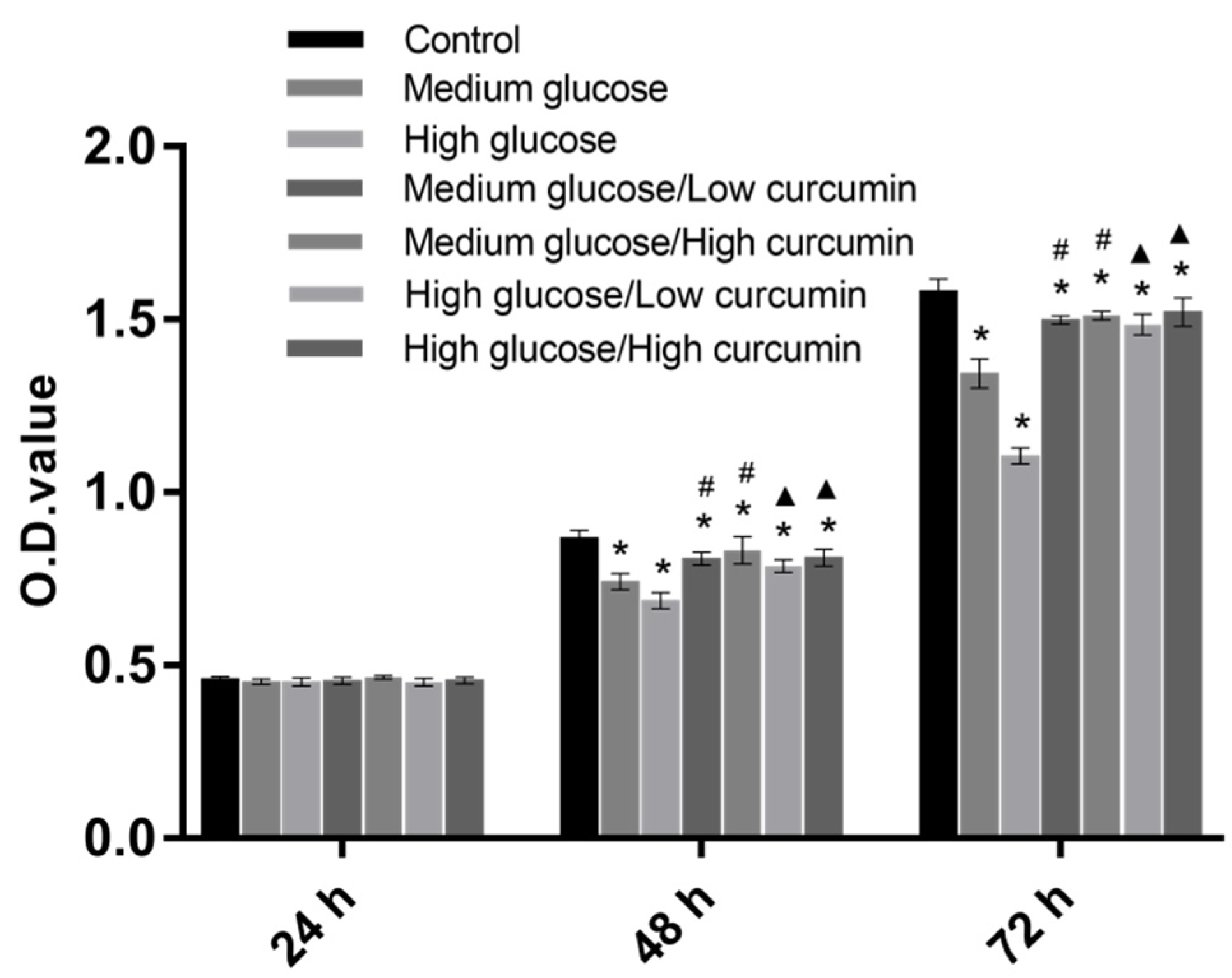
3.1. Effect of Curcumin on the Viability of MC3T3 Cells in High-Glucose Conditions

The proliferation of MC3T3 treated by curcumin under different concentrations of glucose was investigated by using a CCK-8 kit at 24, 48, and 72 h. No differences could be found among each group at 24 h. Cell viability in the control group increased most up to 48 h, and was significantly higher than that in the other groups (p < 0.05). The high-glucose group had the lowest cell viability, indicating that a higher concentration of glucose severely affected cell viability. When curcumin was introduced, cell viabilities in different glucose groups significantly increased (p < 0.05), whereas there were no differences between low and high concentrations of curcumin treatment. At 72 h, the cell viability in the curcumin-treated groups was significantly higher than that in the glucose groups (p < 0.05; Figure 1). Three concentrations of glucose were selected in present study. At 16.5 mmol/L glucose, there was a negative influence on cell viability. No differences were found on the effect of concentrations of curcumin among curcumin-treated groups.

3.2. Effect of Curcumin on Apoptosis of MC3T3 Cells in High-Glucose Conditions

Flow cytometry showed that curcumin treatment had an effect on glucose-induced apoptosis in MC3T3 cells (Figure 2A). The first-quadrant cells were necrotic and consisted of late apoptotic cells, which could be simultaneously stained with Annexin V-FITC and PI. The second-quadrant cells were PI-negative. In the third quadrant, normal living cells were not stained with Annexin V-FITC or PI. The fourth-quadrant cells were only stained with Annexin V-FITC, indicating early apoptotic cells. The early apoptosis rates of the control and glucose groups were 9.03% ± 0.67%, 14.70% ± 0.26%, and 18.70% ± 0.61%, and the curcumin-treated groups were 13.30% ± 0.10%, 12.00% ± 0.85%, 16.80% ± 0.20%, and 13.77% ± 0.21%. Glucose significantly increased the rate of apoptosis (p < 0.05), whereas treatment with both low and high doses of curcumin significantly decreased the apoptosis rate for cells under different concentrations of glucose (p < 0.05). In the high glucose group, the apoptosis rate in the high concentration of the curcumin group was significantly lower than that in the low concentration of the curcumin group (p < 0.05; Figure 2B), whereas there were no differences in the medium glucose group. It can be concluded that treatment with curcumin significantly protected cells from hyperglycemia-triggered apoptosis of MC3T3 cells under high-glucose conditions. This was shown in a dose-dependent manner under a high concentration of glucose, but not under a low concentration of glucose, under which a low concentration of curcumin can achieve an optimal protective effect on cells. This expands on previous observations that curcumin mitigates hyperglycemia-related clinical symptoms [29,30].

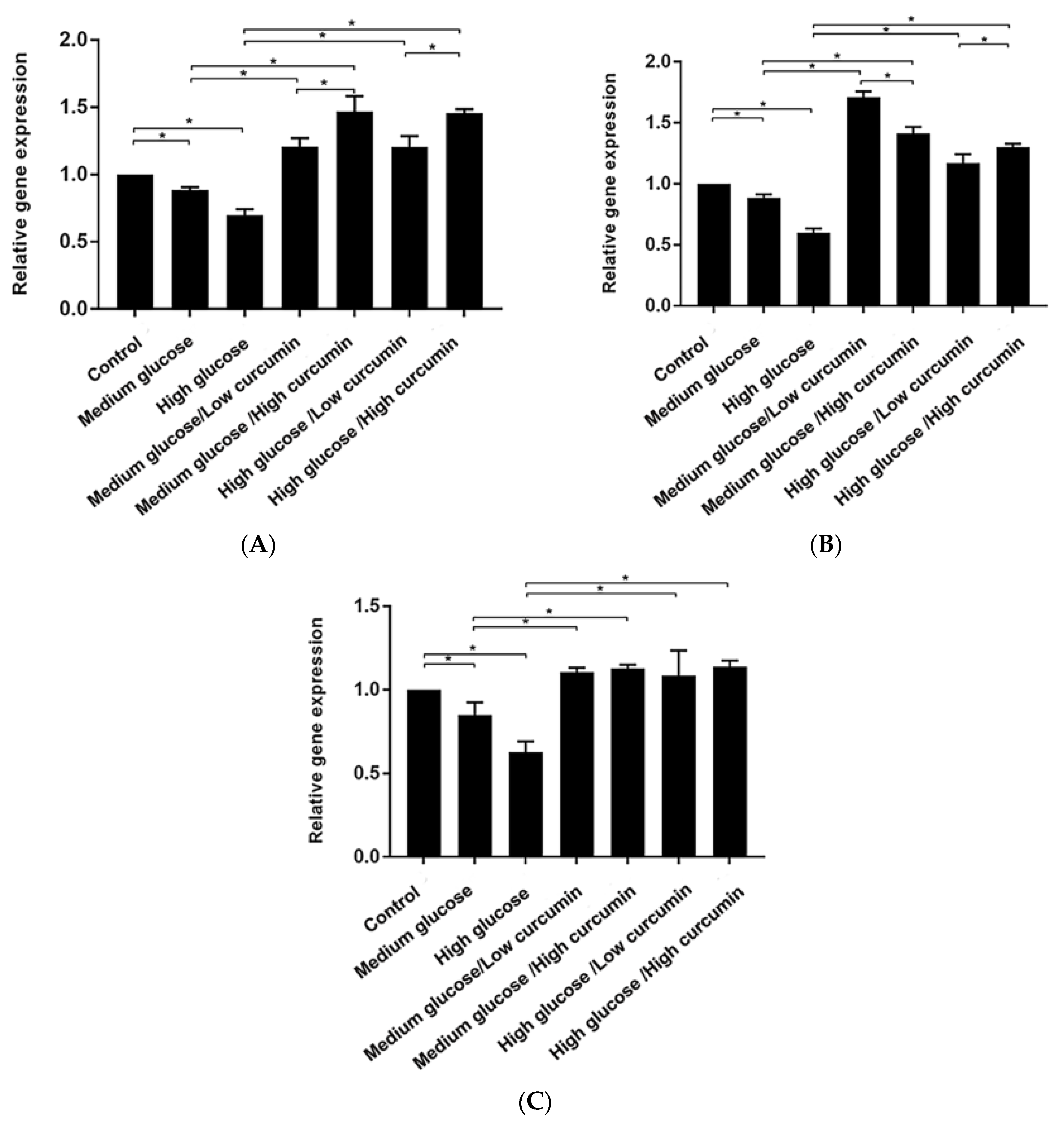
3.3. Effect of Curcumin on Osteogenic Differentiation of MC3T3 Cells in Glucose Conditions

The expressions of osteogenesis-related genes Runx2, Opn, and Col-1 in MC3T3 cells related to different concentrations of glucose and curcumin were evaluated using real-time quantitative PCR (RT-qPCR). Runx2 is crucial for osteoblastogenesis, regulates the differentiation, maturation, and bone formation of osteoblasts, and activates the expression of other osteogenic genes, such as Col-1 during early stages and Opn during late stages [31,32]. Col-1 is an important organic component of the bone matrix and the most crucial extracellular protein in bone, which initially provides a structural framework for inorganic deposition [33]. Opn is a phosphorylated glycoprotein secreted by osteoblasts and can promote biomineralization and bone remodeling [18]. In addition, the specific binding of Opn to Col-1 may naturally localize Opn, influencing the adhesion, differentiation, and function of osteoblasts [34]. In the present study, the expressions of Runx2, Opn, and Col-1 genes were significantly downregulated in the high glucose-treated group and were significantly lower than those of the control and medium glucose-treated groups (p < 0.05), indicating that a higher concentration of glucose severely affected expression of osteogenesis-related genes. When curcumin was introduced, the expressions of Runx2, Opn, and Col-1 genes were significantly upregulated at different concentrations of glucose (p < 0.05). The expressions of Runx2 and Col-1 genes in the high curcumin-treated group were significantly higher than those in the low curcumin-treated group (p < 0.05), whereas there was no significant difference in the expression of Opn between the low and high curcumin-treated groups (Figure 3). The results demonstrated that the osteogenic effect of curcumin was dose-dependent in the early stage of osteogenesis, but not in the late stage. It is possible that curcumin might predominantly upregulate the expression of Runx2, which in turn enhanced Col-1 expression in precursor osteoblasts, whereas Opn has a unique regulation mechanism, which is different from the combination of Col-1 and Runx2. It is interesting to further investigate the mechanism of curcumin upregulating Runx2, Opn, and Col-1 expressions in osteoblasts and the relationship between the expression of each gene. The current results indicated that curcumin could induce differentiation of MC3T3 cells from pre-osteoblasts to osteoblasts in different concentrations of glucose in a dose-dependent manner. Curcumin could serve as an effective method to recover glucose-suppressed osteogenic differentiation in precursor osteoblasts. In addition, 10 μmol/L curcumin was found to be an effective concentration that could promote osteogenic differentiation of MC3T3 under different concentrations of glucose.

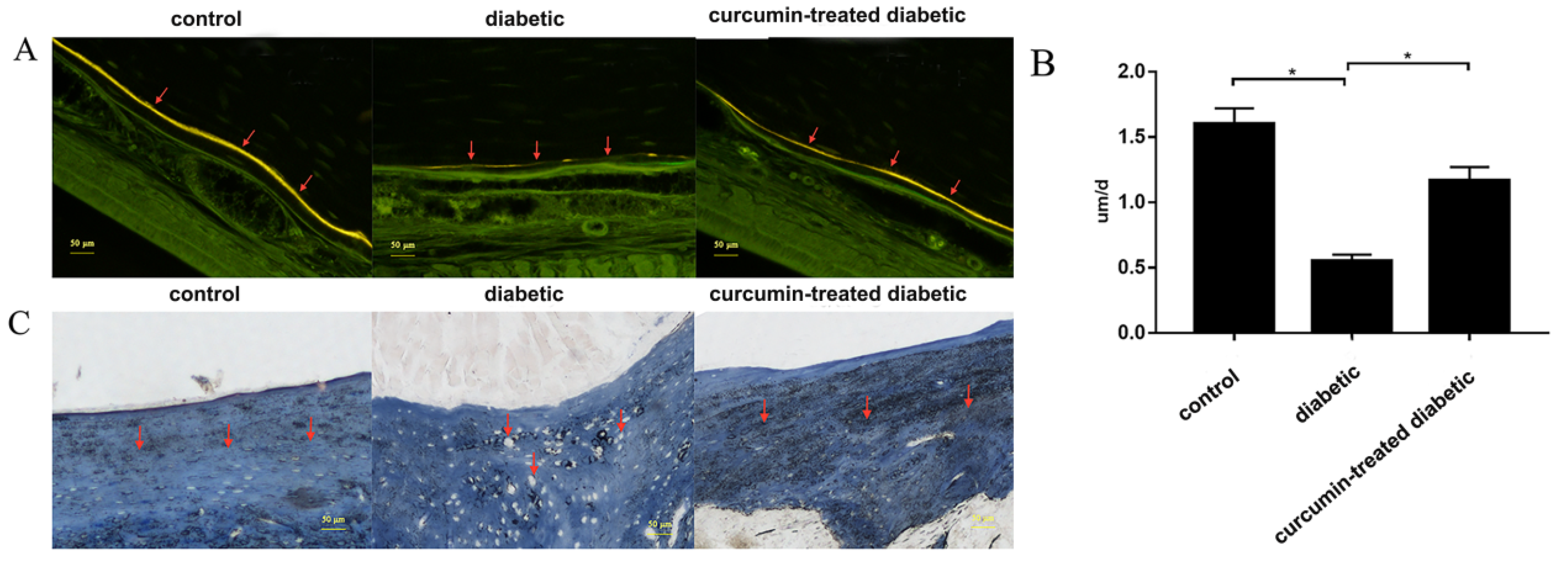
3.4. Curcumin Treatment Improved Alveolar Bone Formation in Diabetic Mice

Diabetes mellitus (DM) is a significant risk factor for osteoporosis. The proliferation and differentiation of osteoblasts in the alveolar bone of diabetic patients remains highly relevant in the daily work of dentists, as they can impact treatment for many oral diseases such as dental implants and oral surgery. Hyperglycemia is often displayed in diabetic patients and has a negative effect on alveolar bone reconstruction [35].
Previous studies have suggested a positive effect of curcumin on bone formation in diabetes and diabetes-related periodontitis [22]. However, few studies have investigated the effectiveness of natural curcumin on alveolar bone formation in diabetic osteoporosis, and there is also a lack of investigations into cortical bone and cancellous bone.
The present study evaluated cortical and cancellous bones by fluorescent double-labeling and Masson staining, respectively. The results revealed that significant differences in alveolar bone loss were observed in curcumin-treated groups when compared with diabetic mice. A general observation from fluorescence images (Figure 4A) was that curcumin treatment significantly enhanced alveolar cortical bone continuity and thickness when compared to diabetic mice, which was similar to that of non-diabetic mice. A general observation from Masson staining (Figure 4C) was that curcumin treatment extremely enhanced alveolar cancellous bone density and mineralization in diabetic mice, which is similar to that of non-diabetic mice. Curcumin treatment significantly enhanced the trabecular bone formation rate compared to diabetic mice (1.60 ± 0.11 um/d (diabetic) vs. 0.55 ± 0.05 um/d (curcumin), p < 0.05), and there was no significant difference observed when compared to the non-diabetic group (1.18 ± 0.11 um/d (non-diabetic)) (Figure 4B). These results show that curcumin treatment could improve alveolar bone structure in diabetes, and this is in agreement with the results of osteogenesis of MC3T3-E1 cells in response to curcumin treatment in vitro.
Diabetic hyperglycemia can trigger excessive production of ROS and increase oxidative stress, which leads to damaged macromolecular substances (nucleic acids and lipids), induction of cell apoptosis, and inhibition of osteogenic differentiation [36]. Bone protective effects of curcumin on diabetic mice may be explained by reducing the production of ROS induced by high glucose, enhancing antioxidant defense, and even further regulating the osteoimmunological RANK/RANKL/OPG pathway, which inhibits the expression of RANKL and promotes the expression of OPG in osteoblasts, results in an increase in bone formation and a decrease in bone resorption, thus offsetting the negative effects caused by high-glucose conditions. However, the specific mechanism remains to be confirmed by further research.

4. Conclusions

The results demonstrated that curcumin could protect cells from high glucose-mediated cytotoxicity, promote osteogenic differentiation of mouse precursor MC3T3 cells in a dose-dependent manner, and significantly improve alveolar bone formation in type 2 diabetic mice. These findings provide new insights into the pathogenesis of diabetes-related osteoporosis and indicate that curcumin may be valuable for prevention and inhibition of diabetes-related osteoporosis, providing a theoretical basis for the clinical treatment of alveolar bone disease in diabetic patients.

Author Contributions

All authors had equal contribution to this paper and research. Conceptualization, J.H. and X.Y.; Data curation, J.H. and A.O.A.; Formal analysis, D.L. and B.Z.; Investigation, J.H. and X.Y.; Supervision, Y.L.; Writing—original draft, J.H.; Writing—review and editing, F.L., A.O.A., and Y.L. All authors have read and agreed to the published version of the manuscript.

Funding

This research was funded by the Liaoning Province Natural Science with grant No. (20180550420), and Liaoning province key research and development guidance plan project with grant No. (2019 JH8/10300015).

Acknowledgments

The authors would like to express their thanks to teachers and students in the Experimental Centre of School and Hospital of Stomatology, China Medical University, for their continuous help and support.

Conflicts of Interest

The authors declare no conflicts of interest.

References

  1. Zhang, N.; Du, S.M.; Ma, G.S. Current lifestyle factors that increase risk of T2DM in China. Eur. J. Clin. Nutr. 2017, 71, 832–838. [Google Scholar] [CrossRef] [PubMed]
  2. Guariguata, L.; Whiting, D.R.; Hambleton, I.; Beagley, J.; Linnenkamp, U.; Shaw, J.E. Global estimates of diabetes prevalence for 2013 and projections for 2035. Diabetes Res. Clin. Pract. 2014, 103, 137–149. [Google Scholar] [CrossRef] [PubMed]
  3. Wu, T.; Qiao, S.; Shi, C.; Wang, S.; Ji, G. Metabolomics window into diabetic complications. J. Diabetes Investig. 2018, 9, 244–255. [Google Scholar] [CrossRef] [PubMed]
  4. De Amorim, L.M.N.; Vaz, S.R.; Cesário, G.; Coelho, A.S.G.; Botelho, P. Effect of green tea extract on bone mass and body composition in individuals with diabetes. J. Funct. Foods 2018, 40, 589–594. [Google Scholar] [CrossRef]
  5. Wang, T.; Cai, L.; Wang, Y.; Wang, Q.; Lu, D.; Chen, H.; Ying, X. The protective effects of silibinin in the treatment of streptozotocin-induced diabetic osteoporosis in rats. Biomed. Pharmacother. 2017, 89, 681–688. [Google Scholar] [CrossRef]
  6. Hamann, C.; Kirschner, S.; Günther, K.P.; Hofbauer, L.C. Bone, sweet bone-osteoporotic fractures in diabetes mellitus. Nat. Rev. Endocrinol. 2012, 8, 297–305. [Google Scholar] [CrossRef]
  7. Schwartz, A.V. Efficacy of osteoporosis therapies in diabetic patients. Calcif. Tissue Int. 2017, 100, 165–173. [Google Scholar] [CrossRef]
  8. Onishi, Y.; Yamada, K.; Zacho, J.; Ekelund, J.; Iwamoto, Y. Insulin degludec/insulin aspart versus biphasic insulin aspart 30 twice daily in Japanese subjects with type 2 diabetes: A randomized controlled trial. J. Diabetes Investig. 2017, 8, 210–217. [Google Scholar] [CrossRef] [Green Version]
  9. Wang, W.; Liu, H.; Xiao, S.; Liu, S.; Li, X.; Yu, P. Effects of insulin plus glucagon-like peptide-1 receptor agonists (GLP-1RAs) in treating type 1 diabetes mellitus: A systematic review and meta-analysis. Diabetes Ther. 2017, 8, 727–738. [Google Scholar] [CrossRef]
  10. Eibich, P.; Green, A.; Hattersley, A.T.; Jennison, C.; Lonergan, M.; Pearson, E.R.; Gray, A.M. Costs and treatment pathways for type 2 diabetes in the UK: A mastermind cohort study. Diabetes Ther. 2017, 8, 1031–1045. [Google Scholar] [CrossRef] [Green Version]
  11. Xie, A.; Li, R.; Tao, J.; Jiang, T.; Yan, H.; Zhang, H.; Yang, Y.; Yang, L.; Yechoor, V.; Chan, L.; et al. Anti-TCRβ mAb in combination with Neurogenin3 gene therapy reverses established overt type 1 diabetes in female NOD mice. Endocrinology 2017, 158, 3140–3151. [Google Scholar] [CrossRef] [PubMed]
  12. Elburki, M.S.; Moore, D.D.; Terezakis, N.G.; Zhang, Y.; Lee, H.M.; Johnson, F.; Golub, L.M. A novel chemically modified curcumin reduces inflammation-mediated connective tissue breakdown in a rat model of diabetes: Periodontal and systemic effects. J. Periodontal. Res. 2017, 52, 186–200. [Google Scholar] [CrossRef] [PubMed]
  13. Alessandra, G.M.; Daniela, F.P.; Jossiele, W.L.; Tamiris, R.S.; Pedro, H.D.; Matheus, H.J.; Vania, L.L.; Daniela, B.R.L. Hyperlipidemia-induced lipotoxicity and immune activation in rats are prevented by curcumin and rutin. Int. Immunopharmacol. 2020, 81, 106217. [Google Scholar] [CrossRef]
  14. Busari, Z.A.; Dauda, K.A.; Morenikeji, O.A.; Afolayan, F.; Oyeyemi, O.T.; Meena, J.; Sahu, D.; Panda, A.K. Antiplasmodial activity and toxicological assessment of curcumin PLGA-encapsulated nanoparticles. Front. Pharmacol. 2017, 8, 622. [Google Scholar] [CrossRef] [PubMed]
  15. Ziwei, M.; Na, W.; Haibing, H.; Xing, T. Pharmaceutical strategies of improving oral systemic bioavailability of curcumin for clinical application. J. Control. Release 2019, 31, 359–380. [Google Scholar] [CrossRef]
  16. Vincenzo, D.L.; Sante, D.G.; Francesco, M.; Paola, F.; Roberto, C.; Erminia, M.; Angela, A.; Massimo, C.; Lucia, C. Eudragit S100 entrapped liposome for curcumin delivery: Anti-oxidative effect in Caco-2 cells. Coatings 2020, 10, 114. [Google Scholar] [CrossRef] [Green Version]
  17. Anita, B.; Inese, M.; Simons, S.; Modra, M.; Martins, K. Curcumin effect on copper transport in HepG2 cells. Medicina 2018, 54, 14. [Google Scholar] [CrossRef] [Green Version]
  18. Abbassy, M.A.; Watari, I.; Soma, K. The effect of diabetes mellitus on rat mandibular bone formation and microarchitecture. Eur. J. Oral. Sci. 2010, 118, 364–369. [Google Scholar] [CrossRef]
  19. Naijil, G.; Anju, T.R.; Jayanarayanan, S.; Paulose, C.S. Curcumin pretreatment mediates antidiabetogenesis via functional regulation of adrenergic receptor subtypes in the pancreas of multiple low-dose streptozotocin-induced diabetic rats. Nutr. Res. 2015, 35, 823–833. [Google Scholar] [CrossRef]
  20. Elburki, M.S.; Rossa, C.J.; Guimaraes-stabili, M.R.; Lee, H.M.; Curylofo, F.A.; Johnson, F.; Golub, L.M. A chemically modified curcumin (CMC 2.24) inhibits nuclear factor κB activation and inflammatory bone loss in murine models of LPS-induced experimental periodontitis and diabetes-associated natural periodontitis. Inflammation 2017, 40, 1436–1449. [Google Scholar] [CrossRef]
  21. Wu, T.J.; Lin, C.Y.; Tsai, C.H.; Huang, Y.L.; Tang, C.H. Glucose suppresses IL-1β-induced MMP-1 expression through the FAK, MEK, ERK, and AP-1 signaling pathways. Environ. Toxicol. 2018, 33, 1061–1068. [Google Scholar] [CrossRef] [PubMed]
  22. Pimentel, S.P.; Casati, M.Z.; Ribeiro, F.V.; Corrêa, M.G.; Franck, F.C.; Benatti, B.B.; Cirano, F.R. Impact of natural curcumin on the progression of experimental periodontitis in diabetic rats. J. Periodontal Res. 2019. [Google Scholar] [CrossRef] [PubMed]
  23. Yu, L.; Zhanzhao, Z. Sustained curcumin release from PLGA microspheres improves bone formation under diabetic conditions by inhibiting the reactive oxygen species production. Drug Des. Dev. Ther. 2018, 12, 1453–1466. [Google Scholar] [CrossRef] [Green Version]
  24. Cirano, F.R.; Pimentel, S.P.; Casati, M.Z.; Corrêa, M.G.; Pino, D.S.; Messora, M.R.; Silva, P.H.F.; Ribeiro, F.V. Effect of curcumin on bone tissue in the diabetic rat: Repair of peri-implant and critical-sized defects. Int. J. Oral. Maxillofac. Surg. 2018, 47, 1495–1503. [Google Scholar] [CrossRef] [PubMed]
  25. Yang, L.; Liu, J.; Shan, Q.; Geng, G.; Shao, P. High glucose inhibits proliferation and differentiation of osteoblast in alveolar bone by inducing pyroptosis. Biochem. Biophys. Res. Commun. 2020, 522, 471–478. [Google Scholar] [CrossRef] [PubMed]
  26. Son, H.E.; Kim, E.J.; Jang, W.G. Curcumin induces osteoblast differentiation through mild-endoplasmic reticulum stress-mediated such as BMP2 on osteoblast cells. Life Sci. 2018, 193, 34–39. [Google Scholar] [CrossRef]
  27. Yang, H.; Gu, Q.; Huang, C.; Shi, Q.; Cai, Y. Curcumin increases rat mesenchymal stem cell osteoblast differentiation but inhibits adipocyte differentiation. Pharmacogn. Mag. 2012, 8, 202–208. [Google Scholar] [CrossRef] [Green Version]
  28. Chang, R.; Sun, L.; Webster, T.J. Short communication: Selective cytotoxicity of curcumin on osteosarcoma cells compared to healthy osteoblasts. Int. J. Nanomed. 2014, 9, 461–465. [Google Scholar] [CrossRef] [Green Version]
  29. Moran, J.M.; Roncero-Martin, R.; Rodriguez-Velasco, F.J.; Calderon-Garcia, J.F.; Rey-Sanchez, P.; Vera, V.; Canal-Macias, M.L.; Pedrera-Zamorano, J.D. Effects of curcumin on the proliferation and mineralization of human osteoblast-like cells: Implications of nitric oxide. Int. J. Mol. Sci. 2012, 13, 16104–16118. [Google Scholar] [CrossRef] [Green Version]
  30. Notoya, M.; Nishimura, H.; Woo, J.T.; Nagai, K.; Ishihara, Y.; Hagiwara, H. Curcumin inhibits the proliferation and mineralization of cultured osteoblasts. Eur. J. Pharmacol. 2006, 534, 55–62. [Google Scholar] [CrossRef]
  31. Corrêa, M.G.; Pires, P.R.; Ribeiro, F.V.; Pimentel, S.Z.; Casarin, R.C.; Cirano, F.R.; Tenenbaum, H.T.; Casati, M.Z. Systemic treatment with resveratrol and/or curcumin reduces the progression of experimental periodontitis in rats. J. Periodontal Res. 2017, 52, 201–220. [Google Scholar] [CrossRef] [PubMed]
  32. Boșca, A.B.; Ilea, A.; Sorițău, O.; Tatomir, C.; Miklášová, N.; Pârvu, A.E.; Mihu, C.M.; Melincovici, C.S.; Fischer-Fodor, E. Modulatory effect of curcumin analogs on the activation of metalloproteinases in human periodontal stem cells. Eur. J. Oral. Sci. 2019, 127, 304–312. [Google Scholar] [CrossRef] [PubMed]
  33. Dong, M.; Jiao, G.; Liu, H.; Wu, W.; Li, S.; Wang, Q.; Xu, D.; Li, X.; Liu, H.; Chen, Y. Biological silicon stimulates collagen type 1 and osteocalcin synthesis in human osteoblast-like cells through the BMP-2/Smad/RUNX2 signaling pathway. Biol. Trace Elem. Res. 2016, 173, 306–315. [Google Scholar] [CrossRef] [PubMed]
  34. Foster, B.L.; Ao, M.; Salmon, C.R.; Chavez, M.B.; Kolli, T.N.; Tran, A.B.; Chu, E.Y.; Kantovitz, K.R.; Yadav, M.; Narisawa, S. Osteopontin regulates dentin and alveolar bone development and mineralization. Bone 2018, 107, 196–207. [Google Scholar] [CrossRef] [PubMed]
  35. Colombo, J.S.; Balani, D.; Sloan, A.J.; Crean, S.J.; Okazaki, J.; Waddington, R.J. Delayed osteoblast differentiation and altered inflammatory response around implants placed in incisor sockets of type 2 diabetic rats. Clin. Oral. Implants Res. 2011, 22, 578–586. [Google Scholar] [CrossRef]
  36. Fakhruddin, S.; Alanazi, W.; Jackson, K.E. Diabetes-induced reactive oxygen species: Mechanism of their generation and role in renal injury. J. Diabetes Res. 2017, 2017, 1–30. [Google Scholar] [CrossRef]
Figure 1. Effects of curcumin on viability of MC3T3 cells in high-glucose conditions. Cell viability was measured using the CCK8 assay after culturing for 24, 48, and 72 h, separately. The bar chart shows the mean optical density (OD) values (n = 3). *p < 0.05 vs the control group; #p < 0.05 vs the medium glucose group; ▲p < 0.05 vs the high-glucose group.
Coatings 10 00258 g001
Figure 2. Effects of curcumin on apoptosis of MC3T3 cells in high-glucose conditions. Cell apoptosis was assessed by staining the cells with Annexin V-FITC/PI double staining kit and subsequent analysis by flow cytometry. (A) Detection of the early apoptosis rate of MC3T3 cells using flow cytometry. (B) The bar chart shows the mean of early apoptosis rate (n = 3). *p < 0.05 vs the control group; #p < 0.05 vs the medium glucose group; ▲p < 0.05 vs the high glucose group; and &p < 0.05 vs the high glucose/low curcumin group.
Coatings 10 00258 g002
Figure 3. Effects of curcumin on osteogenic differentiation of MC3T3 cells in glucose conditions. Expressions of (A) Runx2, (B) Opn, and (C) Col-1 were measured by reverse transcription quantitative polymerase chain reaction (RT-qPCR; n = 3). The results are shown as a relative expression level of the target gene using glyceraldehyde-3-phosphate-dehydrogenase (GAPDH) as the inner reference gene, and analyzed by 2-ΔΔCt. Data are shown as mean ± SD. *p < 0.05.
Coatings 10 00258 g003
Figure 4. Evaluation of the osteogenic effect of curcumin on bone formation in vivo. (A) Fluorescent microscopy images of alveolar bone in non-diabetic (control), diabetic, and curcumin-treated groups, respectively. Alveolar cortical bone continuity and thickness improved in the db/db + C group compared with that in the db/db group, which was similar to that of the db/m group. (B) The bar chart shows the rate of trabecular formation. *p < 0.05. (C) Masson staining images of alveolar bone in non-diabetic (control), diabetic, and curcumin-treated groups, respectively. Alveolar cancellous bone density and mineralization improved in the db/db + C group compared with that in the db/db group, which was similar to that of the db/m group.
Coatings 10 00258 g004
Table 1. Experiment groups and abbreviations.
Experiment GroupsGlucoseCurcuminAbbreviation
Control5.5 mmol/L0 μmol/LC
Medium glucose11 mmol/L0 μmol/LMg
High glucose16.5 mmol/L0 μmol/LHg
Medium glucose/Low curcumin11 mmol/L5 μmol/LMg-Lc
Medium glucose/High curcumin11 mmol/L10 μmol/LMg-Hc
High glucose/Low curcumin16.5 mmol/L5 μmol/LHg-Lc
High glucose/High curcumin16.5 mmol/L10 μmol/LHg-Hc

