Article

Cross-Sector Antimicrobial Resistance and Virulence in Enterococcus spp. from Humans, Animals and the Environment

by
Nicolau Fialho
1,2,3,†,
Joana Monteiro Marques
1,2,4,5,†,
Maria Teresa Barreto-Crespo
6,7 and
Teresa Semedo-Lemsaddek
1,2,8,*
1
Centre for Interdisciplinary Research in Animal Health (CIISA), Faculty of Veterinary Medicine, University of Lisbon, 1300-477 Lisbon, Portugal
2
Associate Laboratory for Animal and Veterinary Sciences (AL4AnimalS), 1300-477 Lisbon, Portugal
3
Faculty of Sciences, University of Lisbon, 1749-016 Lisbon, Portugal
4
National Institute of Agricultural and Veterinary Research, IP (INIAV), 2780-157 Oeiras, Portugal
5
cE3c—Centre for Ecology, Evolution and Environmental Changes & CHANGE, Global Change and Sustainability Institute, Faculty of Sciences, University of Lisbon, 1749-016 Lisbon, Portugal
6
iBET—Instituto de Biologia Experimental e Tecnológica, 2780-157 Oeiras, Portugal
7
Instituto de Tecnologia Química e Biológica António Xavier (ITQB), 2780-157 Oeiras, Portugal
8
Biosystems & Integrative Sciences Institute (BioISI), Faculty of Sciences, University of Lisbon, 1749-016 Lisbon, Portugal
*
Author to whom correspondence should be addressed.
These authors contributed equally to this work.
Antibiotics 2026, 15(7), 657; https://doi.org/10.3390/antibiotics15070657
Submission received: 3 June 2026 / Revised: 19 June 2026 / Accepted: 25 June 2026 / Published: 2 July 2026

Abstract

Background/Objectives: Antimicrobial resistance is a major public health concern requiring integrated surveillance across human, animal, and environmental sectors. Enterococcus spp. are widely distributed opportunistic bacteria with the capacity to acquire and disseminate resistance and virulence determinants. This study aimed to characterize species distribution, phenotypic antimicrobial resistance, resistance genes, and virulence-associated traits in Enterococcus spp. from One Health sources. Methods: Enterococci were recovered from 66 samples collected in Lisbon, Portugal, between late 2022 and early 2024, including healthy and sick humans, healthy and sick animals, canteen food, surface water, and public transport surfaces. RAPD-PCR was used to assess genetic diversity among 90 isolates and select 74 representative enterococci. Species identification, resistance gene screening, and virulence gene detection were performed by PCR. Antimicrobial susceptibility was assessed by disk diffusion against 12 antibiotics using CLSI criteria. Vancomycin resistance was further evaluated by agar dilution and Etest when applicable. Results: Enterococcus faecalis predominated, representing 63/74 isolates (85.1%), followed by Enterococcus faecium (5/74, 6.8%) and other Enterococcus spp. (6/74, 8.1%). Antibiotic resistance was detected across all One Health sectors. Sick Human isolates showed higher resistance than Healthy Human isolates. E. faecium showed higher resistance than E. faecalis, and all E. faecium isolates across sectors were multidrug-resistant. Rifampicin resistance was frequent, as was quinupristin-dalfopristin resistance (excluding intrinsically resistant E. faecalis), while linezolid resistance was not detected. Resistance and virulence determinants were distributed across sectors. Conclusions: Enterococci from human, animal, and environmental sources carried antimicrobial resistance and virulence-associated traits. These findings support integrated One Health surveillance to monitor resistant enterococci across interconnected reservoirs.

1. Introduction

Antimicrobial resistance (AMR) is a major threat to public health, reducing the effectiveness of antimicrobial therapy and increasing morbidity and mortality worldwide [1]. The global burden of AMR is substantial, with an estimated 4.95 million deaths associated with bacterial AMR, and 1.27 million directly attributable deaths [2]. This growing burden threatens the continued effectiveness of antimicrobial therapy and increases the difficulty of treating common infections [3].
Addressing the shared threat posed by AMR requires an integrated approach across human, animal and environmental sectors, as formalized by the One Health framework [3]. The concept recognizes that selection pressures in healthcare, agriculture and the wider environment interact to shape resistance patterns [4,5]. Effective surveillance and control therefore require coordinated action linking public health, veterinary medicine and environmental science and policies.
Enterococci are ubiquitous bacteria with a broad ecological distribution. They colonize the gastrointestinal tracts of wild and domestic animals and occur in soils, plants, insects, food products, hospital environments and the human microbiota [6,7,8,9,10,11,12,13,14]. In clinical settings, enterococci have emerged as opportunistic pathogens and are a major cause of healthcare-associated infections, including urinary tract infections, bacteremia, and infective endocarditis [15].
E. faecalis and E. faecium account for approximately 90 to 95% of enterococcal infections, while other species, including Enterococcus hirae, Enterococcus gallinarum, and Enterococcus casseliflavus, occur less frequently [16].
Enterococci exhibit intrinsic resistance to third-generation cephalosporins and aminoglycosides [17] and can also acquire virulence factors and Antimicrobial Resistance Genes (ARGs) through horizontal gene transfer events, including determinants conferring resistance to last resort antimicrobials [16].
Vancomycin-resistant Enterococcus (VRE) represent a major clinical concern, particularly vancomycin-resistant E. faecium (VREfm), which is frequently associated with hospital outbreaks [17,18]. This organism is classified by the World Health Organization as a high-priority pathogen, and is included in the ESKAPE group of pathogens composed of E. faecium, Staphylococcus aureus, Klebsiella pneumoniae, Acinetobacter baumannii, Pseudomonas aeruginosa, and Enterobacter spp. [17,19].
Vancomycin resistance in enterococci is mediated by the van gene cluster. The vanA and vanB genotypes are most frequently detected in E. faecium [17,20]. Vancomycin-variable Enterococcus (VVE) isolates appear phenotypically susceptible to vancomycin while harboring vanA, but express resistance after exposure to glycopeptides [17,21].
As VRE prevalence has increased, linezolid has been used as a last resort oxazolidinone due to its activity against Gram-positive pathogens through inhibition of protein synthesis [22,23]. However, resistance to linezolid emerged within one year of its introduction and despite remaining relatively low, has been increasing among clinical isolates [17].
Resistance patterns in enterococci are shaped by antimicrobial selection pressures operating across healthcare, agriculture, aquaculture, and environmental settings [24,25]. Comparative and genomic studies have demonstrated differences in species composition and resistance profiles across sources, complicating interpretation when surveillance remains restricted to individual sectors [17,26,27].
In addition to antibiotic resistance, the assessment of virulence determinants is essential for understanding the pathogenic potential and dissemination of enterococcal lineages across the One Health continuum [17]. Virulence factors such as agg, cylA, esp and gelE are commonly identified in enterococci among humans [28], animals [13,16,29,30,31,32], and the environment [33,34,35] across the world.
Enterococci have traditionally been studied within separate human, animal, or environmental sectors, with limited interdisciplinary integration across these domains [36,37]. This compartmentalized approach constrains understanding of resistance reservoirs and transmission pathways across the One Health continuum [17,26,27].
Despite recognition of these limitations, integrated studies comparing species distribution and resistance profiles across sectors remain limited. Existing surveillance and review studies frequently rely on sector-specific datasets, which restrict inference on the dissemination of resistance determinants between interconnected sources [17,26,27].
This study aimed to characterize the species distribution, as well as the phenotypic and genotypic AMR and virulence profiles, of Enterococcus spp. isolated from human, animal, and environmental sources within a One Health framework, in the Lisbon metropolitan area, Portugal. The Lisbon metropolitan area was selected due to its geographical proximity, high degree of urban interconnectedness, and the growing epidemiological relevance of VREfm in Portugal, where increasing levels of VRE have been reported in recent years [38]. We hypothesized that isolates from these interconnected reservoirs would exhibit overlapping AMR and virulence determinants, supporting the ecological interconnectedness of enterococci across One Health sectors.

2. Results and Discussion

2.1. Microbial Collection

Enterococci were isolated from a total of 66 samples from One Health sources (Humans, Animals and Environment), including Healthy (n = 14) and Sick (n = 17) humans with varied enterococcal infections, Healthy (n = 10) and Sick outpatient (n = 12) animals (non-enterococcal infection), Canteen Food (n = 9), Surface Water (n = 1) and Public Transport Surfaces (n = 3). Clinical isolates from enterococcal infections included genitourinary infections (urine, n = 7), tissue infections (biopsy, n = 3), purulent infections (pus, n = 3), systemic infections (blood, n = 2), and unknown infection sites (n = 2). Following isolation in Slanetz-Bartley agar (SBA) and SBA supplemented with vancomycin (SBAvan), culture purity and presumptive genus-level identification were confirmed by successive subcultures on SBA and Bile-Esculin Azide agar (BEA). From this initial purification, 90 isolates were retrieved, 35 from the Human sector, 27 from the Animal sector, and 28 from the Environment sector.
Given the ubiquity of enterococci across One Health sectors, the first analytical objective of this study was to assess microbial diversity and select representative isolates for further characterization. This approach aimed to reduce redundancy while preserving the diversity of enterococcal populations across sectors (humans, animals, and environment) and respective subsectors, ensuring balanced representation in subsequent analyses.
Random Amplification of Polymorphic DNA-Polymerase Chain Reaction (RAPD-PCR) profiles generated with primers OPC15 and (GTG)5 were combined to construct a composite dendrogram based on the average similarity of both analyses. The mean reproducibility of the method was 76.6%, which was adopted as the similarity threshold for cluster definition and is indicated in Supplementary Figure S1.
Following RAPD-PCR clustering and application of the predefined selection criteria, a final subset of representative enterococcal isolates was established for downstream analyses. This collection comprised a total of 74 isolates, including 31 from the Human sector, 22 from the Animal sector and 21 from the Environment sector. Considering subsector distribution, it includes Healthy Humans (n = 14) and Sick Humans (n = 17), Healthy Animals (n = 10) and Sick Animals (n = 12), Canteen Food (n = 9), Surface Water (n = 6), and Public Transport Surfaces (n = 6). Of these isolates, 5 from Healthy Humans, 1 from Sick Animals and 2 from Canteen Food were retrieved from SBAvan plates.

2.2. Molecular Species Identification

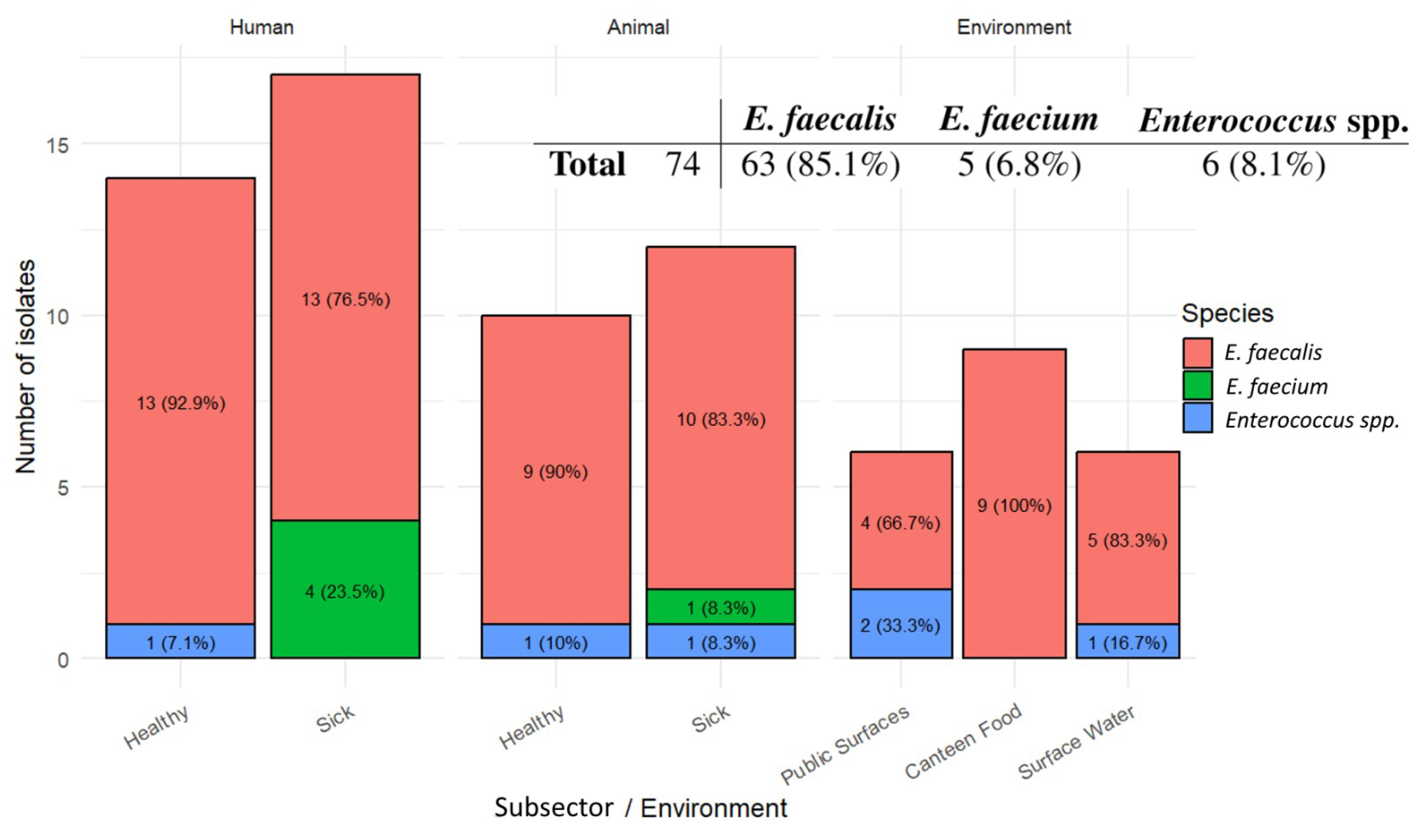
Species identification was performed by Polymerase Chain Reaction (PCR). Species distribution results, by sector and subsectors, are summarized in Figure 1 and detailed by isolate in Supplementary Table S1. Isolates that could not be assigned to E. faecalis, E. faecium, or Enterococcus durans were tested for the genus and classified as Enterococcus spp.
Among the 74 representative isolates analyzed, E. faecalis was the dominant species, accounting for 63 isolates (85.1%), followed by E. faecium with 5 isolates (6.8%), while 6 isolates (8.1%) remained non-determined at the species level and were identified at the genus level. E. durans was not identified in our collection. E. faecalis predominated across all One Health sectors, although its relative dominance was lower in Environmental samples, where other Enterococcus spp. were more frequently recovered, particularly from Surface Water and Public Transport Surfaces. This broader diversity is consistent with the heterogeneous nature of environmental matrices and their exposure to multiple contamination sources [39,40,41,42].
Subsector-specific patterns were evident. In the Human sector, E. faecalis predominated in both Healthy and Sick isolates, while E. faecium was detected only among Sick Human isolates, supporting its recognized association with hospital-associated infections and clinical settings [43,44,45]. A similar pattern was observed in Animals, where E. faecalis remained dominant and E. faecium was detected in only one Sick Animal isolate. This aligns with studies reporting E. faecalis as frequent in animal hosts, while variation in other species, including E. hirae and Enterococcus cecorum, reflects host species, age, and agricultural practices [26,46,47,48].
The predominance of E. faecalis is consistent with its ecological versatility as a generalist commensal, its frequent occurrence in the gastrointestinal tract of humans and animals, and its capacity to persist in soil, water, sediments, plants, food products, and other environmental compartments [11,49,50,51,52,53]. Enterococcal tolerance to environmental stressors, oxidative stress adaptation, virulence traits, and biofilm formation further support persistence across host-associated and abiotic environments [9,15,17,20,36]. Nevertheless, the distribution of enterococci remains context dependent, as hospital settings have seen an increasing role of multidrug-resistant E. faecium, particularly in bloodstream infections and in association with resistance to ampicillin and vancomycin [17,18,50,51,54,55]. Culture-based isolation may also have favored the recovery of E. faecalis, as commonly used media select for bile tolerance, esculin hydrolysis, and robust growth, potentially underrepresenting less tolerant species [31,56,57,58].

2.3. Antibiotic Resistance

2.3.1. Phenotypic Resistance Assay

Antimicrobial susceptibility testing (AST) was performed using the disk diffusion method. Results were interpreted according to breakpoints established by the Clinical and Laboratory Standards Institute (CLSI). Although the European Committee on Antimicrobial Susceptibility Testing (EUCAST) criteria are geographically appropriate for a European dataset, EUCAST breakpoints were unavailable for several antibiotics included in this panel (specifically quinupristin-dalfopristin, rifampicin, and chloramphenicol) and, where available, differ from CLSI in both disk concentrations and interpretive thresholds, limiting direct comparability across compounds and studies. Given these constraints and the broader availability of published reference data using CLSI, this system was applied throughout. The use of CLSI rather than EUCAST is acknowledged as a limitation and should be considered when comparing results with European surveillance datasets.
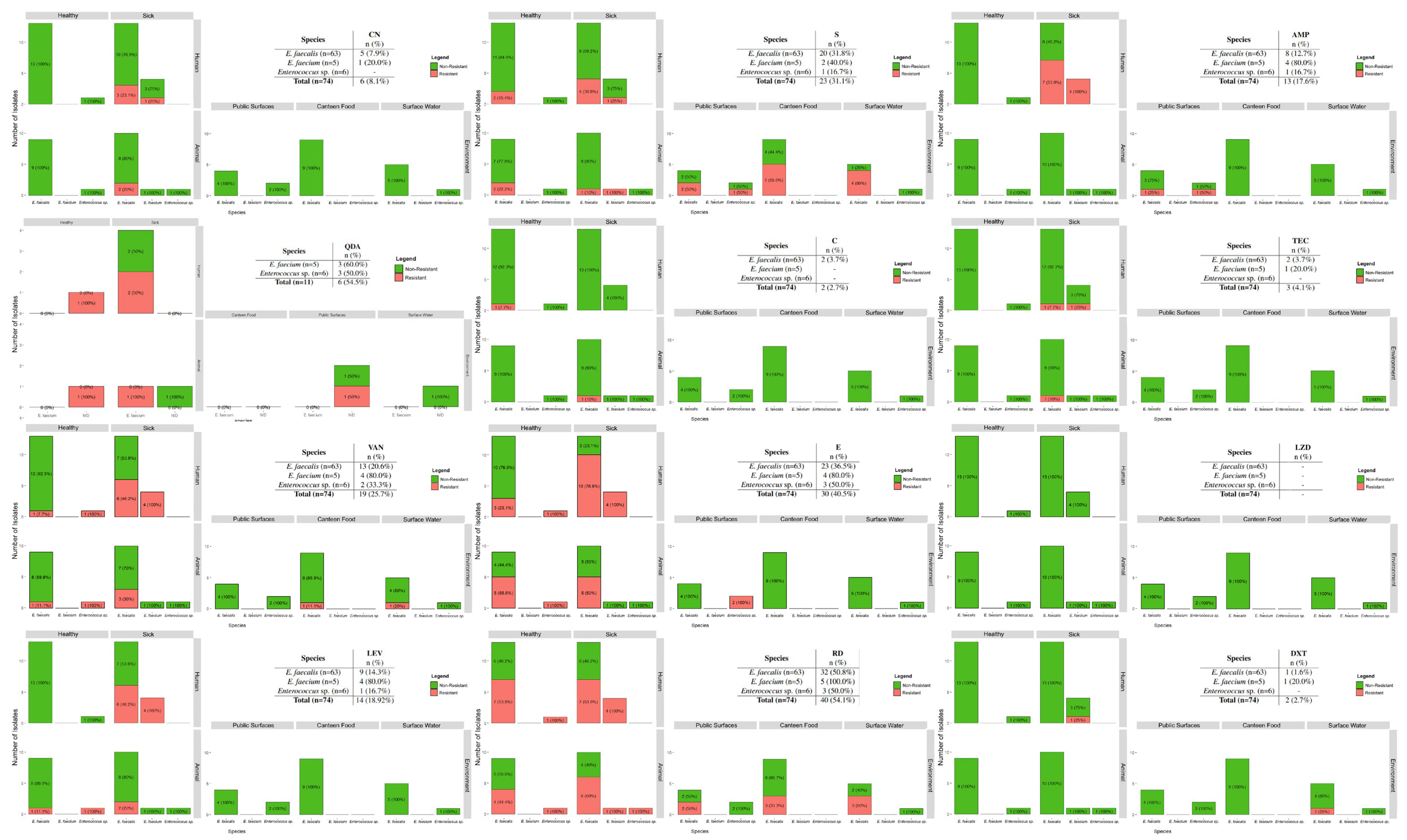
Phenotypic resistance was assessed for each antibiotic by One Health sector and subsector. The distribution of resistance by enterococcal species and One Health subsector is plotted in Figure 2, while resistance frequencies by sector and subsector are presented in Table 1. Resistance frequencies were calculated considering only isolates classified as resistant according to CLSI breakpoints. Antibiotic resistance profiles for individual isolates are provided in Supplementary Table S1.
Across the One Health framework, higher resistance prevalences were consistently observed among isolates from Sick Humans, followed by Sick Animals, whereas isolates from Healthy Humans, Healthy Animals, and Environmental subsectors generally exhibited lower resistance levels. Exceptions were noted for streptomycin, quinupristin-dalfopristin, and doxycycline, for which elevated resistance was detected in specific Environmental subsectors. Overall, E. faecium displayed higher resistance rates than E. faecalis across most antibiotics, reflecting its recognized role as a hospital-adapted and Multidrug-Resistant (MDR) lineage [18,50,51]. The main exception was chloramphenicol, for which resistance was detected exclusively in E. faecalis. These patterns broadly align with existing literature, supporting a gradient of resistance intensity from clinical to non-clinical settings while underscoring species-specific differences in resistance burden.
Aminoglycoside resistance displayed marked heterogeneity across sectors and between compounds. Resistance to gentamicin was uncommon overall and was primarily detected in Sick patients, particularly among Sick Humans. This pattern aligns with the historical role of gentamicin in severe enterococcal infections, where synergy with cell-wall active agents was established and exploited in combination therapy [50,59]. Following recognition of toxicity at high doses and the introduction of alternative agents, gentamicin use declined, although renewed interest has emerged in the context of MDR infections and combination regimens [60,61,62,63]. Consistent with this clinical selectivity, little to no gentamicin resistance was detected in several environmental and food-associated contexts referenced in the literature, including transport-associated surfaces, ready-to-eat foods, cheeses, and surface water [34,64,65,66,67,68], although earlier Portuguese drinking-water data reported high-level gentamicin resistance in a subset of isolates [69]. In contrast to the clinically restricted distribution of gentamicin resistance, resistance to streptomycin was widespread across the One Health continuum in the present dataset and Public Transport Surfaces; and represented one of the most prevalent resistance phenotypes observed. Similar resistance has been repeatedly reported across healthy and clinical human isolates, diverse animal hosts, foods, and environmental waters [28,34,44,47,68,69,70,71,72,73,74,75]. This broad distribution is consistent with the long-standing use of streptomycin in human medicine and its documented use in animal settings, including historical growth promotion, which together have supported the persistence and dissemination of high-level streptomycin resistance determinants across multiple compartments [26,42,50,66,76]. Taken together, aminoglycoside resistance patterns illustrate contrasting selection histories: gentamicin resistance appears more tightly linked to clinical exposure, while streptomycin resistance reflects widespread dissemination across multiple One Health reservoirs [50,77].
Resistance to ampicillin showed a clear sectoral and species-associated pattern. No resistance was detected in Healthy Humans, consistent with reports from Portugal and Tunisia, where resistance remains low in E. faecalis and limited in E. faecium [28,74]. In contrast, resistance increased markedly in Sick Humans, particularly in E. faecium, in line with its established association with clinical infections [44,78]. No resistance was observed in the Animal sector, which contrasts with studies reporting moderate to high prevalence in livestock and companion animals [47,68,79]. This discrepancy may partly reflect the limited sample size in the animal subsector of the present study, which may have been insufficient to detect resistance at the lower prevalence levels described in some populations [75,80]; this should be considered a constraint when interpreting the absence of resistance in this sector. Environmental sources exhibited low prevalence overall, with sporadic detection on Public Transport Surfaces and absence in Food and surface Water, consistent with reports from food and environmental matrices [34,64,66,67,68,69,70]. These patterns reflect the role of ampicillin as a first-line agent and the preferential accumulation of resistance in hospital-adapted E. faecium, while E. faecalis remains largely susceptible [50].
Resistance to glycopeptides showed a marked concentration in clinical settings. Teicoplanin resistance was restricted to Sick Humans and Animals, with no detection in Healthy or Environmental subsectors, consistent with reports describing low prevalence outside clinical contexts [73,74,75]. In contrast, vancomycin resistance reported by disk-diffusion was more widely distributed but remained primarily associated with Sick Humans and Animals, with most resistant isolates belonging to E. faecium, frequently classified as VREfm, in line with its recognized dominance in clinical infections [26,81,82]. Lower prevalence in Healthy Humans and Animals agrees with community and livestock studies reporting limited vancomycin resistance [43,73,74,75,83]. Environmental sources showed low prevalence, with absence on Public Transport Surfaces and sporadic detection in Canteen Food and Surface Water, although higher levels have been reported in wastewater-impacted environments, particularly involving E. faecium [19,34,67]. These patterns reflect the extensive clinical use of glycopeptides and the selective pressure driving the emergence and persistence of vancomycin-resistant enterococci. The historical use of avoparcin, a glycopeptide antibiotic used as a growth promoter in European livestock prior to its ban in 1997, is well recognized as a key driver in the early selection and environmental establishment of VRE reservoirs, contributing to the persistence of resistance determinants in animal and environmental compartments observed to this day [76,84]. Vancomycin resistance was confirmed by Etest in 19 isolates considered resistant in both AST and agar dilution methods, with Minimum Inhibitory Concentrations (MIC) ranging from 2 to 256 μg/mL. Vancomycin Etest MICs among resistant isolates were distributed as follows: 2 μg/mL (n = 4, 1 from Sick Humans, 1 from Sick Animals, 1 from Healthy Animals, 1 from Surface Water), 4 μg/mL (n = 5, 1 from Healthy Animals, 2 from Sick Animals, 1 from Canteen Food), 8 μg/mL (n = 1 from Healthy Humans), 64 μg/mL (n = 1 from Sick Humans), and ≥ 256 μg/mL (n = 8 from Sick Humans) (detailed in Supplementary Table S1. The ten isolates with MIC values below the CLSI resistance threshold for vancomycin (32 μg/mL) were reclassified and interpreted as susceptible or intermediate VVE isolates and were not considered in the VAN-MIC column on Table 1. High-level vancomycin resistance (MIC ≥ 256 μg/mL) was restricted to clinical isolates, being detected in 8 of the 19 enterococci previously classified as VRE and recovered from three different healthcare entities (two hospitals and a clinical laboratory). This finding is consistent with the recognized predominance of VRE in healthcare-associated settings and hospitalized patient populations. An overall prevalence of 12.1% (9/74) was observed, with all resistant isolates originating from the Human sector, primarily from clinical infection samples. At the European level, EARS-Net surveillance highlights the continued clinical importance of VREfm, with a population-weighted mean resistance prevalence of 16.5% among invasive isolates in the EU/EEA in 2024 and an estimated incidence of 1.96 bloodstream infections per 100,000 population [38]. Similarly, Portugal has reported increasing levels of VREfm, rising from 4.3% to 11.0% from 2018 to 2022 among invasive isolates [85]. However, in our study, only 37.5% of high-MIC VRE were E. faecium, whereas the remainder were E. faecalis. This finding contrasts with EARS-Net surveillance trends, where vancomycin resistance is predominantly associated with E. faecium clinical isolates, suggesting potential differences in the epidemiology of VRE within the study population and sampling methods.
Resistance to erythromycin was widespread across sectors, with higher prevalence in Sick Human and Animal isolates and lower but persistent levels in Healthy and Environmental subsectors. In Humans, resistance increased markedly in Sick isolates, consistent with reports of elevated macrolide resistance in clinical enterococci, while remaining lower in Healthy individuals [28,44,74]. In the Animal sector, high resistance was observed in both Healthy and Sick isolates, aligning with reported variability linked to host species and antimicrobial exposure [46,73,75]. Environmental sources showed lower prevalence, although resistance was detected on Public Transport Surfaces, supporting dissemination beyond clinical and agricultural settings, as described in urban and aquatic environments [66,67,68,70]. Joint Wald tests were performed to assess the relationship between each sector for each antibiotic. For erythromycin, the Environmental sector had significantly lower resistance counts compared to both the Human (p = 0.034) and the Animal (p = 0.039) sectors. Species-specific patterns were evident, with E. faecalis accounting for most resistant isolates, consistent with the widespread distribution of macrolide resistance determinants such as the erm(B) gene, frequently associated with mobile genetic elements and co-localized with other ARGs [46,50]. These patterns reflect sustained selective pressure driven by historical and ongoing macrolide use across sectors, supporting the persistence and dissemination of resistance within a One Health framework.
Linezolid resistance was not detected across any sector or subsector, with all Human, Animal, and Environmental isolates remaining phenotypically susceptible, including those from sick hosts. This finding aligns with most reports describing low prevalence of resistance in enterococci, typically below 5% for both E. faecalis and E. faecium [16,17,50]. However, higher prevalence has been reported in specific contexts, including fecal carriage in Switzerland, hospital isolates in Romania, companion animals, and wastewater-associated environments [19,44,47,86], indicating that resistance emergence is context-dependent and influenced by local selective pressures. The sustained efficacy of linezolid reflects its restricted use as a last-resort agent for multidrug-resistant Gram-positive infections, including VRE [50]. Resistance, when present, is mediated by target site mutations and transferable determinants associated with cross-resistance to other antimicrobial classes [17,46,87]. Although not approved for veterinary or agricultural use, the detection of such determinants in animal and environmental isolates suggests indirect selection via co-located ARGs. These results support the continued effectiveness of linezolid, while highlighting the need for ongoing phenotypic and genotypic surveillance across One Health sectors.
Resistance to chloramphenicol was uncommon overall, with very low prevalence and resistant isolates restricted to Healthy subsectors. No resistance was detected among Sick Human or Sick Animal isolates, or in any Environmental subsector. Detection of resistance in Healthy Animal isolates contrasts with some reports of complete susceptibility but remains within the variability described across host species [73,75,88]. This pattern is consistent with contemporary studies reporting low chloramphenicol resistance in enterococci from distinct sources, particularly in Europe [28,74,89], and likely reflects reduced clinical use following recognition of severe toxicity, including aplastic anemia, and restrictions in food-producing animals [90,91,92,93]. Notably, chloramphenicol resistance genes are often transferable and co-located with determinants conferring resistance to other antimicrobial classes, allowing their maintenance through co-selection even in the absence of direct drug exposure [46,94].
Resistance to levofloxacin showed a clear sectoral pattern, with high prevalence among Sick Human isolates, lower but detectable resistance in Animal isolates, and no resistance in Healthy Human or Environmental subsectors. In Sick Humans, resistance affected all E. faecium and a substantial proportion of E. faecalis, consistent with reports of elevated fluoroquinolone resistance in enterococci recovered from infections, particularly E. faecium [26,78]. In Animals, resistance remained within the broad range reported for livestock and companion animals, where prevalence varies with host species and antimicrobial exposure [47,72,75]. The absence of resistance in Canteen Food, Surface Water, and Public Transport Surfaces aligns with food surveillance studies reporting minimal resistance [66,70,95], although higher levels have been described in aquatic environments influenced by agricultural or urban run-off, especially involving E. faecium [67,96]. These patterns reflect the extensive use of fluoroquinolones in human medicine and historical or extra-label use in animals, with regulatory restrictions potentially having reduced selection pressure in non-human sectors [75,97,98,99,100]. Continued monitoring remains important given the clinical relevance of fluoroquinolones and their classification as critically important antimicrobials for human health [101].
Resistance to rifampicin was among the highest observed in this study, with more than half of all isolates showing non-susceptibility and resistance detected across all sectors. Prevalence was particularly high among Sick Human and Animal isolates, and most pronounced in E. faecium, where all isolates were resistant. Human-sector resistance exceeded several European and international reports, where clinical enterococcal resistance typically ranges from approximately 20% to 40% [44,102]. The higher resistance rates observed here may reflect the composition of the clinical sample, which was enriched for severely ill or bacteremic patients at a tertiary referral center, populations known to harbor higher proportions of MDR E. faecium [102]. Regional prescribing patterns, including the use of rifampicin as part of combination regimens for difficult-to-treat infections such as VREfm endocarditis, may also have contributed to elevated local selection pressure [103]. The higher resistance in E. faecium compared with E. faecalis is consistent with published species-specific patterns [102]. In Animals, resistance fell within the broad range reported for livestock and companion animals [47,48,89]. Environmental resistance, particularly in Surface Water, closely matched findings from municipal wastewater effluents and receiving rivers, supporting the view that rifampicin resistance in environmental enterococci primarily reflects contamination from human sources rather than direct environmental selection [50,104]. The high cross-sector prevalence observed supports continued surveillance of rifampicin resistance across One Health settings.
Resistance to quinupristin-dalfopristin was among the highest observed, with its prevalence being only documented for E. faecium and other Enterococcus spp., as E. faecalis exhibits intrinsic resistance to streptogramins and is not a clinical target for this antibiotic. The therapeutically relevant resistance burden therefore lies among E. faecium and isolates from other Enterococcus spp., for which acquired resistance is of genuine clinical concern [50]. With this distinction in mind, resistance in Human isolates aligns with reports from hospital-associated enterococci in Portugal and elsewhere [74], while Animal-sector resistance falls within the range described for livestock and companion animals [47,75]. Although quinupristin-dalfopristin is not approved for veterinary use, historical use of the related streptogramin virginiamycin as a growth promoter likely contributed to the selection of resistant enterococci in animal-associated reservoirs [50,105]. Environmental resistance, detected on Public Transport Surfaces, likely reflects dissemination from human and animal sources, with resistance not being found in other Environmental subsectors, most likely due to the reduced sample size [65,70,71,106]. Given its role as a therapeutic option for VREfm and MDR Gram-positive infections, the widespread acquired resistance in E. faecium warrants continued One Health surveillance of streptogramin resistance [50,52,107].
Resistance to doxycycline was infrequent in this dataset, with resistant isolates restricted to Sick Humans and Surface Water, and no resistance detected in Healthy Humans, Animals, Public Transport Surfaces, or Canteen Food. This diverges substantially from the broader literature, where tetracycline resistance is commonly reported at moderate to high prevalence across One Health sectors [28,48,74]. In Sick Humans, resistance affected only a minority of E. faecalis, whereas studies using tetracycline as a proxy report higher resistance in both carriage and clinical isolates [26,28,44]. The complete absence of resistance in Animals also contrasts with reports of widespread tetracycline resistance in livestock and companion animals [47,80,88]. These discrepancies likely reflect a combination of factors: the relatively limited sample sizes in several subsectors of this study, the host composition of the animal cohort, and importantly, methodological differences between doxycycline- and tetracycline-specific testing, including the marked divergence in interpretive breakpoints between CLSI and EUCAST for the tetracycline class, where CLSI breakpoints are routinely higher, typically by around 4-fold, producing substantially different susceptibility classifications for the same isolate depending on the system applied [108], and the documented variability in genotype–phenotype concordance for doxycycline specifically in Enterococcus, where false-positive resistance rates of up to 27% have been reported due to variability in the tet(M) sequence [109], compounded by known inter-method discrepancies between disk diffusion and agar dilution for doxycycline when testing E. faecalis and E. faecium [110]. Furthermore, tetracycline is more frequently used in clinical and veterinary settings and has historically shown higher resistance rates; doxycycline-specific testing may therefore underestimate the true burden of tetracycline-class resistance. Environmental resistance detected only in Surface Water is consistent with reports linking tetracycline resistance to aquatic environments influenced by agricultural run-off and wastewater discharge [67,69]. These anomalous findings should be interpreted cautiously and underscore the importance of standardized compound-specific testing in One Health surveillance programs.
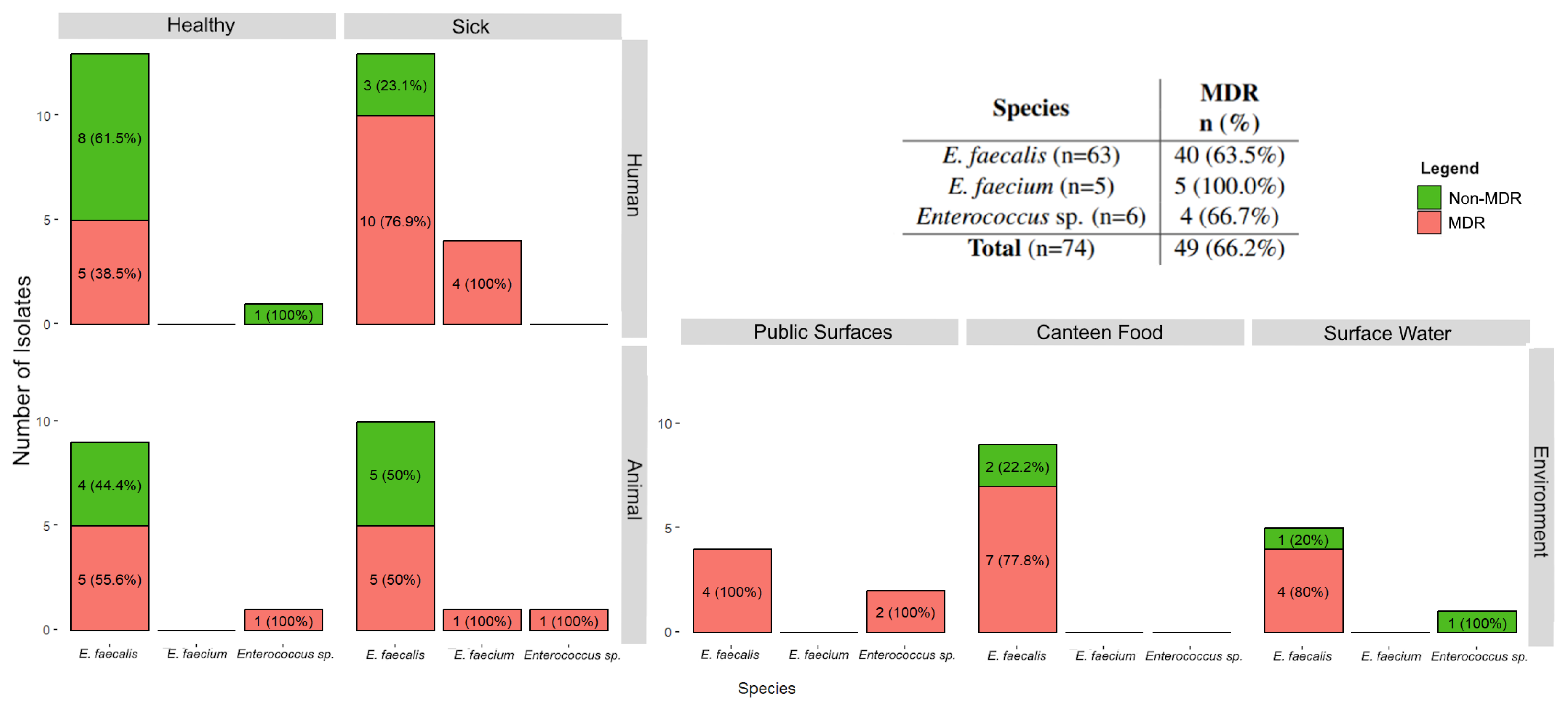
The AMR profiles of the Enterococcus isolates were categorized to identify MDR phenotypes. MDR was defined as non-intrinsic, non-susceptibility to at least one agent in three or more antimicrobial classes [60]. Therefore, both intermediate/susceptible with increased exposure (I) and resistant (R) categories were grouped as non-susceptible for MDR classification and analysis. In addition, the analysis did not include QDA resistance in E. faecalis. The prevalence of MDR is presented by enterococcal species and subsector in Figure 3. Individual isolates classified as MDR are identified in Supplementary Table S1.
Multidrug-resistant isolates were detected across all One Health sectors, with the highest proportions observed among Sick isolates and Public Transport Surfaces. All E. faecium isolates were classified as MDR; while this finding is consistent with the recognized clinical association between this species and multidrug resistance [44,74], it should be noted that the E. faecium sample size in this study was limited, and the finding should be interpreted accordingly rather than as a precise population estimate. In Humans, MDR prevalence was higher among isolates from Sick Humans compared to Healthy humans, consistent with studies reporting greater multidrug resistance in enterococci recovered from infections, particularly E. faecium [28,44,74]. It should be noted that the Sick Humans subsector comprised isolates recovered from a variety of clinical infection types and individuals with different underlying comorbidities, which may have acted as confounding factors in the observed resistance patterns.
In Animals, MDR was common in both Healthy and Sick enterococci and fell within the range reported for livestock and companion animals, where prevalence varies with antimicrobial exposure, host species, production system, and geography [47,75,80]. Environmental MDR was also frequent, with all Public Transport Surfaces isolates classified as MDR and high prevalence in Canteen Food and Surface Water, consistent with reports identifying community surfaces, river water, and wastewater-impacted environments as reservoirs of clinically relevant resistance phenotypes [34,58,69]. The highest numbers of non-susceptibilities were concentrated in Sick Human isolates, while only two isolates, both E. faecalis from Healthy Animals, remained fully susceptible, supporting the role of enterococci as reservoirs and disseminators of AMR within the One Health framework.

2.3.2. Genotypic Resistance Determinants

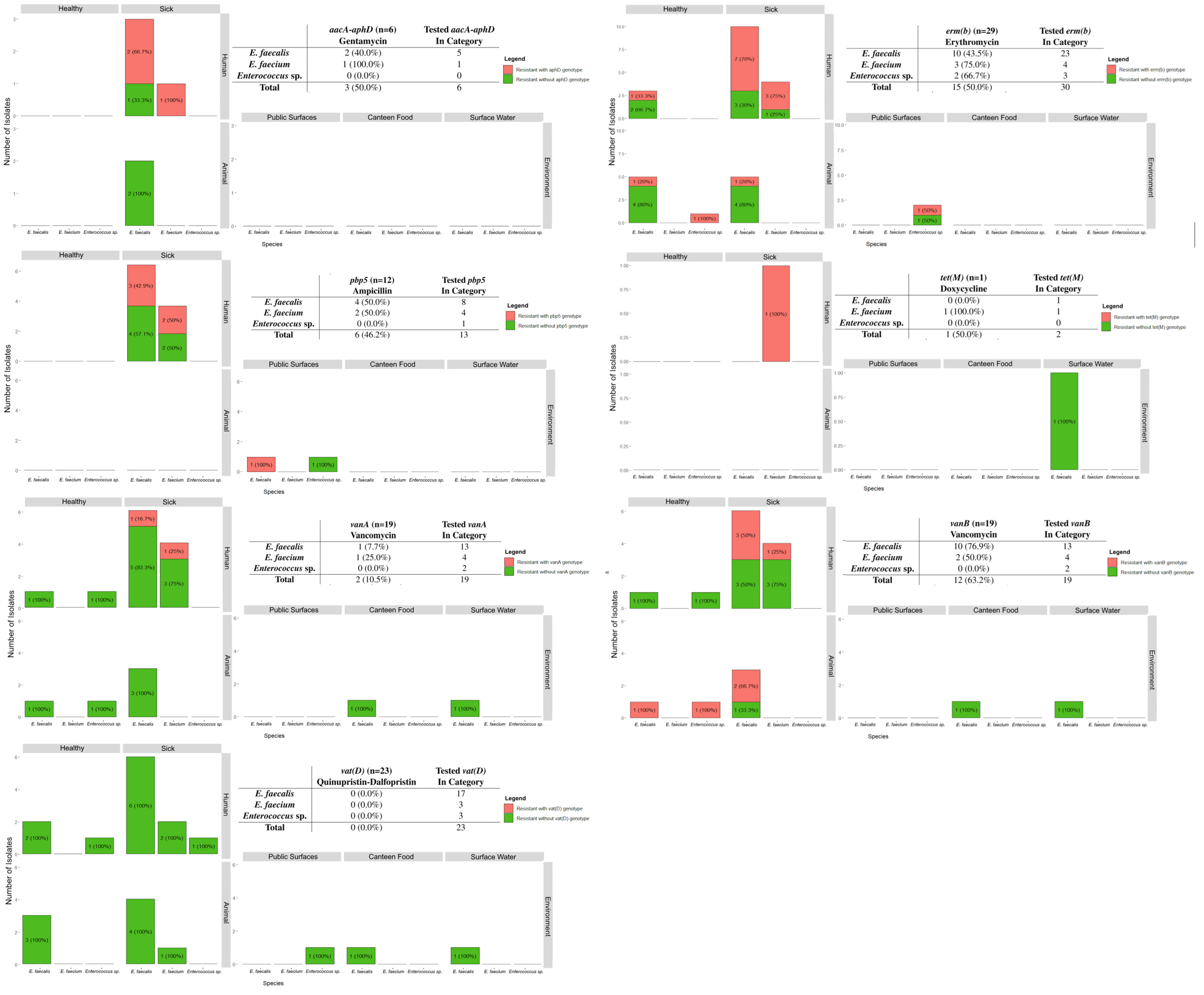
A panel of ARGs (aacA-aphD, erm(B), pbp5, tet(M), vanA, vanB, vanC, vat(D)) was tested by Multiplex PCR using the primers and conditions described below in Section 3.4.2. Isolates were selected for testing according to their phenotypical resistance profile, with the objective of providing a targeted phenotypic-genotypic overview of the most clinically and epidemiologically relevant determinants. The results obtained are summarized in Figure 4, and Supplementary Table S1 lists the ARGs identified for each isolate.
The aacA-aphD gene, a major determinant of High-Level Gentamicin Resistance (HLGR) and resistance to other aminoglycosides [41], was detected exclusively in gentamicin-resistant isolates from the Sick Human subsector. Overall, aacA-aphD was identified in 75% of tested Sick Human isolates, including 66.7% of E. faecalis and all tested E. faecium. This prevalence closely aligns with a report from Sick Human isolates, where approximately 64% of HLGR isolates carried aacA-aphD [111]. In contrast, the gene was not detected in isolates from Sick Animals, consistent with the literature showing species-dependent variability and generally low carriage outside clinical human settings [47].
Resistance to macrolides and related compounds was assessed through detection of the erm(B) gene, which mediates resistance to macrolide, lincosamide, and streptogramin B antibiotics and is frequently associated with mobile genetic elements [105,112]. The erm(B) gene was detected across all One Health sectors, with approximately half of the tested isolates carrying this determinant. In the Human sector, erm(B) carriage was higher among Sick Human isolates, where 70% of E. faecalis and 75% of E. faecium were positive, corresponding to an overall prevalence of 71%. These values are consistent with reports from Portuguese studies of erythromycin-resistant enterococci [111]. Lower prevalences were observed among Healthy Humans and Animal subsectors, although detection across these groups aligns with previous reports from Southern Europe [73,74]. In Environmental subsectors, erm(B) was detected sporadically in Public Transport Surfaces isolates, in agreement with prior findings from public transportation environments in Portugal [68].
Resistance to β-lactams was assessed through the detection of mutations in the chromosomally encoded pbp5 gene, which confers reduced susceptibility to penicillins such as ampicillin and to cephalosporins, particularly in E. faecium [113]. The pbp5 gene was broadly distributed across subsectors and species, being present in 42.9% of tested E. faecalis isolates and in 50% of tested E. faecium isolates from Sick Humans. The gene was also detected in half of the tested Public Transport Surfaces isolates. This widespread distribution is consistent with previous reports across human, animal, and environmental sources [36,47,68,74,96,114]. While there are reports of universal carriage of pbp5 mutations among ampicillin-resistant E. faecium from clinical settings [36], the lower prevalence observed here likely reflects limited sampling. However, while the presence of the pbp5 gene was assessed in this study, it is important to emphasize that β-lactam resistance arises from specific mutations and allelic variations in pbp5, rather than its presence. Consequently, the results presented here reflect gene prevalence rather than resistance potential.
The tet(M) gene, the most prevalent tetracycline resistance determinant in Enterococcus spp. and commonly associated with mobile elements of the Tn916/Tn1545 family [59,105], was detected in only one of the two tested isolates. This isolate belonged to E. faecium from the Sick Human subsector, while the second tested isolate, recovered from Surface Water and identified as E. faecalis, did not carry tet(M). This low resistance and tet(M) detection contrasts with numerous reports describing widespread carriage of tet(M) in Human, Animal, and Environmental sectors, including both Healthy and Sick hosts and diverse environmental matrices [28,47,68,69,70,74,75,115,116].
Acquired glycopeptide resistance was assessed in isolates that exhibited phenotypic resistance to vancomycin by AST, through the detection of the vanA and vanB gene clusters, while intrinsic low-level resistance was evaluated via vanC operon screening. The vanA gene, which confers resistance to vancomycin and teicoplanin and is typically associated with mobile transposons [59,105], was detected exclusively in the Sick Human subsector. Within this group, vanA was identified in 25% of tested E. faecium and 16.7% of tested E. faecalis isolates. This distribution is consistent with the dominance of vanA among VRE in European hospitals [117]. No vanA-carrying isolates were detected among Healthy Humans, Animals, or Environmental subsectors, in line with reports of low or absent carriage in community, animal, and most environmental settings [28,74,80,118,119]. Environmental detection of vanA has been mainly reported in hospital-impacted matrices, such as wastewater and downstream surface waters [34,120], which were not represented among the VRE isolates analyzed here.
The vanB gene cluster, an acquired determinant typically carried on conjugative transposons and conferring resistance to vancomycin but not teicoplanin [46,105], showed a broader distribution. Overall, 42.1% of vancomycin-resistant isolates carried vanB, spanning Human and Animal sectors. In Sick Humans, vanB was detected in approximately half of E. faecalis isolates and in a quarter of E. faecium. Similar moderate prevalences have been reported in European studies for Healthy Humans, yet contrasting with higher rates in some hospital-associated VREfm lineages and lower rates in community and animal reservoirs [121,122]. In the Animal sector, the high proportion of vanB-positive isolates observed contrasts with generally lower prevalences reported in the literature [80,123] and is likely influenced by the limited number of isolates tested. In the Environmental sector, the low prevalence observed is confirmed by the literature [34,70].
In contrast to the acquired operons, the vanC gene cluster, which is chromosomally encoded and confers intrinsic low-level resistance to vancomycin, was not detected in any isolates from this study. This finding is consistent with the established association of vanC with species such as E. gallinarum, and E. casseliflavus/Enterococcus flavescens [46,124], which were not specifically targeted in this study due to their relatively low prevalence and the characteristic yellow pigmentation typically exhibited by these species on agar media. No vanC-positive isolates were detected among E. faecalis or E. faecium in this study, in contrast to sporadic reports of interspecies transfer in wastewater-impacted environments [34].
The vat(D) gene, encoding a streptogramin A acetyltransferase and typically carried on mobile genetic elements [24,46,105], was not detected in any isolate analyzed. This absence is consistent with recent genomic and phenotypic surveys reporting very low prevalence of vat(D) among Enterococcus spp. from human, animal, and environmental sources [26,28,46,125,126,127]. The lack of detection in this study supports the view that vat(D) remains a rare determinant across the One Health continuum.
Overall, the distribution of ARGs observed in this study reflects a clear sectoral stratification for certain determinants, particularly those associated with high-level resistance and clinical selective pressure, such as aacA-aphD and vanA, which were confined to Sick Human isolates. In contrast, genes such as pbp5, vanB, and erm(B) displayed broader distributions across One Health sectors, consistent with their known mobility and persistence in both clinical and non-clinical reservoirs. Intrinsic resistance determinants, exemplified by vanC, remained restricted to non-E. faecalis/non-E. faecium species. Together, these findings support the view that while clinical settings act as focal points for the selection of high-impact resistance determinants, a substantial proportion of the resistome circulates across human, animal, and environmental compartments, supporting the relevance of integrated One Health surveillance.

2.4. Virulence Factor Production

2.4.1. Hemolytic Activity

Hemolytic activity was assessed on blood agar plates supplemented with horse blood under anaerobic incubation. Horse blood was used instead of sheep blood based on previous findings from our group showing that horse erythrocytes are more susceptible to hemolysin-mediated lysis, and that β-hemolysis on horse blood under anaerobic conditions is significantly associated with the detection of cyl genes [128]. Anaerobic incubation was used to minimize oxidation-related erythrocyte lysis not directly attributable to enterococcal hemolysin activity [128].
Hemolytic activity results are listed for each isolate in Supplementary Table S1. Overall, β-hemolysis was detected in 20.3% of enterococcal isolates (15/74). Hemolytic activity was observed across all One Health sectors, with the highest proportion in Environmental isolates (23.8%, 5/21), followed by Human isolates (19.4%, 6/31) and Animal isolates (18.2%, 4/22). All β-hemolytic isolates were identified as E. faecalis, except for one Environmental isolate identified only at the genus level. Previous studies have reported variable prevalences of β-hemolysis among enterococci from One Health sources [129,130].

2.4.2. Virulence Gene Screening

A virulence gene screening was carried out by multiplex PCR for agg (aggregation substance), cylA (cytolysin activator), esp (enterococcal surface protein) and gelE (gelatinase) genes. In general, gelE was the most frequently identified (39.2%, 29/74), followed by esp (36.5%, 27/74), agg (24.4%, 18/74) and cylA (17.6%, 13/74), although these prevalences changed when considering each sector.
The virulence gene screening results are listed for each isolate in Supplementary Table S1. Among the virulence genes screened per sector, esp was the most frequent in Human isolates, being observed in 58.1% (18/31), with most esp-positive isolates associated with infections (Sick Humans; 77.7%, 14/18). Another study on clinical and food isolates in Brazil also observed a higher prevalence of esp (68.4%) among human clinical isolates [131]. In our study, this gene was also present in 27.3% (6/22) of Animal enterococci and 14.3% (3/21) of Environment isolates. The moderate-to-high prevalence of esp in animals and lower prevalence in environmental-associated enterococci, such as food, is consistent with the literature [32,33,131]. Analysis of the esp gene using a joint Wald test revealed a significant association between the sector and gene prevalence, with a significant difference detected between the Environmental and Human sectors (p = 0.0492). No significant associations between sector and the prevalence of the remaining virulence genes were detected.
In contrast, gelE was the most prevalent gene in both Animal and Environment isolates, occurring in 40.9% (9/22) and 42.9% (9/21), respectively. Other authors reported a high prevalence of gelE in animal-associated enterococci [13,16,30,31,32] and environmental sources such as food (only in E. faecalis) [131]. Overall, gelE was also consistently detected across all sectors, including 35.5% (11/31) of Human isolates, which underscores its role as a fitness determinant rather than only an infection-associated gene.
As for the agg gene, it showed a similar prevalence to gelE among Human isolates (35.5%, 11/31), but was less frequent in Animal and Environment isolates, where it was detected in 18.2% (4/22) and 14.3% (3/21), respectively. This finding contrasts with previous reports stating a higher prevalence of gelE compared with agg among human clinical isolates [131], while agg was reported as the most prevalent (64.7%) virulence factor among enterococci isolated from wild animals [132].
The cylA gene was less prevalent overall, occurring in 22.6% (7/31) of Human isolates, 13.6% (3/22) of Animal isolates, and 14.3% (3/21) of Environment isolates. Among the seven cylA-positive human isolates, 71.4% were associated with infections, consistent with the known role of cytolysin in enhancing enterococcal virulence in infection models and the higher mortality observed in infections caused by cytolysin-producing strains compared with non-hemolytic strains [133]. The higher prevalence of cylA in human enterococci compared to environment-associated enterococci has also been observed by [128]. In addition, a surveillance study of surface contamination in small animal veterinary hospitals and another study on canine and feline enterococci both reported cylA as the least prevalent virulence gene, which is in line with our prevalence results in the Environmental and Animal sectors [30,32].
Phenotypically, β-hemolysis was not observed in all cylA-carrying isolates. Of the 13 isolates harboring cylA, only nine exhibited β-hemolytic activity (69.2%), indicating an incomplete genotype–phenotype correspondence between cylA detection and hemolytic phenotype already described by other authors [128,131]. Conversely, most β-hemolytic isolates carried the cylA gene (60%, 9/15), in line with a previously reported correlation between cylA genotype and hemolysis phenotype in animals and humans [16]. Together, these results highlight the importance of combining phenotypic with genotypic methods to assess hemolytic activity.
Overall, 33.8% (25/74) of total isolates had none of the tested virulence genes, representing 25.8% (8/31) of isolates in Humans, 36.4% (8/22) in Animals and 42.9% (9/21) in the Environment. Among isolates carrying at least one virulence gene (66.2%, 49/74), 57.1% (28/49) harbored multiple virulence genes, whereas only 4.1% of all isolates (3/74) carried the full virulence gene panel tested. These findings demonstrate that virulence-associated traits are not restricted to clinical enterococci, but are distributed across human, animal, and environmental sectors. From a One Health perspective, this overlap suggests the presence of similar enterococcal strains and pathogenicity determinants across interconnected settings, reinforcing the importance of integrated surveillance.

2.5. Integrative Analysis

Interpretation of these findings should consider several methodological limitations. The number of isolates within individual subsectors was limited, which may accentuate differences in resistance and MDR prevalence, virulence gene prevalence, and species distribution. Sampling was cross-sectional and geographically restricted, preventing inference on temporal trends or broader regional patterns. The inclusion of animals presenting with general clinical conditions, rather than enterococcal infections specifically, due to sampling constraints and sample availability, limits direct comparisons between the animal and human clinical cohorts. In addition, disk diffusion interpreted using CLSI breakpoints may limit direct comparison with studies using EUCAST criteria or broth microdilution, while culture-based isolation may favor robust species such as E. faecalis and underrepresent less tolerant enterococci [26,50,56,57]. Finally, it is important to highlight that our study design does not allow inference about directionality or transmission of resistance or virulence pathways between sectors.
Despite these constraints, the study provides an integrated assessment of species distribution, phenotypic AMR, and resistance gene prevalence across Human, Animal, and Environmental sectors. The predominance of E. faecalis across sectors is consistent with its role as a successful generalist and gastrointestinal commensal [12,39,40]. In contrast, the concentration of E. faecium and MDR phenotypes among Sick isolates supports its recognized association with healthcare-associated resistance [50,54,134].
Statistical analyses showed a clear association between health status (healthy/sick) and resistance in the Human sector (Table 2). The Cochran-Mantel-Haenszel (CMH) test showed that resistance was significantly higher in Sick Human isolates than in Healthy Human isolates (CMH OR = 5.354, 95% CI [2.939, 9.754]; p ≤ 0.001). On the other hand, no significant differences were observed between Healthy and Sick Animals according to the CMH test (p = 0.961) or among Environmental subsectors according to a binomial Generalized Linear Mixed-Model (GLMM) with a Likelihood-Ratio Test (LRT) (GLMM-LRT; p = 0.357). The absence of significant differences among the Animal and Environmental sub-sectors suggests that antimicrobial resistance is broadly distributed within these sectors, rather than being associated with specific subsectors. This pattern suggests a wide distribution of resistant enterococci between Healthy and Sick Animals, as well as among Public Transport Surfaces, Canteen Food, and Surface Water. In contrast, significant differences observed between Healthy and Sick Humans point to distinct selective pressures within the human sector. The higher resistance levels detected in the Sick Humans subsector likely reflect the greater exposure to antimicrobial agents in clinical settings, particularly considering that all antimicrobials evaluated in this study are approved for use in human medicine.
The pooled Fisher’s exact test, although widely reported in the literature, was used only as an exploratory analysis method because resistance outcomes across antibiotics were non-independent. CMH and GLMM-LRT were considered the primary inferential methods, as they better reflected the structure of the data [135,136] while the small sample size further supported a cautious and rigorous analytical approach. Although pooled Fisher testing suggested significant differences in resistance across One Health sectors, this result was not corroborated by the GLMM analysis (GLMM-LRT, p = 0.357), which accounted for non-independence across antibiotics. This might indicate that AMR was not confined to a single sector but was instead broadly distributed across the One Health continuum. This finding indicates that, while differences in resistance patterns can be observed between Healthy and Sick Humans, no significant overall sector effect was detected. This is very important from a One Health standpoint, as it can suggest that antibiotic resistance is widely distributed across One Health sectors.
Species-level analysis identified E. faecium as the main driver of resistance. Resistance was approximately five times more likely in E. faecium than in E. faecalis (CMH OR = 5.852, 95% CI [2.922, 11.719]; p = 2.38   ×   10 8 ), consistent with evidence identifying E. faecium as the enterococcal species most strongly associated with AMR across multiple drug classes [38,42,74]. The detection of resistance and MDR isolates in Public Transport Surfaces, Canteen Food, and Surface Water further supports the permeability of sector boundaries and reinforces the need for integrated One Health surveillance across clinical, animal, food-chain, and environmental reservoirs [5,17,26,27,34,67,68,76,137].
Utilizing the same statistical methodology, the same categories were tested for virulence genes (Table 3). In contrast with phenotypical resistance, no association between health status (healthy/sick) and presence of virulence genes can be seen in the Human sector (p = 0.249). No association was observed between any of the subsectors within their corresponding sector. However, between sectors, when accounting for the differences between subsectors using a binomial GLMM-LRT, the differences can be considered significant (p = 0.028). Looking further into the difference between sectors, we found a significant difference between the Human and Animal sectors (p = 0.035, GLMM-LRT OR = 0.4702, 95% CI [0.2524, 0.8760]) and the Human and Environmental sectors (p = 0.011, GLMM-LRT OR = 0.3803, 95% CI [0.1977, 0.7315]). The difference between Animal and Environmental sectors was not significant (p = 0.569, GLMM-LRT OR = 0.8089, 95% CI [0.3898, 1.6788]). This might reflect differences between adaptations to Human hosts and non-Human hosts, though this is not apparent in antibiotic resistance differences within the same sectors.
Focusing on the co-occurrence of multidrug resistance and virulence determinants, 34 of the 49 MDR isolates (69.4%) carried at least one virulence gene, whereas 15 (30.6%) did not harbor any of the virulence genes tested. The three isolates that possessed the full set of virulence determinants assessed were also MDR, representing 6.1% (3/49) of all MDR isolates. These findings indicate that virulence genes were common among MDR enterococci, suggesting that resistance and virulence traits may frequently coexist. However, the small number of isolates harboring all virulence genes suggests that the acquisition of multidrug resistance may not necessarily be accompanied by the accumulation of a broader virulence profile, indicating that these determinants may be subject to different selective pressures.

3. Materials and Methods

3.1. Sampling and Microbial Isolation

Enterococcal isolates were recovered from 66 samples obtained from Humans (n = 31 fecal samples from 14 Healthy Humans; 17 individuals with an enterococcal infection), Animals (n = 22 fecal samples from 10 Healthy and 12 out-patient Sick Animals in a veterinary teaching hospital) and Environment (n = 13 from 9 raw and cooked Canteen Food, 1 Surface Water and 3 Public Transport Surfaces). Samples were collected concurrently in all sectors between late 2022 and early 2024 in the Lisbon metropolitan area, Portugal.
Human fecal samples were self-collected by healthy human participants, while animal fecal samples (healthy cats and dogs, and sick outpatient dogs) were collected either by the pet tutors or by veterinary healthcare professionals. Samples from sick animals included only outpatient dogs, which had general clinical conditions (e.g., varied tumors, bone and tissue lesions), and healthy cats and dogs had no known clinical conditions. In cases of healthy animals housed together, fecal samples were collected independently and processed as a composite single sample representative of the household. Samples from humans with varied community-acquired and healthcare-associated enterococcal infections were processed by three partner healthcare entities (two hospitals and one clinical laboratory), and pure enterococcal isolates were provided in cryotubes. Surface water samples were collected and processed by a partner laboratory, and pure enterococcal isolates were provided in selective media plates. Samples from public transport environments were collected from multiple high-contact surfaces frequently touched by passengers, both within vehicles and at stations. Environmental samples were subsequently pooled into composite samples according to surface type and sampling location (e.g., handles, ATMs and ticket machines, similar foodstuffs, or surface water). General patient or individual data was collected (age, gender, geographic location, underlying condition/infection, antibiotic usage in the previous months) in samples from humans and animals whenever compatible with the policies of collaborator institutions (available in Supplementary Table S1). These variables were not considered during participant selection and therefore were not used as inclusion or exclusion criteria.
Fecal and food samples were diluted (1:10) in Maximum recovery broth (MRB) and enriched overnight at 37 ± 2 °C prior to isolation, whereas surface samples were enriched in MRB and spread plated and water was directly inoculated. Samples were inoculated in Slanetz-Bartley agar (SBA, Liofilchem, Roseto degli Abruzzi, Italy) and Slanetz-Bartley agar supplemented with vancomycin (SBAvan) at 6 μg/mL [16], and incubated aerobically at 37 ± 2 °C for up to 48 h. Approximately 20% of characteristic colonies were randomly selected and streaked onto Bile-Esculin Azide (BEA) agar (Liofilchem, Roseto degli Abruzzi, Italy) aiming for presumptive genus confirmation associated with the capacity to hydrolyze esculin [138]. Except when justified, a single presumptive enterococcal isolate per sample was selected and subcultured on Brain–Heart Infusion agar (BHI, Liofilchem, Roseto degli Abruzzi, Italy). Cultures were maintained on BHI at 4 °C for routine handling and preserved at −80 °C in BHI supplemented with 20% glycerol.

3.2. Microbial Diversity by RAPD-PCR

Genomic DNA extraction was performed from pure colonies using the boiling lysis method [139]. The supernatant obtained was used as template DNA for PCR.
Genetic diversity was assessed by RAPD-PCR using primers OPC15 (5′-GACGGATCAG-3′) and (GTG)5 (5′-GTGGTGGTGGTGGTG-3′) in independent reactions [16,140]. Amplification was performed on a thermocycler GTC96S (Cleaver Scientific, Rugby, UK), under the following conditions: 95 °C for 5 min; 35 cycles of 95 °C for 45 s, 57 °C for 45 s, and 72 °C for 45 s; followed by a final extension at 72 °C for 10 min. Replicates (10%) were randomly selected for reproducibility assessment purposes.
Amplification products were resolved on 1% agarose gels prepared in 0.5× Tris-Borate-EDTA buffer (4.5 × 10⁻² M Tris, 4.5 × 10⁻² M boric acid, and 1 × 10⁻³ M Na2EDTA, NYZTech, Lisboa, Portugal). Each well was loaded with 8 μL of amplification mixture, 2 μL of GelRed 10× (Biotium, Fremont, CA, USA), and 2 μL of bromophenol blue 6× (Merck KGaA, Darmstadt, Germany). Electrophoresis was performed at 90 V for 3 h, and results were observed using the ChemiDoc XRS+ device (Bio-Rad, Hercules, CA, USA) and Image Lab 6.1. software (Bio-Rad, Hercules, CA, USA) [16].
RAPD-PCR profiles were analyzed using BioNumerics version 6.6 (BioMérieux, Marcy-l’Étoile, France) as a composite dataset. After gel images normalization, similarities were calculated using the Pearson correlation coefficient, and dendrograms were constructed using the pair group method with the arithmetic mean (UPGMA) method with 3% tolerance. The resulting similarity matrices were averaged to construct a final dendrogram using the UPGMA method. Reproducibility estimates derived from replicate analyses (10 pairs) were used to guide cluster definition and the selection of representative isolates for downstream analyses. Sector-specific dendrograms were subsequently generated to support the selection of representative isolates while preserving balanced representation across One Health sectors and subsectors. Isolate selection followed a hierarchical decision process, prioritizing, in sequence, distinct RAPD-PCR electrophoretic profiles, the sector and subsector of origin, and the initial isolation medium (SBA or SBAvan). Isolates that did not cluster with others above the defined similarity threshold were treated as single-member clusters and retained as representatives for downstream analyses.

3.3. Molecular Species and Genus Identification

Species-level identification was performed by PCR, targeting species-specific regions of the sodA gene using primer pairs specific for E. faecalis, E. faecium, and E. durans, as well as for the genus Enterococcus [141,142,143]. Primer sequences and expected amplicon size are presented in Table 4. The reaction mixtures for each set contained 10 µL of NZYTaq II 2x Green Master Mix (NZYTech, Lisboa, Portugal) including NZYTaq II DNA polymerase, reaction buffer, deoxyribonucleotides (dNTPs), magnesium chloride (MgCl2), and additives, 7 µL of Milli-Q water (Millipore, Merck KGaA, Darmstadt, Germany), 2 µL of DNA and 0.5 µL of forward and reverse primer at 50 pmol/µL (STABVIDA, Caparica, Portugal). Considering all components, the reaction mixtures totaled 20 µL [16].
PCR products were resolved by 1.2% agarose gel electrophoresis (90 V for 2 h) as previously described [16]. In all reactions, E. faecalis ATCC® 29212, E. faecium E300 and E. durans DSMZ® 20633 were used as positive controls and sterile water was added as a negative control. Ten percent of the replicates were randomly selected to guarantee reproducibility.

3.4. Antimicrobial Susceptibility Testing

3.4.1. Phenotypic Analysis

Antimicrobial susceptibility testing was performed using the Kirby–Bauer disk diffusion method using twelve antibiotics representing distinct classes and cellular targets. Listed by alphabetical order of classes, the following antimicrobial list was chosen considering its relevance for enterococcal infections in human medicine [63]: high-level gentamicin (120 μg), high-level streptomycin (300 μg), ampicillin (10 μg), teicoplanin (30 μg), vancomycin (30 μg), erythromycin (15 μg), linezolid (30 μg), chloramphenicol (30 μg), quinupristin-dalfopristin (15 μg), doxycycline (30 μg), levofloxacin (5 μg) and rifampicin (5 μg) [144].
Bacterial suspensions were prepared from overnight-grown cultures and adjusted to a turbidity equivalent to 0.5 McFarland scale using Ringer solution (Oxoid, Hampshire, UK). The suspensions were spread onto agar plates, and antibiotic disks (Liofilchem, Roseto degli Abruzzi, Italy) were equidistantly placed on the agar surface. Plates were incubated at 37 ± 2 °C for 24 h, and inhibition zones were interpreted according to CLSI breakpoints for Enterococcus spp. [144]. To assess reproducibility, 10% of isolates were analyzed in duplicate. E. faecalis ATCC 29212 was used as quality control.
Vancomycin resistance was further confirmed by agar dilution at 6, 16, and 32 μg/mL. The 6 μg/mL concentration was used as a screening concentration for vancomycin resistance, while 16 and 32 μg/mL represented the CLSI intermediate and resistant MIC thresholds, respectively, for Enterococcus spp. Plates were incubated at 37 ± 2 °C for 48 h, and results were interpreted according to CLSI criteria [144]. Only isolates with confirmed vancomycin resistance by both AST and agar dilution methods were subjected to MIC determination by Etest (Liofilchem, Roseto degli Abruzzi, Italy).

3.4.2. Genotypical Analysis

The antimicrobial resistance reported in antimicrobial susceptibility testing, to gentamicin (aacA-aphD), ampicillin (pbp5), vancomycin (vanA, vanB, vanC), erythromycin (erm(B)), quinupristin-dalfopristin (vat(D)) and doxycycline (tet(M)) was confirmed by multiplex PCR [145,146,147,148,149,150]. ARGs were selected considering the resistance phenotypes identified by AST, prioritizing those most frequently observed in enterococci, and the clinical relevance of the corresponding antibiotics in human and veterinary medicine.
PCR preparation was equal to that described in Section 3.3. Primer sequences and respective amplicon size are presented in Table 5. All reactions included positive and negative controls. As positive controls, the following strains were used: E. faecalis V583 (aacA-aphD, pbp5, vanB); E. faecalis H161 (vanA), E. faecalis H32 (tet(M)) and Enterococcus AR3-1V (vanC) from the culture collection of the Microbugs Research Team (Faculty of Veterinary Medicine, University of Lisbon). Additionally, 10% of isolates were analyzed in independent replicate reactions for quality control. Amplification conditions were as follows: 95 °C for 5 min, 35 cycles of 95 °C for 45 s, 55 °C for 45 s, 72 °C for 1 min; 72 °C for 5 min. PCR products were visualized on 1.2% agarose gels by electrophoresis (90 V for 2 h) as previously described [16].

3.5. Virulence Factors

3.5.1. Hemolytic Activity

Metabolically active cultures were inoculated onto Columbia agar plates supplemented with 5% horse blood (Frilabo, Trofa, Portugal) and incubated anaerobically at 37 ± 2 °C for 48 h, using Oxoid AnaerogenTM sachets (Thermo Fisher Scientific, Waltham, MA, USA). Hemolytic activity was evaluated after incubation based on the appearance of the agar around the colonies. A transparent halo was interpreted as β-hemolysis, a greenish halo as α-hemolysis (partial hemolysis), and the absence of any change in the medium as γ-hemolysis (non-hemolytic activity) [128]. E. faecalis MMH594 and E. faecalis ATCC 29212 were included as positive and negative controls, respectively. Reproducibility was guaranteed by 10% replicates randomly selected.

3.5.2. Virulence Genes

Virulence gene screening was performed in all representative isolates by multiplex PCR, targeting genes associated with cell aggregation (agg), immune evasion (esp), gelatinase production (gelE), and cytolysin/hemolysin activity (cylA) [16].
PCR assays were performed using the same reaction mixture described in Section 3.3 and Section 3.4.2. Primer sequences and expected amplicon size are shown in Table 6. Positive (E. faecalis MMH 594) and negative (sterile water) controls were included in every run, and 10% of isolates were randomly selected for independent replicate testing. The amplification protocol consisted of an initial denaturation at 95 °C for 5 min, 35 cycles of 95 °C for 45 s, 55 °C for 45 s, and 72 °C for 45 s, and final extension at 72 °C for 5 min. PCR products were resolved on 1.2% agarose gels by electrophoresis (90 V for 2 h) and visualized as previously reported [16].

3.6. Statistical Analysis

Statistical analyses were performed in R version 4.5.1. Phenotypic resistance determined by AST was analyzed as a binary outcome, resistant/non-resistant, by antibiotic. Virulence gene occurrence was analyzed as a binary outcome, present/absent, by gene. For two-level comparisons, 2 × 2 Fisher’s exact tests were applied per antibiotic or per virulence gene. For comparisons involving more than two levels, Fisher–Freeman–Halton exact tests with Monte Carlo estimation, B = 100,000, were used. Multiplicity across antibiotics or virulence genes was controlled using Holm’s adjustment.
Overall associations across antibiotics or virulence genes were assessed using methods that account for repeated testing across traits. Two-level factors were analyzed using the CMH test stratified by antibiotic or virulence gene. Factors with more than two levels were analyzed using binomial GLMM tests, with significance assessed by LRT. In these models, antibiotic or virulence genes were included as an adjustment factor, as appropriate. Sector-level differences were analyzed using a logistic mixed-effects model with inference based on an LRT. Per-antibiotic and virulence gene sector effects were assessed using joint Wald tests derived from a binomial generalized linear mixed model. Pairwise sector comparisons were reported as model-adjusted odds ratios with 95% confidence intervals and Wald p-values. For comparability with previous studies, pooled Fisher’s exact tests, Pooled-Fisher, were also reported. All tests were two-sided with α = 0.05.

4. Conclusions

An integrated One Health approach is required to address the dissemination of AMR. This framework supports the surveillance of resistant strains and resistance determinants across human, animal, and environmental sectors, while also enabling evaluation of antimicrobial efficacy in clinical settings. In line with the aims and hypothesis of this study, Enterococcus spp. were recovered from all analyzed sectors, supporting the interconnected nature of these reservoirs and the potential circulation of AMR and virulence determinants across One Health compartments.
E. faecalis was the dominant species across all sectors, accounting for 85.1% of isolates and exceeding 66.7% in each environment. E. faecium was less prevalent but was mainly associated with Sick Human and Animal subsectors, consistent with its recognized role in clinical settings. Other Enterococcus spp. were primarily detected in environmental samples, reinforcing the ecological diversity of enterococci across interconnected reservoirs.
Resistance to clinically relevant antimicrobials was observed across all sectors, particularly for streptomycin, rifampicin, and quinupristin-dalfopristin. High resistance to the latter was reported in E. faecium and other Enterococcus spp., whereas rifampicin resistance likely reflects acquired determinants and selective pressures extending beyond human clinical use. The detection of VRE, especially among Sick Humans, remains particularly concerning given the importance of vancomycin as a last-resort antimicrobial.
Despite its lower prevalence, E. faecium exhibited higher resistance levels than E. faecalis, consistent with its enhanced capacity to acquire resistance determinants in healthcare-associated environments. This contributes to the emergence of MDR strains and increases the risk of dissemination across sectors, further supporting the hypothesis of ecological connectivity between human, animal, and environmental reservoirs.
Environmental sources reflected cumulative antimicrobial pressures from both human and animal origins and acted as important reservoirs of resistance. These settings remain underrepresented in current research, particularly high-contact public surfaces outside healthcare environments. Moreover, most studies continue to focus on isolated sectors, limiting the broader understanding of cross-sector transmission dynamics.
Overall, these findings support the study hypothesis and highlight the value of integrated One Health surveillance frameworks to characterize the distribution, persistence, and dissemination of AMR across interconnected reservoirs.

Supplementary Materials

The following supporting information can be downloaded at: https://www.mdpi.com/article/10.3390/antibiotics15070657/s1, Supplementary Figure S1: Dendrogram constructed through RAPD-PCR using (GTG)5 and OPC15 primers, with the threshold average reproducibility shown as the red line; Supplementary Table S1: List of representative Enterococcus isolates, including One Health sector and subsector, and sampling of origin, general participant information, isolation media, enterococcal species, antimicrobial susceptibility testing and MIC (vancomycin) results, hemolytic activity, ARGs and virulence genes identified.

Author Contributions

Conceptualization, M.T.B.-C. and T.S.-L.; methodology, N.F. and J.M.M.; formal analysis, N.F. and J.M.M.; writing—original draft preparation, N.F. and J.M.M.; writing—review and editing, J.M.M., M.T.B.-C. and T.S.-L.; supervision, M.T.B.-C. and T.S.-L.; funding acquisition, M.T.B.-C. and T.S.-L. All authors have read and agreed to the published version of the manuscript.

Funding

This research was funded in whole or in part by Fundação para a Ciência e Tecnologia-Ministério da Educação, Ciência e Inovação, under Grants 2024.03583.BDANA (doctoral fellowship—J.M.M.), UID/276/2025 (CIISA, https://doi.org/10.54499/UID/00276/2025) and LA/P/0059/2020 (AL4ANIMALS). M.T.B.-C. acknowledges the support of the Research Unit iNOVA4Health (UID/4462/2025, UID/PRR/4462/2025—https://doi.org/10.54499/UID/PRR/04462/2025, UID/PRR2/4462/2025), financially supported by Fundação para a Ciência e Tecnologia-Ministério da Educação, Ciência e Inovação, and by União Europeia—NextGenerationEU; and by Fundação para a Ciência e Tecnologia/Ministério da Educação, Ciência e Inovação through the Associate Laboratory LS4FUTURE (LA/P/0087/2020; DOI 10.54499/LA/P/0087/2020).

Institutional Review Board Statement

This study involved only non-invasive fecal sample collection from animals and humans and posed minimal risk to participants. The study was conducted in accordance with the Declaration of Helsinki and approved by the Ethics Committee for Research and Teaching (CEIE) of Faculty of Veterinary Medicine, University of Lisbon (protocol code 036/2025, 24 October 2025). Retrospective approval covered earlier samplings.

Informed Consent Statement

Written informed consent was obtained from all human participants involved in the study, including formal consent of the participating animals by their tutors. Minimal personal and health-related data from animals and humans was self-reported by the human volunteer participants and was processed in accordance with applicable data protection regulations.

Data Availability Statement

The contributions presented in this study are included in the manuscript or as Supplementary Materials. Further inquiries can be directed to the corresponding author.

Acknowledgments

The authors acknowledge the healthcare professionals and laboratory staff of the participating hospitals and clinical laboratory in Lisbon for their availability, collaboration and for providing the clinical isolates and samples used in this study; Óscar Gamboa from the Teaching Veterinary Hospital, Faculty of Veterinary Medicine, University of Lisbon, for his assistance in collecting and providing part of the animal samples used in this study; all volunteer companion animal tutors who provided human and animal fecal samples; Vanessa Pereira, coordinator of the Laboratory of Membrane Processes at iBET, for providing the Enterococcus isolates from water used in this study; and the participating canteen for facilitating access to food samples. The authors also gratefully acknowledge the management and staff of the participating public transport operator for facilitating access to sampling locations and supporting sample collection activities. We further acknowledge the technical support of Daniel Pinto, Lisete Sousa, Bruno Martins, and Joana Cruz. During the preparation of this manuscript, the authors used ChatGPT 5.4 (v.1.2026.153) for language editing and text improvement to enhance clarity. The authors reviewed and edited all generated content and take full responsibility for the content of this publication.

Conflicts of Interest

The authors declare no conflicts of interest.

Abbreviations

The following abbreviations are used in this manuscript:
AMRAntimicrobial Resistance
ARGsAntimicrobial Resistance Genes
ASTAntimicrobial Susceptibility Testing
BEABile-Esculin Azide
BHIBrain-Heart Infusion
CLSIClinical and Laboratory Standards Institute
CMHCochran-Mantel-Haenszel
EUCASTEuropean Committee on Antimicrobial Susceptibility Testing
GLMMGeneralized Linear Mixed-Model
GLMM-LRTGeneralized Linear Mixed-Model with Likelihood-Ratio Tests
HLGRHigh-Level Gentamicin Resistance
LRTLikelihood-Ratio Test
MDRMultidrug-Resistant
MICMinimum Inhibitory Concentration
PCRPolymerase Chain Reaction
RAPD-PCRRandom Amplification of Polymorphic DNA-Polymerase Chain Reaction
SBASlanetz-Bartley agar
SBAvanSlanetz-Bartley agar supplemented with Vancomycin
UPGMAUnweighted Pair Group Method with Arithmetic Mean
VREVancomycin-Resistant Enterococcus
VREfmVancomycin-Resistant Enterococcus faecium
VVEVancomycin-Variable Enterococci

References

  1. Rondon, C.; Garcia, C.; Krapp, F.; Machaca, I.; Olivera, M.; Fernández, V.; Villegas, M.; Vilcapoma, P.; Casapia, M.; Concha-Velasco, F.; et al. Antibiotic Point Prevalence Survey and Antimicrobial Resistance in Hospitalized Patients across Peruvian Reference Hospitals. J. Infect. Public Health 2023, 16, 52–60. [Google Scholar] [CrossRef] [Scilit] [PubMed]
  2. Murray, C.J.L.; Ikuta, K.S.; Sharara, F.; Swetschinski, L.; Robles Aguilar, G.; Gray, A.; Han, C.; Bisignano, C.; Rao, P.; Wool, E.; et al. Global Burden of Bacterial Antimicrobial Resistance in 2019: A Systematic Analysis. Lancet 2022, 399, 629–655. [Google Scholar] [CrossRef] [Scilit] [PubMed]
  3. Cameron, A.; Esiovwa, R.; Connolly, J.; Hursthouse, A.; Henriquez, F. Antimicrobial Resistance as a Global Health Threat: The Need to Learn Lessons from the COVID-19 Pandemic. Glob. Policy 2022, 13, 179–192. [Google Scholar] [CrossRef] [Scilit] [PubMed]
  4. Welburn, S.C.; Beange, I.; Ducrotoy, M.J.; Okello, A.L. The Neglected Zoonoses—The Case for Integrated Control and Advocacy. Clin. Microbiol. Infect. 2015, 21, 433–443. [Google Scholar] [CrossRef] [Scilit] [PubMed]
  5. Boqvist, S.; Söderqvist, K.; Vågsholm, I. Food Safety Challenges and One Health within Europe. Acta Vet. Scand. 2018, 60, 1. [Google Scholar] [CrossRef] [Scilit] [PubMed]
  6. Steinhaus, E.A. A Study of the Bacteria Associated with Thirty Species of Insects. J. Bacteriol. 1941, 42, 757–790. [Google Scholar] [CrossRef] [Scilit] [PubMed]
  7. Mundt, J.O. Occurrence of Enterococci: Bud, Blossom, and Soil Studies. Appl. Microbiol. 1961, 9, 541–544. [Google Scholar] [CrossRef] [PubMed]
  8. Mundt, J.O. Occurrence of Enterococci in Animals in a Wild Environment. Appl. Microbiol. 1963, 11, 136–140. [Google Scholar] [CrossRef] [PubMed]
  9. Franz, C.; Stiles, M.; Schleifer, K.; Holzapfel, W. Enterococci in Foods—A Conundrum for Food Safety. Int. J. Food Microbiol. 2003, 88, 105–122. [Google Scholar] [CrossRef] [Scilit] [PubMed]
  10. Broderick, N.A.; Raffa, K.F.; Goodman, R.; Handelsman, J. Census of the Bacterial Community of the Gypsy Moth Larval Midgut by Using Culturing and Culture-Independent Methods. Appl. Environ. Microbiol. 2004, 70, 293–300. [Google Scholar] [CrossRef] [Scilit] [PubMed]
  11. Švec, P.; Vandamme, P.; Bryndová, H.; Holochová, P.; Kosina, M.; Mašlaňová, I.; Sedláček, I. Enterococcus plantarum Sp. Nov., Isolated from Plants. Int. J. Syst. Evol. Microbiol. 2012, 62, 1499–1505. [Google Scholar] [CrossRef] [Scilit] [PubMed]
  12. Tannock, G.W.; Cook, G. Enterococci as Members of the Intestinal Microflora of Humans. In ASM Press eBooks; Gilmore, M.S., Clewell, D.B., Courvalin, P., Dunny, G.M., Murray, B.E., Rice, L.B., Eds.; Wiley Blackwell: Hoboken, NJ, USA, 2014; pp. 101–132. [Google Scholar]
  13. Pillay, S.; Zishiri, O.T.; Adeleke, M.A. Prevalence of Virulence Genes in Enterococcus Species Isolated from Companion Animals and Livestock. Onderstepoort J. Vet. Res. 2018, 85, 8. [Google Scholar] [CrossRef] [Scilit] [PubMed]
  14. Rodrigues Dos Santos, B.; Cerqueira, A.; Moreira, R.S.; Bär, K.A.; Vizzotto, B.S. Distribution of Vancomycin-Resistant Enterococcus on Hospital Surfaces in Southern Brazil. J. Hosp. Infect. 2024, 145, 228–229. [Google Scholar] [CrossRef] [Scilit] [PubMed]
  15. Codelia-Anjum, A.; Lerner, L.B.; Elterman, D.; Zorn, K.C.; Bhojani, N.; Chughtai, B. Enterococcal Urinary Tract Infections: A Review of the Pathogenicity, Epidemiology, and Treatment. Antibiotics 2023, 12, 778. [Google Scholar] [CrossRef] [Scilit] [PubMed]
  16. Monteiro Marques, J.; Pita, B.; Pinto, D.; Barreto-Crespo, M.T.; Mato, R.; Semedo-Lemsaddek, T. One Health Insights into Enterococcus: Antimicrobial Resistance and Virulence in Companion Animals and Their Tutors. Int. J. Mol. Sci. 2026, 27, 654. [Google Scholar] [CrossRef] [Scilit] [PubMed]
  17. Monteiro Marques, J.; Coelho, M.; Santana, A.R.; Pinto, D.; Semedo-Lemsaddek, T. Dissemination of Enterococcal Genetic Lineages: A One Health Perspective. Antibiotics 2023, 12, 1140. [Google Scholar] [CrossRef] [Scilit] [PubMed]
  18. Zhou, X.; Willems, R.J.L.; Friedrich, A.W.; Rossen, J.W.A.; Bathoorn, E. Enterococcus faecium: From Microbiological Insights to Practical Recommendations for Infection Control and Diagnostics. Antimicrob. Resist. Infect. Control 2020, 9, 130. [Google Scholar] [CrossRef] [Scilit] [PubMed]
  19. Denissen, J.; Reyneke, B.; Waso-Reyneke, M.; Havenga, B.; Barnard, T.; Khan, S.; Khan, W. Prevalence of ESKAPE Pathogens in the Environment: Antibiotic Resistance Status, Community-Acquired Infection and Risk to Human Health. Int. J. Hyg. Environ. Health 2022, 244, 114006. [Google Scholar] [CrossRef] [Scilit] [PubMed]
  20. Krawczyk, B.; Wityk, P.; Gałęcka, M.; Michalik, M. The Many Faces of Enterococcus spp.—Commensal, Probiotic and Opportunistic Pathogen. Microorganisms 2021, 9, 1900. [Google Scholar] [CrossRef] [Scilit] [PubMed]
  21. Downing, M.A.; Xiong, J.; Eshaghi, A.; McGeer, A.; Patel, S.N.; Johnstone, J. Vancomycin-Variable Enterococcal Bacteremia. J. Clin. Microbiol. 2015, 53, 3951–3953. [Google Scholar] [CrossRef] [Scilit] [PubMed]
  22. Diekema, D.J.; Jones, R.N. Oxazolidinones: A Review. Drugs 2000, 59, 7–16. [Google Scholar] [CrossRef] [Scilit] [PubMed]
  23. Rodríguez-Noriega, E.; Hernández-Morfin, N.; Garza-Gonzalez, E.; Bocanegra-Ibarias, P.; Flores-Treviño, S.; Esparza-Ahumada, S.; González-Díaz, E.; Pérez-Gómez, H.R.; Mendoza-Mujica, C.; León-Garnica, G.; et al. Risk Factors and Outcome Associated with the Acquisition of Linezolid-Resistant Enterococcus faecalis. J. Glob. Antimicrob. Resist. 2020, 21, 405–409. [Google Scholar] [CrossRef] [Scilit] [PubMed]
  24. Wright, G.D. Antibiotic Resistance in the Environment: A Link to the Clinic? Curr. Opin. Microbiol. 2010, 13, 589–594. [Google Scholar] [CrossRef] [Scilit] [PubMed]
  25. White, A.; Hughes, J.M. Critical Importance of an One Health Approach to Antimicrobial Resistance. EcoHealth 2019, 16, 404–409. [Google Scholar] [CrossRef] [Scilit] [PubMed]
  26. Zaheer, R.; Cook, S.R.; Barbieri, R.; Goji, N.; Cameron, A.; Petkau, A.; Polo, R.O.; Tymensen, L.; Stamm, C.; Song, J.; et al. Surveillance of Enterococcus spp. Reveals Distinct Species and Antimicrobial Resistance Diversity across an One-Health Continuum. Sci. Rep. 2020, 10, 3937. [Google Scholar] [CrossRef] [Scilit] [PubMed]
  27. Gouliouris, T.; Coll, F.; Ludden, C.; Blane, B.; Raven, K.E.; Naydenova, P.; Crawley, C.; Török, M.E.; Enoch, D.A.; Brown, N.M.; et al. Quantifying Acquisition and Transmission of Enterococcus faecium Using Genomic Surveillance. Nat. Microbiol. 2021, 6, 103–111. [Google Scholar] [CrossRef] [Scilit] [PubMed]
  28. Ben Sallem, R.; Klibi, N.; Klibi, A.; Ben Said, L.; Dziri, R.; Boudabous, A.; Torres, C.; Ben Slama, K. Antibiotic Resistance and Virulence of Enterococci Isolates from Healthy Humans in Tunisia. Ann. Microbiol. 2015, 66, 717–725. [Google Scholar] [CrossRef] [Scilit]
  29. Eaton, T.J.; Gasson, M.J. Molecular Screening of Enterococcus Virulence Determinants and Potential for Genetic Exchange between Food and Medical Isolates. Appl. Environ. Microbiol. 2001, 67, 1628–1635. [Google Scholar] [CrossRef] [Scilit] [PubMed]
  30. KuKanich, K.S.; Ghosh, A.; Skarbek, J.V.; Lothamer, K.M.; Zurek, L. Surveillance of Bacterial Contamination in Small Animal Veterinary Hospitals with Special Focus on Antimicrobial Resistance and Virulence Traits of Enterococci. J. Am. Vet. Med. Assoc. 2012, 240, 437–445. [Google Scholar] [CrossRef] [Scilit] [PubMed]
  31. Iseppi, R.; Messi, P.; Anacarso, I.; Bondi, M.; Sabia, C.; Condo, C.; Niederhausern, S.D. Antimicrobial Resistance and Virulence Traits in Enterococcus Strains Isolated from Dogs and Cats. PubMed 2015, 38, 369–378. [Google Scholar]
  32. Said, L.B.; Dziri, R.; Sassi, N.; Lozano, C.; Slama, K.B.; Ouzari, I.; Torres, C.; Klibi, N. Species Distribution, Antibiotic Resistance and Virulence Traits in Canine and Feline Enterococci in Tunisia. Acta Vet. Hung. 2017, 65, 173–184. [Google Scholar] [CrossRef] [Scilit] [PubMed]
  33. Ferguson, D.M.; Talavera, G.N.; Hernández, L.A.R.; Weisberg, S.B.; Ambrose, R.F.; Jay, J.A. Virulence Genes among Enterococcus faecalis and Enterococcus faecium Isolated from Coastal Beaches and Human and Nonhuman Sources in Southern California and Puerto Rico. J. Pathog. 2016, 2016, 3437214. [Google Scholar] [CrossRef] [Scilit] [PubMed]
  34. Gotkowska-Płachta, A. The Prevalence of Virulent and Multidrug-Resistant Enterococci in River Water and in Treated and Untreated Municipal and Hospital Wastewater. Int. J. Environ. Res. Public Health 2021, 18, 563. [Google Scholar] [CrossRef] [Scilit] [PubMed]
  35. Adegoke, A.A.; Madu, C.E.; Reddy, P.; Fatunla, O.K.; Stenström, T.A.; Okoh, A.I. Virulence and Antibiotic Resistance Genes in Enterococcus from Wastewater for Reuse and Their Health Impact. Microorganisms 2025, 13, 1045. [Google Scholar] [CrossRef] [Scilit] [PubMed]
  36. Rathnayake, I.U.; Hargreaves, M.; Huygens, F. Antibiotic Resistance and Virulence Traits in Clinical and Environmental Enterococcus faecalis and Enterococcus faecium Isolates. Syst. Appl. Microbiol. 2012, 35, 326–333. [Google Scholar] [CrossRef] [Scilit] [PubMed]
  37. Jahan, M.; Zhanel, G.G.; Sparling, R.; Holley, R.A. Horizontal Transfer of Antibiotic Resistance from Enterococcus faecium of Fermented Meat Origin to Clinical Isolates of E. faecium and Enterococcus faecalis. Int. J. Food Microbiol. 2015, 199, 78–85. [Google Scholar] [CrossRef] [Scilit] [PubMed]
  38. European Centre for Disease Prevention and Control (ECDC). Antimicrobial Resistance in the EU/EEA (EARS-Net): Annual Epidemiological Report 2024; ECDC: Stockholm, Sweden, 2025. [Google Scholar]
  39. Fisher, K.; Phillips, C. The Ecology, Epidemiology and Virulence of Enterococcus. Microbiology 2009, 155, 1749–1757. [Google Scholar] [CrossRef] [Scilit] [PubMed]
  40. Franz, C.M.A.P.; Huch, M.; Abriouel, H.; Holzapfel, W.; Gálvez, A. Enterococci as Probiotics and Their Implications in Food Safety. Int. J. Food Microbiol. 2011, 151, 125–140. [Google Scholar] [CrossRef] [Scilit] [PubMed]
  41. Cattoir, V. The Multifaceted Lifestyle of Enterococci: Genetic Diversity, Ecology and Risks for Public Health. Curr. Opin. Microbiol. 2022, 65, 73–80. [Google Scholar] [CrossRef] [Scilit] [PubMed]
  42. Solaiman, S.; Patterson, R.; Davey, K.; Katz, Y.; Payne-Sturges, D.; Sapkota, A.R.; Micallef, S.A. Effects of Season and Water Type on the Distribution and Antimicrobial Resistance of Enterococcus faecalis and E. faecium from Surface and Reclaimed Water. J. Appl. Microbiol. 2022, 133, 477–487. [Google Scholar] [CrossRef] [Scilit] [PubMed]
  43. Kalfopoulou, E.; Huebner, J. Advances and Prospects in Vaccine Development against Enterococci. Cells 2020, 9, 2397. [Google Scholar] [CrossRef] [Scilit] [PubMed]
  44. Iancu, A.-V.; Arbune, M.; Zaharia, E.-A.; Tutunaru, D.; Maftei, N.-M.; Peptine, L.-D.; Țocu, G.; Gurău, G. Prevalence and Antibiotic Resistance of Enterococcus spp.: A Retrospective Study in Hospitals of Southeast Romania. Appl. Sci. 2023, 13, 3866. [Google Scholar] [CrossRef] [Scilit]
  45. Fahim, N.; Masud, R.; Salam, S. Role of Enterococcus in Spreading Antimicrobial Resistance Genes and Its Public Health Significance. Ger. J. Vet. Res. 2025, 5, 95–112. [Google Scholar] [CrossRef] [Scilit]
  46. Torres, C.; Alonso, C.A.; Ruiz-Ripa, L.; León-Sampedro, R.; Campo, R.D.; Coque, T.M. Antimicrobial Resistance in Enterococcus spp. of Animal Origin. Microbiol. Spectr. 2018, 6. [Google Scholar] [CrossRef] [Scilit] [PubMed]
  47. Stępień-Pyśniak, D.; Bertelloni, F.; Dec, M.; Cagnoli, G.; Pietras-Ożga, D.; Urban-Chmiel, R.; Ebani, V.V. Characterization and Comparison of Enterococcus spp. Isolates from Feces of Healthy Dogs and Urine of Dogs with Utis. Animals 2021, 11, 2845. [Google Scholar] [CrossRef] [Scilit] [PubMed]
  48. Osman, M.; Altier, C.; Cazer, C.L. Antimicrobial Resistance among Canine Enterococci in the Northeastern United States, 2007–2020. Front. Microbiol. 2023, 13, 1025242. [Google Scholar] [CrossRef] [Scilit] [PubMed]
  49. Byappanahalli, M.N.; Nevers, M.B.; Korajkic, A.; Staley, Z.R.; Harwood, V.J. Enterococci in the Environment. Microbiol. Mol. Biol. Rev. 2012, 76, 685–706. [Google Scholar] [CrossRef] [Scilit] [PubMed]
  50. Lebreton, F.; Willems, R.J.L.; Gilmore, M.S. Enterococcus Diversity, Origins in Nature, and Gut Colonization. In Enterococci: From Commensals to Leading Causes of Drug Resistant Infection; Massachusetts Eye and Ear Infirmary: Boston, MA, USA, 2014. [Google Scholar]
  51. Gao, W.; Howden, B.P.; Stinear, T.P. Evolution of Virulence in Enterococcus faecium, a Hospital-Adapted Opportunistic Pathogen. Curr. Opin. Microbiol. 2018, 41, 76–82. [Google Scholar] [CrossRef] [Scilit] [PubMed]
  52. Lee, T.; Pang, S.; Abraham, S.; Coombs, G.W. Antimicrobial-Resistant CC17 Enterococcus faecium: The Past, the Present and the Future. J. Glob. Antimicrob. Resist. 2019, 16, 36–47. [Google Scholar] [CrossRef] [Scilit] [PubMed]
  53. Gaca, A.O.; Lemos, J.A. Adaptation to Adversity: The Intermingling of Stress Tolerance and Pathogenesis in Enterococci. Microbiol. Mol. Biol. Rev. 2019, 83, e00008-19. [Google Scholar] [CrossRef] [Scilit] [PubMed]
  54. Willems, R.J.L.; Hanage, W.P.; Bessen, D.E.; Feil, E.J. Population Biology of Gram-Positive Pathogens: High-Risk Clones for Dissemination of Antibiotic Resistance. FEMS Microbiol. Rev. 2011, 35, 872–900. [Google Scholar] [CrossRef] [Scilit] [PubMed]
  55. Khan, A.; Miller, W.R.; Axell-House, D.; Munita, J.M.; Arias, C.A. Antimicrobial Susceptibility Testing for Enterococci. J. Clin. Microbiol. 2022, 60, e00843-21. [Google Scholar] [CrossRef] [Scilit] [PubMed]
  56. Schleifer, K.H.; Kilpper-Balz, R. Transfer of Streptococcus faecalis and Streptococcus faecium to the Genus Enterococcus Nom. Rev. as Enterococcus faecalis Comb. Nov. and Enterococcus faecium Comb. Nov. Int. J. Syst. Bacteriol. 1984, 34, 31–34. [Google Scholar] [CrossRef] [Scilit]
  57. Domig, K.J.; Mayer, H.K.; Kneifel, W. Methods Used for the Isolation, Enumeration, Characterisation and Identification of Enterococcus spp. Int. J. Food Microbiol. 2003, 88, 165–188. [Google Scholar] [CrossRef] [Scilit] [PubMed]
  58. Pesavento, G.; Calonico, C.; Ducci, B.; Magnanini, A.; Lo Nostro, A. Prevalence and Antibiotic Resistance of Enterococcus spp. Isolated from Retail Cheese, Ready-to-Eat Salads, Ham, and Raw Meat. Food Microbiol. 2014, 41, 1–7. [Google Scholar] [CrossRef] [Scilit] [PubMed]
  59. García-Solache, M.; Rice, L.B. The Enterococcus: A Model of Adaptability to Its Environment. Clin. Microbiol. Rev. 2019, 32, e00058-18. [Google Scholar] [CrossRef] [Scilit] [PubMed]
  60. Magiorakos, A.-P.; Srinivasan, A.; Carey, R.B.; Carmeli, Y.; Falagas, M.E.; Giske, C.G.; Harbarth, S.; Hindler, J.F.; Kahlmeter, G.; Olsson-Liljequist, B.; et al. Multidrug-Resistant, Extensively Drug-Resistant and Pandrug-Resistant Bacteria: An International Expert Proposal for Interim Standard Definitions for Acquired Resistance. Clin. Microbiol. Infect. Off. Publ. Eur. Soc. Clin. Microbiol. Infect. Dis. 2012, 18, 268–281. [Google Scholar] [CrossRef] [Scilit] [PubMed]
  61. Pacifici, G.M. Clinical Pharmacology of Gentamicin in Neonates: Regimen, Toxicology and Pharmacokinetics. Med. Express 2015, 2, M150501. [Google Scholar] [CrossRef] [Scilit]
  62. Krause, K.M.; Serio, A.W.; Kane, T.R.; Connolly, L.E. Aminoglycosides: An Overview. Cold Spring Harb. Perspect. Med. 2016, 6, a027029. [Google Scholar] [CrossRef] [Scilit] [PubMed]
  63. Aloke, C.; Achilonu, I. Coping with the ESKAPE Pathogens: Evolving Strategies, Challenges and Future Prospects. Microb. Pathog. 2023, 175, 105963. [Google Scholar] [CrossRef] [Scilit] [PubMed]
  64. Krahulcová, M.; Cverenkárová, K.; Koreneková, J.; Oravcová, A.; Koščová, J.; Bírošová, L. Occurrence of Antibiotic-Resistant Bacteria in Fish and Seafood from Slovak Market. Foods 2023, 12, 3912. [Google Scholar] [CrossRef] [Scilit] [PubMed]
  65. Campos, J.; Mourão, J.; Pestana, N.; Peixe, L.; Novais, C.; Antunes, P. Microbiological Quality of Ready-to-Eat Salads: An Underestimated Vehicle of Bacteria and Clinically Relevant Antibiotic Resistance Genes. Int. J. Food Microbiol. 2013, 166, 464–470. [Google Scholar] [CrossRef] [Scilit] [PubMed]
  66. Rocha, B.; Monteiro, J.M.; Barreto, A.S.; Semedo-Lemsaddek, T. Enterococcus spp. from Azeitão and Nisa PDO-Cheeses: Surveillance for Antimicrobial Drug Resistance. LWT—Food Sci. Technol. 2022, 154, 112622. [Google Scholar] [CrossRef] [Scilit]
  67. Ibekwe, A.M.; Obayiuwana, A.C.; Murinda, S.E. Enterococcus Species and Their Antimicrobial Resistance in an Urban Watershed Affected by Different Anthropogenic Sources. Water 2023, 16, 116. [Google Scholar] [CrossRef] [Scilit]
  68. Lopes, J.; de Lencastre, H.; Conceição, T. Genomic Analysis of Enterococcus faecium from Non-Clinical Settings: Antimicrobial Resistance, Virulence, and Clonal Population in Livestock and the Urban Environment. Front. Microbiol. 2024, 15, 1466990. [Google Scholar] [CrossRef] [Scilit] [PubMed]
  69. Macedo, A.S.; Freitas, A.R.; Abreu, C.; Machado, E.; Peixe, L.; Sousa, J.C.; Novais, C. Characterization of Antibiotic Resistant Enterococci Isolated from Untreated Waters for Human Consumption in Portugal. Int. J. Food Microbiol. 2011, 145, 315–319. [Google Scholar] [CrossRef] [Scilit] [PubMed]
  70. Chajęcka-Wierzchowska, W.; Zadernowska, A.; Łaniewska-Trokenheim, Ł. Diversity of Antibiotic Resistance Genes in Enterococcus Strains Isolated from Ready-to-Eat Meat Products. J. Food Sci. 2016, 81, M2799–M2807. [Google Scholar] [CrossRef] [Scilit] [PubMed]
  71. Gołaś-Prądzyńska, M.; Łuszczyńska, M.; Rola, J.G. Dairy Products: A Potential Source of Multidrug-Resistant Enterococcus faecalis and Enterococcus faecium Strains. Foods 2022, 11, 4116. [Google Scholar] [CrossRef] [Scilit] [PubMed]
  72. Kwon, J.; Ko, H.J.; Yang, M.H.; Park, C.; Park, S.C. Antibiotic Resistance and Species Profile of Enterococcus Species in Dogs with Chronic Otitis Externa. Vet. Sci. 2022, 9, 592. [Google Scholar] [CrossRef] [Scilit] [PubMed]
  73. Abdullahi, I.N.; Lozano, C.; Juárez-Fernández, G.; Höfle, Ú.; Simón, C.; Rueda, S.; Martínez, Á.V.; Álvarez-Martínez, S.; Eguizábal, P.; Martínez-Cámara, B.; et al. Nasotracheal Enterococcal Carriage and Resistomes: Detection of optrA-, poxtA- and cfrD-carrying Strains in Migratory Birds, Livestock, Pets, and in-Contact Humans in Spain. Eur. J. Clin. Microbiol. Infect. Dis. 2023, 42, 569–581. [Google Scholar] [CrossRef] [Scilit] [PubMed]
  74. Almeida-Santos, A.C.; Duarte, B.; Tedim, A.P.; Teixeira, M.J.; Prata, J.C.; Azevedo, R.M.S.; Novais, C.; Peixe, L.; Freitas, A.R. The Healthy Human Gut Can Take It All: Vancomycin-Variable, Linezolid-Resistant Strains and Specific Bacteriocin-Species Interplay in Enterococcus spp. Appl. Environ. Microbiol. 2024, 91, e01699-24. [Google Scholar] [CrossRef] [Scilit] [PubMed]
  75. Cagnoli, G.; Di Paolo, A.; Bertelloni, F.; Salvucci, S.; Buccioni, A.; Marzoni Fecia di Cossato, M.; Ebani, V.V. Occurrence of Antimicrobial-Resistant Enterococcus spp. in Healthy Chickens Never Exposed to Antimicrobial Agents in Central Italy. Antibiotics 2024, 13, 417. [Google Scholar] [CrossRef] [Scilit] [PubMed]
  76. World Health Organization. WHO Guidelines on Use of Medically Important Antimicrobials in Food-Producing Animals; World Health Organization: Geneva, Switzerland, 2017; ISBN 978-92-4-155013-0. [Google Scholar]
  77. Korajkic, A.; McMinn, B.R.; Staley, Z.R.; Ahmed, W.; Harwood, V.J. Antibiotic-Resistant Enterococcus Species in Marine Habitats: A Review. Curr. Opin. Environ. Sci. Health 2020, 16, 92–100. [Google Scholar] [CrossRef] [Scilit] [PubMed]
  78. Monticelli, J.; Di Bella, S.; Giacobbe, D.R.; Amato, G.; Antonello, R.M.; Barone, E.; Brachelente, G.; Busetti, M.; Carcione, D.; Carretto, E.; et al. Trends in the Incidence and Antibiotic Resistance of Enterococcal Bloodstream Isolates: A 7-Year Retrospective Multicenter Epidemiological Study in Italy. Microb. Drug Resist. 2021, 27, 529–535. [Google Scholar] [CrossRef] [Scilit] [PubMed]
  79. Pomba, C.; Rantala, M.; Greko, C.; Baptiste, K.E.; Catry, B.; van Duijkeren, E.; Mateus, A.; Moreno, M.A.; Pyörälä, S.; Ružauskas, M.; et al. Public Health Risk of Antimicrobial Resistance Transfer from Companion Animals. J. Antimicrob. Chemother. 2017, 72, 957–968. [Google Scholar] [CrossRef] [Scilit] [PubMed]
  80. Garcia-Llorens, J.; Monroy, I.; Torres-Boncompte, J.; Soriano, J.M.; Catalá-Gregori, P.; Sevilla-Navarro, S. Tracking the Prevalence of Antibiotic Resistance in Enterococcus Within the Spanish Poultry Industry: Insights from a One Health Approach. Antibiotics 2024, 14, 16. [Google Scholar] [CrossRef] [Scilit] [PubMed]
  81. Ayobami, O.; Willrich, N.; Reuss, A.; Eckmanns, T.; Markwart, R. The Ongoing Challenge of Vancomycin-Resistant Enterococcus faecium and Enterococcus faecalis in Europe: An Epidemiological Analysis of Bloodstream Infections. Emerg. Microbes Infect. 2020, 9, 1180–1193. [Google Scholar] [CrossRef] [Scilit] [PubMed]
  82. O’Riordan, F.; Shiely, F.; Byrne, S.; O’Brien, D.; Ronayne, A.; Fleming, A. Antimicrobial Use and Antimicrobial Resistance in Enterobacterales and Enterococcus faecium: A Time Series Analysis. J. Hosp. Infect. 2022, 120, 57–64. [Google Scholar] [CrossRef] [Scilit] [PubMed]
  83. Tedim, A.P.; Ruíz-Garbajosa, P.; Corander, J.; Rodríguez, C.; Cantón, R.; Willems, R.; Baquero, F.; Coque, T.M. Population Biology of Intestinal Enterococcus Isolates from Hospitalized and Nonhospitalized Individuals in Different Age Groups. Appl. Environ. Microbiol. 2015, 81, 1820–1831. [Google Scholar] [CrossRef] [Scilit] [PubMed]
  84. Endtz, H.P.; van den Braak, N.; Verbrugh, H.A.; van Belkum, A. Vancomycin Resistance: Status Quo and Quo Vadis. Eur. J. Clin. Microbiol. Infect. Dis. 1999, 18, 683–690. [Google Scholar] [CrossRef] [Scilit] [PubMed]
  85. Manageiro, V.; Paiva, J.A.; Caniça, M. EARS-Net-Portugal: Vigilância Da Resistência Aos Antibióticos Em Portugal de 2015 a 2022; Instituto Nacional de Saúde Doutor Ricardo Jorge: Lisboa, Portugal, 2023; Available online: http://hdl.handle.net/10400.18/8747 (accessed on 2 June 2026).
  86. Nüesch-Inderbinen, M.; Biggel, M.; Zurfluh, K.; Treier, A.; Stephan, R. Faecal Carriage of Enterococci Harbouring Oxazolidinone Resistance Genes among Healthy Humans in the Community in Switzerland. J. Antimicrob. Chemother. 2022, 77, 2779–2783. [Google Scholar] [CrossRef] [Scilit] [PubMed]
  87. Dadashi, M.; Sharifian, P.; Bostanshirin, N.; Hajikhani, B.; Bostanghadiri, N.; Khosravi-Dehaghi, N.; van Belkum, A.; Darban-Sarokhalil, D. The Global Prevalence of Daptomycin, Tigecycline, and Linezolid-Resistant Enterococcus faecalis and Enterococcus faecium Strains from Human Clinical Samples: A Systematic Review and Meta-Analysis. Front. Med. 2021, 8, 720647. [Google Scholar] [CrossRef] [Scilit] [PubMed]
  88. Ghosh, A.; KuKanich, K.; Brown, C.E.; Zurek, L. Resident Cats in Small Animal Veterinary Hospitals Carry Multi-Drug Resistant Enterococci and Are Likely Involved in Cross-Contamination of the Hospital Environment. Front. Microbiol. 2012, 3, 19232. [Google Scholar] [CrossRef] [Scilit] [PubMed]
  89. Asadian, M.; Sadeghi, J.; Lari, A.R.; Razavi, S.; Hasannejad Bibalan, M.; Talebi, M. Antimicrobial Resistance Pattern and Genetic Correlation in Enterococcus faecium Isolated from Healthy Volunteers. Microb. Pathog. 2015, 92, 54–59. [Google Scholar] [CrossRef] [Scilit] [PubMed]
  90. Wiest, D.B.; Cochran, J.B.; Tecklenburg, F.W. Chloramphenicol Toxicity Revisited: A 12-Year-Old Patient with a Brain Abscess. J. Pediatr. Pharmacol. Ther. 2012, 17, 182–188. [Google Scholar] [CrossRef] [Scilit] [PubMed]
  91. Schrenk, D. Chemical Contaminants and Residues in Food; Woodhead Publishing: Cambridge, UK, 2012; ISBN 978-0-85709-058-4. [Google Scholar]
  92. Rohana, H.; Hager-Cohen, A.; Azrad, M.; Peretz, A. Trend of Changes in Chloramphenicol Resistance during the Years 2017–2020: A Retrospective Report from Israel. Antibiotics 2023, 12, 196. [Google Scholar] [CrossRef] [Scilit] [PubMed]
  93. Bale, B.I.; Elebesunu, E.E.; Manikavasagar, P.; Agwuna, F.O.; Ogunkola, I.O.; Sow, A.U.; Lucero-Prisno, D.E., III. Antibiotic Resistance in Ocular Bacterial Infections: An Integrative Review of Ophthalmic Chloramphenicol. Trop. Med. Health 2023, 51, 15. [Google Scholar] [CrossRef] [Scilit] [PubMed]
  94. Jung, Y.-H.; Cha, M.-H.; Woo, G.-J.; Chi, Y.-M. Characterization of Oxazolidinone and Phenicol Resistance Genes in Non-Clinical Enterococcal Isolates from Korea. J. Glob. Antimicrob. Resist. 2021, 24, 363–369. [Google Scholar] [CrossRef] [Scilit] [PubMed]
  95. Gaglio, R.; Couto, N.; Marques, C.; Lopes, M.d.F.S.; Moschetti, G.; Pomba, C.; Settanni, L. Evaluation of Antimicrobial Resistance and Virulence of Enterococci from Equipment Surfaces, Raw Materials, and Traditional Cheeses. Int. J. Food Microbiol. 2016, 236, 107–114. [Google Scholar] [CrossRef] [Scilit] [PubMed]
  96. Sadowy, E.; Luczkiewicz, A. Drug-Resistant and Hospital-Associated Enterococcus faecium from Wastewater, Riverine Estuary and Anthropogenically Impacted Marine Catchment Basin. BMC Microbiol. 2014, 14, 66. [Google Scholar] [CrossRef] [Scilit] [PubMed]
  97. Ball, P. Efficacy and Safety of Levofloxacin in the Context of Other Contemporary Fluoroquinolones: A Review. Curr. Ther. Res. Clin. Exp. 2003, 64, 646–661. [Google Scholar] [CrossRef] [Scilit] [PubMed]
  98. Bush, L.; Chaparro-Rojas, F.; Okeh, V.; Etienne, J. Cumulative Clinical Experience from over a Decade of Use of Levofloxacin in Urinary Tract Infections: Critical Appraisal and Role in Therapy. Infect. Drug Resist. 2011, 4, 177. [Google Scholar] [CrossRef] [Scilit] [PubMed]
  99. Velhner, M.; Todorović, D.; Grego, E.; Jovčić, B.; Prunić, B.; Stojanov, I.; Kehrenberg, C. Fluoroquinolone-Resistant and Extended-Spectrum Beta-Lactamase Producing Escherichia Coli Isolates from Free-Living Wild Animals. Vet. Microbiol. 2018, 223, 168–172. [Google Scholar] [CrossRef] [Scilit] [PubMed]
  100. Sitovs, A.; Sartini, I.; Giorgi, M. Levofloxacin in Veterinary Medicine: A Literature Review. Res. Vet. Sci. 2021, 137, 111–126. [Google Scholar] [CrossRef] [Scilit] [PubMed]
  101. Podder, V.; Patel, P.; Sadiq, N.M. Levofloxacin. Available online: https://www.ncbi.nlm.nih.gov/sites/books/NBK545180 (accessed on 3 June 2026).
  102. Castillo-Rojas, G.; Mazari-Hiríart, M.; Ponce, S.; Amieva-Fernández, R.I.; Agis-Juárez, R.A.; Huebner, J.; López-Vidal, Y. Comparison of Enterococcus faecium and Enterococcus faecalis Strains Isolated from Water and Clinical Samples: Antimicrobial Susceptibility and Genetic Relationships. PLoS ONE 2013, 8, e59491. [Google Scholar] [CrossRef] [Scilit] [PubMed]
  103. Rand, K.H.; Houck, H.J.; Silverman, J.A. Daptomycin-Reversible Rifampicin Resistance in Vancomycin-Resistant Enterococcus faecium. J. Antimicrob. Chemother. 2007, 59, 1017–1020. [Google Scholar] [CrossRef] [Scilit] [PubMed]
  104. da Costa, P.M.; Vaz-Pires, P.; Bernardo, F. Antimicrobial Resistance in Enterococcus spp. Isolated in Inflow, Effluent and Sludge from Municipal Sewage Water Treatment Plants. Water Res. 2006, 40, 1735–1740. [Google Scholar] [CrossRef] [Scilit] [PubMed]
  105. Werner, G.; Coque, T.M.; Franz, C.M.A.P.; Grohmann, E.; Hegstad, K.; Jensen, L.; van Schaik, W.; Weaver, K. Antibiotic Resistant Enterococci-Tales of a Drug Resistance Gene Trafficker. Int. J. Med. Microbiol. 2013, 303, 360–379. [Google Scholar] [CrossRef] [Scilit] [PubMed]
  106. Tiwari, A.; Kurittu, P.; Al-Mustapha, A.I.; Heljanko, V.; Johansson, V.; Thakali, O.; Mishra, S.K.; Lehto, K.M.; Lipponen, A.; Oikarinen, S.; et al. Wastewater Surveillance of Antibiotic-Resistant Bacterial Pathogens: A Systematic Review. Front. Microbiol. 2022, 13, 977106. [Google Scholar] [CrossRef] [Scilit] [PubMed]
  107. Allington, D.R.; Rivey, M.P. Quinupristin/Dalfopristin: A Therapeutic Review. Clin. Ther. 2001, 23, 24–44. [Google Scholar] [CrossRef] [Scilit] [PubMed]
  108. Jones, R.N.; Stilwell, M.G.; Wilson, M.L.; Mendes, R.E. Contemporary Tetracycline Susceptibility Testing: Doxycycline MIC Methods and Interpretive Criteria (CLSI and EUCAST) Performance When Testing Gram-Positive Pathogens. Diagn. Microbiol. Infect. Dis. 2013, 76, 69–72. [Google Scholar] [CrossRef] [Scilit] [PubMed]
  109. Anahtar, M.N.; Bramante, J.T.; Xu, J.; Desrosiers, L.A.; Paer, J.M.; Rosenberg, E.S.; Pierce, V.M.; Kwon, D.S. Prediction of Antimicrobial Resistance in Clinical Enterococcus faecium Isolates Using a Rules-Based Analysis of Whole-Genome Sequences. Antimicrob. Agents Chemother. 2022, 66, e0119621. [Google Scholar] [CrossRef] [Scilit] [PubMed]
  110. Traub, W.H.; Geipel, U.; Leonhard, B. Antibiotic Susceptibility Testing (Agar Disk Diffusion and Agar Dilution) of Clinical Isolates of Enterococcus faecalis and E. faecium: Comparison of Mueller-Hinton, Iso-Sensitest, and Wilkins-Chalgren Agar Media. Chemotherapy 1998, 44, 217–229. [Google Scholar] [CrossRef] [Scilit] [PubMed]
  111. Coelho, M. Isolamento e Caracterização de Enterococcus spp. Do Ambiente Hospitalar e de Amostras Clínicas Humanas: Vigilância Da Resistência a Antibióticos. Master’s Thesis, Faculty of Sciences, University of Lisbon, Lisbon, Portugal, 2023. [Google Scholar]
  112. Geraldes, C.; Tavares, L.; Gil, S.; Oliveira, M. Enterococcus Virulence and Resistant Traits Associated with Its Permanence in the Hospital Environment. Antibiotics 2022, 11, 857. [Google Scholar] [CrossRef] [Scilit] [PubMed]
  113. Rice, L.B.; Carias, L.L.; Hutton-Thomas, R.; Sifaoui, F.; Gutmann, L.; Rudin, S.D. Penicillin-Binding Protein 5 and Expression of Ampicillin Resistance in Enterococcus faecium. Antimicrob. Agents Chemother. 2001, 45, 1480–1486. [Google Scholar] [CrossRef] [Scilit] [PubMed]
  114. Freitas, A.R.; Tedim, A.P.; Almeida-Santos, A.C.; Duarte, B.; Elghaieb, H.; Abbassi, M.S.; Hassen, A.; Novais, C.; Peixe, L. High-Resolution Genotyping Unveils Identical Ampicillin-Resistant Enterococcus faecium Strains in Different Sources and Countries: A One Health Approach. Microorganisms 2022, 10, 632. [Google Scholar] [CrossRef] [Scilit] [PubMed]
  115. Tao, S.; Chen, H.; Li, N.; Fang, Y.; Xu, Y.; Liang, W. Association of CRISPR-Cas System with the Antibiotic Resistance and Virulence Genes in Nosocomial Isolates of Enterococcus. Infect. Drug Resist. 2022, 15, 6939–6949. [Google Scholar] [CrossRef] [Scilit] [PubMed]
  116. Fatoba, D.O.; Amoako, D.G.; King, L.; Ismail, A.; Essack, S.Y. Genomic Analysis of Antibiotic-Resistant Enterococcus spp. Reveals Novel Enterococci Strains and the Spread of Plasmid-Borne tet(M), tet(L) and erm(B) Genes from Chicken Litter to Agricultural Soil in South Africa. J. Environ. Manag. 2021, 302, 114101. [Google Scholar] [CrossRef] [Scilit] [PubMed]
  117. Brinkwirth, S.; Ayobami, O.; Eckmanns, T.; Markwart, R. Hospital-Acquired Infections Caused by Enterococci: A Systematic Review and Meta-Analysis, WHO European Region, 1 January 2010 to 4 February 2020. Eurosurveillance 2021, 26, 2001628. [Google Scholar] [CrossRef] [Scilit] [PubMed]
  118. Poeta, P.; Costa, D.; Rodrigues, J.; Torres, C. Study of Faecal Colonization by vanA-Containing Enterococcus Strains in Healthy Humans, Pets, Poultry and Wild Animals in Portugal. J. Antimicrob. Chemother. 2005, 55, 278–280. [Google Scholar] [CrossRef] [Scilit] [PubMed]
  119. Wada, Y.; Irekeola, A.A.; Nur, E.; Yusof, W.; Huey, L.L.; Muhammad, S.L.; Harun, A.; Yean, C.Y.; Zaidah, A.R. Prevalence of Vancomycin-Resistant Enterococcus (VRE) in Companion Animals: The First Meta-Analysis and Systematic Review. Antibiotics 2021, 10, 138. [Google Scholar] [CrossRef] [Scilit] [PubMed]
  120. Gouliouris, T.; Raven, K.E.; Moradigaravand, D.; Ludden, C.; Coll, F.; Blane, B.; Naydenova, P.; Horner, C.; Brown, N.M.; Corander, J.; et al. Detection of Vancomycin-Resistant Enterococcus faecium Hospital-adapted Lineages in Municipal Wastewater Treatment Plants Indicates Widespread Distribution and Release into the Environment. Genome Res. 2019, 29, 626–634. [Google Scholar] [CrossRef] [Scilit] [PubMed]
  121. Falgenhauer, L.; Preuser, I.; Imirzalioglu, C.; Falgenhauer, J.; Fritzenwanker, M.; Mack, D.; Best, C.; Heudorf, U.; Chakraborty, T. Changing Epidemiology of Vancomycin-Resistant Enterococcus Faecium: Results of a Genome-Based Study at a Regional Neurological Acute Hospital with Intensive Care and Early Rehabilitation Treatment. Infect. Prev. Pract. 2021, 3, 100138. [Google Scholar] [CrossRef] [Scilit] [PubMed]
  122. Sun, H.-L.; Liu, C.; Zhang, J.-J.; Zhou, Y.-M.; Xu, Y.-C. Molecular Characterization of Vancomycin-Resistant Enterococci Isolated from a Hospital in Beijing, China. J. Microbiol. Immunol. Infect. 2019, 52, 433–442. [Google Scholar] [CrossRef] [Scilit] [PubMed]
  123. Lozano, C.; González-Barrio, D.; García, J.; Ceballos, S.; Olea, P.P.; Ruiz-Fons, F.; Torres, C. Detection of Vancomycin-Resistant Enterococcus faecalis ST6-vanB2 and E. faecium ST915-vanA in Faecal Samples of Wild Rattus Rattus in Spain. Vet. Microbiol. 2015, 177, 168–174. [Google Scholar] [CrossRef] [Scilit] [PubMed]
  124. Palmer, K.L.; Godfrey, P.; Griggs, A.; Kos, V.N.; Zucker, J.; Desjardins, C.; Cerqueira, G.; Gevers, D.; Walker, S.; Wortman, J.; et al. Comparative Genomics of Enterococci: Variation in Enterococcus faecalis, Clade Structure in E. faecium, and Defining Characteristics of E. gallinarum and E. casseliflavus. mBio 2012, 3, e00318-11. [Google Scholar] [CrossRef] [Scilit] [PubMed]
  125. Werner, G.; Klare, I.; Heier, H.; Hinz, K.; Böhme, G.; Wendt, M.; Witte, W. Quinupristin/Dalfopristin-Resistant Enterococci of the satA (vatD) and satG (vatE) Genotypes from Different Ecological Origins in Germany. Microb. Drug Resist. 2000, 6, 37–47. [Google Scholar] [CrossRef] [Scilit] [PubMed]
  126. De Graef, E.; Decostere, A.; De Leener, E.; Goossens, H.; Baele, M.; Haesebrouck, F. Prevalence and Mechanism of Resistance against Macrolides, Lincosamides, and Streptogramins among Enterococcus faecium Isolates from Food-Producing Animals and Hospital Patients in Belgium. Microb. Drug Resist. 2007, 13, 135–141. [Google Scholar] [CrossRef] [Scilit] [PubMed]
  127. Gouliouris, T.; Raven, K.E.; Ludden, C.; Blane, B.; Corander, J.; Horner, C.S.; Hernandez-Garcia, J.; Wood, P.; Hadjirin, N.F.; Radakovic, M.; et al. Genomic Surveillance of Enterococcus faecium Reveals Limited Sharing of Strains and Resistance Genes between Livestock and Humans in the United Kingdom. mBio 2018, 9, e01780-18. [Google Scholar] [CrossRef] [Scilit] [PubMed]
  128. Semedo, T.; Almeida Santos, M.; Martins, P.; Silva Lopes, M.F.; Figueiredo Marques, J.J.; Tenreiro, R.; Barreto Crespo, M.T. Comparative Study Using Type Strains and Clinical and Food Isolates to Examine Hemolytic Activity and Occurrence of the cyl Operon in Enterococci. J. Clin. Microbiol. 2003, 41, 2569–2576. [Google Scholar] [CrossRef] [Scilit] [PubMed]
  129. De Vuyst, L.; Foulquié Moreno, M.; Revets, H. Screening for Enterocins and Detection of Hemolysin and Vancomycin Resistance in Enterococci of Different Origins. Int. J. Food Microbiol. 2003, 84, 299–318. [Google Scholar] [CrossRef] [Scilit] [PubMed]
  130. Kubašová, I.; Strompfová, V.; Lauková, A. Safety Assessment of Commensal Enterococci from Dogs. Folia Microbiol. 2017, 62, 491–498. [Google Scholar] [CrossRef] [Scilit] [PubMed]
  131. Medeiros, A.W.; Pereira, R.I.; Oliveira, D.V.; Martins, P.D.; d’Azevedo, P.A.; Van der Sand, S.; Frazzon, J.; Frazzon, A.P.G. Molecular Detection of Virulence Factors among Food and Clinical Enterococcus faecalis Strains in South Brazil. Braz. J. Microbiol. 2014, 45, 327–332. [Google Scholar] [CrossRef] [Scilit] [PubMed]
  132. Skowron, K.; Borkowski, W.; Wiktorczyk-Kapischke, N.; Budzyńska, A.; Wilk, M.; Czuba, J.; Fiderewicz, J.; Skonieczna-Kurpiel, J. Katarzyna Grudlewska-Buda Occurrence and Assessment of Antibiotic Resistance and Virulence of Enterococcus spp. Strains Isolated from Fecal Samples of Wild Animals. Sci. Rep. 2025, 15, 16957. [Google Scholar] [PubMed]
  133. Coburn, P.S.; Gilmore, M.S. The Enterococcus faecalis cytolysin: A Novel Toxin Active against Eukaryotic and Prokaryotic Cells. Cell. Microbiol. 2003, 5, 661–669. [Google Scholar] [CrossRef] [Scilit] [PubMed]
  134. Coll, F.; Gouliouris, T.; Blane, B.; Yeats, C.A.; Raven, K.E.; Ludden, C.; Khokhar, F.A.; Wilson, H.J.; Roberts, L.W.; Harrison, E.M.; et al. Antibiotic Resistance Determination Using Enterococcus faecium Whole-Genome Sequences: A Diagnostic Accuracy Study Using Genotypic and Phenotypic Data. Lancet Microbe 2024, 5, e151–e163. [Google Scholar] [CrossRef] [Scilit] [PubMed]
  135. Rosner, B. Fundamentals of Bioestatistics; Cengage: Melbourne, Australia, 2021; ISBN 978-0-357-45290-5. [Google Scholar]
  136. Rothman, K.J. Modern Epidemiology; Lippincott Williams & Wilkins: Philadelphia, PA, USA, 2012; ISBN 978-1-4511-9005-2. [Google Scholar]
  137. Ceric, O.; Tyson, G.H.; Goodman, L.B.; Mitchell, P.K.; Zhang, Y.; Prarat, M.; Cui, J.; Peak, L.; Scaria, J.; Antony, L.; et al. Enhancing the One Health Initiative by Using Whole Genome Sequencing to Monitor Antimicrobial Resistance of Animal Pathogens: Vet-LIRN Collaborative Project with Veterinary Diagnostic Laboratories in United States and Canada. BMC Vet. Res. 2019, 15, 130. [Google Scholar] [CrossRef] [Scilit] [PubMed]
  138. Macfaddin, J.F. Biochemical Tests for Identification of Medical Bacteria; Lippincott Williams & Wilkins: Philadelphia, PA, USA, 2000; ISBN 978-0-683-05318-0. [Google Scholar]
  139. Millar, B.C.; Jiru, X.; Moore, J.E.; Earle, J.A.P. A Simple and Sensitive Method to Extract Bacterial, Yeast and Fungal DNA from Blood Culture Material. J. Microbiol. Methods 2000, 42, 139–147. [Google Scholar] [CrossRef] [Scilit] [PubMed]
  140. Semedo-Lemsaddek, T.; Nóbrega, C.S.; Ribeiro, T.; Pedroso, N.M.; Sales-Luís, T.; Lemsaddek, A.; Tenreiro, R.; Tavares, L.; Vilela, C.L.; Oliveira, M. Virulence Traits and Antibiotic Resistance among Enterococci Isolated from Eurasian Otter (Lutra lutra). Vet. Microbiol. 2013, 163, 378–382. [Google Scholar] [CrossRef] [Scilit] [PubMed]
  141. Jackson, C.R.; Fedorka-Cray, P.J.; Barrett, J.B. Use of a Genus- and Species-Specific Multiplex PCR for Identification of Enterococci. J. Clin. Microbiol. 2004, 42, 3558–3565. [Google Scholar] [CrossRef] [Scilit] [PubMed]
  142. Jurkovic, D.; Krizkova, L.; Dusinsky, R.; Belicova, A.; Sojka, M.; Krajcovic, J.; Ebringer, L. Identification and Characterization of Enterococci from Bryndza Cheese. Lett. Appl. Microbiol. 2006, 42, 553–559. [Google Scholar] [CrossRef] [Scilit] [PubMed]
  143. Ke, D.; Picard, F.J.; Martineau, F.; Ménard, C.; Roy, P.H.; Ouellette, M.; Bergeron, M.G. Development of a PCR Assay for Rapid Detection of Enterococci. J. Clin. Microbiol. 1999, 37, 3497–3503. [Google Scholar] [CrossRef] [Scilit] [PubMed]
  144. Clinical and Laboratory Standards Institute. Performance Standards for Antimicrobial Susceptibility Testing, 36th ed.; Clinical and Laboratory Standards Institute: Wayne, PA, USA, 2026. [Google Scholar]
  145. Lleó, M.; Tafi, M.C.; Signoretto, C.; Cero, C.; Canepari, P. Competitive Polymerase Chain Reaction for Quantification of Nonculturable Enterococcus faecalis Cells in Lake Water. FEMS Microbiol. Ecol. 1999, 30, 345–353. [Google Scholar] [CrossRef] [Scilit] [PubMed]
  146. Soltani, M.; Beighton, D.; Philpott-Howard, J.; Woodford, N. Mechanisms of Resistance to Quinupristin-Dalfopristin among Isolates of Enterococcus faecium from Animals, Raw Meat, and Hospital Patients in Western Europe. Antimicrob. Agents Chemother. 2000, 44, 433–436. [Google Scholar] [CrossRef] [Scilit] [PubMed]
  147. Aminov, R.I.; Garrigues-Jeanjean, N.; Mackie, R.I. Molecular Ecology of Tetracycline Resistance: Development and Validation of Primers for Detection of Tetracycline Resistance Genes Encoding Ribosomal Protection Proteins. Appl. Environ. Microbiol. 2001, 67, 22–32. [Google Scholar] [CrossRef] [Scilit] [PubMed]
  148. Elsayed, S.; Hamilton, N.; Boyd, D.; Mulvey, M. Improved Primer Design for Multiplex PCR Analysis of Vancomycin-Resistant Spp. J. Clin. Microbiol. 2001, 39, 2367–2368. [Google Scholar] [CrossRef] [Scilit] [PubMed]
  149. Macovei, L.; Zurek, L. Ecology of Antibiotic Resistance Genes: Characterization of Enterococci from Houseflies Collected in Food Settings. Appl. Environ. Microbiol. 2006, 72, 4028–4035. [Google Scholar] [CrossRef] [Scilit] [PubMed]
  150. Yean, C.Y.; Yin, L.S.; Lalitha, P.; Ravichandran, M. A Nanoplex PCR Assay for the Rapid Detection of Vancomycin and Bifunctional Aminoglycoside Resistance Genes in Enterococcus Species. BMC Microbiol. 2007, 7, 112. [Google Scholar] [CrossRef] [Scilit] [PubMed]
  151. Carlos, A.R.; Semedo-Lemsaddek, T.; Barreto-Crespo, M.T.; Tenreiro, R. Transcriptional Analysis of Virulence-Related Genes in Enterococci from Distinct Origins. J. Appl. Microbiol. 2010, 108, 1563–1575. [Google Scholar] [CrossRef] [Scilit] [PubMed]
Figure 1. Distribution of E. faecalis, E. faecium and other Enterococcus spp. across One Health sectors and subsectors.
Antibiotics 15 00657 g001
Figure 2. Distribution of Enterococcus spp. exhibiting resistance to each tested antibiotic by species and subsector.
Antibiotics 15 00657 g002
Figure 3. Distribution of Enterococcus exhibiting multidrug resistance. Percentages were calculated based on the total number of MDR isolates within each One Health subsector. The Total row represents the overall percentage of MDR across all enterococci.
Antibiotics 15 00657 g003
Figure 4. Distribution of Enterococcus carriage of genetic resistance determinants (aacA-aphD, erm(B), pbp5, tet(M), vanA, vanB, vanC, vat(D)), by enterococcal species in distinct subsectors.
Antibiotics 15 00657 g004
Table 1. Distribution of resistance across One Health sectors and subsectors to each tested antibiotic.
Sector Antibiotic
SubsectorCN
n (%)
S
n (%)
AMP
n (%)
QDA *
n (%)
C
n (%)
TEC
n (%)
VAN
n (%)
VAN-MIC
n (%)
E
n (%)
LZD
n (%)
LEV
n (%)
RD
n (%)
DXT
n (%)
HumanHealthy
(n = 14)/(nQDA = 1)
0
(0.0)
2
(14.3)
0
(0.0)
1
(100.0)
1
(7.1)
0
(0.0)
2
(14.3)
0
(0.0)
3
(21.4)
0
(0.0)
0
(0.0)
8
(57.1)
0
(0.0)
Sick
(n = 17)/(nQDA = 4)
4
(23.5)
5
(29.4)
11
(64.7)
2
(50.0)
0
(0.0)
2
(11.8)
10
(58.8)
9
(52.9)
14
(82.4)
0
(0.0)
10
(58.8)
11
(64.7)
1
(5.9)
AnimalHealthy
(n = 10)/(nQDA = 1)
0
(0.0)
2
(20.0)
0
(0.0)
1
(100.0)
1
(10.0)
0
(0.0)
2
(20.0)
0
(0.0)
6
(60.0)
0
(0.0)
2
(20.0)
5
(50.0)
0
(0.0)
Sick
(n = 12)/(nQDA = 2)
2
(16.7)
2
(16.7)
0
(0.0)
1
(50.0)
0
(0.0)
1
(8.3)
3
(25.0)
0
(0.0)
5
(41.7)
0
(0.0)
2
(16.7)
8
(66.7)
0
(0.0)
EnvironmentCanteen Food
(n = 9)/(nQDA = 0)
0
(0.0)
5
(55.6)
0
(0.0)
0
(0.0)
0
(0.0)
0
(0.0)
1
(11.1)
0
(0.0)
0
(0.0)
0
(0.0)
0
(0.0)
3
(33.3)
0
(0.0)
Surface Water
(n = 6)/(nQDA = 1)
0
(0.0)
4
(66.7)
0
(0.0)
0
(0.0)
0
(0.0)
0
(0.0)
1
(16.7)
0
(0.0)
0
(0.0)
0
(0.0)
0
(0.0)
3
(50.0)
1
(16.7)
Public Transport Surfaces
(n = 6)/(nQDA = 2)
0
(0.0)
3
(50.0)
2
(33.3)
1
(50.0)
0
(0.0)
0
(0.0)
0
(0.0)
0
(0.0)
2
(33.3)
0
(0.0)
0
(0.0)
2
(33.3)
0
(0.0)
Total
(n = 74)
6
(8.1)
23
(31.1)
13
(17.6)
6/11
(54.5)
2
(2.7)
3
(4.1)
19
(25.7)
9
(12.1)
30
(40.5)
0
(0.0)
14
(18.9)
40
(54.1)
2
(2.7)
Antibiotics tested, by alphabetic order of classes: CN—Gentamicin, S—Streptomycin, AMP—Ampicillin, QDA—Quinupristin-dalfopristin, C—Chloramphenicol, TEC—Teicoplanin, VAN—Vancomycin, E—Erythromycin, LZD—Linezolid, LEV—Levofloxacin, RD—Rifampicin, DXT—Doxycycline. * Quinupristin-dalfopristin antimicrobial susceptibility results exclude E. faecalis, which is intrinsically resistant to this antibiotic. VAN column corresponds to disk-diffusion resistant results and VAN-MIC column refers to VRE whose resistance was confirmed by MIC determination (MIC ≥ 32 μg/mL).
Table 2. Summary of antibiotic resistance comparisons by category, considering sectors, subsectors and species.
CategorySub-CategoryResistant Isolates
(Total n = 192)
p-Value
Pooled-Fisher
p-ValueStatistical TestOR
95% Confidence Interval (CI)
HumansHealthy239.44 × 10−86.33 × 10⁻⁹CMH5.354
[2.939, 9.754]
Sick78
AnimalsHealthy230.8760.961CMH1.086
[0.535, 2.206]
Sick29
EnvironmentCanteen Food150.7570.646Binomial
GLMM-LRT
-
Surface Water13
Public Transport Surfaces11
SectorsHumans1010.0160.357Binomial
GLMM-LRT
-
Animals52
Environment39
Species/genusE. faecalis1499.17 × 10−61.83 × 10⁻⁷Binomial
GLMM-LRT
-
E. faecium29
Enterococcus spp.14
Accounted speciesE. faecalis1491.43 × 10−62.38 × 10⁻⁸CMH5.852
[2.922, 11.719]
E. faecium29
SectorsHumans1010.0310.426Binomial
GLMM-LRT
0.655
[0.231, 1.855]
Animals52
Humans1010.0010.121Binomial
GLMM-LRT
0.460
[0.172, 1.227]
Environment39
Animals520.2490.487Binomial
GLMM-LRT
0.702
[0.258, 1.905]
Environment39
Legend: Resistance total is the sum of resistant isolate–antibiotic outcomes within each category (overall n = 192). p-value (Pooled-Fisher) is the Fisher exact p-value computed after pooling counts across antibiotics. The adjacent p-value reports the overall comparison adjusting for antibiotic—using the Cochran-Mantel-Haenszel (CMH) for two-level contrasts and a binomial Generalized Linear Mixed-Model Likelihood-Ratio Test (GLMM-LRT) for factors with >2 levels. Significant value p < 0.050.
Table 3. Summary of virulence gene comparisons by category, considering sectors, subsectors and species.
CategorySub-CategoryVirulence Genes Identified
(Total n = 87)
p-Value
Pooled-Fisher
p-ValueStatistical TestOR
95% Confidence Interval (CI)
HumansHealthy190.2030.249CMH1.657
[0.788, 3.483]
Sick31
AnimalsHealthy120.3370.455CMH0.604
[0.224, 1.626]
Sick10
EnvironmentCanteen Food100.4160.315Binomial
GLMM-LRT
-
Surface Water3
Public Transport Surfaces5
SectorsHumans500.0060.028Binomial
GLMM-LRT
-
Animals22
Environment18
Species/genusE. faecalis750.6420.563Binomial
GLMM-LRT
-
E. faecium4
Enterococcus spp.8
Accounted
species
E. faecalis750.4490.498CMH0.582
[0.186, 1.823]
E. faecium4
SectorsHumans500.0270.035Binomial
GLMM-LRT
0.4702
[0.252, 0.876]
Animals22
Humans500.0040.011Binomial
GLMM-LRT
0.380
[0.198, 0.732]
Environment18
Animals220.5940.569Binomial
GLMM-LRT
0.809
[0.390, 1.679]
Environment18
Legend: Virulence Genes total is the sum of the presence of Virulence genes-isolate outcomes within each category (overall n = 87). p-value (Pooled-Fisher) is the Fisher exact p-value computed after pooling counts across Virulence Genes. The adjacent p-value reports the overall comparison adjusting for Virulence Genes—using the Cochran-Mantel-Haenszel (CMH) for two-level contrasts and a binomial Generalized Linear Mixed-Model Likelihood-Ratio Test (GLMM-LRT) for factors with >2 levels. Significant value p < 0.050.
Table 4. Primers used for species and genus identification, including sequence and amplicon size.
Species/GenusPrimerPrimer Sequence (5′-3′)Amplicon Size (bp)Reference
E. faecalisFL1
FL2
F-ACTTATGTGACTAACTTAACC
R-TAATGGTGAATCTTGGTTTGG
360[141]
E. faeciumFM1
FM2
F-GAAAAAACAATAGAAGAATTAT
R-TGCTTTTTTGAATTCTTCTTTA
215[141]
E. duransDU1
DU2
F-GCATTATTACCAGTGTTAGTGGTTG
R-TGAATCATATTGGTATGCAGTCCG
186[142]
Enterococcus spp. Ent1
Ent2
F-TACTGACAAACCATTCATGATG
R-AACTTCGTCACCAACGCGAAC
112[143]
Table 5. Primers used for the identification of ARGs, including sequence and product length.
GeneResistance toPrimer Sequence (5′-3′)Amplicon Size (bp)Reference
aacA-aphDHigh-level gentamicinF-GATTGCCAGAACATGAATTACACGA
R-CATAACCACTACCGATTATTTCAAT
156[150]
erm(B)ErythromycinF-GAAAAGGTACTCAACCAAATA
R-AGTAACGGTACTTAAATTGTTTAC
639[149]
pbp5Ampicillin F-CATGCGCAATTAATCGG
R-CATAGCCTGTCGCAAAAC
444[145]
tet(M)Tetracycline, doxycyclineF-ACAGAAAGCTTATTATATAAC
R-TGGCGTGTCTATGATGTTCAC
155[147]
vanAVancomycinF-TTGGGGGTTGCTCAGAGGAG
R-CTTCGTTCAGTACAATGCGG
931[150]
vanBF-AAGCTATGCAAGAAGCCATG
R-CCGACAATCAAATCATCCTC
536[148]
vanCF-GCAGGTTCTGCCTTATGTATGAA
R-ATGAAATGGCGTCACAAGCA
339[150]
vat(D)Quinupristin-dalfopristinF-GCTCAATAGGACCAGGTGTA
R-TCCAGCTAACATGTATGGCG
271 [146]
Table 6. Virulence determinants tested, including gene, biological role in virulence, primers, amplicon size and reference.
GeneRole in VirulencePrimer Sequence (5′-3′)Amplicon Size (bp)Reference
aggAggregation protein with a role in adherence to eukaryotic cells and cell aggregation and conjugationF-CGGTACAGTTGGCAGTGTTTCG
R-GGCTTGTGGGTCTTTGGCAGAG
775[151]
gelEExtracellular metalloendopeptidase.
Hydrolyzes gelatin, collagen, hemoglobin and other compounds
F-ACCCCGTATCATTGGTTT
R-ACGCATTGCTTTTCCATC
419[29]
cylAActivation of cytolysin which lyses a range of eukaryotic and Gram-positive cellsF-CGGGGATTGATAGGCTTCATCC
R-TAACCATCTGGAAAGTCAGCAG
628[151]
espCell wall protein responsible for immune evasion. May be associated with the cyl genes located on a pathogenicity islandF-TTGCTAATGCTAGTCCACGACC
R-GCGTCAACACTTGCATTGCCGAA
933[29]
Adapted from Monteiro Marques et al. [17].
Disclaimer/Publisher’s Note: The statements, opinions and data contained in all publications are solely those of the individual author(s) and contributor(s) and not of MDPI and/or the editor(s). MDPI and/or the editor(s) disclaim responsibility for any injury to people or property resulting from any ideas, methods, instructions or products referred to in the content.