1. Introduction
Mycoplasma pneumoniae (MP) is one of the leading causes of community-acquired pneumonia (CAP) in children, adolescents, and young adults worldwide [
1,
2]. Following a marked decline in pathogen circulation during the COVID-19 pandemic, a significant resurgence of MP has been reported in Europe since mid-2023, affecting otherwise healthy individuals [
3,
4]. Macrolides remain the recommended first line therapy for MP infections; however, a global increase in macrolide-resistant MP (MRMP) strains over the past two decades has raised concern regarding empirical treatment strategies and therapeutic failure [
5,
6]. Notably, severe pneumonia has also been described in patients infected with macrolide-susceptible strains, indicating that disease severity is not exclusively driven by antimicrobial resistance, but is strongly influenced by the host immune response [
7,
8,
9]. Excessive activation of macrophages and T lymphocytes, with subsequent release of pro-inflammatory cytokines, may lead to a dysregulated immune response and significant tissue damage [
10], even in the presence of antibiotic susceptibility. This immunopathogenic process is associated with peri bronchial and interstitial inflammatory infiltrates and can substantially contribute to clinical deterioration. Against this background, molecular characterization of circulating MP strains is essential to accurately distinguish macrolide-susceptible from macrolide-resistant isolates, supporting targeted therapeutic decisions and improved interpretation of the clinical course. Therefore, severe MP pneumonia in otherwise healthy young adults represents a clinically relevant scenario that challenges the disease severity paradigm centered on resistance. In routine practice, severe presentation is frequently interpreted as indirect evidence of macrolide resistance, prompting early escalation to second-line regimens. However, this heuristic may be misleading during the current European resurgence, with relevant implications for antimicrobial stewardship and for interpreting treatment response. Here, we describe a severe MP pneumonia in a previously healthy young adult and use rapid 23S rRNA genotyping to demonstrate that severity does not necessarily predict macrolide resistance, highlighting a practical diagnostic–therapeutic framework to support stewardship.
2. Results
2.1. Clinical Data and Case Definition
An 18-year-old male in previously good health presented with cough, fever and dyspnoea. Arterial blood gas analysis on room air revealed severe hypoxaemia (PaO
2 54.9 mmHg; P/F ratio 110) and mild respiratory alkalosis (pH 7.52, pCO
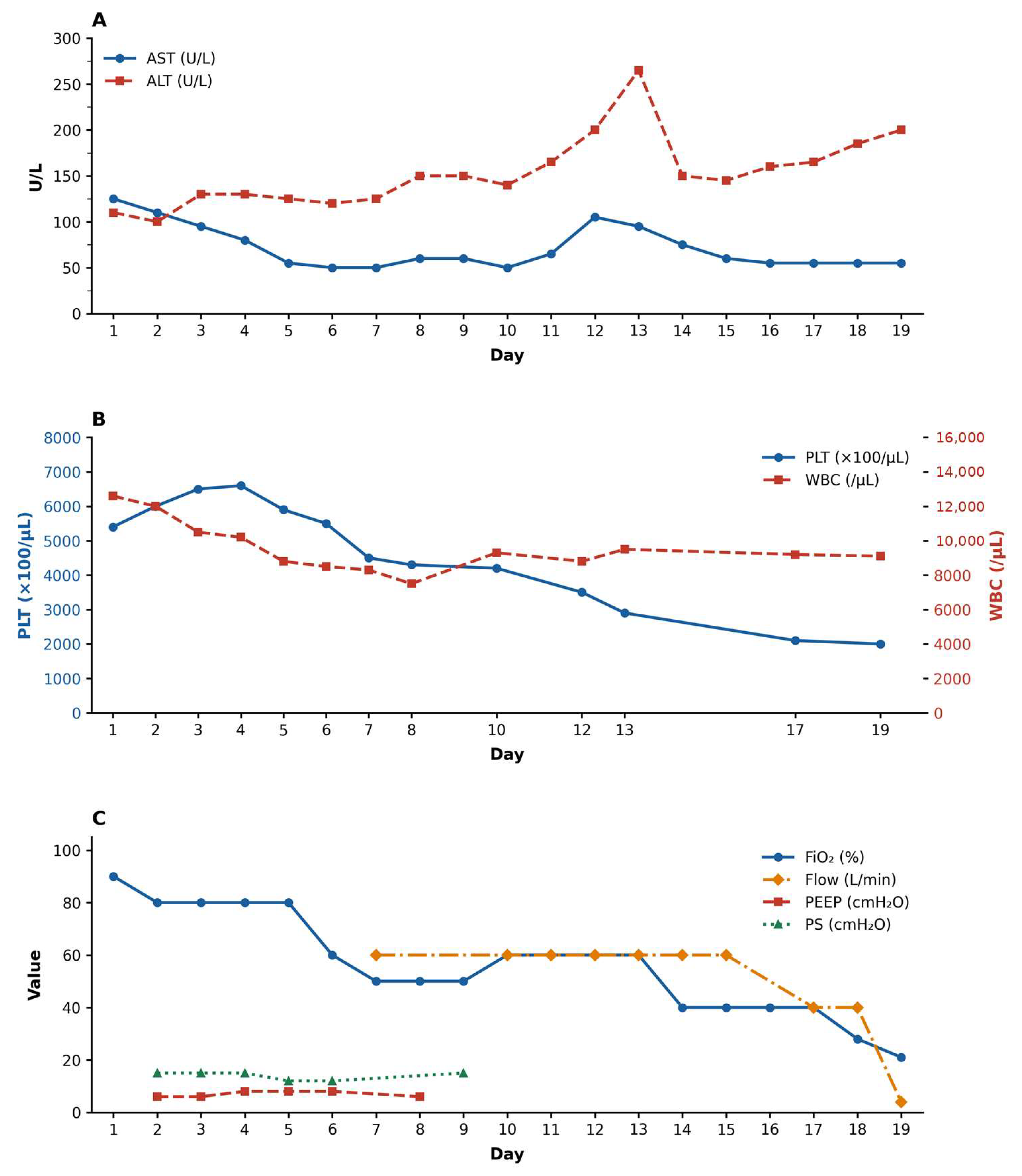
2 28.4 mmHg). Admission laboratory evaluation revealed leukocytosis (12,600 cells/μL), with a predominance of neutrophils (9880 cells/μL), thrombocytosis (539,000 cells/μL), and markedly elevated C-reactive protein (175 mg/L). There was also an increase in transaminases (AST 121 U/L and ALT 109 U/L). Chest computed, tomography revealed diffuse centrilobular micronodules in the middle lobe, the lingula and the basal segment of the left lower lobe, which are consistent with atypical pneumonia. Respiratory support was rapidly escalated from a Venturi mask to a high-flow nasal cannula, and subsequently to non-invasive ventilation with BiPAP. Based on clinical assessment, corticosteroid therapy with dexamethasone was initiated at 4 mg/day for the first 3 days, followed by 6 mg/day, for a total duration of 2 weeks. Anticoagulation with enoxaparin 4000 IU/day was also started. Given the ongoing resurgence and concerns about MRMP, the initial severity also raised the possibility of macrolide resistance, informing early therapeutic escalation pending genotypic confirmation. Empirical antibiotic therapy consisted of levofloxacin 500 mg every 12 h and doxycycline 100 mg every 12 h, both administered for 2 weeks. During hospitalization, respiratory and laboratory parameters improved progressively. Liver enzymes temporarily increased, peaking around day 12, before returning to normal levels. This pattern is consistent with the reported extrapulmonary manifestations of
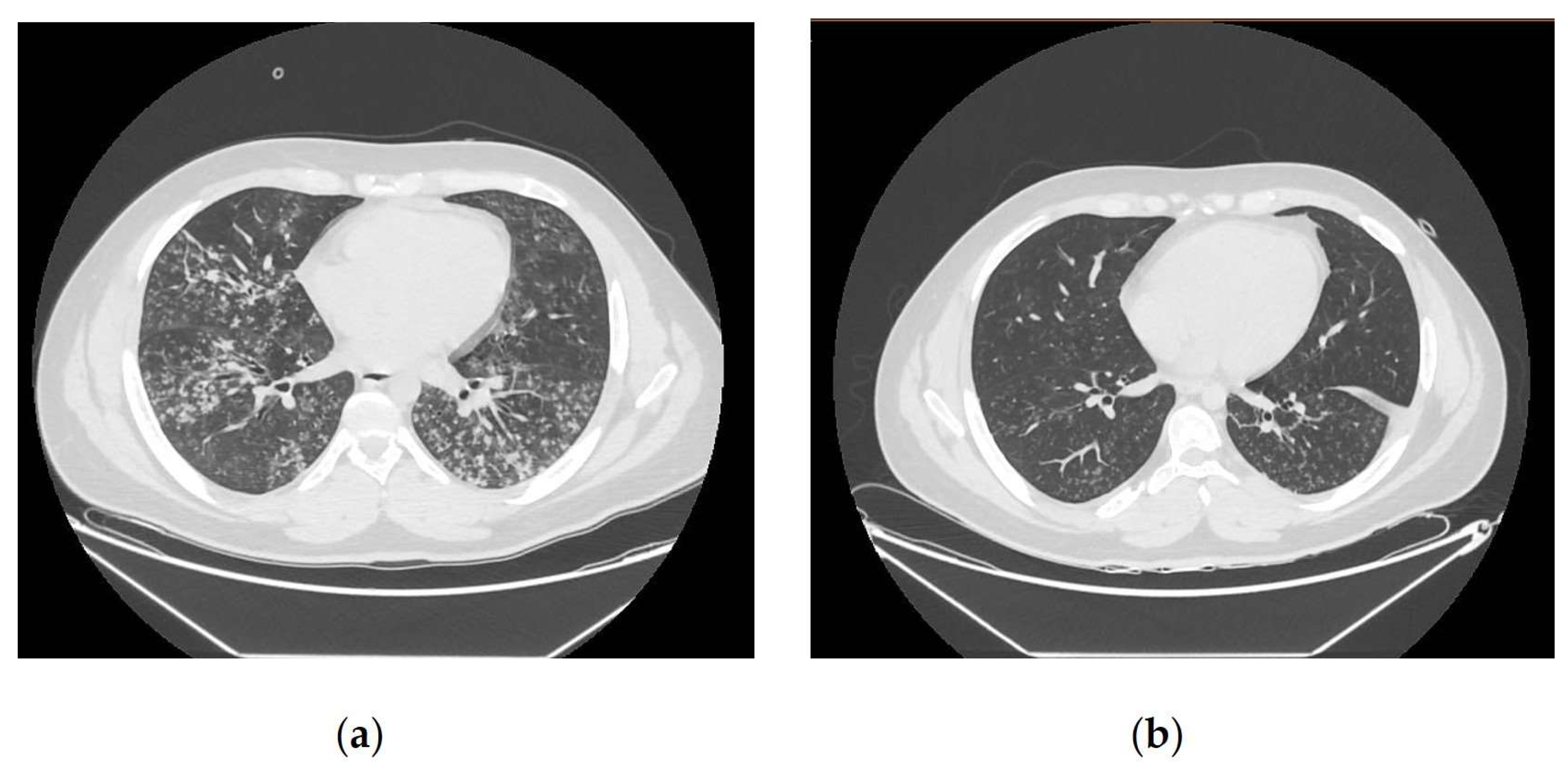
Mycoplasma pneumoniae infection, or a transient drug-related effect. A follow-up chest CT scan performed on day 13 showed a significant reduction in the size and density of centrilobular micronodules, with partial re-aeration of previously affected lung segments (
Figure 1). A progressive reduction in oxygen requirements and ventilatory pressures occurred alongside the normalization of inflammatory and hematological markers (
Figure 2). The patient was discharged on day 20 after full recovery of respiratory function.
2.2. Laboratory Investigations
Multiplex RT-PCR performed on sputum identified
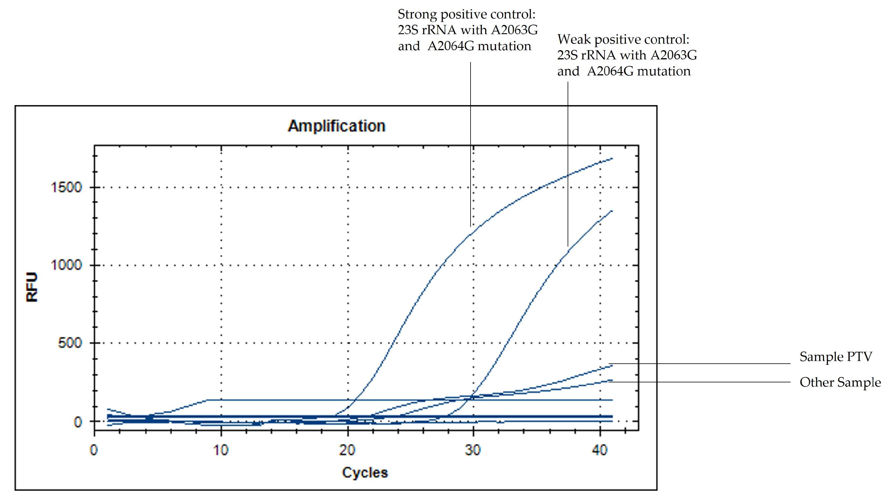
Mycoplasma pneumoniae DNA, and serology results supported an acute infection (IgM 5.9 UR/mL; IgG 196 UR/mL). Due to global concerns regarding macrolide-resistant MP the empirical antimicrobial therapy was optimized early on to include doxycycline and levofloxacin. Targeted molecular analysis showed no evidence of macrolide resistance; RT-PCR was negative for the A2063G and A2064G mutations (
Figure 3), and Sanger sequencing of an 807 bp fragment of the 23S rRNA gene confirmed a wild-type genotype. Overall, molecular testing consistently supported a macrolide-susceptible (wild-type) genotype despite severe disease.
3. Discussion
The re-emergence of
Mycoplasma pneumoniae (MP) in Europe in 2023–2024 is important for understanding severe infections in young, otherwise healthy individuals. This case confirms that severe MP pneumonia is not exclusively associated with macrolide-resistant strains, since the identified isolate carried a fully susceptible 23S rRNA genotype. The discrepancy between clinical severity and macrolide susceptibility indicates a significant role of host-mediated inflammatory mechanisms, which is consistent with previous reports describing cytokine-driven pulmonary injury in MP infections [
7,
9,
11]. A key practical implication is that disease severity alone should not be used as an indicator of resistance. Importantly, this observation does not challenge current recommendations for prompt empirical antibiotic therapy in severe cases. Rather, it aims to support the optimization of these recommendations through early diagnostic clarification, including rapid molecular testing and resistance genotyping.
During periods of heightened alertness for MRMP, this assumption may lead clinicians to prescribe broader regimens more frequently, thereby increasing exposure to fluoroquinolones or tetracyclines, even when macrolide susceptibility is retained. Phenotypic antimicrobial susceptibility testing was not performed because culture-based assays for MP are not routinely available in real-time clinical practice and would not support timely therapeutic decisions.
Rapid genotyping is therefore valuable not only for surveillance, but also for real-time stewardship decisions. The radiological findings observed at presentation, which included confluent centrilobular micronodules involving multiple lobes, together with the rapid improvement seen on follow-up imaging, support the diagnosis of
Mycoplasma pneumoniae (MP) pneumonia, despite macrolide susceptibility, which is characterized by a marked inflammatory component. The close temporal association between clinical stabilization, reduction in ventilatory support, normalization of laboratory parameters and radiological resolution highlights the dynamic and reversible nature of the inflammatory process (see
Figure 1 and
Figure 2). Combining confirmatory genotyping, which enabled rapid identification of the cause, and multiplex RT-PCR also allowed an accurate assessment of macrolide susceptibility.
Although global epidemiological data indicates a significant increase in the prevalence of macrolide-resistant Mycoplasma pneumoniae (MRMP), with marked heterogeneity across Europe, molecular confirmation of antibiotic susceptibility remains essential to avoid misclassification and the unnecessary escalation of antimicrobial therapy. In this case, escalation to doxycycline and levofloxacin was justifiable while MRMP could not be excluded. However, subsequent confirmation of a wild-type 23S rRNA genotype demonstrated how genotyping can rapidly reorient therapeutic rationale, thus preventing resistance misclassification driven solely by clinical severity. Overall, this case supports the practical diagnostic and therapeutic approach of combining syndromic molecular tests (NAATs) with rapid resistance genotyping or confirmatory sequencing for the current resurgence of MP infection. This indicates that clinical severity and antibiotic resistance do not necessarily coincide, so stewardship decisions should be based not only on clinical impression, but also on the infectious agent’s genotype.
This report is limited by its single case nature, which precludes generalization; however, the detailed clinical, radiological, and molecular characterization strengthens the diagnostic and antimicrobial stewardship message in the context of the current MP resurgence, without questioning the validity of existing therapeutic guidelines, but rather emphasizing the importance of early diagnostic clarification to optimize treatment decisions.
4. Materials and Methods
Clinical, laboratory, and radiological data were collected from hospital admission through discharge, including arterial blood gas analyses, complete blood count, inflammatory markers, liver enzymes, and respiratory support parameters. Imaging included chest computed tomography performed on admission and during follow-up. Etiological testing was carried out using a multiplex RT-PCR panel for respiratory pathogens on a sputum sample, followed by serological assays for Mycoplasma pneumoniae IgM and IgG. Macrolide-resistance was evaluated using a two-step molecular workflow. First, RT-PCR screening targeted the A2063G and A2064G mutations of the 23S rRNA gene using a commercial assay, with the Mycoplasma Pneumoniae and Macrolides-Resistant Strain Nucleic Acid Test Kit (Jiangsu Mole Bioscence Co. Taizhou, Jiangsu Province, China). The analysis was extended with Sanger sequencing for the genomic portion of 23S, potentially containing the mutations known in the literature. The RT-PCR was performed on a GeneAmp PCR System 9700 thermocycler (Applied Biosystems, Foster City, CA, USA) under the following conditions: enzymatic activation at 95 °C for 1 min, followed by 30 denaturation steps at 95 °C for 15 s and annealing/extension at 60 °C for 30 s, with the latter two steps repeated for a total of 35 cycles. The resulting RT-PCR amplicon was 807 bp in length (forward primer 5′-GAACGGCGGCCGTAACTATA-3′; reverse primer 5′-GGCGCTACAACTGGAGCATA-3′). Sequencing was performed on a 3500 Dx Genetic Analyzer (Applied Biosystems, Thermo Fisher Scientific, Waltham, MA, USA). Sequence analysis was carried out using BioEdit software version 7.0.5.3 and revealed no mutations in the region of interest. Molecular assays were performed on available clinical specimens using the appropriate controls in accordance with routine diagnostic practices.
5. Conclusions
This case demonstrates that severe, macrolide-susceptible pneumonia associated with the wild-type 23S rRNA genotype can occur in otherwise healthy young adults. During the current resurgence, it is important to note that clinical severity should not be used to infer macrolide resistance. An approach combining syndromic NAAT with rapid resistance genotyping and 23S rRNA sequencing can prevent misclassification based on severity, support rational antibiotic selection, and avoid unnecessary escalation to broad-spectrum antibiotics. However, as this is a single-case report, it is not possible to estimate the proportion of similar cases among hospitalizations related to pneumonia.
Author Contributions
Conceptualization, L.S. and C.F.; methodology, B.B., M.F., V.D., E.P., L.F. and M.I.; clinical investigation, E.P., L.F. and M.I.; data collection, B.B., M.F., V.D., E.P., L.F. and M.I.; data curation, B.B., M.F., V.D., E.P., L.F. and M.I.; interpretation of clinical data, E.P., L.F. and M.I.; writing—original draft preparation, E.P., B.B., M.F. and V.D.; writing—review and editing, L.S., C.F., L.F. and M.I.; supervision, L.S. and C.F. All authors have read and agreed to the published version of the manuscript.
Funding
This work was supported by grants from the Italian Ministry of Health through Ricerca Corrente, Linea 3.
Institutional Review Board Statement
Ethical review and approval were waived for this study because it reports a single clinical case using data collected as part of routine care and fully anonymized for publication, in accordance with institutional policies.
Informed Consent Statement
Informed consent was obtained from all subjects involved in the study.
Data Availability Statement
The data presented in this study are available on request from the corresponding author.
Acknowledgments
We gratefully acknowledge the contributors of the staff of the “L. Spallanzani” biobanking facility: Gianluca Prota, Alberto Rossi, Valentina Antonelli, Claudia Caparrelli and Claudia Maestripieri for their helpful collaboration in receiving and storing isolates; and the technicians of the Microbiology Laboratory of the “L. Spallanzani”: Chiara De Giuli, Chiara Stellitano, Chiara Massimino for their support in molecular biology techniques and sample management.
Conflicts of Interest
The authors declare no conflicts of interest.
Abbreviations
The following abbreviations are used in this manuscript:
| MP | Mycoplasma pneumoniae |
| CAP | Community-acquired pneumonia |
| MRPM | Macrolide-resistant MP |
| AST | Aspartate aminotransferase |
| ALT | Alanine aminotransferase |
| CT | Computed tomography |
| WBC | White blood cell |
| PLT | Platelet |
| FiO2 | Fraction of inspired oxygen |
| PEEP | Positive end expiratory pressure |
| PS | Pressure support |
| BiPAP | Bilevel positive airway pressure |
References
- Waites, K.B.; Talkington, D.F. Mycoplasma pneumoniae and its role as a human pathogen. Clin. Microbiol. Rev. 2004, 17, 697–728. [Google Scholar] [CrossRef] [PubMed]
- Bajantri, B.; Venkatram, S.; Diaz-Fuentes, G. Mycoplasma pneumoniae: A potentially severe infection. J. Clin. Med. Res. 2018, 10, 535–544. [Google Scholar] [CrossRef] [PubMed]
- Meyer Sauteur, P.M.; Beeton, M.L.; European Society of Clinical Microbiology and Infectious Diseases (ESCMID) Study Group for Mycoplasma and Chlamydia Infections (ESGMAC); ESGMAC Mycoplasma pneumoniae Surveillance (MAPS) Study Group. Mycoplasma pneumoniae: Delayed re-emergence after COVID-19 pandemic restrictions. Lancet Microbe 2024, 5, e100–e101. [Google Scholar] [CrossRef] [PubMed]
- Larcher, R.; Boudet, A.; Roger, C.; Villa, F.; Loubet, P. Mycoplasma pneumoniae is back! Is it the next pandemic? Anaesth. Crit. Care Pain Med. 2024, 43, 101338. [Google Scholar] [CrossRef] [PubMed]
- Kim, K.; Jung, S.; Kim, M.; Park, S.; Yang, H.J.; Lee, E. Global trends in the proportion of macrolide-resistant Mycoplasma pneumoniae infections: A systematic review and meta-analysis. JAMA Netw. Open 2022, 5, e2220949. [Google Scholar] [CrossRef] [PubMed]
- Loconsole, D.; De Robertis, A.L.; Sallustio, A.; Centrone, F.; Morcavallo, C.; Campanella, S.; Accogli, M.; Chironna, M. Update on the epidemiology of macrolide-resistant Mycoplasma pneumoniae in Europe: A systematic review. Infect. Dis. Rep. 2021, 13, 811–820. [Google Scholar] [CrossRef] [PubMed]
- Narita, M.; Tanaka, H. Cytokines involved in the severe manifestations of pulmonary diseases caused by Mycoplasma pneumoniae. Pediatr. Pulmonol. 2007, 42, 397. [Google Scholar] [CrossRef] [PubMed]
- Narita, M. Pathogenesis of extrapulmonary manifestations of Mycoplasma pneumoniae infection with special reference to pneumonia. J. Infect. Chemother. 2010, 16, 162–169. [Google Scholar] [CrossRef] [PubMed]
- Khoury, T.; Sviri, S.; Rmeileh, A.A.; Nubani, A.; Abutbul, A.; Hoss, S.; van Heerden, P.V.; Bayya, A.E.; Hidalgo-Grass, C.; Moses, A.E.; et al. Increased rates of intensive care unit admission in patients with Mycoplasma pneumoniae: A retrospective study. Clin. Microbiol. Infect. 2016, 22, 711–714. [Google Scholar] [CrossRef] [PubMed]
- Shi, S.; Zhang, X.; Zhou, Y.; Tang, H.; Zhao, D.; Liu, F. Immunosuppression reduces lung injury caused by Mycoplasma pneumoniae infection. Sci. Rep. 2019, 9, 7147. [Google Scholar] [CrossRef] [PubMed]
- Tsai, T.A.; Tsai, C.K.; Kuo, K.C.; Yu, H.R. Rational stepwise approach for Mycoplasma pneumoniae pneumonia in children. J. Microbiol. Immunol. Infect. 2021, 54, 557–565. [Google Scholar] [CrossRef] [PubMed]
| Disclaimer/Publisher’s Note: The statements, opinions and data contained in all publications are solely those of the individual author(s) and contributor(s) and not of MDPI and/or the editor(s). MDPI and/or the editor(s) disclaim responsibility for any injury to people or property resulting from any ideas, methods, instructions or products referred to in the content. |



 ,
,