Article

Retargeting Gram-Positive-Only Adarotene-Derived Antibacterials to Broad-Spectrum Antibiotics

1
Department of Food, Environmental and Nutritional Sciences (DeFENS), University of Milan, via Celoria 2, 20133 Milan, Italy
2
Department of Biotechnology, Chemistry and Pharmacy, University of Siena, via Aldo Moro 2, 53100 Siena, Italy
3
Department of Science, University Roma Tre, Viale G. Marconi 446, 00146 Rome, Italy
4
Laboratory affiliated to Pasteur Italia-Fondazione Cenci Bolognetti, Department of Biochemical Sciences, Sapienza University of Rome, 00185 Rome, Italy
5
Experimental Hepatology and Drug Targeting (HEVEPHARM), Institute of Biomedical Research of Salamanca (IBSAL), University of Salamanca, 37007 Salamanca, Spain
6
Center for the Study of Liver and Gastrointestinal Diseases (CIBEREHD), Carlos III National Institute of Health, 28029 Madrid, Spain
7
IRCCS Fondazione Santa Lucia, Via Ardeatina 306/354, 00179 Rome, Italy
8
Special Products Line, 03012 Anagni, Italy
*
Authors to whom correspondence should be addressed.
These authors contributed equally to this work.
Antibiotics 2025, 14(9), 956; https://doi.org/10.3390/antibiotics14090956
Submission received: 25 July 2025 / Revised: 6 September 2025 / Accepted: 18 September 2025 / Published: 21 September 2025

Abstract

Background: Bacterial resistance to antibiotics continues to rise globally, posing a significant public health challenge and incurring substantial social and economic burdens. In response, the World Health Organization (WHO) has published a list of priority pathogens for which effective treatment options are critically limited. Several antibiotics are categorized as Gram-positive-only (GPO) agents due to their lack of activity against Gram-negative species. Although these compounds often target conserved bacterial processes, their limited spectrum is largely attributed to poor penetration of the Gram-negative outer membrane (OM). Results: In this study, we designed and synthesized a series of adarotene-derived compounds to evaluate the impact of introducing positively charged groups on their interaction with the Gram-negative OM. One of the newly synthesized derivatives, SPL 207, displayed minimum inhibitory concentration (MIC) values ranging from 8 to 64 µM against a panel of Gram-positive and Gram-negative bacteria. The ability of SPL207 to disrupt outer and inner membrane permeability was evaluated using fluorescence assays and confocal microscopy, revealing that the compound compromises membrane integrity across all tested Gram-negative bacteria. Strong synergistic activity was observed in combination with colistin against three P. aeruginosa colistin-resistant strains. Atomistic details of membrane interference were elucidated by molecular dynamics (MD) simulations, with SPL207 clearly acting as a membrane destabilizer by enhancing Ca2+ ions diffusion and lipids destabilization. Conclusions: Although the observed MIC values remain above clinically acceptable thresholds, these findings provide a promising proof of concept. The further structural optimization of adarotene derivatives may yield novel broad-spectrum agents with improved antimicrobial potency against MDR pathogens.

1. Introduction

The rising prevalence of multidrug-resistant (MDR) pathogens—and those resistant to nearly all available antibiotics—is contributing to a growing number of deaths from bacterial infections worldwide. In addition, this trend is inversely paralleled by a decline in the discovery and development of new antibiotics. It has been predicted that 10 million people worldwide will die per year from antibiotic-resistant infections by 2050 [1,2,3].
From an economic perspective, infections due to MDR pathogens lead to prolonged hospitalization and repeated antibiotic courses, causing estimated extra healthcare costs of about EUR 1.1 billion to the healthcare systems in EU/EEA countries [4]. These costs are set to rise dramatically if novel antimicrobials will not be identified. In the last two decades Gram-negative bacteria have emerged as particularly problematic pathogens. With the launch of the EU4Health 2021–2027 program, and in line with HORIZON 2020, the European Commission has confirmed the fight against communicable diseases in general and, more specifically, against antibiotic resistance as a critical priority. The World Health Organization issued a list of ‘Priority Pathogens’ for which new antibiotics are urgently needed. The vast majority of these pathogens are Gram-negative, including third-generation cephalosporin- and carbapenem-resistant Escherichia coli and Klebsiella pneumoniae, carbapenem-resistant Acinetobacter baumannii and Pseudomonas aeruginosa, and fluoroquinolone-resistant Shigella species and Salmonella enterica ser. Typhi [1,5].
The lack of Gram-negative antibacterial discovery could be traced to the unique structure of their outer membrane (OM), usually composed of lipopolysaccharides (LPS). LPS are made up of lipid A and a long, negatively charged, oligosaccharide. Lipid A molecules contain 4–7 acyl chains (predominantly saturated), attached to a phosphorylated disaccharide. The adjacent negative charges of the LPS are stabilized by divalent cations. These features allow LPS molecules to stack together very tightly, making passive diffusion extremely challenging for most small molecules [6,7]. As many compounds cannot readily passively diffuse across the OM, small molecules permeate the OM through porins and/or the self-promoted uptake (SPU) pathway. Porins are water-filled β-barrels lined interiorly with charged amino acids. They form a narrow channel allowing only certain molecules to rapidly diffuse (e.g., certain β-lactams and fluoroquinolones). Other small molecules can pass through the OM via the SPU pathway by displacing the divalent cations, temporarily destabilizing the LPS layer. Compounds that utilize SPU are typically polycationic, as for instance colistin [8]; however, some compounds likely enter through a combination of both porins and SPU pathways [6].
For the above reasons, many antibiotics are inactive against Gram-negative pathogens, hence they have been termed “Gram-positive-only” (GPO) antibiotics [6]. The spectrum of activity of GPO antibiotics can be extended to Gram-negative bacteria through the addition of chemical groups that facilitate the passage of the pharmacophore across the OM. So far, the unique marketed example of broad-spectrum antibiotic derived from a GPO antibiotic is cefiderocol. This is made of a cephalosporin pharmacophore conjugated to a catechol-type siderophore. During the infection, bacteria experience iron starvation and respond expressing siderophores. The iron-bound catechol moiety is recognized by specific bacterial receptors located on the OM, promoting cefiderocol uptake. Once in the periplasm, the cephalosporin moiety can bind to the penicillin binding protein and inhibit cell wall synthesis [9]. A similar strategy has been recently used for expanding the range of activity of aztreonam, a GPO β-lactam antibiotic [10].
In the past, extensive screening campaigns for new classes of antibiotics gave no relevant results for Gram-negative bacteria. The lack of cell permeability was recognized as a major problem, since no rationale processes existed to improve hit compounds. However, recent structure-activity relationship (SAR) studies have provided some cues useful to transform GPO into broad-spectrum antibiotics. According to these criteria, named the eNTRy rules, compounds are most likely to be accumulated in Gram-negative bacteria if they: (i) contain a non-sterically encumbered ionizable Nitrogen; (ii) have low Three dimensionality; (iii) are relatively Rigid; (iv) have some non-polar functionality [6]. In support of the validity of the eNTRy rules, promising results have been recently published about modifications of bakuchiol, a prenylated phenolic monoterpene with antibacterial activity only against Gram-positive bacteria. Bakuchiol derivatives with antibacterial activity against Gram-negative pathogens and low toxicity against eukaryotic cells were obtained by introducing an aliphatic tail containing up to 3 positively charged amine groups. This tail favors the crossing through the OM, allowing the bakuchiol derivatives to disrupt the OM and cause cell death [11].
In 2018, the ability of atypical retinoids to penetrate and embed lipid bilayers was correlated with their bactericidal activity [12]. Starting from this evidence, we have recently synthesized novel adarotene-like molecules, belonging to the class of the atypical retinoids, endowed with significant antimicrobial activity against MDR strains of Enterococcus faecalis and Staphylococcus aureus. The most promising compound (2, AB473) showed a Minimal Inhibitory Concentration (MIC) of 4 µM (2 μg/mL) for all tested strains and very low cytotoxicity on human cells (Figure 1) [13,14] suggesting a potential high therapeutic index.
However, the great limitation of the adarotene derivatives obtained so far, including AB473, is their poor activity against Gram-negative bacteria (MIC > 128 μM against E. coli, P. aeruginosa, A. baumannii) [12,13]. To overcome this limitation, we designed new adarotene derivatives by following the above-mentioned eNTRy rules [6].
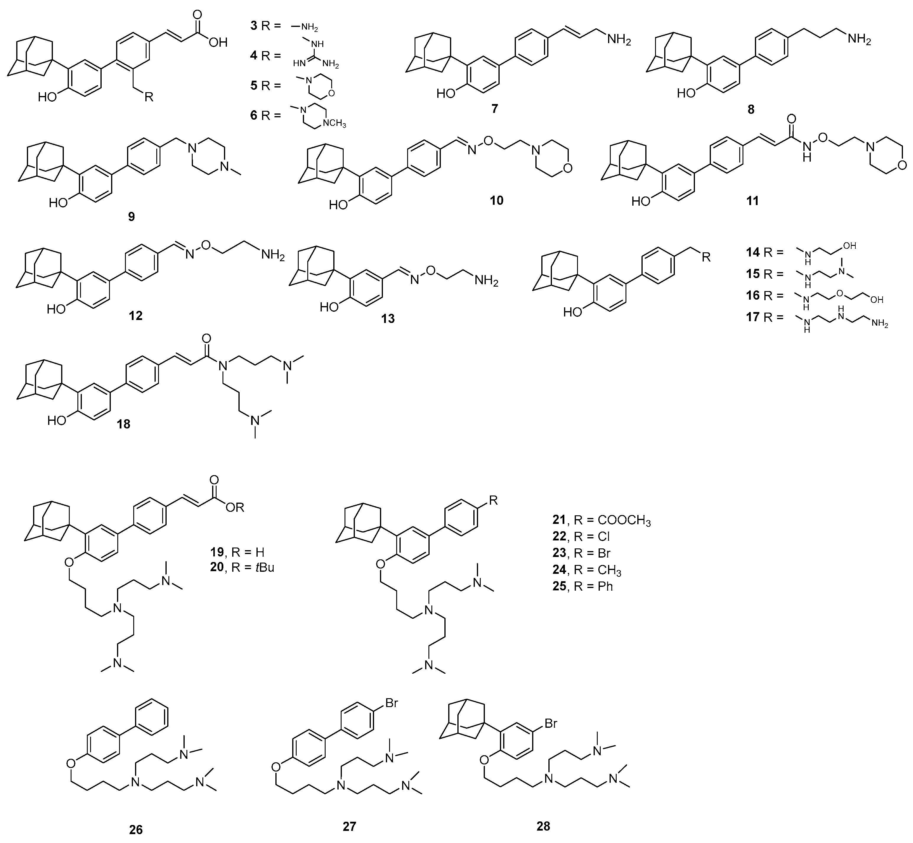
In particular, we aimed to investigate whether various positively charged nitrogen species could enhance the translocation of compounds across the Gram-negative OM. We hypothesized that the incorporated basic cationic moieties could favor the interaction between the derivatives and the negatively charged bacterial cell membrane. The subsequent insertion of the hydrophobic moiety into the phospholipid bilayer could lead to the destruction of the integrity of the bacterial membrane, leakage of intracellular content and bacterial cell death. Beyond enhanced OM crossing, amino moieties could provide other advantages, including higher solubility in aqueous environments (e.g., biological fluids) [15,16]. Thus, a diverse set of amine-containing adarotene-derived compounds were designed and synthesized (Figure 2). Additionally, a shift from simple amino groups to polycationic groups was implemented, with the aim of obtaining molecules capable of penetrating Gram-negative bacteria via the SPU pathway [6].
Bearing in mind that the amino groups need to be embedded within a compound possessing appropriate flexibility, the structural features of the linker (e.g., geometry, orientation, size, and chemical composition) connecting the charged group(s) to the core skeleton, as well as the position of attachment, were carefully modulated. The installation of the selected moieties was designed not only on the biphenyl skeleton, but also on the key functional groups of the parent compound (e.g., the carboxylic group and the phenolic OH), which will serve as sites of attachment of the chains by amide and ether bonds, respectively (Figure 2). To assess membrane integrity disruption, fluorescence assays and microscopy imaging evaluation were planned for the most promising compounds identified by their MIC profiles. A validated P. aeruginosa OM model was developed to enable in silico investigations of ligand penetration. Finally, considering that colistin is one of the last-resort treatments for P. aeruginosa, we aimed to evaluate whether the most promising compounds exhibit synergistic effects against colistin-resistant strains.

2. Results and Discussion

2.1. Chemistry

Adarotene analogs and derivatives were synthesized to explore their structural and biological properties. Given the critical role of protonatable nitrogen-containing side chains in enhancing anti-Gram-negative activity, all synthesized compounds incorporated (poly)aminic residues.
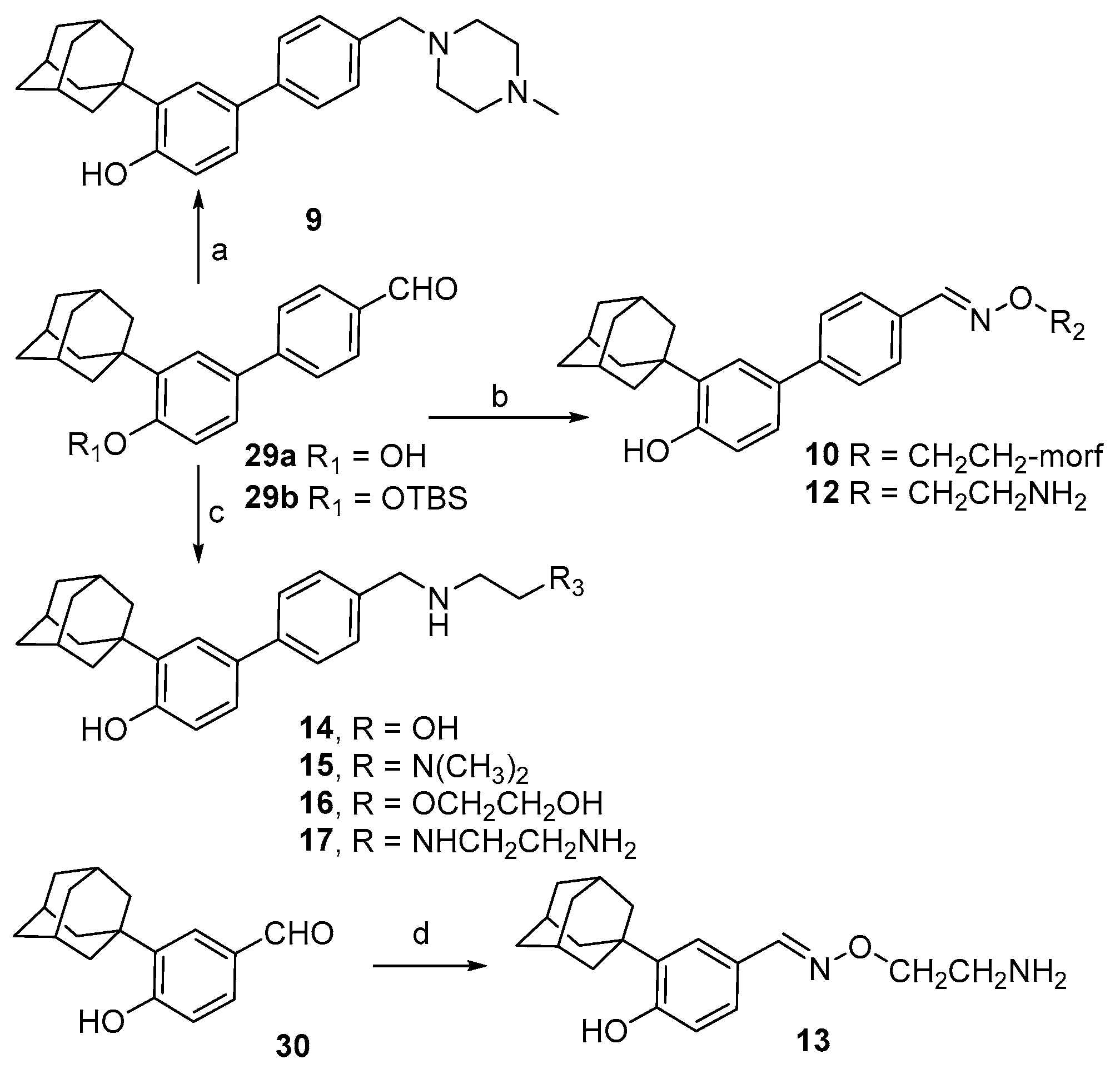
Aldehydes 29a,b [14,17] were used as starting material for the preparation of benzylamines 9 and 1417, as well as oximes 10 and 12 (Scheme 1). Reductive amination of 29a with N-methyl piperazine in presence of formic acid and BF3Et2O afforded derivative 9. Treatment with ethanolamine and N,N-dimethylethylenediamine gave 14 and 15, respectively; longer and more flexible chains were installed by reductive amination using diethylenetriamine and its corresponding alcohol (compound 17 and 16, respectively). Oximes 10 and 12 were prepared by treatment of the suitable aldehyde with the corresponding O-alkylated hydroxylamine in EtOH at reflux temperature. Similarly, aldehyde 30 furnished oxime 13 upon reaction with 2-(aminooxy)-ethan-1-amine hydrochloride.
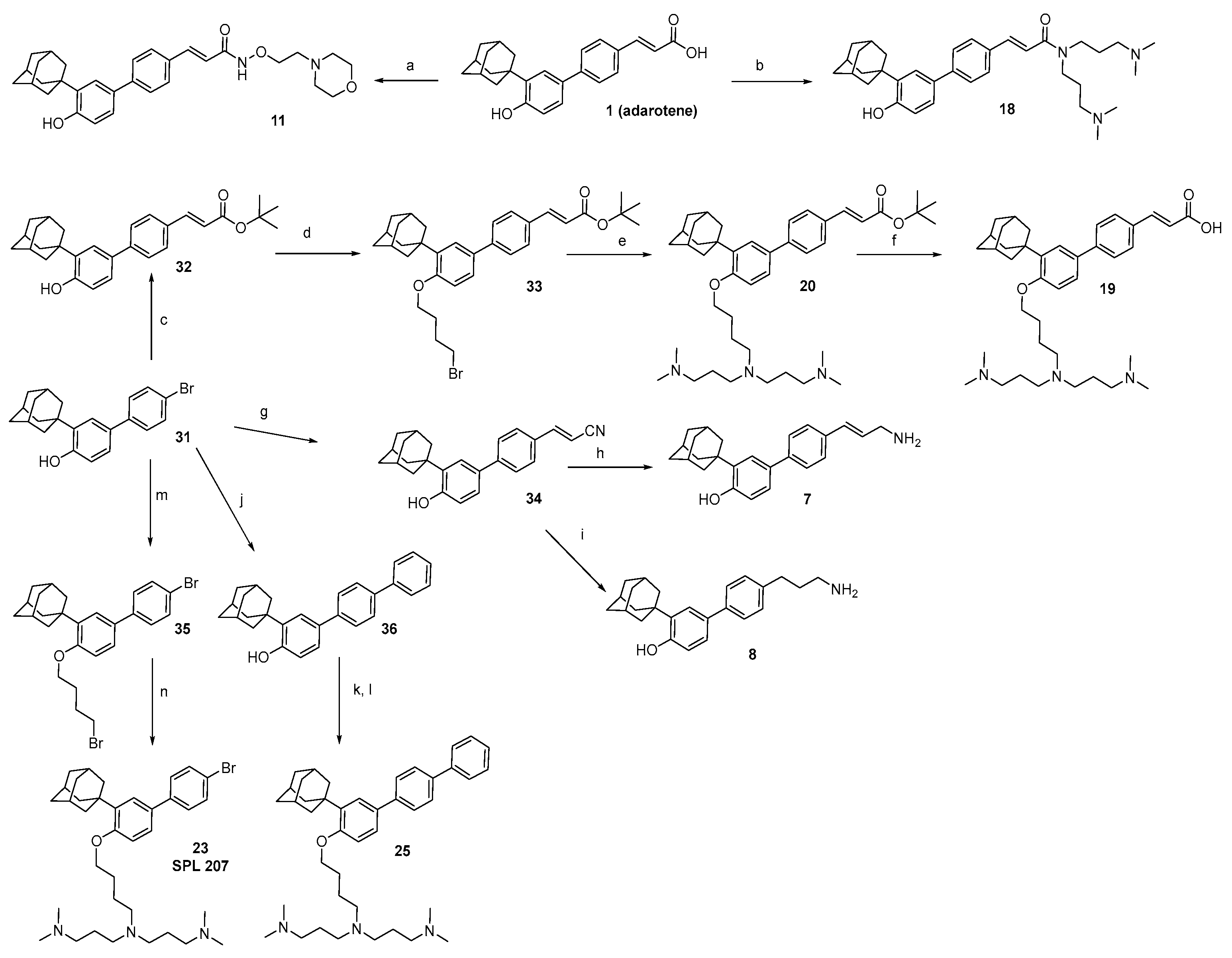
Hydroxamate 11 and amide 18 were obtained from adarotene, by condensation with O-[2-(morpholin-4-yl)ethyl]hydroxylamine and N1-(3-(dimethylamino)propyl)-N3,N3-dimethylpropane-1,3-diamine, respectively. Common brominated intermediate 31 was employed to introduce a polyamine chain on different portions of the biphenyl adarotene-like skeleton (Scheme 2). Heck reaction with t-butyl acrylate promptly afforded t-butyl ester 32, ready for further functionalization. Alkylation of the phenolic residue with 1,4-dibromobutane resulted in compound 33, which was alkylated and final hydrolyzed to give acid 19. Derivative 34 was obtained by Heck reaction with acrylonitrile, whose cyano group was reduced to allyl amine 7 and its saturated analog 8. Suzuki coupling between bromine 31 and phenylboronic acid afforded compound 36, which was O-alkylated with 1,4-dibromobutane and functionalized with N1-(3-(dimethylamino)propyl)-N3,N3-dimethylpropane-1,3-diamine to give derivative 25. Application of the same synthetic route on 31 gave compound 23.
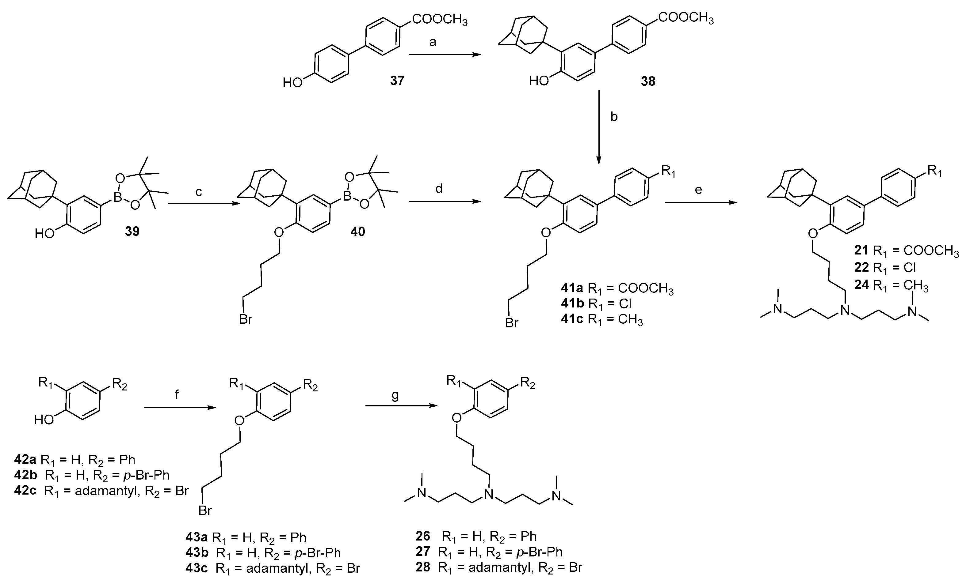
A similar procedure was employed for the O-alkylation of ester 38, obtained by Friedel-Crafts reaction of adamantanol on the biphenyl moiety (Scheme 3). In order to change the substituent at C(4′), treatment of boronate 39 [18] with dibromobutane (compound 40) was followed by Suzuki-Myiaura coupling with chlorobenzene or toluene, to afford 41b and 41c, respectively. Substitution of the terminal bromide with the triamine resulted in the obtainment of compounds 21, 22 and 24. Installation of the basic side chain at the phenolic oxygen was carried out also on biphenyls 42a and 42b, as well as on adamantanyl bromophenol 42c.
According to the eNTRy rules, incorporating protonatable nitrogen-containing groups into the retinoid biphenyl scaffold may facilitate the compound’s passage through the Gram-negative OM [6].
With this rationale, we initially evaluated a set of compounds (36) from our in-house collection, each bearing an amino group on the same carbon that carries the substituent in AB473 (Figure 2, Table 1).
These compounds were tested against S. aureus (Gram-positive) and E. coli (Gram-negative) model strains by determining the MIC (Table 1). Although the compounds 4, 5, and 6 showed mild activity against the Gram-positive strain, they were inactive against the Gram-negative one. These results prompted us to explore the introduction of protonatable chains in other positions of the biphenyl scaffold.
We therefore evaluated adarotene analogs featuring chains that differ in length, polarity, and flexibility in place of the acrylic moiety, aiming to balance the lipophilic portions of the molecule with the protonatable groups (Figure 2).
Compound 7, having a -CH2NH2 group in place of the carboxylic acid moiety of adarotene (Figure 2), retained a good activity against Gram-positive (MIC = 4 µM), not showing however any activity against Gram-negative. The increase in flexibility of the chain (compound 8) did not give any improvement (MIC = 8 µM), whereas the introduction of a methylpyrazine group (compound 9) completely abolished the activity. The derivative obtained by installing a morpholino moiety through an oxime spacer (compound 10) was inactive against both S. aureus and E. coli, while a weak effect against S. aureus (MIC = 32 µM) was obtained using the hydroxamate of adarotene as a spacer for the morpholino group (compound 11). Thus, the morpholino group was replaced with a primary amine (compound 12); however, the compound remained inactive against E. coli while showing potent activity against S. aureus (MIC = 4 µM).
The removal of one of the aromatic rings (compounds 13 vs. 12) produced a further decrease in activity against the Gram-positive strain. The introduction of other secondary and tertiary amino groups (compounds 14, 15, and 16) confirmed a good activity against the Gram-positive bacterium (MIC = 8–64 µM) but still no effect against the Gram-negative one.
Interestingly, compound 17, carrying a polyamine chain in place of the acrylic moiety, showed moderate activity against E. coli, with MIC = 32 µM, retaining also activity against S. aureus (MIC = 16 µM). Moreover, the insertion of a polyamine chain through an amide bond with the carboxylic group of adarotene gave compound 18 with good activity against both E. coli and S. aureus (MIC = 32 µM and 8 µM, respectively).
The antimicrobial activity of compounds 7, 12, and 17 was also tested against other representative Gram-negative pathogens (Table S1). Compounds 7 and 12 were not active (MIC values > 128 μM), while compound 17 showed mild activity against A. baumannii, E. coli, K. pneumoniae and P. aeruginosa strains, with MIC values ranging from 64 µM to 128 µM (Table S1).
Successively, we focused our attention on the position of the polyamine moieties. We thus installed a branched protonatable chain on the phenolic OH. The acid (compound 19) showed a comparable MICs (MIC = 8 and 16 µM) against both S. aureus and E. coli, respectively, while the corresponding t-butyl ester (compound 20) showed reduced activity against E. coli (MIC = 64 µM). Antibacterial activity was improved by directly linking an ester group to the aromatic system. Notably, compound 21 exhibited good activity against S. aureus and E. coli, with MIC values of 4 µM and 8 µM, respectively. Accordingly, we evaluated the effect of introducing various substituents on the aromatic ring while keeping the rest of the molecular structure unchanged (compounds 2225). The same range of activities of compound 21 was retained in compounds 22, 23, and 24, having a chloro, bromo, and methyl group, respectively, in place of the ester moiety.
A similar activity was maintained after the removal of one of the phenyl groups (compound 28), whereas the introduction of a third phenyl group (compound 25) abolished the activity against the Gram-negative strain (MIC = 128 µM). A further simplification of the scaffold with the removal of the adamantyl group (compounds 26, 27) caused an overall reduction in the activity, even against the Gram-positive strain.
This initial screening allowed the identification of compounds capable of inhibiting the growth of a Gram-negative bacterium such as E. coli. For this reason, the active compounds were subsequently tested against another opportunistic Gram-negative pathogen, the more clinically challenging P. aeruginosa. As reported in Table 2, compounds 18, 19, 20, and 22 were inactive (MIC ≥ 128 µM). Compounds 21 and 28 showed a mild activity with MIC = 64 µM, while the most active compound resulted to be compound 23 (MIC = 32 µM), which was also tested against other Gram-negatives (A. baumannii, E. cloacae, E. coli MG1655, and K. pneumoniae; Table 3). The results showed MIC values ranging from 8 µM to 64 µM, confirming compound 23 (hereafter referred to as SPL207) as the most promising candidate for further investigation. As a preliminary assessment of potential cytotoxicity, the effect of the compound SPL207 on eukaryotic cells was tested against the human alveolar lung epithelial A549 cells and the human keratinocyte HaCaT cells (Figures S3 and S4). Compounds 7, 12 and 18 were also included. As reported in Figure S3, for the A549 cell line, the percentage of cell viability remained above 80% upon treatment with all four tested compounds up to a concentration of 16 μM. In the case of the HaCaT cell line (Figure S4), viability remained at 100% up to 16 μM for all four compounds. Treatment with compound 7 did not affect cell viability even at concentrations up to 32 μM. In any case, the concentrations at which an effect on cell viability was observed are higher than the MIC values of the selected molecules against S. aureus and most of the tested Gram-negative strains, except for P. aeruginosa.
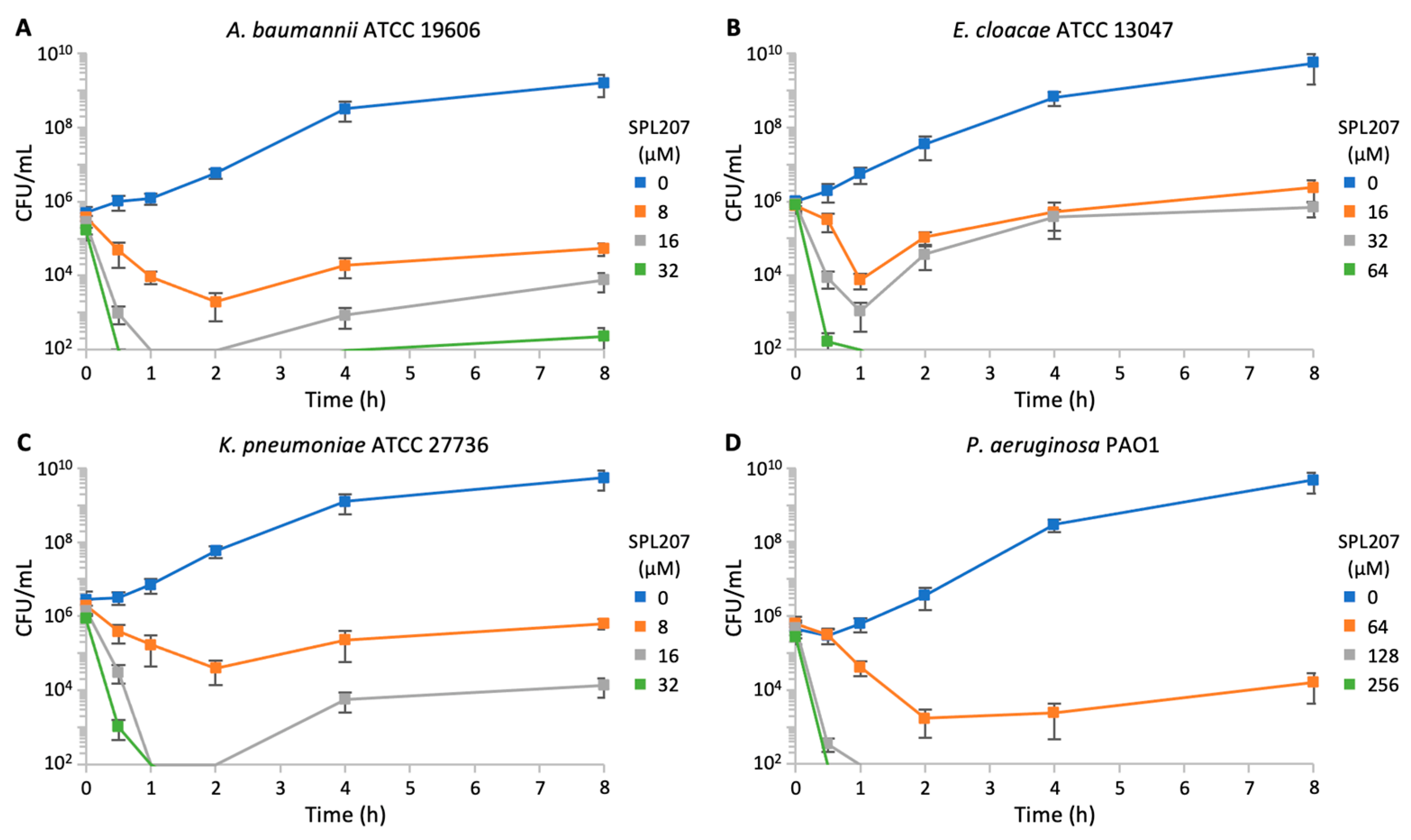
Time-kill assays performed on A. baumannii, E. cloacae, K. pneumoniae, and P. aeruginosa cultures showed a drastic drop of CFU counts after treatment with SPL207 at 2 × MIC and 4 × MIC (Figure 3). Hence, SPL207 displays bactericidal activity.

2.2. Biology and Computational Analysis

2.2.1. Investigation About P. aeruginosa Resistance to SPL207

We reasoned that the antimicrobial activity of SPL207 against P. aeruginosa could be mainly weakened by efflux pumps capable of extruding this compound outside of the cell [19] and/or by low permeability of the OM [20,21]. To test these hypotheses, the activity of SPL207 was assessed in P. aeruginosa mutant strains deleted in efflux pump genes or defective in OM biogenesis.
Since the efflux pumps MexAB-OprM, MexXY, MexCD-OprJ, and MexEF-OprN play a key role in P. aeruginosa resistance to many antimicrobial agents [19,22], MIC assays were performed on PAO1-KP ∆efflux, a P. aeruginosa PAO1 mutant with deletion of the mexAB-oprM, mexXY, mexCD-oprJ, and mexEF-oprN operons [23,24]. As reported in Table 4, SPL207 showed the same MIC in PAO1-KP ∆efflux and its isogenic wild-type strain PAO1-KP, indicating that efflux pumps are not responsible for poor activity of SPL207 in P. aeruginosa.
MIC assays were also performed in a P. aeruginosa PAO1 mutant strains with increased OM permeability. These strains are conditional mutants in which lptE or lptH expression can be induced by arabinose supplementation. LptE and LptH proteins are involved in the transport of lipopolysaccharides to the OM, hence low expression of lptE or lptH affects OM biogenesis and increases OM permeability [25,26].
Preliminary experiments were performed to identify the concentrations of arabinose that support growth of the lptE and lptH conditional mutants. In agreement with literature data [25,26], in the absence of arabinose P. aeruginosa growth was slightly reduced or abolished in the lptE and lptH conditional mutants, respectively, while arabinose supplementation did not affect PAO1 wild-type growth rate (Figure S1A,B). As shown in Table 4, the antimicrobial activity of SPL207 increased when the lptE and lptH conditional mutants were grown in the presence of the lowest arabinose concentrations required to minimize their growth defect (i.e., 0.002% [w/v] and 0.125% [w/v] arabinose for the lptE and lptH conditional mutants, respectively). Conversely, MIC was restored to wild type levels when the conditional mutants were grown with high arabinose concentration (i.e., 0.5% [w/v] for both conditional mutants). Interestingly, increased activity of SPL207 in P. aeruginosa was observed also in the presence of ethylenediaminetetraacetic acid (EDTA), a divalent cation chelating agent that increases OM permeability [27] (Table 4 and Figure S1C).
These results suggest that low OM permeability of P. aeruginosa could account for the limited activity of SPL207 against this pathogen.

2.2.2. Investigation of SPL207 Mechanism of Action and Synergy with Colistin

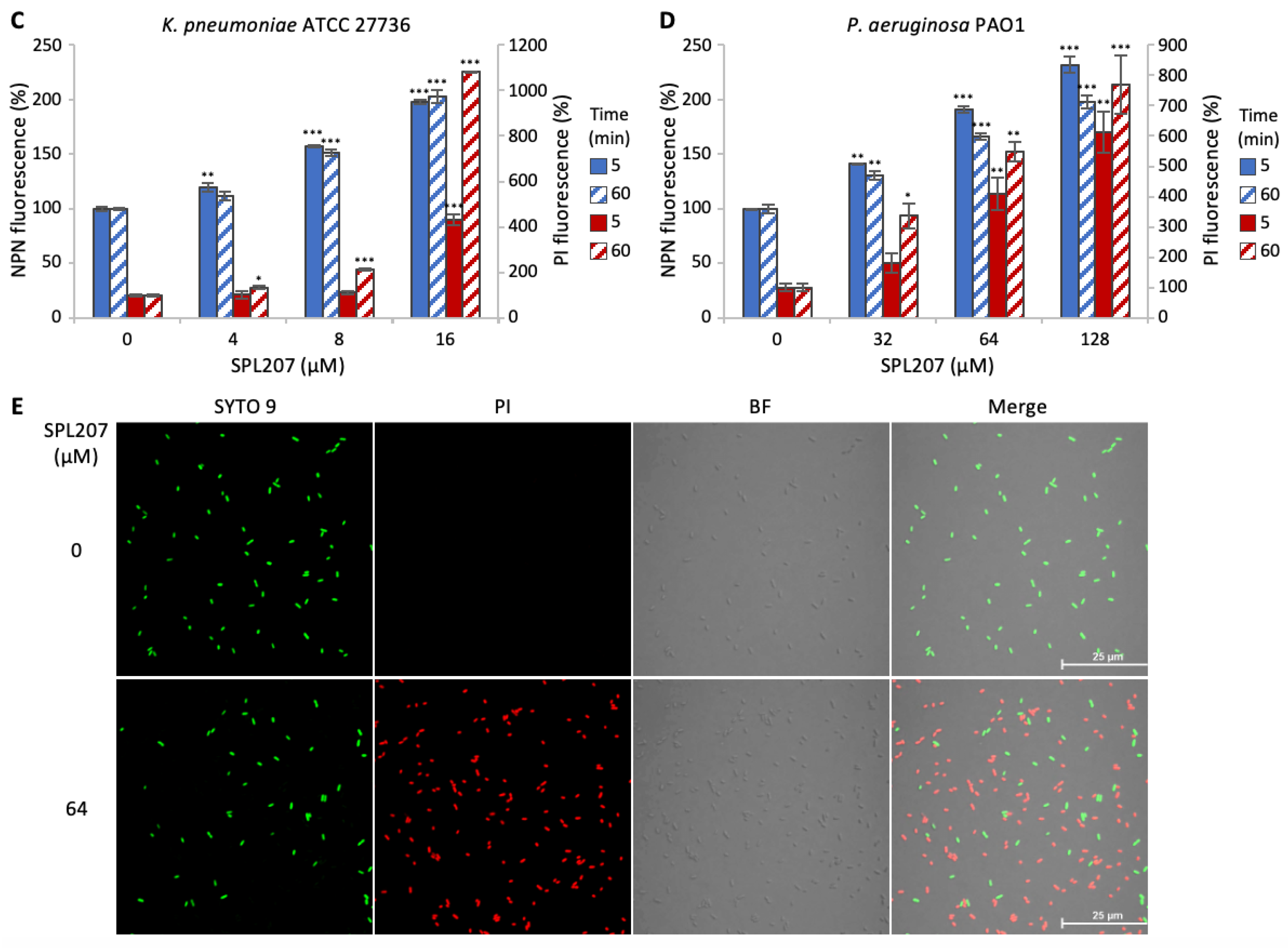
Previous studies showed that adarotene derivatives can penetrate lipid bilayers and permeabilize the membranes of Gram-positives, ultimately causing cell lysis [12,14]. To investigate whether SPL207 shares a similar mechanism of action in Gram-negative bacteria, its effect on membrane integrity was evaluated in A. baumannii, E. cloacae, K. pneumoniae, and P. aeruginosa. Briefly, a potential impact of SPL207 on OM and inner membrane (IM) permeability was evaluated by fluorescent assays based on the probes N-phenyl-1-naphthylamine (NPN) and propidium iodide (PI) [28,29]. In all four strains tested, an increase in NPN and PI fluorescence was observed as the concentration of SPL207 increased (Figure 4A–D). These results indicate that SPL207 impairs the OM and IM integrity in all the Gram-negative bacteria tested.
Additionally, to evaluate a potential loss of P. aeruginosa envelope integrity in response to SPL207, a Live/Dead staining assay [30] was performed. As shown by confocal microscopy imaging, SPL207 treatment strongly increased the fraction of red fluorescent PAO1 cells (Figure 4E), indicative of membrane damage and cell death (only these cells can uptake the red fluorescent dye PI). Impairment of membrane integrity in SPL207 treated cells is in line with the fast-killing activity exerted by this compound (Figure 3D).
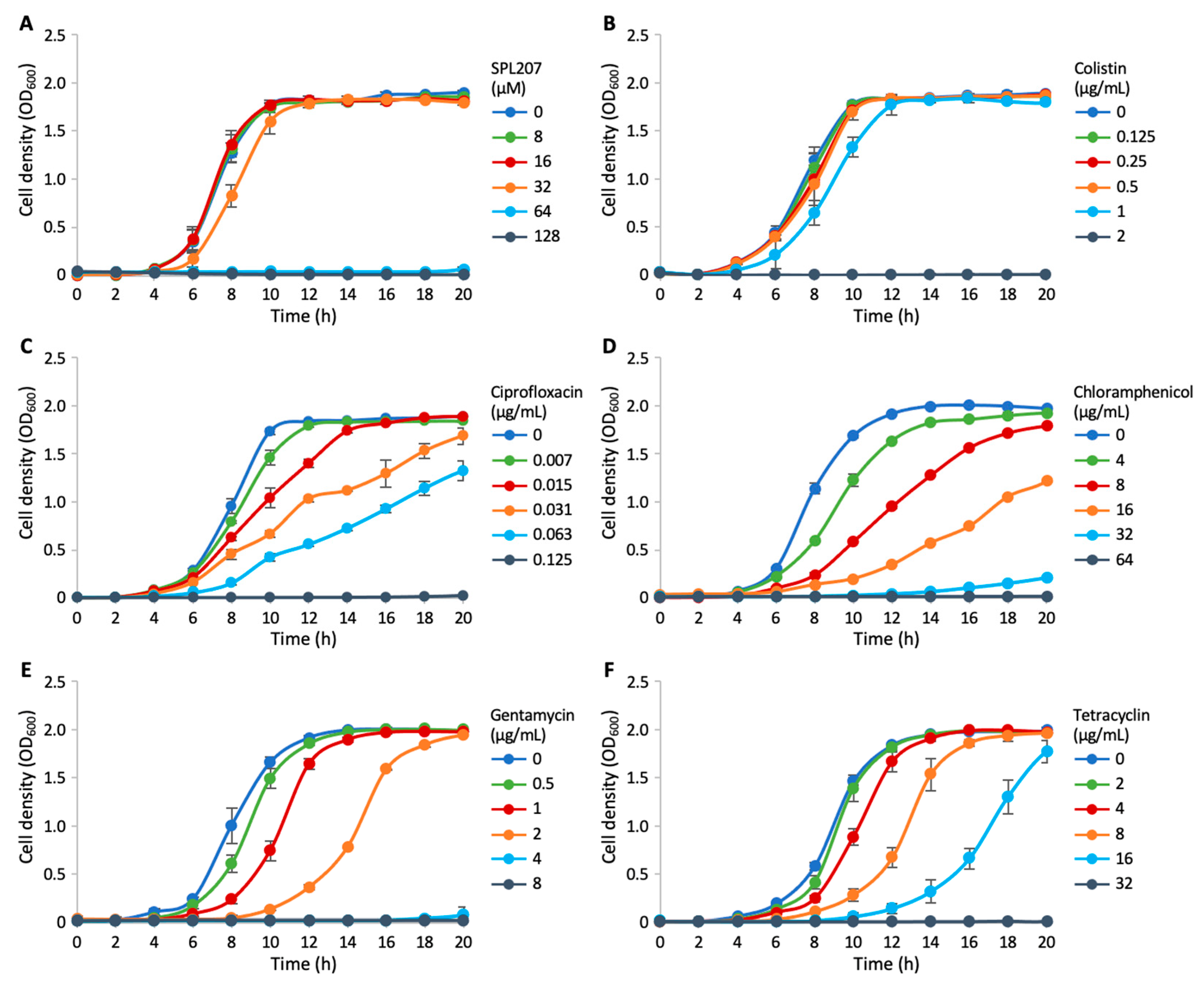
These findings indicate that SPL207 may exert its antibacterial effect via a mechanism akin to that of colistin, involving interaction with the OM and consequent membrane disruption [31]. Interestingly, similar to colistin and in contrast to antibiotics with different mechanisms of action (e.g., ciprofloxacin, chloramphenicol, gentamycin, and tetracycline), SPL207 has no effect on the bacterial growth kinetics when at sub-MICs (Figure 5). The results of the permeability assays and the all-or-nothing effect shared by SPL207 and colistin on P. aeruginosa growth support the mechanism of action of SPL207 as a membrane perturbating agent.
Based on the similarities in the mechanisms of action of SPL207 and colistin, the potential interaction between these two antimicrobials was evaluated. Checkerboard assays revealed that colistin and SPL207 have a synergistic antimicrobial activity against P. aeruginosa, with a Fractional Inhibitory Concentration Index (FICI) = 0.5 (Table 5 and Figure S2; FICI ≤ 0.5 indicates synergy between antimicrobials) [32]. Conversely, checkerboard assays showed no interaction, either positive or negative, between SPL207 and other two anti-P. aeruginosa antibiotics of clinical relevance such as ciprofloxacin or tobramycin. The effect of the colistin-SPL207 combination was investigated also in P. aeruginosa colistin-resistant strains obtained by in vitro evolution experiments [33]. As reported in Table 5 and Figure S2, the MIC of colistin in these strains was higher than 4 μg/mL (corresponding to the colistin breakpoint in P. aeruginosa according to the EUCAST guidelines; i.e., colistin MIC ≤ 4 μg/mL, susceptible strain; colistin MIC > 4 μg/mL, resistant strain) [34]. The results demonstrated strong synergistic activity between colistin and SPL207 against the three colistin-resistant strains tested (FICI ranging from 0.047 to 0.094; Table 5 and Figure S2). It is important to note that, in the presence of SPL207 at concentrations of 8 μM (for PAO1 colR1), 2 μM (for PAO1 colR3), and 4 μM (for PAO1 colR5), the MIC of colistin against the tested strains decreased below the breakpoint [34].
Overall, SPL207 possesses intrinsic antimicrobial activity and acts as a resistance breaker by resensitizing colistin-resistant P. aeruginosa strains to this last-resort antibiotic.

2.2.3. Small Molecule/Membrane Interaction Analysis by MD Simulations

To provide atomistic details of the interaction between the most promising representative SPL207 and the P. aeruginosa OM, extended molecular dynamics (MD) were carried out by adapting a computational protocol validated previously [35].
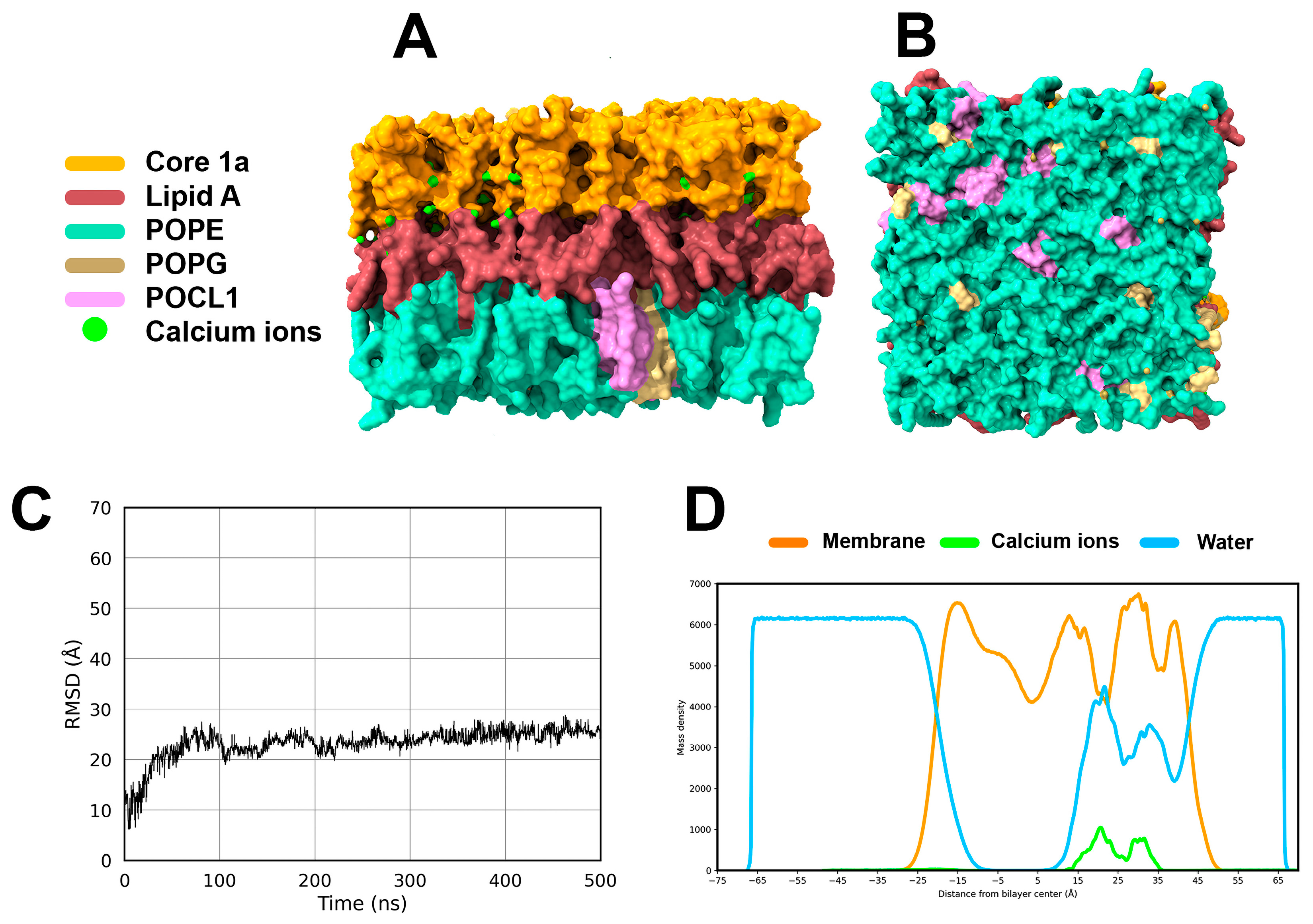
The P. aeruginosa OM computational model was validated by 500 ns of all-atom MD simulations, analyzed through frames clustering, mass density distribution along the z-axis, and root mean square deviation (RMSD) (Figure 6). While the representative MD frame (i.e., the centroid frame of the most populated cluster) shows a homogeneous distribution of POPE, POPG, POCL1 phospholipids in the inner leaflet (Figure 6A,B), the RMSD plot indicates that the membrane model is conformationally stable in the MD simulation time (Figure 6C). Visual inspection of the mass density plot (Figure 6D) highlights the peculiar density of Ca2+ ions in the PA-LPS region, supporting their crucial role in stabilizing the OM of Gram-negative bacteria through electrostatic interactions with anionic components of the LPS [36,37,38]. Overall, this analysis suggests that the P. aeruginosa OM model is coherent with biological data, and it is suitable for ligand penetration studies in silico.
To evaluate the interaction of SPL207 and its parent compound adarotene (1) into the P. aeruginosa OM model, 500 ns of all-atom MD simulations were performed. The pKa analysis of amino groups in SPL207 carried out with the MoKa software version 4.0.12 [39] at pH = 7.4 (Figure 7) led to the selection of the protonated form of the molecule with total formal charge = +2 for this in silico study.
Cluster analysis of MD frames shows that, in the most populated cluster, SPL207 penetrates deeply into the OM (Figure 8A) compared to 1, this latter remaining locked within the PA-LPS region (Figure 8B). This behavior is also confirmed by the mass density plot, showing that 1 is unable to overcome the first phosphate layer of core 1a (orange dashed lines in Figure 8C), while SPL207 successfully reaches the central part of the OM (Figure 8D). In evaluating atomistic details of the binding mode of 1 and SPL207, significant differences were observed. The carboxylic acid group of 1 (Figure 8E) establishes water-bridged H-bond interactions with the phosphate groups within the PA-LPS. The adamantyl moiety and the phenolic –OH group are projected towards the outer space and do not establish significant interactions with the P. aeruginosa OM model. In contrast, the positively charged amino groups of SPL207 (Figure 8F) establish direct and water-bridged H-bonds interactions with negatively charged phosphate groups of the lipid A. This suggests that positively charged groups in SPL207 may play a crucial role in OM embedding. Finally, the adamantyl moiety of SPL207 is positioned towards the hydrophobic tails of POPE, POPG and POCL1 of the P. aeruginosa OM model.

2.2.4. Displacement of Ca2+ Ions and Membrane Destabilization by SPL207

Based on the stabilizing role of Ca2+ ions in bacterial OMs, [34,35,36] it is expected that small molecules acting as membrane destabilizers can displace Ca2+ ions from the LPS. To assess this ability by 1 and SPL207, the mass density of Ca2+ ions within 20 Å of each molecule was monitored along MD trajectories (Figure 9). The mass density peak (MDP) of 1 fails to affect the mass density profile of Ca2+ ions, such as underlined by the comparison with the Ca2+ ions density in the free membrane (Figure 9A). In contrast, SPL207 modifies the mass density profile of Ca2+ ions with respect to the free membrane (Figure 9B) suggesting that SPL207 might displace Ca2+ ions more effectively than 1. To further confirm these results, the mean squared displacement analysis of Ca2+ ions was carried out [40,41,42]. Results in Table 6 clearly show that SPL207 imprints a significant Ca2+ ions displacement in the three axes compared to the baseline displacement in the free PA OM model, which is greater than that observed for 1.
Finally, the membrane z thickness and lipid diffusion analysis were calculated along MD trajectories as descriptors of the physical change that occur in the structure of the PA-OM. Membrane z thickness represents the distances in the z axis between the outer and the inner leaflets of the PA OM model, and its comparison in Figure 10A clearly shows that the free OM and the OM/1 system are highly superimposable, whereas the presence of SPL207 induces a greater movement of phospholipids along the z axis, further confirming SPL207’s ability to destabilize the PA OM in silico.
The analysis of lateral lipid diffusion further allows us to describe the movement of lipids along the xy axis over the MD trajectory. The results show that SPL207 triggers a larger movement of lipids than 1. Moreover, unlike compound 1, SPL207 induces a marked increase in lipid diffusion from the very beginning of the MD production trajectory.
Overall, MD results clearly point to the potential mechanism of action of SPL207 as a membrane destabilizer through improved diffusion of Ca2+ ions and lipids destabilization.

3. Materials and Methods

3.1. Chemistry

All reagents and solvents were reagent grade or were purified by standard methods before use. 1H and 13C NMR spectra were recorded on Bruker AV600 (Billerica, MA, USA) spectrophotometer at 600 and 150 MHz, respectively, or Bruker Neo 400 spectrophotometer at 400 and 100 MHz, respectively. Chemical shifts are reported in ppm (δ) relative to TMS. The coupling constants, J are reported in Hertz (Hz). All compounds were routinely checked by thin layer chromatography (TLC) using precoated silica gel 60 F254, aluminum foil and the spots were detected under UV light at 254 nm and 365 nm or were revealed spraying with 10% phosphomolybdic acid (PMA) in ethanol.
Compounds 3, 5, 6, 39 [18], 4 [13], 1, 29a-b, 34 [17], 30 [43], 31 [44], 32 [45], 42c [46] were prepared according to reported procedures.
3-((3r,5r,7r)-adamantan-1-yl)-4′-((E)-3-aminoprop-1-en-1-yl)-[1,1′-biphenyl]-4-ol (7). To a solution of AlCl3 (49 mg, 0.37 mmol) in dry THF (1.25 mL) 1 M LiAlH4 in THF (0.56 mL) was added at 0 °C and the solution was stirred 10 min, then compound 34 [17] (100 mg, 0.28 mmol) dissolved in THF (1.25 mL) was added at 0 °C. The mixture was stirred for 1 h at 0 °C, then quenched with water and 1M NaOH and extracted with AcOEt. The organic phase was dried with Na2SO4 and concentrated under reduced pressure. The crude was purified by preparative TLC (CH2Cl2: MeOH = 9:1) to afford product in 28% yield (28 mg) as a white solid. Rf = 0.39 (CH2Cl2: MeOH = 9:1). 1H-NMR (400 MHz, DMSO-d6) δ: 7.56–7.49 (2H, m); 7.47–7.39 (2H, m); 7.37–7.27 (2H, m); 6.86 (1H, d, J = 8.3 Hz); 6.54 (1H, d, J = 16.0 Hz); 6.35 (1H, dt, J = 16.0 Hz, J = 5.7 Hz); 3.37 (2H, d, J = 5.7 Hz); 2.22–2.10 (6H, m); 2.09–2.01 (3H, m); 1.82–1.67 (6H, m). 13C-NMR (100 MHz, DMSO-d6) δ: 156.4; 140.1; 136.3; 135.4; 131.2; 130.7; 129.0; 127.0 (×2C); 126.5 (×2C); 125.0; 124.9; 117.3; 43.8; 40.3 (×3C); 37.1 (×3C); 36.7; 28.9 (×3C).
3-((3r,5r,7r)-adamantan-1-yl)-4′-(3-aminopropyl)-[1,1′-biphenyl]-4-ol (8). To a solution of compound 34 (128 mg, 0.359 mmol) in dry THF (2.5 mL) 1 M LiAlH4 in THF (0.05 mL) was added at 0 °C. The mixture was stirred for 2 h at 0 °C, then quenched with water and 1 M NaOH and extracted with AcOEt. The organic phase was dried with Na2SO4 and concentrated under reduced pressure. The crude was purified by column chromatography (CH2Cl2: MeOH = 95:5 with 1% of conc. NH3 aqueous solution) to afford product 8 in 15% yield (20 mg) as a white solid. Rf = 0.39 (CH2Cl2: MeOH = 9:1). 1H-NMR (400 MHz, CH3OH-d4) δ: 7.48–7.43 (2H, m); 7.36 (1H, d, J = 2.3 Hz); 7.27–7.21 (3H, m); 6.78 (1H, d, J = 8.2 Hz); 2.83–2.78 (2H, m); 2.72 (2H, t, J = 7.2 Hz); 2.27–2.20 (6H, m); 2.12–2.05 (3H, m); 1.95–1.79 (8H, m). 13C-NMR (100 MHz, CH3OH-d4) δ: 157.1; 140.5; 137.7; 132.2; 129.7 (×2C); 127.5 (×2C); 126.1; 125.8; 117.6; 41.6 (×3C); 41.4; 38.3 (×3C); 38.0; 33.5; 30.66 (×3C).
3-((3r,5r,7r)-adamantan-1-yl)-4′-((4-methylpiperazin-1-yl)methyl)-[1,1′-biphenyl]-4-ol (9). To a suspension of aldehyde 29b [17] (200 mg, 0.448 mmol) in CH3CN (1 mL) 1-methylpiperazine (49 mg, 0.49 mmol), formic acid (41 mg, 0.90 mmol) and BF3Et2O (0.002 mmol) were added and the mixture was heated at 85 °C for 6 h. The reaction was concentrated and treated with Et2O to obtain a precipitate. Compound 9 was obtained in 38% yield (70 mg) as a white solid upon chromatographic purification (CH2Cl2: MeOH = 20: 1 with 1% of conc. NH3). 1H-NMR (400 MHz, DMSO-d6) δ: 9.42 (1H, s); 7.52–7.45 (2H, m); 7.35–7.23 (4H, m); 6.84 (1H, d, J = 8.1 Hz); 3.46 (2H, s); 2.48–2.20 (8H, m); 2.18–2.10 (9H, m); 2.07–2.00 (3H, m); 1.82–1.70 (6H, m). 13C-NMR (100 MHz, DMSO-d6) δ: 155.8; 139.6; 136.2; 135.8; 130.6; 129.3 (×2C); 125.8 (×2C); 124.7 (×2C); 116.8; 61.8; 54.7 (×2C); 52.6 (×2C); 45.8; 39.9 (×3C); 36.6 (×3C); 36.3; 28.4 (×3C).
(E)-3′-((3r,5r,7r)-adamantan-1-yl)-4′-hydroxy-[1,1′-biphenyl]-4-carbaldehyde O-(2-morpholinoethyl) oxime (10). To a solution of compound 29a [17] (20 mg, 0.06 mmol) in EtOH (0.5 mL) O-(2-morpholinoethyl)hydroxylamine hydrochloride (22 mg, 0.12 mmol) and pyridine (95 mg, 1.2 mmol) were added and the resulting mixture was refluxed for 5 h. The reaction was concentrated and treated with H2O to obtain product 10 as a white precipitate in 67% yield (18 mg) without further purification. Rf = 0.14 (TLC hexane: acetone = 7:3). 1H-NMR (400 MHz, DMSO-d6) δ: 9.54 (1H, s); 8.26 (1H, s); 7.68–7.57 (4H, m); 7.39–7.30 (2H, m); 6.87 (1H, d, J = 8.0 Hz); 4.24 (2H, t, J = 5.8 Hz); 3.63–3.51 (4H, m); 2.63 (2H, t, J = 5.8 Hz); 2.48–2.40 (4H, m); 2.20–2.10 (6H, m); 2.09–2.00 (3H, m); 1.83–1.67 (6H, m). 13C-NMR (100 MHz, DMSO-d6) δ: 156.3; 148.5; 142.3; 135.9;129.8; 129.8; 127.3 (×2C); 126.2 (×2C); 124.9; 124.7; 116.9; 71.3; 66.2 (×2C); 57.0; 53.6 (×2C); 39.8 (×3C); 36.6 (×3C); 36.3; 28.4 (×3C).
(E)-3-(3′-((3r,5r,7r)-adamantan-1-yl)-4′-hydroxy-[1,1′-biphenyl]-4-yl)-N-(2-morpholinoethoxy)acrylamide (11). To a solution of compound 1 (adarotene) [17] (50 mg, 0.13 mmol) in DMF (2 mL) cooled to 0 °C DIPEA (52 mg, 0.40 mmol) and HBTU (51 mg, 0.13 mmol) were added and the resulting mixture was stirred for 10 min. After addition of O-(2-morpholinoethyl)hydroxylamine hydrochloride (24 mg, 0.13 mmol) the reaction was stirred for 48 h at room temperature and then concentrated and treated with H2O to obtain a precipitate. Product 11 was obtained in 24% yield (16 mg) as a sticky solid upon chromatographic purification (DCM: MeOH = 9:1). Rf = 0.26 (DCM: MeOH = 9:1). 1H-NMR (400 MHz, CH3OH-d4) δ: 7.65 (1H, d, J = 15.8 Hz); 7.61–7.56 (4H, m); 7.42 (1H, d, J = 2.3 Hz); 7.31 (1H, dd, J = 2.3 Hz, 8.3 Hz); 6.82 (1H, d, J = 8.3 Hz); 6.46 (1H, d, J = 15.8 Hz); 4.13 (2H, t, J = 5.2 Hz); 3.78–3.71 (4H, m); 2.76 (2H, t, J = 5.2 Hz); 2.68–2.57 (4H, m); 2.27–2.18 (6H, m); 2.12–2.03 (3H, m); 1.92–1.80 (6H, m). 13C-NMR (100 MHz, CH3OH-d4) δ: 166.4; 157.8; 145.0; 142.5; 137.9; 133.7; 132.2; 129.4 (×2C); 127.8 (×2C); 126.2; 126.1; 117.8; 117.1; 74.4; 67.6 (×2C); 57.5; 54.8 (×2C); 41.5 (×3C); 38.3 (×3C); 38.0; 30.6 (×3C).
(E)-3′-((3r,5r,7r)-adamantan-1-yl)-4′-hydroxy-[1,1′-biphenyl]-4-carbaldehyde O-(2-aminoethyl) oxime (12). To a solution of compound 29a [17] (30 mg, 0.09 mmol) in EtOH, (0.5 mL) 2-(aminooxy)ethan-1-amine hydrochloride (27 mg, 0.18 mmol) and pyridine (142 mg, 1.80 mmol) were added and the resulting mixture was refluxed for 5 h. The reaction was concentrated under reduced pressure and the crude was finely shredded and washed with H2O to afford product 12 in 51% yield (18 mg) as a white solid without further purification. Rf = 0.16 (reverse phase TLC MeOH: H2O = 9:1). 1H-NMR (600 MHz, CH3OH-d4) δ: 8.27 (1H, s); 7.22–7.66 (2H, m); 7.64–7.58 (2H, m); 7.43 (1H, d, J = 1.8 Hz); 7.32 (1H, dd, J = 1.8Hz, 8.4 Hz); 6.82 (1H, d, J = 8.4 Hz); 4.39 (2H, t, J = 4.9 Hz); signal overlapped with the solvent, 2.28–2.22 (6H, m); 2.12–2.07 (3H, m); 1.90–1.82 (6H, m). 13C-NMR (150 MHz, CH3OH-d4) δ: 157.9; 151.9; 145.3; 137.9; 132.2; 130.8; 128.7 (×2C); 127.6 (×2C); 126.2; 126.1; 117.8; 70.9; 41.5 (×3C); 40.3; 38.3 (×3C); 38.0; 30.6 (×3C).
(E)-3-((3r,5r,7r)-adamantan-1-yl)-4-hydroxybenzaldehyde O-(2-aminoethyl) oxime (13). To a solution of compound 30 [43] (20 mg, 0.078 mmol) in EtOH, (0.5 mL) 2-(aminooxy)ethan-1-amine hydrochloride (23 mg, 0.156 mmol) and pyridine (123 mg, 1.56 mmol) were added and the resulting mixture was refluxed for 3 h. The reaction was concentrated, diluted with ethyl acetate, washed with H2O, dried with Na2SO4 and again concentrated under reduced pressure. Product 13 was obtained in 37% yield (9 mg) as a sticky solid without further purification. Rf = 0.34 (reverse phase TLC MeOH: H2O = 9:1). 1H-NMR (400 MHz, CH3OH-d4) δ: 8.09 (1H, s); 7.43 (1H, d, J = 2.0 Hz); 7.25 (1H, dd, J = 2.0 Hz, 8.3 Hz); 6.74 (1H, d, J = 8.3 Hz); 4.19 (2H, t, J = 5.2 Hz); 3.03 (2H, t, J = 5.2 Hz); 2.20–2.13 (6H, m); 2.09–2.03 (3H, m); 1.86–1.79 (6H, m). 13C-NMR (100 MHz, CH3OH-d4) δ: 159.8; 151.5: 137.9; 127.1; 126.8; 124.3; 117.5; 74.5; 41.4; 41.4 (×3C); 38.2 (×3C); 37.9; 30.5 (×3C).
3-((3r,5r,7r)-adamantan-1-yl)-4′-(((2-hydroxyethyl)amino)methyl)-[1,1′-biphenyl]-4-ol (14). Ethanolamine (5 mg, 0.09 mmol) was added dropwise to a solution of compound 29a [17] (30 mg, 0.09 mmol) in MeOH (1.5 mL) at 0 °C and the resulting mixture was stirred for 18 h at room temperature. NaBH4 (7 mg, 0.18 mmol) was then gradually added at 0 °C and after stirring for 30 min the reaction was diluted with 1 M HCl. The aqueous phase was washed with CH2Cl2 in order to remove organic impurities, then added with 1 M NaOH and extracted with ethyl acetate. The organic phase was dried over anhydrous Na2SO4 and concentrated under reduced pressure to afford product 14 in 97% yield (32 mg) as a sticky yellow solid without further purification. Rf = 0.32 (reverse phase TLC MeOH: H2O = 9:1). 1H-NMR (600 MHz, CH3OH-d4) δ: 7.54–7.49 (2H, m); 7.41–7.35 (3H, m); 7.26 (1H, dd, J = 2.1 Hz, 8.3 Hz); 6.80 (1H, d, J = 8.3 Hz); 3.83 (2H, s); 3.71 (2H, J = 5.5 Hz); 2.77 (2H, t, J = 8.3 Hz); 2.31–2.17 (6H, m); 2.13–2.05 (3H, m); 1.90–1.79 (6H, m). 13C-NMR (150 MHz, CH3OH-d4) δ: 157.3; 142.3; 138.1; 137.7; 133.0; 129.9 (×2C); 127.5 (×2C); 126.2; 125.9; 117.7; 61.4; 54.0; 51.6; 41.6 (×2C); 38.3 (×3C); 38.0; 30.6 (×3C).
3-((3r,5r,7r)-adamantan-1-yl)-4′-(((2-(dimethylamino)ethyl)amino)methyl)-[1,1′-biphenyl]-4-ol (15). N1,N1-dimethylethane-1,2-diamine (11 mg, 0.12 mmol) was added dropwise to a solution of compound 29a [17] (40 mg, 0.12 mmol) in MeOH (1.3 mL) at 0 °C and the resulting mixture was stirred for 18 h at room temperature. NaBH3CN (15 mg, 0.24 mmol) was then gradually added at 0 °C and after stirring for 40 min the reaction was concentrated under reduced pressure. The residue was treated with H2O and the resulting precipitate was filtered, washed with H2O and dried to afford product 15 in 84% yield (41 mg) as a white solid without further purification. Rf = 0.14 (reverse phase TLC MeOH: H2O = 9:1). 1H-NMR (600 MHz, CH3OH-d4) δ: 8.42 (1H, s); 7.84–7.75 (2H, m); 7.69–7.61 (2H, m); 7.45 (1H, d, J = 2 Hz); 7.34 (1H, dd, J = 2 Hz, 8.2 Hz); 6.82 (1H, d, J = 8.2 Hz); 3.79 (2H, t, J = 6.7 Hz); 2.72 (2H, t, J = 6.7 Hz); 2.41–2.32 (3H, s); 2.31–2.20 (6H, m); 2.13–2.06 (3H, m); 1.92–1.80 (6H, m). 13C-NMR (150 MHz, CDCl3) δ: 157.9; 146.0; 137.8; 134.9; 132.2; 129.9 (×2C); 127.5 (×2C); 126.3; 126.2; 117.8; 60.7; 59.7; signal overlapped with the solvent; 45.8 (×2C); 41.5 (×3C); 38.3 (×3C); 38.0; 30.6 (×3C).
3-((3r,5r,7r)-adamantan-1-yl)-4′-(((2-(2-hydroxyethoxy)ethyl)amino)methyl)-[1,1′-biphenyl]-4-ol (16). 2-(2-aminoethoxy)ethan-1-ol (9 mg, 0.09 mmol) was added dropwise to a solution of compound 29a [17] (30 mg, 0.09 mmol) in MeOH (1.3 mL) at 0 °C and the resulting mixture was stirred for 18 h at room temperature. NaBH3CN (11 mg, 0.18 mmol) was then gradually added at 0 °C and after stirring for 30 min the reaction was concentrated under reduced pressure. The residue was treated with H2O and resulting precipitate was filtered, washed with H2O and dried to afford product 16 in 54% yield (24 mg) as a white solid without further purification. Rf = 0.40 (reverse phase TLC MeOH: H2O = 9:1). 1H-NMR (400 MHz, CH3OH-d4) δ: 7.65–7.55 (2H, m); 7.51–7.43 (2H, m); 7.39 (1H, d, J = 2.1 Hz); 7.28 (1H, dd, J = 2.1 Hz, J = 8.2 Hz); 6.81 (1H, d, J = 8.2 Hz); 4.09 (2H, s); 3.77–3.67 (4H, m); 3.63–3.55 (2H, m); 3.15–3.05 (2H, m). 13C-NMR (100 MHz, CH3OH-d4) δ: 157.6; 143.7; 137.8; 133.0; 132.4; 130.9 (×2C); 127.9 (×2C); 126.2; 126.1; 117.7; 73.4; 68.1; 62.1; 52.7.0; 48.5; 41.5 (×3C); 38.3 (×3C); 38.0; 30.6 (×3C).
3-((3r,5r,7r)-adamantan-1-yl)-4′-(((2-((2-aminoethyl)amino)ethyl)amino)methyl)-[1,1′-biphenyl]-4-ol (17). N1-(2-aminoethyl)ethane-1,2-diamine (24 mg, 0.23 mmol) was added dropwise to a solution of compound 29a [17] (26 mg, 0.07 mmol) in MeOH (1.3 mL) at 0 °C and the resulting mixture was stirred for 18h at room temperature. NaBH4 (8 mg, 0.23 mmol) was then gradually added at 0 °C and after stirring for 30 min the reaction was diluted with 1 M HCl. The aqueous phase was washed with CH2Cl2 to remove organic impurities and added with 1 M NaOH. The resulting precipitate was filtered, washed with H2O and dried. Product 17 was obtained in 49% yield (14 mg) as a white solid upon crystallization from Et2O. Rf = 0.08 (reverse phase TLC MeOH: H2O = 9:1). 1H-NMR (400 MHz, CH3OH-d4) δ: 7.54–7.46 (2H, m); 7.41–7.32 (3H, m); 7.25 (1H, dd, J = 2.2 Hz, 8.2 Hz); 6.79 (1H, d, J = 8.2 Hz); 3.79 (2H, s); 2.81–2.71 (6H, m); 2.71–2.63 (2H, m); 2.27–2.17 (6H, m); 2.11–2.03 (3H, m); 1.89–1.77 (6H, m). 13C-NMR (100 MHz, CH3OH-d4) δ: 157.3; 142.3; 138.4; 137.7; 133.0; 129.9 (× 2C); 127.5 (× 2C); 126.1; 125.9; 117.7; 54.2; 54.0; 52.3; 49.5; 41.7 (× 3C); 38.3 (× 3C); 38.0; 30.6 (× 3C).
(E)-3-(3′-((3r,5r,7r)-adamantan-1-yl)-4′-hydroxy-[1,1′-biphenyl]-4-yl)-N,N-bis(3-(dimethylamino)propyl)acrylamide (18). To a solution of compound 1 (50 mg, 0.134 mmol) in dry CH2Cl2 (2 mL), DIPEA (163 mg, 1.3 mmol), N1-(3-(dimethylamino)propyl)-N3,N3-dimethylpropane-1,3-diamine (32 mg, 0.173 mmol) and BOP (77 mg, 0.173) were added and the resulting mixture was stirred for 48 h at room temperature. The reaction was diluted with CH2Cl2, washed with H2O, dried with Na2SO4 and concentrated under reduced pressure. The resulting precipitate was filtered, washed with H2O and dried. The crude was purified by column chromatography (CH2Cl2: MeOH = 93:7 with 1% of conc. NH3 aqueous solution) to afford product 18 in 54% yield (39 mg) as a sticky solid. Rf = 0.20 (CH2Cl2: MeOH = 95:5 with 1% of conc. NH3 aqueous solution). 1H-NMR (600 MHz, CH3OH-d4) δ: 7.70–7.65 (2H, m); 7.63–7.58 (3H, m); 7.44 (1H, d, J = 2.0 Hz); 7.32 (1H, dd, J = 2.0 Hz, J = 8.1 Hz) 7.15 (1H, d, J = 15.2 Hz); 6.82 (1H, d, J = 8.1 hz); 3.62 (2H, t, J = 7.7 Hz); 3.52 (2H, t, J = 7.2 Hz); 2.54 (2H, m); 2.47–2.42 82H, m); 2.41 (6H, s); 2.34–2.28 (6H, s); 2.27–2.22 (6H, m); 2.12–2.05 83H, m); 1.93–1.87 (4H, m); 1.86–1.83 (6H, m). 13C-NMR (600 MHz, CH3OH-d4) δ: 169.1; 157.8; 144.8; 144.1; 137.9; 134.2; 132.2; 129.6 (×2C); 127.7 (×2C); 126.2; 126.0; 117.8; 117.6; 57.7; 57.2; 47.2; 45.8; 45.5 (×2C); 45.1 (×2C); 41.5 (×3C); 38.3 (×3C); 38.0; 30.6 (×3C); 28.2; 26.2.
tert-butyl (E)-3-(3′-((3r,5r,7r)-adamantan-1-yl)-4′-(4-(bis(3-(dimethylamino)propyl)amino)butoxy)-[1,1′-biphenyl]-4-yl)acrylate (20). To a solution of compound 1 (140 mg, 0.32 mmol) in acetone (5 mL), K2CO3 (225 mg, 1.62 mmol) and 1,4-dibromobuthane (526 mg, 2.43 mmol) were added and the resulting mixture was heated to reflux for 6 h. The reaction was concentrated, diluted with ethyl acetate, washed with H2O, dried over anhydrous Na2SO4 and concentrated again under reduced pressure. Product 33 was obtained in 66% yield (122 mg) as a white solid upon crystallization from hexane. Rf = 0.75 (Hex: AcOEt = 8:2). 1H-NMR (600 MHz, CDCl3) δ: 7.64 (1H, d, J = 16.0 Hz); 7.62–7.55 (4H, m); 7.51 (1H, d, J = 2.1 Hz); 7.43 (1H, dd, J = 2.1 Hz, J = 8.6 Hz); 6.96 ( 1H, d, J = 8.6 Hz); 6.41 (1H, d, J = 16.2 Hz); 4.10 (2H, t, J = 6.2 Hz); 3.56 (2H, t, J = 6.6 Hz); 2.24–2.17 (8H, m); 2.16–2.07 (5H, m); 1.86–1.787 (6H, m); 1.58 (9H, s).
To a solution of the above intermediate 33 (50 mg, 0.09 mmol) in dry DMF (2.5 mL), N1-(3-(dimethylamino)propyl)-N3,N3-dimethylpropane-1,3-diamine (165 mg, 0.90 mmol) was added and the mixture was stirred for 24h. The reaction was diluted with ethyl acetate, washed with H2O, dried with Na2SO4 and concentrated under reduced pressure. Impurities were removed from the crude by dissolving them in Et2O to obtain product 20 in 47% yield (25 mg) as a sticky solid. Rf = 0.32 (reverse phase TLC MeOH: H2O = 9:1). 1H-NMR (600 MHz, CDCl3) δ: 7.68–7.58 (5H, m); 7.50 (1H, d; J = 2.2 Hz); 7.47 (1H, dd, J = 2.2 Hz, J = 8.2 Hz); 7.06 (1H, d, J = 8.2 Hz); 6.45 (1H, d, J = 16.0 Hz); 4.16 (2H, t, J = 5.9 Hz); 3.52–3.43 (4H, m); 3.13 (6H, s); 2.72–2.60 (4H, m); 2.49–2.40 (2H, m); 2.30 (6H, m); 2.25–2.19 (6H, m); 2.16–2.05 (5H, m); 2.04–1.98 (4H, m); 1.90–1.79 (6H, m); 1.72–1.64 (2H, m); 1.54 (9H, s). 13C-NMR (150 MHz, CDCl3) δ: 168.2; 159.2; 144.8; 142.6; 139.6; 134.1; 133.7; 129.7 (×2C); 127.9 (×2C); 126.4; 126.3; 120.5; 113.9; 81.7; 68.1; 65.2; 63.7; 58.5; 51.3; 47.1; 45.3 (×4C); 42.0 (×3C); 38.3 (×4C); 30.6 (×3C); 28.5 (×3C); 27.9; 27.5; 23.7; 21.0.
(E)-3-(3′-((3r,5r,7r)-adamantan-1-yl)-4′-(4-(bis(3-(dimethylamino)propyl)amino)butoxy)-[1,1′-biphenyl]-4-yl)acrylic acid (19). To a solution of compound 20 (20 mg, 0.03 mmol) in dry CH2Cl2 (0.47 mL), trifluoroacetic acid (0.21 mL) was added at 0 °C and the resulting mixture was stirred at 0 °C for 2 h. The reaction was concentrated under reduced pressure. After the azeotropic removal of TFA with toluene, product 19 was obtained in quantitative yield (28 mg) as a sticky solid. Rf = 0.57 (reverse phase TLC MeOH: H2O = 9:1). 1H-NMR (600 MHz, CH3OH-d4) δ: 7.73 (1H, d, J = 16.2 Hz); 7.70–7.61 (4H, m); 7.52 (1H, d, J = 2.1 Hz); 7.49 (1H, dd, J = 2.1 Hz, 8.6 Hz); 7.08 (1H, d, J = 8.6 Hz); 6.52 (1H, d, J = 16.2 Hz); 4.18 (2H, t, J = 6.1 Hz); 3.57–3.49 (4H, m); 3.31–3.26 (2H, m); 3.24–3.15 (10H, m); 2.99–2.92 (6H, m); 2.34–2.28 (2H, m); 2.27–2.18 (8H, m); 2.16–2.08 (5H, m); 2.07–1.99 (2H, m); 1.91–1.84 (6H, m). 13C-NMR (150 MHz, CH3OH-d4) δ: 170.6; 162.6; 146.1; 144.8; 139.8; 134.2; 133.8; 129.9 (×2C); 128.1 (×2C); 126.6; 126.4; 118.8; 114.0; 68.2; 65.8; 62.3; 55.7; 51.4; 49.5; 46.0; 45.8; 43.7 (×3C); 42.1 (×3C); 38.4 (×4C); 30.5 (×3C); 22.8; 21.1; 21.0.
Methyl 3′-((3r,5r,7r)-adamantan-1-yl)-4′-(4-(bis(3-(dimethylamino)propyl)amino)butoxy)-[1,1′-biphenyl]-4-carboxylate (21). To a 0 °C cooled solution of methyl 4′-hydroxy(1,1′-biphenyl)-4-carboxylate 37 (100 mg, 0.438 mmol) and 1-adamantanol (73 mg, 0.48 mmol) in CH2Cl2 (3.5 mL), H2SO4 (0.05 mL) was added and the resulting mixture was stirred for 2 h. The reaction was diluted with cold H2O, extracted with CH2Cl2, dried over anhydrous Na2SO4 and concentrated under reduced pressure. The crude was purified by column chromatography (Hex: AcOEt = 8:2) to afford product 38 in 42% yield (67 mg). Rf = 0.30 (Hex: AcOEt = 9:1). 1H-NMR (600 MHz, CDCl3) δ: 8.14–8.06 (2H, m); 7.68–7.61 (2H, m); 7.52 (1H, d, J = 2.2 Hz); 7.37 (1H, dd, J = 2.2 Hz, 8.2 Hz); 6.77 (1H, d, J = 8.2 Hz); 4.98 (1H, s); 3.97 (3H, s); 2.25–2.20 (6H, m); 2.18–2.11 (3H, m); 1.86–1.81 (6H, m).
To a solution of above intermediate 38 (33 mg, 0.09 mmol) in acetone (1.5 mL), K2CO3 (63 mg, 0.46 mmol) and 1,4-dibromobuthane (136 mg, 0.63 mmol) were added and the resulting mixture was refluxed for 4 h. The reaction was concentrated, diluted with ethyl acetate, washed with H2O, dried with Na2SO4 and concentrated again under reduced pressure. Product 41a was obtained in 53% yield (23 mg) as a white solid upon crystallization from hexane. Rf = 0.36 (Hex: AcOEt = 9:1). 1H-NMR (600 MHz, CDCl3) δ: 8.14–8.05 (2H, m); 7.70–7.60 (2H, m); 7.53 (1H, d, J = 2.2 Hz); 7.46 (1H, dd, J = 2.2 Hz, 8.4 Hz); 6.97 (1H, d, J = 8.4 Hz); 4.11 (2H, t, J = 6.0 Hz); 3.96 (3H, s); 3.56 (2H, t, J = 6.4 Hz); 2.26–2.17 (8H, m); 2.16–2.05 (5H, m); 1.87–1.78 (6H, m).
To a solution of compound 41a (60 mg, 0.12 mmol) in THF (2 mL), N1-(3-(dimethylamino)propyl)-N3,N3-dimethylpropane-1,3-diamine (0.27 mL, 0.4 mmol) was added and the mixture was refluxed for 6 h. The reaction was diluted with ethyl acetate, washed with H2O, dried with Na2SO4 and concentrated under reduced pressure. Product 21 was obtained in 39% yield (4 mg) as a sticky solid upon chromatographic purification (CH2Cl2: MeOH: toluene = 4.5: 1.5: 4 with 1% of conc. NH3). Rf = 0.23 (DCM: MeOH: toluene = 4.5: 1.5: 4 with 1% of conc. NH3). 1H-NMR (400 MHz, CDCl3) δ: 8.13–8.03 (2H, m); 7.68–7.62 (2H, m); 7.52 (1H, d, J = 2.0 Hz); 7.45 (1H, dd, J = 2.0 Hz, 8.3 Hz); 6.95 (1H, d, J = 8.3 Hz); 4.06 (2H, t, J = 6.1 Hz); 3.94 (3H, s); 2.60–2.46 (6H,.m); 2.41–2.33 (2H, m); 2.29 (12H, s); 2.23–2.19 (6H, m); 2.16–2.10 (3H, m); 1.97–1.85 (2H, m); 1.83–1.77 (6H, m); 1.76–1.62 (6H, m). 13C-NMR (100 MHz, CDCl3) δ: 167.3; 158.5; 146.1; 138.7; 131.8; 130.1 (×2C); 128.1; 126.6 (×2C); 125.9; 125.7; 112.4; 67.9; 57.9 (×2C); 53.9; 52.1; 52.0 (×2C); 45.4 (× 4C); 40.7 (×3C); 37.3 (×3C); 29.8; 29.2 (×3C); 27.6; 25.1 (×2C); 24.0.
N1-(4-((3-(adamantan-1-yl)-4′-chloro-[1,1′-biphenyl]-4-yl)oxy)butyl)-N1-(3-(dimethylamino)propyl)-N3,N3-dimethylpropane-1,3-diamine (22). To a solution of compound 39 [18] (463 mg, 1.30 mmol) in dry acetone (12 mL), K2CO3 (898 mg, 6.50 mmol) and 1,4-dibromobuthane (1.10 mL, 9.15 mmol) were added and the resulting mixture was refluxed for 4 h. The reaction was concentrated, diluted with ethyl acetate, washed with H2O, dried over anhydrous Na2SO4. In vacuo concentration followed by flash column chromatography in Hex: AcOEt 9: 1 furnished the intermediate 2-(3-((3r,5r,7r)-adamantan-1-yl)-4-(4-bromobutoxy)phenyl)-4,4,5,5-tetramethyl-1,3,2-dioxaborolane (40) in 76% yield (484 mg). 1H-NMR (600 MHz, CDCl3) δ: 7.32 (1H, d, J = 2.4 Hz); 7.27 (1H, dd, J = 2.4 Hz, 8.9 Hz); 6.74 (1H, d, J = 8.9 Hz); 4.01 (2H, t, J = 5.9 Hz); 3.54 (2H, t, J = 6.6 Hz); 2.23–2.15 (2H, m); 2.14–2.02 (11H, m); 1.85–1.72 (6H, m).
To a suspension of intermediate (40, 100 mg, 0.20 mmol) in THF: H2O 2: 1 (2.4 mL), 1-bromo-4-chlorobenzene (38 mg, 0.20 mmol), K2CO3 (69 mg, 0.50 mmol) and Pd(PPh3)4 (7 mg, 0.006 mmol) were sequentially added under N2 atmosphere, and the mixture was heated 4 h at 80 °C. The reaction was diluted with ethyl acetate, washed with H2O, dried over anhydrous Na2SO4 and concentrated under reduced pressure. The intermediate 41b was obtained in 39% yield (37 mg) as a white solid upon crystallization from hexane. 1H-NMR (600 MHz, CDCl3) δ: 7.53–7.47 (2H, m); 7.44 (1H, d, J = 2.0 Hz); 7.41–7.34 (3H, m); 6.94 (1H, d, J = 8.3 Hz); 4.09 (2H, t, J = 5.9 Hz); 3.55 (2H, t, J = 6.5 Hz); 2.26–2.15 (8H; m); 2.15–2.08 (5H, m); 1.86–1.75 (6H, m).
To a solution of compound 41b (37 mg, 0.09 mmol) in THF (0.8 mL), N1-(3-(dimethylamino)propyl)-N3,N3-dimethylpropane-1,3-diamine (174 µL, 0.78 mmol) was added and the mixture was refluxed for 4 h. The reaction was diluted with ethyl acetate, washed with H2O, dried with Na2SO4 and concentrated under reduced pressure. Product 22 was obtained in 24% yield (11 mg) as a sticky solid upon chromatographic purification (CH2Cl2: MeOH: toluene = 4.5: 1.5: 4 with 1% of conc. NH3). 1H-NMR (400 MHz, CDCl3) δ: 7.54–7.46 (2H, m); 7.43 (1H, d, J = 2.3 Hz); 7.40–7.33 (3H, m); 6.94 (1H, d, J = 8.7 Hz); 4.05 (2H, t, J = 5.9 Hz); 2.59–2.52 (2H, m); 2.52 −2.44 (4H, m); 2.25 (12H, s); 2.22–2.16 (6H, m); 2.15–2.07 (3H, m); 1.95–1.85 (2H, m); 1.84–1.78 (6H, m); 1.77–1.59 (6H, m). 13C-NMR (100 MHz, CDCl3) δ: 158.0; 140.2; 138.7; 132.5; 131.9; 128.8 (×2C); 128.1 (×2C); 125.6; 125.3; 112.3; 67.9; 58.1 (×2C); 54.0; 52.1 (×2C); 45.6 (×4C); 40.7 (×3C); 37.3 (×3C); 29.8; 29.2 (×3C); 27.7; 25.5 (×2C); 24.1.
N1-(4-((3-((3r,5r,7r)-adamantan-1-yl)-4′-methyl-[1,1′-biphenyl]-4-yl)oxy)butyl)-N1-(3-(dimethylamino)propyl)-N3,N3-dimethylpropane-1,3-diamine (24). To a suspension of intermediate (40) in THF: H2O 2: 1 (2.4 mL), 1-bromo-4-methylbenzene (34 mg, 0.20 mmol), K2CO3 (69 mg, 0.50 mmol) and Pd(PPh3)4 (7 mg, 0.006 mmol) were sequentially added under N2 atmosphere, and the mixture was heated 4 h at 80 °C. The reaction was diluted with ethyl acetate, washed with H2O, dried over anhydrous Na2SO4 and concentrated under reduced pressure. The intermediate (3r,5r,7r)-1-(4-(4-bromobutoxy)-4′-methyl-[1,1′-biphenyl]-3-yl)adamantane 41c was obtained in 47% yield (42 mg) as a white solid upon crystallization from hexane. 1H-NMR (400 MHz, CDCl3) δ: 7.51–7.44 (3H, m); 7.38 (1H, d, J = 2.3 Hz, 8.4 Hz); 7.27–7.21 (2H, m); 6.93 (1H, d, J = 8.4 Hz); 4.09 (2H, t, J = 6.0 Hz); 3.55 (2H, t, J = 6.5 Hz); 2.41 (3H, s); 2.23–2.16 (8H, m); 2.15–2.05 (5H, m); 1.88–1.77 (6H, m).
To a solution of compound 41c (42 mg, 0.09 mmol) in THF (0.8 mL) N1-(3-(dimethylamino)propyl)-N3,N3-dimethylpropane-1,3-diamine (206 µL, 0.92 mmol) was added and the mixture was refluxed for 4 h. The reaction was diluted with ethyl acetate, washed with H2O, dried with Na2SO4 and concentrated under reduced pressure. Product 24 was obtained in 25% yield (13 mg) as a sticky solid upon chromatographic purification (CH2Cl2: MeOH: toluene = 4.5: 1.5: 4 with 1% of conc. NH3). 1H-NMR (600 MHz, CDCl3) δ: 7.48–7.41 (3H, m); 7.38–7.33 (1H, m); 7.24–7.18 (2H, m); 6.91 (1H, d, J = 8.5 Hz); 4.02 (2H, t, J = 6.2 Hz); 2.55–2.49 (2H, m); 2.49–2.42 (4H, m); 2.38 (3H, s); 2.31–2.25 (4H, m); 2.22 (12H, s); 2.20–2.14 (6H, m); 2.12–2.04 (3H, m); 1.90–1.83 (2H, m); 1.82–1.75 (6H, m); 1.74–1.56 (6H, m). 13C NMR (101 MHz, CDCl3) δ 157.51, 138.81, 138.32, 136.03, 133.09, 129.34 (×2C), 126.70 (×2C), 125.53, 125.05, 112.17, 67.78, 57.98 (×2C), 53.93, 52.06 (×2C), 45.51 (×4C), 40.66 (×3C), 37.24 (×3C), 37.16, 29.17 (×3C), 27.65, 25.44 (×2C), 24.06, 21.06.
N1-(4-((3-((3r,5r,7r)-adamantan-1-yl)-4′-bromo-[1,1′-biphenyl]-4-yl)oxy)butyl)-N1-(3-(dimethylamino)propyl)-N3,N3-dimethylpropane-1,3-diamine (23). To a solution of 3-((3r,5r,7r)-adamantan-1-yl)-4′-bromo-[1,1′-biphenyl]-4-ol 31 [17,44] (100 mg, 0.26 mmol) in dry acetone (3 mL), K2CO3 (180 mg, 1.3 mmol) and 1,4-dibromobuthane (392 mg, 1.82 mmol) were added and the resulting mixture was refluxed for 6 h. The reaction was concentrated, diluted with ethyl acetate, washed with H2O, dried with Na2SO4 and concentrated again under reduced pressure. Product 35 was obtained in 65% yield (87 mg) as a white solid upon crystallization from hexane. Rf = 0.77 (Hex: AcOEt = 9:1). 1H-NMR (600 MHz, CDCl3) δ: 7.57–7.52 (2H, m); 7.48–7.42 (3H, m); 7.37 (1H, dd, J = 2.6 Hz, 8.7 Hz); 6.95 (1H, d, J = 8.7 Hz); 4.10 (2H, t, J = 6.2 Hz); 3.56 (2H, t, J = 6.5 Hz); 2.25–2.16 (8H, m); 2.15–2.04 (5H, m); 1.86–1.77 (6H, m).
To a solution of compound 35 (40 mg, 0.077 mmol) in dry DMF (1.5 mL), N1-(3-(dimethylamino)propyl)-N3,N3-dimethylpropane-1,3-diamine (144 mg, 0.77 mmol) was added and the mixture was stirred for 18 h. The reaction was diluted with ethyl acetate, washed with H2O, dried with Na2SO4 and concentrated under reduced pressure. Impurities were removed from the crude by dissolving them in Et2O: Hex= 1: 1 to obtain product 23 in 42% yield (20 mg) as a sticky solid. Rf = 0.30 (CH2Cl2: MeOH = 8:2 with 1% of conc. NH3 aqueous solution). 1H-NMR (600 MHz, CH3OH-d4) δ: 7.59–7.53 (2H, m); 7.51–7.47 (2H, m); 7.43 (1H, d, J = 2.3 Hz); 7.41 (1H, dd, J = 2.3 Hz, 8.4 Hz); 7.03 (1H, d, J = 8.4 Hz); 4.10 (2H, t, J = 6.1 Hz); 2.64 (2H, t, J = 7.2 Hz); 2.60–2.54 (4H, m); 2.49–2.43 (4H, m); 2.33 (12H, s); 2.28–2.20 (6H, m); 2.15–2.08 (3H, m); 1.97–1.90 (2H, m); 1.90–1.85 (6H, m); 1.85–1.78 (2H, m); 1.77–1.69 (4H, m). 13C-NMR (150 MHz, CH3OH-d4) δ: 159.3; 142.0; 139.6; 133.1; 132.8 (×2C); 129.3 (×2C); 126.3; 126.1; 121.4; 113.6; 68.8; 58.8 (×2C); 54.7; 53.0 (×2C); 45.3 (×4C); 42.0 (×3C); 38.3 (×3C); 30.8; 30.6 (×3C); 28.7; 25.2 (×2C); 24.8.
N1-(4-((3-((3r,5r,7r)-adamantan-1-yl)-[1,1′:4′,1″-terphenyl]-4-yl)oxy)butyl)-N1-(3-(dimethylamino)propyl)-N3,N3-dimethylpropane-1,3-diamine (25). Compound 31 (100 mg, 0.26 mmol), phenylboronic acid (32 mg, 0.26 mmol), Pd(PPh3)4 (9 mg, 0.008 mmol) and K2CO3 (90 mg, 0.65 mmol) were placed under N2 atmosphere and then H2O (1 mL) and THF (2 mL) were added. The resulting mixture was heated at 80 °C for 4 h. The reaction was diluted with cold water, acidified with 1 M HCl and extracted with ethyl acetate. The organic phase was dried over anhydrous Na2SO4 and concentrated under reduced pressure. The residue was purified by flash column chromatography (petroleum ether: AcOEt = 95: 5) to obtain product 36 in 46% yield (46 mg) as a white solid. Rf = 0.29 (Hex: AcOEt = 9:1). 1H-NMR (600 MHz, CDCl3) δ: 7.70–7.72 (6H, m); 7.55–7.52 (1H, m); 7.51–7.46 (2H, m); 7.40−7.35 (2H, m); 6.77 (1H, d, J = 8.0 Hz); 2.25–2.21 (6H, m); 2.18–2.11 (3H, m); 1.87–1.80 (6H, m).
To a solution of intermediate 36 (46 mg, 0.12 mmol) in acetone (1 mL), K2CO3 (83 mg, 0.6 mmol) and 1,4-dibromobuthane (181 mg, 0.84 mmol) were added and the resulting mixture was refluxed for 6 h. The reaction was concentrated, diluted with ethyl acetate, washed with H2O, dried over anhydrous Na2SO4 and concentrated again under reduced pressure. (3r,5r,7r)-1-(4-(4-bromobutoxy)-[1,1′:4′,1″-terphenyl]-3-yl)adamantane was obtained in 40% yield (25 mg) as a white solid upon crystallization from petroleum ether. Rf = 0.53 (Hex: AcOEt = 9:1). 1H-NMR (400 MHz, CDCl3) δ: 7.72–7.61 (6H, m); 7.54 (1H, d, J = 2.5 Hz); 7.51–7.43 (3H, m); 7.41–7.34 (1H, m); 6.97 (1H, d, J = 8.4 Hz); 4.10 (2H, t, J = 6.0 Hz); 3.56 (2H, t, J = 6.0 Hz); 2.30–2.17 (8H, m); 2.16–2.03 (5H, m); 1.90–1.77 (6H, m).
To a solution of the above intermediate (17 mg, 0.033 mmol) in THF (1 mL), N1-(3-(dimethylamino)propyl)-N3,N3-dimethylpropane-1,3-diamine (62 mg, 0.33 mmol) was added and the mixture was refluxed for 4 h. The reaction was diluted with ethyl acetate, washed with H2O, dried over anhydrous Na2SO4 and concentrated under reduced pressure. Product 25 was obtained in 20% yield (4 mg) as a sticky solid upon chromatographic purification (CH2Cl2: MeOH: toluene = 4.5: 1.5: 4 with 1% of conc. NH3 aqueous solution). 1H-NMR (400 MHz, CDCl3) δ: 7.72–7.60 (6H, m); 7.53 (1H, d, J = 2.4 Hz); 7.51–7.42 (3H, m); 7.41–7.34 (1H, m); 6.97 (1H, d, J = 8.5 Hz); 4.07 (2H, t, J = 6.0 Hz); 2.60–2.53 (2H, m); 2.53–2.46 (4H, m); 2.37–2.30 (4H, m); 2.26 (12H, s); 2.24–2.20 (6H, m); 2.16–2.07 (3H, m); 1.96–1.86 (2H, m); 1.85–1.79 (6H, m); 1.77–1.61 (6H, m). 13C-NMR (600 MHz, CDCl3) δ: 157.8; 141.0; 140.6; 139.2; 132.6; 128.8 (×2C); 127.4 (×2C); 127.2 (×3C); 127.0 (×2C); 125.6; 125.2; 112.2; 67.8, 57.9 (×2C); 53.9; 52.0 (×2C); 45.5 (×4C); 40.6 (×3C); 37.2 (×3C); 29.7; 29.1 (×3C); 27.6; 25.4 (×2C); 24.0.
N1-(4-([1,1′-biphenyl]-4-yloxy)butyl)-N1-(3-(dimethylamino)propyl)-N3,N3-dimethylpropane-1,3-diamine (26). To a solution of 4-hydroxybiphenyl 42a (200 mg, 0.18 mmol) in acetone (6 mL), K2CO3 (815 mg, 5.9 mmol) and 1,4-dibromobuthane (1902 mg, 8.8 mmol) were added and the resulting mixture was refluxed for 3 h. The reaction was concentrated, diluted with ethyl acetate, washed with H2O, dried over anhydrous Na2SO4 and concentrated again under reduced pressure. Product 43a 4-(4-bromobutoxy)-1,1′-biphenyl was obtained in 60% yield (216 mg) as a white solid upon crystallization from hexane. Rf = 0.71 (Hex: AcOEt = 9:1). 1H-NMR (600 MHz, CDCl3) δ: 7.60–7.53 (4H, m); 7.47–7.42 (2H, m); 7.35–7.31 (1H, m); 4.08 (1H, t, J = 6.3 Hz); 3.54 (1H, t, J = 6.6 Hz); 2.16–2.09 (2H, m); 2.04–1.97 (2H, m).
To a solution of intermediate 43a (40 mg, 0.131 mmol) in dry DMF (2 mL), N1-(3-(dimethylamino)propyl)-N3,N3-dimethylpropane-1,3-diamine (245 mg, 1.31 mmol) was added and the mixture was stirred for 48 h. The reaction was diluted with ethyl acetate, washed with H2O, dried with Na2SO4 and concentrated under reduced pressure. Product 26 was obtained in 24% yield (13 mg) as a sticky solid without further purification. Rf = 0.16 (CH2Cl2: MeOH = 9:1 with 1% of conc. NH3 aqueous solution).1H-NMR (600 MHz, CDCl3) δ: 7.60–7.58 (2H, m); 7.56–7.52 (2H, m); 7.46–7.41 (2H, m); 7.35–7.30 (1H, m); 7.01–6.97 (2H, m); 4.04 (2H, t, J = 6.2 Hz); 2.52 (2H, t, J = 7.4 Hz); 2.50–2.34 (4H, m); 2.32–2.28 (4H, m); 2.33 (12H, s); 1.88–1.77 (6H, m); 1.69–1.60 (2H, m). 13C-NMR (150 MHz, CDCl3) δ: 158.8; 141.0; 133.7; 128.8 (×2C); 128.2 (×2C); 126.8 (×2C); 126.7; 114.9 (×2C); 68.10, 58.2 (×2C); 52.3 (×2C); 45.7 (×4C); 27.5; 25.6 (×2C); 23.8.
N1-(4-((4′-bromo-[1,1′-biphenyl]-4-yl)oxy)butyl)-N1-(3-(dimethylamino)propyl)-N3,N3-dimethylpropane-1,3-diamine (27). To a solution of 4′-bromo-(1,1′-biphenyl)-4-ol 42b (100 mg, 0.40 mmol) in acetone (3 mL), K2CO3 (276 mg, 2 mmol) and 1,4-dibromobuthane (605 mg, 2.8 mmol) were added and the resulting mixture was refluxed for 3 h. The reaction was concentrated, diluted with ethyl acetate, washed with H2O, dried with Na2SO4 and concentrated again under reduced pressure. Product 43b was obtained in 65% yield (90 mg) as a white solid upon crystallization from petroleum ether. 1H-NMR (600 MHz, CDCl3) δ: 7.58–7.53 (2H, m); 7.52–7.47 82H, m); 7.46–7.40 (2H, m); 7.01–6.95 (2H, m); 4.06 (2H, t, J = 6.0 Hz); 3.53 (2H, t, J = 6.5 Hz); 2.16–2.07 (2H, m); 2.05–1.97 (2H, m).
To a solution of compound 43b (56 mg, 0.16 mmol) in THF (3 mL), N1-(3-(dimethylamino)propyl)-N3,N3-dimethylpropane-1,3-diamine (300 mg, 1.6 mmol) was added and the mixture was refluxed for 3 h. The reaction was diluted with ethyl acetate, washed with H2O, dried over anhydrous Na2SO4 and concentrated under reduced pressure. Product 27 was obtained in 26% yield (20 mg) as a sticky solid upon chromatographic purification (CH2Cl2: MeOH: toluene = 4.5: 1.5: 4 with 1% of conc. NH3 aqueous solution). 1H-NMR (600 MHz, CDCl3) δ: 7.57–7.54 (2H, m); 7.53–7.48 (2H, m); 7.46–7.42 (2H, m); 7.01–6.95 (2H, m); 4.03 (2H, t, J = 6.5 Hz); 2.52 (2H, t, J = 7.4 Hz); 2.50–2.46 (4H, m); 2.33–2.27 (4H, m); 2.25 (12H, s); 1.87–1.80 (2H, m); 1.70–1.61 (6H, m). 13C-NMR (600 MHz, CDCl3) δ: 158.9; 139.8; 132.3; 131.8 (×2C); 128.3 (×2C); 127.9 (×2C); 120.7; 114.9 (×2C); 67.9; 57.9 (×2C); 53.8; 52.0 (×2C); 45.5 (×4C); 27.8; 25.3 (×2C); 23.6.
N1-(4-(2-((3r,5r,7r)-adamantan-1-yl)-4-bromophenoxy)butyl)-N1-(3-(dimethylamino)propyl)-N3,N3-dimethylpropane-1,3-diamine (28). To a solution of 2-((3r,5r,7r)-adamantan-1-yl)-4-bromophenol 42c [46] (200 mg, 0.65 mmol) in acetone (5 mL), K2CO3 (450 mg, 3.25 mmol) and 1,4-dibromobuthane (983 mg, 4.5 mmol) were added and the resulting mixture was refluxed for 5h. The reaction was concentrated, diluted with ethyl acetate, washed with H2O, dried with Na2SO4 and concentrated again under reduced pressure. The intermediate (3r,5r,7r)-1-(5-bromo-2-(4-bromobutoxy)phenyl)adamantane 43c was obtained in 69% yield (198 mg) as a white solid upon crystallization from hexane. Rf = 0.78 (hex: AcOEt = 9:1). 1H-NMR (600 MHz, CDCl3) δ: 7.32 (1H, d, J = 2.4 Hz); 7.28 (1H, dd, J = 2.4 Hz, J = 8.7 Hz); 6.74 (1H, d, J = 8.7 Hz); 4.01 (2H, t, J = 6.0 Hz); 3.54 (2H, t, J = 6.5 Hz); 2.21–2.14 (2H, m); 2.12–2.09 (9H, m); 1.84–1.74 (6H, m).
To a solution of above compound 43c (126 mg, 0.285 mmol) in THF (5 mL), N1-(3-(dimethylamino)propyl)-N3,N3-dimethylpropane-1,3-diamine (534 mg, 0.4 mmol) was added and the mixture was refluxed for 3h. The reaction was diluted with ethyl acetate, washed with H2O, dried over anhydrous Na2SO4 and concentrated under reduced pressure. Product 28 was obtained in 24% yield (38 mg) as a sticky solid upon chromatographic purification (CH2Cl2: MeOH: toluene = 4.5: 1.5: 4 with 1% of conc. NH3 aqueous solution). 1H-NMR (600 MHz, CDCl3) δ: 7.321(1H, d, J = 2.5 Hz); 7.26 (1H, dd, J = 2.5 Hz, J = 8.6 Hz); 6.74 (1H, d, J = 8.6 Hz); 3.97 (2H, t, J = 6.2 Hz); 2.53 (2H, t, J = 7.2 Hz); 2.48 (4H, t, J = 7.0 Hz); 2.34–2.28 (4H, m); 2.25 (12 H, s); 2.14–2.06 (9H, m); 1.91–1.84 (2H, m); 1.83–1.75 (6H, m); 1.75–1.60 (6H, m). 13C-NMR (150 MHz, CDCl3) δ: 157.3; 140.6; 129.9; 129.4; 113.7; 68.1; 58.1 (×2C); 54.0; 52.2 (×2C); 45.6 (×4C); 40.5 (×3C); 37.4; 37.2 (×3C); 29.1 (×3C); 27.6; 25.6 (×2C); 24.1.

3.2. Bacterial Strains and Growth Conditions

The bacterial strains used in this study are listed in Table S2. All strains were routinely grown at 37 °C in Muller Hinton II broth cation-adjusted (MHB-II) in shaking conditions (200 rpm), or Lysogeny Broth medium (LB) supplemented with 1.5% (w/v) agar. When specified, growth media were supplemented with L-arabinose or EDTA at the indicated concentrations.
Stock solutions of the adarotene derivatives were prepared in dimethyl sulfoxide (DMSO) at concentrations of 12.8 mM; stock solutions of 25% (w/v) L-arabinose, 0.5 M EDTA (pH 8.0), 10 mg/mL colistin, 1 mg/mL tobramycin were prepared in water; the stock solution of 1 mg/mL ciprofloxacin was prepared in 0.1 M HCl.
Growth assays in liquid cultures were performed as follows. Bacterial strains were grown at 37 °C in MHB-II with shaking (for the PAO1 lptE and lptH strains, the medium was supplemented with 0.5% (w/v) L-arabinose). After 16 h, cultures were diluted in fresh medium to an optical density at 600 nm wavelength (OD600) of ≈0.001. 100 µL aliquots were dispensed into each well of 96-well microtiter plates, to which 100 µL of SPL207, colistin, ciprofloxacin, L-arabinose or EDTA at increasing concentrations were added. The OD600 of the cultures was recorded every 2 h using an automated luminometer-spectrophotometer plate reader Spark10M (Tecan). Results were obtained from at least three independent experiments.

3.3. Antimicrobial Assays

The Minimal Inhibitory Concentration (MIC) of the compounds tested in this study was determined with the standard microdilution method, according to Clinical and Laboratory Standards Institute guidelines [47]. Bacterial strains were grown in MHB-II at 37 °C in shaking conditions. After 8 h of growth, the cultures were diluted in fresh medium at an OD600 of ≈0.0005 (ca. 5 × 10⁵ CFU/mL) in 96-well microtiter plates in presence of increasing concentrations of each compound (or the solvent in which the compound was dissolved, as control). When required, L-arabinose or EDTA were added at the concentrations indicated in the text. The MIC values were visually evaluated after 24 h of incubation at 37 °C in static conditions. Results were obtained from at least three independent experiments.
Time–kill assays were performed as reported in [48]. Briefly, bacterial strains were inoculated in MHB-II and incubated at 37 °C with shaking. Overnight cultures were adjusted to an OD600 of ≈0.0005 (ca. 5 × 10⁵ CFU/mL) in fresh medium supplemented or not with increasing concentrations of SPL207. Bacterial cultures were incubated at 37 °C in shaking conditions and 100 µL aliquots were harvest at different time points. The number of CFU/mL was determined by serially diluting the cultures in MHB-II and plating them on MHB-II agar plates for CFU count.

3.4. Cell Viability Assay

The human type II alveolar epithelial cell line A549 and the human immortalized keratinocyte cell line HaCaT were obtained from the American Type Culture Collection (ATCC, Manassas, VA, USA) and AddexBio (San Diego, CA, USA), respectively. Both cell lines were maintained in Dulbecco’s modified Eagle’s medium (DMEM) supplemented with L-glutamine (2mM for A549 or 4mM for HaCaT cells), 10% fetal bovine serum (FBS) and 0.1 mg/mL penicillin-streptomycin.
The effect of compounds 7, 12, SPL207 and 18 on the viability of mammalian cells was determined by the inhibition of 3(4,5-dimethylthiazol-2yl)2,5-diphenyltetrazolium bromide (MTT) reduction to insoluble formazan. Cells suspended in the corresponding culture medium supplemented with glutamine and 2% FBS without antibiotics were plated in triplicate wells of a microtiter plate (4 × 10⁴ cells/well). After overnight incubation at 37 °C in a 5% CO2 atmosphere, the medium was replaced with 100 μL fresh serum-free medium containing the compounds at different concentrations. The plate was incubated for 24 h at 37 °C in a 5% CO2 atmosphere and the culture medium was removed and replaced with Hank’s buffer (136 mM NaCl; 4.2 mM Na2HPO4; 4.4 mM KH2PO4; 5.4 mM KCl; 4.1 mM NaHCO3, pH 7.2, supplemented with 20 mM d-glucose) containing 0.5 mg/mL MTT. After 4 h incubation, the formazan crystals were dissolved by adding 100 μL of acidified isopropanol and viability was determined by absorbance measurements at 570 nm using a microplate reader (Infinite M200; Tecan, Salzburg, Austria). Cell viability was calculated with respect to untreated cells (in DMEM containing 1% DMSO).

3.5. Fluorescent Probe-Permeability Assays

Permeability assays were performed as previously described [49], with minor modifications. Briefly, overnight cultures were diluted in 5 mL of fresh MHB-II and grown for 6 h at 37 °C in shaking conditions. Then, bacteria were harvested by centrifugation, resuspended in 5 mM HEPES (pH 7.2) at an OD600 of ≈1.0 and dispensed in 96-well black microtiter plates in the presence of increasing concentration of SPL207 and NPN or PI at final concentrations of 10 µM or 20 µg/mL, respectively. Fluorescence was measured in an automated luminometer-spectrophotometer plate reader Spark10M (Tecan) after 5 min or 60 min at room temperature (λex 350 nm and λem 420 nm, for NPN; λex 580 nm and λem 620 nm, for PI).
Membrane permeabilization was also visualized by confocal laser scanning microscopy, as previously described [30], with minor modifications. Overnight cultures were diluted in 5 mL of fresh MHB-II and grown for 6 h at 37 °C in shaking conditions. Then, bacteria were harvested by centrifugation, washed with Phosphate-Buffered Saline (PBS, 1X), resuspended in PBS at an OD600 of ≈1.0, and dispensed in 96-well black microtiter plates in the presence of SYTO 9 and PI at final concentrations of 6 µM or 30 µM, respectively, in the presence or in the absence of 64 µM SPL207. After 15 min of incubation at room temperature in the dark, bacteria were washed with PBS and 10 µL aliquots of each sample were spotted on a microscope glass slide covered with 0.5% (w/v) agarose. Imaging was performed with a laser scanning confocal microscope Nikon A1R+, using 40× oil immersion objective. Images were acquired both in bright field and fluorescence channels using the following parameters: λex 488 nm and λem ranging from 495 nm to 560 nm for SYTO 9; λex 561 nm and λem ranging from 580 nm to 720 nm for PI. The NIS-elements software 6.1 was used to acquire and pre-process the images.

3.6. Checkerboard Assays

Checkerboard assays were performed as reported in [48]. Briefly, the antibiotic (i.e., colistin, ciprofloxacin, or tobramycin) was 2-fold diluted in MHB-II along the abscissa (x-axis), while SPL207 (or DMSO as control) was 2-fold diluted in MHB-II along the ordinate (y-axis), allowing the testing of all possible combinations of the two compounds. The strains were grown in MHB-II at 37 °C in shaking conditions. After 8 h of growth, 100 µL of cultures diluted in fresh medium at an OD600 of ≈0.001 (ca. 1 × 10⁶ CFU/mL) was added to each well, previously filled with 100 µL containing the two compounds alone or in combination. After 20 h of incubation in static conditions at 37 °C, the MIC was determined as the lowest concentration of the antibiotic-SPL207 combination that showed no visible bacterial growth. Results were obtained from at least three independent experiments.

3.7. System Preparation for MD Simulations

The asymmetric OM model of P. aeruginosa was built with the CHARMM-GUI Membrane Builder Tool [50,51]. The outer leaflet of the OM consists of 60 molecules of P. aeruginosa type 1 lipid A, core 1a lipopolysaccharide (PA-LPS, 100%) from CHARMM-GUI, while the inner leaflet contains 164 molecules of 1-palmitoyl-2-oleoyl-phosphatidylethanolamine (POPE, 90%), 9 molecules of 1-palmitoyl-2-oleoyl-phosphatidylglycerol (POPG, 5%) and 9 molecules of cardiolipin (POCL1, 5%) for a total of 60 PA-LPS and 182 phospholipid molecules, in agreement with the literature [52,53,54,55,56,57,58,59,60,61,62,63]. The system has a box length of 106.70 Å in the xy dimension and it is surrounded by a water layer with a thickness of 50 Å. 300 Ca2+ ions were added to each system, while Na+ and Cl- ions were further added at a concentration of 0.15 M by the CHARMM-GUI tool, up to charge neutrality. In total, there were approximately 180,349 atoms in each simulation system.
Adarotene 1 and SPL207 were sketched in 2D with the Picto software (version 4.5.4.1, OpenEye Cadence Molecular Sciences, Santa Fe, NM, USA) [64] and converted into a 3D structure with OMEGA (version 4.2.0.1, OpenEye Cadence Molecular Sciences, Santa Fe, NM) [65,66]. The protonation state of the molecule was predicted at pH 7.4 using the pKa prediction software MoKa (Molecular Discovery, version 4.0.12) [39,67,68]; ligand energy minimization was performed with SZYBKI (version 2.5.0.1, OpenEye Cadence Molecular Sciences, Santa Fe, NM, USA) [69]. The two molecules were parameterized by the ligand reader and modeler module of CHARMM-GUI using the standard CHARMM force field (FF) [70]. The Multicomponent Assembler tool was used to randomly distribute the small molecules under investigation within the solvent area [71,72]. Following system construction with CHARMM-GUI, the topology and coordinate files were generated for AMBER [73,74].

3.8. MD Simulations

All-atom MD simulations were run with AMBER22, using a 2 fs time-step and a 10 Å non-bonded cut-off [75,76]. Each system underwent energy minimization for 50.000 steps (1.500 steps using the steepest descent algorithm, followed by 48.500 steps with the conjugate gradient algorithm). Subsequently, heating from 0 to 300 K was achieved over 900 ps at constant volume using the Langevin thermostat with a collision frequency of 2 ps−1, further keeping the temperature at 300 K at constant volume for 100 ps. Box density was equilibrated at constant pressure and constant temperature (300 K) over 1 ns using the Berendsen barostat. Following density equilibration, a preliminary 50 ns MD simulation was conducted at constant pressure. Subsequently, MD trajectories were generated for 500 ns. Analysis of MD trajectories was performed using the CPPTRAJ [77] software (version 6.18.1) and python packages like MDanalysis and LiPyphilic [78,79,80]. Small molecules interactions with the membrane were visually inspected with PyMol [81].

3.9. Statistical Analysis

Statistical analysis was performed with the software GraphPad Prism 5, using one-way analysis of variance (ANOVA) followed by Tukey–Kramer multiple comparison tests. Differences having a p value < 0.05 were considered statistically significant.

4. Conclusions

The search for novel antimicrobials is critical to combating the escalating threat of antibiotic resistance. In this study, we conjugated a retinoid-like scaffold—distinct from any currently used antibiotic class and originally active only against Gram-positive bacteria—with protonatable residues to promote penetration of the Gram-negative OM. We investigated the influence of various functional groups on enhancing permeation through the Gram-negative OM and identified SPL207 as the most promising compound in the series. SPL207 was shown to compromise membrane integrity across all tested Gram-negative species. Notably, it exhibited strong synergistic activity in combination with colistin against colistin-resistant strains. MD simulations provided atomistic insights into SPL207–membrane interactions, supporting the proposed mechanism of OM destabilization. Further studies are warranted to elucidate the structural determinants required to optimize the activity of these adarotene analogs against Gram-negative pathogens.

Supplementary Materials

The following supporting information can be downloaded at: https://www.mdpi.com/article/10.3390/antibiotics14090956/s1, Table S1: In vitro antimicrobial activity of derivatives 7, 12, 17 against Gram-negative bacteria; Table S2: Bacterial strains used in this study; Figure S1: Growth curves of P. aeruginosa strains with arabinose or EDTA; Figure S2: Checkerboard assays with SPL207 and colistin; Figure S3: Cytotoxicity assays on A549 cells; Figure S4: Cytotoxicity assays on HaCaT cells; Figure S5: 1H-NMR and 13C-NMR of tested compounds.

Author Contributions

Conceptualization, S.D. and C.P. (Claudio Pisano); methodology, S.P., L.C., A.F., L.L., M.L.M. and M.M. (Mattia Mori); software, L.C., A.G.T. and M.M. (Mattia Mori); investigation, S.P., L.C., A.F., M.M. (Marta Mellini), M.L.M., B.C., M.R.L., A.G.T., F.C., G.R., L.M., F.S. and C.P. (Cecilia Pinna); resources, C.P. (Claudio Pisano); writing—original draft preparation, S.D.; writing—review and editing, all the authors.; supervision, S.D., C.P. (Claudio Pisano), L.L., M.M. (Mattia Mori) and M.L.M.; project administration, S.D. and C.P. (Claudio Pisano); funding acquisition, M.M. (Mattia Mori), L.C., L.L., G.R. and S.D. All authors have read and agreed to the published version of the manuscript.

Funding

This research was supported by EU funding within the MUR PNRR Extended Partnership initiative on Emerging Infectious Diseases (Project no. PE00000007, INF-ACT) to M.M. (Mattia Mori) and L.C. This work was also partly supported by the MUR with the grants Excellence Departments to the Department of Science of the University Roma Tre (art. 1, commi 314–337 Legge 232/2016), PRIN 2020 to L.L. (Prot. 202089LLEH), PRIN 2022 to L.L. (Prot. 2022C5PNXB), PRIN 2022 to G.R. (Prot. 20224BYR59), and by the European Union NextGenerationEU with the grants Rome Technopole Innovation Ecosystem—PNRR Missione 4 Componente 2 Investimento 1.5 (CUP F83B22000040006 to L.L. and Project ECS 0000024—CUP B83C22002820006 to M.L.M.). M.M. thanks OpenEye Cadence Molecular Sciences for their free academic license.

Data Availability Statement

Data are contained within the article and supplementary materials.

Acknowledgments

This article is based upon work from COST Action EURESTOP, CA21145, supported by COST (European Cooperation in Science and Technology). We thank: Francesco Imperi (Department of Science, University Roma Tre, Rome, Italy) for kindly providing the P. aeruginosa mutant strains PAO1 colR1, colR3, colR5, lptE, and lptH; Keith Poole (Department of Biomedical and Molecular Sciences, School of Medicine, Queen’s University, Kingston, Canada) for kindly providing the P. aeruginosa strains PAO1-KP wild type and ∆efflux. M.R.L. and F.C. thank Sapienza University for their Research Grants.

Conflicts of Interest

Claudio Pisano is employed by Special Products Line. The remaining authors declare that the research was conducted in the absence of any commercial or financial relationships that could be construed as a potential conflict of interest.

Abbreviations

BOPBenzotriazole-1-yl-oxy-tris-(dimethylamino)-phosphoniumhexafluorophosphate
CFUcolony forming units
DIPEAdiisopropylethylamine
DMFN,N-dimethylformamide
EDTAethylenediaminetetraacetic acid
EtOHethanol
FICIfractional inhibitory concentration index
GPOgram-positive only
HBTUN,N,N′,N′-Tetramethyl-O-(1H-benzotriazol-1-yl)uronium hexafluorophosphate
IMinner membrane
LPSlipopolysaccharide
MeOHmethanol
MDmolecular dynamics
MDRmultidrug-resistant
MICminimum inhibitory concentration
MHB-IIMuller-Hinton II (broth)
NPNN-phenyl-1-naphthylamine
ODoptical density
OMouter membrane
PAO1Pseudomonas aeruginosa (strain)
PIpropidium iodide
POPE 3-palmitoyl-2-oleoyl-D-glycero-1-Phosphatidylethanolamine
POPG 3-palmitoyl-2-oleoyl-D-glycero-1-Phosphatidylglycerol
POCL1 POPG + POPG cardiolipin with head group charge = −1
RMSD root mean square deviation
rt room temperature
SAR structure-activity relationship
SPU self-promoted uptake
TEA triethylamine
TFA trifluoroacetic acid
THF tetrahydrofuran

References

  1. Willyard, C. The Drug-Resistant Bacteria That Pose the Greatest Health Threats. Nature 2017, 543, 15. [Google Scholar] [CrossRef]
  2. Naghavi, M.; Vollset, S.E.; Ikuta, K.S.; Swetschinski, L.R.; Gray, A.P.; Wool, E.E.; Robles Aguilar, G.; Mestrovic, T.; Smith, G.; Han, C.; et al. Global Burden of Bacterial Antimicrobial Resistance 1990–2021: A Systematic Analysis with Forecasts to 2050. Lancet 2024, 404, 1199–1226. [Google Scholar] [CrossRef]
  3. Tang, K.W.K.; Millar, B.C.; Moore, J.E. Antimicrobial Resistance (AMR). Br. J. Biomed. Sci. 2023, 80, 11387. [Google Scholar] [CrossRef]
  4. OECD Antimicrobial Resistance Tackling the Burden in the European Union Briefing Note for EU/EEA Countries Contents; 2019. Available online: https://www.oecd.org/en/publications/antimicrobial-resistance-tackling-the-burden-in-the-european-union_33cbfc1c-en.html (accessed on 16 June 2025).
  5. WHO TEAM WHO Bacterial Priority Pathogens List, 2024: Bacterial Pathogens of Public Health Importance to Guide Research, Development and Strategies to Prevent and Control Antimicrobial Resistance; 2024. Available online: https://www.who.int/publications/i/item/9789240093461 (accessed on 16 June 2025).
  6. Richter, M.F.; Hergenrother, P.J. The Challenge of Converting Gram-positive-only Compounds into Broad-spectrum Antibiotics. Ann. N. Y Acad. Sci. 2019, 1435, 18–38. [Google Scholar] [CrossRef]
  7. Perlmutter, S.J.; Geddes, E.J.; Drown, B.S.; Motika, S.E.; Lee, M.R.; Hergenrother, P.J. Compound Uptake into E. Coli Can Be Facilitated by N-Alkyl Guanidiniums and Pyridiniums. ACS Infect. Dis. 2021, 7, 162–173. [Google Scholar] [CrossRef]
  8. Dupuy, F.G.; Pagano, I.; Andenoro, K.; Peralta, M.F.; Elhady, Y.; Heinrich, F.; Tristram-Nagle, S. Selective Interaction of Colistin with Lipid Model Membranes. Biophys. J. 2018, 114, 919–928. [Google Scholar] [CrossRef]
  9. McCreary, E.K.; Heil, E.L.; Tamma, P.D. New Perspectives on Antimicrobial Agents: Cefiderocol. Antimicrob. Agents Chemother. 2021, 65, e0217120. [Google Scholar] [CrossRef] [PubMed]
  10. Liu, R.; Miller, P.A.; Miller, M.J. Conjugation of Aztreonam, a Synthetic Monocyclic β-Lactam Antibiotic, to a Siderophore Mimetic Significantly Expands Activity Against Gram-Negative Bacteria. ACS Infect. Dis. 2021, 7, 2979–2986. [Google Scholar] [CrossRef]
  11. Li, H.; Liu, J.; Liu, C.-F.; Li, H.; Luo, J.; Fang, S.; Chen, Y.; Zhong, R.; Liu, S.; Lin, S. Design, Synthesis, and Biological Evaluation of Membrane-Active Bakuchiol Derivatives as Effective Broad-Spectrum Antibacterial Agents. J. Med. Chem. 2021, 64, 5603–5619. [Google Scholar] [CrossRef] [PubMed]
  12. Kim, W.; Zhu, W.; Hendricks, G.L.; Van Tyne, D.; Steele, A.D.; Keohane, C.E.; Fricke, N.; Conery, A.L.; Shen, S.; Pan, W.; et al. A New Class of Synthetic Retinoid Antibiotics Effective against Bacterial Persisters. Nature 2018, 556, 103–107. [Google Scholar] [CrossRef] [PubMed]
  13. Princiotto, S.; Mazzini, S.; Musso, L.; Arena, F.; Dallavalle, S.; Pisano, C. New Antimicrobials Based on the Adarotene Scaffold with Activity against Multi-Drug Resistant Staphylococcus Aureus and Vancomycin-Resistant Enterococcus. Antibiotics 2021, 10, 126. [Google Scholar] [CrossRef]
  14. Princiotto, S.; Casciaro, B.; Temprano, A.G.; Musso, L.; Sacchi, F.; Loffredo, M.R.; Cappiello, F.; Sacco, F.; Raponi, G.; Fernandez, V.P.; et al. The Antimicrobial Potential of Adarotene Derivatives against Staphylococcus Aureus Strains. Bioorg. Chem. 2024, 145, 107227. [Google Scholar] [CrossRef]
  15. Long, Q.; Zhou, W.; Zhou, H.; Tang, Y.; Chen, W.; Liu, Q.; Bian, X. Polyamine-Containing Natural Products: Structure, Bioactivity, and Biosynthesis. Nat. Prod. Rep. 2024, 41, 525–564. [Google Scholar] [CrossRef]
  16. Chen, H.; Zhong, L.; Zhou, H.; Bai, X.; Sun, T.; Wang, X.; Zhao, Y.; Ji, X.; Tu, Q.; Zhang, Y.; et al. Biosynthesis and Engineering of the Nonribosomal Peptides with a C-Terminal Putrescine. Nat. Commun. 2023, 14, 6619. [Google Scholar] [CrossRef] [PubMed]
  17. Cincinelli, R.; Dallavalle, S.; Nannei, R.; Carella, S.; De Zani, D.; Merlini, L.; Penco, S.; Garattini, E.; Giannini, G.; Pisano, C.; et al. Synthesis and Structure−Activity Relationships of a New Series of Retinoid-Related Biphenyl-4-Ylacrylic Acids Endowed with Antiproliferative and Proapoptotic Activity. J. Med. Chem. 2005, 48, 4931–4946. [Google Scholar] [CrossRef]
  18. Cincinelli, R.; Musso, L.; Guglielmi, M.B.; La Porta, I.; Fucci, A.; Luca D’Andrea, E.; Cardile, F.; Colelli, F.; Signorino, G.; Darwiche, N.; et al. Novel Adamantyl Retinoid-Related Molecules with POLA1 Inhibitory Activity. Bioorg. Chem. 2020, 104, 104253. [Google Scholar] [CrossRef]
  19. Lorusso, A.B.; Carrara, J.A.; Barroso, C.D.N.; Tuon, F.F.; Faoro, H. Role of Efflux Pumps on Antimicrobial Resistance in Pseudomonas Aeruginosa. Int. J. Mol. Sci. 2022, 23, 15779. [Google Scholar] [CrossRef]
  20. Nikaido, H.; Vaara, M. Molecular Basis of Bacterial Outer Membrane Permeability. Microbiol. Rev. 1985, 49, 1–32. [Google Scholar] [CrossRef] [PubMed]
  21. Hancock, R.E.W. Alterations in outer membrane permeability. Annu. Rev. Microbiol. 1984, 38, 237–264. [Google Scholar] [CrossRef] [PubMed]
  22. Poole, K. Pseudomonas Aeruginosa: Resistance to the Max. Front. Microbiol. 2011, 2, 65. [Google Scholar] [CrossRef]
  23. Morita, Y.; Sobel, M.L.; Poole, K. Antibiotic Inducibility of the MexXY Multidrug Efflux System of Pseudomonas Aeruginosa: Involvement of the Antibiotic-Inducible PA5471 Gene Product. J. Bacteriol. 2006, 188, 1847–1855. [Google Scholar] [CrossRef] [PubMed]
  24. Rampioni, G.; Pillai, C.R.; Longo, F.; Bondì, R.; Baldelli, V.; Messina, M.; Imperi, F.; Visca, P.; Leoni, L. Effect of Efflux Pump Inhibition on Pseudomonas Aeruginosa Transcriptome and Virulence. Sci. Rep. 2017, 7, 11392. [Google Scholar] [CrossRef]
  25. Fernández-Piñar, R.; Lo Sciuto, A.; Rossi, A.; Ranucci, S.; Bragonzi, A.; Imperi, F. In Vitro and in Vivo Screening for Novel Essential Cell-Envelope Proteins in Pseudomonas Aeruginosa. Sci. Rep. 2015, 5, 17593. [Google Scholar] [CrossRef]
  26. Lo Sciuto, A.; Martorana, A.M.; Fernández-Piñar, R.; Mancone, C.; Polissi, A.; Imperi, F. Pseudomonas Aeruginosa LptE Is Crucial for LptD Assembly, Cell Envelope Integrity, Antibiotic Resistance and Virulence. Virulence 2018, 9, 1718–1733. [Google Scholar] [CrossRef]
  27. Vaara, M. Agents That Increase the Permeability of the Outer Membrane. Microbiol. Rev. 1992, 56, 395–411. [Google Scholar] [CrossRef]
  28. Loh, B.; Grant, C.; Hancock, R.E. Use of the Fluorescent Probe 1-N-Phenylnaphthylamine to Study the Interactions of Aminoglycoside Antibiotics with the Outer Membrane of Pseudomonas Aeruginosa. Antimicrob. Agents Chemother. 1984, 26, 546–551. [Google Scholar] [CrossRef]
  29. Kwon, J.Y.; Kim, M.K.; Mereuta, L.; Seo, C.H.; Luchian, T.; Park, Y. Mechanism of Action of Antimicrobial Peptide P5 Truncations against Pseudomonas Aeruginosa and Staphylococcus Aureus. AMB Express 2019, 9, 122. [Google Scholar] [CrossRef] [PubMed]
  30. D’Agostino, I.; Ardino, C.; Poli, G.; Sannio, F.; Lucidi, M.; Poggialini, F.; Visaggio, D.; Rango, E.; Filippi, S.; Petricci, E.; et al. Antibacterial Alkylguanidino Ureas: Molecular Simplification Approach, Searching for Membrane-Based MoA. Eur. J. Med. Chem. 2022, 231, 114158. [Google Scholar] [CrossRef] [PubMed]
  31. Nang, S.C.; Li, M.; Harper, M.; Mandela, E.; Bergen, P.J.; Rolain, J.-M.; Zhu, Y.; Velkov, T.; Li, J. Polymyxin Causes Cell Envelope Remodelling and Stress Responses in Mcr-1-Harbouring Escherichia Coli. Int. J. Antimicrob. Agents 2022, 59, 106505. [Google Scholar] [CrossRef]
  32. Doern, C.D. When Does 2 Plus 2 Equal 5? A Review of Antimicrobial Synergy Testing. J. Clin. Microbiol. 2014, 52, 4124–4128. [Google Scholar] [CrossRef]
  33. Lo Sciuto, A.; Imperi, F. Aminoarabinosylation of Lipid A Is Critical for the Development of Colistin Resistance in Pseudomonas Aeruginosa. Antimicrob. Agents Chemother. 2018, 62, e01820-17. [Google Scholar] [CrossRef]
  34. EUCAST Clinical Breakpoints-Breakpoints and Guidance. Available online: https://www.eucast.org/clinical_breakpoints (accessed on 5 June 2025).
  35. de Sanchez Blas, B.; Temprano, A.G.; Cives-Losada, C.; Briz, O.; Lozano, E.; Martinez-Chantar, M.L.; Avila, M.A.; Mori, M.; Ghallab, A.; Hengstler, J.G.; et al. A Novel Noninvasive Test Based on Near-Infrared Fluorescent Cholephilic Probes for Hepatobiliary Secretory Function Assessment. Biomed. Pharmacother. 2025, 187, 118074. [Google Scholar] [CrossRef]
  36. Jiang, X.; Sun, Y.; Yang, K.; Yuan, B.; Velkov, T.; Wang, L.; Li, J. Coarse-Grained Simulations Uncover Gram-Negative Bacterial Defense against Polymyxins by the Outer Membrane. Comput. Struct. Biotechnol. J. 2021, 19, 3885–3891. [Google Scholar] [CrossRef]
  37. Clifton, L.A.; Skoda, M.W.A.; Le Brun, A.P.; Ciesielski, F.; Kuzmenko, I.; Holt, S.A.; Lakey, J.H. Effect of Divalent Cation Removal on the Structure of Gram-Negative Bacterial Outer Membrane Models. Langmuir 2015, 31, 404–412. [Google Scholar] [CrossRef]
  38. Li, J.; Beuerman, R.; Verma, C.S. Dissecting the Molecular Mechanism of Colistin Resistance in Mcr-1 Bacteria. J. Chem. Inf. Model. 2020, 60, 4975–4984. [Google Scholar] [CrossRef] [PubMed]
  39. Milletti, F.; Storchi, L.; Sforna, G.; Cruciani, G. New and Original PKa Prediction Method Using Grid Molecular Interaction Fields. J. Chem. Inf. Model. 2007, 47, 2172–2781. [Google Scholar] [CrossRef] [PubMed]
  40. Maginn, E.J.; Messerly, R.A.; Carlson, D.J.; Roe, D.R.; Elliot, J.R. Best Practices for Computing Transport Properties 1. Self-Diffusivity and Viscosity from Equilibrium Molecular Dynamics [Article v1.0]. Living J. Comput. Mol. Sci. 2018, 1, 6324. [Google Scholar] [CrossRef]
  41. Yeh, I.-C.; Hummer, G. System-Size Dependence of Diffusion Coefficients and Viscosities from Molecular Dynamics Simulations with Periodic Boundary Conditions. J. Phys. Chem. B 2004, 108, 15873–15879. [Google Scholar] [CrossRef]
  42. von Bülow, S.; Bullerjahn, J.T.; Hummer, G. Systematic Errors in Diffusion Coefficients from Long-Time Molecular Dynamics Simulations at Constant Pressure. J. Chem. Phys. 2020, 153, 021101. [Google Scholar] [CrossRef] [PubMed]
  43. Ren, W.; Liu, Y.; Wu, G.; Liu, J.; Lu, X. Stereoregular Polycarbonate Synthesis: Alternating Copolymerization of CO2 with Aliphatic Terminal Epoxides Catalyzed by Multichiral Cobalt(III) Complexes. J. Polym. Sci. A Polym. Chem. 2011, 49, 4894–4901. [Google Scholar] [CrossRef]
  44. Cincinelli, R.; Musso, L.; Giannini, G.; Zuco, V.; De Cesare, M.; Zunino, F.; Dallavalle, S. Influence of the Adamantyl Moiety on the Activity of Biphenylacrylohydroxamic Acid-Based HDAC Inhibitors. Eur. J. Med. Chem. 2014, 79, 251–259. [Google Scholar] [CrossRef]
  45. Giannini, G.; Brunetti, T.; Battistuzzi, G.; Alloatti, D.; Quattrociocchi, G.; Cima, M.G.; Merlini, L.; Dallavalle, S.; Cincinelli, R.; Nannei, R.; et al. New Retinoid Derivatives as Back-Ups of Adarotene. Bioorg. Med. Chem. 2012, 20, 2405–2415. [Google Scholar] [CrossRef] [PubMed]
  46. Kong, L.; Qi, T.; Ren, Z.; Jin, Y.; Li, Y.; Cheng, Y.; Xiao, F. High-Performance Intrinsic Low-k Polymer via the Synergistic Effect of Its Three Units: Adamantyl, Perfluorocyclobutylidene and Benzocyclobutene. RSC Adv. 2016, 6, 68560–68567. [Google Scholar] [CrossRef]
  47. CLSI M07-A9; Methods for Dilution Antimicrobial Susceptibility Tests for Bacteria That Grow Aerobically; Approved Standard—Ninth Edition. Clinical and Laboratory Standards Institute: Wayne, PA, USA, 2012.
  48. Collalto, D.; Fortuna, A.; Visca, P.; Imperi, F.; Rampioni, G.; Leoni, L. Synergistic Activity of Colistin in Combination with Clofoctol against Colistin Resistant Gram-Negative Pathogens. Microbiol. Spectr. 2023, 11, e0427522. [Google Scholar] [CrossRef] [PubMed]
  49. Cervoni, M.; Lo Sciuto, A.; Bianchini, C.; Mancone, C.; Imperi, F. Exogenous and Endogenous Phosphoethanolamine Transferases Differently Affect Colistin Resistance and Fitness in Pseudomonas Aeruginosa. Front. Microbiol. 2021, 12, 778968. [Google Scholar] [CrossRef]
  50. Li, Y.; Liu, J.; Gumbart, J.C. Preparing Membrane Proteins for Simulation Using CHARMM-GUI. In Methods in Molecular Biology; Springer: Berlin/Heidelberg, Germany, 2021; Volume 2302. [Google Scholar]
  51. Jo, S.; Kim, T.; Iyer, V.G.; Im, W. CHARMM-GUI: A Web-Based Graphical User Interface for CHARMM. J. Comput. Chem. 2008, 29, 1859–1865. [Google Scholar] [CrossRef] [PubMed]
  52. Jefferies, D.; Hsu, P.C.; Khalid, S. Through the Lipopolysaccharide Glass: A Potent Antimicrobial Peptide Induces Phase Changes in Membranes. Biochemistry 2017, 56, 1672–1679. [Google Scholar] [CrossRef]
  53. Zhu, Y.; Lu, J.; Han, M.L.; Jiang, X.; Azad, M.A.K.; Patil, N.A.; Lin, Y.W.; Zhao, J.; Hu, Y.; Yu, H.H.; et al. Polymyxins Bind to the Cell Surface of Unculturable Acinetobacter Baumannii and Cause Unique Dependent Resistance. Adv. Sci. 2020, 7, 2000704. [Google Scholar] [CrossRef]
  54. Khondker, A.; Dhaliwal, A.K.; Saem, S.; Mahmood, A.; Fradin, C.; Moran-Mirabal, J.; Rheinstädter, M.C. Membrane Charge and Lipid Packing Determine Polymyxin-Induced Membrane Damage. Commun. Biol. 2019, 2, 67. [Google Scholar] [CrossRef]
  55. Stead, C.M.; Zhao, J.; Raetz, C.R.H.; Trent, M.S. Removal of the Outer Kdo from Helicobacter Pylori Lipopolysaccharide and Its Impact on the Bacterial Surface. Mol. Microbiol. 2010, 78, 837–852. [Google Scholar] [CrossRef]
  56. Berglund, N.A.; Piggot, T.J.; Jefferies, D.; Sessions, R.B.; Bond, P.J.; Khalid, S. Interaction of the Antimicrobial Peptide Polymyxin B1 with Both Membranes of E. Coli: A Molecular Dynamics Study. PLoS Comput. Biol. 2015, 11, e1004180. [Google Scholar] [CrossRef] [PubMed]
  57. Loutet, S.A.; Flannagan, R.S.; Kooi, C.; Sokol, P.A.; Valvano, M.A. A Complete Lipopolysaccharide Inner Core Oligosaccharide Is Required for Resistance of Burkholderia Cenocepacia to Antimicrobial Peptides and Bacterial Survival in Vivo. J. Bacteriol. 2006, 188, 2073–2080. [Google Scholar] [CrossRef] [PubMed]
  58. Jiang, X.; Yang, K.; Han, M.L.; Yuan, B.; Li, J.; Gong, B.; Velkov, T.; Schreiber, F.; Wang, L.; Li, J. Outer Membranes of Polymyxin-Resistant Acinetobacter Baumannii with Phosphoethanolamine-Modified Lipid A and Lipopolysaccharide Loss Display Different Atomic-Scale Interactions with Polymyxins. ACS Infect. Dis. 2020, 6, 2698–2708. [Google Scholar] [CrossRef]
  59. Vergalli, J.; Bodrenko, I.V.; Masi, M.; Moynié, L.; Acosta-Gutiérrez, S.; Naismith, J.H.; Davin-Regli, A.; Ceccarelli, M.; van den Berg, B.; Winterhalter, M.; et al. Porins and Small-Molecule Translocation across the Outer Membrane of Gram-Negative Bacteria. Nat. Rev. Microbiol 2020, 18, 164–176. [Google Scholar] [CrossRef]
  60. López, C.A.; Zgurskaya, H.; Gnanakaran, S. Molecular Characterization of the Outer Membrane of Pseudomonas Aeruginosa. Biochim. Biophys. Acta Biomembr. 2020, 1862, 183151. [Google Scholar] [CrossRef]
  61. Rice, A.; Wereszczynski, J. Atomistic Scale Effects of Lipopolysaccharide Modifications on Bacterial Outer Membrane Defenses. Biophys. J. 2018, 114, 1389–1399. [Google Scholar] [CrossRef]
  62. Akhoundsadegh, N.; Belanger, C.R.; Hancock, R.E.W. Outer Membrane Interaction Kinetics of New Polymyxin B Analogs in Gram-Negative Bacilli. Antimicrob. Agents Chemother. 2019, 63, e00935-19. [Google Scholar] [CrossRef]
  63. Jiang, X.; Yang, K.; Yuan, B.; Han, M.; Zhu, Y.; Roberts, K.D.; Patil, N.A.; Li, J.; Gong, B.; Hancock, R.E.W.; et al. Molecular Dynamics Simulations Informed by Membrane Lipidomics Reveal the Structure-Interaction Relationship of Polymyxins with the Lipid A-Based Outer Membrane of Acinetobacter Baumannii. J. Antimicrob. Chemother. 2020, 75, 3534–3543. [Google Scholar] [CrossRef]
  64. PICTO, version 4.5.4.1; OpenEye, Cadence Molecular Sciences: Santa Fe, NM, USA. Available online: http://www.eyesopen.com (accessed on 23 January 2025).
  65. OMEGA, version 4.2.0.1; OpenEye, Cadence Molecular Sciences: Santa Fe, NM, USA. Available online: http://www.eyesopen.com (accessed on 23 January 2025).
  66. Hawkins, P.C.D.; Skillman, A.G.; Warren, G.L.; Ellingson, B.A.; Stahl, M.T. Conformer Generation with OMEGA: Algorithm and Validation Using High Quality Structures from the Protein Databank and Cambridge Structural Database. J. Chem. Inf. Model. 2010, 50, 572–584. [Google Scholar] [CrossRef] [PubMed]
  67. Milletti, F.; Vulpetti, A. Tautomer Preference in PDB Complexes and Its Impact on Structure-Based Drug Discovery. J. Chem. Inf. Model. 2010, 50, 1062–1074. [Google Scholar] [CrossRef]
  68. Milletti, F.; Storchi, L.; Sfoma, G.; Cross, S.; Cruciani, G. Tautomer Enumeration and Stability Prediction for Virtual Screening on Large Chemical Databases. J. Chem. Inf. Model. 2009, 49, 68–75. [Google Scholar] [CrossRef]
  69. SZYBKI, version 2.5.0.1. OpenEye, Cadence Molecular Sciences: Santa Fe, NM, USA. Available online: http://www.eyesopen.com (accessed on 23 January 2025).
  70. Kim, S.; Lee, J.; Jo, S.; Brooks, C.L.; Lee, H.S.; Im, W. CHARMM-GUI Ligand Reader and Modeler for CHARMM Force Field Generation of Small Molecules. J. Comput. Chem. 2017, 38, 1879–1886. [Google Scholar] [CrossRef]
  71. Kern, N.R. CHARMM-GUI Multicomponent Assembler for Modeling and Simulation of Complex Heterogeneous Biomolecular Systems. Biophys. J. 2019, 116, 290A. [Google Scholar] [CrossRef]
  72. Kern, N.R.; Lee, J.; Kyo Choi, Y.; Im, W. CHARMM-GUI Multicomponent Assembler for Modeling and Simulation of Complex Multicomponent Systems. Biophys. J. 2022, 121, 529A. [Google Scholar] [CrossRef]
  73. Lee, J.; Cheng, X.; Swails, J.M.; Yeom, M.S.; Eastman, P.K.; Lemkul, J.A.; Wei, S.; Buckner, J.; Jeong, J.C.; Qi, Y.; et al. CHARMM-GUI Input Generator for NAMD, GROMACS, AMBER, OpenMM, and CHARMM/OpenMM Simulations Using the CHARMM36 Additive Force Field. J. Chem. Theory Comput. 2016, 12, 405–413. [Google Scholar] [CrossRef]
  74. Brooks, B.R.; Brooks, C.L.; Mackerell, A.D.; Nilsson, L.; Petrella, R.J.; Roux, B.; Won, Y.; Archontis, G.; Bartels, C.; Boresch, S.; et al. CHARMM: The Biomolecular Simulation Program. J. Comput. Chem. 2009, 30, 1545–1614. [Google Scholar] [CrossRef] [PubMed]
  75. Salomon-Ferrer, R.; Case, D.A.; Walker, R.C. An Overview of the Amber Biomolecular Simulation Package. Wiley Interdiscip. Rev. Comput. Mol. Sci. 2013, 3, 198–210. [Google Scholar] [CrossRef]
  76. Case, D.A.; Cheatham, T.E.; Darden, T.; Gohlke, H.; Luo, R.; Merz, K.M.; Onufriev, A.; Simmerling, C.; Wang, B.; Woods, R.J. The Amber Biomolecular Simulation Programs. J. Comput. Chem. 2005, 26, 1668–1688. [Google Scholar] [CrossRef] [PubMed]
  77. Roe, D.R.; Cheatham, T.E. PTRAJ and CPPTRAJ: Software for Processing and Analysis of Molecular Dynamics Trajectory Data. J. Chem. Theory Comput. 2013, 9, 3084–3095. [Google Scholar] [CrossRef]
  78. Smith, P.; Lorenz, C.D. LiPyphilic: A Python Toolkit for the Analysis of Lipid Membrane Simulations. J. Chem. Theory Comput. 2021, 17, 5907–5919. [Google Scholar] [CrossRef]
  79. Gowers, R.; Linke, M.; Barnoud, J.; Reddy, T.; Melo, M.; Seyler, S.; Domański, J.; Dotson, D.; Buchoux, S.; Kenney, I.; et al. MDAnalysis: A Python Package for the Rapid Analysis of Molecular Dynamics Simulations. In Proceedings of the 15th Python in Science Conference, Austin, TX, USA, 11–17 July 2016. [Google Scholar] [CrossRef]
  80. Michaud-Agrawal, N.; Denning, E.J.; Woolf, T.B.; Beckstein, O. MDAnalysis: A Toolkit for the Analysis of Molecular Dynamics Simulations. J. Comput. Chem. 2011, 32, 2319–2327. [Google Scholar] [CrossRef] [PubMed]
  81. Delano, W.L. The PyMOL Molecular Graphics System. CCP4 Newsl. Protein Crystallogr. 2002, 40. Available online: https://legacy.ccp4.ac.uk/newsletters/newsletter40/11_pymol.pdf (accessed on 31 January 2025).
Figure 1. Structure of adarotene (1) and AB473 (2).
Antibiotics 14 00956 g001
Figure 2. Analogs of adarotene with protonatable groups.
Antibiotics 14 00956 g002
Scheme 1. Reagents and conditions: (a) 29b, 1-methylpiperazine, formic acid, BF3Et2O, CH3CN, 6 h, 85 °C, 38%; (b) for 10: 29a, O-(2-morpholinoethyl)hydroxylamine hydrochloride, pyridine, EtOH, 5 h, reflux, 67%; for 12: 29a, 2-(aminooxy)ethan-1-amine hydrochloride, pyridine, EtOH, refluxed for 5 h, 51%; (c) for 14: i. 29a, ethanolamine, MeOH, 0 °C then 18 h at rt, ii. NaBH4, MeOH, 0 °C, 30 min, 97%; for 15: i. 29a, N1,N1-dimethylethane-1,2-diamine, MeOH, 0 °C then 18 h at rt, ii. NaBH3CN, MeOH, 0 °C, 40 min, 84%; for 16: i. 29a, 2-(2-aminoethoxy)ethan-1-ol, MeOH, 0 °C then 18 h at rt, ii. NaBH3CN, MeOH, 0 °C, 30 min, 54%; for 17: i. 29a, N1-(2-aminoethyl)ethane-1,2-diamine, 0 °C then 18 h rt, ii. NaBH4, MeOH, 0 °C, 30 min, 49%; (d) 2-(aminooxy)ethan-1-amine hydrochloride, pyridine, EtOH, 3 h, reflux, 37%.
Antibiotics 14 00956 sch001
Scheme 2. Reagents and conditions: (a) i. DIPEA, HBTU, DMF, 0 °C, 10 min.; ii. O-(2-morpholinoethyl)hydroxylamine hydrochloride, 48 h, rt, 24%; (b) N1-(3-(dimethylamino)propyl)-N3,N3-dimethylpropane-1,3-diamine, CH2Cl2, DIPEA, BOP, 48 h, rt, 54%; (c) tert-butyl acrylate, Pd(OAc)2, TEA, tri-o-tolylphosphine, 110 °C, 1 h, 90%; (d) K2CO3, 1,4-dibromobuthane, acetone, 6 h, reflux, 66%; (e) N1-(3-(dimethylamino)propyl)-N3,N3-dimethylpropane-1,3-diamine, DMF, 24 h, rt, 47%; (f) TFA, CH2Cl2, 2 h, 0 °C, quant.; (g) acrylonitrile, tri-o-tolylphosphine, Pd(OAc)2, TEA, 46%; (h) AlCl3, THF, 1 M LiAlH4 in THF, 1 h, 0 °C, 28%; (i) 1 M LiAlH4 in THF, 2 h, 0 °C, 15%; (j) phenylboronic acid, Pd(PPh3)4, K2CO3, H2O: THF (1: 2), 4 h, 80 °C, 46%; (k) 1,4-dibromobutane, K2CO3, acetone, 6 h, reflux, 40%; (l) N1-(3-(dimethylamino)propyl)-N3,N3-dimethylpropane-1,3-diamine, THF, 4 h, reflux, 20%; (m) 1,4-dibromobutane, K2CO3, acetone, 8 h, reflux, 65%; (n) N1-(3-(dimethylamino)propyl)-N3,N3-dimethylpropane-1,3-diamine, DMF, 16 h, rt, 42%.
Antibiotics 14 00956 sch002
Scheme 3. Reagents and conditions: (a) 1-adamantanol, H2SO4, 2 h, rt, 42%; (b) for 41a: 38, 1,4-dibromobuthane, K2CO3, acetone, 4 h, reflux, 53%; (c) 1,4-dibromobuthane, K2CO3, acetone, 4 h, reflux, 76%; (d) for 41b: 1-bromo-4-chlorobenzene, THF: H2O (2: 1), K2CO3, Pd(PPh3)4, 4 h, 80 °C, 39%; for 41c: 1-bromo-4-methylbenzene, THF: H2O (2: 1), K2CO3, Pd(PPh3)4, 4 h, 80 °C, 47%; (e) for 21: 41a, N1-(3-(dimethylamino)propyl)-N3,N3-dimethylpropane-1,3-diamine, THF, 6 h, reflux, 39%; for 22: 41b, N1-(3-(dimethylamino)propyl)-N3,N3-dimethylpropane-1,3-diamine, THF, 4 h, reflux, 24%; for 24: 41c, N1-(3-(dimethylamino)propyl)-N3,N3-dimethylpropane-1,3-diamine, THF, 4 h, reflux, 25%; (f) for 43a: 42a, 1,4-dibromobuthane, K2CO3, acetone, 3 h, reflux, 60%; for 43b: 42b, 1,4-dibromobuthane, K2CO3, acetone, 3 h, reflux, 65%; for 43c: 42c, 1,4-dibromobuthane, K2CO3, acetone, 5 h, reflux, 69%; (g) for 26: 43a, N1-(3-(dimethylamino)propyl)-N3,N3-dimethylpropane-1,3-diamine, DMF, overnight, rt, 24%; for 27: 43b, N1-(3-(dimethylamino)propyl)-N3,N3-dimethylpropane-1,3-diamine, THF, 3 h, reflux, 26%; for 28: 43c, N1-(3-(dimethylamino)propyl)-N3,N3-dimethylpropane-1,3-diamine, THF, 3 h, reflux, 24%.
Antibiotics 14 00956 sch003
Figure 3. Time-kill curves of (A) A. baumannii ATCC 19606, (B) E. cloacae ATCC 13047, (C) K. pneumoniae ATCC 27736 and (D) P. aeruginosa PAO1 treated with SPL207 at 1× MIC (orange lines), 2× MIC (gray lines) or 4× MIC (green lines). The untreated controls are shown with blue lines. Data are mean values from three independent experiments with standard deviations. The detection limit of the assay was 102 CFU/mL.
Antibiotics 14 00956 g003
Figure 4. Effect of SPL207 on bacterial membrane integrity. (AD) Uptake of NPN (blue bars) or PI (red bars) by the (A) A. baumannii ATCC 19606, (B) E. cloacae ATCC 13047, (C) K. pneumoniae ATCC 27736, and (D) P. aeruginosa PAO1 strains after treatment for 5 min (full bars) or 60 min (striped bars) with increasing concentrations of SPL207 (corresponding to 0.5×, 1×, and 2× MIC of SPL207 for each strain), reported as fluorescence emission relative to untreated samples (control, considered as 100%). Data represent the mean of two independent experiments with standard deviations. Asterisks indicate a statistically significant difference with respect to the untreated samples (* p < 0.05, ** p < 0.01, *** p < 0.001; ANOVA). (E) Confocal microscopy images showing P. aeruginosa PAO1 cells incubated for 15 min in the absence or in the presence of 64 µM SPL207. From left to right: SYTO 9 fluorescence; PI fluorescence; BF, bright field; merge, overlap of bright field and fluorescence images.
Antibiotics 14 00956 g004aAntibiotics 14 00956 g004b
Figure 5. Growth curves of P. aeruginosa PAO1 strain grown in MHB-II supplemented with increasing concentrations of (A) SPL207 (µM), (B) colistin (µg/mL), (C) ciprofloxacin (µg/mL), (D) chloramphenicol (µg/mL), (E) gentamycin (µg/mL), and (F) tetracycline (µg/mL). Data are mean values from three independent biological replicates with standard deviations.
Antibiotics 14 00956 g005
Figure 6. Representation of the P. aeruginosa OM model as obtained by 500 ns of MD simulation. (A,B) the representative frame extracted from the MD simulation is shown in a side and bottom view, respectively. Membrane components are shown as a colored surface: Core 1a (orange); Lipid A (brown); POPG (beige); POPE (light green); POCL1 (purple), while Ca2+ ions are represented as green spheres. (C). RMSD plot of the P. aeruginosa OM model along MD time. (D) mass density profile of water molecules (cyan), OM (orange), and Ca2+ ions (green).
Antibiotics 14 00956 g006
Figure 7. pKa values of the amino groups in SPL207 as predicted by the MoKa software [39].
Antibiotics 14 00956 g007
Figure 8. Analysis of MD trajectories with 1 and SPL207. (A,B) global view of the binding mode of 1 (A) and SPL207 (B) to the P. aeruginosa OM model in the representative MD frames. The OM is represented as sticks, the Core 1a portion is colored orange, the Lipid A is colored brown, POPE is colored light green, POPG is colored beige, and POCL1 is colored purple; Ca2+ ions are represented as green spheres. 1 is shown as blue spheres, SPL207 as red spheres. (C,D) mass density peak analysis for 1 ((C), blue line) and SPL207 ((D), red line). The solid black line indicates the center of the membrane, while the density peaks of the phosphate barrier are represented by orange dashed lines. (E,F) Magnification of the binding mode of 1 ((E), blue sticks) and SPL207 ((F), red sticks) within the P. aeruginosa OM as depicted by MD simulations. Water molecules involved in intermolecular interactions are shown as red spheres, Ca2+ ions as green spheres. Phosphate groups are shown as orange spheres. Polar contacts are highlighted by black dashed lines.
Antibiotics 14 00956 g008
Figure 9. Mass density profile of Ca2+ ions within 20 Å of 1 ((A), blue line) and SPL207 ((B), red line). In green line is represented the mass density of Ca2+ ions in the OM system with 1 and SPL207, respectively, in black line is represented the mass density of Ca2+ ions in the ligand-free OM system.
Antibiotics 14 00956 g009
Figure 10. (A) Membrane z thickness analysis: green = ligand-free OM; blue = OM with 1; red = OM with SPL207. (B) Lipid diffusion analysis: black = ligand-free OM; blue = OM with 1; red = OM with SPL207.
Antibiotics 14 00956 g010
Table 1. In vitro antimicrobial activity of adarotene derivatives against representative Gram-positive (S. aureus) and Gram-negative (E. coli) bacteria.
CompoundMIC (µM) *
S. aureus
ATCC 25923
E. coli
ATCC 25922
24 **>128 **
3>128>128
464>128
532>128
632>128
74>128
88>128
9>128>128
10>128>128
1132>128
124>128
1364>128
148>128
1564>128
1664>128
171632
18832
19816
20864
2148
2244
2344
2448
254128
2632128
276464
2888
* Values represent the mode of at least three independent experiments; ** values taken from [14].
Table 2. MICs of active adarotene derivatives against P. aeruginosa ATCC 27853.
MIC (µM) *
CompoundP. aeruginosa
ATCC 27853
18>128
19128
20128
2164
22128
2332
24128
25>128
26>128
27>128
2864
* Values represent the mode of at least three independent experiments.
Table 3. In vitro antimicrobial activity of the adarotene-derivative compound SPL207.
StrainMIC SPL207
(µM) *
A. baumannii ATCC 196068
A. baumannii ACICU8
E. cloacae ATCC 1304716
E. coli MG16558
K. pneumoniae ATCC 277368
P. aeruginosa PAO164
* Values represent the mode of at least three independent experiments.
Table 4. In vitro antimicrobial activity of SPL207 against P. aeruginosa PAO1 strains grown in MHB-II without or with EDTA (mM) or arabinose (%).
StrainGrowth Medium
Supplemented with
MIC SPL207
(µM) *
PAO1-KP-64
PAO1-KP ∆efflux-64
PAO1 lptE0.002% arabinose16
0.5% arabinose64
PAO1 lptH0.125% arabinose16
0.5% arabinose64
PAO1-64
0.4 mM EDTA32
0.8 mM EDTA16
* Values represent the mode of at least three independent experiments.
Table 5. Effect of the SPL207-colistin combination on the MIC of the indicated P. aeruginosa strains.
StrainColistin MIC (µg/mL) at SPL207 conc. (µM) of:Maximum Fold Change aColSPL207 b MICSPL207Col c MICFICI d
0248163264
PAO1110.50.50.250.250.2540.25160.5
PAO1 colR164884222322160.094
PAO1 colR36442222232240.047
PAO1 colR58810.50.50.50.5160.580.094
a Ratio between the MIC of colistin and the MIC of ColSPL207. b The MIC value of colistin in combination with SPL207 used to calculate the FICI (µg/mL). c The MIC value of SPL207 in combination with colistin used to calculate the FICI (µM). d Fractional inhibitory concentration index. For the PAO1 strain, the MIC of SPL207 was 64 µM; for PAO1 colR1, PAO1 colR3 and PAO1 colR5, the MIC of SPL207 was >128 µM, hence it was considered 256 µM for calculation of the FICI [FICI = (MIC ColSPL207/MIC Col) + (MIC SPL207Col/MIC SPL207)] [32].
Table 6. Diffusion coefficient for Ca2+ ions in PA OM model in the absence and in the presence of SPL207 and 1.
Diffusion Coefficient [10−7 cm2/s]
Systemxy Planez AxisTotal
PA free OM1.36 ± 0.0320.38 ± 0.0251.03 ± 0.051
PA–OM + 12.90 ± 0.0050.65 ± 0.0492.15 ± 0.091
PA-OM + SPL2074.74 ± 1.1320.84 ± 0.0574.73 ± 0.474
Disclaimer/Publisher’s Note: The statements, opinions and data contained in all publications are solely those of the individual author(s) and contributor(s) and not of MDPI and/or the editor(s). MDPI and/or the editor(s) disclaim responsibility for any injury to people or property resulting from any ideas, methods, instructions or products referred to in the content.