Article

Liposomal Fluopsin C: Physicochemical Properties, Cytotoxicity, and Antibacterial Activity In Vitro and over In Vivo MDR Klebsiella pneumoniae Bacteremia Model

by
Mickely Liuti Dealis Gomes
1,
Leandro Afonso
1,
Kawany Roque Basso
1,
Leonardo Cruz Alves
1,
Enri Josué Navia Macías
1,
Sueli Fumie Yamada-Ogatta
2,
Ana Carolina Guidi
3,
João Carlos Palazzo de Mello
3,
Fábio Goulart Andrade
4,
Luís Fernando Cabeça
5,
Martha Viviana Torres Cely
6,* and
Galdino Andrade
1
1
Microbial Ecology Laboratory, Department of Microbiology, State University of Londrina, Londrina 86057-970, Brazil
2
Laboratory of Molecular Biology of Microorganisms, Department of Microbiology, State University of Londrina, Londrina 86057-970, Brazil
3
Laboratory of Animal Experimentation, Department of Pharmacy, State University of Maringá, Maringá 87020-900, Brazil
4
Laboratory of Histopathological Analysis, Department of Histology, State University of Londrina, Londrina 86057-970, Brazil
5
Laboratory of Biomaterials and Organic Biomolecules, Department of Chemistry, Federal Technological University of Paraná, Londrina 86036-700, Brazil
6
Agricultural and Environmental Sciences Institute, Mato Grosso Federal University, Sinop 78550-728, Brazil
*
Author to whom correspondence should be addressed.
Antibiotics 2025, 14(9), 948; https://doi.org/10.3390/antibiotics14090948
Submission received: 25 April 2025 / Revised: 24 July 2025 / Accepted: 14 August 2025 / Published: 19 September 2025
(This article belongs to the Section Novel Antimicrobial Agents)

Abstract

Introduction: Antimicrobial resistance has become a global concern, and few new antimicrobials are currently being developed. Fluopsin C has proven broad-spectrum activity, being a promising candidate for new antimicrobial development. To optimize antimicrobial activity, this research aimed at fluopsin C (Flp) encapsulation in liposomes to achieve controlled release and reduce cytotoxicity. Methods: Liposomal formulations were prepared by extruding formulations based on soy phosphatidylcholine (SPC) or poly (ethylene glycol)-distearoylphosphatidylethanolamine (DSPE-PEG) plus cholesterol, and were characterized by their size, polydispersity index, zeta potential, encapsulation efficiency, shelf-life stability, in vitro release profile, cytotoxicity, and antimicrobial activity against Klebsiella pneumoniae in vitro and in vivo. Results: The results indicated that the DSPE-PEG DMSO+Flp formulation presented superior physicochemical stability and unaltered antimicrobial activity. In vitro, CC50 decreased by 54%. No lethal dose was obtained in mice within the concentration range tested. The most effective doses in vivo were 2 × 2 mg/kg for free fluopsin C and 1 × 2 mg/kg for DSPE-PEG DMSO+Flp, resulting in a 40% reduction in mortality from bacteremia. Only discrete inflammatory infiltration was detected in the liver, while kidney necrosis ranged from discrete to moderate. Encapsulation of fluopsin C in liposomes showed promising features supporting to use against infections by MDR K. pneumoniae.

1. Introduction

Among infections caused by multidrug-resistant (MDR) microorganisms, the World Health Organization (WHO) classified carbapenem-resistant Enterobacterales (CRE), including Klebsiella pneumoniae, as critical in terms of the urgency for developing new antibiotics [1,2]. K. pneumoniae is particularly associated with severe bloodstream infections and secondary pneumonia in patients with viral respiratory infections [3].
The search for new MDR-suppressing antimicrobials includes overcoming current approaches such as combinatory therapies and dosage augmentation, usage reformulations of already commercialized drugs, and developing new drugs with novel modes of action [4,5,6,7,8]. Fluopsin C corresponds chemically to bis(N-methylthioformohydrox-amato) Cu(II)) and is a natural broad-spectrum antimicrobial discovered five decades ago. It is a low-molecular-weight compound produced by the secondary metabolism of Pseudomonas spp. and Streptomyces sp. grown in the presence of copper [9]. This compound can be purified from microbial culture supernatants as thin dark-green-brownish prismatic crystals and shows antibiotic activity against MDR strains, in vitro and in vivo [9,10,11,12]. Kerbauy et al. [11] tested FlpC against 69 MDR-K. pneumoniae isolates, both KPC producers and resistant to colistin and polymyxin B, including under biofilm conditions, resulting in MICs of 1.95 to 3.9 µg/mL.
In addition, a low frequency of Fluopsin C-resistant mutants was observed, as well as an efficient antibiofilm activity [9,10,11]. However, the application of the compound was discouraged due to a high toxicity found in in vitro and in vivo experiments, still considered unviable for clinical purposes [10,11,12,13,14], in addition to the poor pharmacokinetic properties [10].
Several antimicrobials exhibit low or moderate cytotoxicity and remain essential resources to control MDR infections [14]. In this context, we believe that developing formulations for fluopsin C may be a key to enhancing its potential as a new drug development. Liposomal encapsulation is a promising strategy to reduce the compound toxicity. This system-controlled drug release is widely explored in pharmacology, improving treatments for high-risk pathogens while minimizing adverse effects found in conventional formulations [15,16]. This method shows some advantages, include enhancing the therapeutic effects, controlling drug release, reducing toxicity, enhancing pharmacokinetics and solubility, and reducing the doses required to maintain the therapeutic effect [15,17,18,19].
The objectives of this paper were to evaluate the effect of liposomes encapsulation of fluopsin C on the cytotoxicity and the pharmacokinetic properties of this compound.

2. Results

2.1. Characterization of the Fluopsin C-Containing Liposomal Formulations

Four liposomal formulations were developed for comparison of their physicochemical features. All formulations followed the ratio of total lipids: fluopsin C (Flp) at 1:1 Mm and are based on soy phosphatidylcholine (SPC), distearoyl phosphatidyl ethanolamine-polyethyleneglycol (DSPE-PEG), and cholesterol in different concentrations that have been proven over time.
Data obtained from the initial physical–chemical characterization of liposomes containing fluopsin C (Figure 1) showed that at time zero, all formulations presented a polydispersity index (PDI) below 0.3. Regarding the liposomal hydrodynamic diameter, DSPE-PEG-containing formulations were smaller than those containing only SPC. Membrane charges measured by zeta potential (ZP) were higher in formulations with dimethylsulfoxide (DMSO) than in those without this fluopsin C solvent, specifically SPC DMSO+Flp and DSPE-PEG DMSO+Flp, which were negatively charged.
The stability of the formulations was assessed at specific time zero (up to 30 days after production), 3, 6, and 12 months after production. DSPE-PEG formulations presented smaller diameters in the long term, especially when stored in solutions. Overall, ZP charges tended to decrease over time; after 12 months, all formulations showed ZP values above −10 mV. Regarding the PDI, all formulations in all storage conditions did not exceed the value of 0.3, considered a good stability parameter for large-scale production.
The fluopsin C-containing liposomal formulations and their respective liposomal controls were analyzed through scanning electron microscopy (SEM) at time zero, revealing a high degree of deformity in the control formulations (i.e., without incorporation of fluopsin C). In contrast, fluopsin C-containing liposomes displayed a spherical shape with morphological uniformity, consistent with the results determined by dynamic light scattering (DLS).
The incorporation of fluopsin C in formulations DMSO-containing enhanced the best encapsulation efficiency (Table 1), around 80%.
Fluopsin C release kinetics in vitro were monitored for the free compound, and the DMSO-containing formulations (SPC DMSO+Flp and DSPE-PEG DMSO+Flp) (Figure 2). After 11 h, 87% of free fluopsin C was released. In the liposomal formulations, at the same time, the value reached 63% for SPC DMSO+Flp and 52% for DSPE-PEG DMSO+Flp of the available 1 mM. Considering the EE% in the release of liposomal fluopsin C, there was a gradual release of encapsulated material over the first 11 h, 11.88 ± 1.69 µg/h for SPC DMSO+Flp, 9.81 ± 1.88 µg/h for DSPE-PEG DMSO+Flp compared to 19.32 ± 2.44 µg/h for free fluopsin C. After the first 11 h, the release of fluopsin C is minimal, leading to a maximum release in 24 h of 67% for SPC DMSO+Flp, 55% for DSPE-PEG DMSO+Flp and 91% for free fluopsin C, considering the limitations of the release technique by Franz cells.

2.2. Determination of In Vitro Liposomal Fluopsin C Cytotoxicity

Cytotoxicity in LLC-MK2 cells was assessed for free fluopsin C, the liposomal formulations SPC DMSO+Flp and DSPE-PEG DMSO+Flp, and their respective control (Table 2). Compared to the free fluopsin C, DSPE-PEG DMSO+Flp reduced the cytotoxicity 54% and 48% in the CC50 and CC90 values, respectively. Meanwhile, SPC DMSO+Flp reduction observed was 69% and 88% in CC50 and CC90, respectively. The DSPE-PEG DMSO liposomal control did not affect cell viability, even at the highest concentration tested, with 100% of the cells viable (Appendix A Figure A1).

2.3. In Vivo Assessment of Liposomal Fluopsin C Toxicity

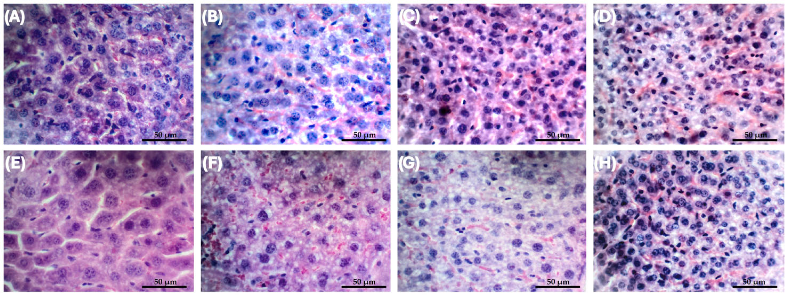
No mortality of Swiss mice was observed in all the concentrations tested; therefore, the lethal dose of DSPE-PEG DMSO+Flp was estimated as >8 mg/kg. In a previous study conducted by our research group, under similar experimental conditions [10], the lethal dose (LD50) of free fluopsin C was estimated at 4 mg/kg. These data indicate that the liposomal formulation reduced the toxicity of the compound in mice. Histopathological analyses were performed for assessment of acute toxicity (single dose, n = 12) and repeated dose toxicity (3 doses every 8 h, n = 12), in healthy animals using the most effective dose of 1 × 2 mg/kg DSPE-PEG DMSO+Flp intravenous [20]. Histopathological analysis of the liver revealed no hemorrhage at any time analyzed. However, mild to moderate necrosis and inflammatory infiltrate were present in all groups, including negative and liposomal controls (Appendix A Table A1). The only significant difference in acute toxicity class occurred for hepatocytes’ nuclei area (p = 0.04), though similar values were found in the control group. Treatments did not alter single-nucleus hepatocyte number. In terms of repeated dose toxicity, there are significant differences in 40 days in the number of double-nuclei hepatocytes, increasing along the tested time. In the same way, this fact occurred over time and was found in all treatments, except the negative control, which maintained similar means throughout the study, suggesting that the liver may recover following the antimicrobial treatment (Figure 3).
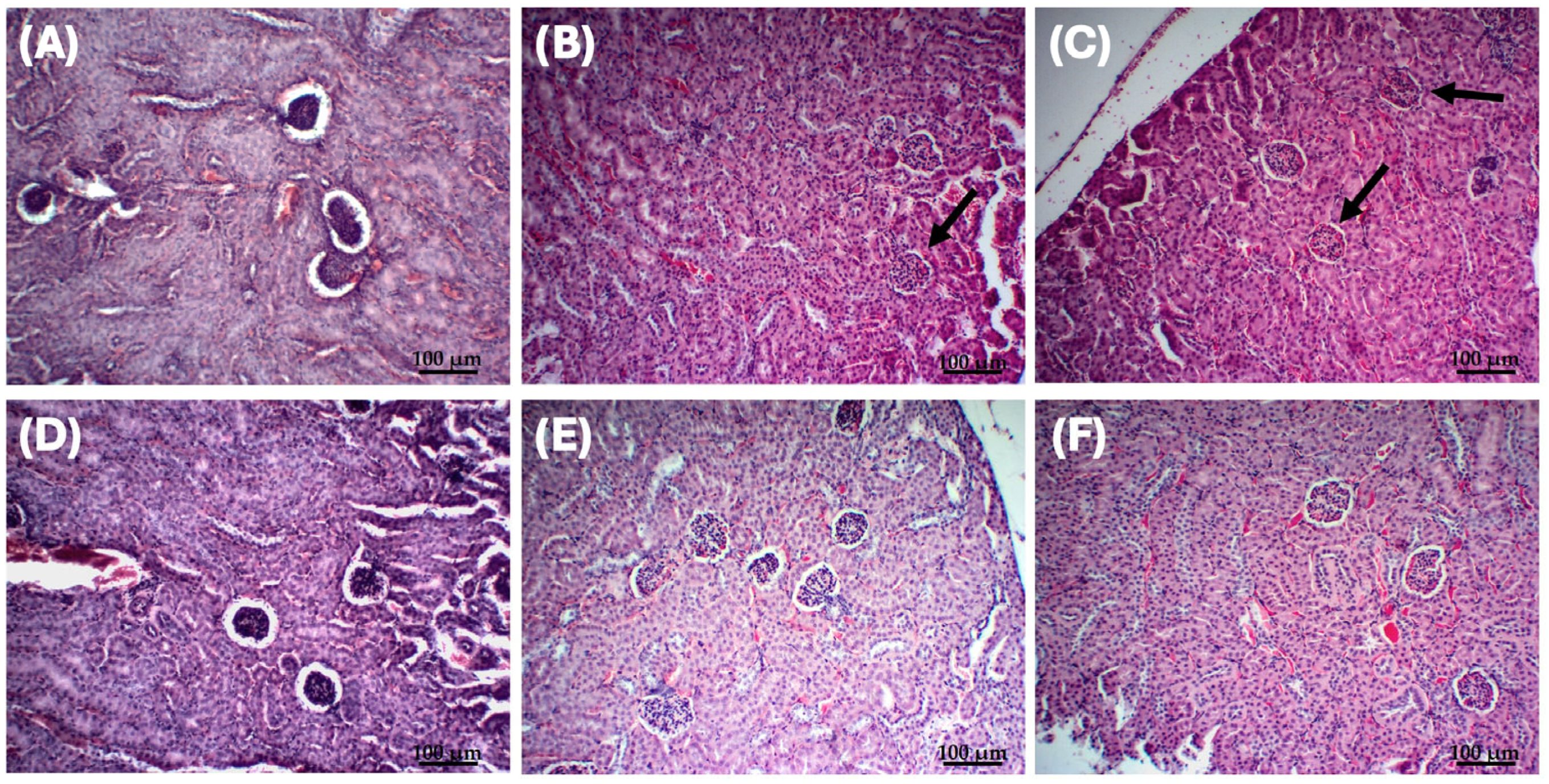
Nephrotoxicity analysis revealed the presence of mild to moderate necrosis, without inflammatory infiltration or hemorrhage in all treatments (Appendix A Table A1). Significant differences were found when comparing Bowman’s space area on day one between the acute toxicity (p = 0.002) and repeated doses toxicity groups (p = 0.003), with the negative control. This indicates that, on the administration day, the Bowman’s space area in the treated groups was contracted, while in the control a space area was found two time bigger. After time zero, no significative differences were observed, indicating the recovery of the organ to normal metrics (Figure 4). Additionally, proximal and distal tubule diameters remained unaltered, with consistent peripheral nucleus placement across all groups and time points.

2.4. Determination of the Minimum Inhibitory Concentration (MIC) and Minimum Bactericidal Concentration (MBC) of Free and Liposomal Fluopsin C

Fluopsin C showed antibacterial activity against the K. pneumoniae strains tested. MIC and MBC values showed no significative difference for both strains, when compared to fluopsin C and fluopsin C combined with liposome. For K. pneumoniae ATCC 10031 [10], the sensitive strain, the MIC and MBC values were both equal to 1.66 µg/mL, while for K. pneumoniae KPN-19, the MDR strain, they were both 3.32 µg/mL. No variance was found in the MIC/MBC tests, performed in triplicate.

2.5. Assessment of the Effect of Free and Liposomal Fluopsin C over the Bacteremia Model in Mice

Lethal bacterial inoculum for 100% of the animals using K. pneumoniae KPN-19 was achieved with 108 UFC/mL, 20 h after infection. Bacteremia was detected from the first blood collection (25 CFU/50 µL blood). Post-mortem organs (lungs, kidneys, and liver) assessment also indicated the generalized presence of the bacteria. This inoculum cell density was standardized for all further experiments.
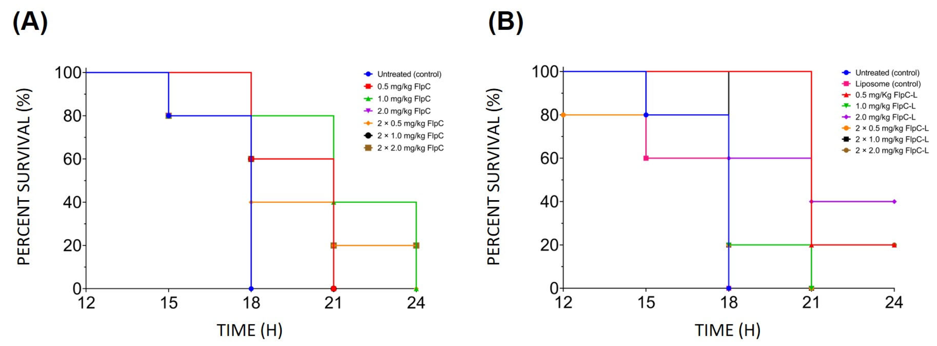
The dose of 2 × 2 mg/kg of free fluopsin C (Figure 5) led to a 20% mortality decrease after 24 h, compared to the other doses. The DSPE-PEG DMSO+Flp formulation provided the best outcome at 1 × 2 mg/kg, reducing mortality by 40% in the bacteremia in mice. Both were considered the most effective doses for free and encapsulated forms.

3. Discussion

Liposomal encapsulation to optimize antimicrobial performance is one of the most promising approaches currently available [15], being particularly beneficial to avoid antimicrobial resistance [21,22]. Fluopsin C showed promise for broad-spectrum antimicrobial activity; however, the pharmacokinetics are low understanding, and high toxicity is a challenge during drug development, still considered unviable for clinical purposes [9,10,11,12,13,14]. Liposomal encapsulation should minimize the high cytotoxicity effects observed of fluopsin C.
In the present study, the DSPE-PEG DMSO+Flp formulation showed the best physical-chemical properties from production to 12 months of storage, especially when stored in solution at low temperature. The formulation preserved the characteristics over time, with predicted variations in size and ZP, but not surpassing the desirable limits to use as an antimicrobial (<500 nm, >±10 mV, and ≤0.3 of PDI) [23]. A PDI value below 0.3 indicates monodispersity, and the homogeneity of size was confirmed using SEM [24].
The decrease in PZ represents a decrease in electrostatic repulsion, with an increased tendency to aggregate, potentially leading to an increase in hydrodynamic diameter. Therefore, the DSPE-PEG DMSO+Flp formulation, both lyophilized and stored in solution, maintained greater PZ stability and, thus, a less variable size. For the SPC DMSO+Flp formulations (in solution and lyophilized), there was a pronounced increase in PZ, resulting in greater agglomeration and a proportional increase in particle diameter [25].
After 11 h, the release of liposomal fluopsin C (DSPE-PEG DMSO+Flp) reached 52%. Similar results were reported, and higher cholesterol content resulted in slower drug release over time, extending the duration of vancomycin and rifampicin release [26]. Cholesterol enhances vesicle stability and reduces membrane permeability, helping minimize cold-induced damage during lyophilization [27,28]. These results are in accordance with the findings of Scriboni et al. [29] that reported values of 30–70% of vancomycin release in 10 h, depending on the formulation tested, which was a similar method used in this paper.
In the in vivo system, a higher difference in release was expected among liposomal formulations, due to the presence of plasmatic proteins, increasing the potential of the DSPE-PEG DMSO+Flp formulation. In the absence of PEG of liposomal formulation, plasmatic proteins opsonize the liposomes, leading to clearance by the mononuclear phagocytic system [28,30]. Previous studies on antimicrobial liposome encapsulation have found similar results to ours, including encapsulation efficiency [31,32,33,34].
The SPC DMSO+Flp formulation with a high concentration of fluopsin C, presented CC50 and CC90 values 69% and 88% higher than free fluopsin C. However, according to other authors, this formulation could lead to fast opsonization, phagocytosis, and removal from circulation by the action of the mononuclear phagocyte system, presenting a potential risk for this formulation [30]. DSPE-PEG DMSO+Flp was identified as the most promising formulation due to the advantages over the immune system, besides achieving CC50 and CC90 values 54% and 48% higher than free fluopsin C, respectively. These advantages were also observed by other Authors [17]. In vivo, there was no mortality associated with the use of this formulation at 8 mg/kg, whereas free fluopsin C showed a DL50 of 4 mg/kg in mice [10].
Liver toxicity was detected as a mild inflammatory infiltrate, which was also found by Navarro et al. [10]. However, complete recovery of the organ was observed 40 days after administration of liposomal fluopsin C (Appendix A Table A1). The nephrotoxicity profile differed from the one found for free fluopsin C [10], with the presence of necrosis (including in the liposomal control), but without other types of lesions.
No alterations in antimicrobial activity were detected between free and liposomal fluopsin C in vitro, indicating that the encapsulation technique did not suppress this activity. Our findings are similar to Kerbauy et al. [11], with 1.95 µg/mL for ATCC 10031 and Navarro et al. [10], with 1 µg/mL for ATCC 10031 and 2 µg/mL for KPN-19. In previous studies by our group, treatment of mice infected with MDR K. pneumoniae using 2 mg/kg of free fluopsin C improved survival by 20% at 96 h [10]. Here, under similar infection conditions, we obtained a 40% reduction in mortality for the same dose at 24 h.

4. Materials and Methods

4.1. Microorganisms

In this study, the target microorganism used was the human pathogen K. pneumoniae KPN-19, isolated from tracheal secretion, and K. pneumoniae ATCC 10031 strain as a control. Both strains were provided by the Laboratory of Basic and Applied Bacteriology of the State University of Londrina, which were used in a previous study [10,11]. KPN-19 strain carries the blaKPC gene, presents porin loss, and resistance to many antibiotics such as imipenem, meropenem, ertapenem, polymyxin B, and colistin [11]. Fluopsin C-producing Pseudomonas aeruginosa LV strain (GenBank CP058323.1) was isolated from citrus canker lesions of orange leaves in an orchard at Astorga, Paraná, Brazil [35,36]. All strains were cryopreserved in 40% (v/v) glycerol solution in liquid nitrogen and deposited in the Laboratory of Microbial Ecology bank of microorganisms.

4.2. Production and Purification of Fluopsin C

P. aeruginosa LV was activated by two consecutive cultures of 48 and 24 h in a nutrient agar plus 100 mg/L of CuCl2·2H2O, and incubated at 28 °C. The fluopsin C production process was performed according to the patented procedure PI 0803350-1 [37], with the modifications proposed by Afonso et al. and Bedoya et al. [9,38]. Purification of fluopsin C was conducted using chromatographic processes, as described by Navarro et al. [10]. High-performance liquid chromatography analysis employed an Agilent 1260 Infinity (Agilent Technologies, Santa Clara, CA, USA) equipped with a multi-wavelength detector and a Zorbax SB-C18 column (4.6 × 250 mm, 5 µm) (Agilent Technologies, Santa Clara, CA, USA), as described by Bedoya et al. [38]. The purity level of fluopsin C used was higher than 80% (Appendix A Figure A2).

4.3. Production and Extrusion of Lipid Vesicles

Four liposomal formulations were used to compare the physicochemical features (Table 3). All formulations followed the amount of total lipids as follows: fluopsin C (Flp) 1:1 mM and are based on soy phosphatidylcholine (SPC), poly(ethylene glycol)-distearoylphosphatidylethanolamine (DSPE-PEG), and cholesterol in different concentrations. Liposomal formulations without fluopsin C were used as control (i.e., liposomal control) (named DSPE-PEG DMSO, DSPE-PEG, SPC DMSO, and SPC).
For each formulation, the respective lipids were aliquoted from chloroform stock solutions according to the concentrations described in Table 3. The samples were left at room temperature for chloroform evaporation, forming a lipid film. DSPE-PEG DMSO+Flp and SPC DMSO+Flp formulations, plus fluopsin C initially, were diluted in DMSO 10% and were added to phosphate buffer (0.1 M, pH 7.4), suspending the emulsion and promoting the incorporation of fluopsin C. The other formulations (DSPE-PEG+Flp and SPC+Flp) had active incorporation of fluopsin C, added during the formation process of the lipid film. After that, phosphate buffer was added to generate liposomal vesicles without DMSO addition.
After resuspended, the formulations were homogenized using a vortex (XH-D Vortex, Global Trade Technology, Jaboticabal, Brazil) for 1 min (min) and sonicated (Ultrasonic Washer L200, Schuster, Santa Maria, Brazil) for 1 min, forming large multilamellar liposomal vesicles. To obtain smaller liposomes, extrusion was performed using a mini extruder (Avanti Polar Lipids, Inc., Alabaster, AL, USA) with polycarbonate membranes (0.4 µm, 19 mm) (Avanti Polar Lipids, Inc., Alabaster, AL, USA), for 15 cycles at room temperature.

4.4. Characterization of Fluopsin C-Containing Liposomes

Fluopsin C-containing liposome formulations were characterized by determination of zeta potential (ZP), polydispersity index (PDI), hydrodynamic diameter, encapsulation efficiency, and fluopsin C release curve. For all tests, 1 mM liposomal fluopsin C was used. The dynamic light scattering (DLS) was used to determine the mean liposome diameter, PDI, and ZP using a NanoPlus particle analyzer (Micromeritics Instrument Corporation, Norcross, GA, USA). PDI below 0.3 was considered monodisperse [24]. Analysis of ZP, PDI, and liposomal diameter was performed four times: 0, 3, 6, and 12 months of storage at 10 °C. Liposomes were stored in solution and lyophilized. All measurements were performed in triplicate.

4.4.1. Encapsulation Efficiency

To determine encapsulation efficiency, the liposomal formulations were centrifuged in a Minispin (Eppendorf F45-12-11 rotor, Merck, Darmstadt, Germany) at 12,100× g (G-Force) for 1 h. The Amicon Ultra-0.5 mL centrifugal filter unit (Ultracel-10 regenerated cellulose membrane, 10,000 NMWL, Merck, Darmstadt, Germany) was used for liposome retention. The measurement of free fluopsin C in solution (non-encapsulated) was performed by spectrophotometry [39] (BioMate3, Thermo, Waltham, MA, USA) at λ = 264 nm, considering the standard method of quantification [40]. The absorbance was compared with a fluopsin C calibration curve, for formulations in the presence or absence of DMSO (range of 13 a 1 μg/mL, r2 = 0.99). The encapsulation efficiency (EE%) was determined by Equation (1) [26], and total fluopsin C was set as 1 mM, and free fluopsin C in the solution was the amount retained in solution after liposomes removal by ultracentrifugation.
E E % = T o t a l   F l u o p s i n   C f r e e   F l u o p s i n   C T o t a l   F l u o p s i n   C × 100

4.4.2. Determination of Free and Liposomal Fluopsin C Release Curve

Free and liposomal fluopsin C released were determined to select the best formulation. Franz cells filled with phosphate plus 10% DMSO v/v in constant agitation were maintained at 37 °C, ensuring sink condition [28]. Fluopsin C release was analyzed by spectrophotometry (BioMate3, Thermo, Waltham, MA, USA) at λ = 264 nm. Aliquots of 300 μL were taken from the Franz cells at 15 and 30 min, 1, 2, 4, 6, 12, and 24 h. The absorbance was used to determine fluopsin C concentration. The experiments were performed in triplicate.

4.5. Analysis of Liposomal Formulations by Scanning Electron Microscopy (SEM)

Samples of lyophilized liposomal formulations were fixed in glutaraldehyde 2.5% fixing solution for 30 min. After that, samples were washed three times with sodium cacodylate buffer 0.1 M, pH 7.2, supplemented with paraformaldehyde solution 2%.
After washing, osmium tetroxide 1% was added, and the samples were incubated for 30 min in the dark to preserve lipid conservation. After that, the samples were washed as described above. For dehydration, samples were treated with serial immersions in different concentrations of ethanol (70%, 90%, and 100%) for 10 min. The last concentration was repeated twice. After that, samples were dried at the critical point using CO2 on a Critical Point Dryer 030 (BalTec Ag, Pfäffikon, Switzerland), coated with gold using the SDC 050 Sputter Coater (BalTec Ag, Switzerland), and observed at FEI quanta 200 scanning electron microscope (FEI Company/Thermo Fisher Scientific, Hillsboro, OR, USA) operating at 30 kV.

4.6. Determination of In Vitro Liposomal Fluopsin C Cytotoxicity

Likewise performed by Navarro et al. [10] and Kerbauy et al. [11], the LLC-MK2 cell line (renal epithelial cells of Macaca mulatta, CCL-7, ATCC, 20–25 passage, Merck Co, São Paulo, Brazil) was grown in a 96-well plate in RPMI medium supplemented with fetal bovine serum 10%, with 2.5 × 10⁴ cells/well and incubated for 24 h at 37.0 °C CO2 5.0% [10,11,41]. A 100% confluence, non-adherent cells in suspension were removed by washing with sterile phosphate-buffered saline (PBS, 0.1 M, pH 7.2).
Formulation DSPE-PEG DMSO+Flp, SPC DMSO+Flp, and the respective control were serially diluted in RPMI medium, at concentrations ranging from 32 to 0.25 μg/mL of fluopsin C, and subsequently added to the cell-containing wells. The treatments were incubated for 24 h under the same conditions mentioned earlier. Cell viability was determined by the MTT test (3-[4,5-dimethylthiazol-2-yl]-2,5-diphenyl-2H-tetrazolium bromide) (Sigma Chemical Co., St. Louis, MO, EUA) reduction method, according to the manufacturer’s recommendation. A non-linear regression method was performed to determine the cytotoxic concentration that inhibits 50% (CC50) and 90% (CC90) of the metabolic activity of the cell population in 24 h.

4.7. Determination of the Minimum Inhibitory and Bactericidal Concentration of Free and Liposomal Fluopsin C

Minimum inhibitory concentration (MIC) and minimum bactericidal concentration (MBC) were determined for DSPE-PEG DMSO+Flp and SPC DMSO+Flp formulations against K. pneumoniae ATCC 10031 and KPN-19 strains. Tested concentrations ranged from 13.3 to 0.2 µg/mL, including for free fluopsin C. Controls consisted of the respective formulations without the incorporation of fluopsin C. The MIC values were determined through the broth microdilution method following the Clinical and Laboratory Standards Institute (CLSI) guidelines of 2021 [42], and incubated for 24 h at 37 °C. For MBC determination, 10 µL aliquots from each well of the microdilution method were plated onto Mueller-Hinton Agar and incubated for 24 h at 37 °C.

4.8. In Vivo Assessments

The in vivo experimental design was carried out as shown in Figure 6.

4.8.1. Animals

The following experiments utilized 42-day-old female Swiss mice, with an average weight of 25 g (total n = 189). These animals were provided by the Central Bioterium of the Biological Sciences Center at the State University of Londrina and the Central Bioterium of the State University of Maringá. Ethical approval for animal use was granted by the respective ethical commissions of the respective universities under the registrations CEUA (Comitê de Ética no Uso de Animais) n° 039.2021 and CEUA n° 9838190121, respectively.
Compliance with regulations concerning animal welfare and management was ensured throughout the study. The animals were randomly allocated to the different experimental groups using a simple randomization protocol. The researchers responsible for administering the treatments/inoculum and assessing the clinical outcomes (including signs of infection and mortality) were blinded as to the identity of the groups. After being assigned to the groups, the animals were kept for a period of 3 days, for adaptation in the new environment, before each experiment. To minimize confounding factors, the cages were kept in ascending order according to the corresponding dose, and the control groups were segregated on another shelf, with greater spacing from the others, avoiding cross-contamination. For the same purpose, the animals were identified with a number according to the cage number, in a painless way.

4.8.2. Lethal Inoculum of K. pneumoniae KPN-19 and Bacteremia Model Establishment

For the determination of the lethal inoculum, n = 5 animals were infected intraperitoneally with K. pneumoniae KPN-19 (0.5 mL) at different cell densities of 107, 108, and 109 CFU/mL. The control group (n = 5) was administered with sterilized saline solution. The animals were observed until 96 h post-inoculation, for a total of n = 20 animals.
After the previous results, the bacteremia model was confirmed by infecting n = 5 animals with 108 CFU/mL, intraperitoneally. 50 µL of caudal blood was collected post-anesthesia (isoflurane 3–5% mg/kg by inhalation) every 2 h after inoculation. Time zero was defined as 10 min after inoculation. The blood samples were plated on blood agar and incubated at 37.0 °C for 24 h. After euthanasia, the lungs, kidneys, and liver were collected. The organs were weighed and macerated, and the product was serially diluted in sterilized PBS. Aliquots were then plated on blood agar and incubated at 37.0 °C for 24 h. The same procedures were performed for control group animals (n = 5). for a total of n = 10 animals.

4.8.3. Lethal Dose for Fluopsin C Liposomal Formulation

In a previous study, we evaluated the lethal dose of free fluopsin C in female Swiss mice [10]. To compare the effect of the liposomal formulation, we used similar experimental conditions. Therefore, the animals were inoculated intravenously with single doses of 0.5, 1, 2, 4, and 8 mg/kg DSPE-PEG DMSO+Flp formulation. The control group received sterilized PBS. The experimental groups consisted of n = 6 animals kept in individual containers, for a total of n = 36 animals. The animals were monitored until 96 h post-administration.

4.8.4. Antibacterial Activity of Liposomal and Free Fluopsin C In Vivo

To determine the protective effects of liposomal and free fluopsin C, n = 5 animals were infected with 0.5 mL 108 CFU/mL of K. pneumoniae KPN-19 intraperitoneally, according to Navarro et al. [10]. Immediately after inoculation, 0.1 mL of free fluopsin C or DSPE-PEG DSMO+Flp formulation was administered intravenously. Different dosages of both compounds were evaluated as single doses of 1 × 0.5, 1 × 1, and 1 × 2 mg/kg and double doses with an 8 h interval of 2 × 0.5, 2 × 1, and 2 × 2 mg/kg, totaling 6 groups for each one. DSPE-PEG DMSO control formulation was used as the liposomal control (n = 5) at 2 × 2 mg/kg dose since there was no antimicrobial activity observed in vitro for the liposomal components. The negative control group (n = 5) consisted of the administration of sterilized saline solutions intraperitoneally and PBS intravenously. The positive control group (n = 5) consisted of the administration of sterilized PBS intravenously, for a total of n = 75 animals.

4.8.5. Histopathological Analysis of the Most Effective Dose

Histopathological analyses were performed for assessment of acute toxicity (single dose, n = 12) and repeated dose toxicity (3 doses every 8 h, n = 12) [20]. Thus, healthy animals were administered DSPE-PEG DMSO+Flp intravenous using the most effective dose (1 × 2 mg/kg). Controls were based on administration of the same liposomal formulation without the addition of fluopsin C (n = 12) and sterilized PBS (n = 12). for a total of n = 48 animals.
The effects of the compounds administered were assessed 1-, 10-, 20-, and 40 days post-treatment. At each point, 3 animals were euthanized with the collection of the liver and kidney, which were immediately fixed using a 4% paraformaldehyde solution. The organs were processed to the obtention of histological slides and stained with hematoxylin/eosin.
Analysis of the organ’s sections was performed in agreement with Navarro et al. [10], consisting of the determination of hepatocyte nucleus area (n = 100); mono- and binucleated number of hepatocytes, number of hepatocytes with condensed chromatin and vacuolization [10]. Nephrotoxic effects were evaluated by determining the Bowman’s space area (n = 10), distal and proximal tubule diameters (n = 50), and nucleus placement of tubules lining cells. The presence of inflammatory infiltrates, hemorrhage, and necrosis was also determined for both organs. This qualitative evaluation of the histological results was carried out by a trained committee unrelated to the experiments, with samples identified by code.

4.9. Statistical Analysis

The results were averaged and are presented with the standard deviation. Liposomal cytotoxicity in vitro assays were analyzed through one-way ANOVA and Dunnett’s multiple comparison test; non-linear regression was also used to infer the CC50 and CC90. The Kaplan–Meier estimator was applied to generate a survival curve. Histological section images were processed using ImageJ 1.54d (Wayne Rasband and contributors National Institutes of Health, Bethesda, MD, USA) and analyzed through two-way ANOVA and Tukey’s multiple comparison test. All tests considered p < 0.05 as indicative of significative differences. Raw data were processed using GraphPad Prism v. 8.4.3 (GraphPad Software, Boston, MA, USA). The study did not have humane endpoints.
All animals were included throughout the experiment, and no experimental units or data points were excluded from the analysis. The experiment was conducted, the results were evaluated, and the data were analyzed by the same team, except for the qualitative evaluation of the histopathological analysis, where an independent analysis-trained committee unrelated to the experiments was formed for this purpose only, with samples identified by code.

5. Conclusions

The present study reported for the first time the effect of encapsulation of fluopsin C, using the liposomal technique. Liposomal fluopsin C (DSPE-PEG DMSO+Flp) was effective in controlling MDR K. pneumoniae, in vitro and in vivo in mice, increasing survival of infected animals. In addition, the reduction of cytotoxicity in vitro and in vivo conditions showed healthy mice and an increase in the in vitro release time of fluopsin C encapsulated. Based on experimental data and previous studies by our group, fluopsin C should be a promising compound for developing a new drug due to its broad activity against MDR. However, further study should be carried out in another animal model, according to preclinical study guidelines, to understand how the antimicrobial activity of fluopsin C does against MDR K. pneumoniae.

Author Contributions

Conceptualization, M.L.D.G., G.A., L.F.C. and S.F.Y.-O.; methodology, K.R.B., L.C.A., A.C.G. and E.J.N.M.; methodology, validation, investigation, writing—original draft preparation, M.L.D.G., S.F.Y.-O., J.C.P.d.M., F.G.A. and L.A.; writing—review and editing, G.A., L.F.C., L.A. and M.V.T.C.; funding acquisition, G.A.; supervision and formal analysis, G.A. and L.F.C. All authors have read and agreed to the published version of the manuscript.

Funding

This research was funded by the Brazilian National Council for Scientific and Technological Development (CNPq), grant number 439754/2018-6 (AMR MCTIC) and 406016/2022-4, PPSUS-Aracauria Foundation-PR.

Institutional Review Board Statement

The animal study protocol was approved by the Ethics Committee of the State University of Londrina (protocol code n° 039.2021, approval data from 5 October 2021) and the State University of Maringá (n° 9838190121 approval data from 18 February 2021).

Informed Consent Statement

Not applicable.

Data Availability Statement

The original contributions presented in the study are included in the article; further inquiries can be directed to the corresponding author.

Acknowledgments

The Electron Microscopy and Microanalysis Laboratory of the State University of Londrina, the Histopathological Analysis Laboratory, the Molecular Biology Laboratory of Microorganisms, and the Basic and Applied Bacteriology Laboratory of the same University, the Laboratory of Biomaterials and Organic Biomolecules of the Federal Technological University of Paraná and, finally, to the Nucleus of Research in Photodynamic Systems of the State University of Maringá.

Conflicts of Interest

The authors declare no conflicts of interest.

Appendix A

Figure A1. Cell viability of LLC-MK2 cells analyzed by the MTT reduction method in the presence of different concentrations of free fluopsin C and liposomal formulations in relation to the untreated cell control. Through ANOVA and Dunnett’s multiple variable tests, the graphs shown in the Figure were obtained for the analyzed samples. The trend observed in the graphs was consistent with the non-linear regression analysis. (A) DSPE-PEG DMSO, (B) SPC DMSO. (C) DSPE-PEG DMSO+F, (D) SPC DMSO+F, and (E) free fluopsin C. C, untreated cell control; ** p ˂ 0.01; *** p ˂ 0.001; **** p ˂ 0.0001.
Antibiotics 14 00948 g0a1
Table A1. Scoring table of qualitative histopathological analysis of liver and kidney after intravenous administration of one dose (acute toxicity) and three doses (repeated dose toxicity) of 2 mg/kg DSPE-PEG DMSO+Flp at times of 1, 10, 20, and 40 days. I: inflammatory infiltrate; H: hemorrhage; N: necrosis. 0—absent; 1—mild; 2—moderate; and 3—severe.
LiverAcute ToxicityRepeated Doses ToxicityLipossomal ControlNegative Control
Time (days)IHNIHNIHNIHN
1101101101002
10101101102002
20002001101001
40002001001002
KidneyAcute ToxicityRepeated Dose ToxicityLipossomal ControlNegative Control
Time (days)IHNIHNIHNIHN
1111001001002
10000001001002
20001002001002
40002001000002
Figure A2. (A) Purified fluopsin C chromatogram at 264 nm using a C18 (5 µm × 4.6 mm × 250 mm). (B) High-purity (89%) fluopsin C molecule was detected at peak 2.
Antibiotics 14 00948 g0a2

References

  1. World Health Organization (WHO). WHO Bacterial Priority Pathogens List, 2024: Bacterial Pathogens of Public Health Importance to Guide Research, Development and Strategies to Prevent and Control Antimicrobial Resistance; World Health Organization: Geneva, Switzerland, 2024; ISBN 978-92-4-010230-9. [Google Scholar]
  2. World Health Organization (WHO). Global Priority List of Antibiotic-Resistant Bacteria to Guide Research, Discovery, and Development of New Antibiotics; World Health Organization: Geneva, Switzerland, 2017; ISBN 9789240026438. [Google Scholar]
  3. Kochan, T.J.; Nozick, S.H.; Medernach, R.L.; Cheung, B.H.; Gatesy, S.W.M.; Lebrun-Corbin, M.; Mitra, S.D.; Khalatyan, N.; Fiorella, K.; Chao, Q.; et al. Genomic surveillance for multidrug-resistant or hypervirulent Klebsiella pneumoniae among United States bloodstream isolates. BMC Infect. Dis. 2022, 22, 603. [Google Scholar] [CrossRef]
  4. Jacobs, D.M.; Safir, M.C.; Huang, D.; Minhaj, F.; Parker, A.; Rao, G.G. Triple Combination Antibiotic Therapy for Carbapenemase-Producing Klebsiella pneumoniae: A Systematic Review. Ann. Clin. Microbiol. Antimicrob. 2017, 16, 76. [Google Scholar] [CrossRef]
  5. Sheu, C.C.; Chang, Y.T.; Lin, S.Y.; Chen, Y.H.; Hsueh, P.R. Infections Caused by Carbapenem-Resistant Enterobacteriaceae: An Update on Therapeutic Options. Front. Microbiol. 2019, 10, 80. [Google Scholar] [CrossRef]
  6. Laws, M.; Shaaban, A.; Rahman, K.M. Antibiotic Resistance Breakers: Current Approaches and Future Directions. FEMS Microbiol. Rev. 2019, 43, 490–516. [Google Scholar] [CrossRef]
  7. Da Silva, A.C.P.; Velasquez, P.A.G. Perfil De Resistência De Klebsiella pneumoniae Isoladas De Pacientes Da Unidade De Terapia Intensiva De Um Hospital No Sudoeste Do Paraná. Discip. Sci.|Saúde 2018, 18, 259–270. [Google Scholar]
  8. Centers for Disease Control and Prevention. Antibiotic Resistence Threats in the United States; CDC: Atlanta, GA, USA, 2013; 114p.
  9. Afonso, L.; Grzegorczyk, K.G.; Salomão, J.M.; Basso, K.R.; Alves, L.C.; Silva, M.C.D.; Chryssafidis, A.L.; Gionco-Cano, B.; Yamada-Ogatta, S.F.; Andrade, G. Fluopsin C Promotes Biofilm Removal of XDR Acinetobacter baumannii and Presents an Additive Effect with Polymyxin B on Planktonic Cells. Antibiotics 2024, 13, 875. [Google Scholar] [CrossRef]
  10. Navarro, M.O.P.; Simionato, A.S.; Pérez, J.C.B.; Barazetti, A.R.; Emiliano, J.; Niekawa, E.T.G.; Andreata, M.F.L.; Modolon, F.; Dealis, M.L.; Araújo, E.J.A.; et al. Fluopsin C for Treating Multidrug-resistant Infections: In vitro Activity against Clinically Important Strains and in vivo Efficacy Against Carbapenemase-producing Kleb pneumoniae. Front. Microbiol. 2019, 10, 2431. [Google Scholar] [CrossRef] [PubMed]
  11. Kerbauy, G.; Vivian, A.C.P.; Glenda, S.C.; Simionato, A.S.; Pelisson, M.; Vespero, E.; Costa, S.F.; Andrade, C.G.T.J.; Barbieri, D.M.; Mello, J.C.P.; et al. Effect of a Metalloantibiotic Produced by Pseudomonas aeruginosa on Klebsiella pneumoniae Carbapenemase (KPC)-producing K. pneumoniae. Curr. Pharm. Biotechnol. 2016, 17, 389–397. [Google Scholar] [CrossRef]
  12. Cardozo, V.F.; Oliveira, A.G.; Nishio, E.K.; Perugini, M.R.E.; Andrade, C.G.T.J.; Silveira, W.D.; Durán, N.; Andrade, G.; Kobayashi, R.K.T.; Nakazato, G. Antibacterial Activity of Extracellular Compounds Produced by a Pseudomonas Strain against Methicillin-resistant Staphylococcus aureus (MRSA) strains. Ann. Clin. Microbiol. Antimicrob. 2013, 12, 12. [Google Scholar] [CrossRef] [PubMed]
  13. Egawa, Y.; Umino, K.; Awataguchi, S.; Kawano, Y.; Okuda, T. Antibiotic YC 73 of Pseudomonas origin. I. Production, Isolation and Properties. J. Antibiot 1970, 23, 267–270. [Google Scholar] [CrossRef]
  14. Sharma, D. Fluopsin C: A potential candidate against the deadly drug-resistant microbial infections in humans. Future Microbiol. 2020, 15, 381–384. [Google Scholar] [CrossRef] [PubMed]
  15. Catalano, A.; Iacopetta, D.; Ceramella, J.; Scumaci, D.; Giuzio, F.; Saturnino, C.; Stefano, A.; Rosano, C.; Sinicropi, M.S. Multidrug resistance (MDR): A widespread phenomenon in pharmacological therapies. Molecules 2022, 27, 616. [Google Scholar] [CrossRef] [PubMed]
  16. Alarfaj, R.E.; Alkhulaifi, M.M.; Al-Fahad, A.J.; Aljihani, S.; Yassin, A.E.B.; Alghoribi, M.F.; Halwani, M.A. Antibacterial efficacy of liposomal formulations containing tobramycin and N-acetylcysteine against tobramycin-resistant Escherichia coli, Klebsiella pneumoniae, and Acinetobacter baumannii. Pharmaceutics 2022, 14, 130. [Google Scholar] [CrossRef] [PubMed]
  17. Alavi, S.E.; Esfahani, M.K.M.; Raza, A.; Adelnia, H.; Shahmabadi, H.E. PEG-grafted liposomes for enhanced antibacterial and antibiotic activities: An in vivo study. NanoImpact 2022, 25, 100384. [Google Scholar] [CrossRef]
  18. Khan, A.A.; Allemailem, K.S.; Almatroodi, S.A.; Almatroudi, A.; Rahmani, A.H. Recent strategies towards the surface modification of liposomes: An innovative approach for different clinical applications. 3 Biotech. 2020, 10, 163. [Google Scholar] [CrossRef]
  19. Li, M.; Du, C.; Guo, N.; Teng, Y.; Meng, X.; Sun, H.; Li, S.; Yu, P.; Galons, H. Composition design and medical application of liposomes. Eur. J. Med. Chem. 2019, 164, 640–653. [Google Scholar] [CrossRef]
  20. Agência Nacional de Vigilância Sanitária (ANVISA). Guia Para a Condução de Estudos Não Clínicos de Toxicologia e Segurança Farmacológica Necessários ao Desenvolvimento de Medicamentos, 2nd ed.; Gerência de Avaliação de Segurança e Eficácia: Brasília, Brasil, 2013; 48p.
  21. Wang, D.Y.; Van der Mei, H.C.; Ren, Y.; Busscher, H.J.; Shi, L. Lipid-based antimicrobial delivery-systems for the treatment of bacterial infections. Front. Chem. 2020, 7, 872. [Google Scholar] [CrossRef]
  22. Eleraky, N.E.; Allam, A.; Hassan, S.B.; Omar, M.M. Nanomedicine fight against antibacterial resistance: An overview of the recent pharmaceutical innovations. Pharmaceutics 2020, 12, 142. [Google Scholar] [CrossRef]
  23. Weaver, E.; Macartney, R.A.; Irwin, R.; Uddin, S.; Hooker, A.; Burke, G.A.; Wylie, M.P.; Lamprou, D.A. Liposomal encapsulation of amoxicillin via microfluidics with subsequent investigation of the significance of PEGylated therapeutics. Int. J. Pharm. 2024, 650, 123710. [Google Scholar] [CrossRef]
  24. Shah, V.M.; Nguyen, D.X.; Patel, P.; Cote, B.; Al-Fatease, A.; Pham, Y.; Huynh, M.G.; Woo, Y.; Alani, A.W.G. Liposomes produced by microfluidics and extrusion: A comparison for scale-up purposes. Nanomed. Nanotechnol. Biol. Med. 2019, 8, 146–156. [Google Scholar] [CrossRef]
  25. Wang, J.; Zhang, L.; Chi, H.; Wang, S. An alternative choice of lidocaine-loaded liposomes: Lidocaine-loaded lipid–polymer hybrid nanoparticles for local anesthetic therapy. Drug Deliv. 2016, 23, 1254–1260. [Google Scholar] [CrossRef]
  26. Gonzalez Gomez, A.; Hosseinidoust, Z. Liposomes for antibiotic encapsulation and delivery. ACS Infect. Dis. 2020, 6, 896–908. [Google Scholar] [CrossRef] [PubMed]
  27. Nsairat, H.; Khater, D.; Sayed, U.; Odeh, F.; Al Bawab, A.; Alshaer, W. Liposomes: Structure, composition, types, and clinical applications. Heliyon 2022, 8, e09394. [Google Scholar] [CrossRef] [PubMed]
  28. Wang, Y.; Grainger, D.W. Lyophilized liposome-based parenteral drug development: Reviewing complex product design strategies and current regulatory environments. Adv. Drug Deliv. Rev. 2019, 151, 56–71. [Google Scholar] [CrossRef]
  29. Scriboni, A.B.; Couto, V.M.; Ribeiro, L.N.D.M.; Freires, I.A.; Groppo, F.C.; De Paula, E.; Franz-Montan, M.; Cogo-Müller, K. Fusogenic liposomes increase the antimicrobial activity of vancomycin against Staphylococcus aureus biofilm. Front. Pharmacol. 2019, 10, 1401. [Google Scholar] [CrossRef] [PubMed]
  30. Liu, P.; Chen, G.; Zhang, J. A review of liposomes as a drug delivery system: Current status of approved products, regulatory environments, and future perspectives. Molecules 2022, 27, 1372. [Google Scholar] [CrossRef]
  31. Werner, J.; Umstätter, F.; Hertlein, T.; Beijer, B.; Kleist, C.; Mühlberg, E.; Zimmermann, S.; Haberkorn, U.; Ohlsen, K.; Fricker, G.; et al. Improved pharmacokinetics and enhanced efficacy of the vancomycin derivative FU002 using a liposomal nanocarrier. Nanomed. Nanotechnol. Biol. Med. 2024, 56, 102731. [Google Scholar] [CrossRef]
  32. Pushparaj Selvadoss, P.; Nellore, J.; Balaraman Ravindrran, M.; Sekar, U. Novel pyochelin-based PEGylated liposomes for enhanced delivery of antibiotics against resistant clinical isolates of Pseudomonas aeruginosa. Artif. Cells Nanomed. Biotechnol. 2017, 46, 2043–2053. [Google Scholar] [CrossRef]
  33. Briuglia, M.L.; Rotella, C.; McFarlane, A.; Lamprou, D.A. Influence of cholesterol on liposome stability and on in vitro drug release. Drug Deliv. Transl. Res. 2015, 5, 231–242. [Google Scholar] [CrossRef]
  34. Fan, X.; Fan, J.; Wang, X.; Wu, P.; Wu, G. S-thanatin functionalized liposome potentially targeting on Klebsiella pneumoniae and its application in sepsis mouse model. Front. Pharmacol. 2015, 6, 249. [Google Scholar] [CrossRef]
  35. Simionato, A.S.; Cano, B.G.; Navarro, M.O.P.; Tavares, E.R.; Ribeiro, R.A.; Hungria, M.; Yamauchi, L.M.; Yamada-Ogatta, S.F.; Andrade, G. Whole-Genome Sequence of Bioactive Compound-Producing Pseudomonas aeruginosa Strain LV. Microbiol. Resour. Announc. 2021, 10, e01120-20. [Google Scholar] [CrossRef] [PubMed]
  36. Rampazo, L.G.L. Evaluation of Biological Agents and their Products on the Incidence of Citrus Canker Foliar Lesions. Master’s Thesis, Universidade Estadual de Londrina, Londrina, Brasil, 2004. [Google Scholar]
  37. Andrade, G. Processo de Produção, Purificação e Obtenção de Substâncias Com Atividades Antibióticas Para o Controle de Doenças Causadas Por Bactérias Em Plantas. Br. Patent PI 0803350-1, 10 September 2008. [Google Scholar]
  38. Bedoya, J.C.; Dealis, M.L.; Silva, C.S.; Niekawa, E.T.G.; Navarro, M.O.P.; Simionato, A.S.; Modolon, F.; Chryssafidis, A.L.; Andrade, G. Enhanced production of target bioactive metabolites produced by Pseudomonas aeruginosa LV strain. Biocatal. Agric. Biotechnol. 2019, 17, 653–664. [Google Scholar] [CrossRef]
  39. Ribeiro, L.N.M.; de Paula, E.; Rossi, D.A.; Monteiro, G.P.; Júnior, E.C.V.; Silva, R.R.; Franco, R.R.; Espíndola, F.S.; Goulart, L.R.; Fonseca, B.B. Hybrid pectin-liposome formulation against multi-resistant bacterial strains. Pharmaceutics 2020, 12, 769. [Google Scholar] [CrossRef] [PubMed]
  40. Lee, S.G.; Jeong, J.H.; Lee, K.M.; Jeong, K.H.; Yang, H.; Kim, M.; Jung, H.; Lee, S.; Choi, Y.W. Nanostructured lipid carrier-loaded hyaluronic acid microneedles for controlled dermal delivery of a lipophilic molecule. Int. J. Nanomed. 2014, 9, 289–299. [Google Scholar] [CrossRef]
  41. Chierrito, D.; Villas-Boas, C.B.; Tonin, R.S.; Fernandez-Limos, F.; Sanches, A.C.C.; Mello, J.C.P. Using cell cultures for the investigation of treatments for Attention Deficit Hyperactivity Disorder: Systematic review. Curr. Neuropharmacol. 2019, 17, 916–925. [Google Scholar] [CrossRef]
  42. Clinical and Laboratory Standards Institute (CLSI). Performance Standards for Antimicrobial Susceptibility Testing, 31st ed; Clinical and Laboratory Standards Institute: Wayne, PA, USA, 2021. [Google Scholar]
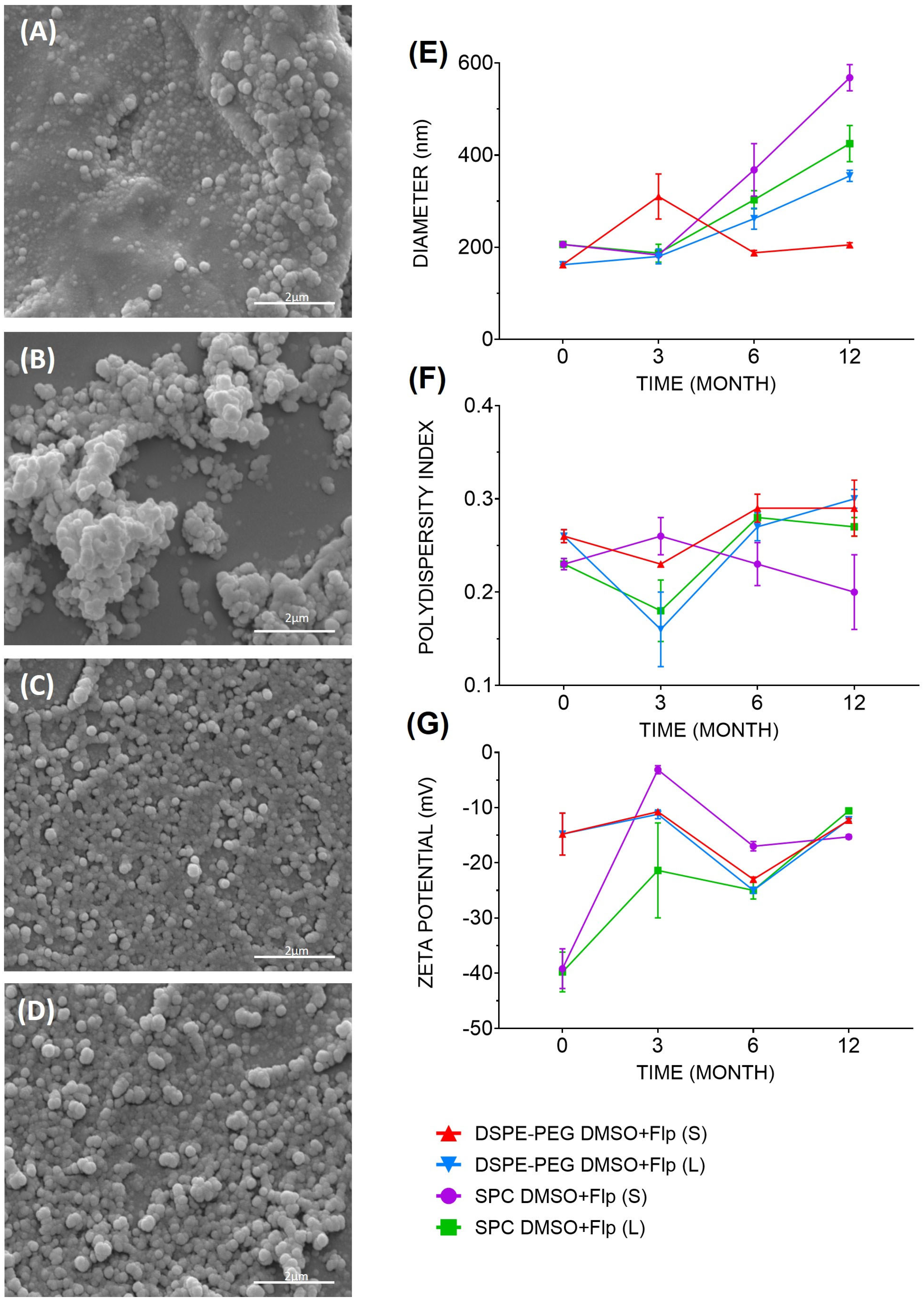
Figure 1. Formulation morphology and their respective controls were analyzed by scanning electron microscopy (SEM) under 30,000× magnification at time zero: (A) DSPE-PEG DMSO, (B) SPC DMSO, (C) DSPE-PEG DMSO+Flp, and (D) SPC DMSO+Flp. Physical–chemical characterization of fluopsin C-containing liposomal formulations. (E) Hydrodynamic diameter (nm), (F) polydispersity index (PDI), and (G) zeta potential (ZP, mV) were obtained by NanoPlus particle analyzer right after production and after 0, 3, 6, and 12 months. Formulations were stored in the form of solution (S) and lyophilized (L).
Antibiotics 14 00948 g001
Figure 2. In vitro release curve of free and liposomal fluopsin C formulations using Franz cells, over 24 h.
Antibiotics 14 00948 g002
Figure 3. Liver analysis of repeated dose toxicity (RDT) and negative control (NC) over 40 days post-treatment, in the number of double-nuclei hepatocytes. (A) NC in day one, (B) NC in 10 days, (C) NC in 20 days, (D) NC in 40 days, (E) RDT in day one, (F) RDT in 10 days, (G) RDT in 20 days, and (H) RDT in 40 days. Color: hematoxylin/eosin (HE). Scale bar: 50 µm.
Antibiotics 14 00948 g003
Figure 4. Nephrotoxicity analysis in the Bowman’s space area on day one and after 10 days post-treatment. Black arrows indicate that Bowman’s space areas in the treated groups were contracted on day one. (A) Negative control on day one, (B) repeated doses toxicity group on day one, (C) acute toxicity group on day one, (D) negative control after 10 days, (E) acute toxicity group after 10 days, and (F) repeated doses toxicity group after 10 days. Color: HE. Scale bar: 100 µm.
Antibiotics 14 00948 g004
Figure 5. Kaplan–Meier survival curve in mouse bacteremia model caused by K. pneumoniae KPN-19 treated intravenously with different doses of (A) fluopsin C (Flp) and (B) liposomal fluopsin C (Flp-L) DSPE-PEG DMSO+Flp formulation.
Antibiotics 14 00948 g005
Figure 6. Timeline of the experiment carried out.
Antibiotics 14 00948 g006
Table 1. Encapsulation efficiency (EE, %) for the different fluopsin C-containing liposomal formulations.
FormulationsEE (%)
SPC+Flp66.04 ± 8.19
SPC DMSO+Flp83.75 ± 1.95
DSPE-PEG+Flp78.04 ± 3.58
DSPE-PEG DMSO+Flp83.67 ± 2.06
Table 2. In vitro cytotoxic concentrations of free and liposomal fluopsin C for 50% (CC50) and 90% (CC90) of the LLC-MK2 cell population by MTT test after 24 h of incubation.
FormulationCC50 (µg/mL)CC90 (µg/mL)
DSPE-PEG DMSO+Flp1.141.26
DSPE-PEG DMSO>32.00>32.00
SPC DMSO+Flp1.261.60
SPC DMSO22.6279.07
Fluopsin C0.740.85
Table 3. Composition of fluopsin C-containing liposomal formulations.
FormulationSPC 1
(mM)
Cholesterol (mM)DSPE-PEG 2 (mM)Fluopsin C (mM)Phosphate Buffer: DMSO 3 (%)
DSPE-PEG+Flp0.540.410.051100:0
DSPE-PEG DMSO+Flp0.540.410.05190:10
SPC+Flp0.70.3-1100:0
SPC DMSO+Flp0.70.3-190:10
1 Soy phosphatidylcholine (SPC); 2 polyethylene glycol 2000 (PEG) conjugated to distearoyl phosphatidyl ethanolamine (DSPE); 3 dimethylsulfoxide (DMSO).
Disclaimer/Publisher’s Note: The statements, opinions and data contained in all publications are solely those of the individual author(s) and contributor(s) and not of MDPI and/or the editor(s). MDPI and/or the editor(s) disclaim responsibility for any injury to people or property resulting from any ideas, methods, instructions or products referred to in the content.