Article

Unique Regulation of Sed-1 β-Lactamase in Citrobacter sedlakii: Insights on Resistance to Third-Generation Cephalosporin

1
Department of Microbiology and Infectious Diseases, Nara Medical University, 840 Shijo-cho, Kashihara 6348521, Nara, Japan
2
Department of Chemistry, Nara Medical University, 88 Shijo-cho, Kashihara 6340813, Nara, Japan
3
Division of Central Clinical Laboratory, Iwate Medical University Hospital, 1-1-1 Idaidori, Yahaba-cho, Shiwa-gun 0283694, Iwate, Japan
*
Author to whom correspondence should be addressed.
These authors contributed equally to this work.
Antibiotics 2025, 14(8), 823; https://doi.org/10.3390/antibiotics14080823
Submission received: 27 June 2025 / Revised: 29 July 2025 / Accepted: 7 August 2025 / Published: 12 August 2025

Abstract

Background: The Citrobacter genus harbors class C (AmpC) and class A β-lactamases. Citrobacter freundii produces an inducible AmpC β-lactamase controlled by the LysR-type transcriptional regulator AmpR and cytosolic amidase AmpD. Citrobacter sedlakii produces the class A β-lactamase Sed-1, whose expression is believed to be regulated by the transcriptional regulator SedR and AmpD. Objectives: C. sedlakii NR2807, isolated in Japan, is resistant to third-generation cephalosporins and displays extended-spectrum β-lactamase characteristics. Here, we sought to understand the mechanism for successful resistance to third-generation cephalosporins by investigating the regulators controlling Sed-1 production. Methods: Plasmids containing blaSed-1 and sedR (pCR2807) or truncated sedR (pCR2807ΔSedR) were constructed and introduced into Escherichia coli. Antibiotic-resistant mutants of NR2807 were obtained, and enzyme kinetics were assessed. Results: The AmpD mutant (pCR2807/ML4953) showed an 8-fold increase in cefotaxime MIC and an 8.46-fold increase in Sed-1 activity compared to the wild-type (pCR2807/ML4947). However, induction of pCR2807/ML4947 also led to a 1.32-fold higher Sed-1 activity, indicating semi-inducibility. Deletion of sedR (pCR2807ΔSedR/ML4947) led to a 4-fold decrease in cefotaxime MIC and 1.93-fold lower Sed-1 activity, confirming SedR as an activator. While wild-type C. sedlakii ATCC51115 is susceptible to third-generation cephalosporins, the AmpD mutation in NR2807 led to Sed-1 overproduction and resistance to this class of antibiotics. Finally, mutagenesis revealed that amino acid substitution in Sed-1 conferred resistance to ceftazidime and extended-spectrum β-lactamase characteristics. Conclusions: Sed-1 producers, though usually susceptible to third-generation cephalosporins, may develop extended-spectrum β-lactamase traits due to AmpD or Sed-1 mutations, thereby requiring careful monitoring.

1. Introduction

Antimicrobial resistance represents a significant global public health concern, with β-lactamase-producing bacteria undermining the effectiveness of β-lactam antibiotics [1,2]. Among Enterobacterales, extended-spectrum β-lactamases (ESBLs) have drawn significant attention due to their ability to hydrolyze third-generation cephalosporins (3GC), thereby limiting the scope of critical antibiotics used in clinical settings [3]. β-Lactamases vary among Citrobacter species: Citrobacter freundii produces a class C β-lactamase (AmpC); whereas Citrobacter amalonaticus, Citrobacter gillenii, Citrobacter koseri, and Citrobacter sedlakii produce class A β-lactamases, CdiA, CKO-1, and Sed-1, respectively (Table 1) [4,5,6,7].
Gram-negative C. freundii expresses an inducible AmpC β-lactamase, which is regulated by the LysR-type transcriptional regulator ampR and the cytosolic N-acetylmuramyl L-alanine amidase AmpD [8,9,10]. Induction of AmpC involves the conversion of ampR from acting as a repressor—in the absence of an inducer—to becoming an activator. Certain AmpR mutants act as constant activators, causing constitutive overproduction of AmpC, regardless of the presence of an inducer (Figure 1, Supplementary Table S1) [8]. Specifically, the Asp135Asn AmpR substitution correlates with substantially higher β-lactamase activity in several Gram-negative bacteria, including C. freundii, Enterobacter cloacae complex, and Pseudomonas aeruginosa (Supplementary Figure S1) [11,12,13,14]. Mutations that permanently inactivate AmpD induce and increase muropeptide content in the cytoplasm and change the conformation of AmpR, repurposing it into a transcriptional activator [15,16]. Constitutive production of AmpC due to mutations in ampR or ampD is linked to successful resistance to 3GC [9,15].
In 2001, C. sedlakii, a member of the C. freundii complex, was first reported to harbor a chromosomally encoded Sed-1 (Table 1) [17]. This enzyme is associated with intrinsic resistance to narrow-spectrum β-lactams such as penicillins and narrow-spectrum cephalosporins. The LysR-type transcriptional regulator sedR is located upstream of blaSed-1. Similarly to AmpC, Sed-1 expression is thought to be regulated by sedR and ampD. However, the regulatory mechanism and strong resistance exhibited by Sed-1-producing bacteria remain poorly understood owing to limited reports of C. sedlakii infections. This is partly due to the difficulty of identifying C. sedlakii isolates using standard automated bacterial identification systems [18]. Understanding the genetic and functional characteristics of Sed-1 is essential for elucidating its role in β-lactam resistance and potential clinical implications.
Here, we sought to understand the mechanism for successful resistance to 3GC by investigating the regulators affecting Sed-1 production. To this end, we focused on C. sedlakii NR2807, which exhibited resistance to 3GC, a characteristic of ESBL-producing bacteria, and C. sedlakii ATCC51115, a 3GC-sensitive reference strain. The objectives of this study were to: (1) confirm the taxonomic identity of NR2807 using various molecular methods; (2) evaluate the genetic and phenotypic features of C. sedlakii strains; (3) characterize genetic variations in blaSed-1, sedR, and ampD; and (4) investigate the regulatory role of sedR and ampD in β-lactamase expression. Additionally, we aimed to determine the functional impact of mutations in blaSed-1 on cephalosporin hydrolysis and substrate specificity by analyzing kinetic parameters.
To facilitate comprehension of specialized terminology used throughout this manuscript, a glossary of technical terms has been provided in the Supplementary Materials (Supplementary Table S2).

2. Results

2.1. Identification of C. sedlakii NR2807

Microscan Walkaway showed that NR2807 was most similar to Citrobacter farmeri (93.15%). Matrix-assisted laser desorption/ionization-time of flight mass spectrometry (MALDI-TOF MS) identified NR2807 as C. amalonaticus with a similarity of 99.9%. Comparison of C. sedlakii ATCC51115, C. amalonaticus FDAARGOS_1489, C. farmeri FDAARGOS 1423, C. freundii ATCC 8090, C. koseri FDAARGOS_86, Citrobacter rodentium NBRC 105723, Citrobacter werkmanii FDAARGOS_1524, Citrobacter youngae NCTC13709, and Citrobacter braakii FDAARGOS 1421 yielded average nucleotide identity (ANI) values of 98.88%, 84.13%, 83.86%, 81.26%, 82.51%, 86.07%, 81.25%, 81.12%, and 81.21%, respectively. Therefore, NR2807 was identified as C. sedlakii and was newly registered as ST1320 (aspC 400, clpX 422, fadD 274, mdh 205, arcA 237, dnaG 196, and lysP 226).

2.2. Antimicrobial Susceptibility and Resistance Gene

NR2807 was resistant to cefotaxime (16 µg/mL) but was inhibited by clavulanic acid (0.5 µg/mL), showing a pattern typical of ESBL-producing strains (Table 2). C. sedlakii ATCC51115 was resistant to ampicillin (>256 µg/mL) but was susceptible to cefotaxime (0.5 µg/mL) and ceftazidime (2 µg/mL), which is typical for class A β-lactamase-producing strains. NR2807 showed higher MIC for β-lactams and 65.47-fold higher β-lactamase activity compared to ATCC51115. Both were susceptible to cefepime, carbapenems, levofloxacin (0.125 µg/mL), and gentamicin (1 µg/mL).
Whole-genome analysis with ResFinder revealed that NR2807 and ATCC51115 harbored blaSed-1 as the only resistance gene, with sedR located upstream of it in the opposite direction. Sed-1 (295 amino acid residues) from NR2807 differs from that of ATCC51115 by two amino acid substitutions (Asp175Gly and Arg228His); whereas SedR (286 amino acid residues) differs by a single amino acid substitution (Thr198Ala) and AmpD (187 amino acid residues) by four amino acid substitutions (Thr50Ala, Arg101His, Asp110Val, and Gln131Leu).

2.3. Effect of Regulator Genes on β-Lactamase Expression

To evaluate the effect of sedR and ampD on Sed-1 production, MIC and β-lactamase activity of Sed-1-producing transformants with wild-type or mutant AmpD were measured in the presence or absence of SedR. β-lactamase activity of the wild-type (pCR2807/ML4947) was 2.78 U/mg and it increased by 1.32-fold when induced by cefoxitin (Table 2); however, that of the AmpD mutant (pCR2807/ML4953) was 8.46-fold higher, reaching 23.50 U/mg. The MIC for cefotaxime was 8-fold higher in pCR2807/ML4953 than in pCR2807/ML4947. These findings indicate that Sed-1 was upregulated by SedR and AmpD. pCR2807ΔSedR/ML4947 showed a 4-fold lower MIC for cefotaxime and 1.93-fold lower β-lactamase activity compared to pCR2807/ML4947. In this case, SedR was not repressing Sed-1, but instead acted as a positive regulator both in the absence and especially in the presence of a β-lactam inducer.
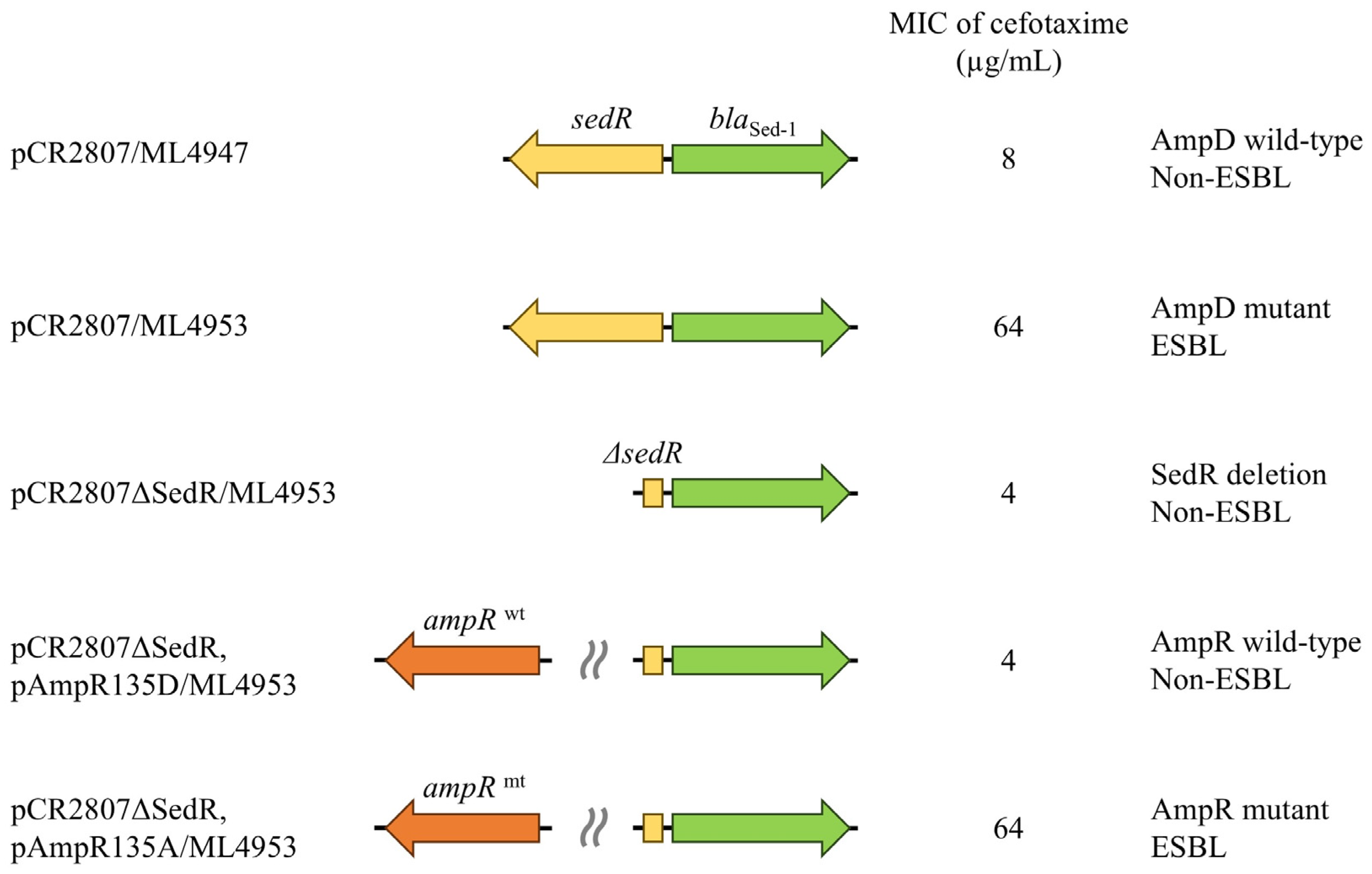
To further elucidate the role of SedR in Sed-1 production, a plasmid carrying ampR was transformed into the sedR-deficient strain (pCR2807ΔSedR/ML4953) (Figure 2). Wild-type AmpR (pAmpR135D), derived from classical AmpC producers, functions as a repressor; whereas mutant AmpR (pAmpR135A) is in a derepressed (active) form. The wild-type ampR strain (pCR2807ΔSedR, pAmpR135D/ML4953) showed 1.66-fold lower Sed-1 activity (2.07 U/mg) compared to pCR2807ΔSedR/ML4953 (3.42 U/mg) (Table 2). In contrast, the mutant ampR strain (pCR2807ΔSedR, pAmpR135A/ML4953) showed 5.87-fold higher Sed-1 activity (20.08 U/mg) and a MIC for 3GC comparable to that of pCR2807/ML4953. These features were confirmed in ATCC51115 transformants. Therefore, SedR functions similarly to the AmpR mutant.

2.4. Characteristics of Various Antibiotic-Resistant Mutants

To investigate the mechanism of successful antibiotic resistance by Sed-1, strains with increased MICs for antibiotics used for selection were analyzed. The antibiotic-resistant mutants appeared at a frequency of ~10−6–10−7 for ceftazidime, cefepime, cefmetazole, aztreonam, and imipenem supplied at 2× or 4× MIC. Ten colonies of the mutants were randomly selected for each condition, and the blaSed-1, sedR, and ampD genes were sequenced and compared to those of the parent C. sedlakii NR2807 strain. Sequence data revealed that only blaSed-1 was altered upon ceftazidime selection. The mutant strains isolated on ceftazidime were found to possess three different sets of mutations in Sed-1 (Pro167Gln; Asp179Gly; and Ile173Met, Pro174Ala, 174_175insS) and drug sensitivity as listed in Table 3. Compared with the NR2807 parent strain, the MIC for ceftazidime was 16-fold higher, while the MIC for cefotaxime was 4- to 8-fold lower in the mutants. Strain NR4574 (Asp179Gly) also displayed a 4-fold lower MIC for aztreonam. Strain NR4575 (Ile173Met, Pro174Ala, 174_175insS) presented a 4-fold lower MIC for piperacillin. Each mutant strain was assumed to possess altered substrate specificity due to amino acid substitutions. To evaluate whether the higher MIC in ceftazidime-resistant mutants was due to mutations in Sed-1, blaSed-1, and sedR of strain NR4573 (Pro167Gln) were cloned into Escherichia coli ML4947 and compared with wild-type transformant pCR2807/ML4947. In line with the original NR4573 strain, pCR4573/ML4947 displayed higher ceftazidime MIC and lower cefotaxime MIC.
No mutations in blaSed-1, sedR, and ampD were found in the other antibiotic-selected mutants. Mutant strain NR4584 selected on 0.5 µg/mL imipenem showed an 8-fold higher MIC for the antibiotics used for selection. Mutant strain NR4062 selected on 8 µg/mL cefmetazole showed 16-fold higher MIC for the antibiotics used for selection, 4-fold higher MIC for cefepime, and 32-fold higher MIC for cefotaxime combined with clavulanic acid. Mutant strains NR4586 and NR5701 selected on 8 µg/mL cefepime and 256 µg/mL aztreonam, respectively, exhibited 4-fold higher MIC for the antibiotic used for selection, along with 8- and 4-fold higher MIC for cefotaxime. Genomic analysis of one representative of each antibiotic mutant strain revealed mutations in various genes (Table 3). Selection with cefepime, aztreonam, and imipenem yielded mutations in cdsA, a phosphatidic acid cytidyl transferase; whereas cefmetazole selection yielded mutations in rseA, an anti-sigma-E factor.

2.5. Kinetic Parameters of Ceftazidime-Resistant Mutants

To evaluate the substrate specificity of wild-type Sed-1 and its ceftazidime-resistant mutants, kinetic parameters were measured for E. coli BL21 transformants harboring wild-type blaSed-1 (NR2807), and ceftazidime-resistant-mutant blaSed-1 (NR4573, NR4574, and NR4575) (Table 4). NR2807 showed lower catalytic efficiency (kcat/Km = 1.31 s−1 mM−1) against ceftazidime, while NR4573 (Pro167Gln), NR4574 (Asp179Gly), and NR4575 (Ile173Met, Pro174Ala, 174_175insS) showed 4.24-, 11.60-, and 30.92-fold higher hydrolytic activity (kcat/Km) than NR2807. On the one hand, Km values were more than 17.78-fold lower in the mutants than in wild-type Sed-1, resulting in substantially higher kcat/Km. On the other hand, they showed 2.11-, 21.83-, and 1.95-fold lower hydrolytic activity against cefotaxime than NR2807, respectively.

3. Discussion

3.1. Genomic and Clinical Significance of Sed-1 in C. Sedlakii

Sed-1 β-lactamase was reported in 2001 as a chromosomally encoded class A β-lactamase in C. sedlakii [17]. Few reports on C. sedlakii isolates followed, probably because the automated bacterial identification systems commonly used in clinical practice failed to identify this species. In this study, Gram-negative rods were identified via whole-genome ANI analysis as C. sedlakii strain NR2807. To the best of our knowledge, this is the first reported clinical isolation of a Sed-1 producer in Japan. According to PubMLST (https://pubmlst.org/bigsdb?db=pubmlst_cfreundii_isolates, accessed on 6 August 2025), only 13 types of STs have been attributed to C. sedlakii, including ST1320, which was newly registered in this study.
Whole-genome analysis showed that blaSed-1 was the only chromosomally encoded β-lactamase in NR2807. As in the type strain ATCC51115, it coded for the regulatory gene sedR. Sed-1 in NR2807 differed from its counterpart in ATCC51115 by amino acid substitutions Asp175Gly and Arg228His. These substitutions have not been related to extended changes in substrate specificity of class A β-lactamases TEM and SHV [19]. Indeed, E. coli transformants (pCR2807/ML4947 and pCR51115/ML4947) harboring these β-lactamases showed similar MICs.

3.2. Functional Impact of AmpD Mutation on Sed-1 Expression and Antibiotic Resistance

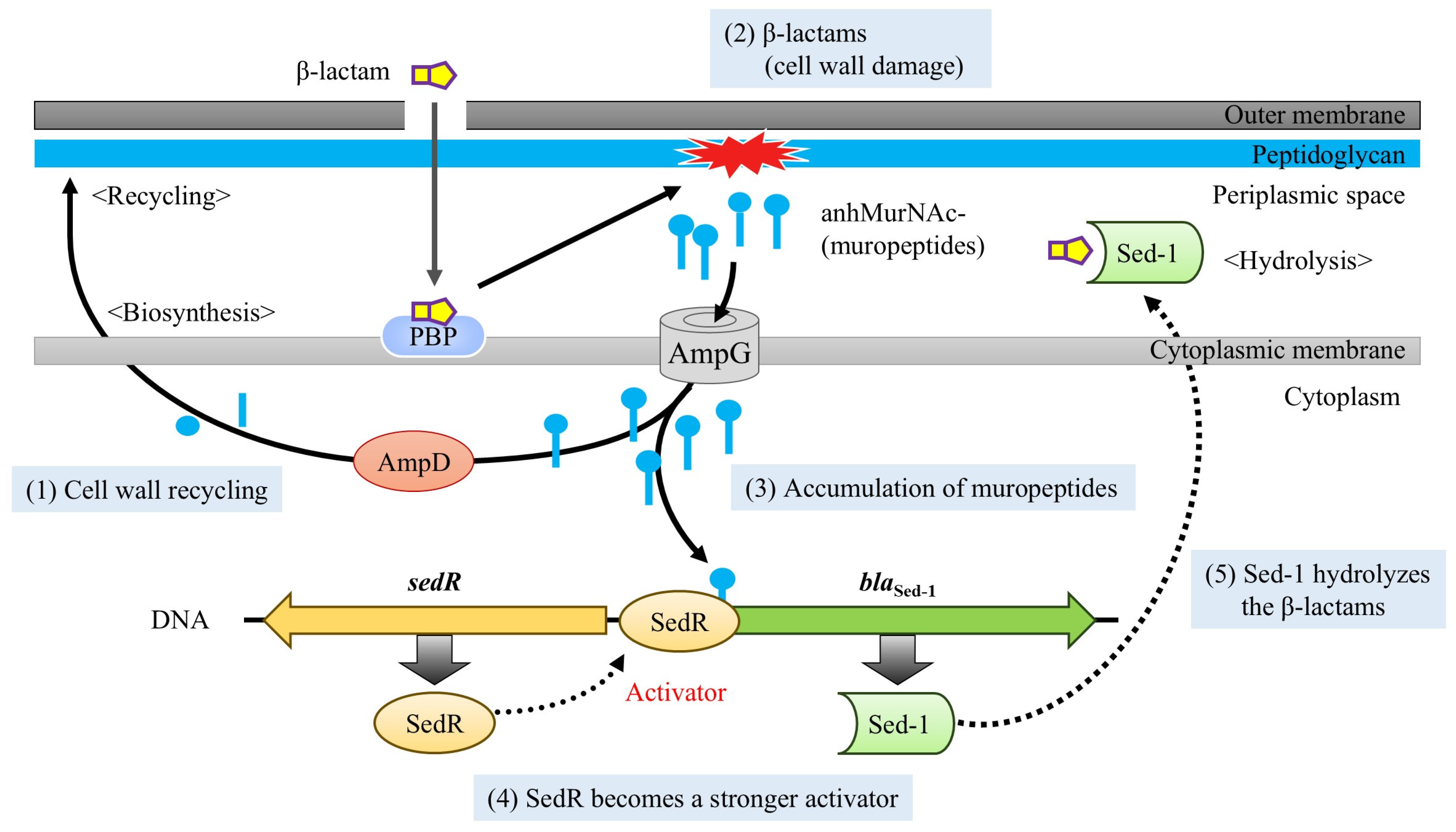
The MIC of NR2807 revealed resistance to a broad range of β-lactams, including 3GC, as well as ESBL-like characteristics; whereas ATCC51115 had an overall lower MIC and no ESBL-like properties (Table 2). Sed-1 production was 65.47-fold higher in NR2807 than in ATCC51115, which may have a significant impact on antimicrobial susceptibility. To elucidate the mechanism regulating Sed-1 expression, blaSed-1sedR was cloned into E. coli with or without the AmpD mutation, and the effect on MIC and enzyme production was assessed. Enzyme activity experiments revealed that pCR2807/ML4947 (AmpD wild-type) and pCR2807/ML4953 (AmpD mutant) increased Sed-1 production by 1.32- and 1.56-fold following induction. Without induction, Sed-1 output was 8.46-fold higher in pCR2807/ML4953 than in pCR2807/ML4957, and the same trend was observed for MIC, suggesting that increased Sed-1 resulted in higher MIC. Accordingly, SedR causes only partial induction of Sed-1, rather than the strong inducibility typically observed in AmpR. Furthermore, Sed-1 expression was strongly influenced by AmpD mutations (Figure 1). In AmpC-producing bacteria, AmpD inactivation leads to the accumulation of cytoplasmic muropeptides, which bind to and activate the transcriptional regulator AmpR. A similar mechanism is likely at play in C. sedlakii, where accumulated muropeptides interact with SedR, shifting it from an inactive to an active transcriptional state. This suggests that SedR requires muropeptide binding to function as a transcriptional activator, and that AmpD mutations indirectly enhance Sed-1 expression through this activation pathway. Therefore, the AmpD mutant mimics an induced state, functionally equivalent to the “Induction” scenario depicted in Figure 1, resulting in elevated β-lactamase activity and increased resistance to 3GC.

3.3. SedR Functions as a Constitutive Activator Similar to Mutant AmpR

The sedR-deficient strain (pCR2807ΔSedR/ML4947) displayed 1.93-fold lower Sed-1 production and lower MIC than the sedR-positive strain (pCR2807/ML4947). Deletion of ampR in inducible AmpC producers resulted in slightly more basal ampC, but no further induction of enzyme synthesis (Figure 1, Supplementary Table S1) [8,20]. In the AmpR (Asp135Ala) mutant, AmpR works as an activator regardless of the presence of an inducer, and its deletion lowers ampC expression [12]. Given that SedR functions similarly to the mutant AmpR, wild-type AmpR, and mutant AmpR were co-inserted in a sedR-deficient strain, and their effect on Sed-1 expression was analyzed. The Sed-1 producer (pCR2807/ML4953) shared the same MIC and enzyme activity as the mutant AmpR-carrying strain (pCR2807ΔSedR, pAmpR135D/ML4953) (Figure 2). Transcriptional regulators of the LysR family are known to act similarly on related genes [21,22]. This result indicates that SedR functions as an activator, akin to mutant AmpR, rather than as a repressor like wild-type AmpR (Figure 3). The amino acid in SedR corresponding to position 135 of AmpR is Asn (Supplementary Figure S1). The Asp135Asn substitution in AmpR has been shown to result in an active form in various bacterial species [8,11,13,14], further confirming the activator-like function of SedR (Supplementary Table S1). Hence, expression of blaSed-1 is regulated by SedR, which acts like the AmpR mutant to activate Sed-1 in the absence of an inducer or aids its production in the presence of an inducer. This mechanism appears unique to Sed-1 compared to other β-lactamases (Figure 1).
ATCC51115 clone strains exhibited similar Sed-1 activity and MIC as NR2807 clone strains, indicating a similar expression mechanism influenced by SedR and AmpD. NR2807 has two amino acid substitutions in Sed-1 compared to ATCC51115, which may have an effect. However, increased Sed-1 production and MIC values were more likely caused by AmpD mutations than by amino acid substitutions. Therefore, NR2807 has become broadly and highly resistant due to the AmpD mutation, whereas ATCC51115 has low Sed-1 production and MIC.

3.4. Ceftazidime-Driven Mutations in Sed-1 Expand Substrate Specificity

To further elucidate the mechanism of β-lactamase expression in NR2807, experiments were conducted to generate a drug-resistant mutant strain. Similar experiments with AmpC producers, such as C. freundii and E. cloacae, yielded mutations in ampD and ampR [11,15,23], but no Sed-1-producing mutants with additional mutations in ampD or sedR could be isolated. These factors could explain the different mutation rates among bacterial species, the role of SedR as an inducible high-producing type, and NR2807 as an AmpD mutant [24]. Here, only the ceftazidime-resistant mutant strain showed an amino acid substitution in Sed-1, which likely expanded substrate specificity and, in this way—rather than via upregulation—improved resistance.
Among the amino acids involved in extended substrate specificity of class A β-lactamase mutants, single substitutions were identified in NR4573 (Pro167Gln) and NR4574 (Asp179Gly) [4,19]. The increased affinity of the mutants for ceftazidime (29.78- and 1213.94-fold decrease in Km, respectively) may have resulted in higher catalytic efficiency and increased MIC. Strain NR4575 had two amino acid substitutions and one insertion (Ile173Met, Pro174Ala, 174_175insS) not previously known to expand substrate specificity of class A β-lactamases. All Class A β-lactamases have a conserved structural feature, the omega loop, which spans residues 161 to 179 or 164 to 179 and forms part of the active site pocket [25,26,27]. The above amino acid substitutions are located in the omega-loop and likely cause a conformational change, thereby increasing enzyme activity against ceftazidime. Interestingly, Sed-1 mutants were obtained in ceftazidime only. No mutations were observed in the regulatory genes of other antibiotics, although mutations were observed in cdsA and rseA [28,29]. These genes are thought to be related to the acquisition of antibiotic resistance through extended substrate specificity.

3.5. ESBL-like Features and Future Risk in Sed-1-Producing C. Sedlakii

In the first report on Sed-1 producing C. sedlakii, Sed-1 showed high catalytic efficiency against narrow-spectrum β-lactams, such as aminopenicillins, carboxypenicillins, and first-generation cephalosporins. Sed-1 producers were resistant to these compounds but were susceptible to 3GC and carbapenems [17]. The wild-type ATCC51115 has a low MIC for extended-spectrum cephalosporins and is not considered an ESBL (Figure 4). Instead, NR2807 exhibited ESBL-like characteristics, such as a high MIC for extended-spectrum cephalosporins. Given reports of C. sedlakii carrying the ESBL gene [18,30], any inquiry into resistance mechanisms by this species will require clarification of ESBLs but also regulatory genes. Furthermore, the catalytic efficiency and MIC exhibited by the ceftazidime mutants obtained in this study clearly suggested they were ESBL producers. Given a history of ESBL types in TEM- and SHV-type Class A β-lactamases, ESBL types may also emerge in Sed-1-producing C. sedlakii [31,32], requiring careful monitoring.

3.6. Study Limitations

This study has some limitations. First, the role of SedR in Sed-1 production is not fully understood. Both strains used in the analysis were active, but there may be mutations that result in even higher output, while others may be suppressive, as in inducible AmpC producers. To elucidate the regulatory mechanism employed by SedR, the function of mutants should be analyzed. Second, several mutations were identified in C. sedlakii strains selected for antibiotics other than ceftazidime, but the cause of resistance to the drug remains unclear. Cloning of each candidate gene would reveal whether its mutation caused increased resistance in Sed-1-producing bacteria. Finally, this study focused on only two strains; more strains need to be collected and analyzed in detail to fully understand the mechanism of Sed-1 production. As this study reveals the possible emergence of ESBL-type Sed-1 producers, such occurrence should be carefully monitored.

4. Materials and Methods

4.1. Bacterial Strains and Antimicrobial Susceptibility Testing

Glucose-fermenting Gram-negative rod-shaped NR2807 was isolated from a blood sample of a hospitalized patient in Japan in 2018. The species was identified by MicroScan WalkAway plus (Beckman Coulter, Inc., Lane Cove West, NSW, Australia), MALDI-TOF MS (VITEK® MS; bioMérieux, North Ryde, NSW, Australia) and ANI analysis using whole-genome sequences (JSpeciesWS, Bremen, Germany) [33]. C. sedlakii ATCC51115 (NBRC105722) was used as the reference strain. To evaluate the effect of sedR and ampD on Sed-1 production, plasmids containing both blaSed-1 and sedR (pCR2807) or blaSed-1 and truncated sedR (pCR2807ΔSedR) were constructed using the Zero Blunt TOPO PCR Cloning Kit (Thermo Fisher Scientific, Waltham, MA, USA) with primers Sed-1down (5′-TGTCTGCGCAGGGTTCTGTTC-3′) and SedRdown (5′-TGGTACGCTGATCCCCGAAC-3′), and Sed-1down and SedRUpR (5′-CGGCGGGATGCGACAGT-3′), respectively. The plasmids were transformed into E. coli ML4947 (AmpD wild-type) and ML4953 (AmpD mutant) [34]. In vitro ceftazidime-resistant mutant strains were isolated from NR2807. The bacterial strains and plasmids used in this study are listed in Table 5. To evaluate the effect of LysR mutation on antimicrobial susceptibility and β-lactamase expression, ampR clone plasmids were transformed into E. coli harboring blaSed-1 and truncated sedR. The ampR clone plasmids pAmpR135D and pAmpR135A used in this study were previously constructed from C. freundii (Supplementary Figure S1) [12].
Antibiotic susceptibility was determined using the agar dilution method according to Clinical and Laboratory Standards Institute guidelines [35].

4.2. Whole-Genome Sequencing and Analysis

Genomic DNA of NR2807 was extracted using QIAGEN Genomic-tip 500/G. Whole genomes were sequenced using MiSeq (Illumina Inc., San Diego, CA, USA) and MinION (Oxford Nanopore Technologies, Oxford, UK), after which they were subjected to hybrid de novo assembly using Unicycler v0.5.0 [36]. Species were identified based on ANI between strain NR2807 (GenBank accession no. BAAHNL01000000.1), C. sedlakii ATCC51115 (GenBank accession no. GCA_000759835.1), C. amalonaticus FDAARGOS_1489 (GenBank accession no. GCF_020099335.1), C. farmeri FDAARGOS 1423 (GenBank accession no. GCA_019048065.1), C. freundii ATCC 8090 (GenBank accession no. CP049015.1), C. koseri FDAARGOS_86 (GenBank accession no. GCA_ 000783445.2), C. rodentium NBRC 105723 (GenBank accession no. GCA_021278985.1), C. werkmanii FDAARGOS_1524 (GenBank accession no. GCA_ 020341495.1), C. youngae NCTC13709 (GenBank accession no. GCA_ 900638065.1), and C. braakii FDAARGOS 1421 (GenBank accession no. GCA_019048805.1). Earlier studies recommended an ANI of 95–96% as a species demarcation cut-off [37,38].
Antimicrobial resistance genes in the genome sequence were identified by searching the ResFinder database (http://genepi.food.dtu.dk/resfinder (accessed on 6 August 2025)) using thresholds of 90% identity and 60% minimum length [39]. Amino acid numbering of Sed-1 corresponded to the class A β-lactamase [40]. Multilocus sequence typing of Citrobacter spp. isolates was performed as previously described [41]. Sequence types were assigned by the PubMLST database (https://pubmlst.org/organisms/citrobacter-spp (accessed on 6 August 2025)).

4.3. Measurement of β-Lactamase Activity and Induction Assays

β-Lactamase activity was measured by a colorimetric assay as previously described [34,42]. Briefly, bacterial strains were cultured in Mueller-Hinton broth to mid-logarithmic phase with shaking at 37 °C, and the protein contents in crude extracts were suspended in 50 mM phosphate buffer (pH 7.0). Enzyme activity was determined at 30 °C using a spectrophotometer (UV-1900i Plus; Shimadzu, Kyoto, Japan) with cephalothin (262 nm) as substrate. One unit of β-lactamase activity was equivalent to the amount of β-lactamase that hydrolyzed 1 μmol of β-lactam in 1 min at 30 °C. Protein concentration was measured using the Bradford assay [43]. All experiments were repeated three times, and enzymatic activity was determined as U/mg of protein. For the induction assay, bacterial strains were cultured in Mueller-Hinton broth to mid-logarithmic phase and subjected to 1/16 × MIC for cefoxitin as the inducer for 1 h [11].

4.4. Selection of Antibiotic-Resistant Strains and Detection of Sequence Changes

Antibiotic-resistant mutants were obtained by plating ~109 colony-forming units (CFU)/mL of late-logarithmic-phase NR2807 grown in Luria–Bertani (LB) broth on LB agar plates containing ceftazidime, cefotaxime, cefepime, cefmetazole, aztreonam, or imipenem at 2× or 4× MIC, as previously described [44]. The mutation frequency was determined by dividing the colony density in CFU/mL on LB agar plates containing the antibiotic by the total colony density in CFU/mL. DNA sequences of blased-1, sedR, and ampD of selected mutants were determined by Sanger sequencing. The primers used are listed in Supplementary Table S3 [17]. Nucleotides and amino acids of selected mutants were compared to those of NR2807. For strains without mutations in these genes, whole-genome sequencing using MiSeq and annotation using DFAST were performed as above. The obtained genome sequences were compared with the sequence of NR2807 to search for mutations.

4.5. Measurement of Kinetic Parameters in NR2807 and Mutants

Amplicons of NR2807 and mutants (NR4573, NR4574, and NR4575) between primers Sed-1F-atg (5′-CTTAAAGAACGGTTTCGCCAGAC-3′) and Sed-1R+BamHI (5′-ATATGGATCCTTACTTTCCTTCCGTCAC-3′) were generated using Q5 Hot Start High-Fidelity 2× Master Mix (New England Biolabs, Ipswich, MA, USA). The amplicons were purified using the High Pure PCR Cleanup Micro Kit (Roche) and digested with BamHI. The resulting fragments underwent kination by T4 Polynucleotide Kinase (TaKaRa Bio, San Jose, CA, USA) and were ligated with a previously described pET28a vector (Merck Millipore, Burlington, MA, USA) [45] using Ligation High Ver. 2 (Toyobo, Osaka, Japan). The resulting plasmids were transformed into One Shot TOP10 Chemically Competent E. coli (Thermo Fisher Scientific). Then, the plasmids were isolated from the transformants using the High Pure Plasmid Isolation Kit (Roche, Basel, Switzerland) and transformed into E. coli BL21(DE3) (Thermo Fisher Scientific) by electroporation. The resulting strains were inoculated into LB broth supplemented with 30 µg/mL kanamycin and incubated at 37 °C. Isopropyl thio-β-D-galactoside (final concentration 0.02 mM) was added when the optical density at 600 nm reached 0.5, and the culture was further incubated for 20 h at 20 °C. Cells were resuspended in buffer A (20 mM Tris/HCl, pH 7.5) and sonicated. The cell-free extract was applied to a Macro-Prep High S Support column (Bio-Rad Laboratories, South Granville, NSW, Australia) and washed with buffer A. The enzymes were eluted with buffer A containing 0.3 M NaCl. Fractions containing Sed-1 or mutant enzymes were pooled, dialyzed against buffer A, and injected into a CM-Toyopearl 650S column (Tosoh, Tokyo, Japan) equilibrated with buffer A. The enzymes were eluted with a linear gradient of NaCl (0–0.25 M) in the same buffer. Enzyme activity was determined with a spectrophotometer (V-730 BIO; JASCO, Hachioji, Japan) at 30 °C in 20 mM HEPES buffer pH7.0. The wavelengths and extinction coefficients used in this study were the same as described previously [45,46]. Protein concentration was determined by Bradford assay [43]. The enzyme was diluted with assay buffer containing 20 µg/mL bovine serum albumin to prevent denaturation. The values of the kinetic parameters (Km and kcat) were obtained by a double-reciprocal (Lineweaver-Burk) plot of initial steady-state velocities at different substrate concentrations.

4.6. Nucleotide Sequence Accession Numbers

Whole-genome DNA sequences of NR2807 were deposited in the GenBank database under accession no. BAAHNL01000000.1

5. Conclusions

In conclusion, the mechanism of Sed-1 production in C. sedlakii appears to differ from that of other β-lactamase-producing bacteria. SedR promotes Sed-1 expression even when not induced and partially promotes it when induced. In particular, the AmpD mutant led to strong expression of Sed-1 and, consequently, elevated resistance. The possibility of ESBL-type emergence in Sed-1 mutant strains has been noted and should be carefully monitored.

Supplementary Materials

The following supporting information can be downloaded at: https://www.mdpi.com/article/10.3390/antibiotics14080823/s1, Figure S1: Amino acid sequence alignment of the N-terminal region of AmpR and SedR (between amino acids 1 and 140); Table S1: Characteristics of inducible AmpC, derepressed AmpC, and Sed-1 producers; Table S2: Glossary of technical terms; Table S3: Primers used in the present study.

Author Contributions

Conceptualization, R.N. and A.N.; methodology, M.W., R.N., K.Y., A.N. and Y.S.; validation, M.W., R.N., K.Y., A.N., Y.S., K.S., S.N. and K.N.; investigation, M.W. and K.Y.; resources, S.N., K.E., K.N. and H.Y.; data curation, M.W., R.N., K.S., K.E. and H.Y.; writing—original draft preparation, M.W. and R.N.; writing—review and editing, M.W., R.N., K.Y., A.N., Y.S., K.S., S.N., K.E., K.N. and H.Y.; visualization, M.W., R.N. and K.Y.; supervision, H.Y.; project administration, H.Y.; funding acquisition, H.Y. All authors have read and agreed to the published version of the manuscript.

Funding

This study was supported by JSPS KAKENHI (Grant No. 22K08606). The funding agency had no role in the study design, data collection, analysis and interpretation, writing of the manuscript, and in the decision to submit the article for publication.

Institutional Review Board Statement

Not applicable.

Informed Consent Statement

Not applicable.

Data Availability Statement

Data will be made available on request.

Acknowledgments

The authors thank Tomoko Asada and Noriko Shirae from Nara Medical University for their excellent technical assistance. We are also particularly grateful to Yuki Yamada from Iwate Medical University Hospital for her continuous support and constructive suggestions.

Conflicts of Interest

The authors declare no conflicts of interest.

Abbreviations

The following abbreviations are used in this manuscript:
3GCThird-generation cephalosporins
CFUColony-forming units
ESBLExtended-spectrum β-lactamase
LBLuria–Bertani

References

  1. Tacconelli, E.; Carrara, E.; Savoldi, A.; Harbarth, S.; Mendelson, M.; Monnet, D.L.; Pulcini, C.; Kahlmeter, G.; Kluytmans, J.; Carmeli, Y.; et al. Discovery, Research, and Development of New Antibiotics: The WHO Priority List of Antibiotic-Resistant Bacteria and Tuberculosis. Lancet Infect Dis. 2018, 18, 318–327. [Google Scholar] [CrossRef]
  2. Melchiorri, D.; Rocke, T.; Alm, R.A.; Cameron, A.M.; Gigante, V. Addressing Urgent Priorities in Antibiotic Development: Insights from WHO 2023 Antibacterial Clinical Pipeline Analyses. Lancet Microbe 2025, 6, 100992. [Google Scholar] [CrossRef] [PubMed]
  3. Husna, A.; Rahman, M.M.; Badruzzaman, A.T.M.; Sikder, M.H.; Islam, M.R.; Rahman, M.T.; Alam, J.; Ashour, H.M. Extended-Spectrum Beta-Lactamases (Esbl): Challenges and Opportunities. Biomedicines 2023, 11, 2937. [Google Scholar] [CrossRef]
  4. Philippon, A.; Slama, P.; Deny, P.; Labia, R. A Structure-Based Classification of Class a Beta-Lactamases, a Broadly Diverse Family of Enzymes. Clin. Microbiol. Rev. 2016, 29, 29–57. [Google Scholar] [CrossRef]
  5. Naas, T.; Aubert, D.; Ozcan, A.; Nordmann, P. Chromosome-Encoded Narrow-Spectrum Ambler Class a Beta-Lactamase Gil-1 from Citrobacter gillenii. Antimicrob. Agents Chemother. 2007, 51, 1365–1372. [Google Scholar] [CrossRef] [PubMed]
  6. Ortiz de la Rosa, J.M.; Bouvier, M.; Poirel, L.; Greub, G.; Blanc, D.; Nordmann, P. Cross-Reaction of Naturally-Produced Beta-Lactamases from Citrobacter farmeri and Citrobacter amalonaticus with Immunological Detection of Ctx-M Enzymes. Diagn. Microbiol. Infect. Dis. 2022, 104, 115760. [Google Scholar] [CrossRef]
  7. Underwood, S.; Avison, M.B. Citrobacter koseri and Citrobacter amalonaticus Isolates Carry Highly Divergent Beta-Lactamase Genes Despite Having High Levels of Biochemical Similarity and 16s rRNA Sequence Homology. J. Antimicrob. Chemother. 2004, 53, 1076–1080. [Google Scholar] [CrossRef]
  8. Bartowsky, E.; Normark, S. Purification and Mutant Analysis of Citrobacter freundii Ampr, the Regulator for Chromosomal Ampc Beta-Lactamase. Mol. Microbiol. 1991, 5, 1715–1725. [Google Scholar] [CrossRef]
  9. Guerin, F.; Isnard, C.; Cattoir, V.; Giard, J.C. Complex Regulation Pathways of Ampc-Mediated B-Lactam Resistance in Enterobacter cloacae Complex. Antimicrob. Agents Chemother. 2015, 59, 7753–7761. [Google Scholar] [CrossRef] [PubMed]
  10. Avison, M.B.; Underwood, S.; Okazaki, A.; Walsh, T.R.; Bennett, P.M. Analysis of Ampc Beta-Lactamase Expression and Sequence in Biochemically Atypical Ceftazidime-Resistant Enterobacteriaceae from Paediatric Patients. J. Antimicrob. Chemother. 2004, 53, 584–591. [Google Scholar] [CrossRef]
  11. Kuga, A.; Okamoto, R.; Inoue, M. Ampr Gene Mutations That Greatly Increase Class C B-Lactamase Activity in Enterobacter cloacae. Antimicrob. Agents Chemother. 2000, 44, 561–567. [Google Scholar] [CrossRef]
  12. Nakano, R.; Nakano, A.; Yano, H.; Okamoto, R. Role of Ampr in the High Expression of the Plasmid-Encoded Ampc Beta-Lactamase Cfe-1. mSphere 2017, 2, e00192-17. [Google Scholar] [CrossRef] [PubMed]
  13. Caille, O.; Zincke, D.; Merighi, M.; Balasubramanian, D.; Kumari, H.; Kong, K.F.; Silva-Herzog, E.; Narasimhan, G.; Schneper, L.; Lory, S.; et al. Structural and Functional Characterization of Pseudomonas aeruginosa Global Regulator Ampr. J. Bacteriol. 2014, 196, 3890–3902. [Google Scholar] [CrossRef] [PubMed]
  14. Juan, C.; Macia, M.D.; Gutierrez, O.; Vidal, C.; Perez, J.L.; Oliver, A. Molecular Mechanisms of Beta-Lactam Resistance Mediated by Ampc Hyperproduction in Pseudomonas aeruginosa Clinical Strains. Antimicrob. Agents Chemother. 2005, 49, 4733–4738. [Google Scholar] [CrossRef]
  15. Kaneko, K.; Okamoto, R.; Nakano, R.; Kawakami, S.; Inoue, M. Gene Mutations Responsible for Overexpression of Ampc B-Lactamase in Some Clinical Isolates of Enterobacter cloacae. J. Clin. Microbiol. 2005, 43, 2955–2958. [Google Scholar] [CrossRef]
  16. Balcewich, M.D.; Reeve, T.M.; Orlikow, E.A.; Donald, L.J.; Vocadlo, D.J.; Mark, B.L. Crystal Structure of the Ampr Effector Binding Domain Provides Insight into the Molecular Regulation of Inducible Ampc Beta-Lactamase. J. Mol. Biol. 2010, 400, 998–1010. [Google Scholar] [CrossRef]
  17. Petrella, S.; Clermont, D.; Casin, I.; Jarlier, V.; Sougakoff, W. Novel Class a Beta-Lactamase Sed-1 from Citrobacter sedlakii: Genetic Diversity of Beta-Lactamases within the Citrobacter Genus. Antimicrob. Agents Chemother. 2001, 45, 2287–2298. [Google Scholar] [CrossRef]
  18. Tasnim, Y.; Stanley, C.; Rahman, M.K.; Awosile, B. Bla Sed-1 Beta-Lactamase-Producing Citrobacter Sedlakii Isolated from Horses and Genomic Comparison with Human-Derived Isolates. J. Appl. Microbiol. 2024, 135, lxae278. [Google Scholar] [CrossRef]
  19. Matagne, A.; Lamotte-Brasseur, J.; Frere, J.M. Catalytic Properties of Class a Beta-Lactamases: Efficiency and Diversity. Biochem. J. 1998, 330 Pt 2, 581–598. [Google Scholar] [CrossRef]
  20. Bennett, P.M.; Chopra, I. Molecular Basis of Beta-Lactamase Induction in Bacteria. Antimicrob. Agents Chemother. 1993, 37, 153–158. [Google Scholar] [CrossRef] [PubMed]
  21. Maddocks, S.E.; Oyston, P.C.F. Structure and Function of the Lysr-Type Transcriptional Regulator (Lttr) Family Proteins. Microbiology (Reading) 2008, 154 Pt 12, 3609–3623. [Google Scholar] [CrossRef] [PubMed]
  22. Kong, K.F.; Jayawardena, S.R.; Indulkar, S.D.; Del Puerto, A.; Koh, C.L.; Hoiby, N.; Mathee, K. Pseudomonas aeruginosa Ampr Is a Global Transcriptional Factor that Regulates Expression of Ampc and Poxb Beta-Lactamases, Proteases, Quorum Sensing, and Other Virulence Factors. Antimicrob. Agents Chemother. 2005, 49, 4567–4575. [Google Scholar] [CrossRef]
  23. Stapleton, P.; Shannon, K.; Phillips, I. DNA Sequence Differences of Ampd Mutants of Citrobacter freundii. Antimicrob. Agents Chemother. 1995, 39, 2494–2498. [Google Scholar] [CrossRef]
  24. Kohlmann, R.; Bahr, T.; Gatermann, S.G. Species-Specific Mutation Rates for Ampc Derepression in Enterobacterales with Chromosomally Encoded Inducible Ampc Beta-Lactamase. J. Antimicrob. Chemother. 2018, 73, 1530–1536. [Google Scholar] [CrossRef]
  25. Arpin, C.; Labia, R.; Andre, C.; Frigo, C.; El Harrif, Z.; Quentin, C. Shv-16, a Beta-Lactamase with a Pentapeptide Duplication in the Omega Loop. Antimicrob. Agents Chemother. 2001, 45, 2480–2485. [Google Scholar] [CrossRef]
  26. Hayes, F.; Hallet, B.; Cao, Y. Insertion Mutagenesis as a Tool in the Modification of Protein Function. Extended Substrate Specificity Conferred by Pentapeptide Insertions in the Omega-Loop of Tem-1 Beta-Lactamase. J. Biol. Chem. 1997, 272, 28833–28836. [Google Scholar] [CrossRef]
  27. Vakulenko, S.B.; Taibi-Tronche, P.; Toth, M.; Massova, I.; Lerner, S.A.; Mobashery, S. Effects on Substrate Profile by Mutational Substitutions at Positions 164 and 179 of the Class a Tem (Puc19) Beta-Lactamase from Escherichia coli. J. Biol. Chem. 1999, 274, 23052–23060. [Google Scholar] [CrossRef] [PubMed]
  28. Laubacher, M.E.; Ades, S.E. The Rcs Phosphorelay Is a Cell Envelope Stress Response Activated by Peptidoglycan Stress and Contributes to Intrinsic Antibiotic Resistance. J. Bacteriol. 2008, 190, 2065–2074. [Google Scholar] [CrossRef] [PubMed]
  29. Sutterlin, H.A.; Zhang, S.; Silhavy, T.J. Accumulation of Phosphatidic Acid Increases Vancomycin Resistance in Escherichia coli. J. Bacteriol. 2014, 196, 3214–3220. [Google Scholar] [CrossRef]
  30. Negrete-Gonzalez, C.; Turrubiartes-Martinez, E.; Briano-Macias, M.; Noyola, D.; Perez-Gonzalez, L.F.; Gonzalez-Amaro, R.; Nino-Moreno, P. Plasmid Carrying Blactx-M-15, Blaper-1, and Blatem-1 Genes in Citrobacter spp. from Regional Hospital in Mexico. Infect. Dis. 2022, 15, 11786337211065750. [Google Scholar] [CrossRef]
  31. Paterson, D.L.; Bonomo, R.A. Extended-Spectrum Beta-Lactamases: A Clinical Update. Clin. Microbiol. Rev. 2005, 18, 657–686. [Google Scholar] [CrossRef]
  32. Liakopoulos, A.; Mevius, D.; Ceccarelli, D. A Review of Shv Extended-Spectrum Beta-Lactamases: Neglected yet Ubiquitous. Front. Microbiol. 2016, 7, 1374. [Google Scholar] [CrossRef]
  33. Richter, M.; Rossello-Mora, R.; Oliver Glockner, F.; Peplies, J. Jspeciesws: A Web Server for Prokaryotic Species Circumscription Based on Pairwise Genome Comparison. Bioinformatics 2016, 32, 929–931. [Google Scholar] [CrossRef]
  34. Nakano, R.; Okamoto, R.; Nakano, Y.; Kaneko, K.; Okitsu, N.; Hosaka, Y.; Inoue, M. Cfe-1, a Novel Plasmid-Encoded Ampc B-Lactamase with an Ampr Gene Originating from Citrobacter freundii. Antimicrob. Agents Chemother. 2004, 48, 1151–1158. [Google Scholar] [CrossRef] [PubMed]
  35. CLSI. Performance Standards for Antimicrobial Susceptibility Testing, 35th ed; Clsi Supplement M100; Clinical and Laboratory Standards Institute: Wayne, PA, USA, 2025. [Google Scholar]
  36. Wick, R.R.; Judd, L.M.; Gorrie, C.L.; Holt, K.E. Unicycler: Resolving Bacterial Genome Assemblies from Short and Long Sequencing Reads. PLoS Comput. Biol. 2017, 13, e1005595. [Google Scholar] [CrossRef] [PubMed]
  37. Goris, J.; Konstantinidis, K.T.; Klappenbach, J.A.; Coenye, T.; Vandamme, P.; Tiedje, J.M. DNA-DNA Hybridization Values and Their Relationship to Whole-Genome Sequence Similarities. Int. J. Syst. Evol. Microbiol. 2007, 57 Pt 1, 81–91. [Google Scholar] [CrossRef]
  38. Chun, J.; Rainey, F.A. Integrating Genomics into the Taxonomy and Systematics of the Bacteria and Archaea. Int. J. Syst. Evol. Microbiol. 2014, 64 Pt 2, 316–324. [Google Scholar] [CrossRef]
  39. Bortolaia, V.; Kaas, R.S.; Ruppe, E.; Roberts, M.C.; Schwarz, S.; Cattoir, V.; Philippon, A.; Allesoe, R.L.; Rebelo, A.R.; Florensa, A.F.; et al. Resfinder 4.0 for Predictions of Phenotypes from Genotypes. J. Antimicrob. Chemother. 2020, 75, 3491–3500. [Google Scholar] [CrossRef] [PubMed]
  40. Ambler, R.P.; Coulson, A.F.; Frere, J.M.; Ghuysen, J.M.; Joris, B.; Forsman, M.; Levesque, R.C.; Tiraby, G.; Waley, S.G. A Standard Numbering Scheme for the Class a Beta-Lactamases. Biochem. J. 1991, 276 Pt 1, 269–270. [Google Scholar] [CrossRef]
  41. Bai, L.; Xia, S.; Lan, R.; Liu, L.; Ye, C.; Wang, Y.; Jin, D.; Cui, Z.; Jing, H.; Xiong, Y.; et al. Isolation and Characterization of Cytotoxic, Aggregative Citrobacter freundii. PLoS ONE 2012, 7, e33054. [Google Scholar] [CrossRef]
  42. Waley, S.G. A Spectrophotometric Assay of Beta-Lactamase Action on Penicillins. Biochem. J. 1974, 139, 789–790. [Google Scholar] [CrossRef]
  43. Bradford, M.M. A Rapid and Sensitive Method for the Quantitation of Microgram Quantities of Protein Utilizing the Principle of Protein-Dye Binding. Anal. Biochem. 1976, 72, 248–254. [Google Scholar] [CrossRef]
  44. Nakano, R.; Yamada, Y.; Nakano, A.; Suzuki, Y.; Saito, K.; Sakata, R.; Ogawa, M.; Narita, K.; Kuga, A.; Suwabe, A.; et al. The Role of Nmcr, Ampr, and Ampd in the Regulation of the Class a Carbapenemase Nmca in Enterobacter ludwigii. Front. Microbiol. 2021, 12, 794134. [Google Scholar] [CrossRef] [PubMed]
  45. Yamamoto, K.; Tanaka, H.; Kurisu, G.; Nakano, R.; Yano, H.; Sakai, H. Structural Insights into the Substrate Specificity of Imp-6 and Imp-1 Metallo-B-Lactamases. J. Biochem. 2022, 173, 21–30. [Google Scholar] [CrossRef] [PubMed]
  46. Laraki, N.; Franceschini, N.; Rossolini, G.M.; Santucci, P.; Meunier, C.; de Pauw, E.; Amicosante, G.; Frere, J.M.; Galleni, M. Biochemical Characterization of the Pseudomonas aeruginosa 101/1477 Metallo-B-Lactamase Imp-1 Produced by Escherichia coli. Antimicrob. Agents Chemother. 1999, 43, 902–906. [Google Scholar] [CrossRef] [PubMed]
Figure 1. Schematic representation of Sed-1 and AmpC β-lactamase regulation. Inducible AmpC producers are repressed by AmpR but increase production when induced. Deletion of the ampR gene results in slightly higher basal expression. Derepressed AmpC producers are constitutively hyperproducing owing to mutated AmpR (Asp135Asn) with or without induction. Deletion of ampR results in lower expression. Sed-1 producers are semi-inducible, mirroring inducible AmpC producers. Deletion of the sedR gene results in reduced expression, akin to derepressed AmpC producers.
Antibiotics 14 00823 g001
Figure 2. Altered drug sensitivity of Sed-1β-lactamase producers is attributed to mutations in regulatory genes. The AmpD mutant strain (pCR2807/ML4953) shows increased expression of Sed-1 and a higher MIC for cefotaxime (64 µg/mL), whereas sedR deletion (pCR2807ΔSedR/ML4953) results in decreased expression and a lower MIC (4 µg/mL). Co-expression of mutant AmpR in the sedR-deficient strain restores the MIC to 64 µg/mL.
Antibiotics 14 00823 g002
Figure 3. Schematic representation of Sed-1 β-lactamase regulation. (1) In the basal state, cell wall degradation products (muropeptides) enter the cytoplasm via AmpG and are processed by AmpD for biosynthesis or recycling. (2) When the cell wall is damaged by β-lactams, (3) muropeptides accumulate in the cytoplasm. (4) Upon interacting with muropeptides, SedR is converted into a stronger activator in a semi-inducible manner, promoting further production of Sed-1. (5) Sed-1 then hydrolyzes β-lactams in the periplasmic space.
Antibiotics 14 00823 g003
Figure 4. Schematic illustration of the evolution of Sed-1 β-lactamase activity in C. sedlakii. (A) In the wild-type strain, Sed-1 exhibits a narrow-spectrum profile and does not show ESBL properties. (B) In the presence of an AmpD mutation, Sed-1 expression is markedly increased, leading to ESBL-like resistance, particularly against third-generation cephalosporins (3GC). (C) Acquisition of Sed-1 structural mutations (e.g., in the omega-loop) can expand substrate specificity, also conferring ESBL-like properties.
Antibiotics 14 00823 g004
Table 1. Chromosomal β-lactamases produced by the Citrobacter genus.
Speciesβ-Lactamase (Class)Regulator GeneExpression
C. freundii complex
C. braakiiAmpC (C)AmpRInducible
C. freundiiAmpC (C)AmpRInducible
C. murliniaeAmpC (C)AmpRInducible
C. youngaeAmpC (C)AmpRInducible
C. werkmaniiAmpC (C)AmpRInducible
C. portucalensisAmpC (C)AmpRInducible
C. gilleniiGIL-1 (A)-Constitutive
C. sedlakiiSed-1 (A)SedRInducible
C. farmeriSed-1 (A)SedRInducible
C. rodentiumSed-1 (A)SedRInducible
C. amalonaticusCdiA (A)CdiR Inducible
C. koseriCKO-1 (A)-Constitutive
C. cronaeAmpC (C)AmpRInducible
C. pasteuriiAmpC (C)AmpRInducible
Table 2. Susceptibility and β-lactamase activity of C. sedlakii strains and their reconstructed transformants.
SpeciesStrainGenesAmpDMIC (µg/mL) aRelative β-Lactamase Activity (U/mg Protein) c
PIPCTXCTX/CLA bCAZFEPCMZCFXATMIPMBasalInduced dInduced/
Basal
C. sedlakiiNR2807blaSed-1, sedRmutant256320.516428640.1258.398.541.02
C. sedlakiiATCC51115blaSed-1, sedRwild-type80.50.2520.1252810.250.130.403.11
E. colipCR2807/ML4947blaSed-1, sedRwild-type2568≤0.064114160.1252.783.681.32
E. colipCR2807/ML4953 blaSed-1, sedRmutant>25664116414640.12523.5036.661.56
E. colipCR2807ΔSedR/ML4947blaSed-1wild-type642≤0.0620.251480.1251.44NT e-
E. colipCR2807ΔSedR/ML4953blaSed-1mutant2564≤0.0620.52480.1253.42NT-
E. colipCR2807ΔSedR, pAmpR135D/ML4953blaSed-1, ampRwtmutant644≤0.064124160.252.076.152.98
E. colipCR2807ΔSedR, pAmpR135A/ML4953blaSed-1, ampRmtmutant>256640.12516414640.2520.0823.501.17
E. colipCR51115/ML4947blaSed-1, sedRwild-type1284≤0.064124160.252.403.791.58
E. colipCR51115/ML4953blaSed-1, sedRmutant256160.516414640.2516.3922.411.37
E. colipCR51115ΔSedR/ML4947blaSed-1wild-type642≤0.0620.251480.251.48NT-
E. colipCR51115ΔSedR/ML4953blaSed-1mutant2562≤0.0620.51480.1253.35NT-
E. colipCR51115ΔSedR, pAmpR135D/ML4953blaSed-1, ampRwtmutant642≤0.06411280.252.854.141.45
E. colipCR51115ΔSedR, pAmpR135A/ML4953blaSed-1, ampRmtmutant>25632≤0.0616412320.2514.8516.711.13
E. coliML4947-wild-type2≤0.06≤0.060.25≤0.0610.5≤0.06≤0.06<0.01NT-
E. coliML4953-mutant4≤0.06≤0.060.5≤0.0610.5≤0.06≤0.06<0.01NT-
a Antibiotics: PIP, piperacillin; CTX, cefotaxime; CLA, clavulanic acid; CAZ, ceftazidime; FEP, cefepime; CMZ, cefmetazole; CFX, cefoxitin; ATM, aztreonam; IPM, imipenem. b MICs were determined in the presence of clavulanic acid (5 µg/mL). c β-Lactamase activities are the geometric mean values of three independent cultures. The standard deviations were within 10%. d 1/16× MIC of cefoxitin was used as the inducer. e NT, Not tested.
Table 3. Mutations and MICs of C. sedlakii NR2807 mutants.
SpeciesStrainsSelection a,bMutation cMIC (µg/mL) a
Sed-1 dSedRAmpDOther Mutated GenesPIPCTXCTX/CLA eCAZFEPCMZCFXATMIPM
C. sedlakiiNR2807-----256320.516428640.125
C. sedlakiiNR4573CAZ 64P167Q---12880.252561281280.125
C. sedlakiiNR4574CAZ 64D179G---12840.25>256128160.125
C. sedlakiiNR4575CAZ 64I173M, P174A, 174_175insS---6480.25>256228320.125
C. sedlakiiNR4586FEP 8---citC, cdsA, ispH>2562560.251616242560.5
C. sedlakiiNR4062CMZ 8---rseA2566416161632641280.25
C. sedlakiiNR5701ATM 256---tsuA, cdsA, ubiD>2561280.25168142560.5
C. sedlakiiNR4584IPM 0.5---pbpA, cdsA256320.516428641
E. colipCR4573/ML4947-P167Q---642≤0.06640.514160.125
E. colipCR2807/ML4947-----2568≤0.064144160.125
a Antibiotics: PIP, piperacillin; CTX, cefotaxime; CLA, clavulanic acid; CAZ, ceftazidime; FEP, cefepime; CMZ, cefmetazole; CFX, cefoxitin; ATM, aztreonam; IPM, imipenem. b Numbers indicate the selected antibiotics concentration (µg/mL). c Abbreviations; P, Pro; Q, Gln; D, Asp; G, Gly; I, Ile; M, Met; A, Ala; ins, insertion; citC, [Citrate [pro-3S]-lyase] ligase; cdsA, phosphatidate cytidylyltransferase; ispH, 4-hydroxy-3-methylbut-2-enyl diphosphate reductase; rseA, anti-sigma-E factor; pbpA, penicillin-binding protein 2; tsuA, thiosulfate utilization transporter; ubiD, 3-octaprenyl-4-hydroxybenzoate carboxy-lyase. d Amino acid numbering corresponds to class A β-lactamase (40). e MICs were determined in the presence of clavulanic acid (5 µg/mL).
Table 4. Kinetic parameters of ceftazidime-resistant mutants of C. sedlakii NR2807.
β-Lactamase and ParameterValue for Antibiotic a
PiperacillinCefotaximeCeftazidimeAztreonam b,c
Wild-type (NR2807)
kcat (s−1)624 ± 31.3230 ± 21.34.55 ± 0.6129.26 ± 0.133
Km (μM)326 ± 29.5264 ± 30.23484 ± 48629.6 ± 2.65
kcat/Km (s−1·mM−1)19108711.31 313
P167Q mutant (NR4573)
kcat (s−1)163 ± 1.24172 ± 3.120.649 ± 0.05424.42 ± 0.0496
Km (μM)43.0 ± 1.56417 ± 9.19117 ± 11.955.1 ± 2.77
kcat/Km (s−1·mM−1)37904135.55 80.2
D179G mutant (NR4574)
kcat (s−1)2.57 ± 0.05510.213 ± 0.00170.0436 ± 0.0013NH
Km (μM)10.5 ± 1.225.34 ± 0.2732.87 ± 0.46NH
kcat/Km (s−1·mM−1)24539.915.2 ND
I173M, P174A, 174_175insS mutant (NR4575)
kcat (s−1)62.5 ± 2.136.06 ± 0.2627.94 ± 0.862NH
Km (μM)84.0 ± 6.5513.6 ± 1.89196 ± 24.1NH
kcat/Km (s−1·mM−1)74444640.5ND
a Km and kcat were calculated as means ± SD from three independent experiments. b NH, no hydrolysis was detected. c ND, not determined.
Table 5. Bacterial strains and plasmids used in this study.
Bacterial Strains or PlasmidsCharacteristics a
Strains
NR2807Clinical isolate of C. sedlakii from Japan
ATCC51115Reference strain of C. sedlakii purchased from NITE Biological Resource Center
NR4573Ceftazidime-resistant mutant of C. sedlakii NR2807 with Sed-1 mutation (P167Q)
NR4574Ceftazidime-resistant mutant of C. sedlakii NR2807 with Sed-1 mutation (D179G)
NR4575Ceftazidime-resistant mutant of C. sedlakii NR2807 with Sed-1 mutation (I173M, P174A, 174_175insS)
ML4947E. coli (F galK2 galT22 hsdR hsdM lacY1 metB1 relA supE44 Rif r), cloning host with AmpD wild type
ML4953E. coli (F galK2 galT22 hsdR hsdM lacY1 metB1 relA supE44 Rif r ampD9), cloning host with AmpD mutant
TOP10E. coli (F mcrA Δ(mrr-hsdRMS-mcrBC) φ80lacZΔM15 ΔlacX74 nupG recA1 araD139 Δ(ara leu)7697 galE15 galK16 rpsL(StrR) endA1 nupG, cloning host for analyzing the Sed-1 β-lactamase production
BL21(DE3)E. coli (F ompT hsdSB(rB mB) gal dcm (DE3)), cloning host for analyzing enzyme kinetic
Plasmids
pCR2807pCR-Blunt II-TOPO containing blased-1 and sedR from NR2807 amplified using Sed-1down and SedRdown
pCR2807ΔSedRpCR-Blunt II-TOPO containing blased-1 from NR2807 amplified using Sed-1down and SedRUpR
pCR51115pCR-Blunt II-TOPO containing blased-1 and sedR from pCR51115 amplified using Sed-1down and SedRdown
pCR51115ΔSedRpCR-Blunt II-TOPO containing blased-1 from pCR51115 amplified using Sed-1down and SedRUpR
pCR4573pCR-Blunt II-TOPO containing blased-1 and sedR from NR4573 amplified using Sed-1down and SedRdown
pAmpR135DpMW219 containing ampR fragment of wild type (AmpR135Asp) obtained from [12]
pAmpR135ApMW219 containing ampR fragment of mutant (AmpR135Ala) obtained from [12]
pCR-Blunt II-TOPOCloning vector purchased from Thermo Fisher Scientific, Km r Zeo r
pMW219Cloning vector purchased from Nippon Gene, Km r
pET-28a (+)Protein expression vector purchased from Novagen, Km r
a Rif r, rifampin resistant; Km r, Kanamycin resistant; Zeo r, Zeocin resistant.
Disclaimer/Publisher’s Note: The statements, opinions and data contained in all publications are solely those of the individual author(s) and contributor(s) and not of MDPI and/or the editor(s). MDPI and/or the editor(s) disclaim responsibility for any injury to people or property resulting from any ideas, methods, instructions or products referred to in the content.