Article

Antimicrobial Activity of Greek Native Essential Oils Against Escherichia coli O157:H7 and Antibiotic Resistance Strains Harboring pNorm Plasmid, mecA, mcr-1 and blaOXA Genes

Department of Public Health, Medical School, University of Patras, 26504 Patras, Greece
*
Authors to whom correspondence should be addressed.
Antibiotics 2025, 14(8), 741; https://doi.org/10.3390/antibiotics14080741
Submission received: 1 July 2025 / Revised: 21 July 2025 / Accepted: 22 July 2025 / Published: 24 July 2025

Abstract

Background/Objectives: The rapid emergence of antibiotic-resistant Escherichia coli in food and clinical environments necessitates new, clean-label antimicrobials. This study assessed eight Greek native essential oils—oregano, thyme, dittany, rosemary, peppermint, lavender, cistus and helichrysum—for activity against six genetically and phenotypically diverse E. coli strains (reference, pNorm, mecA, mcr-1, blaOXA and O157:H7). We aimed to identify oils with broad-spectrum efficacy and clarify the chemical constituents responsible. Methods: Disk-diffusion assays measured inhibition zones at dilutions from 50% to 1.56% (v/v). MIC and MBC values were determined by broth microdilution. GC–MS profiling identified dominant components, and Spearman rank-order correlations (ρ) linked composition to activity. Shapiro–Wilk tests (W = 0.706–0.913, p ≤ 0.002) indicated non-normal data, so strain comparisons used Kruskal–Wallis one-way ANOVA with Dunn’s post hoc and Bonferroni correction. Results: Oregano, thyme and dittany oils—rich in carvacrol and thymol—exhibited the strongest activity, with MIC/MBC ≤ 0.0625% (v/v) against all strains and inhibition zones > 25 mm at 50%. No strain-specific differences were detected (H = 0.30–3.85; p = 0.998–0.571; padj = 1.000). Spearman correlations confirmed that carvacrol and thymol content strongly predicted efficacy (ρ = 0.527–0.881, p < 0.001). Oils dominated by non-phenolic terpenes (rosemary, peppermint, lavender, cistus, helichrysum) showed minimal or no activity. Conclusions: Phenolic-rich EOs maintain potent, strain-independent antimicrobial effects—including against multidrug-resistant and O157:H7 strains—via a multi-target mode that overcomes classical resistance. Their low-dose efficacy and GRAS status support their use as clean-label food preservatives or adjuncts to antibiotics or bacteriophages to combat antimicrobial resistance.

1. Introduction

The global rise of antibiotic-resistant bacteria constitutes one of the most pressing public health challenges of the 21st century, prompting an urgent demand for alternative antimicrobial strategies. According to recent estimates, bacterial antimicrobial resistance (AMR) directly caused 1.27 million fatalities worldwide in 2019, with another 4.95 million deaths attributable to drug-resistant infections [1]. This escalating crisis is predominantly driven by the misuse and overuse of antimicrobials in human healthcare, animal husbandry and agriculture, accelerating the emergence and dissemination of resistant pathogens [2]. The AMR complicates the treatment of common infections and jeopardizes critical medical procedures such as surgical interventions, organ transplants and chemotherapy, significantly increasing the risk of complications and mortality [3].
Antibiotic overuse and abuse in both clinical and agricultural settings have played a crucial role in the epidemic, allowing multidrug-resistant bacteria to arise and spread quickly [4]. The food supply chain represents a critical transmission pathway for AMR [5]. Resistant bacteria, notably Escherichia coli, are frequently detected in animal-derived products, fruits and vegetables, usually resulting from contamination during slaughter, processing, or through the use of untreated animal manure as fertilizer [6]. Additionally, irrigation with dirty water promotes the proliferation of resistant microorganisms. Humans can acquire AMR infections through the consumption of contaminated food or water or via direct contact with animals or contaminated environments [7]. E. coli attracted significant scientific attention and public health concern due to its widespread occurrence as a foodborne pathogen and its remarkable capability to acquire and disseminate antibiotic resistance genes [8]. Some E. coli strains containing critical resistance determinants such as pNorm [9], mcr-1 (colistin resistance) [10] and blaOXA (beta-lactamase resistance) [11], pose a growing threat because of their ability to withstand multiple antibiotic treatments, complicating patient management and increasing morbidity and mortality. In certain experimental contexts or environmental screenings, the mecA gene—typically associated with Staphylococcus aureus—has also been detected in E. coli or transferred to it under laboratory conditions [12], raising concerns about potential horizontal gene transfer events. Additionally, E. coli O157:H7 has emerged as a notorious serotype linked to significant foodborne outbreaks across the world, frequently resulting in serious consequences such as hemolytic–uremic syndrome (HUS) [13].
Essential oils (EOs), which are naturally occurring secondary metabolites extracted from plants, have lately received interest as possible antimicrobial agents due to their complex combinations of bioactive chemicals, particularly terpenoids and phenolic compounds [14]. EOs’ antimicrobial properties are attributed to their ability to disrupt bacterial cell membranes, interfere with cellular processes, and inhibit biofilm formation [15], all of which significantly reduce the likelihood of microbial resistance when compared to conventional antibiotics [14,16]. This makes essential oils extremely promising as natural preservatives and medicinal agents, notably in food preservation [17], clinical disinfection and public health efforts. The Mediterranean area, particularly Greece, has a surprisingly rich botanical variety, with plants generating essential oils high in powerful antimicrobial compounds, including carvacrol, thymol, p-cymene and other terpenoids [18,19]. While numerous research have looked at the usefulness of EOs like oregano and thyme, there is still a significant vacuum in the literature addressing the potential of less-studied oils like rosemary (Rosmarinus officinalis), sage (Salvia fruticosa), cistus (Cistus ladanifer) and helichrysum (Helichrysum italicum) [20]. Preliminary findings show that these oils have antibacterial properties [21]. Nevertheless, their efficacy against multidrug resistance (MDR) organisms such as E. coli is unclear [22]. Recent reviews have reaffirmed the antimicrobial potential of essential oils against MDR foodborne bacteria, emphasizing the importance of species-specific evaluations and the need for standardized experimental approaches to assess their applicability in real-world food systems [23,24]. Further research into their chemical makeup and particular mechanisms of action might reveal crucial information about their potential utility as natural antibacterial agents.
Despite numerous studies evaluating essential oils’ antimicrobial potential against general bacterial populations, little attention has been paid to genetically characterized multidrug-resistant E. coli strains carrying significant resistance genes such as pNorm’s, mcr-1, mecA and blaOXA. This notable research gap warrants further investigation, particularly on how antibiotic resistance determinants influence bacterial susceptibility to EOs. The present study addresses this crucial research gap by systematically assessing the antimicrobial efficacy of eight essential oils extracted from Greek native plants against E. coli O157:H7 and various E. coli strains characterized by the presence of these critical antibiotic resistance genes. Employing standardized and robust methodologies, including disk diffusion assays, minimum inhibitory concentration (MIC) and minimum bactericidal concentration (MBC) determination, this investigation aims to establish clear relationships between resistance gene presence and susceptibility patterns to essential oils. Ultimately, the outcomes of this study hold substantial implications for developing targeted and effective natural antimicrobial interventions, contributing decisively not only to mitigating antibiotic resistance that has infiltrated the food chain but also addressing the significant threat posed by E. coli O157:H7 due to its toxin-producing capabilities. Thus, this holistic and comparative assessment of essential oils’ effectiveness against diverse E. coli strains underscores the innovative potential and strategic relevance of natural antimicrobial solutions in safeguarding global food safety standards.

2. Results

2.1. Disk Diffusion Assay
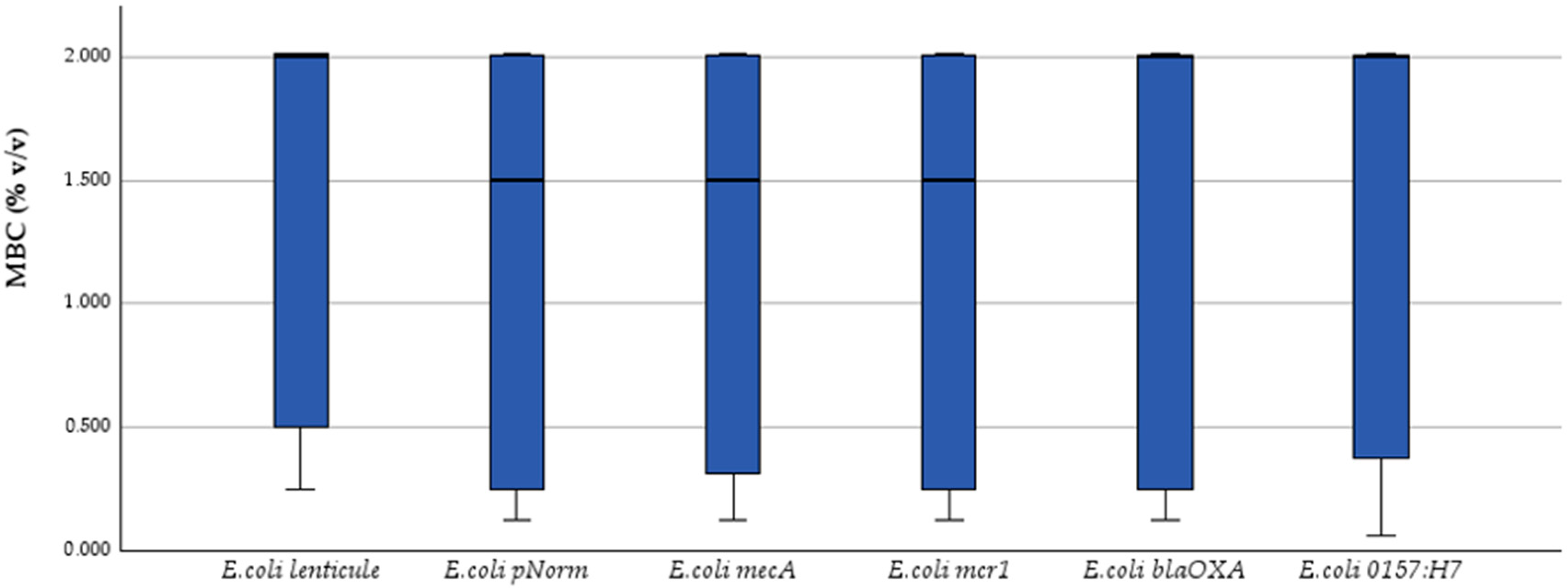
As shown in Table 1, at the highest tested concentration of 50%, all EOs showed some inhibitory effect against the six E. coli bacteria, but the efficacy varied significantly. Oregano (Origanum vulgare) had the most impact, continuously producing inhibition zones greater than 35 mm against resistant organisms like E. coli blaOXA (40.3 ± 1.5 mm) and pNorm (29.7 ± 0.6 mm) at 25% concentration. Dittany (Origanum dictamnus) had strong activity, with zones of 28.3 ± 0.6 mm against the lenticule strain and up to 31.7 ± 1.5 mm against blaOXA. Thyme (Thymus vulgaris) followed closely, especially against pNorm and O157:H7, with zones exceeding 25 mm.
As the concentrations were reduced from 25% to 12.5%, these three essential oils (oregano, thyme and dittany) maintained high action against virtually all E. coli bacteria, albeit with some reduction in zone size. Thyme exhibited a zone of 26.7 ± 0.6 mm at 12.5% against E. coli blaOXA, whereas oregano maintained 20.7 ± 1.2 mm against the mcr-1 strain. At concentrations below 6.25%, the range of effective EOs became significantly smaller. Oregano and thyme were the most robust, producing inhibition zones of 9–13 mm at 3.125% and even 1.56% against certain strains. Oregano formed an 8.4 ± 0.5 mm zone at 1.56% against the lenticule strain, while thyme-maintained activity of 7.5 ± 1.3 mm at the same dilution. Other Eos, including peppermint, lavender and rosemary, had lower effectiveness rates, below 12.5%, resulting in zones of less than 8 mm or none at all.
Cistus (Cistus ladanifer) and helichrysum (Helichrysum italicum) showed little to no action throughout the dilution series. Even at 50% concentration, inhibition zones were either nonexistent or limited (7–8 mm), and in most cases, decreased to 0 mm at concentrations below 6.25%.
Between 24 and 48 h, only minor variations were recorded in the inhibition zone diameters, typically less than ±0.5 mm and never exceeding ±1 mm. No further changes were observed after 48 h, indicating that the maximum antimicrobial effect was reached within the first day of exposure and remained stable thereafter. To contextualize the antimicrobial activity of ΕOs, three conventional antibiotics were included as internal controls in the disk diffusion assays. The inhibition zones and interpretations are shown in Table 2. Results confirmed expected resistance profiles for strains and susceptibility in reference strains, validating assay performance.

2.2. MIC and MBC

The MIC and MBC values of the tested EOs against the six E. coli strains are listed in Table 3. Among all evaluated EOs, oregano had the strongest antibacterial activity, with MIC values ranging from 0.03125% to 0.125% across all strains. Its bactericidal concentrations were likewise consistently low, with MBCs ranging from 0.0625% to 0.25%, showing a significant bactericidal rather than just bacteriostatic activity. The lowest MIC was found against E. coli lenticule and blaOXA strains (0.03125%), whereas the lowest MBC (0.0625%) was found against E. coli O157:H7. Thyme and dittany were the next most effective EOs, with thyme and dittany having MICs ranging from 0.125% to 0.5% throughout the tested strains. Both oils had consistent MBC values, indicating dependable bactericidal activity. Thyme had an MIC and MBC of 0.125% against pNorm and mecA strains, which matched oregano’s effectiveness in these specific circumstances. In contrast, peppermint, lavender, rosemary and cistus had significantly higher MIC and MBC values, ranging from 1% to >2%, indicating lesser efficiency and the requirement for greater doses to accomplish microbial control. Lavender and cistus, for instance, had MBCs greater than 2% against various bacteria, indicating inadequate bactericidal activity even at high dosages. Helichrysum was discovered to be mostly inactive, with MIC and MBC values unknown (ND) for five of six strains and only modest activity against E. coli O157:H7 (MIC = 2%, MBC > 2%).

2.3. Statistical Analysis

Shapiro–Wilk tests (α = 0.05) confirmed significant departures from normality for every response variable (W = 0.706–0.913, p ≤ 0.002). Consequently, inter-strain comparisons were performed with the non-parametric Kruskal–Wallis test.
Kruskal–Wallis one-way ANOVA on ranks revealed no significant strain-to-strain variation for any end-point: MIC (H = 0.30, p = 0.998) (Figure 1), MBC (H = 0.84, p = 0.975) (Figure 2) and inhibition-zone diameters at 50% (Figure 3), 25%, 12.5%, 6.25%, 3.125% and 1.56% v/v dilutions (H = 1.55, 1.15, 3.38, 3.85, 2.55 and 2.28; p = 0.907–0.571). Dunn post hoc contrasts with Bonferroni correction were uniformly non-significant (padj = 1.000), confirming the absence of strain-specific effects.
Thus, both bacteriostatic (MIC) and bactericidal (MBC) activities, as well as diffusion-based inhibition zones, remained essentially strain-independent, underscoring the functional stability of the tested phenolic-rich essential oils across genetically and serotypically diverse E. coli populations.
Spearman rank-order correlations showed that the phenolic monoterpenes carvacrol and thymol are the main determinants of antibacterial potency. Carvacrol content displayed very strong positive correlations with all inhibition-zone diameters (ρ = 0.771–0.881, p < 0.001) and very strong negative correlations with both MIC (ρ = –0.858, p < 0.001) and MBC (ρ = –0.851, p < 0.001). Thymol followed a similar pattern, correlating positively with inhibition zones (ρ = 0.527–0.703, p < 0.001) and negatively with MIC (ρ = –0.699) and MBC (ρ = –0.710, both p < 0.001). In contrast, terpenes such as α-pinene and 1,8-cineole were inversely related to antimicrobial efficacy: α-pinene was negatively associated with zone diameters (e.g., 50% dilution: ρ = –0.520, p < 0.001) and positively associated with MIC (ρ = 0.599, p < 0.001) and MBC (ρ = 0.651, p < 0.001), while 1,8-cineole correlated positively with MIC (ρ = 0.222, p < 0.001) and MBC (ρ = 0.132, p = 0.001) and negatively with zone diameters (ρ = –0.342, p ≤ 0.017). p-Cymene showed moderate positive correlations with inhibition zones (ρ = 0.437–0.645, p ≤ 0.002) but was not significantly linked to MIC/MBC. Menthol, menthone, linalool and linalyl acetate exhibited weak correlations (|ρ| < 0.30, p > 0.05), indicating minimal contribution to the overall activity.

3. Discussion

This study demonstrated that oregano, thyme and dittany essential oils exhibited the most potent antimicrobial activity against all six E. coli strains tested, including the virulent O157:H7 and antibiotic-resistant isolates. These three oils consistently showed the lowest MICs and MBCs across the panel. In contrast, rosemary, peppermint and lavender oils had more moderate effects, requiring higher concentrations to inhibit and kill E. coli. Helichrysum italicum and Cistus ladanifer oils were essentially ineffective against E. coli, with no significant growth suppression observed at practical doses.
These findings align with the literature, which consistently ranks oregano and thyme oils among the most active agents against Gram-negative pathogens such as E. coli [25]. For example, oregano oil has been reported to achieve MICs as low as 0.05% w/w against Gram-negative bacteria, markedly lower than those of thyme oil [26]. Although somewhat less potent, thyme oil still shows significant efficacy. Previous studies have found an MIC of ~0.10% v/v and an MBC of ~0.20% v/v against E. coli O157:H7, whereas in our work, we observed markedly lower thresholds (MIC and MBC = 0.0625%), underscoring the superior potency of the oils tested. Notably, whole thyme oil often outperforms its major constituent alone. Thyme oil has been shown to exert greater antibacterial power against E. coli than pure thymol, an effect attributed to synergistic interactions with its minor components [27]. Dittany was similarly potent and its performance is supported by the literature as well. Mitropoulou et al. found O. dictamnus EO to inhibit a broad range of bacteria, with an MIC of ~0.27% v/v for E. coli, comparable to oregano’s effectiveness [28]. Peppermint, rosemary and lavender oils showed intermediate efficacy. These oils did inhibit E. coli growth, but at higher concentrations (generally in the range of 0.5–2% v/v) compared to the phenolic-rich oils. For instance, οne previous study reported a peppermint oil MIC of 1% (v/v) against E. coli [29], whereas in our work, the minimum inhibitory and bactericidal concentration for peppermint remained consistently at 2% (v/v) across all six strains. Accordingly, rosemary EO often exhibits little to no [30] antibacterial activity within the of ~0.2–0.6% v/v, which is consistently linked to its chemical profile [31]. The literature reports MIC values for lavender oil against E. coli that are an order of magnitude higher than oregano. One study found lavender’s MIC against E. coli to be as high as 0.5–1%, aligning with our findings [32]. In contrast, several of the remaining EOs exhibited only limited or no inhibitory activity against E. coli. Both cistus [33] and helichrysum oils failed to meaningfully suppress growth, a finding that accords with previous reports [34].
The major bioactive constituents—chiefly carvacrol and thymol—exert a multifaceted antimicrobial effect, primarily by disrupting membrane permeability and core cellular functions [35,36]. Their hydrophobic character enables them to insert into the lipid bilayer, causing leakage of intracellular contents, collapse of the electrochemical gradient and, ultimately, cell lysis [37,38]. This observation agrees with reports that oregano oil exhibits potent bactericidal activity against a range of MDR pathogens [39], including E. coli [40], precisely because it inflicts extensive damage on bacterial membranes and associated proteins. Overall, EO efficacy was unaffected by the presence of resistance genes, underscoring their potential value as alternative antimicrobials when conventional antibiotics fail. Recent mechanistic hypotheses suggest that EO constituents may also disrupt quorum sensing, efflux pump activity and metabolic enzyme function in MDR bacteria [41]—complementing their primary membrane-targeting effects and explaining their sustained efficacy even in resistant strains [14,15,16].
The chemical composition of the EOs proved decisive for their antimicrobial performance. It is worth noting that the variability observed in previous studies on EO antimicrobial efficacy can be attributed to differences in plant chemotypes, geographic and seasonal origin, extraction techniques and testing methodologies. These inconsistencies often limit comparability across studies. In contrast, the present work utilized standardized EO concentrations, triplicate replicates and chemically profiled oils, while assessing activity against well-characterized E. coli strains with defined resistance genes. This approach enhances both the reproducibility and the translational relevance of our findings. The particularly high levels of phenolic monoterpenes, in oregano, dittany and thyme oils largely explain their superiority. Gas Chromatography–Mass Spectrometry (GC–MS) profiling showed that our oregano oil contained carvacrol as the dominant constituent (>80–90%), whereas thyme oil was rich in thymol and p-cymene (together ~ 60%). Dittany oil likewise displayed a substantial carvacrol fraction (≈55%) alongside related monoterpenes such as p-cymene (~14%). These phenolic compounds are well documented for their strong antibacterial effects [42]. In our dataset, Spearman analysis revealed a clear positive correlation between an oil’s carvacrol/thymol content and its antimicrobial efficacy, in line with earlier reports. Oregano oils enriched in carvacrol consistently show superior bactericidal action [43], while studies on dittany confirm that carvacrol is the principal contributor to its bioactivity [44]. Beyond the intrinsic strength of the main phenolics, the complex EO matrix can generate synergistic or additive effects [45,46].
The less effective oils (rosemary, peppermint, lavender) have chemical profiles dominated by terpene hydrocarbons or esters rather than phenolic compounds. For example, peppermint oils often contain menthol and menthone as key components [47]; these have moderate antimicrobial effects but again are less potent than carvacrol. Lavender oil’s major constituents include linalool and linalyl acetate [48], while rosemary oil is rich in camphor, α-pinene and 1,8-cineole (eucalyptol) [49]. These compounds, while not inert, generally exhibit milder antimicrobial activity than phenolics.
The inactivity of cistus and helichrysum oils can likewise be attributed to their composition. Although some cistus chemotypes have been reported to inhibit Gram-negative pathogens, including Salmonella and E. coli, those preparations contained high levels of phenolic constituents [50], whereas our chemotype was dominated by less active terpenes, hence its modest effect. Similarly, helichrysum oil consists mainly of non-phenolic terpenes (e.g., neryl acetate, α-pinene, γ-curcumene) [51], to which E. coli is essentially tolerant. A commercial H. italicum oil showed noteworthy activity against Gram-positive bacteria (Staphylococcus aureus, Bacillus spp., MIC 5–10 µg mL−1) but none against E. coli [34]. The most likely explanation is that its major constituents either cannot penetrate the Gram-negative outer membrane effectively or lack sufficient intrinsic potency. In general, oils lacking appreciable phenolic terpenoids failed to act strongly against this Gram-negative pathogen [52], consistent with the broader observation that Gram-negative bacteria are relatively refractory to many plant extracts because the lipopolysaccharide (LPS) barrier restricts entry [53].
An important outcome of this study is that the essential oils retained full efficacy across all tested resistance genotypes and pathogenic backgrounds. The oils’ antimicrobial activity was essentially unaffected by the presence of antibiotic resistance genes such as pNorm, mecA, mcr-1 or blaOXA, and was equally potent against the enterohemorrhagic E. coli O157:H7 strain. This breadth is notable since the agri-food continuum is now recognized as a significant reservoir and transmission route for antibiotic-resistant bacteria [6], and recent O157:H7 outbreaks [54] highlight the importance of new control methods.
Resistance genes typically confer protection against specific antibiotics, yet these mechanisms cannot withstand essential oils’ simultaneous disruption of membranes, proteins and DNA [55]. For instance, cinnamon and thyme oils have shown strong inhibitory activity against E. coli isolates harboring mcr-1, comparable to their activity on colistin-sensitive strains [56]. Additionally, certain thyme oils have been observed to act synergistically with colistin on mcr-1-positive E. coli, restoring colistin susceptibility through membrane permeabilization [57]. Similarly, the presence of the blaOXA β-lactamase gene did not diminish the oils’ bactericidal action. This is expected, as enzymes like OXA-type carbapenemases target β-lactam drug molecules [58] but have no effect on the complex mixture of phytochemicals in an essential oil. In essence, there is no cross-resistance between conventional antibiotics and essential oils because their modes of action are entirely different [59]. Our data exemplify this; even a strain artificially equipped with mecA was not better protected against the oils. Although the mecA gene is conventionally associated with methicillin resistance in S. aureus [60], its sporadic detection in environmental Gram-negative bacteria has been reported, suggesting possible horizontal gene transfer events [12,61]. However, to date, there is no conclusive evidence of stable integration or functional expression of mecA in clinical E. coli isolates [62]. In the present study, the inclusion of an mecA-positive E. coli strain—acquired for research purposes—was intended to model a hypothetical but epidemiologically relevant scenario of resistance gene dissemination. Our goal was not to demonstrate mecA activity in E. coli, but to evaluate whether essential oils retain their antimicrobial effects across genetically diverse resistance backgrounds. Indeed, even when bacteria are serially exposed to sublethal doses of EOs, they rarely develop stable resistance, unlike with antibiotics [63]. It is also noteworthy that the highly pathogenic E. coli O157:H7 strain was as sensitive to the oils as non-O157 strains. Yet, our results indicate that oregano, thyme and dittany oils were uniformly lethal to O157:H7 at low concentrations, just as they were to the other isolates. Numerous works have shown oregano and thyme EOs to be highly effective against E. coli O157:H7 in vitro and even in food matrices [64].
The effective suppression of E. coli, including the O157:H7 serotype, by natural EOs suggests they can serve as clean-label preservatives [65], reducing reliance on synthetic chemicals. Consumer demand is already shifting toward “cleaner” ingredient lists free of nitrites or sorbates [66], creating a niche for plant-derived antimicrobials [67]. Oregano and thyme oils, rich in potent phenolic compounds and sourced from edible herbs, are generally recognized as safe (GRAS/FDA approved) at low doses and have been piloted as antimicrobial food additives [17]. For instance, edible coatings enriched with thyme or oregano oil can be applied to fresh meat [68], fresh fruits [69], cheese or produce [70], forming a thin film that slows pathogen growth. EOs can also be incorporated into active-packaging systems—antimicrobial sachets or films that slowly volatilize the oil and provide continuous protection during storage [71]. However, despite their promising antimicrobial properties, the direct application of essential oils in food systems is not without limitations. Factors such as volatility, oxidation, interactions with food components (e.g., proteins and fats) and strong sensory impact (e.g., flavor, aroma) may affect their stability and consumer acceptability [72]. These aspects must be carefully addressed in future formulation strategies to enhance their practical applicability.
These oils’ ability to suppress both pathogenic and resistance-encoded bacteria at low MIC/MBC thresholds (≤0.0625% v/v) distinguishes them as very versatile antimicrobials. At such minimal concentrations, they can be incorporated into foods, contact surfaces or active-packaging systems with negligible sensory or toxicological impact—advantages seldom matched by conventional chemical preservatives. Crucially, their multi-target mode of action bypasses specific resistance mechanisms, creating a robust barrier against the spread of resistant organisms throughout the food chain. Moreover, our work underpins the potential for these oils to be used alone or in combination with existing—now less effective—antibiotics to restore or enhance their efficacy. Emerging strategies even explore synergistic applications of essential oils with bacteriophages [72], leveraging phages’ targeted lytic activity [73] together with oils’ broad-spectrum membrane disruption [74]. In an era when the agri-food sector urgently needs clean-label, effective alternatives to synthetic sanitizers and failing antibiotics, these essential oils offer a timely, innovative solution for strengthening food safety systems and combating antimicrobial resistance.

4. Materials and Methods

4.1. Essential Oils

This study used eight EOs (Table 4) sourced from local Greek aromatic plants. All oils were received from Vessel Essential Oils, a recognized distillation and processing firm that specializes in organically and bio-grown plant materials. The plant material utilized in EO manufacturing was sourced from a variety of locales around Greece, including mountainous and coastal areas, each with its own microclimate, which contributes to the range and richness of essential oil composition. To retain volatile components, each essential oil was extracted using steam distillation shortly after plant gathering. To retain bioactive components, oils were kept in amber glass vials at 4 °C after extraction.

4.2. GC–MS Chemical Profiling of Essential Oils

The chemical composition of each EO was determined as part of standard internal quality control procedure. Analyses were performed on an HP-5MS capillary column (30 m × 0.25 mm, 0.25 μm) with helium as the carrier gas (1 mL/min) and electron impact ionization at 70 eV [GC–MS system: 6890 Network GC System coupled to a 5975B MSD, Agilent Technologies, Santa Clara, CA, USA]. Retention indices were calculated using a C8–C20 alkane standard series, and compound identification was based on Wiley/NIST05 libraries. Results are expressed as relative area percentages and presented in Table 5.

4.3. Bacterial Strains

Six distinct Escherichia coli strains were employed in this study to assess the antimicrobial efficacy of the selected essential oils. These contained one standard reference strain, one surrogate non-toxigenic O157:H7 strain, and four genetically defined MDR strains, each with its own antibiotic resistance gene. The standard non-resistant E. coli strain was acquired as a certified reference material in the form of LENTICULE® discs [CRM09001M, lot no. BCCH4935, Supelco, Sigma-Aldrich, Buchs, Switzerland], which included freeze-dried E. coli NCTC 9001 (batch SQNL).
The four MRD strains were chosen for their known resistance genotypes and were all laboratory-constructed reference strains provided by Dr. Uli Klümper (Environmental Scences, Technical University of Dresden, Institute of Hydrobiology, Dresden, Germany) as part of the NORMAN project [75]. These strains are commonly used as standardized ARG-positive controls in studies quantifying antimicrobial resistance. The pNorm strain (E. coli TOP10 harboring the pNORM1 plasmid) has a collection of synthetic resistance genes—intI1, blaTEM, vanA, sul1, qnrS1, ctx-m-32 and 16S rRNA—built on a pEX-A2 plasmid backbone and selected with ampicillin. Eurofins Genomics [Eurofins Genomics, Ebersberg, Germany] developed the plasmid, which is commonly utilized in resistance quantification investigations. An mecA-positive E. coli strain was added due to the uncommon but proven horizontal transfer of the methicillin resistance gene from S. aureus to Enterobacterales. Gene presence was verified using PCR. The mcr-1-positive E. coli strain linked with plasmid-mediated colistin resistance was obtained from a reputable source. Before performing antimicrobial testing, the mcr-1 gene was molecularly confirmed. A blaOXA-positive E. coli with an OXA-type β-lactamase was revived from a cryopreserved stock and tested using molecular tests for inclusion in the study.
The E. coli O157:H7 strain, although representing a clinically relevant Shiga toxin-producing serotype, was employed here in its non-toxigenic surrogate form due to biosafety considerations. It was included because to its frequent involvement in foodborne outbreaks and major importance in food safety evaluations. The strain was purchased as a lyophilized tablet with catalog number LGC-ATCC-43895 and batch number 17F13-LGC-ATCC-43895 [LGC Standards, Middlesex, UK; Easy-Tab™ format] and revived using normal microbiological techniques.
Strains were grown in Tryptic Soy Broth [TSB, Oxoid, Basingstoke, Hampshire, UK] and incubated at 37 °C for 18–24 h to achieve logarithmic growth. Bacterial suspensions were adjusted to an optical density of OD625 = 0.08–0.13, comparable to 0.5 McFarland standard (~108 CFU/mL), and utilized immediately for antimicrobial susceptibility testing after strain viability and integrity were verified.

4.4. Disk Diffusion Method

The antibacterial activity of the EOs was evaluated using the disk diffusion method, as described by EUCAST [76]. Suspensions of the E. coli strains were prepared to contain approximately 108 colony-forming units per milliliter (CFU/mL) and spread evenly onto Mueller–Hinton agar plates. Each EO was diluted in dimethyl sulfoxide (DMSO) to yield six final concentrations (% v/v): 50%, 25%, 12.5%, 6.25%, 3.125% and 1.56%. Sterile paper disks (6 mm diameter) were impregnated with 10 μL of each EO concentration and placed aseptically onto the inoculated agar plates. Pure DMSO served as the negative control. Antibiotic disks, including ciprofloxacin (5 μg), gentamicin (10 μg) and cefotaxime (30 μg), were used as positive controls, where appropriate. Additionally, ampicillin (10 μg), erythromycin (15 μg) and oxytetracycline (30 μg) were included to assess phenotypic resistance patterns across all E. coli strains. Interpretations were based on EUCAST v2025 breakpoints for ampicillin; no official breakpoints exist for erythromycin and oxytetracycline in Enterobacterales, and therefore, results for these agents are reported without formal classification. Plates were incubated at 37 °C for 24, 48 and 72 h, and the diameters of inhibition zones (in mm) around each disk were measured, including the disk diameter. Each test was performed in triplicate to ensure reliability and reproducibility of the results.

4.5. MIC and MBC Determination

The minimum inhibitory concentration (MIC) and minimum bactericidal concentration (MBC) of each EO were determined using the standard EUCAST [76] broth microdilution method, adapted for EOs. EOs were diluted in DMSO and then serially two-fold diluted directly in Mueller–Hinton broth (MHB) to achieve concentrations ranging from 2% to 0.00049% (v/v), through twelve sequential dilutions. Bacterial suspensions adjusted to 0.5 McFarland standard (~108 CFU/mL) were diluted to achieve a final inoculum of approximately 5 × 10⁵ CFU/mL in each well. Microtiter plates were incubated at 37 °C for 24 h. The MIC was defined as the lowest concentration that showed no visible bacterial growth. For MBC determination, 100 μL from wells without visible growth were spread onto Mueller–Hinton agar plates and incubated at 37 °C for a further 24 h. The MBC was defined as the lowest concentration resulting in no colony formation. All tests were conducted in triplicate.

4.6. Statistical Analysis

All antimicrobial experiments, including disk diffusion, MIC and MBC assays, were performed in triplicate to ensure reproducibility. Data were initially assessed for normality using the Kolmogorov–Smirnov test and visual inspection of Q–Q plots. Given the non-parametric nature of the data, appropriate non-parametric tests were applied. The Kruskal–Wallis test was used to evaluate potential differences in antimicrobial activity across the six E. coli strains for each EO. When required, pairwise comparisons were conducted using Dunn’s post hoc test with Bonferroni correction. Additionally, Spearman’s rank correlation analysis was employed to explore potential associations between the dominant chemical components of the EOs and their antimicrobial activity. All statistical analyses were carried out using IBM SPSS Statistics, version 29.0. The level of statistical significance was set at p < 0.05.

5. Conclusions

This comprehensive evaluation of eight Greek native essential oils against six genetically and phenotypically diverse E. coli strains, including multidrug-resistant isolates (pNorm, mecA, mcr-1, blaOXA) and the highly virulent O157:H7, demonstrates that carvacrol- and thymol-rich oils (oregano, thyme, dittany) achieve consistent bacteriostatic and bactericidal effects at remarkably low MIC/MBC thresholds (≤0.0625% v/v). This strain-independent potency reflects a multi-target mode of action—membrane disruption, protein denaturation and nucleic acid damage—that overcomes classical resistance mechanisms and affords no cross-protection from antibiotic resistance genes. By contrast, oils dominated by non-phenolic terpenes (rosemary, peppermint, lavender, cistus, helichrysum) required far higher doses or proved inactive. These findings not only validate phenolic-rich essential oils as robust, clean-label antimicrobial agents for food and surface applications but also lay the groundwork for innovative synergies with conventional antibiotics or bacteriophages, offering a timely strategy to reinforce food safety barriers and combat antimicrobial resistance.

Author Contributions

Conceptualization, R.F. and A.V.; methodology, R.F., Z.A. and A.V.; software, R.F.; validation, R.F., Z.A. and A.V.; formal analysis, R.F.; investigation, R.F. and A.V.; resources, A.V.; data curation, R.F. and A.V.; writing—original draft preparation, R.F. and A.V.; writing—review and editing, R.F. and A.V.; visualization, R.F. and A.V.; supervision, A.V.; project administration, R.F. and A.V.; funding acquisition, A.V. All authors have read and agreed to the published version of the manuscript.

Funding

This research was supported by the Hellenic Foundation for Research and Innovation (HFRI) under the 5th Call for HFRI PhD Fellowships (Fellowship Number: 19437).

Institutional Review Board Statement

Not applicable.

Informed Consent Statement

Not applicable.

Data Availability Statement

The data presented in this study are available on request from the corresponding author.

Acknowledgments

The authors would like to thank Vessel Essential Oils [Anthemounta, Farmakeika, Thermi, 57001, Thessaloniki, Greece] for their generous donation of all essential oil samples used in this study. This contribution was instrumental in enabling the experimental procedures. We also wish to acknowledge the contribution of the NORMAN qPCR reference strains for the quantification of ARGs kindly provided by Uli Klümper from the Technical University of Dresden, Germany.

Conflicts of Interest

The authors declare no conflicts of interest.

Abbreviations

The following abbreviations are used in this manuscript:
AMRAntimicrobial Resistance
EOEssential Oil
MICMinimum Inhibitory Concentration
MBCMinimum Bactericidal Concentration
CC-MSGass Chromatography–Mass Spectrometry
LPSLipopolysaccharide
GRASGenerally Recognized As Safe
FDAFood and Drug Administration
M.HMonoterpene Hydrocarbons
O.MOxygenated Monoterpenes
S.HSesquiterpene Hydrocarbons
O.SOxygenated Sesquiterpenes

References

  1. Murray, C.J.; Ikuta, K.S.; Sharara, F.; Swetschinski, L.; Robles Aguilar, G.; Gray, A.; Han, C.; Bisignano, C.; Rao, P.; Wool, E.; et al. Global burden of bacterial antimicrobial resistance in 2019: A systematic analysis. Lancet 2022, 399, 629–655. [Google Scholar] [CrossRef]
  2. Ahmed, S.K.; Hussein, S.; Qurbani, K.; Ibrahim, R.H.; Fareeq, A.; Mahmood, K.A.; Mohamed, M.G. Antimicrobial resistance: Impacts, challenges, and future prospects. J. Med. Surg. Public Health 2024, 2, 100081. [Google Scholar] [CrossRef]
  3. Ferraz, M.P. Antimicrobial Resistance: The Impact from and on Society According to One Health Approach. Societies 2024, 14, 187. [Google Scholar] [CrossRef]
  4. Manyi-Loh, C.; Mamphweli, S.; Meyer, E.; Okoh, A. Antibiotic Use in Agriculture and Its Consequential Resistance in Environmental Sources: Potential Public Health Implications. Molecules 2018, 23, 795. [Google Scholar] [CrossRef] [PubMed]
  5. Ye, Z.; Li, M.; Jing, Y.; Liu, K.; Wu, Y.; Peng, Z. What Are the Drivers Triggering Antimicrobial Resistance Emergence and Spread? Outlook from a One Health Perspective. Antibiotics 2025, 14, 543. [Google Scholar] [CrossRef] [PubMed]
  6. Antimicrobial Resistance in the Environment and the Food Supply: Causes and How It Spreads|Antimicrobial Resistance|CDC. Available online: https://www.cdc.gov/antimicrobial-resistance/causes/environmental-food.html (accessed on 27 June 2025).
  7. Antimicrobial Resistance, Food, and Food Animals|Food Safety|CDC. Available online: https://www.cdc.gov/food-safety/foods/antimicrobial-resistance.html (accessed on 27 June 2025).
  8. Ramos, S.; Silva, V.; de Lurdes Enes Dapkevicius, M.; Caniça, M.; Tejedor-Junco, M.T.; Igrejas, G.; Poeta, P. Escherichia coli as Commensal and Pathogenic Bacteria among Food-Producing Animals: Health Implications of Extended Spectrum β-Lactamase (ESBL) Production. Animals 2020, 10, 2239. [Google Scholar] [CrossRef] [PubMed]
  9. Hembach, N.; Schmid, F.; Alexander, J.; Hiller, C.; Rogall, E.T.; Schwartz, T. Occurrence of the mcr-1 colistin resistance gene and other clinically relevant antibiotic resistance genes in microbial populations at different municipal wastewater treatment plants in Germany. Front. Microbiol. 2017, 8, 1282. [Google Scholar] [CrossRef] [PubMed]
  10. Xie, J.; Liang, B.; Xu, X.; Yang, L.; Li, H.; Li, P.; Qiu, S.; Song, H. Identification of mcr-1-positive multidrug-resistant Escherichia coli isolates from clinical samples in Shanghai, China. J. Glob. Antimicrob. Resist. 2022, 29, 88–96. [Google Scholar] [CrossRef] [PubMed]
  11. Loucif, L.; Chelaghma, W.; Bendjama, E.; Cherak, Z.; Khellaf, M.; Khemri, A.; Rolain, J.M. Detection of blaOXA-48 and mcr-1 Genes in Escherichia coli Isolates from Pigeon (Columba livia) in Algeria. Microorganisms 2022, 10, 975. [Google Scholar] [CrossRef] [PubMed]
  12. Tyasningsih, W.; Ramandinianto, S.C.; Ansharieta, R.; Witaningrum, A.M.; Permatasari, D.A.; Wardhana, D.K.; Effendi, M.H.; Ugbo, E.N. Prevalence and antibiotic resistance of Staphylococcus aureus and Escherichia coli isolated from raw milk in East Java, Indonesia. Vet. World 2022, 15, 2021. [Google Scholar] [CrossRef] [PubMed]
  13. Jangid, H.; Kumar, D.; Kumar, G.; Kumar, R.; Mamidi, N. An Emerging Foodborne Pathogen Spotlight: A Bibliometric Analysis and Scholarly Review of Escherichia coli O157 Research. Antibiotics 2024, 13, 60. [Google Scholar] [CrossRef] [PubMed]
  14. Yammine, J.; Chihib, N.E.; Gharsallaoui, A.; Dumas, E.; Ismail, A.; Karam, L. Essential oils and their active components applied as: Free, encapsulated and in hurdle technology to fight microbial contaminations. A review. Heliyon 2022, 8, e12472. [Google Scholar] [CrossRef] [PubMed]
  15. Maggio, F.; Rossi, C.; Serio, A.; Chaves-Lopez, C.; Casaccia, M.; Paparella, A. Anti-biofilm mechanisms of action of essential oils by targeting genes involved in quorum sensing, motility, adhesion, and virulence: A review. Int. J. Food Microbiol. 2025, 426, 110874. [Google Scholar] [CrossRef] [PubMed]
  16. Kovačević, Z.; Čabarkapa, I.; Šarić, L.; Pajić, M.; Tomanić, D.; Kokić, B.; Božić, D.D. Natural Solutions to Antimicrobial Resistance: The Role of Essential Oils in Poultry Meat Preservation with Focus on Gram-Negative Bacteria. Foods 2024, 13, 3905. [Google Scholar] [CrossRef] [PubMed]
  17. Bibow, A.; Oleszek, W. Essential Oils as Potential Natural Antioxidants, Antimicrobial, and Antifungal Agents in Active Food Packaging. Antibiotics 2024, 13, 1168. [Google Scholar] [CrossRef] [PubMed]
  18. Alexopoulos, A.; Kimbaris, A.C.; Plessas, S.; Mantzourani, I.; Theodoridou, I.; Stavropoulou, E.; Polissiou, M.G.; Bezirtzoglou, E. Antibacterial activities of essential oils from eight Greek aromatic plants against clinical isolates of Staphylococcus aureus. Anaerobe 2011, 17, 399–402. [Google Scholar] [CrossRef] [PubMed]
  19. Sakkas, H.; Papadopoulou, C. Antimicrobial activity of basil, oregano, and thyme essential oils. J. Microbiol. Biotechnol. 2017, 27, 429–438. [Google Scholar] [CrossRef] [PubMed]
  20. Moussa, M.M.; Mazrou, R.M.; Hassan, F.A.S. How Sage and Rosemary Essential Oils Regulate Postharvest Senescence and Extend the Vase Life of Cut Gladiolus Spikes. Horticulturae 2024, 10, 638. [Google Scholar] [CrossRef]
  21. Fokas, R.; Koukousianou, V.; Strataki, A.; Sinou, P.S.; Lamari, F.N.; Vantarakis, A. Comparative invitro evaluation of the antimicrobial properties of essential oils from Lamiaceae, Cistaceae, and Asteraceae families against Enterococcus faecalis. J. Food Sci. Nutr. Res. 2024, 7, 188–197. [Google Scholar] [CrossRef]
  22. Soliman, A.M.; Shaaban, M.T.; Dawood, A.S.; Shaheen, M.N.; Salama, H.S. Microbiological studies on Escherichia coli and Klebsiella pneumoniae causing vaginal and urinary tract inflammation; prevalence, antibiotics-resistance and natural products susceptibility. Microb. Pathog. 2024, 196, 107000. [Google Scholar] [CrossRef] [PubMed]
  23. Sobhy, M.; Abdelkarim, E.A.; Hussein, M.A.; Aziz, T.; Al-Asmari, F.; Alabbosh, K.F.; Cui, H.; Lin, L. Essential oils as antibacterials against multidrug-resistant foodborne pathogens: Mechanisms, recent advances, and legal considerations. Food Biosci. 2025, 64, 105937. [Google Scholar] [CrossRef]
  24. Lupia, C.; Castagna, F.; Bava, R.; Naturale, M.D.; Zicarelli, L.; Marrelli, M.; Statti, G.; Tilocca, B.; Roncada, P.; Britti, D.; et al. Use of Essential Oils to Counteract the Phenomena of Antimicrobial Resistance in Livestock Species. Antibiotics 2024, 13, 163. [Google Scholar] [CrossRef] [PubMed]
  25. Fournomiti, M.; Kimbaris, A.; Mantzourani, I.; Plessas, S.; Theodoridou, I.; Papaemmanouil, V.; Kapsiotis, I.; Panopoulou, M.; Stavropoulou, E.; Bezirtzoglou, E.E.; et al. Antimicrobial activity of essential oils of cultivated oregano (Origanum vulgare), sage (Salvia officinalis), and thyme (Thymus vulgaris) against clinical isolates of Escherichia coli, Klebsiella oxytoca, and Klebsiella pneumoniae. Microb. Ecol. Health Dis. 2015, 26, 23289. [Google Scholar] [CrossRef] [PubMed]
  26. Mihaylova, S.; Tsvetkova, A.; Stamova, S.; Ermenlieva, N.; Tsankova, G.; Georgieva, E.; Peycheva, K.; Panayotova, V.; Voynikov, Y. Antibacterial Effects of Bulgarian Oregano and Thyme Essential Oils Alone and in Combination with Antibiotics Against Klebsiella pneumoniae and Pseudomonas aeruginosa. Microorganisms 2025, 13, 843. [Google Scholar] [CrossRef] [PubMed]
  27. Santurio, F.; de Jesus, K.; Pantella, F.; Adriel, R.; Schlemmer, B.; Fries, M.; Lucy, L.; Scientiae, A.; Flores Santurio, D.; Pantella Kunz de Jesus, F.; et al. Sistema de Información Científica Antimicrobial Activity of the Essential Oil of Thyme and of Thymol against Escherichia coli Strains. Acta Sci. Vet. 2014, 42, 1–4. [Google Scholar]
  28. Mitropoulou, G.; Fitsiou, E.; Stavropoulou, E.; Papavassilopoulou, E.; Vamvakias, M.; Pappa, A.; Oreopoulou, A.; Kourkoutas, Y. Composition, antimicrobial, antioxidant, and antiproliferative activity of Origanum dictamnus (dittany) essential oil. Microb. Ecol. Health Dis. 2015, 26, 26543. [Google Scholar] [CrossRef] [PubMed]
  29. El Maimouni, M.A.; El Amrani, S.; Lairini, S.; Bouslamti, R.; Er-Rajy, M.; Mujwar, S.; Chalkha, M.; Chebbac, K.; El Akhal, F.; El Ouali Lalami, A. Gas Chromatography-Mass Spectrometry Profiling, Antioxidant and Antibacterial Evaluations of Essential Oils Extracted from Mentha piperita and Mentha pulegium. Trop. J. Nat. Prod. Res. 2024, 8, 9386–9398. [Google Scholar] [CrossRef]
  30. Puvača, N.; Milenković, J.; Galonja Coghill, T.; Bursić, V.; Petrović, A.; Tanasković, S.; Pelić, M.; Ljubojević Pelić, D.; Miljković, T. Antimicrobial Activity of Selected Essential Oils against Selected Pathogenic Bacteria: In Vitro Study. Antibiotics 2021, 10, 546. [Google Scholar] [CrossRef] [PubMed]
  31. Kabotso, D.E.K.; Neglo, D.; Gaba, S.E.; Danyo, E.K.; Dayie, A.D.; Asantewaa, A.A.; Kotey, F.C.N.; Dayie, N.T.K.D. In Vitro Evaluation of Rosemary Essential Oil: GC-MS Profiling, Antibacterial Synergy, and Biofilm Inhibition. Pharmaceuticals 2024, 17, 1653. [Google Scholar] [CrossRef] [PubMed]
  32. Zych, S.; Adaszyńska-Skwirzyńska, M.; Szewczuk, M.A.; Szczerbińska, D. Interaction between Enrofloxacin and Three Essential Oils (Cinnamon Bark, Clove Bud and Lavender Flower)—A Study on Multidrug-Resistant Escherichia coli Strains Isolated from 1-Day-Old Broiler Chickens. Int. J. Mol. Sci. 2024, 25, 5220. [Google Scholar] [CrossRef] [PubMed]
  33. Chtibi, H.; Harboul, K.; Benali, T.; Bouyahya, A.; Abdelamjid, K.; Louahlia, S.; Chew, J.; Ming, L.C.; Hammani, K. Comparative Study of Antibacterial Activity of Cistus ladanifer L. Leaves Extracted by Ultrasound-Assisted Extraction and Maceration. Prog. Microbes Mol. Biol. 2023, 6, a0000394. [Google Scholar] [CrossRef]
  34. Djihane, B.; Wafa, N.; Elkhamssa, S.; Pedro, D.H.J.; Maria, A.E.; Mohamed Mihoub, Z. Chemical constituents of Helichrysum italicum (Roth) G. Don essential oil and their antimicrobial activity against Gram-positive and Gram-negative bacteria, filamentous fungi and Candida albicans. Saudi Pharm. J. 2017, 25, 780–787. [Google Scholar] [CrossRef] [PubMed]
  35. Bhavaniramya, S.; Vishnupriya, S.; Al-Aboody, M.S.; Vijayakumar, R.; Baskaran, D. Role of essential oils in food safety: Antimicrobial and antioxidant applications. Grain Oil Sci. Technol. 2019, 2, 49–55. [Google Scholar] [CrossRef]
  36. Kachur, K.; Suntres, Z. The antibacterial properties of phenolic isomers, carvacrol and thymol. Crit. Rev. Food Sci. Nutr. 2020, 60, 3042–3053. [Google Scholar] [CrossRef] [PubMed]
  37. Dong, H.; You, Y.; Wang, N.; Wang, M.; Song, T.; He, Y.; Zou, Y.; He, Y.; Peng, T.; Mei, L. Development of amphipathic derivatives of thymol and carvacrol as potent broad-spectrum antibacterial agents. Eur. J. Med. Chem. 2024, 276, 116716. [Google Scholar] [CrossRef] [PubMed]
  38. Helander, I.M.; Alakomi, H.L.; Latva-Kala, K.; Mattila-Sandholm, T.; Pol, I.; Smid, E.J.; Gorris, L.G.M.; Von Wright, A. Characterization of the Action of Selected Essential Oil Components on Gram-Negative Bacteria. J. Agric. Food Chem. 1998, 46, 3590–3595. [Google Scholar] [CrossRef]
  39. Lu, M.; Dai, T.; Murray, C.K.; Wu, M.X. Bactericidal Property of Oregano Oil Against Multidrug-Resistant Clinical Isolates. Front. Microbiol. 2018, 9, 2329. [Google Scholar] [CrossRef] [PubMed]
  40. Scandorieiro, S.; de Camargo, L.C.; Lancheros, C.A.C.; Yamada-Ogatta, S.F.; Nakamura, C.V.; de Oliveira, A.G.; Andrade, C.G.T.J.; Duran, N.; Nakazato, G.; Kobayashi, R.K.T. Synergistic and additive effect of oregano essential oil and biological silver nanoparticles against multidrug-resistant bacterial strains. Front. Microbiol. 2016, 7, 181296. [Google Scholar] [CrossRef] [PubMed]
  41. Zhang, S.; Wang, J.; Ahn, J. Advances in the Discovery of Efflux Pump Inhibitors as Novel Potentiators to Control Antimicrobial-Resistant Pathogens. Antibiotics 2023, 12, 1417. [Google Scholar] [CrossRef] [PubMed]
  42. Marchese, A.; Arciola, C.R.; Barbieri, R.; Silva, A.S.; Nabavi, S.F.; Sokeng, A.J.T.; Izadi, M.; Jafari, N.J.; Suntar, I.; Daglia, M.; et al. Update on Monoterpenes as Antimicrobial Agents: A Particular Focus on p-Cymene. Materials 2017, 10, 947. [Google Scholar] [CrossRef] [PubMed]
  43. Bairamis, A.; Sotiropoulou, N.S.D.; Tsadila, C.; Tarantilis, P.; Mossialos, D. Chemical Composition and Antimicrobial Activity of Essential Oils and Hydrosols from Oregano, Sage and Pennyroyal against Oral Pathogens. Appl. Sci. 2024, 14, 3238. [Google Scholar] [CrossRef]
  44. Tzortzakis, N. Origanum dictamnus Essential Oil in Vapour or Aqueous Solution Application for Pepper Fruit Preservation against Botrytis cinerea. Agronomy 2024, 14, 257. [Google Scholar] [CrossRef]
  45. Ferreira, A.P.; Almeida-Aguiar, C.; Costa, S.P.G.; Neves, I.C. Essential Oils Encapsulated in Zeolite Structures as Delivery Systems (EODS): An Overview. Molecules 2022, 27, 8525. [Google Scholar] [CrossRef] [PubMed]
  46. Cho, T.J.; Park, S.M.; Yu, H.; Seo, G.H.; Kim, H.W.; Kim, S.A.; Rhee, M.S. Recent advances in the application of antibacterial complexes using essential oils. Molecules 2020, 25, 1752. [Google Scholar] [CrossRef] [PubMed]
  47. Beigi, M.; Torki-Harchegani, M.; Pirbalouti, A.G. Quantity and chemical composition of essential oil of peppermint (Mentha × piperita l.) leaves under different drying methods. Int. J. Food Prop. 2018, 21, 267–276. [Google Scholar] [CrossRef]
  48. Khatri, P.K.; Paolini, M.; Larcher, R.; Ziller, L.; Magdas, D.A.; Marincas, O.; Roncone, A.; Bontempo, L. Botanical characterization and authentication of lavender essential oil using its volatile organic compounds and compound-specific carbon and hydrogen isotope ratio analysis. Food Control 2023, 154, 110002. [Google Scholar] [CrossRef]
  49. Aziz, E.; Batool, R.; Akhtar, W.; Shahzad, T.; Malik, A.; Shah, M.A.; Iqbal, S.; Rauf, A.; Zengin, G.; Bouyahya, A.; et al. Rosemary species: A review of phytochemicals, bioactivities and industrial applications. S. Afr. J. Bot. 2022, 151, 3–18. [Google Scholar] [CrossRef]
  50. Boy, F.R.; Benito, M.J.; Córdoba, M.D.G.; Rodríguez, A.; Casquete, R. Antimicrobial Properties of Essential Oils Obtained from Autochthonous Aromatic Plants. Int. J. Environ. Res. Public Health 2023, 20, 1657. [Google Scholar] [CrossRef] [PubMed]
  51. Furlan, V.; Bren, U. Helichrysum italicum: From Extraction, Distillation, and Encapsulation Techniques to Beneficial Health Effects. Foods 2023, 12, 802. [Google Scholar] [CrossRef] [PubMed]
  52. Ergüden, B. Phenol group of terpenoids is crucial for antibacterial activity upon ion leakage. Lett. Appl. Microbiol. 2021, 73, 438–445. [Google Scholar] [CrossRef] [PubMed]
  53. Snyder, D.S.; McIntosh, T.J. The lipopolysaccharide barrier: Correlation of antibiotic susceptibility with antibiotic permeability and fluorescent probe binding kinetics. Biochemistry 2000, 39, 11777–11787. [Google Scholar] [CrossRef] [PubMed]
  54. Investigation Update: E. coli Outbreak, Onions Served at McDonald’s-November 13, 2024|E. coli Infection|CDC. Available online: https://www.cdc.gov/ecoli/outbreaks/investigation-update-e-coli-o157-2024.html (accessed on 29 June 2025).
  55. Nazzaro, F.; Fratianni, F.; De Martino, L.; Coppola, R.; De Feo, V. Effect of Essential Oils on Pathogenic Bacteria. Pharmaceuticals 2013, 6, 1451. [Google Scholar] [CrossRef] [PubMed]
  56. Foda, A.M.; Kalaba, M.H.; El-Sherbiny, G.M.; Moghannem, S.A.; El-Fakharany, E.M. Antibacterial activity of essential oils for combating colistin-resistant bacteria. Expert Rev. Anti-Infect. Ther. 2022, 20, 1351–1364. [Google Scholar] [CrossRef] [PubMed]
  57. Ben Selma, W.; Farouk, A.; Ban, Z.; Ferjeni, M.; Alsulami, T.; Ali, H.; Boukadida, J. Thymus algeriensis essential oil: Phytochemical investigation, bactericidal activity, synergistic effect with colistin, molecular docking, and dynamics analysis against Gram-negative bacteria resistant to colistin. Heliyon 2024, 10, e38281. [Google Scholar] [CrossRef] [PubMed]
  58. Avci, F.G.; Tastekil, I.; Jaisi, A.; Ozbek Sarica, P.; Sariyar Akbulut, B. A review on the mechanistic details of OXA enzymes of ESKAPE pathogens. Pathog. Glob. Health 2022, 117, 219. [Google Scholar] [CrossRef] [PubMed]
  59. Yap, P.S.X.; Yiap, B.C.; Ping, H.C.; Lim, S.H.E. Essential Oils, A New Horizon in Combating Bacterial Antibiotic Resistance. Open Microbiol. J. 2014, 8, 6. [Google Scholar] [CrossRef] [PubMed]
  60. Pournajaf, A.; Ardebili, A.; Goudarzi, L.; Khodabandeh, M.; Narimani, T.; Abbaszadeh, H. PCR-based identification of methicillin-resistant Staphylococcus aureus strains and their antibiotic resistance profiles. Asian Pac. J. Trop. Biomed. 2014, 4, S293–S297. [Google Scholar] [CrossRef] [PubMed]
  61. Kassem, I.I.; Esseili, M.A.; Sigler, V. Occurrence of mecA in nonstaphylococcal pathogens in surface waters. J. Clin. Microbiol. 2008, 46, 3868–3869. [Google Scholar] [CrossRef] [PubMed]
  62. Miragaia, M. Factors contributing to the evolution of Meca-mediated β-lactam resistance in staphylococci: Update and new insights from whole genome sequencing (WGS). Front. Microbiol. 2018, 9, 396897. [Google Scholar] [CrossRef] [PubMed]
  63. Visan, A.I.; Negut, I. Coatings Based on Essential Oils for Combating Antibiotic Resistance. Antibiotics 2024, 13, 625. [Google Scholar] [CrossRef] [PubMed]
  64. Selim, S. Antimicrobial Activity of Essential Oils Against Vancomycin-Resistant Enterococci (Vre) and Escherichia coli O157:H7 in Feta Soft Cheese and Minced Beef Meat. Braz. J. Microbiol. 2011, 42, 187. [Google Scholar] [CrossRef]
  65. Santiesteban-López, N.A.; Gómez-Salazar, J.A.; Santos, E.M.; Campagnol, P.C.B.; Teixeira, A.; Lorenzo, J.M.; Sosa-Morales, M.E.; Domínguez, R. Natural Antimicrobials: A Clean Label Strategy to Improve the Shelf Life and Safety of Reformulated Meat Products. Foods 2022, 11, 2613. [Google Scholar] [CrossRef] [PubMed]
  66. Asioli, D.; Aschemann-Witzel, J.; Caputo, V.; Vecchio, R.; Annunziata, A.; Næs, T.; Varela, P. Making sense of the “clean label” trends: A review of consumer food choice behavior and discussion of industry implications. Food Res. Int. 2017, 99, 58–71. [Google Scholar] [CrossRef] [PubMed]
  67. Gurtler, J.B.; Garner, C.M. A Review of Essential Oils as Antimicrobials in Foods with Special Emphasis on Fresh Produce. J. Food Prot. 2022, 85, 1300–1319. [Google Scholar] [CrossRef] [PubMed]
  68. Gaba, A.B.M.; Hassan, M.A.; Abd El-Tawab, A.A.; Abdelmonem, M.A.; Morsy, M.K. Protective Impact of Chitosan Film Loaded Oregano and Thyme Essential Oil on the Microbial Profile and Quality Attributes of Beef Meat. Antibiotics 2022, 11, 583. [Google Scholar] [CrossRef] [PubMed]
  69. Sarengaowa; Hu, W.; Feng, K.; Xiu, Z.; Jiang, A.; Lao, Y. Thyme oil alginate-based edible coatings inhibit growth of pathogenic microorganisms spoiling fresh-cut cantaloupe. Food Biosci. 2019, 32, 100467. [Google Scholar] [CrossRef]
  70. Pires, A.; Pietruszka, H.; Bożek, A.; Szkolnicka, K.; Gomes, D.; Díaz, O.; Cobos, A.; Pereira, C. Sheep’s Second Cheese Whey Edible Coatings with Oregano and Clary Sage Essential Oils Used as Sustainable Packaging Material in Cheese. Foods 2024, 13, 674. [Google Scholar] [CrossRef] [PubMed]
  71. Casalini, S.; Baschetti, M.G.; Cappelletti, M.; Guerreiro, A.C.; Gago, C.M.; Nici, S.; Antunes, M.D. Antimicrobial activity of different nanocellulose films embedded with thyme, cinnamon, and oregano essential oils for active packaging application on raspberries. Front. Sustain. Food Syst. 2023, 7, 1190979. [Google Scholar] [CrossRef]
  72. Fokas, R.; Giormezis, N.; Vantarakis, A. Synergistic Approaches to Foodborne Pathogen Control: A Narrative Review of Essential Oils and Bacteriophages. Foods 2025, 14, 1508. [Google Scholar] [CrossRef] [PubMed]
  73. Fokas, R.; Kotsiri, Z.; Vantarakis, A. Can Bacteriophages Be Effectively Utilized for Disinfection in Animal-Derived Food Products? A Systematic Review. Pathogens 2025, 14, 291. [Google Scholar] [CrossRef] [PubMed]
  74. Yap, P.S.X.; Yusoff, K.; Lim, S.H.E.; Chong, C.M.; Lai, K.S. Membrane Disruption Properties of Essential Oils—A Double-Edged Sword? Processes 2021, 9, 595. [Google Scholar] [CrossRef]
  75. Cacace, D.; Fatta-Kassinos, D.; Manaia, C.M.; Cytryn, E.; Kreuzinger, N.; Rizzo, L.; Karaolia, P.; Schwartz, T.; Alexander, J.; Merlin, C.; et al. Antibiotic resistance genes in treated wastewater and in the receiving water bodies: A pan-European survey of urban settings. Water Res. 2019, 162, 320–330. [Google Scholar] [CrossRef] [PubMed]
  76. Eucast: Disk Diffusion Methodology. Available online: https://www.eucast.org/ast_of_bacteria/disk_diffusion_methodology (accessed on 27 June 2025).
Figure 1. MIC (% v/v) box-plots for six E. coli strains; Kruskal–Wallis p = 0.998.
Antibiotics 14 00741 g001
Figure 2. MBC (% v/v) box-plots for the same strains; Kruskal–Wallis p = 0.975.
Antibiotics 14 00741 g002
Figure 3. Inhibition-zone diameter at 50% (v/v) dilution; Kruskal–Wallis p = 0.907.
Antibiotics 14 00741 g003
Table 1. Inhibition zones of selected essential oils against E. coli strains, determined by disk diffusion assay.
Essential OilE. coli (lenticule)
24 h48 h and 72 h
50%25%12.5%6.25%3.125%1.56%50%25%12.5%6.25%3.125%1.56%
Peppermint10.0 ± 0.09.3 ± 1.57.5 ± 0.57.0 ± 0.06.5 ± 0.50.0 ± 0.09.6 ± 0.49.0 ± 0.07.7 ± 0.37.2 ± 0.26.3 ± 0.36.1 ± 0.6
Oregano22.1 ± 0.919.4 ± 0.518.4 ± 0.511.2 ± 1.19.2 ± 1.38.4 ± 0.521.4 ± 0.519.1 ± 0.917.5 ± 1.711.1 ± 1.09.1 ± 1.28.2 ± 0.8
Dittany28.3 ± 0.624.0 ± 1.719.0 ± 0.012.7 ± 1.29.7 ± 1.59.3 ± 0.627.7 ± 0.621.7 ± 2.915.0 ± 2.012.0 ± 1.710.0 ± 1.09.7 ± 1.5
Rosemary11.0 ± 1.011.7 ± 2.18.7 ± 0.66.3 ± 0.67.3 ± 0.60.0 ± 0.010.7 ± 0.610.0 ± 1.09.3 ± 0.67.3 ± 0.47.0 ± 0.60.0 ± 0.0
Thyme24.0 ± 3.617.7 ± 1.215.0 ± 0.611.0 ± 0.68.3 ± 0.67.5 ± 1.323.3 ± 2.915.7 ± 1.214.3 ± 2.511.7 ± 2.38.0 ± 0.07.5 ± 1.3
Lavender10.0 ± 0.010.0 ± 1.77.3 ± 0.67.0 ± 0.60.0 ± 0.00.0 ± 0.09.3 ± 0.69.0 ± 1.07.7 ± 0.67.0 ± 0.00.0 ± 0.00.0 ± 0.0
Cistus12.7 ± 0.611.5 ± 2.10.0 ± 0.00.0 ± 0.00.0 ± 0.00.0 ± 0.010.7 ± 2.90.0 ± 0.00.0 ± 0.00.0 ± 0.00.0 ± 0.00.0 ± 0.0
Helichrysum7.0 ± 0.06.5 ± 0.76.5 ± 0.00.0 ± 0.00.0 ± 0.00.0 ± 0.07.0 ± 0.00.0 ± 0.00.0 ± 0.00.0 ± 0.0 0.0 ± 0.00.0 ± 0.0
Essential OilE. coli (pNorm)
24 h48 h and 72 h
50%25%12.5%6.25%3.125%1.56%50%25%12.5%6.25%3.125%1.56%
Peppermint8.3 ± 0.67.3 ± 0.67.0 ± 0.00.0 ± 0.00.0 ± 0.00.0 ± 0.08 ±7.3 ± 0.67 ± 0.00.0 ± 0.00.0 ± 0.00.0 ± 0.0
OreganoOMZ29.7 ± 0.626.7 ± 2.917.7 ± 0.614 ± 1.010.7 ± 0.6OMZ29.3 ± 1.224.3 ± 0.617 ± 0.012.7 ± 0.610 ± 0.0
Dittany31.7 ± 2.928.3 ± 1.514.3 ± 0.611.3 ± 0.610 ± 0.08.3 ± 0.629.3 ± 1.227.3 ± 2.513.7 ± 0.611.3 ± 0.69.7 ± 0.68.7 ± 0.6
Rosemary11 ± 1.78 ± 0.67.3 ± 0.67.0 ± 0.00.0 ± 0.00.0 ± 0.011 ± 1.78.3 ± 0.67 ± 0.67 ± 0.00.0 ± 0.00.0 ± 0.0
ThymeOMZ36.7 ± 4.217.7 ± 2.512 ± 0.010 ± 0.08 ± 0.0OMZ36.7 ± 4.217.7 ± 2.512 ± 0.010 ± 0.08 ± 0.0
Lavender9.7 ± 1.28 ± 0.07.0 ± 0.07.0 ± 0.07.0 ± 0.00.0 ± 0.09.7 ± 1.28 ± 0.07 ± 0.07 ± 0.07 ± 0.00.0 ± 0.0
Cistus7 ± 0.07 ± 0.00.0 ± 0.00.0 ± 0.00.0 ± 0.00.0 ± 0.07.0 ± 0.07.0 ± 0.00.0 ± 0.00.0 ± 0.00.0 ± 0.00.0 ± 0.0
Helichrysum7 ± 0.07 ± 0.00.0 ± 0.00.0 ± 0.00.0 ± 0.00.0 ± 0.07.0 ± 0.07.0 ± 0.00.0 ± 0.00.0 ± 0.00.0 ± 0.00.0 ± 0.0
Essential OilE. coli (mecA)
24 h48 h and 72 h
50%25%12.5%6.25%3.125%1.56%50%25%12.5%6.25%3.125%1.56%
Peppermint10 ± 0.010.7 ± 1.29.3 ± 1.27.7 ± 1.27.7 ± 1.57.5 ± 0.010 ± 0.010.7 ± 1.28.3 ± 0.67.7 ± 1.28 ± 0.07 ± 0.0
Oregano26.3 ± 3.223 ± 1.020 ± 0.015.7 ± 2.112.3 ± 1.510 ± 0.026.7 ± 2.923 ± 2.020.7 ± 3.115 ± 1.012 ± 0.010 ± 0.0
Dittany21.7 ± 1.518.3 ± 1.514.7 ± 0.611.3 ± 1.28.7 ± 0.67 ± 0.021 ± 1.717.3 ± 2.513.3 ± 0.610.3 ± 0.68 ± 1.07 ± 0.0
Rosemary9 ± 1.08.3 ± 0.67.7 ± 0.67 ± 0.00.0 ± 0.00.0 ± 0.08 ± 0.68.3 ± 1.57.3 ± 0.60.0 ± 0.00.0 ± 0.00.0 ± 0.0
Thyme23.3 ± 2.919.3 ± 1.213.7 ± 1.211.3 ± 0.610 ± 0.07.7 ± 0.620.3 ± 0.617.3 ± 2.512.7 ± 1.510.7 ± 0.610 ± 0.08 ± 0.0
Lavender10 ± 0.09.3 ± 1.28 ± 1.07 ± 0.07 ± 0.07 ± 0.09.7 ± 0.69.3 ± 0.68.3 ± 1.57 ± 0.07 ± 0.07 ± 0.0
Cistus10.3 ± 2.58 ± 0.07 ± 0.07 ± 0.07 ± 0.00.0 ± 0.010.7 ± 3.88 ± 0.07 ± 0.00.0 ± 0.00.0 ± 0.00.0 ± 0.0
Helichrysum0.0 ± 0.00.0 ± 0.00.0 ± 0.00.0 ± 0.00.0 ± 0.0.0.0 ± 0.00.0 ± 0.00.0 ± 0.00.0 ± 0.00.0 ± 0.00.0 ± 0.00.0 ± 0.0
Essential OilE. coli (mcr-1)
24 h48 h and 72 h
50%25%12.5%6.25%3.125%1.56%50%25%12.5%6.25%3.125%1.56%
Peppermint10.3 ± 0.69.3 ± 0.68 ± 1.07.7 ± 0.67 ± 0.07 ± 0.09.7 ± 0.68.7 ± 0.67.7 ± 0.67.7 ± 0.67 ± 0.07 ± 0.0
Oregano24 ± 1.022 ± 1.020.7 ± 1.217.3 ± 0.613.3 ± 0.610.3 ± 0.624.7 ± 0.621 ± 1.020 ± 0.016 ± 1.712.7 ± 0.610 ± 0.0
Dittany22 ± 1.021 ± 1.016 ± 1.710 ± 0.09 ± 0.08.3 ± 0.621 ± 1.020.3 ± 0.613.7 ± 0.69.3 ± 0.68 ± 0.08 ± 0.0
Rosemary9 ± 0.08.7 ± 0.68 ± 0.07.3 ± 0.67 ± 0.00.0 ± 0.08.7 ± 0.69 ± 1.08.3 ± 0.67.3 ± 0.60.0 ± 0.00.0 ± 0.0
Thyme23.7 ± 0.619.3 ± 1.20.0 ± 0.010 ± 0.09.7 ± 0.68.3 ± 0.622.7 ± 1.515.7 ± 4.916 ± 1.710 ± 0.09.7 ± 0.68.7 ± 0.6
Lavender10 ± 0.09 ± 0.08 ± 0.07.3 ± 0.67 ± 0.07 ± 0.09.7 ± 0.69 ± 0.08 ± 0.07 ± 0.07 ± 0.07 ± 0.0
Cistus0.0 ± 0.07 ± 0.07 ± 0.07 ± 0.00.0 ± 0.00.0 ± 0.00.0 ± 0.07 ± 0.07 ± 0.07 ± 0.00.0 ± 0.00.0 ± 0.0
Helichrysum0.0 ± 0.00.0 ± 0.00.0 ± 0.00.0 ± 0.00.0 ± 0.00.0 ± 0.00.0 ± 0.00.0 ± 0.00.0 ± 0.00.0 ± 0.00.0 ± 0.00.0 ± 0.0
Essential OilE. coli (blaOXA)
24 h48 h and 72 h
50%25%12.5%6.25%3.125%1.56%50%25%12.5%6.25%3.125%1.56%
Peppermint10 ± 1.08.3 ± 0.68 ± 0.08 ± 0.08 ± 0.07 ± 0.08.7 ± 0.67.7 ± 0.67.3 ± 0.67.3 ± 0.67 ± 0.07 ± 0.0
OreganoOMZ40.3 ± 1.530.7 ± 3.818.3 ± 0.613.7 ± 0.610 ± 0.0OMZ38.3 ± 2.932.3 ± 2.518.3 ± 0.613.3 ± 0.610.3 ± 0.6
Dittany35.7 ± 3.531.7 ± 1.521.3 ± 3.111.7 ± 0.69.2 ± 0.68.3 ± 0.635 ± 1.031.7 ± 1.519 ± 1.710.7 ± 1.29.16 ± 0.68.3 ± 0.6
Rosemary8.7 ± 0.68 ± 0.08 ± 0.08 ± 0.07 ± 0.00.0 ± 0.08.3 ± 0.68 ± 0.08 ± 0.07 ± 0.07 ± 0.00.0 ± 0.0
ThymeOMZ37.7 ± 2.526.7 ± 0.614.7 ± 0.610.7 ± 0.69.7 ± 0.6OMZ35.7 ± 1.226 ± 3.613.7 ± 0.611.3 ± 0.69.3 ± 0.6
Lavender12.7 ± 1.2 10.3 ± 0.68.7 ± 0.68.3 ± 0.67 ± 0.07 ± 0.011 ± 0.08.7 ± 0.68 ± 0.08 ± 0.07 ± 0.07 ± 0.0
Cistus9.7 ± 1.58 ± 0.08 ± 0.07.3 ± 0.67.3 ± 0.67 ± 0.09.3 ± 1.28 ± 0.08 ± 0.07 ± 0.07 ± 0.00.0 ± 0.0
Helichrysum0.0 ± 0.08 ± 0.07 ± 0.07 ± 0.07 ± 0.07 ± 0.00.0 ± 0.07.7 ± 0.67.3 ± 0.67 ± 0.07 ± 0.07 ± 0.0
Essential OilE. coli O157:H7
24 h48 h and 72 h
50%25%12.5%6.25%3.125%1.56%50%25%12.5%6.25%3.125%1.56%
Peppermint0.0 ± 0.08.7 ± 0.68 ± 1.07 ± 0.07 ± 0.07 ± 0.00.0 ± 0.08.3 ± 0.68 ± 1.07 ± 0.07 ± 0.07 ± 0.0
Oregano28 ± 1.023.3 ± 1.221 ± 1.717.3 ± 0.610.3 ± 0.610 ± 0.024.7 ± 0.621.3 ± 1.220 ± 1.016 ± 1.010 ± 0.09.7 ± 0.6
Dittany23.7 ± 4.719 ± 1.016.3 ± 1.512.3 ± 0.69 ± 1.07.7 ± 0.622.7 ± 3.818.3 ± 1.515.3 ± 1.511 ± 1.08.7 ± 0.68 ± 0.0
Rosemary7.7 ± 0.68.3 ± 1.57.7 ± 0.67 ± 0.00.0 ± 0.00.0 ± 0.08 ± 0.08 ± 1.77.7 ± 0.67 ± 0.00.0 ± 0.00.0 ± 0.0
Thyme25.3 ± 1.522.3 ± 1.515.5 ± 2.113 ± 1.010 ± 0.08 ± 0.023.7 ± 1.221.7 ± 2.114.5 ± 0.712 ± 1.09.3 ± 0.68 ± 0.0
Lavender11.3 ± 0.69.7 ± 1.28.7 ± 1.27.7 ± 0.67 ± 0.07 ± 0.010.3 ± 0.69 ± 1.08.7 ± 0.67.7 ± 0.67 ± 0.07 ± 0.0
Cistus7.5 ± 0.77 ± 0.07.3 ± 0.60.0 ± 0.00.0 ± 0.00.0 ± 0.07 ± 0.07 ± 0.07.3 ± 0.60.0 ± 0.00.0 ± 0.00.0 ± 0.0
Helichrysum0.0 ± 0.07 ± 0.07 ± 0.07 ± 0.00.0 ± 0.00.0 ± 0.00.0 ± 0.07 ± 0.07 ± 0.07 ± 0.00.0 ± 0.00.0 ± 0.0
Values represent mean ± standard deviation (SD) from three independent experiments (n = 3). EO concentrations (% v/v): 50%, 25%, 12.5%, 6.25%, 3.125% and 1.56%. “OMZ” (Over Maximum Zone): inhibition zone > 50 mm.
Table 2. Antimicrobial susceptibility of E. coli strains to antibiotics.
Strain (E. coli)AmpicillinErythromycinOxytetracycline
Zones (mm)InterpretationZones (mm)InterpretationZones (mm)Interpretation
lenticule23.0S13.0ND27.0ND
pNorm14.0R15.0ND34.0ND
mecA0.0R0.0ND0.0ND
mcr-10.0R0.0ND0.0ND
blaOXA0.0R11.0ND33.0ND
O157:H720.0S16.0ND26.0ND
Interpretation based on EUCAST v.2025 breakpoints for ampicillin only. For erythromycin and oxytetracycline, no EUCAST criteria exist for Enterobacterales. Abbreviations: S = Susceptible (inhibition zone ≥ breakpoint according to EUCAST v.2025). R = Resistant (inhibition zone < breakpoint according to EUCAST v.2025). ND = Not Defined (no official EUCAST breakpoint available for this antibiotic–strain combination).
Table 3. Minimum inhibitory concentration and minimum bactericidal concentration values (% v/v) of essential oils against E. coli strains.
EOsE. coli LenticuleE. coli (pNorm)E. coli (mecA)E. coli (mcr-1)E. coli (blaOXA)E. coli O157:H7
MICMBCMICMBCMICMBCMICMBCMICMBCMICMBC
Peppermint1%2%1%2%2%2%2%2%2%2%2%2%
Oregano0.03125%0.25%0.0625%0.25%0.125%0.125%0.125%0.125%0.03125%0.125%0.0625%0.0625%
Dittany0.125%0.5%0.25%0.5%0.5%0.5%0.25%0.25%0.25%0.25%0.5%0.5%
Rosemary2%2%2%2%2%2%2%2%2%2%2%2%
Thyme0.5%0.5%0.125%0.125%0.125%0.125%0.25%0.25%0.25%0.25%0.25%0.25%
Lavender2%>2%0.5%1%1%1%1%1%1%2%2%2%
Cistus2%>2%2%>2%2%>2%2%>2%2%>2%2%>2%
HelichrysumNDNDNDNDNDNDNDNDNDND2%>2%
“>2%” indicates that the bactericidal concentration exceeded the highest concentration tested (2% v/v). “ND” = Not Determined.
Table 4. Botanical information, plant parts used and geographical origin of the essential oils studied.
Essential OilBotanical NamePlant FamilyHarvest RegionPlant Part Used
PeppermintMentha × piperitaLamiaceaeGreeceAerial parts
OreganoOriganum vulgare subsp. hirtum (91.3% carvacrol)LamiaceaeGreeceAerial parts of flowering plant
DittanyOriganum dictamnusLamiaceaeCrete, GreeceAerial parts of flowering plant
RosemaryRosmarinus officinalisLamiaceaeGreeceAerial parts
ThymeThymus vulgarisLamiaceaeGreeceFlowering tops
LavenderLavandula angustifolia cv. Hemus and Lavandula angustifolia cv.LamiaceaeNea Tenedos, Northern GreeceDried flowers and flowering tops
CistusCistus ladaniferCistaceaeNea Tenedos, Northern GreeceLeaves
HelichrysumHelichrysum italicumAsteraceaeVavdos, Halkidiki, Central MacedoniaFlowering tops
Table 5. List of volatile compounds identified in ΕOs by GC–MS analysis and their chemical classification.
EOsVolatile Metabolites (%)Total (%)Density (g/mL) 25 °C
PeppermintMenthol (46), Menthone (24.7), 1.8-Cineole (6.7), Menthyl Acetate (5.4)910.896
M.H. 2.50%, O.M. 88.60%
OreganoCarvacrol (91.30)97.290.95
M.H. 2.38%, O.M. 93.44%, S.H. 0.96%, O.S. 0.44%
DittanyCarvacrol (54.81), p-Cymene (13.99), γ-Terpinene (8.33)95.060.95
M.H. 32.57%, O.M. 57.76%, S.H. 3.70%
RosemaryCamphor (26.91), α-Pinene (14.76), 1,8-Cineole (13.41), Camphene (7.31), β-Myrcene (5.15)93.220.879
M.H. 39.65%, O.M. 50.98%, S.H. 1.09%
Thymep-Cymene (33.53), Thymol (26.59), 1,8-Cineole (6.25), Limonene (5.32)90.590.9189
M.H. 47.61%, O.M. 39.06%, S.H. 2.28%
LavenderLinalyl Acetate (31.82), Linalool (29.27), Cis-β-Ocimene (6.47), Terpinen-4-ol (5.05)97.31%0.885–0.905
M.H. 11.46%, O.M. 57.76%, S.H. 6.18%
Cistusα-Pinene (38.94)71.740.962
M.H. 52.88%, O.M. 12.82%, S.H. 0.64%, O.S. 2.67%
Helichrysumγ-Curcumene (14.94), α-Pinene (14.94), Neryl acetate (14.14), β-Selinene (5.09) 79.170.9
M.H. 18.23%, O.M. 19.71%, S.H. 37.30%
Data were based on reports provided by Vessel Essential Oils’ manufacturers, with only dominant components (>5%) reported and minor ones omitted for clarity. Abbreviations: M.H.: monoterpene hydrocarbons; O.M.: oxygenated monoterpenes; S.H.: sesquiterpene hydrocarbons; O.S.: oxygenated sesquiterpenes.
Disclaimer/Publisher’s Note: The statements, opinions and data contained in all publications are solely those of the individual author(s) and contributor(s) and not of MDPI and/or the editor(s). MDPI and/or the editor(s) disclaim responsibility for any injury to people or property resulting from any ideas, methods, instructions or products referred to in the content.

