Article

First Report of Stenotrophomonas maltophilia from Canine Dermatological Infections: Unravelling Its Antimicrobial Resistance, Biofilm Formation, and Virulence Traits

by
Ria Rajeev
1,†,
Porteen Kannan
1,*,
Sureshkannan Sundaram
1,
Sandhya Bhavani Mohan
2,
Sivachandiran Radjendirane
1,†,
Chaudhary Jeetendrakumar Harnathbhai
3,
Anbazhagan Subbaiyan
4,
Viswanathan Naveenkumar
5,
Nithya Quintoil Mohanadasse
6,
Wilfred Ruban Savariraj
7,
Charley A. Cull
8 and
Raghavendra G. Amachawadi
9,*
1
Department of Veterinary Public Health and Epidemiology, Madras Veterinary College, Tamil Nadu Veterinary and Animal Sciences University (TANUVAS), Chennai 600051, India
2
Department of Clinics, Madras Veterinary College, Tamil Nadu Veterinary and Animal Sciences University (TANUVAS), Chennai 600051, India
3
Department of Veterinary Public Health and Epidemiology, College of Veterinary Science and Animal Husbandry, Kamdhenu University, Anand 388001, India
4
ICMR-National Animal Resource Facility for Biomedical Research (NARFBR), Hyderabad 500101, India
5
Veterinary Clinical Complex, Veterinary College and Research Institute (VC&RI), Tamil Nadu Veterinary and Animal Sciences University (TANUVAS), Udumalpet 642205, India
6
Department of Veterinary Public Health and Epidemiology, Rajiv Gandhi Institute of Veterinary Education and Research (RIVER), Puducherry 605009, India
7
Department of Livestock Products Technology, Karnataka Veterinary, Veterinary College, Animal and Fisheries Sciences University, Bengaluru 560024, India
8
Midwest Veterinary Services, Inc., Oakland, NE 68045, USA
9
Department of Clinical Sciences, College of Veterinary Medicine, Kansas State University, Manhattan, KS 66506, USA
*
Authors to whom correspondence should be addressed.
These authors contributed equally to this work.
Antibiotics 2025, 14(7), 639; https://doi.org/10.3390/antibiotics14070639
Submission received: 7 May 2025 / Revised: 19 June 2025 / Accepted: 20 June 2025 / Published: 23 June 2025
(This article belongs to the Special Issue Antimicrobial Resistance and Infections in Veterinary Settings)

Abstract

Background/Objectives: The present study was aimed at documenting S. maltophilia occurrence in dogs with skin ailments, investigating its virulence, biofilm-forming ability, antimicrobial susceptibility, and zoonotic potential to inform preventive and therapeutic strategies against multidrug resistant S. maltophilia infections. Methods: Skin swabs (n = 300) were collected from dogs with dermatological ailments. Isolation was performed using selective media and confirmed with molecular methods, validated by MALDI Biotyper. Antimicrobial susceptibility testing and efflux activity assessment were conducted. Resistance genes related to sulfonamides, quinolones, and β-lactams were screened. Virulence was assessed by biofilm formation, motility, and virulence gene profiling. Results: In total, 15 S. maltophilia (5%) isolates were identified. All 15 isolates were susceptible to trimethoprim-sulfamethoxazole, enrofloxacin, gatifloxacin, levofloxacin, minocycline, and tigecycline, but resistant to cefpodoxime and aztreonam. The following resistance genes qnr (93.3%), blaOXA-48 (46.7%), blaKPC (33.3%), blaNDM (33.3%), blaCTX-M (20%), blaSHV (20%), and blaTEM (6.7%) were detected. All 15 isolates displayed high efflux activity. Overall, 9 isolates (60%) were strong biofilm producers, and 6 (40%) were moderate. Virulence genes such as virB, motA, rmlA, and fliC were present in all 15 isolates, with others varying in frequency. All isolates exhibited swimming motility. Heat map clustering showed diverse profiles, with no identical isolate patterns. Correlation analysis indicated positive associations between several antimicrobial resistance and virulence genes. Conclusions: This study underscores the zoonotic potential of S. maltophilia from dogs, advocating for a One Health approach to mitigate infection risks and limit the spread of virulent multidrug resistant pathogens.

Graphical Abstract

1. Introduction

The genus Stenotrophomonas, belonging to the class Gammaproteobacteria, consists of non-fermenting gram-negative bacilli (NFGNB) that occur ubiquitously in environments such as soil, water, plant rhizospheres, and the bodies of animals and humans [1]. Stenotrophomonas maltophilia is the primary pathogen within this genus, implicated in various infections in humans. This opportunistic pathogen causes nosocomial infections, including respiratory, wound, urinary tract, ocular, and soft tissue infections, and ranks as the third most frequently isolated unusual NFGNB after Pseudomonas aeruginosa and Acinetobacter baumannii [2].
S. maltophilia expresses numerous virulence factors that facilitate host colonization, such as adhesins (lipopolysaccharides, flagella, type 1 fimbriae, type IV pili) and extracellular enzymes (proteases, esterases, lipases, gelatinase, hemolysin, siderophores, and cytotoxins) [3]. Its biofilm-forming ability on abiotic surfaces and host tissues diminishes the efficacy of drugs used to treat hospital-acquired infections [3]. Additionally, quorum sensing via autoinducers regulates gene expression based on cell density [4].
S. maltophilia is intrinsically resistant to a wide range of antibiotics, including β-lactams, fluoroquinolones, tetracyclines, chloramphenicol, and aminoglycosides. This resistance is mediated by two β-lactamases, L1 (a metallo-β-lactamase) and L2 (a clavulanic acid-susceptible cephalosporinase) [1]. Aminoglycoside resistance is also supported by modifying enzymes [AAC (6′)-Iz, APH (3′)-Iic, AAC (6′)-Iak] and efflux pumps (SmeABC, SmeIJK, SmeYZ) [1]. Additionally, resistance genes such as sul1, sul2, and dfrA associated with Class 1 integrons confer resistance to trimethoprim/sulfamethoxazole (TMP/SMX). TMP/SMX is the drug of choice for treating S. maltophilia infections, with alternate options like levofloxacin, minocycline, tigecycline, and ceftazidime used for TMP/SMX-resistant cases [2].
S. maltophilia has been isolated from animals with chronic respiratory disease and urinary infections, including horses, cats, dogs, and ball pythons [5,6]. However, only one study in India has reported its occurrence in animals, specifically in sarcoptic pig skin ulcers [7], with no prior studies in dogs. This study aimed to document S. maltophilia occurrence in dogs with skin conditions, evaluating its virulence, biofilm-forming capacity, and antimicrobial susceptibility to assess zoonotic potential and develop treatment strategies for biofilm-associated multidrug resistant S. maltophilia infections.

2. Results

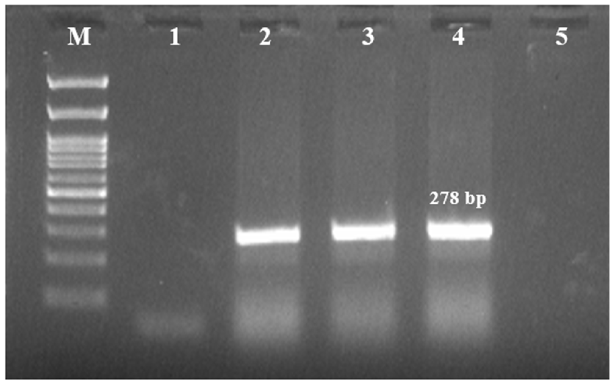
2.1. Prevalence of S. maltophilia

A total of 15 isolates (5%, 15/300) showed characteristic colonies of S. maltophilia and were species-identified by morphological, biochemical, molecular (Figure 1), and MALDI-TOF MS methods (Table 1).

2.2. Characterization of Antimicrobial Resistance in S. maltophilia

All S. maltophilia isolates were susceptible to six antimicrobials: cotrimoxazole, enrofloxacin, gatifloxacin, levofloxacin, minocycline, and tigecycline. However, all isolates were resistant to cefpodoxime and aztreonam. Additionally, 14 isolates (93.33%) were resistant to imipenem and ceftriaxone, while 10 isolates (66.67%) exhibited resistance to piperacillin-tazobactum (Table 2). Molecular characterization revealed the presence of various ARGs in the isolates (Figure 2), with the qnr gene being most prevalent (93.33%). Other detected ARGs included blaOXA-48 (n = 7, 46.67%), blaKPC (n = 5, 33.33%), blaNDM (n = 5, 33.33%), blaCTX-M (n = 3, 20%), blaSHV (n = 3, 20%), and blaTEM (n = 1, 6.67%) (Table 2). In addition to this, evaluation of the S. maltophilia isolates for bacterial pump efflux activity by the Etbr agar cartwheel method showed no fluorescence on the agar plates indicating active efflux activity (Figure 3) (Table 2).

2.3. Virulence Characterization of S. maltophilia

The crystal violet staining assay revealed that 9 of the S. maltophilia isolates (60%) were strong biofilm producers, while 6 isolates (40%) were moderate biofilm producers (Figure 4) (Table 2). Biofilm strength was categorized based on OD values as Negative: < 0.044; Weak: 0.044 < A ≤ 0.088; Moderate: 0.088 < A ≤ 0.176 and Strong: A > 0.176, where A-OD Value of Sample.
Virulence gene analysis revealed all the isolates harbored virB (100 %), motA (100 %), rmlA (100 %), and fliC (100 %) genes followed by pilU (93.3 %), gspD (80 %), papD (73.3 %), hgbB (73.3 %), picNl (66.6 %), afaD (53.3 %), and hlyIII (40 %) genes, respectively. However, the frpC and fimH genes were not found in any of the isolates (Table 2).
Swimming motility was assessed on modified LB plates, revealing that all 15 isolates exhibited swimming motility. Based on the diameter of the motility zone, 11 isolates (73.3%) were classified as strong, while 4 isolates (26.67%) were classified as moderate (Figure 5).

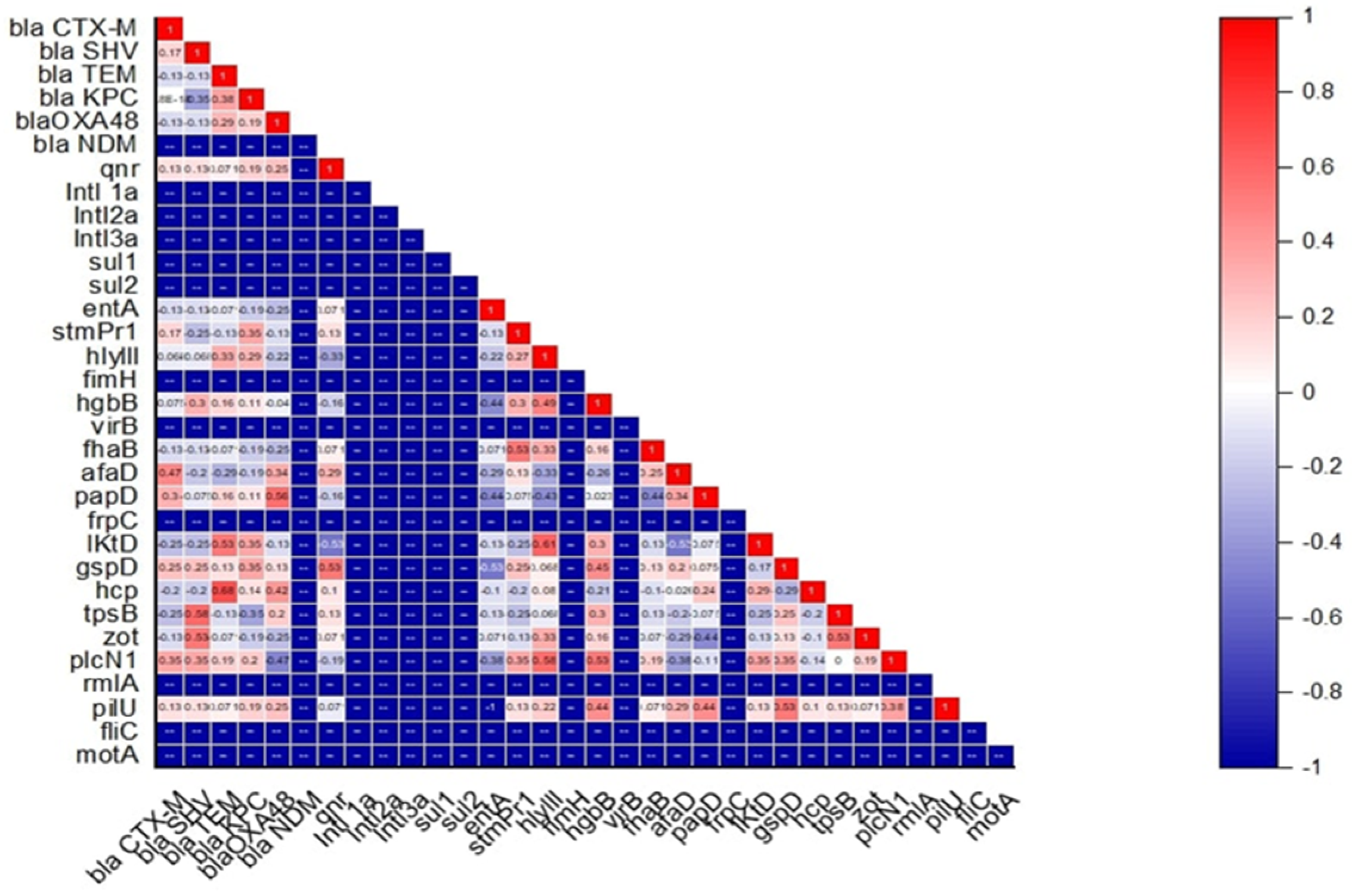
2.4. Association Between Antimicrobial Resistance, Virulence, Motility Pattern, and Biofilm-Forming Ability in S. maltophilia

The link between antimicrobial resistance, virulence, motility pattern, and biofilm-forming ability of S. maltophilia isolated from skin swabs of dogs with dermatological ailments was established by hierarchical clustering (dendrogram). The heatmap showed the grouping of isolates into mainly three clusters (A, B, and C) (Figure 6). Cluster A consisted of 4 isolates namely SM 13, SM 7, SM 1, and SM 6 which harbored virB, motA, rmlA, fliC, pilU, hgbB, picNl, and hlyIII virulent genes; blaNDM ARG and resistant to cefpodoxime, aztreonam, imipenem, and ceftriaxone in common. Cluster B consisted of 5 isolates namely SM 10, SM 14, SM 9, SM 5, and SM 8 which harbored virB, motA, rmlA, fliC, pilU, picNl, and gspD virulent genes; qnr ARG and resistant to cefpodoxime, aztreonam, imipenem, piperacillin-tazobactum, and ceftriaxone in common. Cluster C consisted of 5 isolates namely SM 3, SM 12, SM 4, SM 11, and SM 15 which harbored virB, motA, rmlA, fliC, pilU, and papD virulent genes; qnr and blaO ARGs and resistant to cefpodoxime, aztreonam, imipenem, and ceftriaxone in common. Isolate SM 2 remained outgroup. Interestingly, among the 15 isolates, none of the isolates harbored the same pattern of antimicrobial resistance, virulence, motility pattern, and biofilm-forming ability. Further association between antimicrobial resistance and virulence gene was established. A moderate positive correlation was observed between blaSHV with zot and tpsB (+0.53 to +0.58), blaTEM with lktD and hcp (+0.53 to +0.68), blaOXA-48 with papD (+0.56) and qnr with gspD (+0.53) (Figure 7). On the other hand, a weak negative correlation was seen between blaKPC with tpsB (−0.35), blaOXA-48 with plcN1 (−0.47), and qnr with hylIII and lktD (−0.33 to −0.53) (Figure 7).

3. Discussion

S. maltophilia has emerged as an important opportunistic pathogen which has been increasingly reported worldwide [1]. Although traditionally considered as a human pathogen, it has been reported in dogs, horses, cats, dogs, and ball pythons [5,6]. Earlier studies have reported that animal strains of S. maltophilia shared phylogenetic traits with some of the most successful human strains [8]. In this study, 15 S. maltophilia isolates were obtained from the skin swabs of dogs with dermatological ailments. These results indicate its involvement either as a primary pathogen or as part of a polymicrobial infection. Further studies would be necessary to confirm whether S. maltophilia is a direct cause of the dermatological problems or a secondary invader exploiting the diseased environment. This is the first study globally to document S. maltophilia from dogs with dermatological ailments.
An antibiotic susceptibility test revealed that the isolates were susceptible to cotrimoxazole, enrofloxacin, gatifloxacin, levofloxacin, minocycline, tigecycline, and ticarcillin-clavulanate. As fluoroquinolones and tetracyclines are commonly used in both human and veterinary medicine, the findings of this study support their potential use for treatment in cases involving S. maltophilia [2]. Furthermore, the isolates were found to be resistant to cefpodoxime, aztreonam, imipenem, ceftriaxone, and piperacillin-tazobactam. Carbapenem resistance is particularly worrisome, as they are considered a last-resort antibiotic for treating MDR infections [9]. A limitation of this study is the lack of specific CLSI breakpoints for S. maltophilia for several antibiotics, including enrofloxacin, gatifloxacin, imipenem, piperacillin-tazobactam, ceftriaxone, cefpodoxime, aztreonam, and tigecycline. In the absence of established breakpoints for S. maltophilia, we interpreted antimicrobial susceptibility results based on the available literature and expert recommendations, which may not fully reflect clinical susceptibility patterns. Further studies are needed to refine and standardize breakpoints for S. maltophilia to improve the accuracy and relevance of antimicrobial resistance data for this pathogen.
The resistance to β-lactams and carbapenems might be attributed to intrinsic resistance mechanisms typical of S. maltophilia or acquired resistance genes [10]. The molecular characterization of ARGs in S. maltophilia isolates demonstrates a high prevalence of the qnr gene (93.33 %) and a concerning presence of multiple β-lactamase genes, including blaOXA-48 (46.67 %), blaKPC (33.33%), blaNDM (33.33%), blaCTX-M (20%), blaSHV (20%), and blaTEM (6.67%). There was a broad spectrum of β-lactamase enzymes, capable of hydrolyzing a wide range of β-lactam antibiotics, including carbapenems, cephalosporins, and penicillins, which are often considered last-resort treatments for MDR infections. While this represents the first investigation of S. maltophilia specifically from canine dermatological sources, our findings can be meaningfully contextualized within the broader literature. Our detection of qnr (93.3%) aligns with previous studies reporting a high prevalence of quinolone resistance mechanisms in S. maltophilia clinical isolates [11,12]. The presence of multiple β-lactamase genes including blaOXA-48 (46.7%), blaKPC (33.3%), and blaNDM (33.3%) is consistent with emerging trends of carbapenemase-producing S. maltophilia in healthcare settings [13,14,15], though the co-occurrence of multiple resistance genes in our canine isolates suggests potential horizontal gene transfer mechanisms warranting further investigation.
Over-expression of the bacterial efflux pump systems is one of the mechanisms by which bacteria exhibit multi-drug resistance [16]. All the isolates showed high efflux activity resulting in MDR phenotype. These findings are in line with the AST results and presence of ARGs which showed that the 15 isolates were resistant to more than 3 antibiotics tested and 14 out of 15 isolates harbored multiple ARGs. These findings point to a complex resistance profile that complicates treatment options for infections caused by these isolates. Therefore, targeted therapeutic strategies are crucial for understanding the genetic basis of resistance and managing the spread of resistant bacteria in both animal and human populations.
Among S. maltophilia virulence factors, biofilm plays an important role in the survivability and virulence. In this study, all S. maltophilia isolates were able to produce biofilm and most of them were strong biofilm producers (60%) followed by moderate biofilm producers (40%) which is comparable to rates reported in clinical S. maltophilia isolates from human infections, where biofilm formation rates typically range from 70 to 90% [17,18,19]. The strong biofilm-forming isolates identified in this study are likely to exhibit increased resistance to standard treatments, as biofilms create a physical barrier that limits the penetration of antibiotics and reduces their efficacy. The slightly lower rates in our study may reflect source-specific variations or environmental factors affecting canine skin isolates. In the study of motility, most of the isolates exhibited strong or moderate levels of swimming patterns; however, none of the isolates were able to swarm which was in line with previous findings [20,21].
The screening of virulence genes in S. maltophilia showed a high prevalence of virB, motA, rmlA, and fliC in 100% of the isolates. Gene virB encodes components of the Type IV secretion system (T4SS), which is involved in the translocation of effector proteins into host cells, contributing to host cell manipulation and immune evasion [22]. Furthermore, motA and fliC are associated with motility, colonization, and biofilm formation, making them essential for bacterial persistence in diverse environments [3]. Also, rmlA is responsible for biosynthesis of rhamnose, a sugar required for the production of lipopolysaccharides (LPS) in the bacterial outer membrane, which plays a role in immune evasion and biofilm formation [23]. Moderate prevalence of pilU (93.3%) encoding for type IV pili, which is essential for bacterial adherence to host tissues and surfaces and translocation; gspD (80%) encoding a component of the Type II secretion system (T2SS); papD (73.3%) involved in the assembly of P pili associated with bacterial adherence; hgbB (73.3%) encoding for a protein involved in iron acquisition, which is crucial in iron-limited environments; and plcN1 (66.6%) responsible for bacterial immune evasion and tissue invasion, further emphasizing the bacterium’s ability to establish infections in various host environments were recorded [3,22,23]. The lower prevalence of virulence genes such as afaD (53.3%) and hlyIII (40%) encoding for adhesins and haemolysin protein were observed among the isolates.
The presence of multiple virulence genes, particularly those related to secretion systems (T4SS and T2SS), motility, adhesion, and iron acquisition, paints a picture of S. maltophilia as a highly adaptable pathogen capable of surviving in diverse environments and host tissues. These virulence factors likely contribute to the bacterium’s ability to persist in chronic infections, form biofilms, and resist host immune responses. The high prevalence of motA and fliC suggests that motility is a critical trait for environmental colonization and infection initiation, while the presence of secretion systems (virB and gspD) underscores the bacterium’s capacity to secrete effector molecules that manipulate host cells and degrade tissues [22].
A heat map was established to study the link between antimicrobial resistance, virulence, motility pattern, and biofilm-forming ability of S. maltophilia which showed that the isolates have a clonal connection. The hierarchical clustering exhibited diverse profiles with no two isolates sharing the same pattern of traits. This could be attributable to the adaptive versatility of S. maltophilia, which enables it to thrive in different environmental niches and cause various infections. These clusters suggests that the pathogenic potential of S. maltophilia may vary significantly between isolates, even when they are from same source.
The correlation matrix analysis revealed interesting associations between ARGs and virulence factors, suggesting potential co-regulation or functional linkages between these traits. The moderate positive correlation between blaSHV, a β-lactamase gene conferring resistance to β-lactam antibiotics, and the virulence genes tpsB (Type VI secretion system) and zot (zonula occludens toxin) (+0.53 to +0.58), suggests that some resistance mechanisms may be linked with the bacterium’s ability to deliver virulence factors to host cells. The association between blaTEM and lktD (leukotoxin D) and hcp (Type VI secretion system protein) (+0.53 to +0.68) points to a possible synergy between resistance to β-lactams and the secretion of toxins or immune-modulating proteins. Similarly, blaOXA-48 a carbapenemase gene, correlates positively with papD (+0.56), a gene involved in the assembly of pili, which is important for bacterial adhesion to host surfaces suggesting that increased resistance to carbapenems may enhance the bacterium’s capacity to adhere to host tissues, potentially making infections harder to treat. The positive correlation between the quinolone resistance gene qnr and gspD (Type II secretion system) suggests that resistance to fluoroquinolones may be linked with the bacterium’s ability to secrete enzymes and toxins, further complicating treatment. A weak negative correlation was seen between blaKPC with tpsB (−0.35), blaOXA-48 with plcN1 (−0.47) and qnr with lktD and hylIII (−0.33 to −0.53) suggesting that resistance of said genes are less dependent on certain virulence mechanisms. The correlation between antimicrobial resistance and virulence genes observed in our study echoes findings from human clinical isolates, where similar associations between resistance mechanisms and pathogenicity factors have been documented [24,25]. This suggests that the pathogenic potential and resistance profiles of S. maltophilia may be conserved across different host species, supporting the zoonotic concern we highlight.

4. Materials and Methods

4.1. Isolation and Characterization of S. maltophilia

4.1.1. Sample Collection

The present study was carried out from January 2024 to March 2024 in the Teaching Veterinary Clinical Complex, Madras Veterinary College, Chennai. A total of 300 skin swabs were collected aseptically from dogs presented with skin ailments and transported to the Bacteriology Laboratory of the Department of Veterinary Public Health and Epidemiology for further processing.

4.1.2. Bacterial Isolation, Identification, and Molecular Confirmation

Skin swabs were inoculated in 5 mL Luria Bertani Broth and incubated at 37 °C for 24 h. The enriched inoculum was then streaked onto Stenotrophomonas selective agar with vancomycin (5 mg), imipenem (32 mg), and amphotericin B (2.5 mg) and incubated overnight at 37 °C. Characteristic S. maltophilia colonies appeared as dark green with a blue halo. Presumptive colonies underwent gram staining, oxidase, catalase, and glucose fermentation tests for confirmation. PCR targeting species-specific 23S rRNA [26] was used for molecular characterization, following a 25 µL reaction protocol with a specific thermal cycle (Table 3). Optimization of PCR was carried out using reference strain S. maltophilia (MCC2083) obtained from the National Centre for Microbial Resource, Pune, India.

4.1.3. Matrix-Assisted Laser Desorption Ionization–Time of Flight Mass Spectrometry (MALDI-TOF MS) Identification

Bacterial isolates were prepared for MALDI-TOF MS following the ethanol-formic acid extraction method according to the manufacturer’s instructions. A loopful of bacteria was suspended in 300 µL distilled water with 900 µL ethanol, centrifuged at 17,000× g for 2 min, and the supernatant discarded. After repeated centrifugation and ethanol removal, the pellet was air-dried and resuspended in 5–50 µL of formic acid–water (70:30). An equal volume of acetonitrile was added, centrifuged, and 1 µL of the supernatant was placed on the MALDI target plate. After drying, it was overlaid with 1 µL matrix solution (α-Cyano-4-hydroxycinnamic acid). Mass spectra were analyzed on a Microflex LT mass spectrometer (Bruker) with Biotyper software (V 3.0) using Bruker bacterial test standard for identification.
Table 3. List of primers used for the detection of S. maltophilia.
GenePrimer SequenceCyclic ConditionNo. of CyclesAmplicon
Size (bp)
Reference
StepTemperature and Time
Species specific PCR
23S rRNAF-GCTGGATTGGTTCTAGGAAAACGC
R-ACGCAGTCACTCCTTGCG
Initial denaturation94 °C–5 min 278[26]
Denaturation94 °C–45 s30
Annealing68 °C–45 s
Extension72 °C–45 s
Final extension72 °C–10 min
Antimicrobial resistance genes
blaSIMF-GTACAAGGGATTCGGCATCG
R-TGGCCTGTTCCCATGTGAG
Initial denaturation 95 °C–4 min 569[27]
Denaturation95 °C–45 s30
Annealing58 °C–60 s
blaVIMF-GTTTGGTCGCATATCGCAAC
R-AATGCGCAGCACCAGGATAG
382
Extension72 °C–40 s
Final Extension72 °C–5 min
blaTEMF-TCCGCTCATGAGACAATAACC
R-TTGGTCTGACAGTTACCAATGC
Initial denaturation 94 °C–5 min 931[28]
Denaturation 94 °C–30 s35
Annealing60 °C–15 s
blaCTX-MF-ATGTGCAGYACCAGTAARGTKATGGC
R-TGGGTRAARTARGTSACCAGAAYCAGCGG
Extension72 °C–30 s593
Final Extension72 °C–5 min
blaSHVF-AGCCGCTTGAGCAAATTAAAC
R-ATCCCGCAGATAAATCACCAC
Initial denaturation 95 °C–5 min 713[29]
Denaturation 94 °C–45 s35
Annealing53 °C–45 s
Extension72 °C–1 min
Final Extension72 °C–10 min
blaKPCF-CGTCTAGTTCTGCTGTCTTG
R-CTTGTCATCCTTGTTAGGCG
Initial denaturation 94 °C–10 min 798[30]
Denaturation 94 °C–30 s30
Annealing52 °C–40 s
Extension72 °C–50 s
Final Extension72 °C–5 min
blaNDMF-TCGCATAAAACGCCTCTG
R-GAAACTGTCGCACCTCAT
Initial denaturation 95 °C–6 min 1001[31]
Denaturation 95 °C–45 s32
Annealing55 °C–45 s
Extension72 °C–60 s
Final Extension72 °C–7 min
qnrF-ACACAGAACGGCTGGACTGC
R-TTCAACGACGTGGAGCTGT
Initial denaturation 95 °C–5 min 817[32]
Denaturation 95 °C–60 s30
Annealing55 °C–60 s
Extension68 °C–60 s
Final Extension68 °C–5 min
Sul1F-TAGCGAGGGCTTTACTAAGC
R-ATTCAGAATGCCGAACACCG
Initial denaturation95 °C–5 min 437[33]
Denaturation95 °C–1 min35
Annealing55 °C–60 s
Sul2F-CCTGTTTCGTCCGACACAGA
R-GAAGCGCAGCCGCAATTCAT
956
Extension72 °C–1 min
Final Extension72 °C–10 min
blaOXA-48F-GCGTGGTTAAGGATGAACAC
R-CATCAAGTTCAACCCAACCG
Initial denaturation 95 °C–5 min 438[34]
Denaturation 95 °C–45 s35
Annealing60 °C–45 s
Extension72 °C–1 min
Final Extension72 °C–8 min
Virulence genes
entAF: CGTTCGCACTCGACGTGAC
R: CGAACTGACGGTAACGATCACG
Initial denaturation 94 °C–5 min 251[35]
Denaturation 94 °C–30 s34
Annealing60 °C–30 s
Extension72 °C–30 s
Final Extension72 °C–5 min
stmPr1F: TGAAAGCAAATGCGCCGTTG
R: GTGATGGCGTCGGTGATGTC
Initial denaturation 94 °C–5 min 852
Denaturation 94 °C–30 s34
Annealing60 °C–30 s
Extension72 °C–30 s
Final Extension72 °C–5 min
hlyIII F: CGTCCATTGCTTCGATCCGTG
R: GACGAAGTGGCAGACGCTG
Initial denaturation 94 °C–5 min 607
Denaturation 94 °C–30 s34
Annealing60 °C–30 s
Extension72 °C–30 s
Final Extension72 °C–5 min
fimH F: GATCCGCCTGAACTGCCAG
R: CTGGCAGTTCAGGCGGATC
Initial denaturation 94 °C–5 min 576
Denaturation 94 °C–30 s34
Annealing60 °C–30 s
Extension72 °C–30 s
Final Extension72 °C–5 min
hgbBF: GGACATCCAGAACATGGGTGC
R: GGATCGATCGTGTACGGACC
Initial denaturation 94 °C–5 min 1239
Denaturation 94 °C–30 s34
Annealing60 °C–30 s
Extension72 °C–30 s
Final Extension72 °C–5 min
virBF: GCATCATGCAGAACGAGCTG
R: GACGGCTCGTACTTCTGCAC
Initial denaturation 95 °C–5 min 1075
Denaturation 95 °C–45 s35
Annealing60 °C–45 s
Extension72 °C–1 min
Final Extension72 °C–8 min
frpCF: CCAGTTCAACCTGTCGATGCTG
R: CACCGAACAGGTTGTCCCAG
Initial denaturation 95 °C–5 min 653
Denaturation 95 °C–45 s35
Annealing60 °C–45 s
Extension72 °C–1 min
Final Extension72 °C–8 min
afaDF: GAAGCGCCTGACTGCCTTTTG
R: GATCACGTTGTAAGGCCGCC
Initial denaturation 95 °C–5 min 328
Denaturation 95 °C–45 s35
Annealing60 °C–45 s
Extension72 °C–1 min
Final Extension72 °C–8 min
fhaBF: GTATCGCACAACCGCTTCCAG
R: CGTCGTTGATGACCTTCTGCAC
Initial denaturation 95 °C–5 min 1744
Denaturation 95 °C–45 s35
Annealing60 °C–45 s
Extension72 °C–1 min
Final Extension72 °C–8 min
papDF: CACGCGAGTGATCTATCCGG
R: GTGATGAAGCGCACCTGGTC
Initial denaturation 95 °C–5 min 579
Denaturation 95 °C–45 s35
Annealing60 °C–45 s
Extension72 °C–1 min
Final Extension72 °C–8 min
gspDF: GTCGACACCGATATCGGTGG
R: GGTAGACCACATGCAGGTTGC
Initial denaturation 94 °C–5 min 694
Denaturation 94 °C–30 s32
Annealing60 °C–15 s
Extension72 °C–30 s
Final Extension72 °C–5 min
hcpF: GACGGCAACGCGATCAATTAC
R: GTTCTTGGTTGCACTCCACTG
Initial denaturation 94 °C–5 min 201
Denaturation 94 °C–30 s32
Annealing60 °C–15 s
Extension72 °C–30 s
Final Extension72 °C–5 min
zotF: GCGTCAGTACACCGATGGTTG
R: GCAGGCAGTGTCCAGCATG
Initial denaturation 94 °C–5 min 431
Denaturation 94 °C–30 s32
Annealing60 °C–15 s
Extension72 °C–30 s
Final Extension72 °C–5 min
plcN1F: GTGACCGATATCGGCCGAC
R: CTGGAAGTGGCGGTGGAAG
Initial denaturation 94 °C–5 min 1779
Denaturation 94 °C–30 s34
Annealing62 °C–30 s
Extension72 °C–30 s
Final Extension72 °C–5 min
pilUF: CGACCACCATCGATTTCACTTCG
R: GACAGGTCCATCAGCAGCTG
Initial denaturation 94 °C–5 min 778
Denaturation 94 °C–30 s34
Annealing60 °C–30 s
Extension72 °C–30 s
Final Extension72 °C–5 min
fliCF: CGATCTCCGAGCGCTTCG
R: GAACAGCTGGCTGGAGAACG
Initial denaturation 94 °C–5 min 296
Denaturation 94 °C–30 s34
Annealing60 °C–30 s
Extension72 °C–30 s
Final Extension72 °C–5 min
rmlAF: CTCAGCGTGCTGATGCTGG
R: GATGAAGTTGGAGGCTTCCAGC
Initial denaturation 94 °C–5 min 600
Denaturation 94 °C–30 s34
Annealing60 °C–30 s
Extension72 °C–30 s
Final Extension72 °C–5 min
tpsBF: GTGGACATCGTGATGAAGCGC
R: CTTGCCGATGAAGTGACGGTG
Initial denaturation 94 °C–5 min 822
Denaturation 94 °C–30 s34
Annealing54 °C–30 s
Extension72 °C–30 s
Final Extension72 °C–5 min
motAF: CGTTGGATTCCTGGTCGTCATC
R: GAGCCCATGGTGATGACGATG
Initial denaturation 94 °C–5 min 558
Denaturation 94 °C–30 s34
Annealing54 °C–30 s
Extension72 °C–30 s
Final Extension72 °C–5 min
lktDF: GCACATCCGTGATGCAGTCG
R: CGAGATTCTCGTCCTGCATGG
Initial denaturation94 °C–5 min 1235
Denaturation 94 °C–30 s34
Annealing54 °C–30 s
Extension72° C–30 s
Final Extension72 °C–5 min

4.2. Screening of the Isolates for the Presence of Antimicrobial Resistance

4.2.1. Antimicrobial Susceptibility Testing (AST)

All isolates underwent antimicrobial sensitivity testing using the Kirby–Bauer disc diffusion method against 12 antibiotics: trimethoprim-sulfamethoxazole (23.75 µg/1.25 µg), enrofloxacin (5 µg), gatifloxacin (5 µg), levofloxacin (5 µg), imipenem (30 µg), piperacillin-tazobactam (100 µg/10 µg), ticarcillin-clavulanate (75 µg/10 µg), ceftriaxone (30 µg), cefpodoxime (10 µg), aztreonam (30 µg), minocycline (30 µg), and tigecycline (15 µg). Isolates were grown in Luria Bertani Broth at 37 °C for 16 h, spread on Mueller–Hinton agar plates, and allowed to dry. Antibiotic discs were placed on the plates within 15 min, and plates were incubated overnight at 37 °C. Zones of inhibition were measured and classified as sensitive, intermediate, or resistant per CLSI guidelines [36].

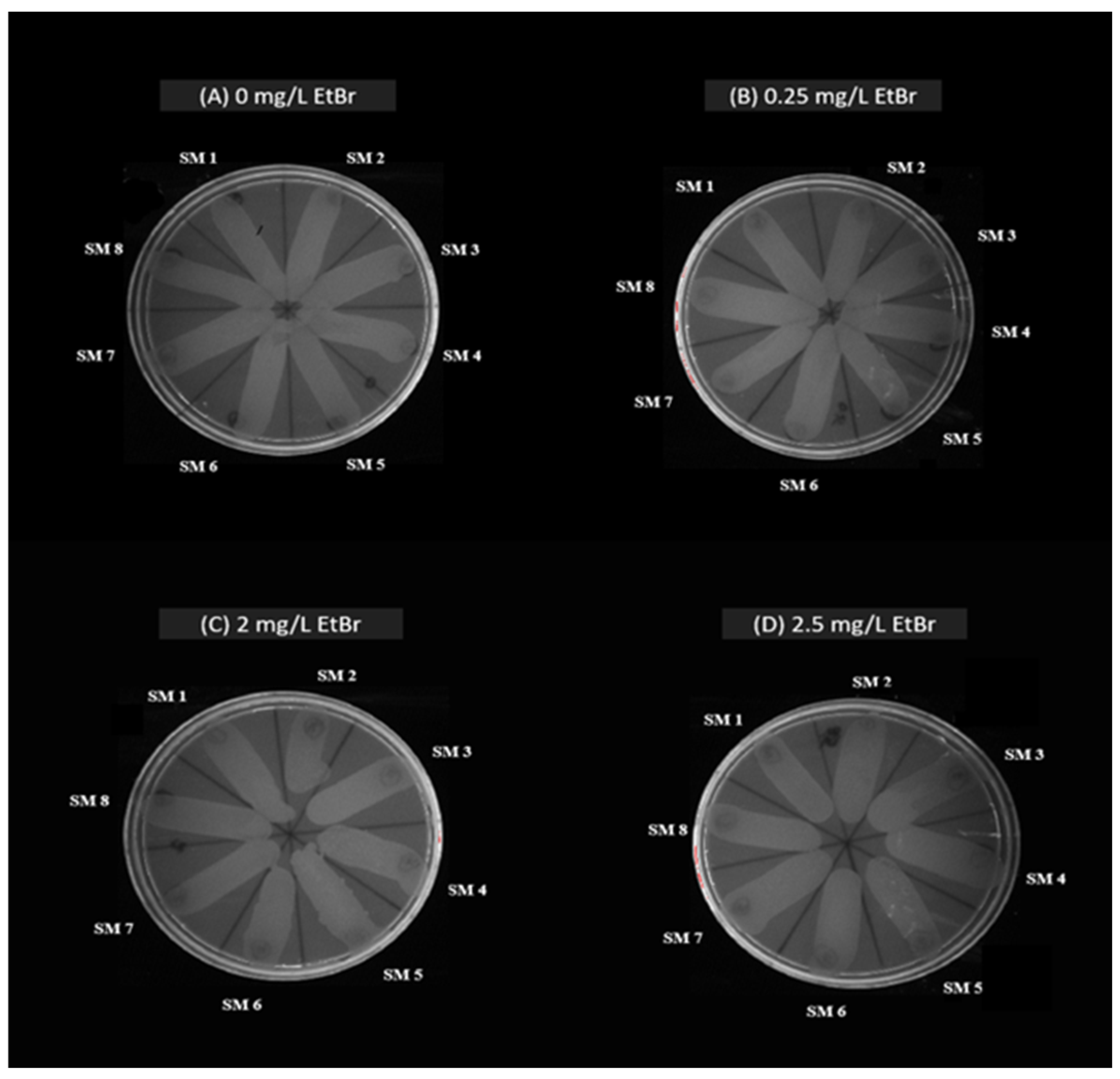
4.2.2. Assessment of Bacterial Efflux Pump Activity in S. maltophilia

Efflux pump activity in S. maltophilia was assessed using the ethidium bromide agar cartwheel method [16]. Trypticase Soy agar plates with ethidium bromide (0.0–2.5 mg/L) were divided into 12 radial sectors. Bacterial cultures (0.5 McFarland) were swabbed from center to edge and incubated at 37 °C for 16 h. Fluorescence was then observed under a gel documentation system.

4.2.3. Antimicrobial Resistance Genes (ARGs)

Screening of antimicrobial genes in S. maltophilia was carried out by PCR with cyclic conditions as described in Table 3. The ARGs targeted were blaCTX-M and blaTEM [28], blaSHV [29], blaOXA-48 [34], blaKPC [30], blaNDM [31], blaVIM, and blaSIM [27] encoding for β-lactam resistance, qnr [32] encoding for quinolone resistance, and sul-1 and sul-2 [33] encoding for sulphonamide resistance.

4.3. Characterization of the Virulence Properties of S. maltophilia

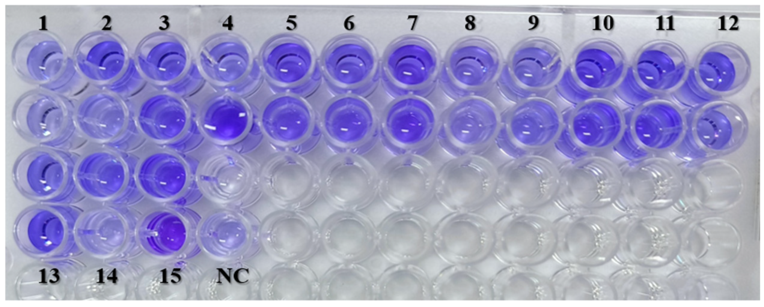
4.3.1. Evaluation of Biofilm-Forming Ability

Biofilm-forming ability of isolates was assessed by crystal violet staining [37,38]. Cultures (1.0 McFarland, diluted 1:100) were incubated in microtiter plates at 37 °C for 24 h. After washing and fixing, biofilms were stained, destained, and quantified by OD at 492 nm. Biofilm strength was categorized based on OD values as Negative: < Ac; Weak: Ac < A ≤ 2Ac; Moderate: 2Ac < A ≤ 4Ac, and Strong: A > 4Ac, where Ac-OD Value of Negative control; A-OD Value of Sample.

4.3.2. Motility Assay

Swimming motility of S. maltophilia was assessed on modified LB agar (0.3% w/v) plates [20]. In total, 10 µL of cultures (0.5 McFarland) were inoculated at the center and incubated at 37 °C for 24 h. Halo diameters were recorded as high (>10 mm), moderate (>7 to ≤10 mm), or low motility (> 6 to ≤7 mm) [35].

4.3.3. Molecular Characterization of Virulence Genes

The screening of 20 virulence genes in S. maltophilia isolates was conducted through multiplex PCR which included set I (entA, stmPr1, hlyIII, fimH, hgbB), set II (virB, frpC, afaD, fhaB, papD), set III (gspD, hcp, zot), set IV (pilU, fliC, rmlA), set V (tpsB, motA, lktD), and picN1 as described by [35] (Table 3).

4.4. Statistical Analysis

A heatmap with hierarchical clustering was generated to visualize the distribution of antimicrobial resistance, virulence, motility, and biofilm formation in S. maltophilia isolates [39]. Spearman’s correlation between resistance and virulence genes was analyzed in R (Version 4.3.1) using pheatmap and rcorr packages, with significance at p < 0.05. Data visualization employed Origin 2024 (trial version).

5. Conclusions

In summary, our study documented the presence of multidrug-resistant virulent S. maltophilia isolates in dogs with dermatological ailments at the Teaching Veterinary Hospital, Madras Veterinary College, India. It highlights significant genetic diversity among isolates and is the first report of S. maltophilia in dogs from India. The varying profiles of antimicrobial resistance, virulence gene expression, motility, and biofilm-forming ability emphasize the need for vigilant antimicrobial stewardship in veterinary settings. The potential for zoonotic transmission from infected pets necessitates a One Health approach to mitigate risks associated with this opportunistic pathogen and promote health in both humans and animals.

Author Contributions

Conceptualization, P.K.; methodology, R.R., P.K., S.S., S.B.M., C.J.H., R.G.A. and A.S.; software, V.N.; formal analysis, investigation, data curation, R.R., P.K., N.Q.M., W.R.S., R.G.A. and S.R.; writing—original draft preparation, R.R. and S.R.; writing—review and editing, P.K., N.Q.M., W.R.S., C.A.C. and R.G.A.; supervision, P.K., S.S. and S.B.M. All authors have read and agreed to the published version of the manuscript.

Funding

This research received no external funding.

Institutional Review Board Statement

The protocols for this study were approved by the Expert Research Review Committee, TANUVAS, Chennai, Tamil Nadu, India (No. 685/Edn.cell/C1/2023). The sample collection was conducted by a well-trained veterinary practitioner from the Teaching Veterinary Clinical Complex, Madras Veterinary College, Chennai, India, as part of routine veterinary care, with informed oral consent obtained from the owners. All procedures adhered to the ethical standards of the institution and complied with the guidelines and animal welfare regulations set by the Committee for Control and Supervision of Experiments on Animals (2018) and the Breeding of and Experiments on Animals (Control and Supervision) Rules 2006. As the sample collection was part of routine veterinary practice aimed at ensuring animal well-being, this study is classified as non-experimental research. Furthermore, all methods were performed in accordance with the relevant ARRIVE guidelines and regulations.

Informed Consent Statement

The sample collection was done with informed oral consent from the owners by a qualified veterinary practitioner from the Teaching Veterinary Clinical Complex, Madras Veterinary College, Chennai, India, as part of routine veterinary care diagnostics.

Data Availability Statement

All data supporting the findings of this study are contained within this manuscript and will be made available upon request.

Acknowledgments

The authors acknowledge the Tamil Nadu Veterinary and Animal Sciences University (TANUVAS), India, for providing the facilities for the smooth conduct of the research. This manuscript is part of the M.V.Sc. thesis submitted by the first author to TANUVAS, India.

Conflicts of Interest

Author Charley A. Cull was employed by Midwest Veterinary Services, Inc., Oakland, Nebraska, 68045, USA. The remaining authors declare that the research was conducted in the absence of any commercial or financial relationships that could be construed as potential conflicts of interest. The funders had no role in the design of this study; in the collection, analyses, or interpretation of data; in the writing of this manuscript; or in the decision to publish the results.

Abbreviations

The following abbreviations are used in this manuscript:
AMRAntimicrobial resistance
ARGsAntimicrobial resistant genes
ASTAntimicrobial susceptibility testing
ATAztreonam
CLSIClinical and Laboratory Standards Institute
CPDCefpodoxime
CTRCeftriaxone
EtbrEthidium bromide
IPMImipenem
lktDLeukotoxin D
LPSLipopolysaccharides
MALDI-TOF MSMatrix-assisted laser desorption/ionization time-of-flight mass spectrometry
MDRMultidrug resistant
NCNegative control
NFGNBNon-fermenting gram-negative bacilli
ODOptical density
PCRPolymerase Chain Reaction
PITPiperacillin-tazobactam
S. maltophiliaStenotrophomonas maltophilia
T2SSType II secretion system
T4SSType IV secretion system
TCCTicarcillin-clavulanate
TMP/SMXTrimethoprim/sulfamethoxazole
zotZonula occludens toxin

References

  1. Brooke, J.S. Advances in the microbiology of Stenotrophomonas maltophilia. Clin. Microbiol. Rev. 2021, 34, e0003019. [Google Scholar] [CrossRef] [PubMed]
  2. Gibb, J.; Wong, D.W. Antimicrobial treatment strategies for Stenotrophomonas maltophilia: A focus on novel therapies. Antibiotics 2021, 10, 1226. [Google Scholar] [CrossRef]
  3. Mikhailovich, V.; Heydarov, R.; Zimenkov, D.; Chebotar, I. Stenotrophomonas maltophilia virulence: A current view. Front. Microbiol. 2024, 15, 1385631. [Google Scholar] [CrossRef]
  4. Bhaumik, R.; Aungkur, N.Z.; Anderson, G.G. A guide to Stenotrophomonas maltophilia virulence capabilities, as we currently understand them. Front. Cell. Infect. Microbiol. 2023, 13, 1322853. [Google Scholar] [CrossRef]
  5. Albini, S.; Abril, C.; Franchini, M.; Hüssy, D.; Filioussis, G. Stenotrophomonas maltophilia isolated from the airways of animals with chronic respiratory disease. Schweiz. Arch. Tierheilkd. 2009, 151, 323–328. [Google Scholar] [CrossRef] [PubMed]
  6. Kralova-Kovarikova, S.; Husnik, R.; Honzak, D.; Kohout, P.; Fictum, P. Stenotrophomonas maltophilia urinary tract infections in three dogs: A case report. Vet. Med. 2012, 57, 380–383. [Google Scholar] [CrossRef]
  7. Thenissery, A.; Chandran, R.; Ravindran, R.; Ravishankar, C.; Piriyath, J.H. Isolation and identification of emergent multidrug resistant Stenotrophomonas maltophilia from skin ulcers of Sarcoptes infested pigs. Vet. Rec. Case Rep. 2022, 10, e312. [Google Scholar] [CrossRef]
  8. Jayol, A.; Corlouer, C.; Haenni, M.; Darty, M.; Maillard, K.; Desroches, M.; Lamy, B.; Jumas-Bilak, E.; Madec, J.Y.; Decousser, J.W. Are animals a source of Stenotrophomonas maltophilia in human infections? Contributions of a nationwide molecular study. Eur. J. Clin. Microbiol. Infect. Dis. 2018, 37, 1039–1045. [Google Scholar] [CrossRef]
  9. Sheu, C.C.; Chang, Y.T.; Lin, S.Y.; Chen, Y.H.; Hsueh, P.R. Infections caused by carbapenem-resistant Enterobacteriaceae: An update on therapeutic options. Front. Microbiol. 2019, 10, 80. [Google Scholar] [CrossRef]
  10. Mojica, M.F.; Humphries, R.; Lipuma, J.J.; Mathers, A.J.; Rao, G.G.; Shelburne, S.A.; Fouts, D.E.; Van Duin, D.; Bonomo, R.A. Clinical challenges treating Stenotrophomonas maltophilia infections: An update. JAC Antimicrob. Resist. 2022, 4, dlac040. [Google Scholar] [CrossRef]
  11. Sánchez, M.B.; Hernández, A.; Rodríguez-Martínez, J.M.; Martínez-Martínez, L.; Martínez, J.L. Predictive analysis of transmissible quinolone resistance indicates Stenotrophomonas maltophilia as a potential source of a novel family of Qnr determinants. BMC Microbiol. 2018, 8, 148. [Google Scholar] [CrossRef]
  12. Huang, Y.W.; Hu, R.M.; Yang, T.C. Role of the pcm-mph operon in resistance to several antibiotics in Stenotrophomonas maltophilia. J. Antimicrob. Chemother. 2013, 68, 1987–1993. [Google Scholar] [CrossRef] [PubMed]
  13. Cha, M.K.; Kang, C.I.; Kim, S.H.; Cho, S.Y.; Ha, Y.E.; Chung, D.R.; Peck, K.R.; Song, J.H. Emergence of fluoroquinolone-resistant Stenotrophomonas maltophilia in blood isolates causing bacteremia: Molecular epidemiology and microbiologic characteristics. Diagn. Microbiol. Infect. Dis. 2016, 85, 210–212. [Google Scholar] [CrossRef] [PubMed]
  14. Windels, E.M.; Michiels, J.E.; Van den Bergh, B.; Fauvart, M.; Michiels, J. Antibiotics: Combatting tolerance to stop resistance. mBio 2019, 10, e02095-19. [Google Scholar] [CrossRef]
  15. Adegoke, A.A.; Stenström, T.A.; Okoh, A.I. Stenotrophomonas maltophilia as an emerging ubiquitous pathogen: Looking beyond contemporary antibiotic therapy. Front. Microbiol. 2017, 8, 2276. [Google Scholar] [CrossRef] [PubMed]
  16. Martins, M.; McCusker, M.P.; Viveiros, M.; Couto, I.; Fanning, S.; Pagès, J.M.; Amaral, L. A simple method for assessment of MDR bacteria for over-expressed efflux pumps. Open Microbiol. J. 2013, 7, 72–82. [Google Scholar] [CrossRef]
  17. Pompilio, A.; Piccolomini, R.; Picciani, C.; D’Antonio, D.; Savini, V.; Di Bonaventura, G. Factors associated with adherence to and biofilm formation on polystyrene by Stenotrophomonas maltophilia: The role of cell surface hydrophobicity and motility. FEMS Microbiol. Lett. 2008, 287, 41–47. [Google Scholar] [CrossRef]
  18. Bostanghadiri, N.; Ghalavand, Z.; Fallah, F.; Yadegar, A.; Ardebili, A.; Tarashi, S.; Pournajaf, A.; Mardaneh, J.; Shams, S.; Hashemi, A. Characterization of phenotypic and genotypic diversity of Stenotrophomonas maltophilia strains isolated from selected hospitals in Iran. Front. Microbiol. 2019, 10, 1191. [Google Scholar] [CrossRef]
  19. Bostanghadiri, N.; Ardebili, A.; Ghalavand, Z.; Teymouri, S.; Mirzarazi, M.; Goudarzi, M.; Ghasemi, E.; Hashemi, A. Antibiotic resistance, biofilm formation, and biofilm-associated genes among Stenotrophomonas maltophilia clinical isolates. BMC Res. Notes 2021, 14, 151. [Google Scholar] [CrossRef]
  20. Amoli, R.I.; Nowroozi, J.; Sabokbar, A.; Rajabniya, R. Isolation of Stenotrophomonas maltophilia from clinical samples: An investigation of patterns motility and production of melanin pigment. Asian Pac. J. Trop. Biomed. 2017, 7, 826–830. [Google Scholar] [CrossRef]
  21. Yinsai, O.; Deeudom, M.; Duangsonk, K. Genotypic diversity, antibiotic resistance, and virulence phenotypes of Stenotrophomonas maltophilia clinical isolates from a Thai University Hospital Setting. Antibiotics 2023, 12, 410. [Google Scholar] [CrossRef] [PubMed]
  22. Nas, M.Y.; White, R.C.; DuMont, A.L.; Lopez, A.E.; Cianciotto, N.P. Stenotrophomonas maltophilia encodes a VirB/VirD4 type IV secretion system that modulates apoptosis in human cells and promotes competition against heterologous bacteria, including Pseudomonas aeruginosa. Infect. Immun. 2019, 87, 10–1128. [Google Scholar] [CrossRef] [PubMed]
  23. Azimi, A.; Aslanimehr, M.; Yaseri, M.; Shadkam, M.; Douraghi, M. Distribution of smf-1, rmlA, spgM and rpfF genes among Stenotrophomonas maltophilia isolates in relation to biofilm-forming capacity. J. Glob. Antimicrob. Resist. 2020, 23, 321–326. [Google Scholar] [CrossRef] [PubMed]
  24. Youenou, B.; Favre-Bonté, S.; Bodilis, J.; Brothier, E.; Dubost, A.; Muller, D.; Nazaret, S. Comparative genomics of environmental and clinical Stenotrophomonas maltophilia strains with different antibiotic resistance profiles. Genome Biol. Evol. 2015, 7, 2484–2505. [Google Scholar] [CrossRef]
  25. Brooke, J.S. Stenotrophomonas maltophilia: An emerging global opportunistic pathogen. Clin. Microbiol. Rev. 2012, 25, 2–41. [Google Scholar] [CrossRef]
  26. Gallo, S.W.; Ramos, P.L.; Ferreira, C.A.S.; Oliveira, S.D. A specific polymerase chain reaction method to identify Stenotrophomonas maltophilia. Mem. Inst. Oswaldo Cruz. 2013, 108, 390–391. [Google Scholar] [CrossRef]
  27. Saleh, R.O.; Hussen, B.M.; Mubarak, S.M.H.; Mostafavi, S.K.S. High diversity of virulent and multidrug-resistant Stenotrophomonas maltophilia in Iraq. Gene Rep. 2021, 23, 101124. [Google Scholar] [CrossRef]
  28. Sakthikarthikeyan, S.; Sivakumar, M.; Manikandan, R.; Senthilkumar, P.; Sureshkumar, V.; Malmarugan, S. Prevalence and Molecular Characterization of Multidrug-resistant ESBL-producing E. coli in Commercial Poultry. Indian J. Anim. Res. 2024, B-5166, 1–6. [Google Scholar] [CrossRef]
  29. Ibrahim, M.E.; Algak, T.B.; Abbas, M.; Elamin, B.K. Emergence of blaTEM, blaCTX-M, blaSHV and blaOXA genes in multidrug-resistant Enterobacteriaceae and Acinetobacter baumannii in Saudi Arabia. Exp. Ther. Med. 2021, 22, 1450. [Google Scholar] [CrossRef]
  30. Poirel, L.; Walsh, T.R.; Cuvillier, V.; Nordmann, P. Multiplex PCR for detection of acquired carbapenemase genes. Diagn. Microbiol. Infect. Dis. 2011, 70, 119–123. [Google Scholar] [CrossRef]
  31. Rizi, K.S.; Jamehdar, S.A.; Sasan, M.S.; Ghazvini, K.; Aryan, E.; Safdari, H.; Farsiani, H. Detection of extended-spectrum beta-lactamases and carbapenemases in clinical isolates of Stenotrophomonas maltophilia in the northeast of Iran. Gene Rep. 2023, 34, 101857. [Google Scholar] [CrossRef]
  32. Malekan, M.; Tabaraie, B.; Akhoundtabar, L.; Afrough, P.; Behrouzi, A. Distribution of class I integron and smqnr resistance gene among Stenotrophomonas maltophilia isolated from clinical samples in Iran. Avicenna J. Med. Biotechnol. 2017, 9, 138–141. [Google Scholar] [PubMed]
  33. Hu, L.F.; Chen, G.S.; Kong, Q.X.; Gao, L.P.; Chen, X.; Ye, Y.; Li, J.B. Increase in the prevalence of resistance determinants to trimethoprim/sulfamethoxazole in clinical Stenotrophomonas maltophilia isolates in China. PLoS ONE 2016, 11, e0157693. [Google Scholar] [CrossRef] [PubMed]
  34. Doyle, D.; Peirano, G.; Lascols, C.; Lloyd, T.; Church, D.L.; Pitout, J.D.D. Laboratory detection of Enterobacteriaceae that produce carbapenemases. J. Clin. Microbiol. 2012, 50, 3877–3880. [Google Scholar] [CrossRef]
  35. Cruz-Córdova, A.; Mancilla-Rojano, J.; Luna-Pineda, V.M.; Escalona-Venegas, G.; Cázares-Domínguez, V.; Ormsby, C.; Franco-Hernández, I.; Zavala-Vega, S.; Hernández, M.A.; Medina-Pelcastre, M.; et al. Molecular epidemiology, antibiotic resistance and virulence traits of Stenotrophomonas maltophilia strains associated with an outbreak in a Mexican tertiary care hospital. Front. Cell. Infect. Microbiol. 2020, 10, 50. [Google Scholar] [CrossRef]
  36. CLSI. Performance Standards for Antimicrobial Susceptibility Testing, 34th ed.; Clinical and Laboratory Standards Institute: Berwyn, PA, USA, 2024; pp. 88–89. [Google Scholar]
  37. Di Bonaventura, G.; Spedicato, I.; Antonio, D.; Robuffo, I.; Piccolomini, R. Biofilm formation by Stenotrophomonas maltophilia: Modulation by quinolones, trimethoprim-sulfamethoxazole, and ceftazidime. Antimicrob. Agents Chemother. 2004, 48, 151–160. [Google Scholar] [CrossRef]
  38. Pompilio, A.; Pomponio, S.; Crocetta, V.; Gherardi, G.; Verginelli, F.; Fiscarelli, E.; Dicuonzo, G.; Savini, V.; Antonio, D.; Di Bonaventura, G. Phenotypic and genotypic characterization of Stenotrophomonas maltophilia isolates from patients with cystic fibrosis: Genome diversity, biofilm formation, and virulence. BMC Microbiol. 2011, 11, 159. [Google Scholar] [CrossRef]
  39. Kolde, R. Pheatmap: Pretty heatmaps. R Package Version 2012, 1, 726. [Google Scholar]
Figure 1. PCR amplification of 23S rRNA gene of S. maltophilia. M—100 bp ladder; 1—No template control; 2—Positive control (S. maltophilia–MCC2083); 3, 4—Positive samples; 5—Negative sample.
Antibiotics 14 00639 g001
Figure 2. PCR amplification of AMR genes in S. maltophilia. M—100 bp ladder; 1—No template control; 2—blaSHV gene (713 bp); 3—qnr gene (817 bp); 4—blaOXA-48 gene (438 bp); 5—blaNDM gene (1001 bp); 6—blaCTX-M gene (593 bp); 7—blaKPC gene (798 bp); 8—blaTEM gene (931 bp).
Antibiotics 14 00639 g002
Figure 3. Efflux pump activity of S. maltophilia strains (SM 1 TO SM 15) obtained from canine samples. (A) 0 mg/L concentration of ethidium bromide; (B) 0.25 mg/L concentration of ethidium bromide; (C) 2 mg/L concentration of ethidium bromide; (D) 2.5 mg/L concentration of ethidium bromide.
Antibiotics 14 00639 g003
Figure 4. Microplate method to evaluate biofilm-forming ability. NC—Negative control; 1–15—Isolates SM1 to SM15.
Antibiotics 14 00639 g004
Figure 5. Swimming motility of S. maltophilia in modified LB media.
Antibiotics 14 00639 g005
Figure 6. Heat map analysis with hierarchical clustering (dendrogram) of S. maltophilia isolates obtained from canine samples.
Antibiotics 14 00639 g006
Figure 7. Correlation matrix analysis of antimicrobial resistance and virulence genes of S. maltophilia isolates obtained from canine samples.
Antibiotics 14 00639 g007
Table 1. Identification of S. maltophilia.
IsolatesDate of CollectionType of SampleGram’s StainingCatalase TestOxidase TestGlucose Fermentation Test23S rRNAMALDI-TOF SCORE
SM 112.01.24Skin swabNegativePositiveNegativeNegativePositive2.25
SM 218.01.24Skin swabNegativePositiveNegativeNegativePositive2
SM 321.01.24Skin swabNegativePositiveNegativeNegativePositive2.06
SM 402.02.24Skin swabNegativePositiveNegativeNegativePositive2.17
SM 507.02.24Skin swabNegativePositiveNegativeNegativePositive2
SM 609.02.24Skin swabNegativePositiveNegativeNegativePositive2.2
SM 717.02.24Skin swabNegativePositiveNegativeNegativePositive2.26
SM 817.02.24Skin swabNegativePositiveNegativeNegativePositive2.15
SM 919.02.24Skin swabNegativePositiveNegativeNegativePositive2.23
SM 1026.02.24Skin swabNegativePositiveNegativeNegativePositive2.27
SM 1111.03.24Skin swabNegativePositiveNegativeNegativePositive2.26
SM 1211.03.24Skin swabNegativePositiveNegativeNegativePositive2.24
SM 1313.03.24Skin swabNegativePositiveNegativeNegativePositive2.26
SM 1416.03.24Skin swabNegativePositiveNegativeNegativePositive2.23
SM 1516.03.24Skin swabNegativePositiveNegativeNegativePositive2.17
Table 2. Characterization of the antimicrobial, virulence, and antibiofilm properties of S. maltophilia.
Strain NumberPhenotypic Resistance PatternAMR GenesVirulence GenesBiofilm ActivityEfflux Activity
SM 1IPM, CTR, CPD, ATblaKPC, blaNDM, qnrvirB, motA, rmlA, fliC, pilU, gspD, hgbB, plcN1, hlyIII, lktDSTRONGACTIVE
SM 2CPD, ATqnrvirB, motA, rmlA, fliC, entAMODERATEACTIVE
SM 3IPM, PIT, TCC, CTR, CPD, ATblaKPC, blaOXA-48, qnrvirB, motA, rmlA, fliC, pilU, gspD, papD, afaDMODERATEACTIVE
SM 4IPM, PIT, TCC, CTR, CPD, ATblaSHV, blaOXA-48,qnrvirB, motA, rmlA, fliC, pilU, gspD, papD, hgbB plcN1, tpsBSTRONGACTIVE
SM 5IPM, PIT, CTR, CPD, ATblaKPC, qnrvirB, motA, rmlA, fliC, pilU, gspD, papD, hgbB, plcN1, stmPr1MODERATEACTIVE
SM 6IPM, CTR, CPD, ATblaTEM, blaKPC
blaNDM, qnr
virB, motA, rmlA, fliC, pilU, gspD, papD, hgbB, plcN1, hlyIII, lktD, hcpMODERATEACTIVE
SM 7IPM, PIT, CTR, CPD, ATblaNDMvirB, motA, rmlA, fliC, pilU, papD, hgbB, plcN1, hlyIII, lktDSTRONGACTIVE
SM 8IPM, PIT, CTR, CPD, ATqnrvirB, motA, rmlA, fliC, pilU, gspD,
hgbB, plcN1, afaD, hlyIII, fhaB, stmPr1
STRONGACTIVE
SM 9IPM, PIT, TCC, CTR, CPD, ATblaCTX-M, blaKPC, blaOXA-48, blaNDM, qnrvirB, motA, rmlA, fliC, pilU gspD, papD, hgbB plcN1, afaD, hlyIII, stmPr1STRONGACTIVE
SM 10IPM, PIT, CTR, CPD, ATblaCTX-M, qnrvirB, motA, rmlA, fliC, pilU, gspD, papD, plcN1, afaDMODERATEACTIVE
SM 11IPM, TCC, CTR, CPD, ATblaOXA-48, qnrvirB, motA, rmlA, fliC, pilU, gspD, papD, hgbB, afaDSTRONGACTIVE
SM 12IPM, PIT, CTR, CPD, ATblaOXA-48, qnrvirB, motA, rmlA, fliC, pilU, papD, afaD, hcpMODERATEACTIVE
SM 13IPM, PIT, TCC, CTR, CPD, ATblaSHV, blaNDM, qnrvirB, motA, rmlA, fliC, pilU, gspD, hgbB, plcN1, hlyIII, tpsB, zotSTRONGACTIVE
SM 14IPM, PIT, CTR, CPD, ATblaCTX-M, blaSHV, qnrvirB, motA, rmlA, fliC, pilU, gspD, papD, hgbB, plcN1, afaDSTRONGACTIVE
SM 15IPM, TCC, CTR, CPD, ATqnrvirB, motA, rmlA, fliC, pilU, gspD, papD, hgbB, afaD tpsBSTRONGACTIVE
IPM—imipenem, CTR—ceftriaxone, CPD—cefpodoxime, AT—aztreonam, PIT—piperacillin-tazobactam, TCC—ticarcillin-clavulanate.
Disclaimer/Publisher’s Note: The statements, opinions and data contained in all publications are solely those of the individual author(s) and contributor(s) and not of MDPI and/or the editor(s). MDPI and/or the editor(s) disclaim responsibility for any injury to people or property resulting from any ideas, methods, instructions or products referred to in the content.

