Abstract
Background/Objectives: Antibiotic therapy faces challenges from rising acquired and biofilm-related antibiotic resistance rates. High resistance levels to commonly used antibiotics have been observed in methicillin-resistant Staphylococcus aureus (MRSA) strains among cystic fibrosis (CF) patients, indicating an urgent need for new antibacterial agents. This study aimed to identify potential novel therapeutics with antibacterial and antibiofilm activities against an MRSA CF strain by screening, for the first time, the Drug Repurposing Compound Library (MedChem Express). Methods/Results: Among the 3386 compounds, a high-throughput screening-based spectrophotometric approach identified 2439 (72%), 654 (19.3%), and 426 (12.6%) drugs active against planktonic cells, biofilm formation, and preformed biofilm, respectively, although to different extents. The most active hits were 193 (5.7%), against planktonic cells, causing a 100% growth inhibition; 5 (0.14%), with excellent activity against biofilm formation (i.e., reduction ≥ 90%); and 4, showing high activity (i.e., 60% ≤ biofilm reduction < 90%) against preformed biofilms. The potential hits belonged to several primary research areas, with “cancer” being the most prevalent. After performing a literature review to identify other, already published biological properties that could be relevant to the CF lung environment (i.e., activity against other CF pathogens, and anti-inflammatory and anti-virulence potential), the most interesting hits were the following: 5-(N,N-Hexamethylene)-amiloride (diuretic), Toremifene (anticancer), Zafirlukast (antiasthmatic), Fenretide (anticancer), and Montelukast (antiasthmatic) against planktonic S. aureus cells; Hemin against biofilm formation; and Heparin, Clemastine (antihistaminic), and Bromfenac (nonsteroidal anti-inflammatory) against established biofilms. Conclusions: These findings warrant further in vitro and in vivo studies to confirm the potential of repurposing these compounds for managing lung infections caused by S. aureus in CF patients.
1. Introduction
Staphylococcus aureus is the most prevalent bacterium isolated from the sputum of cystic fibrosis (CF) patients during their first decade of life. The incidence of methicillin-sensitive S. aureus (MSSA) is significantly lower than that of methicillin-resistant S. aureus (MRSA) in most CF patients []. Chronic lung infections caused by methicillin-resistant S. aureus (MRSA) strains lead to worse clinical outcomes, including a more rapid decline in lung function (measured as forced expiratory volume in 1 s, FEV1) and increased mortality rates [,].
Antibiotic therapy presents challenges due to rising rates of antibiotic resistance, particularly among biofilm-related infections []. MRSA strains show high resistance to commonly used antibiotics in CF patients, such as ciprofloxacin, clindamycin, erythromycin, and gentamicin. This underscores the pressing need for new antibacterial agents [].
Traditional drug discovery processes are often lengthy, labor-intensive, and expensive. In contrast, drug repurposing offers a more dynamic, cost-effective, and feasible approach to quickly addressing the declining drug discovery pipeline. This strategy involves exploring new applications for already approved pharmaceuticals, thus reducing the time required for drug development, lowering costs, and minimizing the inherent risks of drug innovation [].
This study aimed to identify potential novel therapeutics against S. aureus by screening the Drug Repurposing Compound Library (MedChem Express) for antibacterial and antibiofilm activities for the first time.
2. Results
In this study, we screened the Drug Repurposing Compound Library from Med-Chem Express to identify compounds with antibacterial and antibiofilm activity against S. aureus. The library contains 3386 bioactive compounds, including 2342 that have already been launched and 1044 that have reached clinical trial stages in the USA. Among these are 1 drug in Phase I, 606 in Phase II, 372 in Phase III, and 65 in Phase IV (Figure 1A). These compounds have various therapeutic indications in several research areas: 973 for “cancer” (Phase I: 0.1%, Phase II: 30.6%, Phase III: 17.6%, Phase IV: 1.3%, and Launched: 50.4%), 570 for “infection” (Phase II: 7.9%, Phase III: 5.1%, Phase IV: 0.9%, and Launched: 86.1%), 558 for “neurological disease” (Phase II: 15.6%, Phase III: 10%, Phase IV: 1.3%, and Launched: 73.1%), 427 for “inflammation/immunology” (Phase II: 19.2%, Phase III: 8%, Phase IV: 1.6%, and Launched: 71.2%), 109 for “endocrinology” (Phase II: 4.6%, Phase III: 4.6%, Phase IV: 0.9%, and Launched: 89.9%), 298 for “cardiovascular disease” (Phase II: 10.4%, Phase III: 7.7%, Phase IV: 1%, and Launched: 80.9%), and 286 for “metabolic disease” (Phase II: 16.8%, Phase III: 14.3%, Phase IV: 5.6%, and Launched: 63.3%) (Figure 1B).
Figure 1.
Drug Repurposing Compound Library (MedChem Express) structure: clinical information (A) and research area (B). Reported are the number of drugs and the relative percentages. Some drugs belong to more research areas.
2.1. HTS Assay Validation
The screening quality was assessed using the Z-factor, a standard measure of the robustness and feasibility of a high-throughput screening (HTS). The Z-factor quantifies the difference in response between the positive and negative controls in relation to the combined standard deviations of both controls.
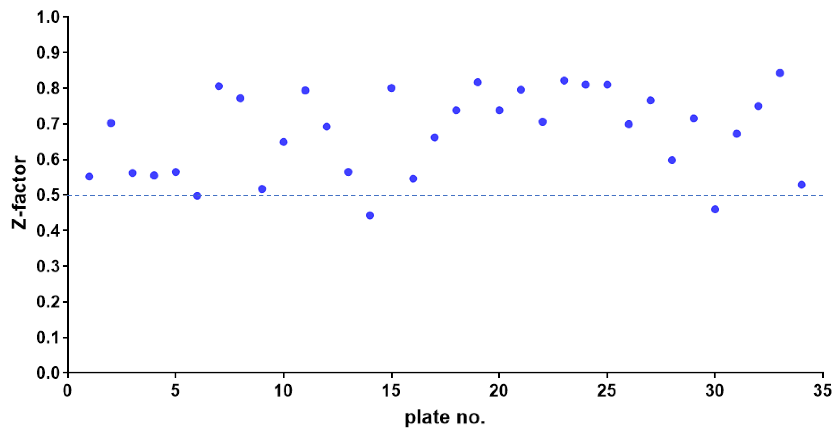
The average Z-factor between the negative and positive controls in the 96-well test plates was 0.675, ranging from 0.500 to 0.843. This value is well above the 0.500 threshold, indicating that the assay can reliably distinguish between positive and negative controls (Figure 2). In addition to the Z-factor, the coefficient of variation was less than 10%, and the signal-to-background ratio exceeded 10-fold, further demonstrating the effectiveness of our anti-S. aureus drug screening assay.
Figure 2.
Z-factor plot from high-throughput screening performed in 96-well plates. Solid dots represent the Z-factor from thirty-four 96-well plates. The dashed line indicates a threshold of 0.5.
2.2. Identification of Hits Inhibiting S. aureus Growth
We identified compounds that are effective against the MRSA Sa2 strain through HTS using a 96-well microtiter plate format. A total of 3386 compounds from our compound library were initially tested at a single concentration of 0.1 mM to identify active hits. Based on spectrophotometric measurements taken from the supernatant, the results were expressed as the percentage of bacterial growth inhibition compared to untreated control samples.
This primary screen initially identified 2439 out of 3386 (72%) compounds able to affect S. aureus Sa2 growth. Among those, 248 hits had “bacterial” as their primary target and were excluded since this work aimed to find new repositionable drugs. Most (146 out of 248; 58.9%) of these excluded compounds showed high (i.e., 60% ≤ growth reduction < 90%) or excellent activity (i.e., growth reduction ≥ 90%), therefore confirming the robustness of HTS.
The remaining 2191 (64.7%) hits showed potential for repurposing against S. aureus, although to different extents: 617 (18.2%) with low activity (i.e., 10% < growth reduction < 25%); 809 (23.9%) with moderate activity (i.e., 25% ≤ growth reduction < 60%); 350 (10.3%) with high activity (i.e., 60% ≤ growth reduction < 90%); and, more interestingly, 415 (12.2%) compounds with excellent activity (i.e., growth reduction ≥ 90%).
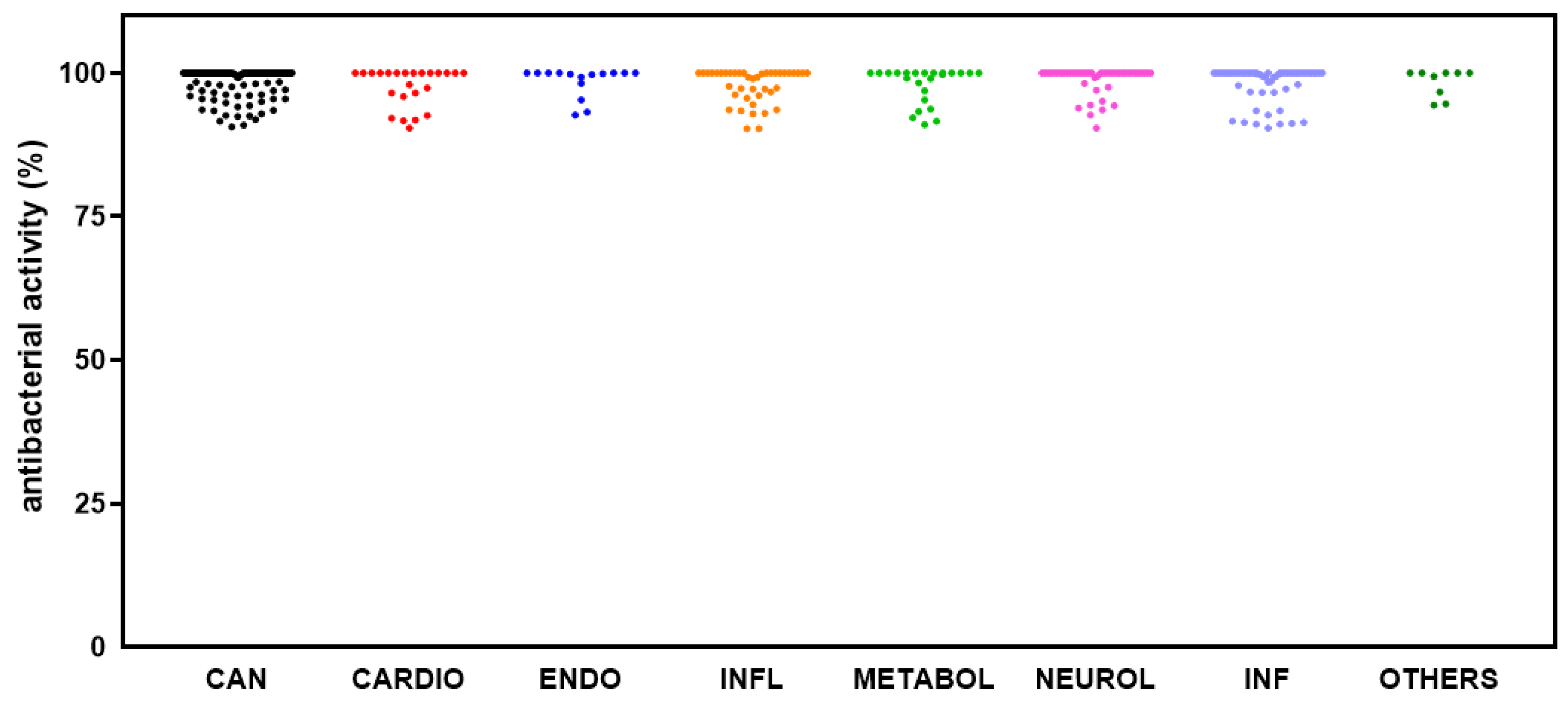
The 415 hits showing excellent activity based on spectrophotometric readings were re-assessed by cell viable count, confirming the efficacy of 364 (10.7%) compounds, listed in Table S1 (in the Supplementary Materials) and graphed in Figure 3. Among those, it is worth noting that 193 hits caused a 100% inhibition rate. Based on their known pharmacological profiles, the hits were grouped into classes based on the primary research area (some compounds belonged to multiple areas). Most of the hits belonged to “cancer” (83 out of 193; 43.0%) as a primary research area, followed by “infection” (other than bacterial) (31; 16.1%), “neurological disease” (28; 14.5%), “inflammation” (17; 8.8%), “metabolic disease” and “cardiovascular disease” (12 each; 6.2%), “endocrinology”, and “others” (5 each; 2.6%) (Figure 3). Dunnett’s multiple comparison tests found no statistically significant differences in antibacterial activity among research areas.
Figure 3.
HTS of the compound library to identify active hits against planktonic S. aureus Sa2 cells. The antibacterial activity of compounds in several research areas (CAN, cancer; CARDIO, cardiovascular; ENDO, endocrinology; INFL, inflammation; METABOL, metabolic disease; NEUROL, neurological disease; INF, infection; OTHERS, various research areas) is shown as a percentage, referred to as the untreated control.
2.3. Identification of Hits Active Against S. aureus Biofilm Formation
The compound library also underwent a 96-well microtiter plate-based HTS to identify hits that could affect biofilm formation by the MRSA Sa2 strain. The results were expressed as the percentage of inhibition of biofilm biomass formation compared to that in untreated controls, estimated from spectrophotometric readings after a crystal violet assay. The compounds significantly affecting biofilm formation, with no known antibacterial activity and inactive against S. aureus Sa2 (i.e., growth inhibition ≤ 10%), were considered potential antibiofilm hits.
HTS revealed that 654 out of 3386 (19.3%) compounds can reduce biofilm formation by S. aureus Sa2, although to different extents: 128 out of 654 (19.6%) with low activity (i.e., 10% < biofilm reduction < 25%), 344 (52.6%) with moderate activity (i.e., 25% ≤ biofilm reduction < 60%); 168 (25.7%) with high activity (i.e., 60% ≤ biofilm reduction < 90%); and 14 (2.1%) compounds with excellent activity (i.e., biofilm reduction ≥ 90%). Overall, hits belonged to several primary research areas, with “cancer” being the most prevalent, as shown in Figure 4. Dunnett’s multiple comparison tests found no statistically significant differences in activity against biofilm formation among research areas.
Figure 4.
HTS of the compound library to identify active hits against biofilm formation by S. aureus Sa2 strain. The antibiofilm activity of compounds in several research areas (CAN, cancer; CARDIO, cardiovascular; ENDO, endocrinology; INFL, inflammation; METABOL, metabolic disease; NEUROL, neurological disease; INF, infection; OTHERS, various research areas) is shown as a percentage referred to as the untreated control. The magnitude of the antibiofilm activity is categorized as follows: low (i.e., 10% < biofilm reduction < 25%), moderate (i.e., 25% ≤ biofilm reduction < 60%), high (i.e., 60% ≤ biofilm reduction < 90%), and excellent (i.e., biofilm reduction ≥ 90%).
The fourteen hits showing excellent activity were retested in a secondary screen—carried out in two independent experiments, each in triplicate—confirming an antibiofilm potential for 5 compounds (Table S2 in the Supplementary Materials). These repositionable hit candidates comprised three anticancer agents (Tipifarnib, Olaparib, and Acefylline), Hemin (“cardiovascular disease” research area), and TMC647055 (choline salt) (“infection” research area).
2.4. Identification of Hits Active Against Preformed Biofilm by S. aureus
Each of the 3386 compounds was tested at a single concentration of 0.1 mM to pinpoint hits effective towards preformed, 24 h old biofilm formed by the MRSA Sa2 strain. The results were expressed as the percentage of biofilm biomass dispersion compared to that in untreated controls, estimated from spectrophotometric readings after a crystal violet assay. The compounds with significant activity against mature biofilms, with no known antibacterial activity, and not active against S. aureus Sa2, were considered potential antibiofilm hits.
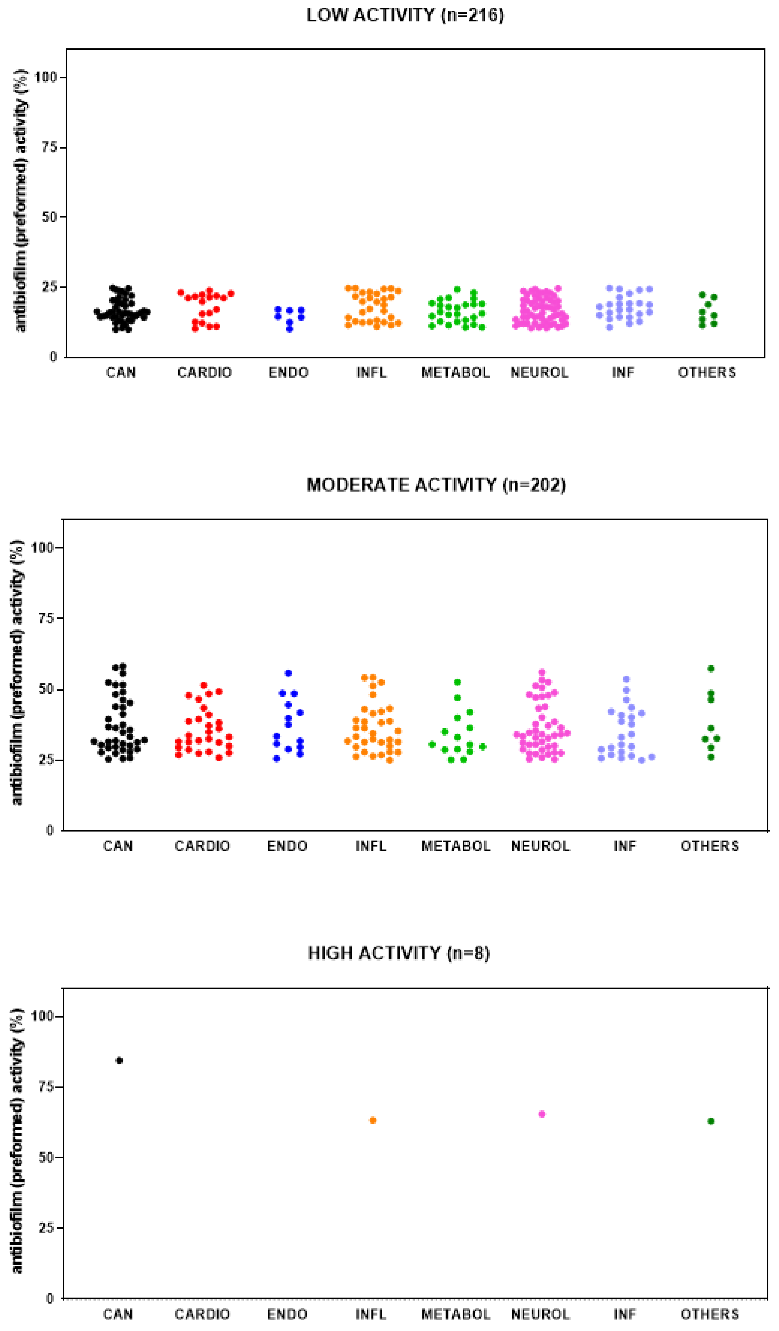
The primary screen identified 426 out of 3386 (12.6%) compounds with the potential for repurposing against mature biofilms by S. aureus Sa2, although to different extents. Most of the hits showed low (i.e., 10% < biofilm reduction < 25%) (216 out of 426; 56.1%) or moderate (i.e., 25% ≤ biofilm reduction < 60%) (202, 40.7%) activity, whereas only 8 (3.1%) showed high activity (i.e., 60% ≤ biofilm reduction < 90%). No compound was able to disperse at least 90% of biofilm biomass.
Overall, hits belonged to several primary research areas, although those with low and moderate activity mainly belonged to “neurological disease” and “cancer” (Figure 5). Dunnett’s multiple comparison tests found no statistically significant differences in activity against mature biofilm among research areas.
Figure 5.
HTS of the compound library to identify active hits against preformed biofilm by S. aureus Sa2 strain. The antibiofilm activity of compounds in several research areas (CAN, cancer; CARDIO, cardiovascular; ENDO, endocrinology; INFL, inflammation; METABOL, metabolic disease; NEUROL, neurological disease; INF, infection; OTHERS, various research areas) is shown as a percentage referred to as the untreated control. The magnitude of the antibiofilm activity is categorized as follows: low (i.e., 10% < biofilm reduction < 25%), moderate (i.e., 25% ≤ biofilm reduction < 60%), high (i.e., 60% ≤ biofilm reduction < 90%), and excellent (i.e., biofilm reduction ≥ 90%).
The eight hits showing high activity in the primary screening were re-assessed in a secondary screen—carried out in two independent experiments, each in triplicate—confirming four repositionable hit candidates, comprising Flumatinib (mesylate) with anticancer activity, the anti-inflammatory Bromfenac (sodium hydrate), Clemastine (fumarate) (“neurological disease”), and Heparin (lithium salt) (Table S3 in the Supplementary Materials).
3. Discussion
In the present study, we screened the Drug Repurposing Compound Library (MedChem Express) to identify hits with relevant potential for antibacterial and antibiofilm activities against an S. aureus strain causing a long-term pulmonary infection in a CF patient.
The library we tested in the present study consists of 3386 bioactive compounds, most of which have already been launched and used as drugs with several therapeutic indications, including cancer and neurodegenerative, infectious, and cardiovascular diseases. The library structure could explain the surprisingly high hit rates we observed during the first HTS: 72% (2440 out of 3386) for antibacterial activity, 19.3% (654 out of 3386) for activity against biofilm formation, and 12.6% (426 out of 3386) for activity against preformed biofilm. Indeed, less than 0.1% of hit rates were reported in other HTS studies evaluating large, random chemical libraries of small synthetic molecules that commonly contain numerous non-drug-like molecules [,]. The calculation of the Z-factor, the coefficient of variation, and the signal-to-background ratio validated the robustness and feasibility of our anti-S. aureus drug HTS assay.
An HTS was initially performed to identify compounds active against planktonic cells of the S. aureus Sa2 strain. Among the 3386 compounds screened, 2191 compounds—with a primary target different from “bacterial”—caused a decrease in Sa2 strain growth, although with varying effectiveness. We focused on selecting 193 compounds with the highest potential for repurposing since they exhibited maximum activity (i.e., 100% growth reduction), as confirmed by viable cell counts.
A revision of the scientific literature indicated that 135 out of 193 compounds were never reported to be active against S. aureus in previous studies and could be further investigated for repurposing potential. In our attempt to individuate drugs with a high repurposing potential relevant to the CF lung infection, the selected 135 potential hits underwent a literature review to reveal other biological effects critical to the interplay between the host and pathogens that shape the course of the disease, i.e., activity against other CF pathogens (e.g., Pseudomonas aeruginosa, Burkholderia cepacia, Haemophilus influenzae) and anti-inflammatory and anti-virulence potential []. In this way, 37 compounds were identified and are listed in Table 1.
Here, we reported antibacterial activity for 31 out of 37 selected compounds for the first time. Previous studies referred to the activity of the remaining 6 compounds against species other than S. aureus: the anticancer compound Toremifene and the antiasthmatic Zafirlukast vs. Porphyromonas gingivalis [,]; Zafirlukast, the anticancer Napabucasin, and the antipsychotic Perphenazine vs. oral streptococci [,,]; Zafirlukast and the anticancer compound Linsitinib vs. Mycobacterium tuberculosis [,]; and Perphenazine vs. Neisseria meningitidis, Enterobacteriaceae, and Listeria monocytogenes [].
Opportunistic polymicrobial bacterial airway infection is a hallmark of CF lung disease and early mortality []. From birth, the airways of individuals with CF are susceptible to infections by microbial opportunists. Over time, more concerning bacterial species often appear in respiratory secretions throughout their shortened lifetimes. The failure of bacterial clearance leads to a dominant chronic inflammation, resulting in a toxic pro-inflammatory local microenvironment that damages the lung and the innate immunity, further facilitating infections and resulting in the predominant cause of morbidity and mortality in CF patients. In this sense, it is worth noting the previously reported activity of the diuretic 5-(N,N-Hexamethylene)-amiloride and Perphenazine, respectively, against Pseudomonas cepacia [] and H. influenzae [], as these species pose the potential to cause pulmonary exacerbations and lung function decline in individuals suffering from CF [,].
Other recent evidence suggests that viridans group streptococci (VGS) may play a crucial role in maintaining a stable microbial ecology within the CF lung, enhancing virulence associated with polymicrobial interactions and directly contributing to the pathology of the lung. Streptococcus pneumoniae has recently been associated with an increased severe decline in FEV1 []. In addition, VGS may have the ability to aggregate with other important CF bacterial pathogens, such as S. aureus and P. aeruginosa, promoting initial attachment and eventual colonization with these pathogens and the development of multispecies biofilms in the CF airways []. In this regard, the previously reported activity of Perphenazine against S. pneumoniae [] and Toremifene against Streptococcus mutans [] adds value to their potential for repurposing, warranting further studies.
The prolonged use of antibiotics has been essential in improving the survival rates of CF patients. However, this approach leads to complications, such as increased adaptive antibiotic resistance, adverse effects, and allergic reactions. An attractive alternative recently explored for treating bacterial infections is targeting or reducing the production of virulence factors, such as inhibiting quorum sensing, biofilm formation, iron uptake, and efflux pumps [,]. Unlike antimicrobial therapies, this strategy does not affect bacterial growth and is less likely to lead to resistance, exerting low selective pressure. In this sense, five drugs among the hits we identified in the present study have previously been shown to exhibit anti-virulence potential against S. aureus and P. aeruginosa, increasing their repurposing potential in CF patients (Table 1). S. aureus hemolysis activity has been observed to be significantly affected in over 200 clinical isolates by the antihistamine compound Loratadine [], and by the antiparasitic Miltefosine in the invertebrate Galleria mellonella and a murine model of pneumonia []. The virulence potential of S. aureus is also decreased by the synthetic estrogen Diethylstilbestrol—causing a reduction in alpha-toxin, coagulase, deoxyribonuclease, and penicillinase production []—and Loratadine, affecting biofilm formation, pigmentation [], and exotoxin production []. In a rat subcutaneous catheter model, biofilm formation was also inhibited by the anticancer compound Toremifene []. Miltefosine and the anti-hypercholesterolemic agent Lovastatin showed anti-virulence potential in P. aeruginosa, respectively, inhibiting phospholipase C/sphingomyelinase with a protective effect against murine pneumonia [] and decreasing swarming motility []. Other studies revealed the synergistic activity of Toremifene and the anticancer compound Mitotane combined with polymyxin B against multidrug-resistant P. aeruginosa [,] and the percutaneous enhancer Laurocapram combined with cephalosporins against MRSA [].
The main hallmark of CF pathophysiology is excessive inflammation and the inability to resolve lung infections, contributing to morbidity and, eventually, mortality. Therefore, anti-inflammatory properties could be desirable to improve the repurposing potential of the hits found in the present study. Most of the 37 selected hit compounds have been reported for their anti-inflammatory potential in the literature, although in clinical settings or models not pertinent to infectious diseases (e.g., inflammatory bowel disease [], allergic dermatitis [], pulmonary arterial hypertension [], osteoarthritis [,], asthma [], Parkinson’s disease [], cigarette smoke [], vascular diseases [], thyroid eye disease [], and amyotrophic lateral sclerosis []) (Table 1). Conversely, a few hits have previously been reported for anti-inflammatory potential in models that could be relevant to CF lung infections, thus increasing their potential for repurposing: 5-(N,N-Hexamethylene)-amiloride in lipopolysaccharide (LPS)-exposed alveolar epithelial cells [], Zafirlukast in LPS-exposed mice [], GW 501,516 (Cardarine)—an aromatic ether under investigation for lipid metabolism’s disorder (phase 2)—and the anticancer compound Ricolinostat, which protect against LPS-activated macrophage inflammation [,], Bardoxololone—a semisynthetic triterpenoids under investigation for lymphoma—Omaveloxolone—a potent anti-inflammatory for treating Friedreich’s ataxia—and Verteporfin—used as a photosensitizer in photodynamic therapy—that alleviates LPS-triggered acute lung injury in mice [,,], and Vandertanib—approved for unresectable and disseminated diseases—in SARS-CoV2-infected mice [] (Table 1). Interestingly, the literature has already reported anti-inflammatory properties in CF patients or models for three hits. Specifically, Fenretide increases IL-1β expression in Cftr-knockout mice, improving their ability to combat P. aeruginosa lung infection []; in addition, it normalizes the fatty acid imbalance by reducing arachidonic acid and increasing docosahexaenoic acid in CF patients []. Zafirlukast, a leukotriene receptor antagonist licensed for asthma prevention, significantly improves the NIH clinical score observed in a pilot study involving CF patients [], probably due to ameliorated lung tissue pathology and reduced inflammatory cell infiltration reported in LPS-induced lung inflammation, both in vitro and in mice []. Finally, the quinoline Montelukast, approved for asthma and allergic rhinitis, decreases eosinophil cationic protein and IL-8 serum and sputum levels, as well as the sputum levels of myeloperoxidase, in CF patients [].
Bacterial persistence in CF lungs is influenced by local host defense impairments and the pathogens’ ability to adapt to a challenging environment due to selective pressures, such as hyperinflammation, oxidative stress, limited nutrients, anaerobiosis, increased acidity, and antibiotic exposure. Biofilm formation is a key adaptive strategy for bacteria, allowing them to persist even when antibiotics are administered. Currently, CF is considered a biofilm-associated disease, and controlling these biofilms is crucial for effective antimicrobial strategies. Several studies reported direct S. aureus biofilm visualization in CF patients by scanning electron microscopy and fluorescence electron in situ hybridization using a specific peptide nucleic acid and further viability evaluation by confocal laser scanning microscopy []. Biofilm formation was a common trait of both MRSA and MSSA strains in CF patients, and high biofilm-formation ability has been associated with fewer pulmonary exacerbations and, conversely, exacerbations negatively impacted biofilm formation [].
In this sense, in the second step of the present study, the compound library also underwent a 96-well microtiter plate-based HTS to identify hits that could affect biofilm formation and preformed, mature, biofilm by the MRSA Sa2 strain. Our findings indicated that 654 compounds affect biofilm formation, although to different extents. Notably, five compounds, i.e., the anticancer Tipifarnib and Olaparib, the bronchodilator Acefylline, Hemin, and the NS5b polymerase inhibitor TMC647055, under investigation for treating hepatitis C—exhibited, for the first time, the highest potential for repurposing due to excellent activity against biofilm formation, which resulted in a reduction of ≥90% compared to the untreated control (Table 2). A careful revision of the literature aimed at identifying additional biological properties of interest in managing CF patients revealed that Olaparib, Acefylline, and Hemin were previously reported for anti-inflammatory activity in a LPS-induced acute lung injury model [,,]. Hemin can also affect S. aureus virulence by downregulating hemolysin expression []. In disagreement with our findings, Tipifarnib and Hemin have previously been shown to inhibit S. aureus growth [,].
Regarding hits active against preformed biofilms, four compounds exhibited the highest activity, i.e., 60% ≤ biofilm reduction < 90% (Table 3). The antibiofilm activity exhibited by Clemastine (fumarate), a histamine receptor H1 antagonist, is probably due to a decrease in the transcriptional level of the biofilm formation-relevant fnbB, icaA, and icaB genes in S. aureus []. Conflicting findings come from the literature concerning the antibiofilm potential of Heparin, a highly sulfated glycosaminoglycan with uneven chain length, routinely used in central venous catheters to prevent thrombosis. Our findings disagree with a previous study reporting that Heparin augments biofilm formation in S. aureus [,], probably due to extracellular DNA-binding proteins on the S. aureus surface that mediate the incorporation of Heparin into the biofilm matrix []. A similar pro-biofilm effect was observed for Escherichia coli [], Staphylococcus epidermidis [], and in patients with colorectal cancer where Heparin increased the biofilm formed by intestinal flora, thus providing a protective layer in the intestinal tract of patients []. In agreement with our findings, heparinoids, glycosaminoglycans chemically and pharmacologically related to Heparin, were found to suppress biofilm formation in Cutibacterium acnes by inhibiting the AI-2-mediated QS []. No evidence of activity against preformed biofilms has been previously published for the anticancer compound Flumatinib (mesylate) and the nonsteroidal anti-inflammatory drug Bromfenac (sodium hydrate). All five hits showing high potential for dispersing established biofilm have also been reported to trigger anti-inflammatory activity [,,,], although only for Heparin under experimental settings relevant to CF patients, such as human bronchial cells [] and LPS-induced lung injury [].
Table 1.
Compounds (n = 37) showing maximum activity against planktonic cells (i.e., 100% growth reduction) of S. aureus Sa2 at HTS. Classification, therapeutic category, and mechanism of action are provided, along with other biological properties relevant to cystic fibrosis (CF).
Table 1.
Compounds (n = 37) showing maximum activity against planktonic cells (i.e., 100% growth reduction) of S. aureus Sa2 at HTS. Classification, therapeutic category, and mechanism of action are provided, along with other biological properties relevant to cystic fibrosis (CF).
| Compound | Classification Therapeutic Category and Indication(s) Mechanism(s) of Action | Antibacterial Activity | Anti-Virulence Activity | Anti-Inflammatory Activity |
|---|---|---|---|---|
| Perphenazine |
|
|
| |
| Miltefosine |
|
|
| |
| Diethylstilbestrol |
|
|
| |
| Selexipag |
|
| ||
| AZD-9056 |
|
| ||
| Vortioxetine |
|
| ||
| Lovastatin |
|
|
| |
| Fenretinide |
|
| ||
| Napabucasin |
|
|
| |
| Loratadine |
|
|
| |
| Toremifene |
|
|
| |
| Etifoxine |
|
| ||
| 5-(N,N-Hexamethylene)-amiloride |
|
|
| |
| Zafirlukast |
|
|
| |
| Mitoquinone (mesylate) |
|
| ||
| Crisaborole |
|
| ||
| Linsitinib |
|
|
| |
| GW 501516 (Cardarine) |
|
| ||
| Darapladib |
|
| ||
| Bardoxolone |
|
| ||
| Meisoindigo |
|
| ||
| Epinastine |
|
| ||
| Salirasib |
|
| ||
| Omaveloxolone |
|
| ||
| Incyclinide |
|
| ||
| Glecaprevir |
|
| ||
| Isradipine |
|
| ||
| Laurocapram |
|
| ||
| Masitinib |
|
| ||
| Mitotane |
|
| ||
| Montelukast |
|
| ||
| Ricolinostat |
|
| ||
| Vandetanib |
|
| ||
| Verteporfin |
|
| ||
| Vigabatrin |
|
| ||
| Diroximel (fumarate) |
|
| ||
| Infigratinib |
|
|
Data source: PubChem (https://pubchem.ncbi.nlm.nih.gov/, accessed on 9 April 2025). The literature review was performed using PubMed (https://pubmed.ncbi.nlm.nih.gov/, accessed on 9 April 2025) using the following keywords: [compound name] and “antibacterial”, “aureus”, “aeruginosa”, “inflammatory”, “inflammatory lung”, “virulence”, and “cystic fibrosis”.
Table 2.
Compounds (n = 5) showing excellent activity against biofilm formation (i.e., biofilm reduction ≥ 90%) by S. aureus Sa2 at HTS. Classification, therapeutic category, and mechanism of action are provided, along with other biological properties relevant to cystic fibrosis (CF).
Table 2.
Compounds (n = 5) showing excellent activity against biofilm formation (i.e., biofilm reduction ≥ 90%) by S. aureus Sa2 at HTS. Classification, therapeutic category, and mechanism of action are provided, along with other biological properties relevant to cystic fibrosis (CF).
| Compound | Classification Therapeutic Category and Indication(s) Mechanism(s) of Action | Antibacterial Activity | Anti-Virulence Activity | Anti-Inflammatory Activity |
|---|---|---|---|---|
| Tipifarnib |
|
|
| |
| Olaparib |
|
| ||
| Acefylline |
|
|
| |
| Hemin |
|
|
|
|
| TMC647055 (Choline salt) |
|
Data source: PubChem (https://pubchem.ncbi.nlm.nih.gov/, accessed on 9 April 2025). The literature review was performed using PubMed (https://pubmed.ncbi.nlm.nih.gov/, accessed on 9 April 2025) using the following keywords: [compound name] and “antibacterial”, “aureus”, “aeruginosa”, “inflammatory”, “inflammatory lung”, “virulence” and “cystic fibrosis”.
Table 3.
Compounds (n = 4) showing activity against established biofilm (i.e., 60% ≤ biofilm dispersion < 90%) by S. aureus Sa2 at HTS. Classification, therapeutic category, and mechanism of action are provided, along with other biological properties relevant to cystic fibrosis (CF).
Table 3.
Compounds (n = 4) showing activity against established biofilm (i.e., 60% ≤ biofilm dispersion < 90%) by S. aureus Sa2 at HTS. Classification, therapeutic category, and mechanism of action are provided, along with other biological properties relevant to cystic fibrosis (CF).
| Compound | Classification Therapeutic Category and Indication(s) Mechanism(s) of Action | Antibacterial Activity | Anti-Virulence Activity | Anti-Inflammatory Activity |
|---|---|---|---|---|
| Clemastine (fumarate) |
|
|
| |
| Heparin |
|
|
|
|
| Flumatinib (mesylate) |
| |||
| Bromfenac (sodium hydrate) |
|
|
|
Data source: PubChem (https://pubchem.ncbi.nlm.nih.gov/, accessed on 9 April 2025). The literature review was performed using PubMed (https://pubmed.ncbi.nlm.nih.gov/, accessed on 9 April 2025) using the following keywords: [compound name] and “antibacterial”, “aureus”, “aeruginosa”, “inflammatory”, “inflammatory lung”, “virulence” and “cystic fibrosis”.
4. Materials and Methods
4.1. Compound Library
The “Drug Repurposing Compound Library” was purchased from MedChem Express (cat. no. HY-L035; Monmouth Junction, NJ, USA). The library was provided in a 96-well plate format with aliquots of 10 mM stocks of drugs in DMSO or water, stored at −80 °C.
4.2. Bacterial Strain and Growth Conditions
HTS was conducted using the S. aureus Sa2, an MRSA strain isolated from a respiratory specimen in a CF patient, which caused chronic infection. Some colonies were grown on Tryptone Soy Agar (TSA; Oxoid, Milan, Italy) after overnight incubation at 37 °C. These colonies were then suspended in sterile saline 0.9% to achieve an optical density at 550 nm (OD550) of 0.3. This suspension was diluted 1:10 in sterile saline to reach a final 1–2 × 10⁷ CFU/mL concentration. This standardized inoculum was utilized for all assays unless otherwise indicated.
4.3. Antibacterial HTS Assay
The library was screened at a single concentration point to identify antibacterial compounds effective against the S. aureus Sa2 strain. Specifically, 5 µL of a standardized inoculum (corresponding to 0.5–1 × 10⁵ CFU/well) was added to each well of a 96-well polystyrene microtiter plate containing 100 µL of Tryptone Soy Broth (TSB; Oxoid) along with 1 µL of a 10 mM compound stock solution from the MedChem library. This setup achieved a final drug concentration of 0.1 mM. Uninoculated samples containing 1% (v/v) DMSO (the final background concentration in each well) were used as blanks. A negative control was also prepared with 50% (v/v) DMSO to ensure 100% killing of the bacteria. The contents of each well were mixed by pipetting, and the plates were incubated at 37 °C in an aerobic atmosphere. After a 24 h incubation period, the survival rate of the planktonic cells was assessed spectrophotometrically by measuring the OD550 of broth culture supernatants using a microplate reader (Tecan Infinite® M PLEX; Tecan Group Ltd., Mannedorf, Switzerland). This value was corrected by subtracting the average OD550 value of the uninoculated wells (blanks).
The growth rate percentage was calculated by comparing it to the inoculated control sample (which had not been treated), set at 100% growth. The antibacterial activity of the compounds in the library was categorized based on the percentage of growth reduction compared to the untreated control sample as follows: (i) low efficacy—10% < x < 25%; (ii) moderate efficacy—25% ≤ x < 60%; (iii) high efficacy—60% ≤ x < 90%; and (iv) excellent efficacy—90% ≤ x ≤ 100%. Only drugs causing a ≥90% reduction in bacterial burden, subsequently confirmed by cell viable count, were considered potential anti-S. aureus hit compounds and underwent further characterization. In the cell viable count, serial dilutions of the sample prepared in sterile saline were plated on TSA; after incubation for 24 h at 37 °C, cell viability was determined by CFU counting. DMSO at the final concentration of 0.1 mM did not show any activity against S. aureus Sa2, as assessed by cell viable count, thus indicating that the antibacterial effects observed were due to the compound only.
4.4. HTS Assay Validation
The results from each HTS microplate were validated by calculating the Z-factor. To validate the degree of separation, the Z-factor and the percent inhibition of the positive and negative controls were determined using the following formula:
where σp and σn are the standard deviations of the positive and negative controls, respectively, and μp and μn are the corresponding mean values. A Z-factor between 0.5 and 1.0 indicates an excellent assay and statistically reliable separation between the positive and negative controls.
4.5. Biofilm Inhibition and Dispersion HTS Assays
To assess the efficacy of the drugs in preventing biofilm formation, a standardized inoculum of 5 µL (1–2 × 10⁷ CFU/mL) was added to each TC-treated microplate (Falcon; Diagramma, Città Sant’Angelo, Italy) well. Each well contained 100 µL of TSB with 1 µL of a 10 mM compound stock solution from the MediChem library, achieving a final drug concentration of 0.1 mM. Control samples were prepared with TSB only. The microplate was incubated at 37 °C for 24 h, under an aerobic atmosphere and statically, after which planktonic cells were gently removed by washing each well twice with 100 µL PBS (Sigma-Aldrich Italia s.r.l.; Milan, Italy). The samples were then fixed at 60 °C for 1 h, and biofilm biomass was quantified spectrophotometrically. For the quantification, 100 µL of 1% (w/v) Hucker’s crystal violet (Sigma-Aldrich s.r.l.) was added to each well. After a 5 min incubation at room temperature, the wells were washed with tap water. Following drying at 37 °C, 100 µL of 33% (v/v) glacial acetic acid (Sigma-Aldrich Italia s.r.l.) was added to dissolve the stained dye for 15 min. The biofilm biomass was measured by recording the absorbance at 492 nm using a Tecan Infinite® M PLEX microplate reader.
To evaluate the effectiveness of the drugs against preformed biofilms, 24 h old biofilms were treated with 100 µL of TSB containing the selected drug at a final concentration of 0.1 mM, or with TSB only for the controls. These biofilm samples were incubated at 37 °C for another 24 h, under an aerobic atmosphere and statically, and washed with PBS. Finally, biofilm biomass was measured using the crystal violet staining method as previously described.
A potential “antibiofilm hit” had to affect biofilm formation or disperse preformed biofilms, whereas it had no antibacterial activity (growth reduction ≤ 10%). The magnitude of the hit’s activity against biofilm formation or preformed biofilms was categorized as follows: low (i.e., 10% < biofilm reduction < 25%), moderate (i.e., 25% ≤ biofilm reduction < 60%), high (i.e., 60% ≤ biofilm reduction < 90%), and excellent (i.e., biofilm reduction ≥ 90%).
4.6. Statistical Analysis
All assays were conducted in triplicate and repeated twice (n = 6). Statistical analysis was performed using GraphPad Prism software (version 7.0; GraphPad Software, San Diego, CA, USA). The data were tested for normal distribution using the Shapiro–Wilk test. Ordinary one-way ANOVA with Dunnett’s multiple comparisons test was applied when assessing differences among three or more groups of unpaired data. The statistical analysis assumed a confidence level of ≥95%, thus considering p values < 0.05 statistically significant.
5. Conclusions
The present study aimed to identify compounds with high repurposing potential against planktonic and sessile cells of an S. aureus strain causing chronic lung infection in a CF patient. In this regard, the HTS of a library compound performed in 96-well microtiter plates with a spectrophotometric assessment of the results revealed a helpful platform. We identified 193 hits causing 100% killing of planktonic cells, 5 causing biofilm reduction of at least 90%, and 4 effectively dispersing from 60 to 90% of established biofilm. Most hits were anticancer agents, followed by anti-infective (other than antibacterial) and anti-inflammatory drugs. Also considering other, already published, biological properties that could be relevant to the CF lung environment, among the hits active against planktonic S. aureus cells, those with the most interesting repurposing potential were 5-(N,N-Hexamethylene)-amiloride (active against other CF pathogens and with anti-inflammatory potential), Toremifene (active against other CF pathogens and with anti-virulence potential), and Zafirlukast, Fenretide, and Montelukast (with anti-inflammatory properties already observed in CF patients). Among hits affecting biofilm formation, Hemin also showed anti-virulence and anti-inflammatory effects. Heparin, Clemastine, and Bromfenac are the most interesting hits that significantly affect established biofilms.
Further in vitro and in vivo studies are needed to confirm the potential of repurposing these compounds for managing S. aureus lung infections in CF patients.
Supplementary Materials
The following supporting information can be downloaded at https://www.mdpi.com/article/10.3390/antibiotics14040402/s1, Table S1: Hits (n = 364) showing excellent activity (i.e., growth reduction ≥ 90% vs. unexposed control) against S. aureus Sa2 strain, based on spectrophotometric readings and cell viable count; Table S2: Hits (n = 5) showing excellent activity (i.e., biofilm reduction ≥ 90% vs. unexposed control) against biofilm formation by S. aureus Sa2 strain, based on spectrophotometric readings; Table S3: Hits (n = 4) showing high activity (i.e., 60% ≤ biofilm reduction < 90% vs. unexposed control) against preformed biofilm by S. aureus Sa2 strain, based on spectrophotometric readings.
Author Contributions
A.P.: Conceptualization, Data curation, Formal analysis, Funding acquisition, Investigation, Methodology, Project administration, Validation, Writing—original draft, Writing—review and editing; V.L.: Investigation, Writing—review and editing; V.P.: Investigation, Writing—review and editing; G.D.B.: Conceptualization, Data curation, Formal analysis, Funding acquisition, Methodology, Supervision, Writing—original draft, Writing—review and editing. All authors have read and agreed to the published version of the manuscript.
Funding
This work was partly funded by the “G. D’ANNUNZIO” UNIVERSITY OF CHIETI-PESCARA, grant number FAR “ex-60%”, 2022.
Institutional Review Board Statement
Not applicable.
Informed Consent Statement
Not applicable.
Data Availability Statement
The data are contained within the article or the Supplementary Materials.
Conflicts of Interest
The authors declare no conflicts of interest.
Abbreviations
The following abbreviations are used in this manuscript:
| CF | Cystic Fibrosis |
| MSSA | Methicillin-Sensitive Staphylococcus aureus |
| MRSA | Methicillin-Resistant Staphylococcus aureus |
| FEV1 | Forced Expiratory Volume in 1 s |
| HTS | High-Throughput Screening |
| VGS | Viridans Group Streptococci |
References
- Craft, K.M.; Nguyen, J.M.; Berg, L.J.; Townsend, S.D. Methicillin-resistant Staphylococcus aureus (MRSA): Antibiotic-resistance and the biofilm phenotype. MedChemComm 2019, 10, 1231–1241. [Google Scholar] [CrossRef] [PubMed]
- Dasenbrook, E.C.; Merlo, C.A.; Diener-West, M.; Lechtzin, N.; Boyle, M.P. Persistent methicillin-resistant Staphylococcus aureus and rate of FEV1 decline in cystic fibrosis. Am. J. Respir. Crit. Care Med. 2008, 178, 814–821. [Google Scholar] [CrossRef] [PubMed]
- Dasenbrook, E.C.; Checkley, W.; Merlo, C.A.; Konstan, M.W.; Lechtzin, N.; Boyle, M.P. Association between respiratory tract methicillin-resistant Staphylococcus aureus and survival in cystic fibrosis. JAMA 2010, 303, 2386–2392. [Google Scholar] [CrossRef]
- Xu, X.; Zhang, X.; Zhang, G.; Abbasi Tadi, D. Prevalence of antibiotic resistance of Staphylococcus aureus in cystic fibrosis infection: A systematic review and meta-analysis. J. Glob. Antimicrob. Resist. 2024, 36, 419–425. [Google Scholar] [CrossRef]
- Farha, M.A.; Brown, E.D. Drug repurposing for antimicrobial discovery. Nat. Microbiol. 2019, 4, 565–577. [Google Scholar] [CrossRef] [PubMed]
- Nilakantan, R.; Immermann, F.; Haraki, K. A novel approach to combinatorial library design. Comb. Chem. High Throughput Screen. 2002, 5, 105–110. [Google Scholar] [CrossRef]
- Langer, T.; Hoffmann, R.; Bryant, S.; Lesur, B. Hit finding: Towards ‘smarter’ approaches. Curr. Opin. Pharmacol. 2009, 9, 589–593. [Google Scholar] [CrossRef]
- Bhagirath, A.Y.; Li, Y.; Somayajula, D.; Dadashi, M.; Badr, S.; Duan, K. Cystic fibrosis lung environment and Pseudomonas aeruginosa infection. BMC Pulm. Med. 2016, 16, 174. [Google Scholar] [CrossRef]
- Gerits, E.; Defraine, V.; Vandamme, K.; De Cremer, K.; De Brucker, K.; Thevissen, K.; Cammue, B.P.; Beullens, S.; Fauvart, M.; Verstraeten, N.; et al. Repurposing Toremifene for Treatment of Oral Bacterial Infections. Antimicrob. Agents Chemother. 2017, 61, e01846-16. [Google Scholar] [CrossRef]
- Gerits, E.; Van der Massen, I.; Vandamme, K.; De Cremer, K.; De Brucker, K.; Thevissen, K.; Cammue, B.P.A.; Beullens, S.; Fauvart, M.; Verstraeten, N.; et al. In vitro activity of the antiasthmatic drug zafirlukast against the oral pathogens Porphyromonas gingivalis and Streptococcus mutans. FEMS Microbiol. Lett. 2017, 364, fnx005. [Google Scholar] [CrossRef]
- Mortensen, I.; Kristiansen, J.E.; Christensen, A.V.; Hvidberg, E.F. The antibacterial effect of some neuroleptics on strains isolated from patients with meningitis. Pharmacol. Toxicol. 1992, 71, 449–451. [Google Scholar] [CrossRef] [PubMed]
- Kuang, X.; Yang, T.; Zhang, C.; Peng, X.; Ju, Y.; Li, C.; Zhou, X.; Luo, Y.; Xu, X. Repurposing Napabucasin as an Antimicrobial Agent against Oral Streptococcal Biofilms. BioMed Res. Int. 2020, 2020, 8379526. [Google Scholar] [CrossRef]
- Pinault, L.; Han, J.S.; Kang, C.M.; Franco, J.; Ronning, D.R. Zafirlukast inhibits complexation of Lsr2 with DNA and growth of Mycobacterium tuberculosis. Antimicrob. Agents Chemother. 2013, 57, 2134–2140. [Google Scholar] [CrossRef]
- Wang, H.; Bi, J.; Zhang, Y.; Pan, M.; Guo, Q.; Xiao, G.; Cui, Y.; Hu, S.; Chan, C.K.; Yuan, Y.; et al. Human Kinase IGF1R/IR Inhibitor Linsitinib Controls the In Vitro and Intracellular Growth of Mycobacterium tuberculosis. ACS Infect. Dis. 2022, 8, 2019–2027. [Google Scholar] [CrossRef] [PubMed]
- VanDevanter, D.R.; LiPuma, J.J.; Konstan, M.W. Longitudinal bacterial prevalence in cystic fibrosis airways: Fact and artifact. J. Cyst. Fibros. 2024, 23, 58–64. [Google Scholar] [CrossRef] [PubMed]
- Cohn, R.C.; Rudzienski, L. In vitro suppression of Pseudomonas cepacia after limited exposure to subinhibitory concentrations of amiloride and 5-(N,N-hexamethylene) amiloride. Pediatr. Pulmonol. 1994, 17, 366–369. [Google Scholar] [CrossRef]
- LiPuma, J.J. The changing Microbial Epidemiology in cystic fibrosis. Clin. Microbiol. Rev. 2010, 23, 299–323. [Google Scholar] [CrossRef]
- Gutiérrez Santana, J.C.; Coria Jiménez, V.R. Burkholderia cepacia complex in cystic fibrosis: Critical gaps in diagnosis and therapy. Ann. Med. 2024, 56, 2307503. [Google Scholar] [CrossRef]
- Paganin, P.; Fiscarelli, E.V.; Tuccio, V.; Chiancianesi, M.; Bacci, G.; Morelli, P.; Dolce, D.; Dalmastri, C.; De Alessandri, A.; Lucidi, V.; et al. Changes in cystic fibrosis airway microbial community associated with a severe decline in lung function. PLoS ONE 2015, 10, e0124348. [Google Scholar] [CrossRef]
- Rickard, A.H.; Gilbert, P.; High, N.J.; Kolenbrander, P.E.; Handley, P.S. Bacterial coaggregation: An integral process in the development of multi-species biofilms. Trends Microbiol. 2003, 11, 94–100. [Google Scholar] [CrossRef]
- Carullo, G.; Di Bonaventura, G.; Rossi, S.; Lupetti, V.; Tudino, V.; Brogi, S.; Butini, S.; Campiani, G.; Gemma, S.; Pompilio, A. Development of Quinazolinone Derivatives as Modulators of Virulence Factors of Pseudomonas aeruginosa Cystic Fibrosis Strains. Molecules 2023, 28, 6535. [Google Scholar] [CrossRef] [PubMed]
- Mudgil, U.; Khullar, L.; Chadha, J.; Prerna Harjai, K. Beyond antibiotics: Emerging antivirulence strategies to combat Pseudomonas aeruginosa in cystic fibrosis. Microb. Pathog. 2024, 193, 106730. [Google Scholar] [CrossRef] [PubMed]
- Zheng, J.; Shang, Y.; Wu, Y.; Zhao, Y.; Chen, Z.; Lin, Z.; Li, P.; Sun, X.; Xu, G.; Wen, Z.; et al. Loratadine inhibits Staphylococcus aureus virulence and biofilm formation. iScience 2022, 25, 103731. [Google Scholar] [CrossRef]
- Koch, G.; Wermser, C.; Acosta, I.C.; Kricks, L.; Stengel, S.T.; Yepes, A.; Lopez, D. Attenuating Staphylococcus aureus Virulence by Targeting Flotillin Protein Scaffold Activity. Cell Chem. Biol. 2017, 24, 845–857.e6. [Google Scholar] [CrossRef] [PubMed]
- Yotis, W. Effects of diethylstilbestrol on the production of various extracellular products of Staphylococcus aureus. Experientia 1977, 33, 325–326. [Google Scholar] [CrossRef]
- Balogh, H.; Anthony, A.K.; Stempel, R.; Vossen, L.; Federico, V.A.; Valenzano, G.Z.; Blackledge, M.S.; Miller, H.B. Novel anti-virulence compounds disrupt exotoxin expression in MRSA. Microbiol. Spectr. 2024, 12, e0146424. [Google Scholar] [CrossRef]
- De Cremer, K.; Delattin, N.; De Brucker, K.; Peeters, A.; Kucharíková, S.; Gerits, E.; Verstraeten, N.; Michiels, J.; Van Dijck, P.; Cammue, B.P.; et al. Oral administration of the broad-spectrum antibiofilm compound toremifene inhibits Candida albicans and Staphylococcus aureus biofilm formation in vivo. Antimicrob. Agents Chemother. 2014, 58, 7606–7610. [Google Scholar] [CrossRef]
- Wargo, M.J.; Gross, M.J.; Rajamani, S.; Allard, J.L.; Lundblad, L.K.; Allen, G.B.; Vasil, M.L.; Leclair, L.W.; Hogan, D.A. Hemolytic phospholipase C inhibition protects lung function during Pseudomonas aeruginosa infection. Am. J. Respir. Crit. Care Med. 2011, 184, 345–354. [Google Scholar] [CrossRef]
- Hennessy, E.; Mooij, M.J.; Legendre, C.; Reen, F.J.; O’Callaghan, J.; Adams, C.; O’Gara, F. Statins inhibit in vitro virulence phenotypes of Pseudomonas aeruginosa. J. Antibiot. 2013, 66, 99–101. [Google Scholar] [CrossRef]
- Hussein, M.H.; Schneider, E.K.; Elliott, A.G.; Han, M.; Reyes-Ortega, F.; Morris, F.; Blastovich, M.A.T.; Jasim, R.; Currie, B.; Mayo, M.; et al. From Breast Cancer to Antimicrobial: Combating Extremely Resistant Gram-Negative “Superbugs” Using Novel Combinations of Polymyxin B with Selective Estrogen Receptor Modulators. Microb. Drug Resist. 2017, 23, 640–650. [Google Scholar] [CrossRef]
- Tran, T.B.; Wang, J.; Doi, Y.; Velkov, T.; Bergen, P.J.; Li, J. Novel Polymyxin Combination With Antineoplastic Mitotane Improved the Bacterial Killing Against Polymyxin-Resistant Multidrug-Resistant Gram-Negative Pathogens. Front. Microbiol. 2018, 9, 721. [Google Scholar] [CrossRef]
- Wang, N.; Li, W.; Yu, H.; Huang, W.; Qiao, Y.; Wang, Q.; Wei, Y.; Deng, X.; Wang, J.; Cui, M.; et al. Laurocapram, a transdermal enhancer, boosts cephalosporin’s antibacterial activity against Methicillin-resistant Staphylococcus aureus. Biochem. Pharmacol. 2024, 227, 116404. [Google Scholar] [CrossRef]
- Verhaar, A.P.; Wildenberg, M.E.; Velde, A.A.; Meijer, S.L.; Vos, A.C.; Duijvestein, M.; Peppelenbosch, M.P.; Hommes, D.W.; van den Brink, G.R. Miltefosine suppresses inflammation in a mouse model of inflammatory bowel disease. Inflamm. Bowel Dis. 2013, 19, 1974–1982. [Google Scholar] [CrossRef] [PubMed]
- Heo, M.J.; Choi, S.Y.; Lee, C.; Choi, Y.M.; An, I.S.; Bae, S.; An, S.; Jung, J.H. Perphenazine Attenuates the Pro-Inflammatory Responses in Mouse Models of Th2-Type Allergic Dermatitis. Int. J. Mol. Sci. 2020, 21, 3241. [Google Scholar] [CrossRef] [PubMed]
- Geerdink, L.M.; Bertram, H.; Hansmann, G. First-in-child use of the oral selective prostacyclin IP receptor agonist selexipag in pulmonary arterial hypertension. Pulm. Circ. 2017, 7, 551–554. [Google Scholar] [CrossRef] [PubMed]
- Aizman, E.; Blacher, E.; Ben-Moshe, O.; Kogan, T.; Kloog, Y.; Mor, A. Therapeutic effect of farnesylthiosalicylic acid on adjuvant-induced arthritis through suppressed release of inflammatory cytokines. Clin. Exp. Immunol. 2014, 175, 458–467. [Google Scholar] [CrossRef]
- Hu, H.; Yang, B.; Li, Y.; Zhang, S.; Li, Z. Blocking of the P2X7 receptor inhibits the activation of the MMP-13 and NF-κB pathways in the cartilage tissue of rats with osteoarthritis. Int. J. Mol. Med. 2016, 38, 1922–1932. [Google Scholar] [CrossRef]
- Townsend, E.A.; Guadarrama, A.; Shi, L.; Roti Roti, E.; Denlinger, L.C. P2X7 signaling influences the production of pro-resolving and pro-inflammatory lipid mediators in alveolar macrophages derived from individuals with asthma. Am. J. Physiol. Lung Cell. Mol. Physiol. 2023, 325, L399–L410. [Google Scholar] [CrossRef]
- Tian, Q.; Yang, X.; Du, J.; Huang, H.; Liu, W.; Zhao, P. Translocator Protein Ligand Etifoxine Attenuates MPTP-Induced Neurotoxicity. Front. Mol. Neurosci. 2022, 15, 850904. [Google Scholar] [CrossRef]
- Yang, D.; Xu, D.; Wang, T.; Yuan, Z.; Liu, L.; Shen, Y.; Wen, F. Mitoquinone ameliorates cigarette smoke-induced airway inflammation and mucus hypersecretion in mice. Int. Immunopharmacol. 2021, 90, 107149. [Google Scholar] [CrossRef]
- Fan, T.; Wang, W.; Wang, Y.; Zeng, M.; Liu, Y.; Zhu, S.; Yang, L. PDE4 inhibitors: Potential protective effects in inflammation and vascular diseases. Front. Pharmacol. 2024, 15, 1407871. [Google Scholar] [CrossRef] [PubMed]
- Gulbins, A.; Horstmann, M.; Daser, A.; Flögel, U.; Oeverhaus, M.; Bechrakis, N.E.; Banga, J.P.; Keitsch, S.; Wilker, B.; Krause, G.; et al. Linsitinib, an IGF-1R inhibitor, attenuates disease development and progression in a model of thyroid eye disease. Front. Endocrinol. 2023, 14, 1211473. [Google Scholar] [CrossRef] [PubMed]
- Ketabforoush, A.H.M.E.; Chegini, R.; Barati, S.; Tahmasebi, F.; Moghisseh, B.; Joghataei, M.T.; Faghihi, F.; Azedi, F. Masitinib: The promising actor in the next season of the Amyotrophic Lateral Sclerosis treatment series. Biomed. Pharmacother. 2023, 160, 114378. [Google Scholar] [CrossRef]
- Haddad, J.J.; Land, S.C. Amiloride blockades lipopolysaccharide-induced proinflammatory cytokine biosynthesis in an IkappaB-alpha/NF-kappaB-dependent mechanism. Evidence for the amplification of an antiinflammatory pathway in the alveolar epithelium. Am. J. Respir. Cell Mol. Biol. 2002, 26, 114–126. [Google Scholar] [CrossRef]
- Xue, T.; Zhang, Q.; Zhang, T.; Meng, L.; Liu, J.; Chai, D.; Liu, Y.; Yang, Z.; Jiao, R.; Cui, Y.; et al. Zafirlukast ameliorates lipopolysaccharide and bleomycin-induced lung inflammation in mice. BMC Pulm. Med. 2024, 24, 456. [Google Scholar] [CrossRef]
- Zhang, W.B.; Yang, F.; Wang, Y.; Jiao, F.Z.; Zhang, H.Y.; Wang, L.W.; Gong, Z.J. Inhibition of HDAC6 attenuates LPS-induced inflammation in macrophages by regulating oxidative stress and suppressing the TLR4-MAPK/NF-κB pathways. Biomed. Pharmacother. 2019, 117, 109166. [Google Scholar] [CrossRef] [PubMed]
- Lim, H.J.; Kwak, H.J. Selective PPARδ Agonist GW501516 Protects Against LPS-Induced Macrophage Inflammation and Acute Liver Failure in Mice via Suppressing Inflammatory Mediators. Molecules 2024, 29, 5189. [Google Scholar] [CrossRef]
- Pei, X.; Zhang, X.J.; Chen, H.M. Bardoxolone treatment alleviates lipopolysaccharide (LPS)-induced acute lung injury through suppressing inflammation and oxidative stress regulated by Nrf2 signaling. Biochem. Biophys. Res. Commun. 2019, 516, 270–277. [Google Scholar] [CrossRef]
- Wu, Y.; Zhang, Y.; Ge, L.; He, S.; Zhang, Y.; Chen, D.; Nie, Y.; Zhu, M.; Pang, Q. RTA408 alleviates lipopolysaccharide-induced acute lung injury via inhibiting Bach1-mediated ferroptosis. Int. Immunopharmacol. 2024, 142 Pt B, 113250. [Google Scholar] [CrossRef]
- Cen, H.; Sun, M.; Zheng, B.; Peng, W.; Wen, Q.; Lin, Z.; Zhang, X.; Zhou, N.; Zhu, G.; Yu, X.; et al. Hyaluronic acid modified nanocarriers for aerosolized delivery of verteporfin in the treatment of acute lung injury. Int. J. Biol. Macromol. 2024, 267 Pt 1, 131386. [Google Scholar] [CrossRef]
- Puhl, A.C.; Gomes, G.F.; Damasceno, S.; Fritch, E.J.; Levi, J.A.; Johnson, N.J.; Scholle, F.; Premkumar, L.; Hurst, B.L.; Lee-Montiel, F.; et al. Vandetanib Blocks the Cytokine Storm in SARS-CoV-2-Infected Mice. ACS Omega 2022, 7, 31935–31944. [Google Scholar] [CrossRef] [PubMed]
- Guilbault, C.; De Sanctis, J.B.; Wojewodka, G.; Saeed, Z.; Lachance, C.; Skinner, T.A.; Vilela, R.M.; Kubow, S.; Lands, L.C.; Hajduch, M.; et al. Fenretinide corrects newly found ceramide deficiency in cystic fibrosis. Am. J. Respir. Cell Mol. Biol. 2008, 38, 47–56. [Google Scholar] [CrossRef] [PubMed]
- Guilbault, C.; Wojewodka, G.; Saeed, Z.; Hajduch, M.; Matouk, E.; De Sanctis, J.B.; Radzioch, D. Cystic fibrosis fatty acid imbalance is linked to ceramide deficiency and corrected by fenretinide. Am. J. Respir. Cell Mol. Biol. 2009, 41, 100–106. [Google Scholar] [CrossRef]
- Conway, S.P.; Etherington, C.; Peckham, D.G.; Whitehead, A. A pilot study of zafirlukast as an anti-inflammatory agent in the treatment of adults with cystic fibrosis. J. Cyst. Fibros. 2003, 2, 25–28. [Google Scholar] [CrossRef] [PubMed][Green Version]
- Schmitt-Grohé, S.; Zielen, S. Leukotriene receptor antagonists in children with cystic fibrosis lung disease: Anti-inflammatory and clinical effects. Paediatr. Drugs 2005, 7, 353–363. [Google Scholar] [CrossRef]
- Jean-Pierre, V.; Boudet, A.; Sorlin, P.; Menetrey, Q.; Chiron, R.; Lavigne, J.P.; Marchandin, H. Biofilm Formation by Staphylococcus aureus in the Specific Context of Cystic Fibrosis. Int. J. Mol. Sci. 2022, 24, 597. [Google Scholar] [CrossRef]
- Wieneke, M.K.; Dach, F.; Neumann, C.; Görlich, D.; Kaese, L.; Thißen, T.; Dübbers, A.; Kessler, C.; Große-Onnebrink, J.; Küster, P.; et al. Association of Diverse Staphylococcus aureus Populations with Pseudomonas aeruginosa Coinfection and Inflammation in Cystic Fibrosis Airway Infection. mSphere 2021, 6, e0035821. [Google Scholar] [CrossRef]
- Kapoor, K.; Singla, E.; Sahu, B.; Naura, A.S. PARP inhibitor, olaparib ameliorates acute lung and kidney injury upon intratracheal administration of LPS in mice. Mol. Cell. Biochem. 2015, 400, 153–162. [Google Scholar] [CrossRef]
- Elgazar, A.A.; El-Domany, R.A.; Eldehna, W.M.; Badria, F.A. Theophylline-based hybrids as acetylcholinesterase inhibitors endowed with anti-inflammatory activity: Synthesis, bioevaluation, in silico and preliminary kinetic studies. RSC Adv. 2023, 13, 25616–25634. [Google Scholar] [CrossRef]
- Cheng, X.; Yin, M.; Sun, X.; Zhang, Z.; Yao, X.; Liu, H.; Xia, H. Hemin attenuated LPS-induced acute lung injury in mice via protecting pulmonary epithelial barrier and regulating HO-1/NLRP3-mediated pyroptosis. Shock 2023, 59, 744–753. [Google Scholar] [CrossRef]
- Schmitt, J.; Joost, I.; Skaar, E.P.; Herrmann, M.; Bischoff, M. Haemin represses the haemolytic activity of Staphylococcus aureus in an Sae-dependent manner. Microbiology 2012, 158 Pt 10, 2619–2631. [Google Scholar] [CrossRef]
- Ladan, H.; Nitzan, Y.; Malik, Z. The antibacterial activity of haemin compared with cobalt, zinc and magnesium protoporphyrin and its effect on potassium loss and ultrastructure of Staphylococcus aureus. FEMS Microbiol. Lett. 1993, 112, 173–177. [Google Scholar] [CrossRef] [PubMed]
- Weber, L.; Hagemann, A.; Kaltenhäuser, J.; Besser, M.; Rockenfeller, P.; Ehrhardt, A.; Stuermer, E.; Bachmann, H.S. Bacteria Are New Targets for Inhibitors of Human Farnesyltransferase. Front. Microbiol. 2021, 12, 628283. [Google Scholar] [CrossRef]
- Shang, Y.; Guo, J.; Zhao, Y.; Chen, J.; Meng, Q.; Qu, D.; Zheng, J.; Yu, Z.; Wu, Y.; Deng, Q. Clemastine Inhibits the Biofilm and Hemolytic of Staphylococcus aureus through the GdpP Protein. Microbiol. Spectr. 2022, 10, e0054121. [Google Scholar] [CrossRef]
- Shanks, R.M.; Donegan, N.P.; Graber, M.L.; Buckingham, S.E.; Zegans, M.E.; Cheung, A.L.; O’Toole, G.A. Heparin stimulates Staphylococcus aureus biofilm formation. Infect. Immun. 2005, 73, 4596–4606. [Google Scholar] [CrossRef] [PubMed]
- Najarzadeh, Z.; Zaman, M.; Sereikaite, V.; Strømgaard, K.; Andreasen, M.; Otzen, D.E. Heparin promotes fibrillation of most phenol-soluble modulin virulence peptides from Staphylococcus aureus. J. Biol. Chem. 2021, 297, 100953. [Google Scholar] [CrossRef] [PubMed]
- Mishra, S.; Horswill, A.R. Heparin Mimics Extracellular DNA in Binding to Cell Surface-Localized Proteins and Promoting Staphylococcus aureus Biofilm Formation. mSphere 2017, 2, e00135-17. [Google Scholar] [CrossRef]
- Wu, D.; Li, X.; Yu, Y.; Gong, B.; Zhou, X. Heparin stimulates biofilm formation of Escherichia coli strain Nissle 1917. Biotechnol. Lett. 2021, 43, 235–246. [Google Scholar] [CrossRef]
- Chen, Q.; Xu, L.; Wu, T.; Li, J.; Hua, L. Analysis of abnormal intestinal flora on risk of intestinal cancer and effect of heparin on formation of bacterial biofilm. Bioengineered 2022, 13, 894–904. [Google Scholar] [CrossRef]
- Hamada, S.; Minami, S.; Gomi, M. Heparinoid enhances the efficacy of a bactericidal agent by preventing Cutibacterium acnes biofilm formation via quorum sensing inhibition. J. Microorg. Control 2024, 29, 27–31. [Google Scholar] [CrossRef]
- Kida, T.; Kozai, S.; Takahashi, H.; Isaka, M.; Tokushige, H.; Sakamoto, T. Pharmacokinetics and efficacy of topically applied nonsteroidal anti-inflammatory drugs in retinochoroidal tissues in rabbits. PLoS ONE 2014, 9, e96481. [Google Scholar] [CrossRef] [PubMed]
- Yi, N.Y.; Newman, D.R.; Zhang, H.; Morales Johansson, H.; Sannes, P.L. Heparin and LPS-induced COX-2 expression in airway cells: A link between its anti-inflammatory effects and GAG sulfation. Exp. Lung Res. 2015, 41, 499–513. [Google Scholar] [CrossRef]
- Chimenti, L.; Camprubí-Rimblas, M.; Guillamat-Prats, R.; Gomez, M.N.; Tijero, J.; Blanch, L.; Artigas, A. Nebulized Heparin Attenuates Pulmonary Coagulopathy and Inflammation through Alveolar Macrophages in a Rat Model of Acute Lung Injury. Thromb. Haemost. 2017, 117, 2125–2134. [Google Scholar] [CrossRef] [PubMed]
- Motawi, T.K.; El-Maraghy, S.A.; Kamel, A.S.; Said, S.E.; Kortam, M.A. Modulation of p38 MAPK and Nrf2/HO-1/NLRP3 inflammasome signaling and pyroptosis outline the anti-neuroinflammatory and remyelinating characters of Clemastine in EAE rat model. Biochem. Pharmacol. 2023, 209, 115435. [Google Scholar] [CrossRef] [PubMed]
- Tou, J.S.; Urbizo, C. Diethylstilbestrol inhibits phospholipase D activity and degranulation by stimulated human neutrophils. Steroids 2008, 73, 216–221. [Google Scholar] [CrossRef]
- Talmon, M.; Rossi, S.; Pastore, A.; Cattaneo, C.I.; Brunelleschi, S.; Fresu, L.G. Vortioxetine exerts anti-inflammatory and immunomodulatory effects on human monocytes/macrophages. Br. J. Pharmacol. 2018, 175, 113–124. [Google Scholar] [CrossRef]
- Planagumà, A.; Pfeffer, M.A.; Rubin, G.; Croze, R.; Uddin, M.; Serhan, C.N.; Levy, B.D. Lovastatin decreases acute mucosal inflammation via 15-epi-lipoxin A4. Mucosal Immunol. 2010, 3, 270–279. [Google Scholar] [CrossRef]
- Williams, J.P.; Hernady, E.; Johnston, C.J.; Reed, C.M.; Fenton, B.; Okunieff, P.; Finkelstein, J.N. Effect of administration of lovastatin on the development of late pulmonary effects after whole-lung irradiation in a murine model. Radiat. Res. 2004, 161, 560–567. [Google Scholar] [CrossRef]
- Ravi, L.I.; Li, L.; Wong, P.S.; Sutejo, R.; Tan, B.H.; Sugrue, R.J. Lovastatin treatment mitigates the pro-inflammatory cytokine response in respiratory syncytial virus infected macrophage cells. Antivir. Res. 2013, 98, 332–343. [Google Scholar] [CrossRef]
- Yu, H.; Valerio, M.; Bielawski, J. Fenretinide inhibited de novo ceramide synthesis and proinflammatory cytokines induced by Aggregatibacter actinomycetemcomitans. J. Lipid Res. 2013, 54, 189–201. [Google Scholar] [CrossRef]
- Wang, J.; Feng, H.; Li, Z.; Zhang, X. Napabucasin prevents brain injury in neuronal neonatal rat cells through suppression of apoptosis and inflammation. Microb. Pathog. 2019, 128, 337–341. [Google Scholar] [CrossRef] [PubMed]
- Cheng, K.C.; Hsu, J.Y.; Fu, L.S.; Chang, W.C.; Chu, J.J.; Chi, C.S. Influence of cetirizine and loratadine on granulocyte-macrophage colony-stimulating factor and interleukin-8 release in A549 human airway epithelial cells stimulated with interleukin-1beta. J. Microbiol. Immunol. Infect. 2006, 39, 206–211. [Google Scholar] [PubMed]
- Lv, S.L.; Zeng, Z.F.; Gan, W.Q.; Wang, W.Q.; Li, T.G.; Hou, Y.F.; Yan, Z.; Zhang, R.X.; Yang, M. Lp-PLA2 inhibition prevents Ang II-induced cardiac inflammation and fibrosis by blocking macrophage NLRP3 inflammasome activation. Acta Pharmacol. Sin. 2021, 42, 2016–2032. [Google Scholar] [CrossRef]
- He, J.X.; Zhu, C.Q.; Liang, G.F.; Mao, H.B.; Shen, W.Y.; Hu, J.B. Targeted-lung delivery of bardoxolone methyl using PECAM-1 antibody-conjugated nanostructure lipid carriers for the treatment of lung inflammation. Biomed. Pharmacother. 2024, 178, 116992. [Google Scholar] [CrossRef] [PubMed]
- Ye, B.; Xiong, X.; Deng, X.; Gu, L.; Wang, Q.; Zeng, Z.; Gao, X.; Gao, Q.; Wang, Y. Meisoindigo, but not its core chemical structure indirubin, inhibits zebrafish interstitial leukocyte chemotactic migration. Pharm. Biol. 2017, 55, 673–679. [Google Scholar] [CrossRef]
- Ye, Y.; Jin, T.; Zhang, X.; Zeng, Z.; Ye, B.; Wang, J.; Zhong, Y.; Xiong, X.; Gu, L. Meisoindigo Protects Against Focal Cerebral Ischemia-Reperfusion Injury by Inhibiting NLRP3 Inflammasome Activation and Regulating Microglia/Macrophage Polarization via TLR4/NF-κB Signaling Pathway. Front. Cell. Neurosci. 2019, 13, 553. [Google Scholar] [CrossRef]
- Ishikawa, S.; Sawamoto, A.; Okuyama, S.; Nakajima, M. T-Cell Activation-Inhibitory Assay to Screen Caloric Restriction Mimetics Drugs for Drug Repositioning. Biol. Pharm. Bull. 2021, 44, 550–556. [Google Scholar] [CrossRef]
- Wu, J.J.; Yuan, X.M.; Huang, C.; An, G.Y.; Liao, Z.L.; Liu, G.A.; Chen, R.X. Farnesyl thiosalicylic acid prevents iNOS induction triggered by lipopolysaccharide via suppression of iNOS mRNA transcription in murine macrophages. Int. Immunopharmacol. 2019, 68, 218–225. [Google Scholar] [CrossRef]
- Alyousef, A.A.; Divakar, D.D.; Muzaheed. Chemically modified tetracyclines an emerging host modulator in chronic periodontitis patients: A randomized, double-blind, placebo-controlled, clinical trial. Microb. Pathog. 2017, 110, 279–284. [Google Scholar] [CrossRef]
- Farghaly, T.A.; Alqurashi, R.M.; Masaret, G.S.; Abdulwahab, H.G. Recent Methods for the Synthesis of Quinoxaline Derivatives and their Biological Activities. Mini Rev. Med. Chem. 2024, 24, 920–982. [Google Scholar] [CrossRef]
- Capecchi, P.L.; Laghi Pasini, F.; Ceccatelli, L.; Di Perri, T. Isradipine inhibits PMN leukocyte function. A possible interference with the adenosine system. Immunopharmacol. Immunotoxicol. 1993, 15, 133–149. [Google Scholar] [CrossRef] [PubMed]
- Alnfakh, Z.A.; Al-Mudhafar, D.H.; Al-Nafakh, R.T.; Jasim, A.E.; Hadi, N.R. The anti-inflammatory and antioxidant effects of Montelukast on lung sepsis in adult mice. J. Med. Life 2022, 15, 819–827. [Google Scholar] [CrossRef] [PubMed]
- Prieto, J.M.; Rapún-Araiz, B.; Gil, C.; Penadés, J.R.; Lasa, I.; Latasa, C. Inhibiting the two-component system GraXRS with verteporfin to combat Staphylococcus aureus infections. Sci. Rep. 2020, 10, 17939. [Google Scholar] [CrossRef] [PubMed]
- Raza, M.; Al-Shabanah, O.A.; El-Hadiyah, T.M.H.; Qureshi, S. Thermoregulatory and In-vivo Anti-inflammatory Effects of Vigabatrin In Rat and Mice. Sci. Pharm. 2000, 68, 379–388. [Google Scholar] [CrossRef]
- He, Y.; Gong, G.; Quijas, G.; Lee, S.M.; Chaudhuri, R.K.; Bojanowski, K. Comparative activity of dimethyl fumarate derivative IDMF in three models relevant to multiple sclerosis and psoriasis. FEBS Open Bio 2025. [Google Scholar] [CrossRef] [PubMed]
- Rajendran, R.; Rajendran, V.; Böttiger, G.; Stadelmann, C.; Shirvanchi, K.; von Au, L.; Bhushan, S.; Wallendszus, N.; Schunin, D.; Westbrock, V.; et al. The small molecule fibroblast growth factor receptor inhibitor infigratinib exerts anti-inflammatory effects and remyelination in a model of multiple sclerosis. Br. J. Pharmacol. 2023, 180, 2989–3007. [Google Scholar] [CrossRef]
- Marcuzzi, A.; De Leo, L.; Decorti, G.; Crovella, S.; Tommasini, A.; Pontillo, A. The farnesyltransferase inhibitors tipifarnib and lonafarnib inhibit cytokines secretion in a cellular model of mevalonate kinase deficiency. Pediatr. Res. 2011, 70, 78–82. [Google Scholar] [CrossRef]
- Voynikov, Y.; Valcheva, V.; Momekov, G.; Peikov, P.; Stavrakov, G. Theophylline-7-acetic acid derivatives with amino acids as anti-tuberculosis agents. Bioorg. Med. Chem. Lett. 2014, 24, 3043–3045. [Google Scholar] [CrossRef]
- Rosett, W.; Hodges, G.R. Antimicrobial activity of heparin. J. Clin. Microbiol. 1980, 11, 30–34. [Google Scholar] [CrossRef]
- Zappala, C.; Chandan, S.; George, N.; Faoagali, J.; Boots, R.J. The antimicrobial effect of heparin on common respiratory pathogens. Crit. Care Resusc. 2007, 9, 157–160. [Google Scholar] [CrossRef]
- Patti, J.M.; Allen, B.L.; McGavin, M.J.; Höök, M. MSCRAMM-mediated adherence of microorganisms to host tissues. Annu. Rev. Microbiol. 1994, 48, 585–617. [Google Scholar] [CrossRef] [PubMed]
- Yin, Z.; Wang, Y.; Whittell, L.R.; Jergic, S.; Liu, M.; Harry, E.; Dixon, N.E.; Kelso, M.J.; Beck, J.L.; Oakley, A.J. DNA replication is the target for the antibacterial effects of nonsteroidal anti-inflammatory drugs. Chem. Biol. 2014, 21, 481–487. [Google Scholar] [CrossRef] [PubMed]
Disclaimer/Publisher’s Note: The statements, opinions and data contained in all publications are solely those of the individual author(s) and contributor(s) and not of MDPI and/or the editor(s). MDPI and/or the editor(s) disclaim responsibility for any injury to people or property resulting from any ideas, methods, instructions or products referred to in the content. |
© 2025 by the authors. Licensee MDPI, Basel, Switzerland. This article is an open access article distributed under the terms and conditions of the Creative Commons Attribution (CC BY) license (https://creativecommons.org/licenses/by/4.0/).