Article

Genomic Insights into Colistin and Tigecycline Resistance in ESBL-Producing Escherichia coli and Klebsiella pneumoniae Harboring blaKPC Genes in Ecuador

1
Facultad de Ciencias Médicas Enrique Ortega Moreira, Carrera de Medicina, Universidad Espíritu Santo, Samborondón 092301, Ecuador
2
Programa de Microbiología y Micología, Instituto de Ciencias Biomédicas, Facultad de Medicina, Universidad de Chile, Santiago 9170022, Chile
3
Molecular Genetics and Antimicrobial Resistance Unit, International Center for Bacterial Genomics, Universidad El Bosque, Bogotá 111321, Colombia
4
Division of Infectious Diseases and Center for Antimicrobial Resistance and Microbial Genomics, McGovern Medical School, University of Texas Health Science Center, Houston, TX 77030, USA
5
Center for Infectious Diseases, School of Public Health, University of Texas Health Science Center, Houston, TX 77030, USA
6
Unidad de Investigaciones en Biomedicina, Zurita & Zurita Laboratorios, Quito 170104, Ecuador
*
Author to whom correspondence should be addressed.
Antibiotics 2025, 14(2), 206; https://doi.org/10.3390/antibiotics14020206
Submission received: 4 October 2024 / Revised: 22 November 2024 / Accepted: 3 December 2024 / Published: 17 February 2025

Abstract

:
Introduction: Escherichia coli (E. coli) and Klebsiella pneumoniae (K. pneumoniae) are resistant to third-generation cephalosporins (3GCs), carbapenems, colistin, and tigecycline, making them a major public health priority, mainly within the developing world. However, their genomic epidemiology and possible determinants of resistance remain to be elucidated. Thus, this study aimed to perform a genomic characterization of E. coli and K. pneumoniae, both of which are resistant to last-line antibiotics, isolated from humans, poultry, and a dairy farm environment within Ecuador. Methods: This study analyzed nine 3GC-resistant E. coli isolates harboring the mcr-1 gene (six from poultry farms, two from human infections, and one from dairy farm compost), together with ten isolated colistin- and carbapenem-resistant K. pneumoniae clinical samples. Results: The E. coli isolates of human origin belonged to ST609 and phylogroup A, while the poultry and compost isolates belonged to phylogroups A, B1, E, and F. Diverse STs of the K. pneumoniae isolates included ST13 (five isolates), ST258 (four isolates), and ST86 (one isolate). Within the E. coli isolates, blaCTX-M-55, blaCTX-M-65, blaCTX-M-15, and blaCTX-M-2 genes were identified. This study also identified blaCMY-2 and blaKPC-3 (the latter in a carbapenem-susceptible isolate). In E. coli, the plasmid-borne mcr-1.1 gene was identified across all E. coli isolates within an IncI2 plasmid. Tigecycline-reduced susceptibility or resistance was related to missense amino acid substitutions coded in the marA and acrA genes. Within K. pneumoiae, blaCTX-M-15 and blaCTX-M-65, on the one hand, and blaKPC-2 and blaKPC-3, on the other, were associated with 3GC and carbapenem resistance, respectively. The blaKPC-2 allele was identified in a ~10 kb Tn4401 transposon (tnpR–tnpA–istA–istB–blaKPC-2–tnpA). In K pneumoniae, sequence data and phenotypic analysis linked a nonsense amino acid substitution coded in the mgrB (K3*) gene and missense amino acid substitutions coded in the marA, acrA, arnB, eptA, pmrB, pmrJ, and phoQ genes to colistin resistance. Meanwhile, tigecycline resistance was linked to nonsense and missense amino acid substitutions coded within the ramR sequence. Additionally, this study identified several integron structures, including Int191 (5′CS-dfrA14-3′CS), which was the most prevalent integron (Int) among E. coli and K. pneumoniae isolates in this study, followed by Int0 (5′CS-3′CS) and Int18 (5′CS-dfrA1-3′CS). Conclusions: These results contribute to the genomic epidemiology of MDR E. coli and K. pneumoniae in our setting and to the worldwide epidemiology in the One Health approach.

1. Introduction

In recent years, the emergence of Enterobacterales strains that are resistant to all β-lactam antibiotics has restricted therapeutic options to only last-line antibiotics, including colistin and tigecycline [1,2]. Furthermore, the emergence of Escherichia coli (E. coli) and Klebsiella pneumoniae (K. pneumoniae) strains that are resistant to multiple antibiotics, including third-generation cephalosporins (3GCs), carbapenems, colistin, and tigecycline, has markedly affected the ability to treat patients, particularly within developing countries [3].
The introduction of 3GCs as a response to the increasing resistance to classic antibiotics (first generations of β-lactams, tetracyclines, quinolones, folate pathway inhibitors, and sulfonamides) has provided a degree of hope with respect to the treatment of complex infections driven by Enterobacterales. However, the extended use of 3GCs to treat human and animal infections has led to their utility becoming compromised, mostly due to the emergence of extended-spectrum β-lactamase (ESBL) genes [4,5]. Consequently, in response to the dissemination of the ESBL genes blaTEM, blaSHV, and blaCTX-M among Enterobacterales, carbapenems have become the gold standard for treating multidrug-resistant (MDR) infections. Unfortunately, during the last decade, the acquisition of genes coding for enzymes capable of hydrolyzing carbapenems (e.g., blaKPC and blaNDM) has become an urgent public health issue, jeopardizing the treatment of severe infections caused by Enterobacterales [5].
The lack of reliable therapeutic options to treat carbapenem-resistant Enterobacterales (CRE) has led to the utilization of older compounds, such as polymyxins. Indeed, colistin (polymyxin E) has been employed in veterinary medicine for decades, mainly for prophylaxis and the treatment of infections caused by Enterobacterales and also as a growth-promoting agent in some countries [6,7]. Since polymyxins have been employed against CRE, resistance has gradually emerged (mostly due to amino acid substitutions coded within chromosomal genes) [8], becoming a major public health priority, particularly within developing countries [9]. Furthermore, reports on mobile elements carrying colistin resistance determinants (e.g., mobile colistin resistance, mcr genes) are scarce [10].
In 2005, tigecycline was approved by the Food and Drug Administration (FDA) for the treatment of skin/soft tissue and intra-abdominal infections [11]. Due to tigecycline’s in vitro activity against CRE, this antibiotic is typically employed as salvage therapy for CRE [12] (mostly in combination with other agents), with resistance in Enterobacterales remaining minimal. Nonetheless, resistance to tigecycline has been characterized as involving mutations in ribosomal proteins and the presence of tet(A) and tet(M) efflux pumps, among others [13]. Recently, the presence of the mobile determinant tetX4, coding for a tetracycline-inactivating enzyme, has been described as driving tigecycline resistance within animal isolates [14]. In Ecuador alone, blaCTX-M, blaKPC, and mcr-1 genes in E. coli have been reported within isolates from humans, food, poultry, pigs, dog feces, and the environment [15,16,17,18,19,20,21,22,23,24,25]. However, this issue needs research and community communication strategies [26]. Furthermore, carbapenem-resistant K. pneumoniae was reported in Ecuador in 2010 [27], with colistin-resistant K. pneumoniae required to be reported through the Ecuadorian national surveillance program and reported in accordance with the One Health approach [28]. However, the genomic epidemiology of Enterobacterales resistant to colistin, a last-resort antibiotic among other new alternatives, together with potential determinants of resistance, remains to be elucidated.
Consequently, the aim of this study was to perform genomic characterization of E. coli and K. pneumoniae within human, poultry, and environmental isolates that were resistant to last-line antibiotics within Quito, Ecuador.

2. Results

2.1. Antibiotic Susceptibility Profile

The antibiotic susceptibility profiles are summarized in Table 1. The isolates (nine E. coli and ten K. pneumoniae) exhibited resistance to colistin, with MIC values ranging from 4 to ≥16. All E. coli isolates were resistant to ampicillin and ceftriaxone, and all, excluding one isolate, were resistant to ciprofloxacin. Two E. coli isolates (1409 and 4536) exhibited reduced susceptibility to tigecycline, and all were susceptible to carbapenems and amikacin. The majority of the K. pneumoniae isolates (excluding one isolate—238) exhibited resistance to most antibiotics that were analyzed, with reduced susceptibility to tigecycline (Table 1 and Figure 1).

2.2. Acquired Resistance Genetic Determinants and Mutations Related to Antibiotic Resistance Profiles

Among the ESBL genes, blaCTX-M-55, blaCTX-M-65, blaCTX-M-15, and blaCTX-M-2 were identified. Additionally, blaCMY-2 and blaKPC-3 (the latter within a carbapenem-susceptible isolate) were annotated. The plasmid-borne mcr-1.1 gene was identified across all E. coli isolates. The isolates with resistance or reduced susceptibility to tigecycline harbored missense mutations within the efflux system regulatory genes marA and acrA.
The K. pneumoniae isolates harbored the ESBL genes blaCTX-M-15, blaCTX-M-65, blaKPC-2, and blaKPC-3, encoding serin-carbapenemases. The data analysis suggested a nonsense mutation in mgrB (K3*), along with several missense mutations in arnB, eptA, pmrB, pmrJ, and phoQ, which are linked to colistin resistance and are involved in the lipid A-Ara4N pathway. Tigecycline resistance in the K. pneumoniae isolates was related to nonsense and missense mutations in the ramR gene, the regulatory gene for the system. Additionally, this study identified several integrons, including Int191 (5’CS-dfrA14-3’CS), which was the most prevalent integron among the E. coli and K. pneumoniae isolates, followed by Int0 (5’CS-3’CS) and Int18 (5’CS-dfrA1-3’CS) (Supplementary Data).

2.3. Phylogenetic Analysis

The E. coli isolates from humans belonged to sequence type (ST) 609 and phylogroup A, while the poultry and compost isolates belonged to differing STs and phylogroups A, B1, E, and F. Conversely, the K. pneumoniae isolates were ST13 (n = 5), ST258 (n = 4), and ST86 (n = 1). The enterobase-based SNPtree of the E. coli isolates revealed that the human samples were closely related and had no genetic relationship with the isolates from compost or poultry. Regarding K. pneumoniae, the phylogenetic tree revealed three clusters that were in agreement with the ST (Figure 1 and Supplementary Table S1).

2.4. Serotypes and Virulence of the E. coli Isolates

The clinical isolates of E. coli were serotyped as O9:H4, with the compost isolate identified as H25, while the poultry isolates belonged to the O115:H28, O2:H4, H28, H34, H20, and O157:H21 serotypes. The virulome of the E. coli isolates included 14 genes, with each isolate harboring 2–10 genes (Table 2). We clarified the most prevalent virulence gene and the isolate harboring the highest number of virulence genes. The most prevalent virulence gene among the E. coli isolates was gad (glutamate decarboxylase). Additionally, the isolate with the highest number of virulence genes was isolate 136A, which originated from avian sources and contained the greatest diversity of these genes. However, such virulence factor profiles did not identify human-related intestinal or extraintestinal E. coli pathotypes.

2.5. Plasmid Analysis of the mcr-1 Gene

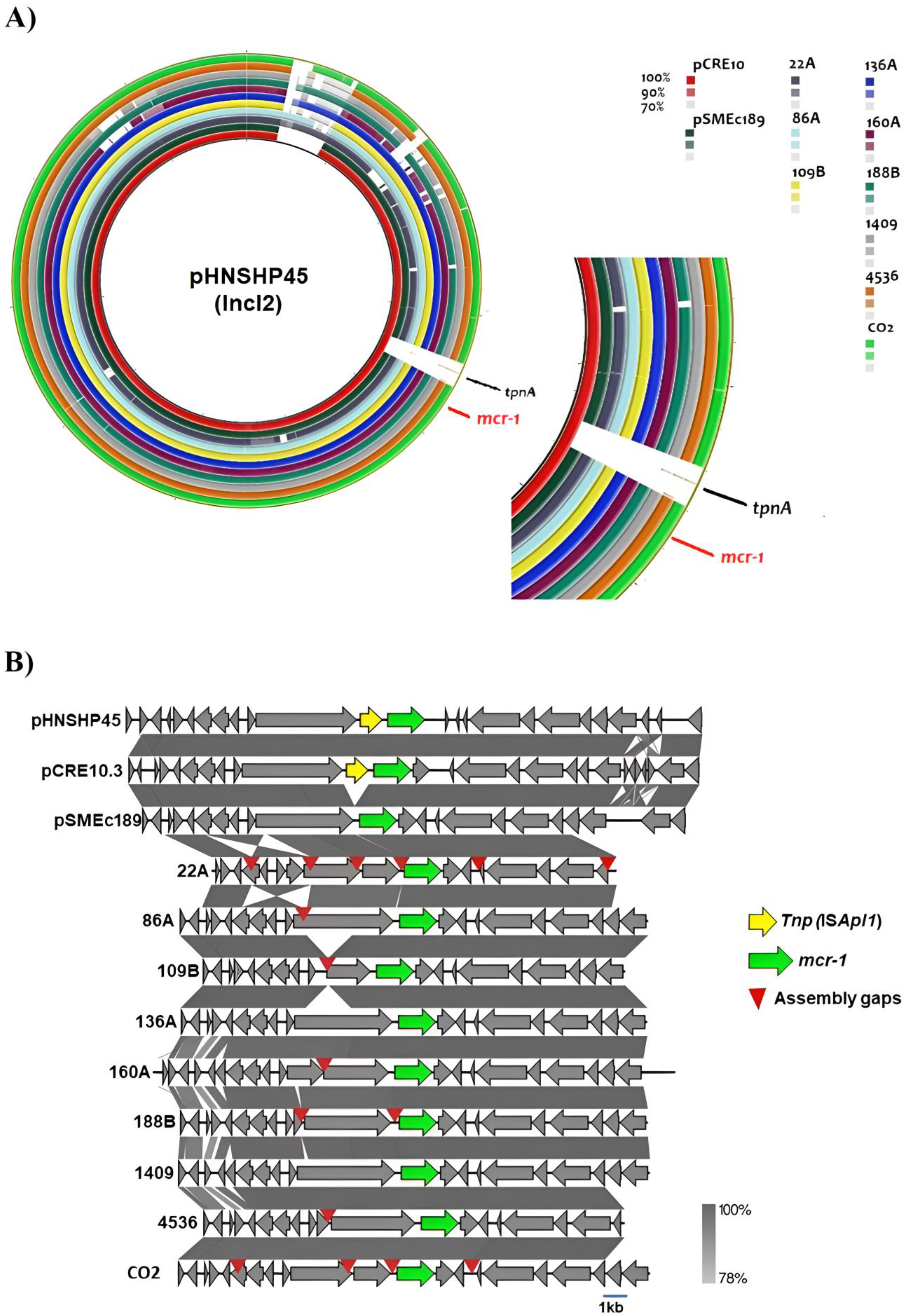
The PlasmidFinder® tool was able to identify 1–8 co-existing plasmids within each isolate. Interestingly, plasmids belonging to the IncI2 type were detected across all E. coli isolates. Plasmid and locus alignment located the mcr-1.1 gene in the IncI2 plasmid, sharing >90% similarity with the 64,015 bp pHNSHP45 plasmid (GenBank nucleotide KP347127.1) (Figure 2A).
All of the E. coli J53 transconjugants selected for colistin resistance produced amplicons with mcr-1-specific primers [20], and none of them produced amplicons following PCR-based replicon typing (PBRT). Additionally, the mcr-1.1 gene was harbored within identical genetic environments for all isolates, where Tnp (ISApl1) is absent (Figure 2B).

2.6. Genetic Environment of Carbapenem Resistance Genes

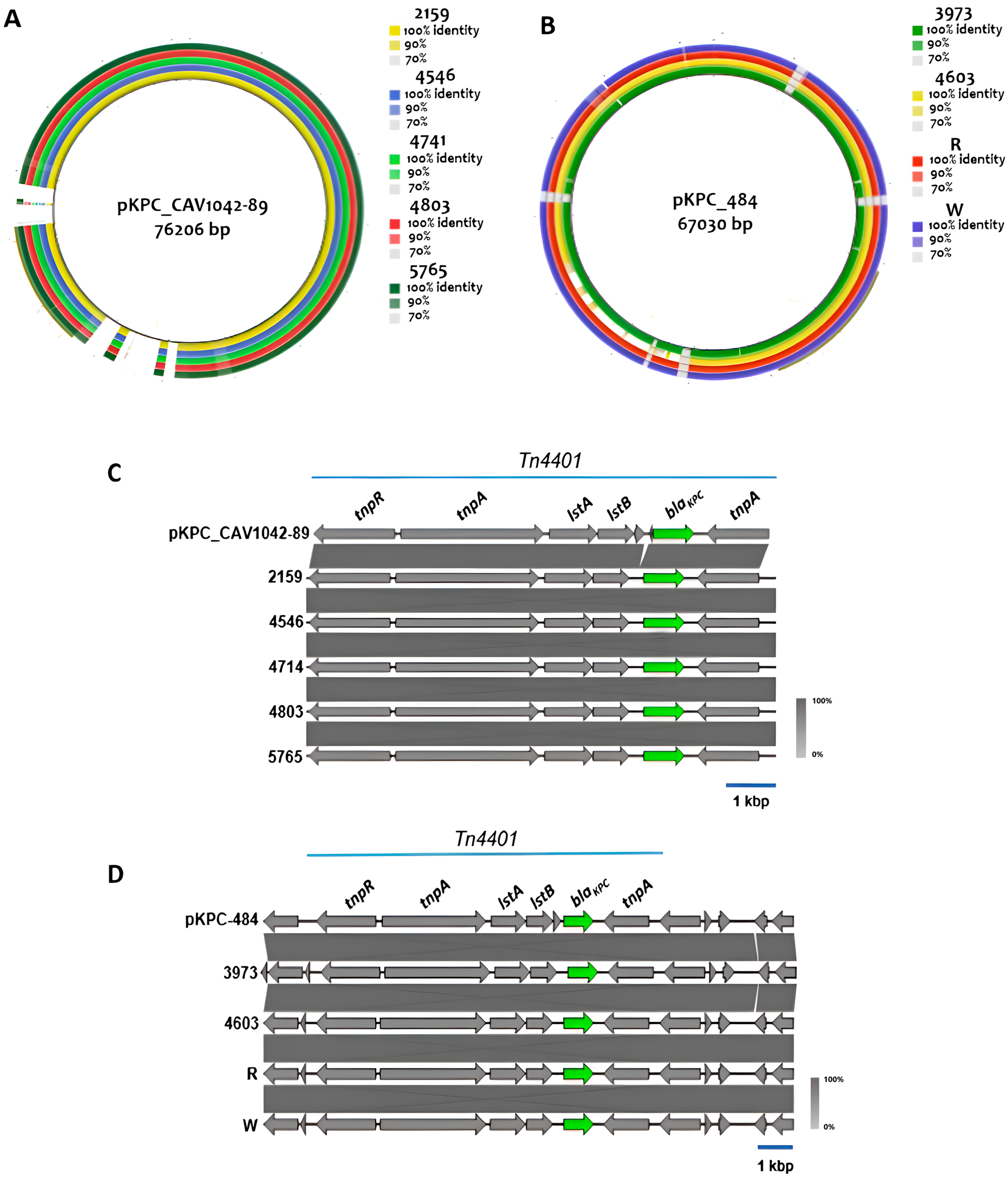
The blaKPC-2 and blaKPC-3 genes were identified within the transposon Tn4401 (Figure 3). Genomic segments containing blaKPC-3 in the five K. pneumoniae ST13 isolates exhibited high sequence identity with plasmid pKPC-CAV1042-89 (GenBank nucleotide CP018669), harbored by a K. pneumoniae ST244 strain obtained from a patient with a urinary tract infection in Ecuador (Figure 3A, left rings, and Figure 3B). Conversely, the genetic context of Tn4401 within the four K. pneumoniae ST258 isolates harboring blaKPC-2 showed high homology with the pKPC-484 plasmid (GenBank nucleotide CP008798.1) from another previously described clinical ST258 strain (Figure 3A, right rings, and Figure 3C). Although E. coli 136A was susceptible to carbapenems, sequence analysis revealed the presence of the blaKPC3 gene within its genome.

3. Discussion

In this study, all E. coli isolates retained susceptibility to amikacin and carbapenems. Conversely, all carbapenem-resistant K. pneumoniae isolates were also resistant to amikacin. The human and poultry isolates belonged to phylogroups A and B1. Previously, multi-resistant E. coli belonging to these phylogenetic groups were identified within human-dwelling commensal flora [29]. Moreover, such phylogroups are the most frequently isolated within healthy poultry [30].
Interestingly, none of the E. coli isolates were classified in phylogroups B2 or D, commonly associated with extraintestinal infections. However, phylogroup F, a less virulent sister group of phylogroup B2—previously found within 14 environmental and clinical samples—was identified in poultry and compost [31]. These datasets suggest that the isolates are not directly related to human infection. This inference was supported by MLST analysis since it did not demonstrate epidemic clones among the E. coli isolates. This was not the case with the K. pneumoniae epidemic clones ST13 (n = 5) and ST258 (n = 3), which were highly represented. K. pneumoniae ST13 and especially ST258 have been reported globally as important XDR pathogens for humans and as being a major source of nosocomial infections [32,33,34]. Several serotypes harboring between 2 and 10 virulence factors were identified within E. coli isolates. However, none of the isolates presented serotypes typically involved in infections or a classic combination of virulence factors linked to intestinal or extraintestinal pathogenic status. Nonetheless, within the poultry isolates, the hlyF, iss, and iroN genes, coding for siderophores, were detected. These genes have been previously reported as highly prevalent in ESBL-producing E. coli isolated from poultry, and it is suspected that they have a role in human extraintestinal infections.
Moreover, the iutA and ompT genes—frequently found in E. coli—were not detected in this study. Colistin (polymyxin E) is an antibiotic of critical importance to human health. Consequently, its use has recently been restricted in most countries due to complex bacterial infections in humans [35]. The use of colistin as a feed additive for animals was banned in China in 2016, following the first report of the mcr-1 gene being correlated with food animal production [36]. This initiative was followed by other nations. Within Ecuador, the use of colistin on farms was banned in 2019 [37]. However, reports of colistin-resistant Enterobacterales, especially E. coli, have increased in recent years [38]. Such reports mostly associate colistin resistance with plasmids carrying mcr genes via horizontal gene transfer [39].
All plasmids reconstructed in this study were highly homologous, suggesting that IncI2-type plasmids are important platforms for the spread of mcr-1.1 in Ecuador. Previously, IncI2-type plasmids were identified as the most frequent disseminators of mcr-1 globally [40,41]. Within these plasmids, mcr-1 is typically associated with insertion sequences that allow its transposition to other plasmids or the chromosome.
This genetic feature was previously reported in Japan (GenBank accession, AP019686.1). Similar to previous reports, colistin resistance in K. pneumoniae isolates (MIC ≥ 16) from Ecuador was not linked to mcr genes. However, this study identified chromosomal mutations that are likely to be involved in lipid A modifications. The first pathway of this resistance includes a mutation in the mgrB gene (3K*) that upregulates the two-component regulatory systems, namely, PhoP/PhoQ and pmrA/pmrB. The phoP/phoQ system indirectly activates pmrA/pmrB, conferring colistin resistance by regulating pmrCAB expression (transfer of phosphoethanolamine transferase, PEtN-to-lipid A) and pmrHFIJKLM (transfer of L-Ara4N to lipid A) operons [42]. The second pathway involves missense mutations within the arnA, phoQ, arnT, eptA, pmrB, and pmrJ genes that are also part of the regulon for the two-component systems mentioned above. Currently, it is known that a complex combination of mutations in TCRS leads to elevated MIC levels for colistin resistance [43].
This study also found that E. coli human isolates exhibit reduced susceptibility to tigecycline. Within such isolates, mutations in marA (S103G, H137Y) and acrR (V29fs) were identified. Similarly to tetracyclines, tigecycline binds to 30S subunits and interrupts the transcription process. Nevertheless, tigecycline is not affected by common tetracycline resistance mechanisms, such as acquired tetA genes. However, the tetX3 and tetX4 genes (capable of degrading all tetracyclines) have been reported to confer high-level acquired tigecycline resistance [44]. Moreover, tigecycline resistance in E. coli is frequently the result of the constitutive expression of resistance–nodulation–cell division (RND)-type efflux pumps [45]. One of the main structures is the tripartite efflux system AcrAB–TolC, which allows the efflux of multiple antibiotics. Upregulation of this system apparently leads to tigecycline resistance in Enterobacterales [46]. The acrA and acrB genes are located in an identical operon regulated by the local repressor acrR. Additionally, global regulators such as marA are involved in E. coli MDR regulation. This gene is regulated by the repressors marR and acrB, which are activated in the absence of marR [47].
All K. pneumoniae isolates in this study presented reduced susceptibility to TGC. Mutations in ramR G25S are possibly related to MIC = 4, K194* to MIC = 2, and S157P to MIC = 8 (Supplementary Data, Table S1). Within K. pneumoniae, ramA acts as an AraC/XylS transcriptional activator that controls the AcrAB–TolC efflux pump, and ramA expression is downregulated by ramR. Therefore, mutations in ramR can cause chromosomal resistance to tigecycline [48]. Additionally, genetic and phenotypic analysis of the E. coli isolates suggested that this decreased susceptibility to tigecycline within the E. coli isolates could be related to missense mutations in marA and frameshift mutation across acrR genes. This study presents a variety of mutations linked to phenotypic resistance to colistin in K. pneumoniae, together with resistance to tigecycline in E. coli and K. pneumoniae. However, functional validation is still required to establish their contributions to such resistance levels.
Regarding the genetic environment of blaKPC, all K. pneumoniae strains analyzed in this study harbored blaKPC-2 and blaKPC-3 alleles inside a ~10 kb Tn4401 transposon, with the genetic context tnpR–tnpA–istA–istB–blaKPC–tnpA. To date, two basic structures have been described as carrying blaKPC genes—the Tn4401 transposon and the Tn3–Tn4401 chimera—both found to be linked to its successful mobilization [49]. However, in South America, blaKPC genes are predominantly linked to Tn4401 [50]. In this study, the identification of blaKPC genes inside of a Tn4401 module in K. pneumoniae suggests that these strains are reservoirs and could disseminate carbapenem resistance in clinical settings. Conversely, only one isolate for E. coli presented a carbapenemase resistance gene. However, this strain was sensitive to carbapenems, and the genetic environment for blaKPC-3 could not be established. Therefore, we believe this gene to be non-functional.
blaTEM genes, especially blaTEM-1, were identified in most of the isolates. However, these genes encode lactamases that are not able to inactivate 3GCs. Therefore, it is not practical to estimate the contribution of these enzymes to the overall resistance observed in the strains producing CTX-M or KPC enzymes. The same rationale is considered for other blaTEM variants, blaSHV, and blaOXA lactamases, although some variants may be from the ESBL group, like those registered in this genomic description.
The genomic epidemiology of mcr-1 genes in Latin America needs to be enhanced. At first glance, plasmids belonging to the IncI2 incompatibility group are an important genetic backbone for mcr-1 gene mobilization in our setting. In Latin America, IncI2-like plasmids have been reported in Chile, Argentina, Venezuela, Bolivia, and Uruguay [51,52,53]. However, the data need to be enhanced with more local studies in the region to reveal their epidemiological implications. The mcr-1 gene’s epidemiology is an example of this. Furthermore, the same issue arises with all the genes and genetic structures reported in this study [54].

Limitations

The isolates were selected for their resistance to colistin, encompassing both E. coli and K. pneumoniae. These represent the full spectrum of resistant isolates collected, thanks to collaboration between various groups in Quito, Ecuador. It was only possible to include K. pneumoniae isolates from human clinical environments. Additionally, these isolates were not addressed in terms of prevalence or outbreaks. Our aim was to identify the mechanisms of resistance to colistin and associated resistances, focusing on phenotypic profiles through genomic data.
Our motivation for sharing this work was to contribute data on the molecular epidemiology of antibiotic resistance in Ecuador, a context where available information is limited. Our study falls within the “One Health” approach, which emphasizes the interdependence among human, animal, and environmental health. A relevant finding is the identification of the mcr-1 gene in a common plasmid genetic environment among isolates from humans, animals, and the environment, reinforcing this approach. A global epidemiological map of bacterial resistance is valuable to the extent that it reaches a level of detail that allows for the identification of relationships between ecological, evolutionary, and clinical aspects. Each finding equally contributes to the understanding of genomic epidemiology, as genetic determinants spread among interconnected bacterial communities across the biome, sharing an inherent potential for dissemination, accelerated by current human community connectivity.
The scientific method is built around hypotheses that can be confirmed or refuted, and the methodology adopted must be capable of addressing these questions. Therefore, the inherent limitations of the techniques employed should not be seen as criteria for questioning the validity of the results obtained. Every technology presents limitations, but these do not undermine its robustness in resolving the questions posed when considering its strengths and weaknesses. If we were to only consider the limitations of each method, no methodology would be useful. In this context, the application of short-read massive sequencing adopted in this study has been established as a fundamental tool in microbiology at the genomic level. It is worth noting that this technique is critical for sequencing the human genome, which attests to its reliability.
Our study did not establish new breakpoints but relied on these pre-existing interpretations to ensure consistency with the original analyses. The primary goal of our work was to confirm the resistance profiles reported by the contributing institutions and validate the isolates for whole-genome sequencing. Consequently, our analysis focused on identifying and characterizing the genetic elements associated with resistance, as described in this manuscript.
The isolates characterized through whole-genome sequencing in this report were initially isolated in separate studies. These isolates were selected based on the criteria established by the respective research groups that donated them. Consequently, this introduces inherent biases related to each study’s phenotypic screening cut-offs. Given the isolates’ diverse origins—spanning environmental, veterinary, and human clinical contexts—specific criteria were employed in accordance with the distinct focus of each field. Therefore, we recommend that prevalence data be interpreted with consideration of these criteria and the specific conditions of the prior studies. Nevertheless, it is important to note that for the selection of extended-spectrum β-lactamase (ESBL)-producing isolates, all studies consistently employed the double-disk synergy test, as recommended by CLSI methodology.
In our case, we utilized the VITEK AST card to reconfirm ESBL production for the specific purpose of selecting isolates that carried the genetic elements of interest for whole-genome sequencing. This approach ensured that we focused on strains containing the genes we aimed to characterize and describe in detail in this study.
The susceptibility testing covered all of our available resources. Therefore, antibiotics such as ceftazidime–avibactam were not tested. However, in the specific case of this therapeutic combination, the absence of metallo-carbapenemases or avibactam-insensitive blaKPC-type carbapenemases provides valuable clues about the sensitivity of the isolates through genomic analysis. It is worth noting that the E. coli isolates were sensitive to carbapenems, indicating an option currently available for our community.

4. Materials and Methods

4.1. Initial Characterization of the Isolates

The isolates were initially characterized in the scope of different studies. In brief, this study considered nine 3GC-resistant E. coli isolates harboring the mcr-1 gene (six from poultry farms, two from human infections, and one from the compost of a dairy farm), together with ten colistin/carbapenem-resistant K. pneumoniae, isolated from clinical samples:
  • All isolates from poultry were identified in 2014 in a previous study conducted by our group [22].
  • The clinical isolates were selected from a 2016 sample collection of 4000 Enterobacteriaceae gathered by Zurita&Zurita Laboratories.
  • The compost isolate was identified during an analysis of the prevalence of blaCTX-M in an agricultural productive unit in Quito, Ecuador, conducted in 2016 (unpublished data).

4.2. Sample Collection and Preparation for Sequencing

The isolates, obtained from three independent projects, had their characteristics—such as colistin resistance, carbapenem resistance, the presence of the mcr-1 gene, and ESBL production—identified and used as inclusion criteria in the present study. Upon completion of these projects, the isolates were stored in cryovials at ultralow temperatures for subsequent genome sequencing and phenotypic analysis, which were confirmed in this study. Prior to sequencing, the isolates were reactivated by inoculation in a liquid medium. Single colonies were isolated on selective media: TBX medium (BIO-RAD, Hercules, CA, USA) supplemented with cefotaxime (3 µg/mL) for E. coli and MacConkey medium (Difco, Detroit, MI, USA) supplemented with imipenem (1 µg/mL) for K. pneumoniae. A single colony was selected for phenotypic analysis and cultured in LB broth, from which DNA was extracted using a commercial column-based total DNA extraction kit. The obtained DNA was used for PCR and sequencing purposes.

4.3. Antibiotic Susceptibility Testing

Antimicrobial susceptibility testing for colistin (COL) was established using the microdilution method employing GNX2F plates (Thermo Scientific™, West Palm Beach, FL, USA), while the minimum inhibitory concentration (MIC) breakpoint was 2 (EUCAST). The presence of the mcr-1 gene was established using the PCR method [20]. MIC values for ampicillin (AMP), piperacillin–tazobactam (TPZ), cefoxitin (FOX), ceftazidime (CAZ), ceftriaxone (CRO), cefepime (FEP), doripenem (DOR), ertapenem (ERT), imipenem (IMI), meropenem (MEM), amikacin (AK), gentamicin (GEN), ciprofloxacin (CIP), and tigecycline (TIG) were all obtained through the Vitek® 2 system via the AST-N272 card (Biomérieux™, Marcy-l’Étoile, France). This card additionally allowed the identification of ESBL production. The results were evaluated with breakpoints and recommendations from the CLSI guide [52]. Tigecycline MIC interpretation was carried out using the EUCAST 2019 clinical breakpoint for E. coli and ECOFF for K. pneumoniae.

4.4. Whole-Genome Sequencing

The whole-genome sequencing of 19 isolates was performed using the MiSeq® platform (Illumina, San Diego, CA, USA). Genomic libraries were prepared using the NexteraXT® kit (Illumina, San Diego, CA, USA). Raw data were processed using the CLC Genomics™ WorkBench® tool, version 8.5. First, a quality filter was applied to the reads, excising regions of low quality and possible contamination with adapters, followed by discarding reads with <25 nucleotides. The quality-filtered reads were used to perform de novo assemblies for each isolate.

4.5. Mating Assay and PCR-Based Replicon Typing (PBRT)

The mating assay and PBRT were carried out using the UNIETAR laboratory protocol. In brief, the E coli J53 strain, resistant to sodium azide (200 µg/mL), was used as a receptor. Mating was carried out in LB broth by mixing every E. coli isolate and the same receptor strain, followed by incubation for 18 h at 35 °C. The transconjugants were selected from LB agar plates supplemented with sodium azide (200 µg/mL) and colistin (2 µg/mL). The transconjugants were evaluated using colony PCR for mcr-1 gene 20 and PBRT using the PBRT 2.0 kit (Diatheva, Fano, Italy).

4.6. Plasmid Analysis

PlasmidFinder v.2.1, along with the map-to-reference tool available in Genious Prime software R10.0 and the cgview_comparison_tool (https://github.com/paulstothard/cgview_comparison_tool, accessed on 19 October 2023), was used for plasmid reconstruction.

4.7. Data Management and Statistical Analysis

Whole-genome sequencing of the isolates formed part of the Bioproject “Colistin-resistant E. coli and K. pneumoniae from Ecuador”, conducted at Universidad del Bosque, Bogotá, Colombia.
The identification of species was carried out using the Strain Seeker® program [53]. Annotation of each genome was performed using the RAST® tool [54]. Sequences are available from NCBI under accession number PRJNA507384.
Phylogenetic analysis was carried out using Center for Genomic Epidemiology (CGE) tools: NDtree 1.2 (NDtree constructs phylogenetic trees from single-end or pair-end reads), TreeViewer 1.0, and MLST tool (multi-locus sequence typing from an assembled genome or from a set of reads) using default settings, as well as VirulenceFinder (identification of acquired virulence genes) and SeroTypeFinder (prediction of serotypes in total or partial sequenced isolates of E. coli). These tools are available at https://www.genomicepidemiology.org/, accessed on 19 October 2023. An enterobase-based SNPtree was used for the MLST E. coli analysis.
Resistance genes were identified through the ResFinder® service (CGE), and integrons were identified using IntFinder (https://github.com/kalilamali/Integrons, accessed on 19 October 2023). The sequences of the laboratory standard strain E. coli K-12 (GenBank accession, NC_000913.3) and the ATCC strain of K. pneumoniae ATCC BAA-2146 (GenBank accession: NZ_CP006659.2) were used as references for putative mutation discovery using Genious Prime software. Genetic environment reconstruction was carried out using the map-to-reference tool.

5. Conclusions

This study investigated nine 3GC-resistant E. coli isolates carrying the mcr-1 gene (sourced from poultry farms, human infections, and dairy farm compost), alongside ten clinical isolates of colistin- and carbapenem-resistant K. pneumoniae. The human-origin E. coli isolates were classified as ST609 (phylogroup A), while those from the poultry and compost represented phylogroups A, B1, E, and F. The diverse K. pneumoniae sequence types included ST13, ST258, and ST86. The key resistance genes identified in E. coli included blaCTX-M-55, blaCTX-M-65, blaCTX-M-15, and blaCTX-M-2, with blaCMY-2 and blaKPC-3 also detected. All mcr-1.1 genes were plasmid-borne within IncI2 plasmids. Reduced tigecycline susceptibility in E. coli was linked to marA and acrA gene mutations.
In K. pneumoniae, 3GC resistance was associated with blaCTX-M-15 and blaCTX-M-65, while carbapenem resistance was mediated by blaKPC-2 and blaKPC-3. Colistin resistance correlated with genetic alterations in mgrB, marA, acrA, arnB, eptA, pmrB, pmrJ, and phoQ, and tigecycline resistance with ramR mutations. This study also identified integron structures, with Int191(dfrA14) being the most prevalent across isolates. These findings enhance our understanding of multidrug-resistant E. coli and K. pneumoniae within the genomic epidemiology context, supporting the global One Health framework.

Supplementary Materials

The following supporting information can be downloaded at https://www.mdpi.com/article/10.3390/antibiotics14020206/s1: Table S1: This supplementary table contains detailed data on the genetic determinants and their relationship with the antibiotics tested for each isolate.

Author Contributions

J.Z., D.O.-P. and C.A.A. designed the study. D.O.-P., C.A.A. and J.Z. performed the experiments. R.R., F.D.C. and D.O.-P. conducted bioinformatic analysis. L.D., J.R., J.Z. and C.A.A. provided critical input. D.O.-P., F.D.C. and J.Z. interpreted the results and wrote the manuscript. All authors have read and agreed to the published version of the manuscript.

Funding

This study was supported by Unidad de Investigaciones en Biomedicina, Zurita & Zurita Laboratorios, grant MIC-021; Universidad El Bosque, International Center for Bacterial Genomics; and the University of Texas, Medical School at Houston.

Institutional Review Board Statement

Not applicable.

Informed Consent Statement

Not applicable.

Data Availability Statement

Publicly available datasets were analyzed in this study. This data can be found here: [https://www.ncbi.nlm.nih.gov] [PRJNA507384]. accessed on 15 January 2023.

Acknowledgments

The authors want to thank Pedro Barba for his technical support and Christian Vinueza for his administrative support and the poultry isolates’ donation.

Conflicts of Interest

The authors declare no conflict of interest. The funders had no role in the design of the study; in the collection, analyses, or interpretation of data; in the writing of the manuscript; or in the decision to publish the results.

References

  1. Doi, Y. Treatment Options for Carbapenem-resistant Gram-negative Bacterial Infections. Clin. Infect. Dis. 2019, 69 (Suppl. 7), S565–S575. [Google Scholar] [CrossRef] [PubMed] [PubMed Central]
  2. Lutgring, J.D.; Limbago, B.M. The Problem of Carbapenemase-Producing-Carbapenem-Resistant Enterobacteriaceae. J. Infect. Dis. 2019, 215, 1–2. [Google Scholar] [CrossRef] [PubMed]
  3. Mączyńska, B.; Frej-Mądrzak, M.; Sarowska, J.; Woronowicz, K.; Choroszy-Król, I.; Jama-Kmiecik, A. Evolution of Antibiotic Resistance in Escherichia coli and Klebsiella pneumoniae Clinical Isolates in a Multi-Profile Hospital over 5 Years (2017–2021). J. Clin. Med. 2023, 12, 2414. [Google Scholar] [CrossRef] [PubMed] [PubMed Central]
  4. Peirano, G.; Pitout, J.D.D. Molecular Epidemiology of Extended-Spectrum β-Lactamase-Producing Enterobacteriaceae in Canada. Front. Microbiol. 2019, 10, 1640. [Google Scholar]
  5. Wise, M.G.; Karlowsky, J.A.; Mohamed, N.; Hermsen, E.D.; Kamat, S.; Townsend, A.; Brink, A.; Soriano, A.; Paterson, D.L.; Moore, L.S.P.; et al. Global trends in carbapenem- and difficult-to-treat-resistance among World Health Organization priority bacterial pathogens: ATLAS surveillance program 2018–2022. J. Glob. Antimicrob. Resist. 2024, 37, 168–175. [Google Scholar] [CrossRef] [PubMed]
  6. Latifi, F.; Pakzad, R.; Asadollahi, P.; Hematian, A.; Pakzad, I. Worldwide Prevalence of Colistin Resistance among Enterobacteriaceae: A Systematic Review and Meta-Analysis. Clin. Lab. 2023, 69, 657–669. [Google Scholar] [CrossRef] [PubMed]
  7. Gharaibeh, S.; Shatnawi, A. The Role of Polymyxins in the Treatment of Gram-Negative Infections. Antibiotics 2019, 8, 116. [Google Scholar]
  8. Mondal, A.H.; Khare, K.; Saxena, P.; Debnath, P.; Mukhopadhyay, K.; Yadav, D. A Review on Colistin Resistance: An Antibiotic of Last Resort. Microorganisms 2024, 12, 772. [Google Scholar] [CrossRef] [PubMed]
  9. Stefaniuk, E.; Tyski, S. Epidemiology of Polymyxin Resistance in Enterobacteriaceae. J. Clin. Microbiol. 2019, 57, e00523–e00619. [Google Scholar]
  10. Gostyńska, A.; Piwowarczyk, L.; Nadolna, M.; Jelińska, A.; Dettlaff, K.; Ogrodowczyk, M.; Popielarz-Brzezińska, M.; Stawny, M. Toward Safe Pharmacotherapy: The Interplay between Meropenem and Parenteral Nutrition Admixtures. Antibiotics 2021, 10, 217. [Google Scholar] [CrossRef]
  11. Karakonstantis, S.; Kritsotakis, E.I.; Gikas, A. Treatment options for K. pneumoniae, P. aeruginosa and A. baumannii co-resistant to carbapenems, aminoglycosides, polymyxins and tigecycline: An approach based on the mechanisms of resistance to carbapenems. Infection 2020, 48, 835–851. [Google Scholar] [CrossRef] [PubMed] [PubMed Central]
  12. Anyanwu, M.U.; Nwobi, O.C.; Okpala, C.O.R.; Ezeonu, I.M. Mobile Tigecycline Resistance: An Emerging Health Catastrophe Requiring Urgent One Health Global Intervention. Front. Microbiol. 2022, 13, 808744. [Google Scholar] [CrossRef] [PubMed] [PubMed Central]
  13. Yaghoubi, S.; Zekiy, A.O.; Krutova, M.; Gholami, M.; Kouhsari, E.; Sholeh, M.; Ghafouri, Z.; Maleki, F. Tigecycline antibacterial activity, clinical effectiveness, and mechanisms and epidemiology of resistance: Narrative review. Eur. J. Clin. Microbiol. Infect. Dis. 2022, 41, 1003–1022. [Google Scholar] [CrossRef] [PubMed] [PubMed Central]
  14. Zhai, W.; Tian, Y.; Lu, M.; Zhang, M.; Song, H.; Fu, Y.; Ma, T.; Sun, C.; Bai, L.; Wang, Y.; et al. Presence of Mobile Tigecycline Resistance Gene tet(X4) in Clinical Klebsiella pneumoniae. Microbiol. Spectr. 2022, 10, e0108121. [Google Scholar] [CrossRef]
  15. Ortega-Paredes, D.; de Janon, S.; Villavicencio, F.; Ruales, K.J.; De La Torre, K.; Villacís, J.E.; Wagenaar, J.A.; Matheu, J.; Bravo-Vallejo, C.; Fernández-Moreira, E.; et al. Broiler Farms and Carcasses Are an Important Reservoir of Multi-Drug Resistant Escherichia coli in Ecuador. Front. Vet. Sci. 2020, 7, 547843. [Google Scholar] [CrossRef] [PubMed] [PubMed Central]
  16. Zurita, J.; Sevillano, G.; Paz YMiño, A.; Haro, N.; Larrea-Álvarez, M.; Alcocer, I.; Ortega-Paredes, D. Dominance of ST131, B2, blaCTX-M-15, and papA-papC-kpsMII-uitA among ESBL Escherichia coli isolated from bloodstream infections in Quito, Ecuador: A 10-year surveillance study (2009–2019). J. Appl. Microbiol. 2023, 134, lxad269. [Google Scholar] [CrossRef] [PubMed]
  17. Ortega-Paredes, D.; Barba, P.; Mena-López, S.; Espinel, N.; Crespo, V.; Zurita, J. High quantities of multidrug-resistant Escherichia coli are present in the Machángara urban river in Quito, Ecuador. J. Water Health 2020, 18, 67–76. [Google Scholar] [CrossRef] [PubMed]
  18. Zurita, J.; Yánez, F.; Sevillano, G.; Ortega-Paredes, D.; Paz, Y.; Miño, A. Ready-to-eat street food: A potential source for dissemination of multidrug-resistant Escherichia coli epidemic clones in Quito, Ecuador. Lett. Appl. Microbiol. 2020, 70, 203–209. [Google Scholar] [CrossRef] [PubMed]
  19. Zurita, J.; Solís, M.B.; Ortega-Paredes, D.; Barba, P.; Paz YMiño, A.; Sevillano, G. High prevalence of B2-ST131 clonal group among extended-spectrum β-lactamase-producing Escherichia coli isolated from bloodstream infections in Quito, Ecuador. J. Glob. Antimicrob. Resist. 2019, 19, 216–221. [Google Scholar] [CrossRef] [PubMed]
  20. Ortega-Paredes, D.; Barba, P.; Zurita, J. Colistin-resistant Escherichia coli clinical isolate harbouring the mcr-1 gene in Ecuador. Epidemiol. Infect. 2016, 144, 2967–2970. [Google Scholar] [CrossRef] [PubMed] [PubMed Central]
  21. Zurita, J.; Ortega-Paredes, D.; Barba, P. First Description of Shigella sonnei Harboring blaCTX-M-55 Outside Asia. J. Microbiol. Biotechnol. 2016, 26, 2224–2227. [Google Scholar] [CrossRef] [PubMed]
  22. Vinueza-Burgos, C.; Ortega-Paredes, D.; Narváez, C.; De Zutter, L.; Zurita, J. Characterization of cefotaxime resistant Escherichia coli isolated from broiler farms in Ecuador. PLoS ONE 2019, 14, e0207567. [Google Scholar] [CrossRef] [PubMed] [PubMed Central]
  23. Ortega-Paredes, D.; Haro, M.; Leoro-Garzón, P.; Barba, P.; Loaiza, K.; Mora, F.; Fors, M.; Vinueza-Burgos, C.; Fernández-Moreira, E. Multidrug-resistant Escherichia coli isolated from canine faeces in a public park in Quito, Ecuador. J. Glob. Antimicrob. Resist. 2019, 18, 263–268. [Google Scholar] [CrossRef] [PubMed]
  24. Ortega-Paredes, D.; Barba, P.; Mena-López, S.; Espinel, N.; Zurita, J. Escherichia coli hyperepidemic clone ST410-A harboring blaCTX-M-15 isolated from fresh vegetables in a municipal market in Quito-Ecuador. Int. J. Food Microbiol. 2018, 280, 41–45. [Google Scholar] [CrossRef] [PubMed]
  25. Barba, P.; Ortega-Paredes, D.; Molina Cuasapaz, G.; Zurita, J. Occurrence of extended-spectrum beta-lactamase producing Escherichia coli in a dairy farm from Quito, Ecuador. API 2017. [Google Scholar] [CrossRef]
  26. Ortega-Paredes, D.; Larrea-Álvarez, C.M.; Torres-Elizalde, L.; de Janon, S.; Vinueza-Burgos, C.; Hidalgo-Arellano, L.; Šefcová, M.A.; Molina-Cuasapaz, G.; Fernandez-Moreira, E.; Larrea-Álvarez, M. Antibiotic Resistance Awareness among Undergraduate Students in Quito, Ecuador. Antibiotics 2022, 11, 197. [Google Scholar] [CrossRef] [PubMed] [PubMed Central]
  27. Zurita, J.; Alcocer, I.; Ortega-Paredes, D.; Barba, P.; Yauri, F.; Iñiguez, D.; Mora, M. Carbapenemhydrolysing β-lactamase KPC-2 in Klebsiella pneumoniae isolated in Ecuadorian hospitals. J. Glob. Antimicrob. Resist. 2013, 1, 229–230. [Google Scholar] [CrossRef] [PubMed]
  28. Bastidas-Caldes, C.; Guerrero-Freire, S.; Ortuño-Gutiérrez, N.; Sunyoto, T.; Gomes-Dias, C.A.; Ramírez, M.S.; Calero-Cáceres, W.; Harries, A.D.; Rey, J.; de Waard, J.H.; et al. Colistin resistance in Escherichia coli and Klebsiella pneumoniae in humans and backyard animals in Ecuador. Rev. Panam. Salud Publica 2023, 47, e48. [Google Scholar] [CrossRef] [PubMed] [PubMed Central]
  29. Lagerstrom, K.M.; Hadly, E.A. Under-Appreciated Phylogroup Diversity of Escherichia coli within and between Animals at the Urban-Wildland Interface. Appl. Environ. Microbiol. 2023, 89, e0014223. [Google Scholar] [CrossRef] [PubMed] [PubMed Central]
  30. Ovi, F.; Zhang, L.; Nabors, H.; Jia, L.; Adhikari, P. A compilation of virulence-associated genes that are frequently reported in avian pathogenic Escherichia coli (APEC) compared to other E. coli. J. Appl. Microbiol. 2023, 134, lxad014. [Google Scholar] [CrossRef] [PubMed]
  31. Riley, L.W. Distinguishing Pathovars from Nonpathovars: Escherichia coli. Microbiol. Spectr. 2020, 8, 10–128. [Google Scholar] [CrossRef] [PubMed] [PubMed Central]
  32. Mendes, G.; Santos, M.L.; Ramalho, J.F.; Duarte, A.; Caneiras, C. Virulence factors in carbapenem-resistant hypervirulent Klebsiella pneumoniae. Front. Microbiol. 2023, 14, 1325077. [Google Scholar] [CrossRef] [PubMed] [PubMed Central]
  33. Cejas, D.; Elena, A.; Guevara Nuñez, D.; Sevilla Platero, P.; De Paulis, A.; Magariños, F.; Alfonso, C.; Berger, M.A.; Fernández-Canigia, L.; Gutkind, G.; et al. Changing epidemiology of KPC-producing Klebsiella pneumoniae in Argentina: Emergence of hypermucoviscous ST25 and high-risk clone ST307. J. Glob. Antimicrob. Resist. 2019, 18, 238–242. [Google Scholar] [CrossRef] [PubMed]
  34. Ernst, C.M.; Braxton, J.R.; Rodriguez-Osorio, C.A.; Zagieboylo, A.P.; Li, L.; Pironti, A.; Manson, A.L.; Nair, A.V.; Benson, M.; Cummins, K.; et al. Adaptive evolution of virulence and persistence in carbapenem-resistant Klebsiella pneumoniae. Nat. Med. 2020, 26, 705–711. [Google Scholar] [CrossRef] [PubMed] [PubMed Central]
  35. Shi, X.; Li, Y.; Yang, Y.; Shen, Z.; Wu, Y.; Wang, S. Global impact of mcr-1-positive Enterobacteriaceae bacteria on “one health”. Crit. Rev. Microbiol. 2020, 46, 565–577. [Google Scholar] [CrossRef] [PubMed]
  36. Liu, Y.Y.; Wang, Y.; Walsh, T.R.; Yi, L.X.; Zhang, R.; Spencer, J.; Doi, Y.; Tian, G.; Dong, B.; Huang, X.; et al. Emergence of plasmid-mediated colistin resistance mechanism MCR-1 in animals and human beings in China: A microbiological and molecular biological study. Lancet Infect. Dis. 2016, 16, 161–168. [Google Scholar] [CrossRef] [PubMed]
  37. Agrocalidad. Agrocalidad Prohíbe El Uso Del Antibiótico Colistina En Animales 2019. Available online: http://www.agrocalidad.gob.ec/agrocalidad-prohibe-el-uso-del-antibiotico-colistina-en-animales/ (accessed on 18 January 2023).
  38. Liu, J.H.; Liu, Y.Y.; Shen, Y.B.; Yang, J.; Walsh, T.R.; Wang, Y.; Shen, J. Plasmid-mediated colistin-resistance genes: mcr. Trends Microbiol. 2024, 32, 365–378. [Google Scholar] [CrossRef] [PubMed]
  39. Hussein, N.H.; Al-Kadmy, I.M.S.; Taha, B.M.; Hussein, J.D. Mobilized colistin resistance (mcr) genes from 1 to 10: A comprehensive review. Mol. Biol. Rep. 2021, 48, 2897–2907. [Google Scholar] [CrossRef] [PubMed]
  40. Rodríguez-Santiago, J.; Cornejo-Juárez, P.; Silva-Sánchez, J.; Garza-Ramos, U. Polymyxin resistance in Enterobacterales: Overview and epidemiology in the Americas. Int. J. Antimicrob. Agents. 2021, 58, 106426. [Google Scholar] [CrossRef] [PubMed]
  41. Yang, J.; Wu, R.; Xia, Q.; Yu, J.; Yi, L.X.; Huang, Y.; Deng, M.; He, W.Y.; Bai, Y.; Lv, L.; et al. The evolution of infectious transmission promotes the persistence of mcr-1 plasmids. mBio 2023, 14, e0044223. [Google Scholar] [CrossRef] [PubMed] [PubMed Central]
  42. Tietgen, M.; Sedlaczek, L.; Higgins, P.G.; Kaspar, H.; Ewers, C.; Göttig, S. Colistin Resistance Mechanisms in Human and Veterinary Klebsiella pneumoniae Isolates. Antibiotics 2022, 11, 1672. [Google Scholar] [CrossRef] [PubMed] [PubMed Central]
  43. Liu, Y.; Lin, Y.; Wang, Z.; Hu, N.; Liu, Q.; Zhou, W.; Li, X.; Hu, L.; Guo, J.; Huang, X.; et al. Molecular Mechanisms of Colistin Resistance in Klebsiella pneumoniae in a Tertiary Care Teaching Hospital. Front. Cell. Infect. Microbiol. 2021, 11, 673503. [Google Scholar] [CrossRef] [PubMed] [PubMed Central]
  44. Cheng, Q.; Cheung, Y.; Liu, C.; Xiao, Q.; Sun, B.; Zhou, J.; Chan, E.W.C.; Zhang, R.; Chen, S. Structural and mechanistic basis of the high catalytic activity of monooxygenase Tet(X4) on tigecycline. BMC Biol. 2021, 19, 262. [Google Scholar] [CrossRef] [PubMed] [PubMed Central]
  45. Korczak, L.; Majewski, P.; Iwaniuk, D.; Sacha, P.; Matulewicz, M.; Wieczorek, P.; Majewska, P.; Wieczorek, A.; Radziwon, P.; Tryniszewska, E. Molecular mechanisms of tigecycline-resistance among Enterobacterales. Front. Cell. Infect. Microbiol. 2024, 14, 1289396. [Google Scholar] [CrossRef] [PubMed] [PubMed Central]
  46. Wang, J.; Wu, H.; Mei, C.Y.; Wang, Y.; Wang, Z.Y.; Lu, M.J.; Pan, Z.M.; Jiao, X. Multiple Mechanisms of Tigecycline Resistance in Enterobacteriaceae from a Pig Farm, China. Microbiol. Spectr. 2021, 9, e0041621. [Google Scholar] [CrossRef] [PubMed] [PubMed Central]
  47. Sundaramoorthy, N.S.; Sivasubramanian, A.; Nagarajan, S. Simultaneous inhibition of MarR by salicylate and efflux pumps by curcumin sensitizes colistin resistant clinical isolates of Enterobacteriaceae. Microb. Pathog. 2020, 148, 104445. [Google Scholar] [CrossRef] [PubMed]
  48. Xu, Q.; Sheng, Z.; Hao, M.; Jiang, J.; Ye, M.; Chen, Y.; Xu, X.; Guo, Q.; Wang, M. RamA upregulates multidrug resistance efflux pumps AcrAB and OqxAB in Klebsiella pneumoniae. Int. J. Antimicrob. Agents 2021, 57, 106251. [Google Scholar] [CrossRef] [PubMed]
  49. Huang, J.; Hu, X.; Zhao, Y.; Shi, Y.; Ding, H.; Wu, R.; Zhao, Z.; Ji, J. Comparative Analysis of blaKPC Expression in Tn4401 Transposons and the Tn3-Tn4401 Chimera. Antimicrob. Agents Chemother. 2019, 63, e02434–e02518. [Google Scholar] [CrossRef] [PubMed]
  50. Reyes, J.A.; Melano, R.; Cárdenas, P.A.; Trueba, G. Mobile genetic elements associated with carbapenemase genes in South American Enterobacterales. Braz. J. Infect. Dis. 2020, 24, 231–238. [Google Scholar] [CrossRef] [PubMed] [PubMed Central]
  51. Lentz, S.A.M.; Dalmolin, T.V.; Barth, A.L.; Martins, A.F. mcr-1 Gene in Latin America: How Is It Disseminated Among Humans, Animals, and the Environment? Front. Public Health 2021, 9, 648940. [Google Scholar] [CrossRef] [PubMed] [PubMed Central]
  52. De La Cadena, E.; Mahecha, M.; Velandia, A.M.; García-Betancur, J.C.; Rojas, L.J.; Porras, J.; Pallares, C.; Villegas, M.V. Identification of mcr-1 Genes and Characterization of Resistance Mechanisms to Colistin in Escherichia coli Isolates from Colombian Hospitals. Antibiotics 2023, 12, 488. [Google Scholar] [CrossRef] [PubMed] [PubMed Central]
  53. Wang, R.; van Dorp, L.; Shaw, L.P.; Bradley, P.; Wang, Q.; Wang, X.; Jin, L.; Zhang, Q.; Liu, Y.; Rieux, A.; et al. The global distribution and spread of the mobilized colistin resistance gene mcr-1. Nat. Commun. 2018, 9, 1179. [Google Scholar] [CrossRef] [PubMed] [PubMed Central]
  54. WHO. WHO Bacterial Priority Pathogens List, 2024: Bacterial Pathogens of Public Health Importance to Guide Research, Development and Strategies to Prevent and Control Antimicrobial Resistance; World Health Organization: Geneva, Switzerland, 2024. [Google Scholar]
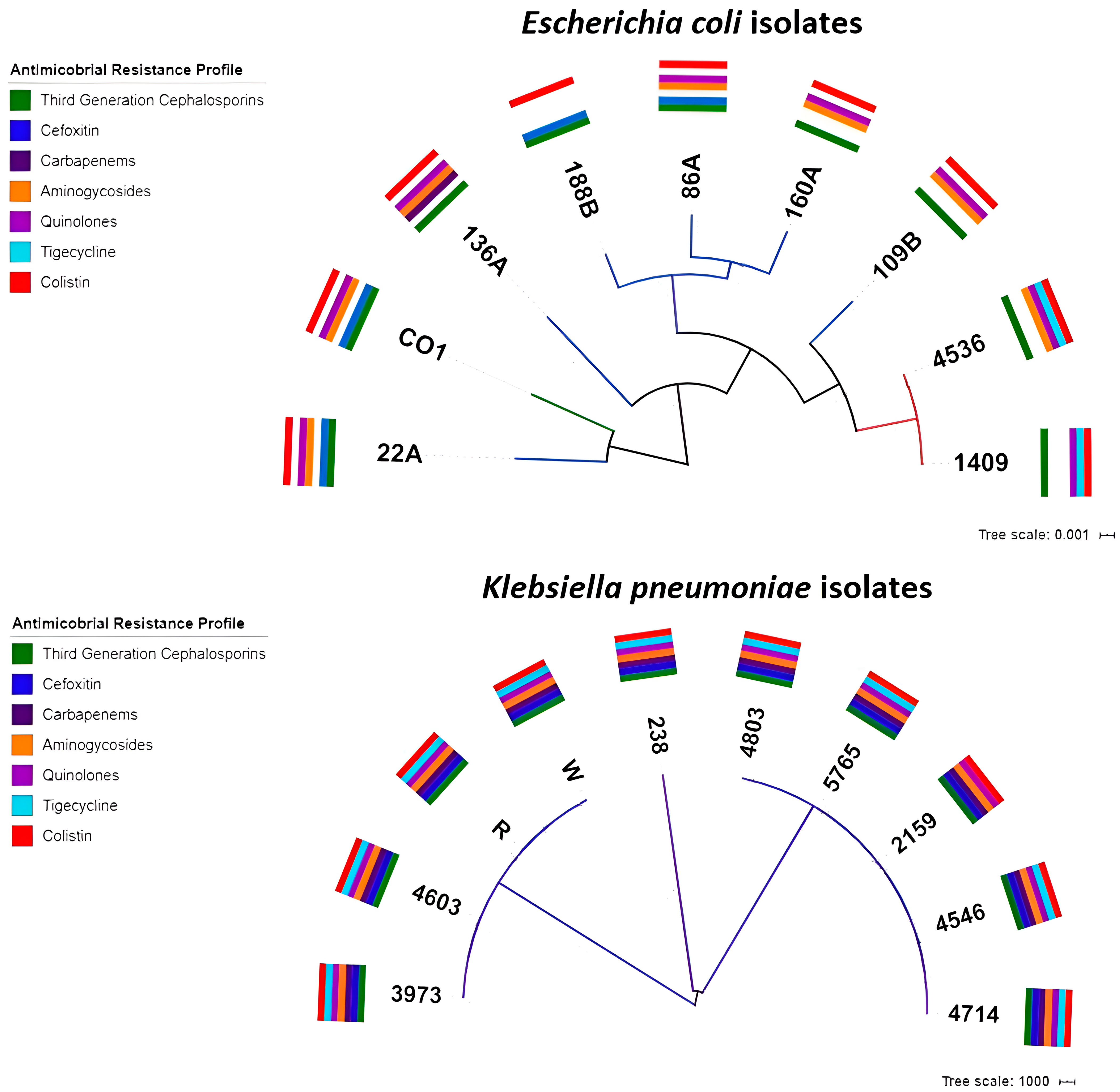
Figure 1. SNP tree determined for the E. coli and K. pneumoniae isolates. Colored bars indicate the antibiotic resistance profile of each isolate.
Antibiotics 14 00206 g001
Figure 2. Plasmid reconstruction and alignment. Alignment of mcr-1 harboring IncI2 plasmid references pHNSHP45, pCRE10, and pSMEc189 with reconstructed plasmids from E. coli isolates harboring mcr-1 genes was performed. The zoomed view shows the mcr-1 neighborhood. Graphic alignment of the mcr-1.1 genetic context (A). ISApl1 transposase and mcr-1 genes are highlighted. Red arrowheads represent gaps between contigs in the draft plasmids (B).
Antibiotics 14 00206 g002
Figure 3. Ring alignment of K. pneumoniae ST13 isolate draft genomes against the previously described plasmid pKPC-CAV1042-89 (A) and ST258 strain daft genomes against pKPC-484 (B). Alignment of Tn4401 loci from ST13 isolates with the pKPC-CAV1042-89 reference (C) and from ST258 strains with the pKPC-484 reference (D).
Antibiotics 14 00206 g003
Table 1. Antibiotic resistance profiles.
IsolateSpeciesDateOriginAgeLocationAMPTPZFOXCAZCROFEPDORERTIMIMEMAKGENCIPTIGCOL
1409E. coli10 February 2016Peritoneal liquid14Quito16≤4816≥642≤0.12≤0.5≤0.25≤0.25≤2≤1≥424
4536E. coli10 June 2016Wound secretion72Quito3264≤416≥642≤0.12≤0.5≤0.25≤0.258≥16≥414
CO2 (CO1)E. coli2016Compost dairy farm-Quito≥32≤41616≥64≥64≤0.12≤0.5≤0.25≤0.25≤2≥16≥4≤0.54
86AE. coli2014Poultry-Yaruquí≥32≤4164≥64≥64≤0.12≤0.5≤0.25≤0.25≤2≥16≥4≤0.58
109BE. coli2014Poultry-El Chota≥32≤48≤1≥642≤0.12≤0.5≤0.25≤0.25≤2≥16≥4≤0.54
136AE. coli2014Poultry-Santo Domingo16≤4≤4≤116≤1≤0.12≤0.5≤0.25≤0.25≤2≥16≥4≤0.58
22AE. coli2014Poultry-Ascazubi≥32≥128≥641616≤1≤0.12≤0.5≤0.25≤0.25≤2≥16≥4≤0.58
160AE. coli2014Poultry-Guayllabamba≥32≤4832≥6440.5≤0.5≤0.25≤0.254≥16≥4≤0.5≥16
188BE. coli2014Poultry-Ibarra16≤4≥6448≤1≤0.12≤0.5≤0.25≤0.25≤2≤1≤0.25≤0.58
1220672 (W)K. pneumoniae12 February 2016Urine61Quito≥32≥128≥64≥64≥64≥64≥8≥8≥16≥16≥64≥16≥44≥16
1204191 (R)K. pneumoniae12 February 2016Blood52Quito≥32≥128≥64≥64≥64≥64≥8≥8≥16≥16≥64≥16≥44≥16
238K. pneumoniae2 June 2016Cervical abscess54Quito16≤4≤4≤1≤1≤1≤0.12≤0.5≤0.25≤0.25≤2≤1≤0.524
2159K. pneumoniae3 March 2016Wound secretion37Quito≥32≥12832≥64≥64≥64≥8≥8≥16≥1616≥1622≥16
3973K. pneumoniae19 May 2016Tracheal secretion55Quito≥32≥128≥64≥64≥64≥64≥8≥8≥16≥16≥64≥16≥44≥16
4546K. pneumoniae10 June 2016Surgical wound discharge56Quito≥32≥128≥64≥64≥64≥64≥8≥88≥16≥64≥16≥42≥16
4603K. pneumoniae13 June 2016Tracheal secretion64Quito≥32≥128≥64≥64≥64≥64≥8≥8≥16≥16≥64≥16≥4≥8≥16
4714K. pneumoniae17 June 2016Blood52Quito≥32≥128≥64≥64≥64≥64≥8≥88≥16≥64≥16≥42≥16
4803K. pneumoniae21 June 2016Tracheal secretion95Quito≥32≥128≥64≥64≥64≥64≥8≥8≥16≥16≥64≥16≥42≥16
5765K. pneumoniae26 July 2016Sputum71Quito≥32≥128≥64≥64≥64≥64≥8≥88≥16≥64≥16≥42≥16
Resistance profiles for colistin-resistant E. coli and K. pneumoniae isolates. Ampicillin (AMP), piperacillin–tazobactam (TPZ), cefoxitin (FOX), ceftazidime (CAZ), ceftriaxone (CRO), cefepime (FEP), doripenem (DOR), ertapenem (ERT), imipenem (IMI), meropenem (MEM), amikacin (AK), gentamicin (GEN), ciprofloxacin (CIP), tigecycline (TIG), and colistin (COL). Resistance MIC data are in bold. Tigecycline MIC interpretation was carried out using the EUCAST 2019 clinical breakpoint for E. coli and ECOFF for K. pneumoniae.
Table 2. Serotypes and virulence factors in the E. coli isolates.
IsolateSerotypegadissmchFcmalpfAiroNaireilAihacelBcclireAcbaastATotal per Isolate
1409 ͳO9:H4XX 2
4536 ͳO9:H4XX X 3
CO2 αH25XXXXX XX 7
86A *0115:H28XX XX X5
109B *02:H4 XXXX X 5
136A *H28XXXX XXXX X X 10
22A *H34X X XXX 5
160A *0157:H21XXX XX X 6
188A *H20X X 2
Total per gene87544233211111
ͳ, human isolates; α, compost isolate; *, poultry isolates; gad, glutamate decarboxylase; iss, increased serum survival; mchF, ABC transporter protein MchF; cma, Colicin M; lpfA, long polar fimbriae; iroN, enterobactin siderophore receptor protein; air, enteroaggregative immunoglobulin repeat protein; eilA, salmonella hilA homolog; iha, adherence protein; celB, endonuclease colicin E2; ccl, cloacin; ireA, siderophore receptor; cba, colicin B; astA, EAST-1 heat-stable toxin.
Disclaimer/Publisher’s Note: The statements, opinions and data contained in all publications are solely those of the individual author(s) and contributor(s) and not of MDPI and/or the editor(s). MDPI and/or the editor(s) disclaim responsibility for any injury to people or property resulting from any ideas, methods, instructions or products referred to in the content.