Article

Synergistic Antibiofilm Effects of Chestnut and Linden Honey with Lavender Essential Oil Against Multidrug-Resistant Otitis Media Pathogens

1
Department of Pharmacognosy, Faculty of Pharmacy, University of Pécs, 7624 Pécs, Hungary
2
Department of Agricultural Biology, Institute of Biology, University of Pécs, 7624 Pécs, Hungary
3
Department of Medical Microbiology and Immunology, Medical School, University of Pécs, 7624 Pécs, Hungary
*
Author to whom correspondence should be addressed.
Antibiotics 2025, 14(2), 146; https://doi.org/10.3390/antibiotics14020146
Submission received: 16 December 2024 / Revised: 24 January 2025 / Accepted: 30 January 2025 / Published: 2 February 2025
(This article belongs to the Special Issue Antibiofilm Activity against Multidrug-Resistant Pathogens)

Abstract

:
Background/Objectives: Bacterial resistance to antibiotics is a major problem in healthcare, complicated by the ability of bacteria to form biofilms. Complementary therapy for infectious diseases can rely on natural substances with antibacterial activity, e.g., essential oils and honeys. The aim of the study was to investigate the effects of linden and chestnut honeys, lavender essential oil, and their combinations against the multidrug-resistant otitis media pathogens Haemophilus influenzae, H. parainfluenzae, Moraxella catarrhalis, Pseudomonas aeruginosa, and Streptococcus pneumoniae. The efficacy of these natural substances was compared with each other and antibiotics used in clinical practice. Methods: Microscopic pollen analysis and physicochemical traits were used to confirm the botanical origin of honey samples. The antibiotic sensitivity of bacteria was tested with a disk diffusion assay. Minimum inhibitory concentrations were determined using a microdilution assay. A 24 h immature biofilm eradication test was performed with a crystal violet assay. The efficacy of combinations was tested with a checkerboard titration method. The DNA release of damaged bacterial cells was measured using a membrane degradation assay. Results: Lavender essential oil displayed more potent antibacterial activity compared to the honey samples. However, honey–essential oil combinations showed higher inhibition rates for biofilm eradication, with P. aeruginosa being the most resistant bacterium. The combined use of chestnut honey and lavender oil resulted in a higher degree of membrane degradation in a shorter time, and their synergistic effect was proven with checkerboard titration. Conclusions: The combination of linden or chestnut honey with lavender essential oil was shown to be effective in the eradication of a 24 h immature biofilm formed by H. parainfluenzae, M. catarrhalis, and S. pneumoniae.

1. Introduction

Bacterial resistance to antibiotics appears in three main forms: intrinsic, acquired, and adaptive resistance. Through intrinsic resistance, the bacterium naturally resists the effects of certain antibiotics due to its innate structural and functional characteristics (the structure of the bacterial cell membrane, efflux pumps, and enzymatic processes, e.g., penicillin-degrading beta-lactamase) [1,2]. In case of acquired resistance, a bacterium previously sensitive to an antibiotic loses its sensitivity, which can develop through genomic mutations or horizontal gene transfer (HGT). During HGT, bacterial cells are able to transfer resistance-carrying genes to each other; thus, it is not necessary to develop resistance from generation to generation, which complicates treatment of the infection [3]. Adaptive resistance is a phenomenon in which bacteria are able to adapt to environmental stressors and the presence of an antibiotic without undergoing genetic change. In contrast to intrinsic and acquired resistance, adaptive resistance ceases when the trigger is removed [4].
Biofilm formation can be observed in many bacterial strains and has given bacteria a huge advantage, as bacterial biofilms provide optimal conditions and protection for the entire bacterial community, thus contributing greatly to the development of antibiotic resistance [5,6]. The dense polymer matrix of biofilms provides physical protection for the bacterium against the antibiotic; its cohesive structure mainly consists of extracellular polysaccharides, DNA fragments, and proteins [7]. Until recently, only biomaterial bound to the surface has been considered a biofilm, but, today, free-floating, aggregated bacterial cells are also recognized as biofilms [8]. Bacterial colonies that do not adhere to the surface have also been observed in children with persistent soft tissue infections or serous otitis media. In the latter case, a bacterial biofilm formed not only on the mucous membrane but also in the fluid of the middle ear [9,10].
Acute otitis media is the most common childhood illness and the most frequent reason for antibiotics to be prescribed in this age group to treat the infection [11]. It is a disease that occurs at least once in 50–85% of children by the age of three [12]. In many cases, it is a condition that develops after an upper respiratory viral infection, which can be accompanied by ear pain, hearing loss, fever, and the appearance of purulent ear discharge. As a result of the infection, the Eustachian tube loses its normal function, i.e., drainage of accumulated fluid, ventilation of the middle ear, and equalization of pressure. The accumulation of fluid caused by a blocked ear horn provides favorable conditions for the growth of bacteria [13]. The infection is often caused by biofilm-forming bacteria, which can reduce the success of antibiotic therapy, thereby slowing down the recovery process. The most common multidrug-resistant, biofilm-forming pathogens causing otitis media include Gram-negative Haemophilus influenzae, H. parainfluenzae, Moraxella catarrhalis, and Pseudomonas aeruginosa and Gram-positive Streptococcus pneumoniae [14]. Patients with acute otitis media have a high risk of the infection re-flaring, so it is very important to keep the ear canal clean and isolated and to promote the regeneration of the eardrum. Plant-based active ingredients with antibacterial activity (e.g., essential oil, honey) can play a significant role as a complementary treatment accompanying antibiotic therapies, as they can prevent the re-emergence of inflammation and superinfection.
Essential oils (EOs) are mainly composed of volatile hydrocarbons and oxygen-containing compounds, which are classified based on their biosynthetic pathways. Terpenoids are produced via the mevalonate pathway, while phenylpropanoids are synthesized through the shikimate pathway [15]. Many EOs penetrate cell membranes and, thanks to this, are able to influence the functioning of molecular targets [16]. Due to its anti-inflammatory, soothing, and antiseptic effects, lavender essential oil (Leo) can be used in dermatology to treat eczema and psoriasis. During upper respiratory infections, inhalation of Leo disinfects the airways [17]. Leo distilled during the early flowering period was proven to have a more significant anti-inflammatory effect compared to Leo distilled in the main flowering period or after flowering [18]. Its main components include the monoterpenes linalool, linalyl acetate, 1,8-cineole, terpinen-4-ol, β-ocimene, and camphor [19].
Honey is a natural medicinal substance that contains the active ingredients of the nectar source plant and the special enzymes and hormones of bees. The bactericidal hydrogen peroxide produced through the dilution of honey, the plant-derived polyphenols that increase the permeability of the bacterial cell membrane, the peptides synthesized by bees (e.g., defensin-1 peptide), the slightly acidic pH (bacteriostatic effect), and the high sugar content (osmotic stress) all contribute to honey’s antibacterial activity [20,21,22,23,24]. The strong antibacterial effect of linden honey (Lh) and chestnut honey (Ch) has been confirmed by several research groups, suggesting that their use as adjunctive therapy may also be effective in the case of otitis media [25,26,27,28,29,30]. In terms of polyphenols, lindenine was identified as a marker compound in Lhs, accompanied by higher amounts of ferulic acid and methyl syringate [31,32,33], while kynurenic acid content was prominent in Chs [34,35,36].
The therapeutic use and combination of medicinal substances that contain different types of active components are justified by their diverse and complex active ingredient composition [37]. Honeys and EOs might achieve their antibacterial effect through several modes of action, which in turn might hinder acquired bacterial resistance to these complex mixtures of compounds [38,39]. The basic question of our research is how effectively the combinations Leo–Lh or Leo–Ch can eradicate the biofilm formed by the most common multidrug-resistant otitis media bacterial strains in comparison to antibiotics and Leo, Lh, or Ch applied alone. To achieve these objectives, minimum inhibitory concentrations (MICs) were measured using microdilution assay, and biofilm eradication was investigated with crystal violet staining. To investigate the mode of action, a membrane degradation study was performed, and the level of interaction between honey and essential oil was determined through checkerboard titration.

2. Results

2.1. Pollen Profile and Physicochemical Parameters of Honey Samples

Based on the melissopalynological analysis, our Lh and Ch samples can clearly be regarded as varietal honeys, as they contained the pollen of the supposed nectar source plant in the percentage described in the literature (Table 1) [40]. In the case of Lh, linden (Tilia spp.) pollen (Figure 1A) was present in the largest proportion, but a significant part of pollen grains was identified as acacia (Robinia pseudoacacia) and oilseed rape (Brassica napus) pollen. Complying with requirements regarding Ch, chestnut (Castanea sativa) pollen (Figure 1B) was present in more than 90% in our honey sample. The sensory and physicochemical properties of the honey samples (Table 2) also supported their monofloral origin, in accordance with the results of the microscopic pollen analysis.

2.2. Antibiotic Sensitivity of Test Bacteria

Table 3 and Table S1 summarize the resistance profile of the pathogens included in the study. Based on these results, amikacin was used for H. influenzae and H. parainfluenzae bacteria, imipenem for M. catarrhalis and S. pneumoniae, and gentamicin for P. aeruginosa.

2.3. Minimum Inhibitory Concentrations (MICs)

The MIC values of the studied samples (Lh, Ch, and Leo) and the antibiotic controls are summarized in Table 4. The most resistant bacterium was P. aeruginosa in the case of both the honey samples and the essential oil, while Gram-negative Haemophilus species and Gram-positive S. pneumoniae bacteria were the most sensitive in the case of honeys and essential oil, respectively. Antibiotics were more effective against each bacterial strain included in the study compared to Lh, Ch, and Leo. Comparing the efficacy of honeys and Leo, the antibacterial activity of Leo was more significant. The Tween40 used for the emulsification of Leo had no antibacterial effect.

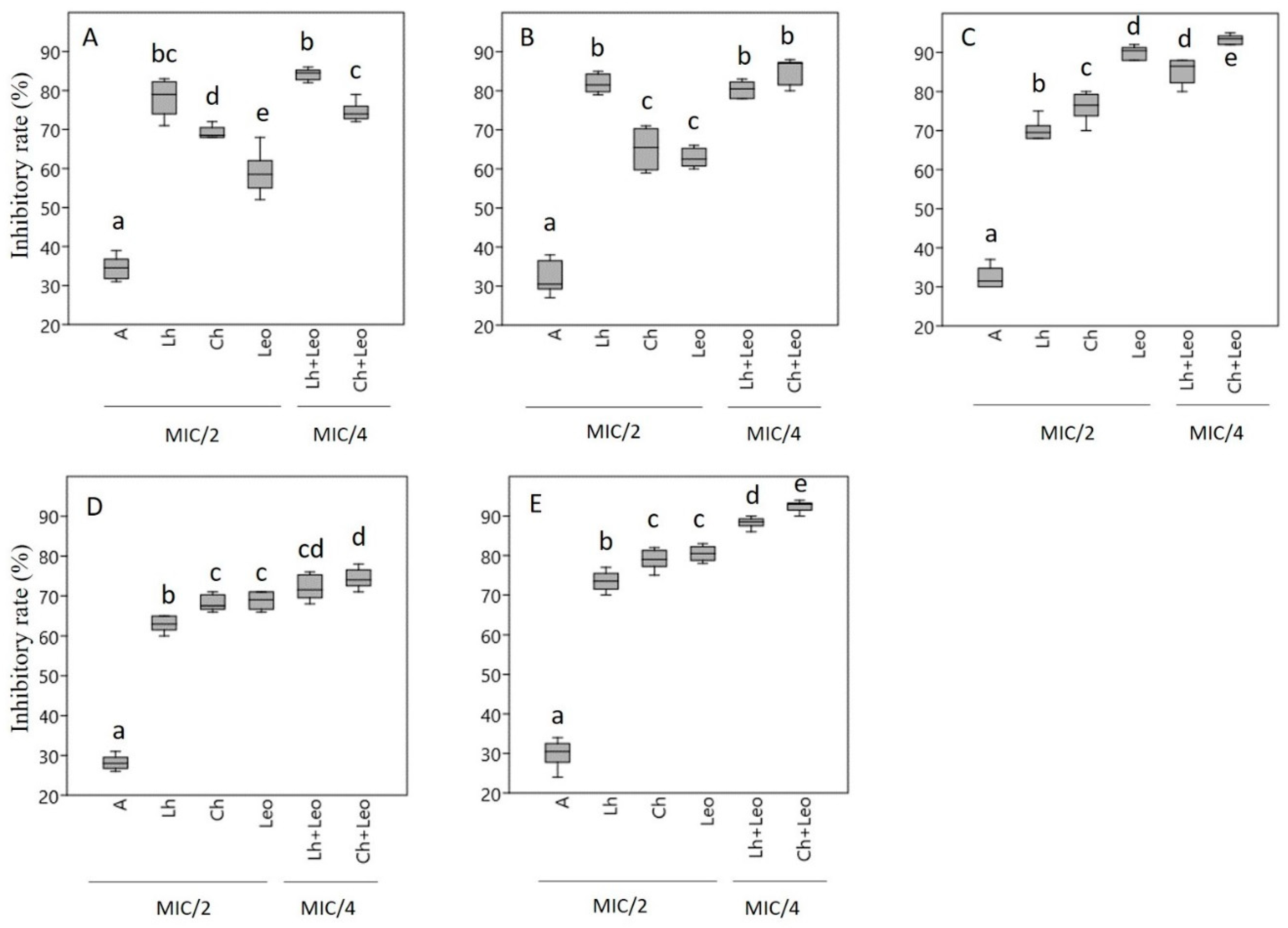
2.4. Biofilm Eradication Activity

In this experiment, Lh, Ch, Leo, and the antibiotics were used at MIC/2 concentration when tested alone, but in the case of honey–essential oil combinations, MIC/4 values were used, as our goal was to reveal the effect of the combinations on biofilm eradication, not to destroy the bacterial cells. Based on our observations, all samples had antibiofilm activity, but they had a different effect on the biofilm eradication of the five bacterial strains included in the study (Figure 2). The biofilm formation of H. influenzae (Figure 2A) and H. parainfluenzae (Figure 2B) was inhibited to a greater degree by the honey samples, with Lh displaying higher inhibition rates compared to Leo. In the case of M. catarrhalis (Figure 2C), however, the essential oil had a higher inhibition rate compared to the honey samples, and here Ch was the stronger inhibitor from the honey samples. Similar tendencies were observed in the case of P. aeruginosa (Figure 2D) and S. pneumoniae (Figure 2E) bacteria, where Ch and Leo exerted the same level of inhibition, while the inhibition activity of Lh was somewhat lower. For each bacterial strain, the combinations had the strongest antibiofilm activity (Ch + Leo inhibition rate: 74–93%). The antibiotic had the lowest inhibitory effect on biofilm formation (inhibition rate: 28–34%). Gram-negative P. aeruginosa was the most resistant, while the Gram-positive S. penumoniae bacterium was the most sensitive. Biofilm formation was not affected by the emulsifier (Tween40) used in the case of essential oil.

2.5. Checkerboard Assay

The results of checkerboard titration are summarized in Table 5 and Table 6. Table 5 shows the combination MIC values of the test substances, which were used to calculate the FICI values. Based on the FICI values, the interactions between the test substances were determined, taking into account the following value ranges: FICI ≤ 0.5: synergist; 0.5–1: additive; >1: negative interaction; >4: antagonist [41].
Based on the FICI values, Ch used together with Leo resulted in a synergistic effect against both Gram-negative and Gram-positive bacteria. When Leo was combined with Lh, an additive effect was detected (Table 6).

2.6. Membrane Degradation Assay

The checkerboard titration supported the synergistic effect of Ch and Leo (Table 6), so membrane degradation studies were performed with these two samples and their combinations. In addition, Ch was chosen for these experiments because of its high antibacterial and antibiofilm activity against S. pneumoniae (Gram-positive) and P. aeruginosa (Gram-negative) bacteria (Table 4, Figure 2).
Using Ch and Leo separately at MIC × 2 concentrations, bacterial DNA release was above 60% (60.7–68.4%) for both bacterial strains included in the study. In the case of the combination, half of the concentration of the two natural substances (MIC, 1:1 ratio) resulted in DNA leakage of 67.9% (P. aeruginosa) and 69.8% (S. pneumoniae) (Table 7). In the study of DNA release kinetics, Ch and Leo were used separately at MIC × 2 concentrations at different time intervals (20, 40, 60, and 90 min) (Table 8). For the combination, in this case too, we worked with the MIC concentration for both samples (1:1 ratio). In each measurement, bacterial DNA was detected already at 20 min (34.4–53.6%), and the highest values were obtained at 90 min (66.6–71.2%). In this study, as well, the most DNA was released during treatment with the combination (90th minute: 70.9 and 71.2%). Based on our results, P. aeruginosa proved to be a more resistant bacterium compared to S. pneumoniae (Table 7 and Table 8).

3. Discussion

Investigating the antibacterial and biofilm inhibition activities of natural substances is vital to explore new therapeutic options to curb the increasing spread of antibiotic resistance. In our study, we applied a novel approach by exploring the possibility of using two natural substances, honey and essential oil, together. Chestnut and linden honey, which were included in our experiment, have proven antibacterial and biofilm formation inhibitory effects against respiratory bacteria. Combining them with Leo not only resulted in an increased antibacterial effect, but the anti-inflammatory effect of lavender essential oil can also contribute to the success of the complementary therapy. Both honey and essential oil have been hypothesized to achieve their antibacterial efficacy through several modes of action.
Essential oils are multicomponent, lipophilic liquids with characteristic aroma, as they comprise volatile compounds. Up to 20–60 different bioactive components can be present in essential oils [42]. Their mechanisms of action are diverse; some essential oils can kill bacteria (bactericidal effect), but there are some that only slow down their division (bacteriostatic effect) [43]. The general bacteriostatic effect of essential oils is based on several mechanisms. They cause the disintegration of the bacterial outer membrane or the phospholipid bilayer. They increase the permeability of the bacterial cell membrane and promote the leakage of intracellular components from the cell, which leads to an upset in the ionic balance. In addition, they can interfere with the cellular metabolism (glucose and fatty acid metabolism) and enzyme kinetics of pathogens [44,45]. According to some researchers, there is a significant difference between the sensitivity of Gram-negative and Gram-positive bacteria. Essential oil components with hydrophobic properties are more effective on Gram-positive bacteria [46,47]. This phenomenon can be explained by the fact that Gram-positive bacteria have a thick peptidoglycan layer linked to apolar molecules through which lipophilic components can easily pass; meanwhile, in the cell envelope of Gram-negative bacteria, proteins linked to the outer membrane and lipopolysaccharides prevent this [48]. Understanding the mechanism of action of a given essential oil or one of its components is of paramount importance, as this allows us to develop new therapeutic procedures and identify new areas of indication. The antibacterial effect of lavender is well-known, and numerous studies have supported the antibacterial and anti-inflammatory effects of Leo [18,49,50]. However, there are few studies on the antibacterial and anti-biofilm effects of Leo against pathogens that cause upper respiratory tract diseases. The activity of Leo against Haemophilus strains was first described by our research group. Due to the overuse of antibiotics and the increasing phenomenon of antibiotic resistance, which can largely be attributed to the ability of bacteria to form biofilms, research is directed towards bacteria that cause problems in the nosocomial area [51,52,53]. Leo has been shown to be effective against both MRSA (methicillin-resistant Staphylococcus aureus) and VRE (vancomycin-resistant Enterococcus faecalis) at concentrations below 1% [54]. In the study by de Rapper, the efficacy of Leo was tested in combination with antibiotics and antifungals, and the individual MIC value of Leo was also determined using the microdilution method. The MIC values obtained against the bacteria P. aeruginosa and S. aureus were 1 mg/mL, and values against the fungus (C. albicans) were 3 mg/mL. Another research group measured 2.33 mg/mL as the MIC value of Leo against the pathogen P. aeruginosa [55]. The MIC value of Leo against P. aeruginosa in our study was 2.5 mg/mL, which shows that our data are comparable with the results of other research groups [56]. A veterinary study indicated that Leo may be effective as a topical treatment for canine otitis externa promoted by biofilm-producing P. aeruginosa [57]. The antipseudomonal effect of L. angustifolia, L. intermedia, L. pyrenaica, and L. stoechas essential oils was confirmed by Végh et al. (2012) [58]. The anti-Pseudomonas activity of essential oils obtained from L. sumian and L. grosso was supported by Donadu’s research group. Their results confirmed that L. sumian essential oil showed lower antimicrobial activity (MIC: 16% solution) than L. grosso (MIC: 8% solution). None of the Leos tested showed cytotoxic effects at the tested concentrations [59].
In biofilm inhibition studies, P. aeruginosa was the most resistant pathogen in each test system. This may be due to the fact that this bacterium has three exopolysaccharides: Psl, Pel, and alginate. Psl is a neutral pentasaccharide that typically contains D-glucose, D-mannose, and L-rhamnose moieties. Psl acts as a signaling molecule and promotes the production of c-di-GMP (bis-(3′-5′)-cyclic dimeric guanosine monophosphate), the levels of which, when elevated, result in thicker and more robust biofilms [60]. In addition, Psl protects biofilm bacteria from antimicrobial agents and neutrophil phagocytosis [61]. Pel is a cationic polysaccharide polymer of partially deacetylated N-acetyl-D-glucosamine and N-acetyl-D-galactosamine. Pel promotes the tolerance of bacteria embedded in biofilms to aminoglycoside antibiotics [62]. Alginic acids are mixtures of uronic acid polymers, the main components of which are β-D-mannuronic acid and α-L-guluronic acid. The salts of alginic acids are alginates. Alginate has a wide range of functions, including biofilm maturation, protection against phagocytosis, and reduced diffusion of antibiotics through the biofilm [63]. The aforementioned units contribute to the increased resistance of P. aeruginosa biofilm to treatment. The other pathogens included in the study (Haemophilus spp., M. catarrhalis, S. pneumoniae) do not have such resistant biofilms, so higher inhibition rate values were detected for these pathogens.
Among the possible mechanisms of action underlying the effectiveness of Leo, in addition to membrane-degrading activity, the inhibition of quorum sensing (QS) should also be mentioned. Although we do not have enough data regarding the anti-QS effect of Leo, a study written in 2014 supports this fact [64]. The antibacterial effect of vaporizing Leo was also confirmed against Staphylococcus strains (S. hominis, S. haemolyticus, S. epidermidis, S. aureus) in hospital environments. The data showed that Leo reduced the number of bacteria in all hospital areas compared to the untreated control [65].
Based on the microbiological studies conducted to date, it can be concluded that similar studies related to Haemophilus strains and M. catharralis and S. pneumoniae bacteria are not available. Furthermore, it should be emphasized that few studies investigate the biofilm inhibitory effect of Leo [66]. In addition to the above, another novelty of our work is that we also examined the biofilm eradication activity of Leo in combination with two types of honey. Honeys, just like essential oils, are promising antibacterial and antiviral agents due to their diverse plant-derived compounds and multiple modes of action.
The antimicrobial activities of honey have recently been reviewed by Luca et al. (2024) [67]. Honey’s antibacterial activity was reported against numerous bacteria, including Acinetobacter, Escherichia, Klebsiella, Pseudomonas, Staphylococcus, and Streptococccus strains. The antifungal effect of honey was investigated mainly against various Candida and Aspergillus species. The antiviral activity of honeys requires further research; however, honey seems to be active against SARS-CoV-2, human immunodeficiency virus, herpes simplex virus types 1 and 2, viral hepatitis virus, and others [68,69]. The antimicrobial properties of honey are attributed partly to plant-derived compounds, like polyphenols [23], and partly to bee-derived components, like defensin-1 peptide [20]. The antibacterial activity of honey is usually in correlation with its hydrogen peroxide content [70], e.g., in acacia, rapeseed, and sunflower honeys harvested in Europe but not in linden honey. The synergistic effect of polyphenols and hydrogen peroxide has been proposed to determine a honey’s antibacterial qualities [71]. New Zealand’s manuka honey is characterized by non-peroxide antibacterial activity, which can be attributed to its methylglyoxal content [72]. In addition, the physicochemical traits of honeys, like acidity (low pH), high sugar concentration, low water content, phenolic acids and flavonoids, as well as proteins, all contribute to honey’s antibacterial properties. It must be emphasized that individually none of these factors would be sufficient to achieve antimicrobial activity, as their complex interaction is required [67]. Exposure to honey can induce physiological changes in bacteria related to membrane integrity and polarization and can lead to metabolic disruption [29]. High levels of carbohydrates in honey can cause the dehydration of bacterial cells through osmosis [67]. Damage to the cytoplasmic membrane can result in the easy release of nucleic acids, amino acids, K+, proteins, and inorganic phosphates from the bacterial cell, leading to cell lysis and death [73]. Lh and Ch contain higher proportions of the polyphenolic components ferulic acid and gallic acid, which can cause irreversible damage to membrane properties of various pathogenic bacteria (e.g., P. aeruginosa), such as physicochemical characteristics or intra- and extracellular permeability [74,75,76]. Increased pore formation, local tearing, or a decrease in negative surface charge may occur [23,77]. Membrane-bound efflux pumps (proteins) fulfill many functions in bacterial cells; for example, they promote the development of resistance to antibacterial agents, biofilm stabilization, and protection against oxidative stress [78,79,80]. Efflux pump inhibitors are used to prevent antibiotic resistance of multidrug-resistant bacterial strains with increased efflux activity. These compounds significantly reduce tolerance and biofilm formation and thus increase the effectiveness of antibiotics [81,82]. Phenolic compounds found in honey (e.g., ferulic, gallic, caffeic and salicylic acids, quercetin, kaempferol, galangin) can act as efflux pump inhibitors by inhibiting ATPases involved in antibiotic efflux [79,83]. In order to increase their chances of survival, bacterial cells constantly communicate with each other using signal molecules. It has been observed that the QS mechanism (local cell density sensing) of bacteria directly affects the development of antibiotic resistance through the expression of different efflux pumps [84]. QS activity is significantly inhibited by many unifloral honeys (e.g., by degrading the N-acyl homoserine lactone signaling molecule in Gram-negative bacteria); however, the extent of the inhibitory effect depends significantly on the type of honey, the place of collection, or the different phenolic acid content. For example, chestnut and linden honeys show higher QS inhibitory activity than orange or rosemary honeys [79,85,86].
There are a few studies available on the combination of honey and essential oil. Imtara and his research group used checkerboard titration to explore the possible synergistic effect of oregano (Origanum vulgare) essential oil and different honey samples on E. coli, P. aeruginosa, S. faecalis, and Staphylococcus aureus by calculating fractional inhibitory concentrations. The most pronounced synergistic effect was achieved with the combination of oregano essential oil and Euphorbia honey against the pathogens E. coli and Streptococcus faecalis [87]. Another study supports the synergistic effect of combining eucalyptus honey and eucalyptus essential oil against the pathogens E. coli, Proteus mirabilis, Salmonella Typhimurium, Bacillus subtilis, Staphylococcus aureus, Listeria monocytogenes, Candida albicans, Trichophyton rubrum, and Aspergillus niger. The study tested honey and essential oil separately and in combination. The results confirmed that the combination of the two substances was more effective [37]. The research team of Qasem tested the combination of chamomile essential oil and chamomile honey against similar pathogens. The results showed that the most sensitive pathogens were S. aureus and L. monocytogenes, and the most resistant ones were E. coli, P. mirabilis, S. Typhimurium, and P. aeruginosa. In all cases, the combination of chamomile honey and chamomile essential oil showed higher efficacy compared to the use of honey or essential oil separately [88]. Increased efficacy of honey–essential oil combinations can be attributed to the fact that complexity makes it difficult for bacteria to develop tolerance, as adapting to several different active ingredients is a much more time-consuming and complicated task.
Our study demonstrated the synergistic effect of Ch and Leo against otitis media pathogens through a series of in vitro experiments. Using only in vitro assays poses a limitation for the usability of our data; thus, further experiments should be planned to test the honey–essential oil combinations in either in vivo or clinical settings. Further research should be directed to clarifying which constituents of honeys and essential oils are responsible for their respective antibacterial and antibiofilm effect and how these bioactive compounds can enhance each other’s efficacy in combination, resulting in synergistic effects. The current study revealed the membrane degrading potential of Lh, Ch, and Leo. This is, however, only one of the mechanisms of honeys’ and essential oils’ antibacterial action; thus, further experiments are needed to shed light on whether these complex natural substances or their bioactive compounds are able to inhibit other defense lines of bacteria, such as efflux pumps and quorum-sensing mechanisms. In addition, the study could be broadened to include several other bacterial strains causing multiple types of infections to reveal the width of the antibacterial spectrum of honeys and essential oils.

4. Materials and Methods

4.1. Honey and Essential Oil Samples

For our investigations, linden (Tilia spp.) and chestnut (Castanea sativa Mill.) honeys were purchased in 2021 from Hungarian apiaries in the South Transdanubian region; Lh was purchased from the Reith Apiary (Baranya County) and Ch was purchased from the Németh Apiary (Zala County). The aerial parts of lavender (Lavandula angustifolia Mill.) were collected before the flowering period in Bolhó village (N46.0408 E17.3034, Somogy County, Hungary) in 2021. The plant material was prepared for essential oil extraction, as described in our previous publication [18]. The herba was dried at room temperature for 1 week, and then the essential oil was extracted through hydrodistillation [89]. The lavender essential oil (Leo) distilled in the pre-bloom period was selected for this study based on our previous experiments, where Leo from this phenophase exhibited greater antibacterial and anti-inflammatory activity compared to Leo obtained during and after the flowering period [18,90]. The chemical composition of Leo was characterized in Balázs et al. (2025), with the main components being linalool (31.02%) and linalyl acetate (23.66%), followed by terpinen-4-ol (6.44%), α-terpineol (5.54%), and geranyl acetate (3.45%) [90].

4.2. Melissopalynological Analysis

Following the work of Nagy-Radványi et al. (2024—A) [91], we chose microscopic pollen analysis as one of the most effective methods to determine the botanical origin of honeys. Honey pollen samples were prepared according to the methodology developed by Von der Ohe et al. (2004) [92]. Ten grams of honey was measured into a 50 mL centrifuge tube and then mixed with 20 mL of distilled water using a Combi-spin FVL-2400N vortex (Biocenter Ltd., Szeged, Hungary). The centrifuge tube was placed into a Neofuge 15R centrifuge (Lab-Ex Ltd., Budapest, Hungary) and centrifuged at 3000 rpm for 10 min. After removing the supernatant, the remaining sediment was supplemented with 10 mL of distilled water, and the mixture was centrifuged again at 3000 rpm for 5 min. After the second centrifugation, 250 µL of distilled water was added to the sediment remaining in the centrifuge tube and vortexed, and then 20 µL of this pollen suspension was pipetted onto slides on an OTS 40 heating plate (Tiba Ltd., Győr, Hungary) set to 40 °C. A piece of Kaiser’s glycerin jelly with fuchsine (Merck Life Science Ltd., Budapest, Hungary) was placed onto the pollen preparations, thus staining the pollen grains. The pollen preparations were covered with a coverslip and analyzed with a Nikon Eclipse E200 light microscope connected to a Michrome 20 MP CMOS camera (Auro-Science Consulting Ltd., Budapest, Hungary) at 400× magnification using the 4.3.0.605 version of TCapture software [93]. Five hundred pollen grains were counted for each honey sample, and the plant species or family the particular pollen grain belonged to was recorded. The relative frequency of pollen types was given as a percentage of all pollen grains.

4.3. Physicochemical Parameters of Honey

The physicochemical properties of the honey samples were determined based on the research of Nagy-Radványi et al. (2024—A) [91]. For the determination of pH and electrical conductivity, we used a DSZ-708 multiparameter analyzer (Simex Ltd., Budapest, Hungary). The pH of the honey samples was measured according to the Hungarian Food Codex, and the pH meter was calibrated using buffer solutions with pH values of 4 and 9 [94]. For the determination of electrical conductivity, 20% (w/w) aqueous honey solutions were prepared, while for pH measurement, 10 g of honey was dissolved in 75 mL of carbon-dioxide-free distilled water. The electrical conductivity of honey, which is related to its mineral content, was measured at 20 °C, and the results were expressed in milli-Siemens per centimeter (mS/cm) [95].
To analyze the color intensity of varietal honeys, 50% (w/w) aqueous honey solutions were prepared, placed in an ultrasonic water bath (45–50 °C) for 5 min, and then filtered (0.45 µm pore size, Agilent Technologies, Milan, Italy). Spectrophotometric analysis was carried out using a Shimadzu UV-1800 spectrophotometer (Shimadzu Schweiz GmbH, Reinach, Switzerland). The results, expressed in milliabsorbance units (mAU), were calculated from the difference in absorbance values measured at 450 nm and 720 nm [96].

4.4. Bacterial Strains

In our research, multidrug-resistant bacteria that play a significant role in the development of otitis media were used. The pathogens Moraxella catarrhalis (DSM 9143), Pseudomonas aeruginosa (ATCC 27853), and Streptococcus pneumoniae (DSM 20566) were cultured using Brain Heart Infusion (BHI) Broth (Sigma-Aldrich Ltd., Budapest, Hungary) as the liquid medium. For culturing two members of the Haemophilus genus, H. influenzae (DSM 4690) and H. parainfluenzae (DSM 8978), a specialized growth medium was prepared. Specifically, 3750 µL of Mueller-Hinton II Broth (MHB, OXOID Ltd., London, UK) was supplemented with 750 µL of NAD solution (1 mg/mL) and 500 µL of Haemophilus Supplement B (Diagon Ltd., Budapest, Hungary). The bacterial cultures were incubated in a shaking incubator (at 60 rpm) at 37 °C for 12 h using a C25 Incubator Shaker (New Brunswick Scientific, Edison, NJ, USA) [97].

4.5. Determination of Antibiotic Sensitivity of Test Bacteria

The antibiotic sensitivity of the microorganisms was evaluated using the Kirby–Bauer disk diffusion method, based on the work of Nagy-Radványi et al. (2024—B) [98] and following the guidelines of the Clinical and Laboratory Standards Institute (CLSI) and the Manual of Clinical Microbiology [99,100]. Susceptibility to the following antibiotics (OXOID Ltd.) was examined: amikacin (30 µg), ampicillin (10 µg), amoxicillin/clavulanic acid (20/10 µg), ceftazidime (10 µg), ciprofloxacin (5 µg), erythromycin (15 µg), gentamicin (30 µg), imipenem (10 µg), levofloxacin (5 µg), oxacillin (1 µg), penicillin (1 µg), piperacillin–tazobactam (100/10 µg), and vancomycin (5 µg) [101]. A filter paper disk impregnated with the specified antibiotic concentration was placed onto the surface of the culture medium (in the case of P. aeruginosa, M. catarrhalis and S. pneumoniae: Mueller-Hinton Agar, Sigma-Aldrich Ltd., Budapest, Hungary; in case of Haemophilus spp.: chocolate agar) that had been inoculated with a bacterial suspension (equal to the visual turbidity of a no. 0.5 McFarland standard). After 15 min, the Petri dishes were incubated at 35 ± 2 °C for 16–18 h, after which the inhibition zones were measured under visible light.

4.6. Determination of Minimum Inhibitory Concentration (MIC)

To determine the MIC, the microdilution method was used in accordance with the guidelines of the CLSI [102]. The procedure was carried out on 96-well microtiter plates, and, to ensure accuracy, all tests were performed in six replicates. For linden and chestnut honey, the following dilution series was prepared using BHI broth: 22.5, 20, 17.5, 15, 12.5, and 10% (w/w). To emulsify the essential oil, a Tween40-containing medium was used, and the following dilution series was prepared: 10, 5, 2.5, 1.25, 0.625, and 0.3125 mg/mL. To each well, 100 µL of a 105 CFU/mL bacterial suspension prepared in the appropriate growth medium was added, followed by 100 µL of honey solution or essential oil solution at varying concentrations. During our study, an antibiotic specific to each bacterial strain was used as the positive control. In the case of Haemophilus spp., amikacin (Likacin 250 mg/mL solution for injection (Lisapharma S.P.A., Erba, Italy), in the case of M. catharralis and S. pneumoniae, imipenem (Imipenem/Cilastatin Kabi 500 mg/500 mg powder for solution for infusion), and in the case of P. aeruginosa, gentamicin (Gentamicin Sandoz 80 mg injection, Sandoz) antibiotics were used. Bacterial cells were also treated with BHI solution containing 1% Tween40 without the addition of essential oil in order to exclude the effect of Tween40. The plates were incubated for 24 h at 37 °C, and then absorbance values were measured at 600 nm using a microplate reader (BMG Labtech, Budapest, Hungary) [98]. We considered the lowest honey or essential oil concentration as the MIC value, in which case no bacterial growth was visible in the wells of the microtiter plate.

4.7. Biofilm Eradication Assay

In order to investigate the effect of honey samples and Leo on biofilm eradication, biofilms were formed on 96-cell microtiter plates, similarly to Balázs et al. (2023) [103]. In these experiments, MIC/2 concentrations determined in previous MIC studies were applied for honey and essential oil samples, while MIC/4 concentrations were used for combinations.
To initiate biofilm formation, 200 µL of bacterial suspension at 108 CFU/mL was pipetted into individual wells of the microtiter plates. As a negative control, BHI medium was applied, while untreated bacterial suspensions (108 CFU/mL) served as the positive control. During a 24 h incubation period (37 °C), bacterial cells formed adherent and cohesive biofilms (24 h immature biofilm). Unattached cells were washed out with physiological saline, followed by the treatment step. Treatments involved adding 200 µL of honey or essential oil solutions or 100 µL of each solution for the combination (MIC/4 concentrations). Tween40-containing medium was used to emulsify the essential oil. After incubation (24 h at 37 °C), the wells were washed again with physiological saline to remove non-adherent cells, and the adhered cells were fixed with 99% methanol (Molar Chemicals Ltd., Halásztelek, Hungary) for 15 min at room temperature (23 °C). After removing the methanol, the biofilms were stained with 0.1% crystal violet for 20 min. The crystal violet binds to negatively charged molecules in the extracellular matrix of biofilms, enabling the quantitative determination of total biofilm biomass. As a final step, the dye bound to the biofilm was dissolved in 33% acetic acid. Absorbance was measured at 595 nm using a SPECTROstar Nano microplate reader (BMG Labtech, Budapest, Hungary) [104,105]. In the series of experiments, the following antibiotic controls were used: H. influenzae and H. parainfluenzae—amikacin, M. catarrhalis and S. pneumoniae—imipenem, and P. aeruginosa—gentamicin.
The inhibition rate was calculated using the formula [106]
Inhibition rate = (1 − S/C) × 100%
where C is the absorbance of the control and S is the absorbance of the sample.

4.8. Checkerboard Assay

The combination studies were performed using the checkerboard titration method. This method is particularly suitable for studying the combination effect of substances with antibacterial effects. The selected bacterial strains were P. aeruginosa and S. pneumoniae; thus, the interaction between different substances was tested in the case of both Gram-negative and Gram-positive pathogens. BHI medium (Sigma-Aldrich Ltd.) was used for cultivation; the germ count was 105 CFU/mL. Furthermore, 96-well microtiter plates were used for the studies, and 3 parallel measurements were performed. In this study, Leo was combined with Lh and Ch for both pathogens. In each study design, a halving dilution series was prepared at concentrations corresponding to predetermined MIC values, which were as follows: MIC × 2, MIC, MIC/2, MIC/4, and MIC/8. Subsequently, according to the test layouts, 100 μL of bacterial suspension and then 50 μL of essential oil sample (substance A) and 50 μL of honey sample (substance B) were applied to the cells of the microtiter plates. In parallel, a bacterial suspension without samples was applied to a microtiter plate as the positive control, and only BHI medium was used as the negative control. The prepared microtiter plates were incubated (for 24 h at 37 °C), and then their absorbance was measured at a wavelength of 600 nm with a plate reader (BMG Labtech, Budapest, Hungary; Bio-Tek Ltd., Winooski, Vermont, USA). Following the test, combination MIC values were calculated. The concentration at which the absorbance of the positive control was reduced to ±10% was considered the combination MIC value. From the combination MIC values calculated in this way, the fractional inhibitory concentration index (FICI) was calculated, which is a dimensionless numerical value that helps to evaluate the combination effect of pharmacons.
FICI = FICA + FICB = M I C   A   c o m b i n a t i o n M I C   A + M I C   B   c o m b i n a t i o n M I C   B

4.9. Membrane Degradation Assay

During the study of membrane degradation, bacterial suspensions (108 CFU/mL) were prepared in phosphate buffer solution (PBS), and untreated bacterial cells were used as controls. Samples (Ch, Leo, and honey—essential oil combination) with concentrations of MIC/4, MIC/2, MIC, MIC × 2, and MIC × 4 were added to the cells (incubation time: 1 h). The time dependence of DNA release was also investigated; in this test, the bacterial cells were suspended in PBS containing honey or essential oil with a concentration of MIC × 2. In the case of the combination, Ch and Leo were used in MIC concentration (1:1 ratio). Treatments lasted 0, 20, 40, 60, and 90 min. In both cases, these steps were followed by centrifugation (Neofuge 15R, Lab-Ex Ltd., Budapest, Hungary) at 12,000× g for 2 min. The absorbance of the supernatant liquid containing nucleic acid was determined at 260 nm with a Metertech SP-8001 (Abl&e-Jasco Ltd., Budapest, Hungary) spectrophotometer, and the results were expressed as a percentage compared to the control [107].

4.10. Statistical Analysis

Statistical analyses were carried out using Microsoft Excel® 2016 MSO (16.0.4266.1001 version) (Microsoft Corp., Redmond, WA, USA) and the PAST software package version 3.11 [108]. The effects of honey types, essential oil, and their combinations were compared with each other using one-way ANOVA. If the null hypothesis of the ANOVA was rejected, we used Tukey’s test to establish a difference between the given groups (p < 0.05).

5. Conclusions

Based on our results and a few other studies conducted so far, it can be stated that the combined use of essential oil and honey may be promising for suppressing pathogens and eradicating biofilms. Demonstrating the synergistic effect of Ch and Leo supports that combinations of the two natural substances could be used in adjunctive therapy for otitis media. Our in vitro experiments provide a good starting point for the development of an ear drop or ear spray with the honey–essential oil combination that would speed up the healing time by preventing the infection from becoming chronic.

Supplementary Materials

The following supporting information can be downloaded at https://www.mdpi.com/article/10.3390/antibiotics14020146/s1, Table S1: Antibiotic sensitivity of test bacteria (1-5) measured by disk diffusion assay.

Author Contributions

Conceptualization, Á.F. and V.L.B.; methodology, V.D.Á., B.K., V.L.B. and L.N.-R.; software, M.K. and V.L.B.; validation, M.K. and Á.F.; formal analysis, M.K.; investigation, V.D.Á., B.K. and L.N.-R.; resources, V.D.Á. and L.N.-R.; data curation, M.K. and V.D.Á.; writing—original draft preparation, V.L.B., Á.F., B.K. and G.H.; writing—review and editing, Á.F., B.K. and V.L.B.; visualization, M.K.; supervision, Á.F., L.N.-R. and V.L.B.; project administration, M.K., Á.F. and V.L.B.; funding acquisition, Á.F., V.L.B., G.H. and V.D.Á. All authors have read and agreed to the published version of the manuscript.

Funding

This research was supported by the Fund of National Research, Development and Innovation Office NKFIH PD 147156 (Balázs V.L.) and NKFIH K18 128217 (Horvath G.), the Research Grant of the Faculty of Pharmacy University of Pécs, GYTK KA-2024-05 (Farkas Á.), the University Research Scholarship Program of the Ministry for Culture and Innovation from the Source of the National Research, Development and Innovation Fund (Nagy-Radványi L.: EKÖP-24-4-I-PTE-113; Balázs V.L.: EKÖP-24-4-II-PTE-114), and the Kriszbacher Ildikó Talent Management Programme of the University of Pécs: PTE-KITEP-2023-262 and PTE-KITEP-2024-385 (Ángyán V.D.).

Institutional Review Board Statement

Not applicable.

Informed Consent Statement

Not applicable.

Data Availability Statement

The original contributions presented in this study are included in the article. Further inquiries can be directed to the corresponding author.

Conflicts of Interest

The authors declare no conflicts of interest.

References

  1. Webber, M.A.; Piddock, L.J. The importance of efflux pumps in bacterial antibiotic resistance. J. Antimicrob. Chemother. 2003, 51, 9–11. [Google Scholar] [CrossRef]
  2. Varela, M.F.; Stephen, J.; Lekshmi, M.; Ojha, M.; Wenzel, N.; Sanford, L.M.; Hernandez, A.J.; Parvathi, A.; Kumar, S.H. Bacterial Resistance to Antimicrobial Agents. Antibiotics 2021, 10, 593. [Google Scholar] [CrossRef]
  3. Moura de Sousa, J.; Lourenço, M.; Gordo, I. Horizontal gene transfer among host-associated microbes. Cell Host Microbe 2023, 31, 513–527. [Google Scholar] [CrossRef]
  4. D’Aquila, P.; De Rango, F.; Paparazzo, E.; Passarino, G.; Bellizzi, D. Epigenetic-Based Regulation of Transcriptome in Escherichia coli Adaptive Antibiotic Resistance. Microbiol. Spectr. 2023, 11, e0458322. [Google Scholar] [CrossRef]
  5. López, D.; Vlamakis, H.; Kolter, R. Biofilms. Cold Spring Harb. Perspect. Biol. 2010, 2, a000398. [Google Scholar] [CrossRef] [PubMed]
  6. Lv, X.; Wang, L.; Mei, A.; Xu, Y.; Ruan, X.; Wang, W.; Shao, J.; Yang, D.; Dong, X. Recent Nanotechnologies to Overcome the Bacterial Biofilm Matrix Barriers. Small 2023, 19, e2206220. [Google Scholar] [CrossRef] [PubMed]
  7. Zhang, L.; Bera, H.; Wang, H.; Wang, J.; Guo, Y.; Shi, C.; Cun, D.; Moser, C.; Høiby, N.; Yang, M. Combination and nanotechnology based pharmaceutical strategies for combating respiratory bacterial biofilm infections. Int. J. Pharm. 2022, 616, 121507. [Google Scholar] [CrossRef]
  8. Kragh, K.N.; Tolker-Nielsen, T.; Lichtenberg, M. The non-attached biofilm aggregate. Commun. Biol. 2023, 6, 898. [Google Scholar] [CrossRef] [PubMed]
  9. Van Hoecke, H.; De Paepe, A.S.; Lambert, E.; Van Belleghem, J.D.; Cools, P.; Van Simaey, L.; Deschaght, P.; Vaneechoutte, M.; Dhooge, I. Haemophilus influenzae biofilm formation in chronic otitis media with effusion. Eur. Arch. Oto-Rhino-Laryngol. 2016, 273, 3553–3560. [Google Scholar] [CrossRef] [PubMed]
  10. Sauer, K.; Stoodley, P.; Goeres, D.M.; Hall-Stoodley, L.; Burmølle, M.; Stewart, P.S.; Bjarnsholt, T. The biofilm life cycle: Expanding the conceptual model of biofilm formation. Nat. Rev. Microbiol. 2022, 20, 608–620. [Google Scholar] [CrossRef] [PubMed]
  11. Scott, A.M.; Clark, J.; Julien, B.; Islam, F.; Roos, K.; Grimwood, K.; Little, P.; Del Mar, C.B. Probiotics for preventing acute otitis media in children. Cochrane Database Syst. Rev. 2019, 6, CD012941. [Google Scholar] [CrossRef] [PubMed]
  12. Gaddey, H.L.; Wright, M.T.; Nelson, T.N. Otitis Media: Rapid Evidence Review. Am. Fam. Physician 2019, 100, 350–356. [Google Scholar]
  13. Danishyar, A.; Ashurst, J.V. Acute Otitis Media; StatPearls Publishing: Treasure Island, FL, USA, 2024. [Google Scholar]
  14. Paramasivan, S.; Bassiouni, A.; Shiffer, A.; Dillon, M.R.; Cope, E.K.; Cooksley, C.; Ramezanpour, M.; Moraitis, S.; Ali, M.J.; Bleier, B.; et al. The international sinonasal microbiome study: A multicentre, multinational characterization of sinonasal bacterial ecology. Allergy 2020, 75, 2037–2049. [Google Scholar] [CrossRef]
  15. de Sousa, D.P.; Damasceno, R.O.S.; Amorati, R.; Elshabrawy, H.A.; de Castro, R.D.; Bezerra, D.P.; Nunes, V.R.V.; Gomes, R.C.; Lima, T.C. Essential Oils: Chemistry and Pharmacological Activities. Biomolecules 2023, 13, 1144. [Google Scholar] [CrossRef]
  16. Sharifi-Rad, J.; Sureda, A.; Tenore, G.C.; Daglia, M.; Sharifi-Rad, M.; Valussi, M.; Tundis, R.; Sharifi-Rad, M.; Loizzo, M.R.; Ademiluyi, A.O.; et al. Biological activities of essential oils: From plant chemoecology to traditional healing systems. Molecules 2017, 22, 70. [Google Scholar] [CrossRef]
  17. Kim, N.S.; Lee, D.S. Comparison of different extraction methods for the analysis of fragrances from Lavandula species by gas chromatography–mass spectrometry. J. Chromatogr. A 2002, 982, 31–47. [Google Scholar] [CrossRef]
  18. Pandur, E.; Balatinácz, A.; Micalizzi, G.; Mondello, L.; Horváth, A.; Sipos, K.; Horváth, G. Anti-inflammatory effect of lavender (Lavandula angustifolia Mill.) essential oil prepared during different plant phenophases on THP-1 macrophages. BMC Complement. Med. Ther. 2021, 21, 287. [Google Scholar] [CrossRef] [PubMed]
  19. Koulivand, P.H.; Ghadiri, M.K.; Gorji, A. Lavender and the nervous system. Evid. -Based Complement. Altern. Med. 2013, 2013, 681304. [Google Scholar] [CrossRef]
  20. Keitel, U.; Schilling, E.; Knappe, D.; Al-Mekhlafi, M.; Petersen, F.; Hoffmann, R.; Hauschildt, S. Effect of antimicrobial peptide from Apis mellifera hemolymph and its optimized version Api88 on biological activities of human monocytes and mast cells. Innate Immun. 2013, 19, 355–367. [Google Scholar] [CrossRef]
  21. Kurek-Górecka, A.; Górecki, M.; Rzepecka-Stojko, A.; Balwierz, R.; Stojko, J. Bee Products in Dermatology and Skin Care. Molecules 2020, 25, 556. [Google Scholar] [CrossRef] [PubMed]
  22. Liu, Y.; Han, L.; Yang, H.; Liu, S.; Huang, C. Effect of apigenin on surface associated characteristics and adherence of Streptococcus mutans. Dent. Mater. J. 2020, 39, 933–940. [Google Scholar] [CrossRef] [PubMed]
  23. Olas, B. Honey and its phenolic compounds as an effective natural medicine for cardiovascular diseases in humans? Nutrients 2020, 12, 283. [Google Scholar] [CrossRef] [PubMed]
  24. Khatami, S.H.; Vakili, O.; Ahmadi, N.; Soltani Fard, E.; Mousavi, P.; Khalvati, B.; Maleksabet, A.; Savardashtaki, A.; Taheri-Anganeh, M.; Movahedpour, A. Glucose oxidase: Applications, sources, and recombinant production. Biotechnol. Appl. Biochem. 2021, 69, 939–950. [Google Scholar] [CrossRef]
  25. Balázs, V.L.; Nagy-Radványi, L.; Filep, R.; Kerekes, E.; Kocsis, B.; Kocsis, M.; Farkas, Á. In vitro antibacterial and antibiofilm activity of Hungarian honeys against respiratory tract bacteria. Foods 2021, 10, 16–32. [Google Scholar] [CrossRef] [PubMed]
  26. Farkas, Á.; Balázs, V.L.; Kőszegi, T.; Csepregi, R.; Kerekes, E.; Horváth, G.; Szabó, P.; Gaál, K.; Kocsis, M. Antibacterial and Biofilm Degradation Effects of Hungarian Honeys Linked with Botanical Origin, Antioxidant Capacity and Mineral Content. Front. Nutr. 2022, 9, 953470. [Google Scholar] [CrossRef] [PubMed]
  27. Sakač, M.; Jovanov, P.; Marić, A.; Četojević-Simin, D.; Novaković, A.; Plavšić, D.; Škrobot, D.; Kovač, R. Antioxidative, Antibacterial and Antiproliferative Properties of Honey Types from the Western Balkans. Antioxidants 2022, 11, 1120. [Google Scholar] [CrossRef] [PubMed]
  28. Oliveira, A.; Sousa, J.C.; Silva, A.C.; Melo, L.D.; Sillankorva, S. Chestnut Honey and Bacteriophage Application to Control Pseudomonas aeruginosa and Escherichia coli Biofilms: Evaluation in an ex vivo Wound Model. Front. Microbiol. 2018, 9, 1725. [Google Scholar] [CrossRef]
  29. Combarros-Fuertes, P.; Estevinho, L.M.; Teixeira-Santos, R.; Rodrigues, A.G.; Pina-Vaz, C.; Fresno, J.M.; Tornadijo, M.E. Antibacterial action mechanisms of honey: Physiological Effects of Avocado, Chestnut, and Polyfloral Honey upon Staphylococcus aureus and Escherichia coli. Molecules 2020, 25, 1252. [Google Scholar] [CrossRef] [PubMed]
  30. Cumhur, A.; Hakan, A.; Tuğrul, Y.; Ahmet, T.; Nuray, T.D. Determination of the superior quality properties of randomly selected chestnut honey samples from the Sinop region. Spectrosc. Lett. 2023, 56, 353–363. [Google Scholar] [CrossRef]
  31. Qiao, J.; Chen, L.; Kong, L.; Dong, J.; Zhou, Z.; Zhang, H. Characteristic Components and Authenticity Evaluation of Rape, Acacia, and Linden Honey. J. Agric. Food Chem. 2020, 68, 9776–9788. [Google Scholar] [CrossRef]
  32. Farkas, Á.; Horváth, G.; Kuzma, M.; Mayer, M.; Kocsis, M. Phenolic compounds in Hungarian acacia, linden, milkweed and goldenrod honeys. Curr. Res. Food Sci. 2023, 6, 100526. [Google Scholar] [CrossRef]
  33. Skadiņš, I.; Labsvārds, K.D.; Grava, A.; Amirian, J.; Tomsone, L.E.; Ruško, J.; Viksna, A.; Bandere, D.; Brangule, A. Antimicrobial and Antibiofilm Properties of Latvian Honey against Causative Agents of Wound Infections. Antibiotics 2023, 12, 816. [Google Scholar] [CrossRef] [PubMed]
  34. Alissandrakis, E.; Tarantilis, P.A.; Pappas, C.; Harizanis, P.C.; Polissiou, M. Investigation of Organic Extractives from Unifloral Chestnut (Castanea sativa L.) and Eucalyptus (Eucalyptus globulus Labill.) Honeys and Flowers to Identification of Botanical Marker Compounds. LWT-Food Sci. Technol. 2011, 44, 1042–1051. [Google Scholar] [CrossRef]
  35. Mayda, N.; Ozok, A.; Sorkun, K. Some Characteristic Properties of Chestnut and Rhododendron Honeys in Turkey. J. Biol. Chem. 2018, 1, 135–145. [Google Scholar] [CrossRef]
  36. Turska, M.; Paluszkiewicz, P.; Turski, W.A.; Parada-Tuska, J. A Review of the Health Benefits of Food Enriched with Kynurenic Acid. Nutrients 2022, 14, 4182. [Google Scholar] [CrossRef] [PubMed]
  37. Assaggaf, H.M.; Naceiri, M.H.; Rajab, B.S.; Attar, A.A.; Hamed, M.; Sheikh, R.A.; Omari, N.E.; Menyiy, N.E.; Belmehdi, O.; Mahmud, S.; et al. Singular and Combined Effects of Essential Oil and Honey of Eucalyptus Globulus on Anti-Inflammatory, Antioxidant, Dermatoprotective, and Antimicrobial Properties: In Vitro and In Vivo Findings. Molecules 2022, 27, 5121. [Google Scholar] [CrossRef]
  38. Almasaudi, S. The antibacterial activities of honey. Saudi J. Biol. Sci. 2021, 28, 2188–2196. [Google Scholar] [CrossRef]
  39. Millezi, A.F.; Piccoli, R.H.; Oliveira, J.M.; Pereira, M.O. Anti-biofilm and Antibacterial Effect of Essential Oils and Their Major Compounds. J. Essent. Oil Bear. Plants 2016, 19, 624–631. [Google Scholar] [CrossRef]
  40. Oddo, L.P.; Piro, R.; Bruneau, É.; Guyot-Declerck, C.; Ivanov, T.; Piskulová, J.; Flamini, C.; Lheritier, J.; Morlot, M.; Russmann, H.; et al. Main European unifloral honeys: Descriptive sheets. Apidologie 2004, 35, 38–81. [Google Scholar] [CrossRef]
  41. Odds, F.C. Synergy, antagonism, and what the chequerboard puts between them. J. Antimicrob. Chemother. 2003, 52, 1. [Google Scholar] [CrossRef] [PubMed]
  42. Swamy, M.K.; Akhtar, M.S.; Sinniah, U.R. Antimicrobial Properties of Plant Essential Oils against Human Pathogens and Their Mode of Action: An Updated Review. Evid. Based Complement. Altern. Med. 2016, 2016, 3012462. [Google Scholar] [CrossRef] [PubMed]
  43. Pezantes-Orellana, C.; German, B.F.; De la Cruz, C.M.; Montalvo, J.L.; Orellana-Manzano, A. Essential oils: A systematic review on revolutionizing health, nutrition, and omics for optimal well-being. Front. Med. 2024, 16, 1337785. [Google Scholar] [CrossRef] [PubMed]
  44. Burt, S. Essential oils: Their antibacterial properties and potential applications in foods—A review. Int. J. Food Microbiol. 2004, 94, 223–253. [Google Scholar] [CrossRef] [PubMed]
  45. Cho, T.; Park, S.M.; Yu, H.; Seo, G.; Kim, H.; Kim, S.A.; Rhee, M. Recent Advances in the Application of Antibacterial Complexes Using Essential Oils. Molecules 2020, 25, 1752. [Google Scholar] [CrossRef]
  46. Plesiat, P.; Nikaido, H. Outer membranes of Gram-negative bacteria are permeable to steroid probes. Mol. Microbiol. 1992, 6, 1323–1333. [Google Scholar] [CrossRef] [PubMed]
  47. Angane, M.; Swift, S.; Huang, K.; Butts, C.A.; Quek, S.Y. Essential Oils and Their Major Components: An Updated Review on Antimicrobial Activities, Mechanism of Action and Their Potential Application in the Food Industry. Foods 2022, 11, 464. [Google Scholar] [CrossRef] [PubMed]
  48. Nikaido, H. Prevention of drug access to bacterial targets: Permeability barriers and active efflux. Science 1994, 264, 382–388. [Google Scholar] [CrossRef]
  49. Huang, M.Y.; Liao, M.H.; Wang, Y.K.; Huang, Y.S.; Wen, H.C. Effect of lavender essential oil on LPS-stimulated inflammation. Am. J. Chin. Med. 2012, 40, 845–859. [Google Scholar] [CrossRef]
  50. Todorova, D.; Yavorov, N.; Lasheva, V.; Damyanova, S.; Kostova, I. Lavender Essential Oil as Antibacterial Treatment for Packaging Paper. Coatings 2023, 13, 32. [Google Scholar] [CrossRef]
  51. Kollef, M.H.; Torres, A.; Shorr, A.F.; Martin-Loeches, I.; Micek, S.T. Nosocomial Infection. Crit. Care Med. 2021, 49, 169–187. [Google Scholar] [CrossRef]
  52. Sousa, S.A.; Feliciano, J.R.; Pita, T.; Soeiro, C.F.; Mendes, B.L.; Alves, L.G.; Leitão, J.H. Bacterial Nosocomial Infections: Multidrug Resistance as a Trigger for the Development of Novel Antimicrobials. Antibiotics 2021, 10, 942. [Google Scholar] [CrossRef] [PubMed]
  53. Shivshankar, P.; Collum, S.; Bi, W. Haemophilus influenzae is associated with fibrotic phenotype of COVID-19 and idiopathic pulmonary fibrosis. ERJ Open Res. 2022, 8, 236. [Google Scholar] [CrossRef]
  54. Cavanagh, H.M.; Wilkinson, J.M. Biological activities of lavender essential oil. Phytother. Res. 2002, 16, 301–308. [Google Scholar] [CrossRef] [PubMed]
  55. Nikolić, M.; Jovanović, K.K.; Marković, T.; Marković, D.; Gligorijević, N.; Radulović, S.; Soković, M. Chemical composition, antimicrobial, and cytotoxic properties of five Lamiaceae essential oils. Ind. Crops Prod. 2014, 61, 225–232. [Google Scholar] [CrossRef]
  56. de Rapper, S.; Viljoen, A.; van Vuuren, S. The In Vitro Antimicrobial Effects of Lavandula angustifolia Essential Oil in Combination with Conventional Antimicrobial Agents. Evid. Based Complement. Altern. Med. 2016, 2016, 2752739. [Google Scholar] [CrossRef]
  57. Mourão, A.; Serrano, I.; Cunha, E.; Tavares, L.; Lourenco, A.; Oliveira, M. In vitro efficacy of lavender oil, otological gel and gentamicin to eradicate biofilm produced by Pseudomonas aeruginosa. Vet. Dermatol. 2024, 35, 726–735. [Google Scholar] [CrossRef] [PubMed]
  58. Végh, A.; Bencsik, T.; Molnár, P.; Böszörményi, A.; Lemberkovics, É.; Kovács, K.; Kocsis, B.; Horváth, G. Composition and antipseudomonal effect of essential oils isolated from different lavender species. Nat. Prod. Commun. 2012, 7, 1393–1396. [Google Scholar] [CrossRef]
  59. Donadu, M.; Usai, D.; Pinna, A.; Porcu, T.; Mazzarello, V.; Fiamma, M.; Marchetti, M.; Cannas, S.; Delogu, G.; Zanetti, S.; et al. In vitro activity of hybrid lavender essential oils against multidrug resistant strains of Pseudomonas aeruginosa. J. Infect. Dev. Ctries. 2018, 12, 9–14. [Google Scholar] [CrossRef] [PubMed]
  60. Irie, Y.; Borlee, B.R.; O’Connor, J.R.; Hill, P.J.; Harwood, C.S.; Wozniak, D.J.; Parsek, M.R. Self-produced exopolysaccharide is a signal that stimulates biofilm formation in Pseudomonas aeruginosa. Proc. Natl. Acad. Sci. USA 2012, 109, 20632–20636. [Google Scholar] [CrossRef] [PubMed]
  61. Mishra, M.; Byrd, M.S.; Sergeant, S.; Azad, A.K.; Parsek, M.R.; McPhail, L.; Schlesinger, L.S.; Wozniak, D.J. Pseudomonas aeruginosa Psl polysaccharide reduces neutrophil phagocytosis and the oxidative response by limiting complement-mediated opsonization. Cell. Microbiol. 2012, 14, 95–106. [Google Scholar] [CrossRef]
  62. Jennings, L.K.; Storek, K.M.; Ledvina, H.E.; Coulon, C.; Marmont, L.S.; Sadovskaya, I.; Secor, P.R.; Tseng, B.S.; Scian, M.; Filloux, A.; et al. Pel is a cationic exopolysaccharide that cross-links extracellular DNA in the Pseudomonas aeruginosa biofilm matrix. Proc. Natl. Acad. Sci. USA 2015, 112, 11353–11358. [Google Scholar] [CrossRef] [PubMed]
  63. Hay, I.D.; Rehman, Z.U.; Moradali, M.F.; Wang, Y.; Rehm, B.H.A. Microbial alginate production, modification and its applications. Microb. Biotechnol. 2013, 6, 637–650. [Google Scholar] [CrossRef]
  64. Yap, P.S.X.; Krishnan, T.; Yiap, B.C.; Hu, C.P.; Chan, K.-G.; Lim, S.H.E. Membrane disruption and anti-quorum sensing effects of synergistic interaction between Lavandula angustifolia (lavender oil) in combination with antibiotic against plasmid-conferred multi-drug-resistant Escherichia coli. J. Appl. Microbiol. 2014, 116, 1119–1128. [Google Scholar] [CrossRef] [PubMed]
  65. Gismondi, A.; Di Marco, G.; Redi, E.L.; Ferrucci, L.; Cantonetti, M.; Canini, A. The antimicrobial activity of Lavandula angustifolia Mill. essential oil against Staphylococcus species in a hospital environment. J. Herb. Med. 2021, 26, 100426. [Google Scholar] [CrossRef]
  66. Leigh-de Rapper, S.; van Vuuren, S.F. Odoriferous Therapy: A Review Identifying Essential Oils against Pathogens of the Respiratory Tract. Chem. Biodivers. 2020, 17, 6. [Google Scholar] [CrossRef]
  67. Luca, L.; Pauliuc, D.; Oroian, M. Honey microbiota, methods for determining the microbiological composition and the antimicrobial effect of honey—A review. Food Chem. 2024, 23, 101524. [Google Scholar] [CrossRef]
  68. Al-Hatamleh, M.A.I.; Hatmal, M.M.; Sattar, K.; Ahmad, S.; Mustafa, M.Z.; Bittencourt, M.D.C.; Mohamud, R. Antiviral and immunomodulatory effects of phytochemicals from honey against COVID-19: Potential mechanisms of action and future directions. Molecules 2020, 25, 5017. [Google Scholar] [CrossRef] [PubMed]
  69. Asma, S.T.; Bobis, O.; Bonta, V.; Acaroz, U.; Shah, S.R.A.; Istanbullugil, F.R.; Arslan Acaroz, D. General nutritional profile of bee products and their potential antiviral properties against mammalian viruses. Nutrients 2022, 14, 3579. [Google Scholar] [CrossRef] [PubMed]
  70. Farkasovska, J.; Bugarova, V.; Godocikova, J.; Majtan, V.; Majtan, J. The role of hydrogen peroxide in the antibacterial activity of different floral honeys. Eur. Food Res. Technol. 2019, 245, 2739–2744. [Google Scholar] [CrossRef]
  71. Sawicki, T.; Starowicz, M.; Kłębukowska, L.; Hanus, P. The profile of polyphenolic compounds, contents of total phenolics and flavonoids, and antioxidant and antimicrobial properties of bee products. Molecules 2022, 27, 1301. [Google Scholar] [CrossRef] [PubMed]
  72. Johnston, M.; McBride, M.; Dahiya, D.; Owusu-Apenten, R.; Nigam, P.S. Antibacterial activity of Manuka honey and its components: An overview. AIMS Microbiol. 2018, 4, 655–664. [Google Scholar] [CrossRef] [PubMed]
  73. Al-Sayaghi, A.M.; Al-Kabsi, A.M.; Abduh, M.S.; Saghir, S.A.M.; Alshawsh, M.A. Antibacterial Mechanism of Action of Two Types of Honey against Escherichia coli through Interfering with Bacterial Membrane Permeability, Inhibiting Proteins, and Inducing Bacterial DNA Damage. Antibiotics 2022, 11, 1182. [Google Scholar] [CrossRef] [PubMed]
  74. Borges, A.; Ferreira, C.; Saavedra, M.J.; Simoes, M. Antibacterial activity and mode of action of ferulic and gallic acids against pathogenic bacteria. Microb. Drug Resist. 2013, 19, 256–265. [Google Scholar] [CrossRef] [PubMed]
  75. Zou, S.; Tao, H.; Chang, Y.-N. Characterization of antioxidant activity and analysis of phenolic acids and flavonoids in linden honey. Food Sci. Technol. 2022, 42, e76621. [Google Scholar] [CrossRef]
  76. Kim, J.; Uy, N.P.; Kim, D.; Lee, S. Analysis of Phenolic Acid Content and Antioxidant Activity of Chestnut Honey from Different Regions of Korea. Natl. Prod. Sci. 2023, 29, 127–131. [Google Scholar] [CrossRef]
  77. Anand, S.; Deighton, M.; Livanos, G.; Morrison, P.D.; Pang, E.C.K.; Mantri, N. Antimicrobial Activity of Agastache Honey and Characterization of Its Bioactive Compounds in Comparison with Important Commercial Honeys. Front. Microbiol. 2019, 10, 263. [Google Scholar] [CrossRef] [PubMed]
  78. Bogomolnaya, L.M.; Andrews, K.D.; Talamantes, M.; Maple, A.; Ragoza, Y.; Vazquez-Torres, A.; Andrews, H. Polymenis The ABC-type efflux pump MacAB protects Salmonella enterica serovar typhimurium from oxidative stress. mBio 2013, 4, e00630-13. [Google Scholar] [CrossRef] [PubMed]
  79. Khataybeh, B.; Jaradat, Z.; Ababneh, Q. Anti-bacterial, anti-biofilm and anti-quorum sensing activities of honey: A review. J. Ethnopharmacol. 2023, 317, 116830. [Google Scholar] [CrossRef]
  80. Nishino, K.; Yamasaki, S.; Nakashima, R.; Zwama, M.; Hayashi-Nishino, M. Function and inhibitory mechanisms of multidrug efflux pumps. Front. Microbiol. 2021, 12, 737288. [Google Scholar] [CrossRef] [PubMed]
  81. Babin, B.M.; Atangcho, L.; van Eldijk, M.B.; Sweredoski, M.J.; Moradian, A.; Hess, S.; Tolker-Nielsen, T.; Newman, D.K.; Tirrell, D.A. Selective proteomic analysis of antibiotic-tolerant cellular subpopulations in pseudomonas aeruginosa biofilms. mBio 2017, 8, e01593-17. [Google Scholar] [CrossRef]
  82. Zimmermann, S.; Klinger-Strobel, M.; Bohnert, J.A.; Wendler, S.; Rödel, J.; Pletz, M.W.; Löffler, B.; Tuchscherr, L. Clinically approved drugs inhibit the Staphylococcus aureus multidrug norA efflux pump and reduce biofilm formation. Front. Microbiol. 2019, 10, 2762. [Google Scholar] [CrossRef] [PubMed]
  83. Jaganathan, S.K. Can flavonoids from honey alter multidrug resistance? Med. Hypotheses 2011, 76, 535–537. [Google Scholar] [CrossRef]
  84. Preda, V.G.; Sandulescu, O. Communication is the key: Biofilms, quorum sensing, formation and prevention. Discoveries 2019, 7, e100. [Google Scholar] [CrossRef]
  85. Truchado, P.; Gil-Izquierdo, A.; Tomás-Barberán, F.; Allende, A. Inhibition by chestnut honey of N-Acyl-L-homoserine lactones and biofilm formation in Erwinia carotovora, Yersinia enterocolitica, and Aeromonas hydrophila. J. Agric. Food Chem. 2009, 57, 11186–11193. [Google Scholar] [CrossRef] [PubMed]
  86. Nazzaro, F.; Fratianni, F.; Coppola, R. Quorum sensing and phytochemicals. Int. J. Mol. Sci. 2013, 14, 12607–12619. [Google Scholar] [CrossRef] [PubMed]
  87. Imtara, H.; Elamine, Y.; Lyoussi, B. Honey Antibacterial Effect Boosting Using Origanum vulgare L. Essential Oil. Evid.-Based Complement. Altern. Med. 2018, 7842583, 14. [Google Scholar] [CrossRef] [PubMed]
  88. Qasem, A.; Assaggaf, H.; Montesano, D.; Khalil, Z.; Al-Mijalli, S.H.; Baaboua, A.E.; El Omari, N.; El Menyiy, N.; Bakrim, S.; Sheikh, R.A.; et al. Determination of Chemical Compounds and Investigation of Biological Properties of Matricaria chamomilla Essential Oils, Honey, and Their Mixture. Molecules 2022, 27, 5850. [Google Scholar] [CrossRef]
  89. Katekar, V.P.; Rao, A.B.; Sardeshpande, V.R. A hydrodistillation-based essential oils extraction: A quest for the most effective and cleaner technology. Sustain. Chem. Pharm. 2023, 36, 101270. [Google Scholar] [CrossRef]
  90. Balázs, V.L.; Bordás, B.; Nagy-Radványi, L.; Ormai, E.; Kocsis, M.; Kocsis, B.; Farkas, Á.; Micalizzi, G.; Mondello, L.; Bencsik-Kerekes, E.; et al. Efficacy of lavender essential oil against respiratory tract bacteria is influenced by harvesting time. Sci. Rep. 2025; under review. [Google Scholar]
  91. Nagy-Radványi, L.; Balázs, V.L.; Kocsis, B.; Csikós, E.; Ángyán, V.D.; Szabó, P.; Biró, V.; Kocsis, M.; Farkas, Á. Antibacterial activity of Hungarian varietal honeys against respiratory pathogens as a function of storage time. Sci. Rep. 2024, 14, 10200. [Google Scholar] [CrossRef]
  92. Von Der Ohe, W.; Oddo, L.P.; Piana, M.L.; Morlot, M.; Martin, P. Harmonized Methods of Melissopalynology. Apidologie 2004, 35, 18–25. [Google Scholar] [CrossRef]
  93. TCapture V4.3.0.605. 2022. Available online: https://www.tucsen.com/uploads/TCapture4.3.0.605.zip (accessed on 20 October 2022).
  94. Codex STAN 12-1981; Revised Codex Standard for Honey. Codex Alimentarius Commission: Rome, Italy, 2001.
  95. Bogdanov, S.; Lüllmann, C.; Martin, P. Harmonised methods of the European Honey Commission. Apidologie 1997, extra issue, 1–59. [Google Scholar]
  96. Beretta, G.; Granata, P.; Ferrero, M.; Orioli, M.; Facino, R.M. Standardization of antioxidant properties of honey by a combination of spectrophotometric/fluorimetric assays and chemometrics. Anal. Chim. Acta 2005, 533, 185–191. [Google Scholar] [CrossRef]
  97. Hindler, J.A.; Jorgensen, J.H. Susceptibility test methods: Fastidious bacteria. In Manual of Clinical Microbiology, 10th ed.; Versalovic, J., Ed.; ASM Press: Washington, DC, USA, 2011; pp. 1180–1187. [Google Scholar]
  98. Nagy-Radványi, L.; Ormai, E.; Koloh, R.; Ángyán, V.D.; Kocsis, B.; Bencsik-Kerekes, E.; Szabó, P.; Csikós, E.; Farkas, Á.; Horváth, G.; et al. Biofilm Inhibition Activity of Fennel Honey, Fennel Essential Oil and Their Combination. Microorganisms 2024, 12, 2309. [Google Scholar] [CrossRef]
  99. CLSI Document M 2-A10; Performance Standards for Antimicrobial Disk Susceptibility Tests. 10th ed, Approved Standard. CLSI (Clinical and Laboratory Standards Institute): Wayne, PA, USA, 2009.
  100. CLSI Document M 100-S20; Performance Standards for Antimicrobial Susceptibility Testing; 19th Informational Supplement. CLSI (Clinical and Laboratory Standards Institute): Wayne, PA, USA, 2010.
  101. Patel, J.B.; Tenover, F.C.; Turnidge, J.D.; Jorgensen, J.H. Susceptibility test methods: Dilution and disk diffusion methods. In Manual of Clinical Microbiology, 10th ed.; Versalovic, J., Ed.; ASM Press: Washington, DC, USA, 2011; Volume 1, pp. 1122–1143. [Google Scholar]
  102. CLSI Document M07-A9; Methods for Dilution Antimicrobial Susceptibility Tests for Bacteria That Grow Aerobically; Approved Standard, 9th ed. Clinical and Laboratory Standards Institute: Wayne, PA, USA, 2012.
  103. Balázs, V.L.; Nagy-Radványi, L.; Bencsik-Kerekes, E.; Koloh, R.; Szabó, D.; Kocsis, B.; Kocsis, M.; Farkas, Á. Antibacterial and Antibiofilm Effect of Unifloral Honeys against Bacteria Isolated from Chronic Wound Infections. Microorganisms 2023, 11, 509. [Google Scholar] [CrossRef] [PubMed]
  104. Peeters, E.; Nelis, H.J.; Coenye, T. Comparison of multiple methods for quantification of microbial biofilms grown in microtiter plates. J. Microbiol. Methods 2008, 72, 157–165. [Google Scholar] [CrossRef] [PubMed]
  105. Kerekes, E.; Deák, É.; Takó, M.; Tserennadmid, R.; Petkovits, T.; Vágvölgyi, C.; Krisch, J. Anti-biofilm formatting and anti-quorum sensing activity of selected essential oils and their main components on food related microorganisms. J. Appl. Microbiol. 2013, 115, 933–942. [Google Scholar] [CrossRef] [PubMed]
  106. Sun, Y.; Chen, S.; Zhang, C.; Liu, Y.; Ma, L.; Zhang, X. Effects of sub-minimum inhibitory concentrations of lemon essential oil on the acid tolerance and biofilm formation of Streptococcus mutans. Arch. Oral Biol. 2018, 87, 235–241. [Google Scholar] [CrossRef] [PubMed]
  107. Bennis, S.; Chami, F.; Chami, N.; Bouchikhi, T.; Remmal, A. Surface Alteration of Saccharomyces Cerevisiae Induced by Thymol and Eugenol. Lett. Appl. Microbiol. 2004, 38, 454–458. [Google Scholar] [CrossRef]
  108. Hammer, Ø.; Harper, D.A.; Ryan, P.D. PAST: Paleontological statistics software package for education and data analysis. Palaeontol. Electron. 2001, 4, 1–9. [Google Scholar]
Figure 1. Microscopic pollen images of linden pollen (A) and chestnut pollen (B).
Antibiotics 14 00146 g001
Figure 2. The biofilm degradation effects of linden and chestnut honeys and lavender essential oil alone and in combination against five bacteria. H. influenzae (A), H. parainfluenzae (B), M. catarrhalis (C), P. aeruginosa (D), and S. pneumoniae (E); antibiotic (A), linden honey (Lh), chestnut honey (Ch), lavender essential oil (Leo), combination of linden honey and lavender essential oil (Lh + Leo), combination of chestnut honey and lavender essential oil (Ch + Leo). Data were expressed using box plots with minimum to maximum values presented by vertical lines, with the median in the plot as a horizontal line. Different lowercase letters above the boxes indicate significant differences between the means of inhibitory rates of different components against the given bacteria, according to the Tukey’s test (p < 0.05), based on results of 6 parallel measurements (n = 6).
Antibiotics 14 00146 g002
Table 1. Pollen spectrum of linden honey and chestnut honey.
Pollen Type—Relative Frequency (%)
Honey SampleTilia spp.Robinia pseudoacaciaBrassica napusCastanea sativaOther
Linden36.025.720.46.211.7
Chestnut0.24.31.493.11.0
Table 2. Sensory characteristics and physicochemical parameters of honey samples. Data are presented as means ± standard deviation (n = 3).
Honey Type,
Plant Name
Sensory Characteristics
(Color, Odor, and Consistency)
ABS450–720
(mAU)
Electrical Conductivity
(mS/cm)
pH
Linden,
Tilia spp.
Light amber, strong odor, semisolid, fine, granulated107 ± 1.40.528 ± 0.014.1 ± 0.01
Chestnut,
Castanea sativa
Dark amber, intense odor, semisolid, fine, granulated215.3 ± 1.40.598 ± 0.014.7 ± 0.01
Table 3. Antibiotic sensitivity of test bacteria according to the disk diffusion assay.
Antibiotics12345
Amikacin-RSSS
AmpicillinRRSRR
Amoxicillin/
clavulanic acid
-SSRR
CiprofloxacinRSSSS
Ceftazidim-SS--
Eritromicin--R-S
GentamicinSSSSS
ImipenemSSSSS
Levofloxacin--R--
OxacillinR----
PenicillinR----
Piperacillin/
tazobactam
-SS--
VankomicinS----
1: Moraxella catarrhalis, 2: Pseudomonas aeruginosa, 3: Streptococcus pneumoniae, 4: Haemophilus influenzae, 5: H. parainfluenzae; R: resistant, S: sensitive, “-”: no data.
Table 4. The minimum inhibitory concentrations (MICs) of linden and chestnut honey, lavender essential oil, and the antibiotic controls (amikacin for H. influenzae and H. parainfluenzae; imipenem for M. catarrhalis and S. pneumoniae; gentamicin for P. aeruginosa) against the investigated bacterial strains.
MIC Values (mg/mL)
S. pneumoniaeH. influenzaeH. parainfluenzaeM. catarrhalisP. aeruginosa
Linden honey142.86111.11111.11142.86176.47
Chestnut honey142.86111.11111.11111.11176.47
Lavender essential oil0.310.630.631.252.50
Antibiotics 0.00080.00310.00160.00020.0063
Table 5. Combination MIC values of test samples (µg/mL).
Bacterial StrainsTest SamplesCombination MIC
P. aeruginosaLeo0.3125
Lh88.235
Leo0.3125
Ch44.1175
S. pneumoniaeLeo0.0775
Lh71.43
Leo0.0775
Ch35.715
Leo: lavender essential oil; Lh: linden honey; Ch: chestnut honey.
Table 6. FICI values and combination effects of test samples.
Bacterial StrainsTest SamplesFICICombination Effect
P. aeruginosaLeo0.625additive effect
Lh
Leo0.375synergistic effect
Ch
S. pneumoniaeLeo0.75additive effect
Lh
Leo0.5synergistic effect
Ch
Leo: lavender essential oil; Lh: linden honey; Ch: chestnut honey.
Table 7. Effect of chestnut honey, lavender essential oil, and their combination on the release of DNA in Gram-negative (P. aeruginosa) and Gram-positive (S. pneumoniae) bacteria.
DNA Release from Bacterial Cells (%)
SamplesConcentrationsP. aeruginosaS. pneumoniae
Chestnut honey
(Ch)
MIC/211.7 ± 1.214.3 ± 1.3
MIC26.9 ± 2.3 31.6 ± 1.7
MIC × 260.7 ± 2.566.3 ± 2.3
MIC × 4100100
Lavender essential oil
(Leo)
MIC/215.6 ± 1.818.1 ± 1.3
MIC36.5 ± 2.242.2 ± 2.1
MIC × 267.1 ± 2.568.4 ± 2.3
MIC × 4100100
CombinationMIC Ch + MIC Leo
(1:1 ratio)
67.9 ± 2.869.8 ± 3.0
Data are means ± standard deviations of six independent determinations (n = 6).
Table 8. Kinetics of 260 nm absorbing material released from Gram-negative (P. aeruginosa) and Gram-positive (S. pneumoniae) bacteria treated with chestnut honey, lavender essential oil, and their combination.
DNA Release from Bacterial Cells (%)
SamplesTime (min)P. aeruginosaS. pneumoniae
Chestnut honey (Ch)
conc.: MIC × 2
000
2034.4 ± 2.1 39.2 ± 2.2
4055.2 ± 2.358.9 ± 2.1
6060.7 ± 2.566.3 ± 2.3
9066.6 ± 2.268.0 ± 2.6
Lavender essential oil (Leo)
conc.: MIC × 2
000
2040.3 ± 2.249.1 ± 2.0
4062.8 ± 2.564.4 ± 2.6
6067.1 ± 2.568.4 ± 2.3
9069.2 ± 2.370.5 ± 2.8
Combination
conc.: MIC Ch + MIC Leo
(1:1 ratio)
000
2046.2 ± 2.453.6 ± 1.9
4063.7 ± 2.268.0 ± 2.1
6067.9 ± 2.869.8 ± 3.0
9070.9 ± 2.671.2 ± 2.2
Data are means ± standard deviations of six independent determinations (n = 6).
Disclaimer/Publisher’s Note: The statements, opinions and data contained in all publications are solely those of the individual author(s) and contributor(s) and not of MDPI and/or the editor(s). MDPI and/or the editor(s) disclaim responsibility for any injury to people or property resulting from any ideas, methods, instructions or products referred to in the content.

