Article

Genomic Characterization of Multiple Antibiotic-Resistant Enterococcus in Farm Animals in Ningxia Province, China

1
Department of Preventive Veterinary Medicine, College of Veterinary Medicine, Northwest Agricultural and Forestry University, Xianyang 712100, China
2
Key Laboratory for Prevention and Control of Major Ruminant Diseases, Ministry of Agriculture and Rural Affairs, Xianyang 712100, China
3
Ningxia Supervision Institute for Veterinary Drugs and Animal Feedstuffs, Yinchuan 750004, China
4
Lanzhou Veterinary Research Institute, Chinese Academy of Agricultura Sciences, Lanzhou 730050, China
*
Authors to whom correspondence should be addressed.
These authors contributed equally to this work.
Antibiotics 2025, 14(11), 1137; https://doi.org/10.3390/antibiotics14111137
Submission received: 4 September 2025 / Revised: 16 October 2025 / Accepted: 17 October 2025 / Published: 10 November 2025

Abstract

Background/Objectives: In an era of increasing bacterial resistance, Enterococcus, as a reservoir of antibiotic resistance genes, poses a serious threat to public health. Methods: This study conducted antibiotic susceptibility tests, whole-genome sequencing, and bioinformatics analysis on 89 Enterococcus isolates from chickens, pigs, cattle, and sheep in Ningxia Autonomous Region. Results: The resistance rates of Enterococcus to clindamycin, cefoxitin, sulfamethoxazole, and tamoxifen were all above 95%, and 96.6% (86/89) of the isolates were multi-antibiotic resistant. There were significant differences in resistance phenotypes among different species, with Enterococcus from pigs showing significantly higher resistance than those from other animals. optrA was commonly found in Enterococcus from pigs, accounting for 61.5% (8/13). ST480, ST16, ST116, and ST300 were the main MLST types, and ST16 was one of the important pathogenic Enterococcus types. Conclusions: The study revealed the occurrence of inter-species transmission events of Enterococcus. In conclusion, this study comprehensively described the resistance spectrum, sequence characteristics, and transmission features of resistance genes in Enterococcus isolated from farm animals, and emphasized the possibility of the spread of resistance genes carried by Enterococcus from farm animals to humans.

1. Introduction

Enterococcus are the natural intestinal flora of humans and animals [1]. Due to their prevalence in human and animal feces and their persistence in the environment, Enterococcus are important key indicator bacteria for antibiotics resistance surveillance systems in humans and veterinarians [2,3]. In addition, Enterococcus harboring virulence genes are considered opportunistic pathogens, which have become one of the major causes of nosocomial and community-acquired human infections, including sepsis, endocarditis, and urinary tract infections [4,5,6,7]. During the COVID-19 pandemic, the reports of diseases caused by antibiotics-resistant Enterococcus have also increased, further increasing the threat of COVID-19 to human life and health [8,9,10].
In addition, antimicrobial resistance genes in Enterococcus can be transferred both intra- and intra-species through mobile genetic elements such as plasmids, transposons (e.g., IS1216, Tn1546), and integrative conjugative elements. These mechanisms facilitate horizontal gene transfer and accelerate the dissemination of resistance determinants. Pheromone-responsive plasmids (e.g., rep9 family) and broad-host-range elements facilitate efficient horizontal gene transfer among Enterococcus populations and even between genera under selective pressure in livestock environments [11]. There is a risk of transmission to humans through the food chain. At present, due to the extensive use of antibiotics by human doctors and veterinarians, the resistance of Enterococcus to vancomycin, macrolides and other antibiotics has increased rapidly [12,13,14]. In 2024, the World Health Organization listed vancomycin-resistant Enterococcus faecium as one of the key bacterial pathogens. In-depth analysis of Enterococcus resistance genes revealed the frequent presence of various alleles (e.g., poxtA, optrA, cfr) as well as IS (e.g., IS1216), optrA, poxtA and cfr genes as part of a plasmid or as a rotor complex; It is well known that the probability of transferring these genes to other bacteria is very high [15,16]. More research is therefore needed to determine the risk of emergence and transmission of antibiotic-resistant Enterococcus from animals to humans.
As a big breeding province in northwest China, Ningxia has a very developed breeding industry. The main breeding animals are pigs, chickens, cattle and sheep. The use of antibiotics is inevitable in the farming process [17]. However, there is no detailed report on the antibiotic resistance and genomic characteristics of Enterococcus isolated from farmed animals. In this study, the whole genome sequencing was used to comprehensively analyze the antimicrobial resistance spectrum of Enterococcus in Ningxia, including the phylogenetic group multilocus sequence type (MLST), antibiotic resistance genes and plasmid types. Our study provides the first comprehensive overview of the antibiotic resistance profile and genomic sequence characteristics of Enterococcus in Ningxia, and highlights the possibility of transmission from farmed animals to humans as spreaders of antibiotic resistance genes.

2. Results

2.1. Strain Source

A total of 540 strains of Enterococcus faecalis, 214 strains of Enterococcus faecium, and other Enterococcus 754 strains were isolated and identified from 25 large-scale farms in various municipalities of Ningxia, China, from 2019 to 2023 (Table S1). Randomly selected 89 Enterococcus strains were collected from all over Ningxia province, including species including: pig 13, cattle 28, sheep 8 and chicken 40 (Figure 1C). The 89 strains of Enterococcus were E. faecalis 58, E. faecium 19 and Enterococcus 11 (Figure 1B). The sampling area covered the whole Ningxia province municipalities the sampling map is shown in (Figure 1A).

2.2. Results of Antibiotic Sensitivity Test

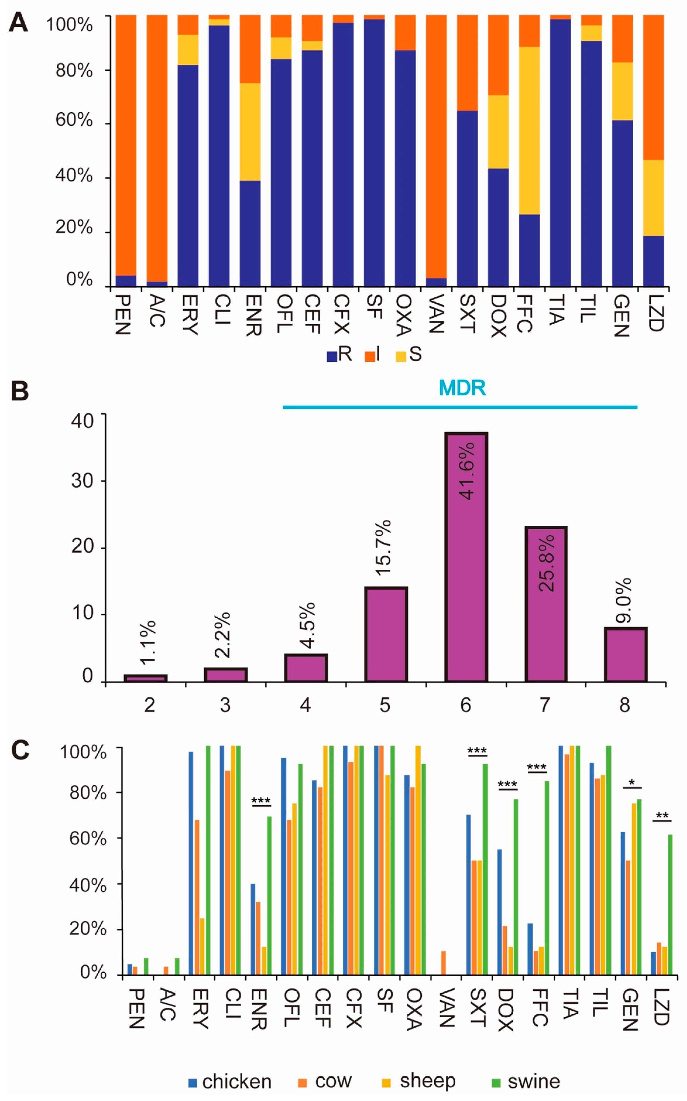
The results of the antibiotic sensitivity test showed that the resistance rate of Enterococcus isolates to clindamycin, cefoxitin, sulfisoxazole and tamoxifen was over 95%, followed by ceftiofur 87.6%, benzoxiline 87.6%, ofloxacin 84.3%, erythromycin 82.0%, etc., and the rate of vancomycin was more sensitive to vancomycin with a resistance rate of 3.4% (Figure 2A, Table 1).
Resistance analysis for different species showed that the overall resistance level was the highest for Enterococcus of porcine origin, followed by chicken, sheep and cattle origin in descending order. We found that the resistance to linezolid was significantly higher in swine-derived Enterococcus than in other animal sources, ranging from 10% to 14.2% in other animal sources, whereas the resistance of swine-derived Enterococcus to linezolid reached 61.5% (Figure 2C). The resistance phenotypes of Enterococcus of porcine origin and Enterococcus of chicken origin were also closer to each other. The number of multi-resistant strains was 86, accounting for 96.6% of the total, of which 7 strains were resistant to all 8 classes of antibiotics (Figure 2B, Table S2).

2.3. Results of Characteristic Genes

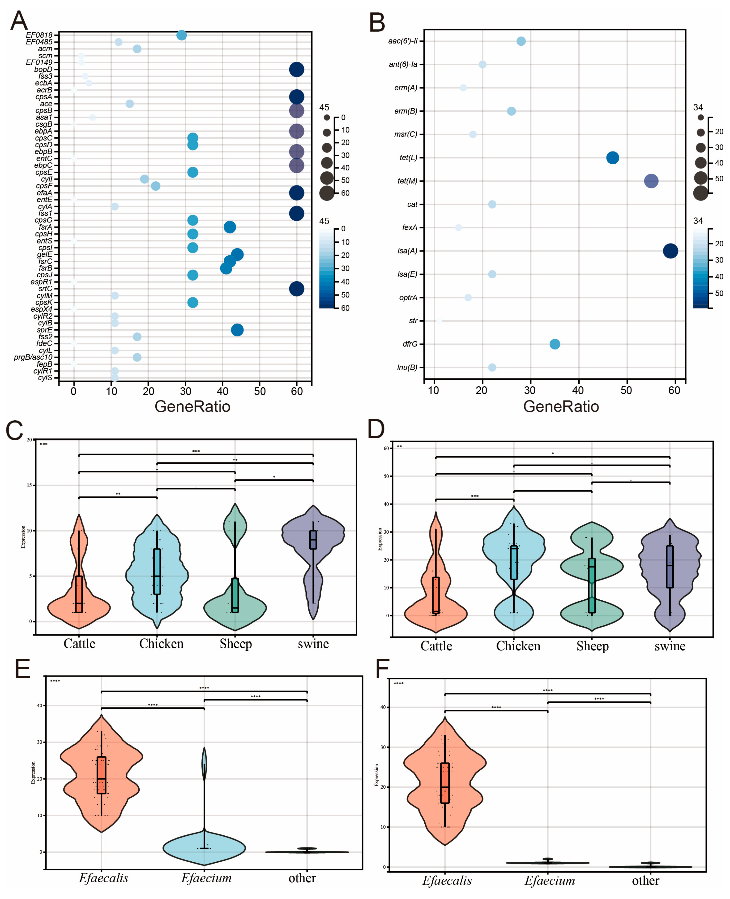
The virulence genes showed that the bacterial hair manipulator genes ebpA, ebpB and ebpC; the fsrA, fsrB and fsrC genes of the virulence regulator fsr system are the most important virulence genes; the polymorphisms and colonization/invasion virulence genes srtC and biofilm-formation-associated virulence cpsA were found to be much more prevalent than those of other virulence genes in Enterococcus (Figure 3A). Five strains carried the plasmid-transferred virulence gene asa1, which mediates the production of aggregates and plays a key role in the adhesion of Enterococcus to renal and endocardial cells, as well as in the invasion of Enterococcus [18].
Of all the ARGs analyzed, 27 ARG types were detected in this study, and among these 27 ARG types, we collated the top16 ARGs (Table S3). these included aminoglycoside resistance genes aph(3′)-III, aac(6′)-II, ant(6)-Ia, macrolide resistance genes erm(A), erm(B), msr(C), tetracycline resistance genes tet(L), tet(M), chloramphenicol resistance genes cat, fexA, lincomycin resistance genes lsa(A), lsa(E), linezolid resistance genes optrA, streptomycin resistance genes str, methicillin resistance genes dfrG, and lincomycin resistance genes lun(B) (Figure 3B). Surprisingly, 17 isolates carrying the oprtA gene were found in Ningxia. Linezolid is banned for animal use in China, but the rates of linezolid resistance and optrA carriage in Enterococcus from pigs in this study were much higher than normal values. Significant associations were observed between optrA and linezolid resistance (p < 0.05), and between erm(B) and erythromycin resistance (p < 0.01).
The analysis according to the source of different species showed that the carrying rate of resistance genes of Enterococcus pigs was significantly higher than that of other species (Figure 3C), and there was no significant difference in the number of virulence genes carried among different species (Figure 3D). The results of antibiotic resistance genes were consistent with the results of combined antibiotic susceptibility, suggesting that porcine Enterococcus is the main reservoir of antibiotic resistance genes, and we should pay more attention to the antibiotic resistance of porcine Enterococcus. The results showed that the number of antibiotic resistance genes (Figure 3E) and virulence genes (Figure 3F) carried by E. faecalis was significantly higher than that of other Enterococcus species. E. faecalis showed the characteristics of high virulence and high antibiotic resistance in this study, which should be paid more attention in the cultivation process.

2.4. Results of MLST Sequence Typing

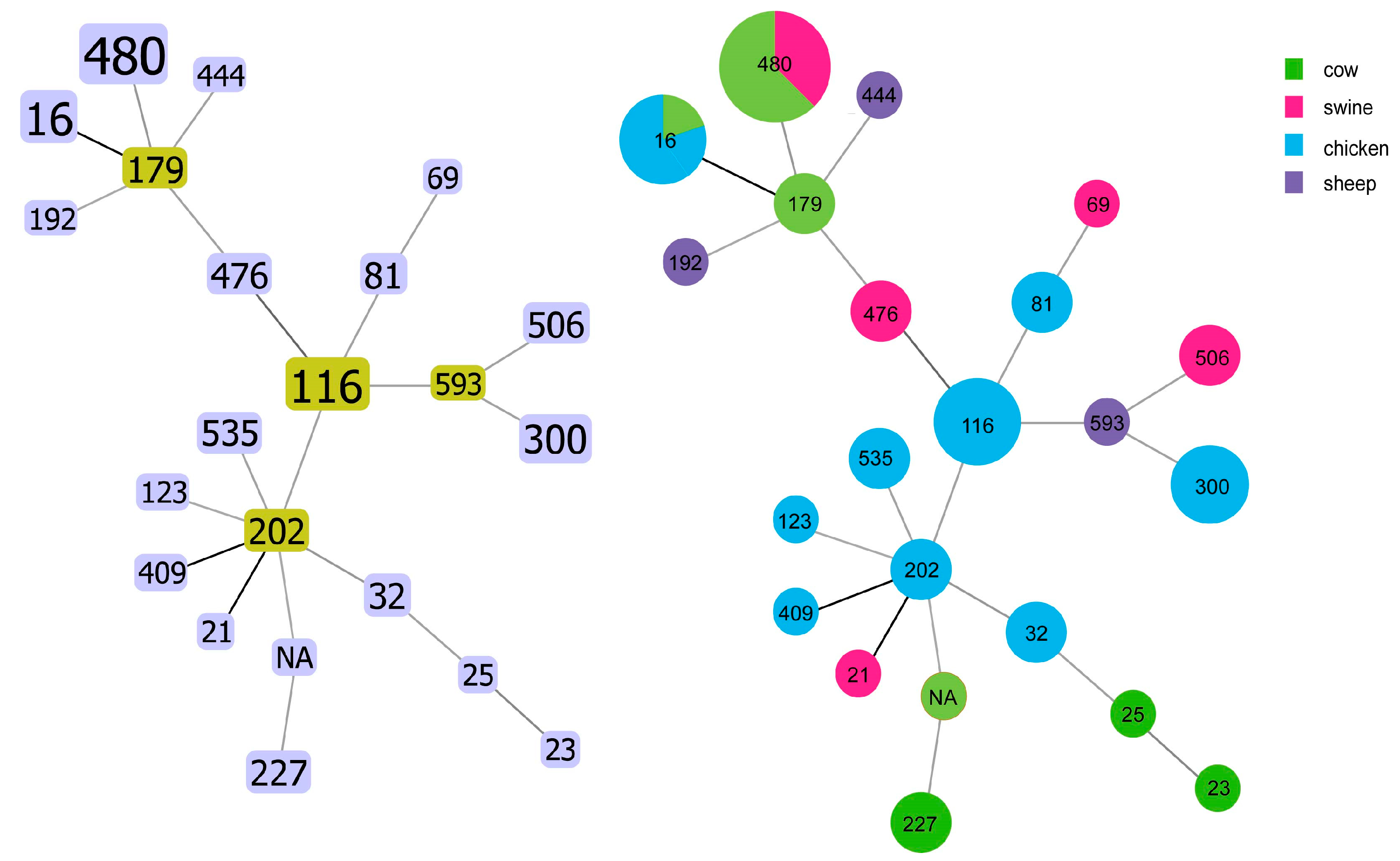
Among the five ST types (i.e., ST506, ST476, ST593, ST179, and ST535), all isolates possessed the optrA-encoding gene (Figure 4). A total of 29 strains (35.6%) had combinations of known ST alleles. We further evaluated the clonality and branching of all 89 Enterococcus isolates. Our results suggest that resistance genes such as optrA are spread horizontally through Enterococcus in large numbers.

2.5. Phylogenetic Tree of SNP and Characteristics of Antimicrobial Resistance Genes and Plasmids Carried by Enterococcus

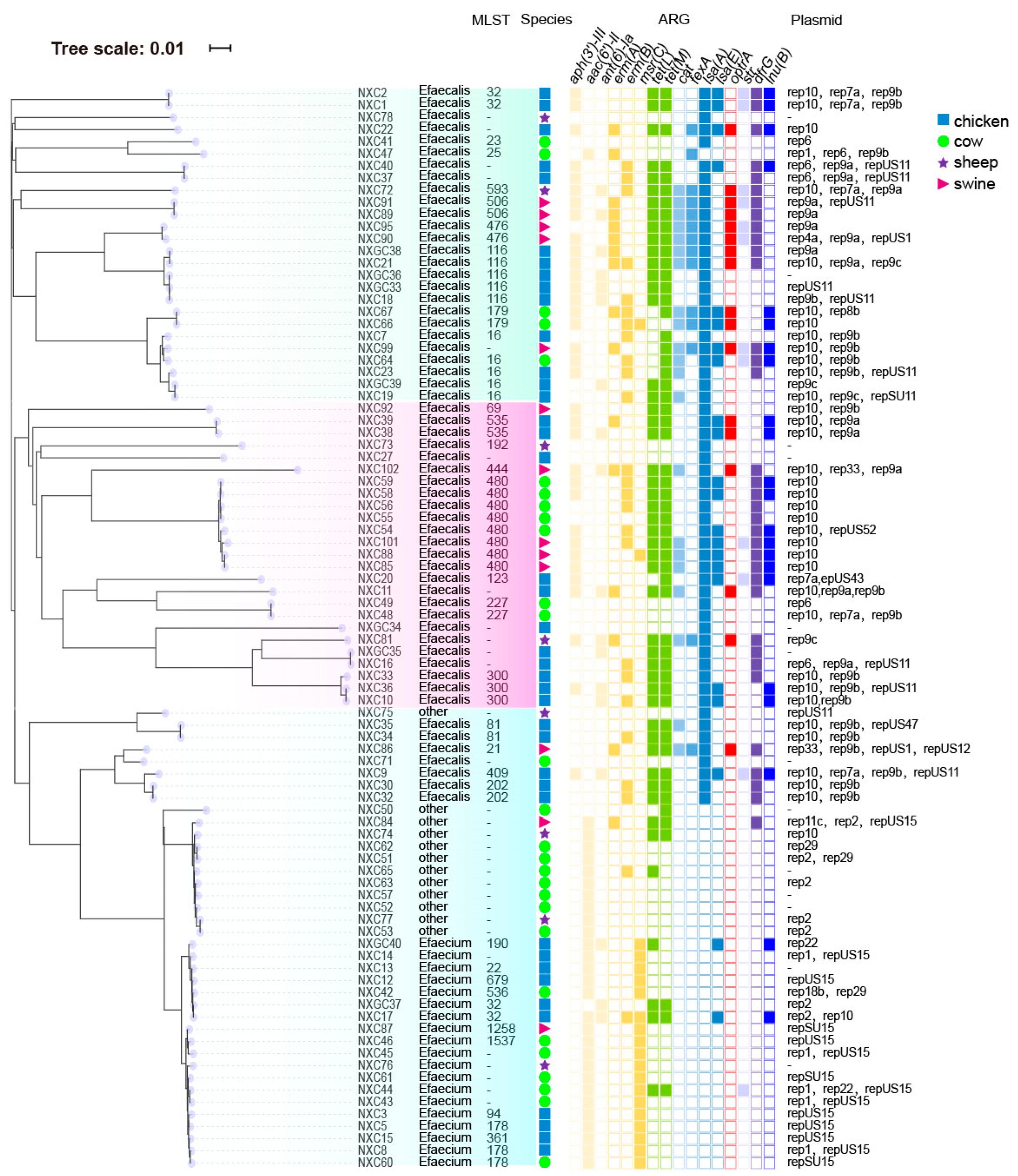
SNP phylogenetic analysis of 89 Enterococcus strains showed that the whole phylogenetic tree was divided into three branches, and most isolates from the same species clustered together. Species was the main reason for the clustering of Enterococcus phylogenetic tree. For example, the difference between NXC77 and NXC53 SNPS was less than 10, which suggested the occurrence of cross-species transmission events in Enterococcus (Figure 5). The 89 Enterococcus isolates were divided into three branches. Clade C1 was dominated by ST16 and ST116 and mainly carried plasmids rep10, rep9a and rep9b. Clade C2 was dominated by ST480 and mainly carried plasmid rep10. Both clades carried a large number of antibiotic resistance genes. Clade C3 was dominated by E. faecium and mainly carried plasmids repUS15 and rep2. The number of antibiotic resistance genes in clade C3 was significantly less than that in clade C1 and C2.

2.6. Co-Existence of Plasmids with Antibiotic-Resistant Genes

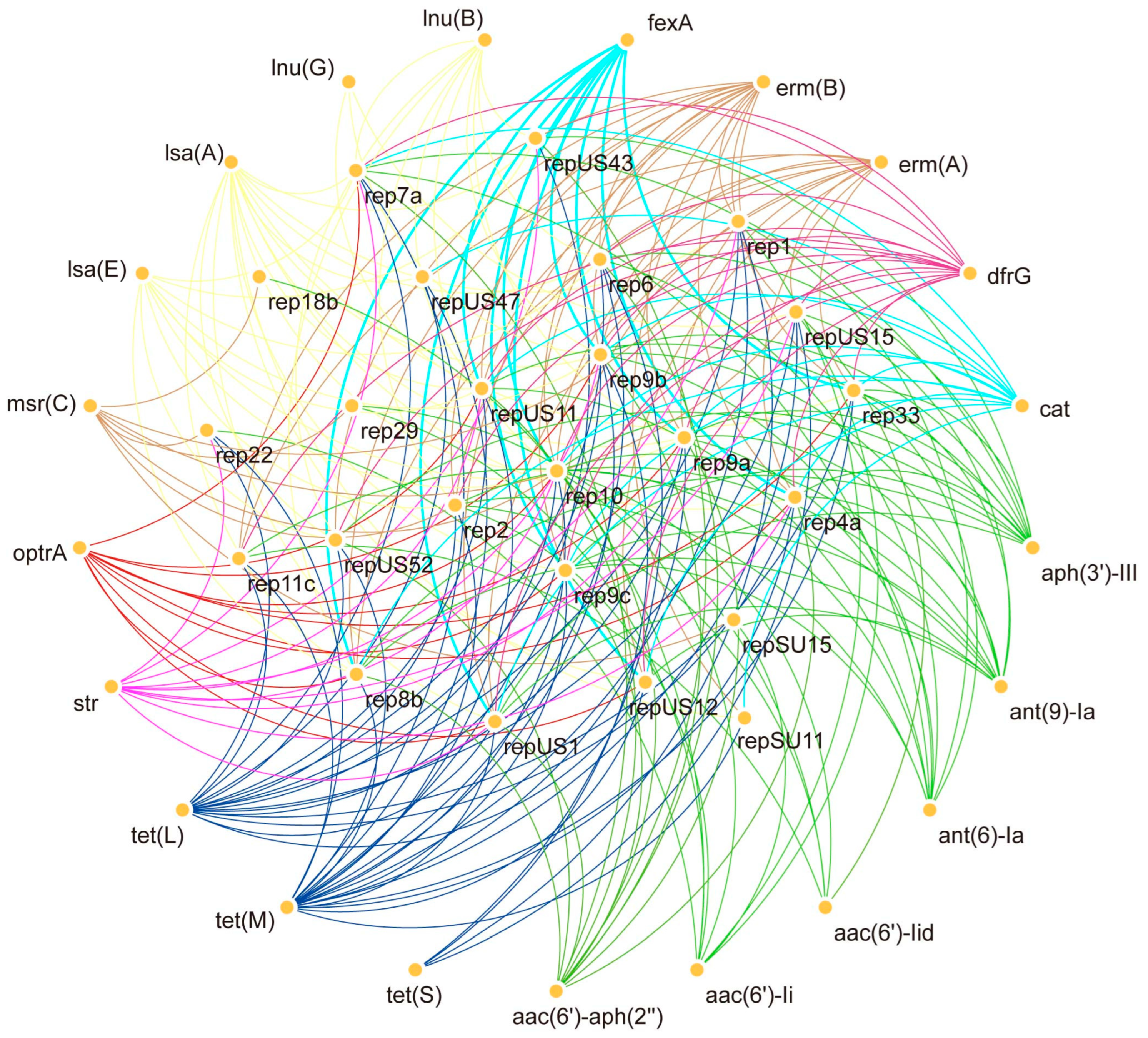
In 89 Enterococcus strains we observed that plasmid rep10 could coexist with all resistance genes, while rep9a, rep9b and rep9c, could coexist with a large number of resistance genes but not with msr(c), aac(6′)-li and aac(6′)-lid. At the same time, we observed that strains carrying the optrA resistance gene contained the plasmids rep9 (Figure 6). This also suggests to us the importance of rep9 for optrA transmission.

3. Discussion

The origin of antibiotic resistance is still unclear, and studies have reported the emergence of methicillin-resistant Staphylococcus aureus well before methicillin began to be used in the clinic. This suggests that the emergence of bacterial resistance is not necessarily related to the high use of antibiotics. The initial generation of resistance genes does not serve the function of making bacteria resistant, but rather functions such as metabolism, signaling and detoxification [19,20]. The resistance mechanism is an important driver of microbial evolution that has continued from ancient times to the present [1]. However, when humans apply new antibiotics as clinical drugs, this move puts unprecedented selection pressure on microbes. Understanding and controlling the spread and development of bacterial resistance is therefore one of the most urgent tasks for human health management in the 21st century.
Enterococcus are important key indicator bacteria for human and veterinary antibiotic resistance surveillance systems [5]. This study revealed the antibiotic resistance profile of Enterococcus in Ningxia region, and the results showed that Enterococcus exhibited a high level of antimicrobial resistance in Ningxia region, in which Enterococcus resistance to clindamycin, cefoxitin, sulfisoxazole, and tamifluorfen was more than 95%. It is noteworthy that most of the strains in this study (86/89, 96.6%) were resistant to antibiotics of class 3 and above, which implies that it is even more difficult to treat the diseases caused by them. There were some differences in strain resistance between species, with the resistance rates of pig- and chicken-origin Enterococcus being significantly higher than those of cattle and sheep, which may be related to farming patterns and medication habits. We found that the resistance of Enterococcus of porcine origin to linezolid was significantly higher than that of other animal sources, this is in line with the results of the antibiotic-resistant gene. Linezolid is banned for use in animals in China, but 61.5% of Enterococcus isolated from pigs carried the linezolid resistance gene optrA. This suggests that optrA is more prone to spread among Enterococcus from pigs. Enterococcus isolated from pigs is an important reservoir of antibiotic resistance genes. As an important economic animal, pigs may become an important reservoir of antibiotic resistance genes in the breeding process, and then spread to humans and other organisms through the food chain or other ways.
In this study, optrA was unevenly distributed among Enterococcus species and plasmid families, with the entire optrA present in E. faecalis and identified on rep9 variants of the RepA_N plasmid [21], which are narrow host range plasmids thought to be specific for E. faecalis. rep9 family plasmids contain several characteristic pheromone responsive plasmids, such as pAD1. Coupling systems based on sex pheromones have been described as very efficient transfer vectors that can facilitate ARGs transfer. All these observations may explain the high detection rate of rep9 replicon in optrA-positive E. faecalis in the present study, so further investigation of cross-host transmission of this plasmid type is warranted. Previous studies have shown that pheromone-responsive conjugative plasmids, particularly those in the rep9 family, play a central role in mediating the horizontal transfer of optrA among Enterococcus spp. Such plasmids contain mobilization genes and sex pheromone systems that enable efficient intra- and interspecies gene transfer, especially under selective pressure from phenicols or macrolides. These mechanisms likely explain the high frequency of optrA-rep9 associations observed in our isolates and their potential contribution to ARG dissemination in livestock environments [22,23].
The high resistance rate in the optrA-positive group in this study was observed in multiple types of farm samples in China. Our results indicated not only a high resistance rate but also a large number of ARGs in the optrA-positive Enterococcus, indicating that optrA-positive Enterococcus pose a higher risk to public health [24,25]. Previous studies have shown that increased daily pork consumption and hospitalization are associated with increased risk of optrA carriage in these healthy populations [26]. Despite the heterogeneity in population structure of optrA-positive E. faecalis observed in the community, a high degree of homology was observed among optrA-positive E. faecalis isolates from the community and other sources, including clinics, pigs, chickens, pets, and the environment, suggesting that multiple sources, including animals and clinics, contribute to human optrA carriage. The results of this study suggest that the mechanisms for generating Enterococcus resistance may be complex, with multiple antibiotic metabolism pathways possible on the basis of multiple resistance phenotypes [27,28,29,30,31].
OptrA-positive Enterococcus bacteria have been repeatedly reported in clinical, food and livestock samples from Asia, Europe and Latin America. However, the detection rates and plasmid profiles vary across different regions. Comparative data from other regions show variable prevalence of optrA-positive Enterococcus isolates, ranging from 2 to 8% in Europe to over 15% in some Asian and South American livestock populations. The high detection rate observed in Ningxia is consistent with regions where florfenicol and macrolide use in animal production remains common, suggesting that antibiotic usage patterns and farm management practices are key factors influencing ARG dissemination worldwide [32,33].
The phylogenetic tree of 89 strains of Enterococcus can be divided into three branches. The composition of different branch strains is mainly determined by the species, and the strains within the same species have a closer genetic relationship, which is similar to the results of previous studies [34]. Some strains have SNP differences of less than 10, indicating that there have been cross-species transmission events in the Ningxia region. Plasmid-mediated horizontal gene transfer can significantly increase the incidence of ARGs [35]. The risk of possible infection by multidrug-resistant Enterococcus from poultry is primarily associated with E. faecalis. The extent of this risk remains unknown and appears to be restricted to specific lineages, such as ST16. In branch C1, ST16 is the dominant ST type, which has been reported as an important pathogenic type of Enterococcus and can carry a large number of virulence genes. At the same time, it is more likely to form strong or moderate biofilms than other ST types. In previous reports, ST16 is the most common type of Enterococcus causing hospital-acquired and linezolid-resistant fecal Enterococcus [28].
A total of 52 virulence genes were detected in Ningxia Enterococcus. Among them, we found that the hyphal manipulator genes ebpA, ebpB, and ebpC; the genes fsrA, fsrB, and fsrC of the virulence regulator fsr system; and the genes srtC, a polymorphism associated with colonization/invasion virulence, and cpsA, a biofilm-formation-associated virulence gene, were far more prlent than the other virulence genes [36,37]. These data also emphasize the importance of Enterococcus as a repository of virulence genes and the potential for Enterococcus to harm humans through the food chain [38]. Combined with the resistance gene results, we found that swine Enterococcus carried more virulence and resistance genes compared to other Enterococcus. This also suggests that we should be more rational in the use of drugs in swine farming.
However, there are still some limitations in this study, such as the small sample size of sequencing, which may lead to errors in the results. However, the results still have important reference value. Meanwhile, the isolates carried a large number of antibiotic resistance genes and virulence genes, which are seriously harmful to the farming industry. It also suggests the urgency of epidemiologic research and surveillance of Enterococcus to prevent future public health risks.
Our findings highlight that swine-derived Enterococcus acts as an important reservoir of antimicrobial resistance genes in livestock ecosystems. To mitigate dissemination, we recommend: (1) promoting prudent antibiotic use policies in veterinary medicine; (2) implementing molecular surveillance systems across the farm–environment–clinical interface; (3) improving manure management and biosecurity practices; and (4) integrating antimicrobial-use data into national risk assessment models. Future research should focus on plasmid-mediated transmission dynamics, environmental persistence, and One Health–based intervention strategies.

4. Materials and Methods

4.1. Source of Samples

A total of 540 strains of E. faecalis, 214 strains of E. faecium, and other Enterococcus 754 strains were isolated and identified from 25 large-scale farms in various municipalities of Ningxia, China, from 2019 to 2023, using the Merieux Company of France Lyon VITEK 2 COMPACT Fully Automated Microbial Identification System. In total, 89 strains of Enterococcus were randomly selected, of which 40 strains were of chicken origin, 28 strains were of bovine origin, 8 strains were of sheep origin, and 13 strains were of porcine origin. Isolation and identification were performed following ISO 6888-1:2018 and ISO 21528-2:2017 guidelines for Enterococcus detection using bile esculin agar and VITEK 2 Compact identification [39].

4.2. Antimicrobial Susceptibility Testing

Enterococcus antimicrobial susceptibility testing, using the Clinical and Laboratory Standards Institute (CLSI) recommended broth microdilution method as the reference to determine the minimum inhibitory concentration [40], was performed on 89 isolates, including 18 antibiotics: Penicillin (P), Amoxicillin/clavulanic acid (A/C), Clindamycin (CLI), Cefoxitin, Cefotiofur (CEF), Erythromycin (EM), Sulfamethoxazole, Trimethoprim/sulfamethoxazole (SXT), Telmicin (TIL), Enrofloxacin (ENR), Ofloxacin (OFL), Vancomycin (VAN), doxycycline (DOX), Gentamicin, Tymiocin (TIA), Florfenicol (FFC), Linezolid (LZD), oxacillin (OXA). Staphylococcus aureus ATCC 29213 and E. faecalis ATCC 29212 were used as quality control strains. For antibiotics not included in the CLSI guidelines, such as enrofloxacin and florfenicol, interpretation criteria were based on published veterinary studies to reflect their practical importance in animal husbandry [41]. Data were analyzed using one-way ANOVA followed by Tukey’s HSD test for mean separation at the 0.05 significance level. Different letters above the bars indicate statistically significant differences among groups.

4.3. Whole Genome Sequencing and Results Analysis

To further investigate the genetic characteristics of the 89 Enterococcus strains in this trial, the bacterial genome was extracted using a Genomic DNA extraction kit (OMEGA, Tianjin, China). Short sequences were obtained using NovaSeq 6000 (2 × 150 bp-paired end reads) system from Illumina, San Diego, CA, USA. The quality control of the raw sequencing data was initially checked using FastQC v0.11.9, and then fastp v0.23.2 was used to remove adapters and perform quality trimming (retaining bases with Q ≥ 20 and reads with a length ≥ 50 bp). The average sequencing depth of each sample was ≥30×, and ≥80% of the bases reached Q30. SPAdes 3.15.5 was used to assemble the sequencing data [42]. Subsequent data analysis was performed using ResFinder 4.1 (https://cge.food.dtu.dk/services/ResFinder/, accessed on 18 April 2025) for prediction of acquired AMR genes (90% concordance and 60% coverage), VirulenceFinder (https://cge.food.dtu.dk/services/VirulenceFinder/ accessed on 18 April 2025) for prediction of virulence genes (90% concordance and 60% coverage), PlasmidFinder (https://cge.food.dtu.dk/services/PlasmidFinder/ accessed on 18 April 2025) for identification of plasmid replicons (95% concordance and 60% coverage). MLST types were analyzed using (https://cge.food.dtu.dk/services/MLST/ accessed on 18 April 2025) for MLST types. All of the aforementioned websites were accessed on 1 May 2024. Correlation of resistance genes with plasmids was analyzed using Cytoscape_v3.7.0 software [43,44,45].

4.4. SNP Phylogenetic Analysis

Phylogenetic tree analysis of the 89 Enterococcus producing strains sequenced in this study. The genome was first annotated using Prokka 1.2.0, the core genome alignment was obtained using the Roary program3.11.2, and the maximum likelihood tree was assembled using the FastTree program2.1.10. Visualization and annotation were performed through iTOL (https://itol.embl.de, accessed on 18 April 2025) for the beautification of the evolutionary tree. Finally, the SVG format picture of the evolution tree was imported into the AI software to adjust the format [46,47].

4.5. Statistical Analysis

All statistical analyses were performed using GraphPad Prism 9.0 (GraphPad Software, San Diego, CA, USA). Data were expressed as mean ± standard deviation (SD). Differences in antimicrobial resistance rates among animal species were evaluated using one-way ANOVA, followed by Tukey’s HSD test for mean separation. Statistical significance was determined at p < 0.05.

5. Conclusions

In summary, this study used WGS combined with biological analysis methods to provide accurate information on the antibiotic resistance status and pathogenicity of Enterococcus of multiple animal origins in Ningxia region of China. E. faecalis was the predominant species among animal-derived Enterococcus in Ningxia, exhibiting high resistance to clindamycin, cefoxitin, and sulfamethoxazole. The frequent detection of optrA on rep9-family plasmids in pig isolates highlights swine as an important reservoir of linezolid resistance genes with potential zoonotic risk.

Supplementary Materials

The following supporting information can be downloaded at: https://www.mdpi.com/article/10.3390/antibiotics14111137/s1. Table S1 Isolation of Enterococcus in Ningxia Province, China. Table S2 Antibiotics resistance spectrum of Enterococcus. Table S3 Enterococcus virulence gene antibiotics resistance genes statistics.

Author Contributions

Conceptualization, Q.Y., Y.D. and J.W.; methodology, H.Z., Z.Y., Q.S. and Q.Y.; investigation, S.C. (Shengling Cui), Q.S., W.Z. (Weimin Zhang) and J.W.; resources, Q.Y., T.T. and L.Z.; data curation, H.Z. and L.Z.; writing—original draft preparation, H.Z. and W.Z. (Wen Zhang); writing—review and editing, J.W.; visualization, H.Z., W.Z. (Wen Zhang), Q.Y. and J.W.; supervision, J.W.; funding acquisition, H.Z., S.C. (Shengli Chen), H.H., W.Z. (Wen Zhang) and J.W. All authors have read and agreed to the published version of the manuscript.

Funding

This work is financially supported by the National Natural Science Foundation of China (Grant No. 32473124), the Foundation of State Key Laboratory of Pathogen and Biosecurity of China (Grant No. SKLPBS2449) and the Key Laboratory of Veterinary Etiological Biology, Ministry of Agricultural and Rural Affairs, China (Grant Number XKQ2025003).

Data Availability Statement

The complete genome sequences of 89 Enterococcus isolates have been deposited in the GenBank database under accession number: PRJNA1130870.

Conflicts of Interest

The authors declare no conflicts of interest.

References

  1. Torres, C.; Alonso, C.A.; Ruiz-Ripa, L.; León-Sampedro, R.; Del Campo, R.; Coque, T.M. Antimicrobial Resistance in Enterococcus spp. of animal origin. Microbiol. Spectr. 2018, 6, 185–227. [Google Scholar] [CrossRef]
  2. Farnleitner, A.H.; Ryzinska-Paier, G.; Reischer, G.H.; Burtscher, M.M.; Knetsch, S.; Kirschner, A.K.; Dirnböck, T.; Kuschnig, G.; Mach, R.L.; Sommer, R. Escherichia coli and enterococci are sensitive and reliable indicators for human, livestock and wildlife faecal pollution in alpine mountainous water resources. J. Appl. Microbiol. 2010, 109, 1599–1608. [Google Scholar] [CrossRef]
  3. European Centre for Disease Prevention and Control publishes Annual epidemiological report 2011. Euro Surveill. 2011, 16, 20012.
  4. Radhouani, H.; Igrejas, G.; Carvalho, C.; Pinto, L.; Gonçalves, A.; Lopez, M.; Sargo, R.; Cardoso, L.; Martinho, A.; Rego, V.; et al. Clonal lineages, antibiotic resistance and virulence factors in vancomycin-resistant enterococci isolated from fecal samples of red foxes (Vulpes vulpes). J. Wildl. Dis. 2011, 47, 769–773. [Google Scholar] [CrossRef] [PubMed]
  5. Iweriebor, B.C.; Obi, L.C.; Okoh, A.I. Macrolide, glycopeptide resistance and virulence genes in Enterococcus species isolates from dairy cattle. J. Med. Microbiol. 2016, 65, 641–648. [Google Scholar] [CrossRef]
  6. Iweriebor, B.C.; Obi, L.C.; Okoh, A.I. Virulence and antimicrobial resistance factors of Enterococcus spp. isolated from fecal samples from piggery farms in Eastern Cape, South Africa. BMC Microbiol. 2015, 15, 136. [Google Scholar] [CrossRef]
  7. Todokoro, D.; Suzuki, T.; Kobayakawa, S.; Tomita, H.; Ohashi, Y.; Akiyama, H. Postoperative Enterococcus faecalis endophthalmitis: Virulence factors leading to poor visual outcome. Jpn. J. Ophthalmol. 2017, 61, 408–414. [Google Scholar] [CrossRef]
  8. Buetti, N.; Tabah, A.; Loiodice, A.; Ruckly, S.; Aslan, A.T.; Montrucchio, G.; Cortegiani, A.; Saltoglu, N.; Kayaaslan, B.; Aksoy, F.; et al. Different epidemiology of bloodstream infections in COVID-19 compared to non-COVID-19 critically ill patients: A descriptive analysis of the Eurobact II study. Crit. Care 2022, 26, 319. [Google Scholar] [CrossRef] [PubMed]
  9. Posteraro, B.; De Angelis, G.; Menchinelli, G.; D’Inzeo, T.; Fiori, B.; De Maio, F.; Cortazzo, V.; Sanguinetti, M.; Spanu, T. Risk Factors for Mortality in Adult COVID-19 Patients Who Develop Bloodstream Infections Mostly Caused by Antimicrobial-Resistant Organisms: Analysis at a Large Teaching Hospital in Italy. J. Clin. Med. 2021, 10, 1752. [Google Scholar] [CrossRef]
  10. Cultrera, R.; Barozzi, A.; Libanore, M.; Marangoni, E.; Pora, R.; Quarta, B.; Spadaro, S.; Ragazzi, R.; Marra, A.; Segala, D.; et al. Co-Infections in Critically Ill Patients with or without COVID-19: A Comparison of Clinical Microbial Culture Findings. Int. J. Environ. Res. Public Health 2021, 18, 4358. [Google Scholar] [CrossRef]
  11. Kojima, A.; Morioka, A.; Kijima, M.; Ishihara, K.; Asai, T.; Fujisawa, T.; Tamura, Y.; Takahashi, T. Classification and antimicrobial susceptibilities of Enterococcus species isolated from apparently healthy food-producing animals in Japan. Zoonoses Public Health 2010, 57, 137–141. [Google Scholar] [CrossRef]
  12. Xu, X.; Lin, D.; Yan, G.; Ye, X.; Wu, S.; Guo, Y.; Zhu, D.; Hu, F.; Zhang, Y.; Wang, F.; et al. vanM, a new glycopeptide resistance gene cluster found in Enterococcus faecium. Antimicrob. Agents Chemother. 2010, 54, 4643–4647. [Google Scholar] [CrossRef] [PubMed]
  13. Cattoir, V.; Leclercq, R. Twenty-five years of shared life with vancomycin-resistant enterococci: Is it time to divorce? J. Antimicrob. Chemother. 2013, 68, 731–742. [Google Scholar] [CrossRef]
  14. Ahmed, M.O.; Baptiste, K.E. Vancomycin-Resistant Enterococci: A Review of Antimicrobial Resistance Mechanisms and Perspectives of Human and Animal Health. Microb. Drug Resist. 2018, 24, 590–606. [Google Scholar] [CrossRef]
  15. Hegstad, K.; Mikalsen, T.; Coque, T.M.; Werner, G.; Sundsfjord, A. Mobile genetic elements and their contribution to the emergence of antimicrobial resistant Enterococcus faecalis and Enterococcus faecium. Clin. Microbiol. Infect. 2010, 16, 541–554. [Google Scholar]
  16. Gordoncillo, M.J.; Donabedian, S.; Bartlett, P.C.; Perri, M.; Zervos, M.; Kirkwood, R.; Febvay, C. Isolation and molecular characterization of vancomycin-resistant Enterococcus faecium from swine in Michigan, USA. Zoonoses Public Health 2013, 60, 319–326. [Google Scholar] [CrossRef]
  17. Liu, Y.; Liu, K.; Lai, J.; Wu, C.; Shen, J.; Wang, Y. Prevalence and antimicrobial resistance of Enterococcus species of food animal origin from Beijing and Shandong Province, China. J. Appl. Microbiol. 2013, 114, 555–563. [Google Scholar]
  18. Kiruthiga, A.; Padmavathy, K.; Shabana, P.; Naveenkumar, V.; Gnanadesikan, S.; Malaiyan, J. Improved detection of esp, hyl, asa1, gelE, cylA virulence genes among clinical isolates of Enterococci. BMC Res. Notes 2020, 13, 170. [Google Scholar] [CrossRef]
  19. Mikalsen, T.; Pedersen, T.; Willems, R.; Coque, T.M.; Werner, G.; Sadowy, E.; van Schaik, W.; Jensen, L.B.; Sundsfjord, A.; Hegstad, K. Investigating the mobilome in clinically important lineages of Enterococcus faecium and Enterococcus faecalis. BMC Genom. 2015, 16, 282. [Google Scholar]
  20. Zou, J.; Tang, Z.; Yan, J.; Liu, H.; Chen, Y.; Zhang, D.; Zhao, J.; Tang, Y.; Zhang, J.; Xia, Y. Dissemination of Linezolid Resistance Through Sex Pheromone Plasmid Transfer in Enterococcus faecalis. Front. Microbiol. 2020, 11, 1185. [Google Scholar] [CrossRef] [PubMed]
  21. Shang, Y.; Li, D.; Shan, X.; Schwarz, S.; Zhang, S.M.; Chen, Y.X.; Ouyang, W.; Du, X.D. Analysis of two pheromone-responsive conjugative multiresistance plasmids carrying the novel mobile optrA locus from Enterococcus faecalis. Infect. Drug Resist. 2019, 12, 2355–2362. [Google Scholar] [CrossRef]
  22. Dönhöfer, A.; Franckenberg, S.; Wickles, S.; Berninghausen, O.; Beckmann, R.; Wilson, D.N. Structural basis for TetM-mediated tetracycline resistance. Proc. Natl. Acad. Sci. USA 2012, 109, 16900–16905. [Google Scholar]
  23. Parent, R.; Roy, P.H. The chloramphenicol acetyltransferase gene of Tn2424: A new breed of cat. J. Bacteriol. 1992, 174, 2891–2897. [Google Scholar] [CrossRef]
  24. Morroni, G.; Brenciani, A.; Antonelli, A.; D’Andrea, M.M.; Di Pilato, V.; Fioriti, S.; Mingoia, M.; Vignaroli, C.; Cirioni, O.; Biavasco, F.; et al. Characterization of a Multiresistance Plasmid Carrying the optrA and cfr Resistance Genes From an Enterococcus faecium Clinical Isolate. Front. Microbiol. 2018, 9, 2189. [Google Scholar] [CrossRef] [PubMed]
  25. Cavaco, L.M.; Bernal, J.F.; Zankari, E.; Léon, M.; Hendriksen, R.S.; Perez-Gutierrez, E.; Aarestrup, F.M.; Donado-Godoy, P. Detection of linezolid resistance due to the optrA gene in Enterococcus faecalis from poultry meat from the American continent (Colombia). J. Antimicrob. Chemother. 2017, 72, 678–683. [Google Scholar]
  26. Mendes, R.E.; Deshpande, L.M.; Jones, R.N. Linezolid update: Stable in vitro activity following more than a decade of clinical use and summary of associated resistance mechanisms. Drug Resist. Updates 2014, 17, 1–12. [Google Scholar] [CrossRef]
  27. Wang, Y.; Lv, Y.; Cai, J.; Schwarz, S.; Cui, L.; Hu, Z.; Zhang, R.; Li, J.; Zhao, Q.; He, T.; et al. A novel gene, optrA, that confers transferable resistance to oxazolidinones and phenicols and its presence in Enterococcus faecalis and Enterococcus faecium of human and animal origin. J. Antimicrob. Chemother. 2015, 70, 2182–2190. [Google Scholar] [CrossRef]
  28. Nüesch-Inderbinen, M.; Raschle, S.; Stevens, M.J.A.; Schmitt, K.; Stephan, R. Linezolid-resistant Enterococcus faecalis ST16 harbouring optrA on a Tn6674-like element isolated from surface water. J. Glob. Antimicrob. Resist. 2021, 25, 89–92. [Google Scholar] [CrossRef] [PubMed]
  29. Khan, A.A.; Nawaz, M.S.; Khan, S.A.; Steele, R. Detection and characterization of erythromycin-resistant methylase genes in Gram-positive bacteria isolated from poultry litter. Appl. Microbiol. Biotechnol. 2002, 59, 377–381. [Google Scholar] [PubMed]
  30. Ben Said, L.; Dziri, R.; Sassi, N.; Lozano, C.; Ben Slama, K.; Ouzari, I.; Torres, C.; Klibi, N. Species distribution, antibiotic resistance and virulence traits in canine and feline enterococci in Tunisia. Acta Vet. Hung. 2017, 65, 173–184. [Google Scholar] [CrossRef]
  31. Almeida, L.M.; Lebreton, F.; Gaca, A.; Bispo, P.M.; Saavedra, J.T.; Calumby, R.N.; Grillo, L.M.; Nascimento, T.G.; Filsner, P.H.; Moreno, A.M.; et al. Transferable Resistance Gene optrA in Enterococcus faecalis from Swine in Brazil. Antimicrob. Agents Chemother. 2020, 64, 10–1128. [Google Scholar] [CrossRef]
  32. Barlow, R.S.; McMillan, K.E.; Duffy, L.L.; Fegan, N.; Jordan, D.; Mellor, G.E. Antimicrobial resistance status of Enterococcus from Australian cattle populations at slaughter. PLoS ONE 2017, 12, e0177728. [Google Scholar] [CrossRef] [PubMed]
  33. Nascimento, C.A.; Tanomaru-Filho, M.; Faria-Junior, N.B.; Faria, G.; Guerreiro-Tanomaru, J.M. Antimicrobial activity of root canal irrigants associated with cetrimide against biofilm and planktonic Enterococcus faecalis. J. Contemp. Dent. Pract. 2014, 15, 603–607. [Google Scholar] [CrossRef]
  34. Osman, M.; Yassine, I.; Hassan, J.; Ericson, J.E.; Schiff, S.J.; Ghorbani Tajani, A.; Bieder, B.; Hamza, M.; Kassem, I.I. Critical genomic insights into vancomycin-resistant Enterococcus faecium in Lebanon. Microbiol. Spectr. 2025, 13, e0017125. [Google Scholar] [CrossRef]
  35. Antimicrobial Resistance Collaborators. Global burden of bacterial antimicrobial resistance in 2019: A systematic analysis. Lancet 2022, 399, 629–655.
  36. Kim, H.J.; Youn, H.Y.; Kang, H.J.; Moon, J.S.; Jang, Y.S.; Song, K.Y.; Seo, K.H. Prevalence and Virulence Characteristics of Enterococcus faecalis and Enterococcus faecium in Bovine Mastitis Milk Compared to Bovine Normal Raw Milk in South Korea. Animals 2022, 12, 1407. [Google Scholar] [CrossRef]
  37. Stępień-Pyśniak, D.; Bertelloni, F.; Dec, M.; Cagnoli, G.; Pietras-Ożga, D.; Urban-Chmiel, R.; Ebani, V.V. Characterization and Comparison of Enterococcus spp. Isolates from Feces of Healthy Dogs and Urine of Dogs with UTIs. Animals 2021, 11, 2845. [Google Scholar] [CrossRef] [PubMed]
  38. Molale, L.G.; Bezuidenhout, C.C. Virulence determinants and production of extracellular enzymes in Enterococcus spp. from surface water sources. Water Sci. Technol. 2016, 73, 1817–1824. [Google Scholar] [CrossRef]
  39. Ogura, A.; Iwasaki, M.; Ogihara, H.; Teramura, H. Evaluation of the Novel Dry Sheet Culture Method for the Enumeration of Enterobacteriaceae. Biocontrol. Sci. 2018, 23, 235–240. [Google Scholar] [CrossRef]
  40. Boulianne, M.; Arsenault, J.; Daignault, D.; Archambault, M.; Letellier, A.; Dutil, L. Drug use and antimicrobial resistance among Escherichia coli and Enterococcus spp. isolates from chicken and turkey flocks slaughtered in Quebec, Canada. Can. J. Vet. Res. 2016, 80, 49–59. [Google Scholar]
  41. Humphries, R.; Bobenchik, A.M.; Hindler, J.A.; Schuetz, A.N. Overview of Changes to the Clinical and Laboratory Standards In-stitute Performance Standards for Antimicrobial Susceptibility Testing, M100, 31st Edition. J. Clin. Microbiol. 2021, 59, e0021321. [Google Scholar] [CrossRef] [PubMed]
  42. Stępień-Pyśniak, D.; Hauschild, T.; Dec, M.; Marek, A.; Urban-Chmiel, R.; Kosikowska, U. Phenotypic and genotypic characterization of Enterococcus spp. from yolk sac infections in broiler chicks with a focus on virulence factors. Poult. Sci. 2021, 100, 100985. [Google Scholar] [CrossRef] [PubMed]
  43. Margos, G.; Binder, K.; Dzaferovic, E.; Hizo-Teufel, C.; Sing, A.; Wildner, M.; Fingerle, V.; Jolley, K. PubMLST.org—The new home for the Borrelia MLSA database. Ticks Tick Borne Dis. 2015, 6, 869–871. [Google Scholar] [CrossRef]
  44. Zhong, Z.; Zhang, W.; Song, Y.; Liu, W.; Xu, H.; Xi, X.; Menghe, B.; Zhang, H.; Sun, Z. Comparative genomic analysis of the genus Enterococcus. Microbiol. Res. 2017, 196, 95–105. [Google Scholar] [CrossRef]
  45. Dong, N.; Lin, D.; Zhang, R.; Chan, E.W.; Chen, S. Carriage of blaKPC-2 by a virulence plasmid in hypervirulent Klebsiella pneumoniae. J. Antimicrob. Chemother. 2018, 73, 3317–3321. [Google Scholar] [CrossRef]
  46. Tyson, G.H.; Sabo, J.L.; Rice-Trujillo, C.; Hernandez, J.; McDermott, P.F. Whole-genome sequencing based characterization of antimicrobial resistance in Enterococcus. Pathog. Dis. 2018, 76, fty018. [Google Scholar] [CrossRef]
  47. Agius, J.E.; Phalen, D.N.; Rose, K.; Eden, J.S. Genomic Insights into the Pathogenicity of a Novel Biofilm-Forming Enterococcus sp. Bact. (Enterococcus lacertideformus) Identified Reptiles. Front. Microbiol. 2021, 12, 635208. [Google Scholar]
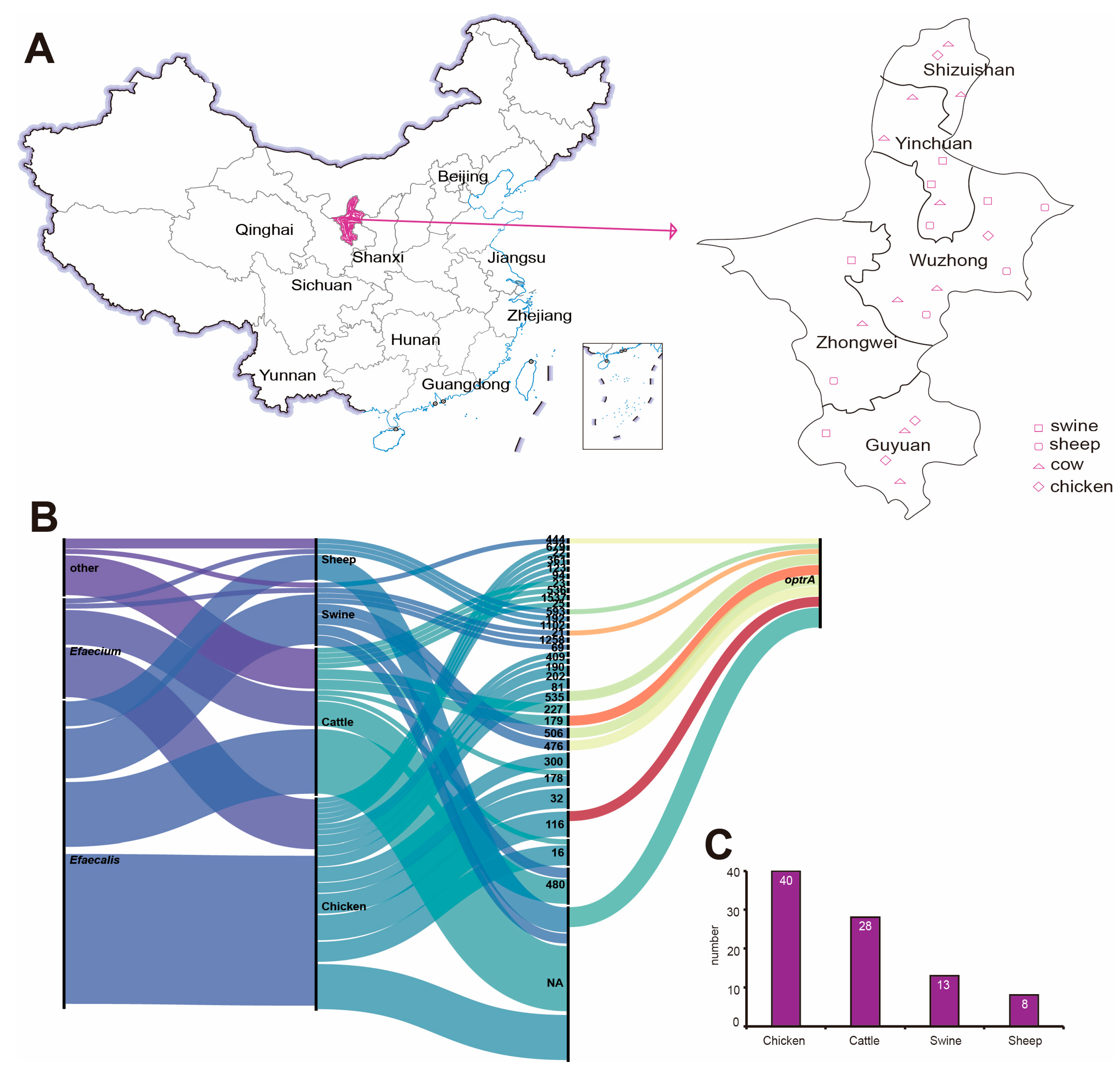
Figure 1. Enterococcus sampling map and basic information statistics. (A) Enterococcus sampling map. (B) Sankey map of genetic information of Enterococcus. (C) Enterococcus host statistics histogram.
Antibiotics 14 01137 g001
Figure 2. Antibiotic susceptibility results. Figure (A) presents the overall antibiotic resistance results of Enterococcus. S indicates sensitive, I indicates moderately sensitive, and R indicates resistant. Figure (B) shows multi drug-resistant strains. Figure (C) presents the antibiotic resistance situations of different types of Enterococcus (* p ≤ 0.05, ** p ≤ 0.01, *** p ≤ 0.001. p < 0.05, ANOVA followed by Tukey’s HSD test).
Antibiotics 14 01137 g002
Figure 3. Genetic characteristic information. (A) Bubble plot showing the carriage status of virulence genes. (B) Bubble plot depicting the carriage status of drug resistance genes. (C) Diagram of the differences in the carriage of drug resistance genes among different species. (D) Diagram of the differences in the carriage of virulence genes among different species. (E) Diagram of the differences in the carriage of drug resistance genes among different taxa. (F) Diagram of the differences in the carriage of virulence genes among different taxa. (* p ≤ 0.05, ** p ≤ 0.01, *** p ≤ 0.001, **** p ≤ 0.0001).
Antibiotics 14 01137 g003
Figure 4. E. faecalis MLST phylogenetic tree.
Antibiotics 14 01137 g004
Figure 5. Phylogenetic tree of Enterococcus SNPS. From left to right, phylogenetic tree, MLST typing, species, resistance gene, and carrying plasmid. The whole thing is divided into three branches: C1 in green, C2 in pink and C3 in blue.
Antibiotics 14 01137 g005
Figure 6. Bar graph of Coexistence network of plasmids and antibiotics resistance genes carried by Enterococcus.
Antibiotics 14 01137 g006
Table 1. Overall Antibiotics susceptibility results of Enterococcus.
Antibiotic Type DrugNameOverall Sensitivity
RIS
PenicillinBenzylpenicillin87.6%012.4%
Penicillin4.5%095.5%
Augmentin2.2%097.8%
ClindamycinClindamycin96.6%2.2%1.1%
QuinolonesEnrofloxacin39.3%36.0%24.7%
Ofloxacin84.3%7.9%7.9%
beta-lactamsCeftiofur87.6%3.4%9.0%
Cefoxitin97.8%02.2%
SulfonamidesSulfamethoxazole98.9%01.1%
GlycopeptidesVancomycin3.4%096.6%
SulfonamidesCotrimoxazole65.2%036.0%
TetracyclineDoxycycline43.8%27.0%29.2%
Florfenicol27.0%61.8%11.2%
MacrolidesErythromycin82.0%11.2%6.7%
Taimiocin98.9%01.1%
Temicosin91.0%5.6%3.4%
AminoglycosidesGentamycin61.8%21.3%16.9%
OxazolidinonesLinezolid19.1%28.1%52.8%
Note: S, sensitive; I, moderately sensitive; R, drug resistance.
Disclaimer/Publisher’s Note: The statements, opinions and data contained in all publications are solely those of the individual author(s) and contributor(s) and not of MDPI and/or the editor(s). MDPI and/or the editor(s) disclaim responsibility for any injury to people or property resulting from any ideas, methods, instructions or products referred to in the content.