Article

Genomic Characterization and Antimicrobial Resistance Profile of Streptococcus uberis Strains Isolated from Cows with Mastitis from Northwestern Spain

by
Emiliano J. Quinto
1,
Paz Redondo del Río
1,
Beatriz de Mateo Silleras
1,
Alberto Prieto
2,3,
Gonzalo López-Lorenzo
2,
Carlos M. Franco
4,* and
Beatriz I. Vázquez
4
1
Department of Nutrition and Food Science (Research Group on Nutrition and Microbial Dynamics), Faculty of Medicine, University of Valladolid, 47005 Valladolid, Spain
2
Department of Animal Pathology (INVESAGA Group), Faculty of Veterinary Medicine, Campus Terra, University of Santiago de Compostela, 27002 Lugo, Spain
3
Instituto de Biodiversidade Agraria e Desenvolvemento Rural (IBADER), Campus Terra, University of Santiago de Compostela, 27002 Lugo, Spain
4
Hygiene, Inspection and Food Control Laboratory, Analytical Chemistry, Nutrition and Bromatology Department, Faculty of Veterinary Medicine, Campus Terra, University of Santiago de Compostela, 27002 Lugo, Spain
*
Author to whom correspondence should be addressed.
Antibiotics 2025, 14(11), 1059; https://doi.org/10.3390/antibiotics14111059
Submission received: 10 September 2025 / Revised: 17 October 2025 / Accepted: 17 October 2025 / Published: 23 October 2025

Abstract

Background/Objectives: Streptococcus uberis is a Gram-positive bacterium and a major cause of bovine mastitis. The use of antimicrobial treatments raises concerns about resistance. This study aimed to characterize S. uberis isolates from one of the ten largest milk-producing regions in Europe. Methods: Thirty-six isolates from 36 cows with mastitis were identified using MALDI-TOF and VITEK®MS. Susceptibility to 9 antibiotics (penicillin G, ampicillin, tetracycline, erythromycin, clindamycin, cefotaxime, ceftriaxone, levofloxacin, and moxifloxacin) was determined with VITEK®2. Whole-genome sequencing was performed using MinION Mk1C. Results: Alleles were identified for 7 loci: arcC, ddl, gki, recP, tdk, tpi, and yqiL. Only 10 isolates had alleles for all the loci. The loci with the highest number of alleles were ddl and tdk (33/36 strains), while arcC had the fewest (19/36). Four isolates were assigned to known sequence types (ST6, ST307, and ST184), and novel alleles were detected in 32 of the 36 isolates. Twelve isolates showed phenotypic resistance to one or more of the following antibiotics: tetracycline, erythromycin, clindamycin, and ceftriaxone. The lnu was the most frequently detected resistance gene (27 out of 102 total gene appearances). A total of 19 virulence factors were identified. All strains were predicted to be capable of infecting human hosts. Conclusions: Streptococcus uberis is a potential reservoir of antimicrobial resistance genes. The use of antimicrobials to treat bovine mastitis has reduced the susceptibility of this microorganism to several antibiotics, underscoring the importance of monitoring antimicrobial use in veterinary practice. The results also highlight the high genetic diversity of the isolates, suggesting a strong capacity to adapt to different environmental conditions.

1. Introduction

Owing to the H5N1 influenza outbreaks in the early 2000s, the American Veterinary Medical Association established a One Health Initiative Task Force in 2006, and the American Medical Association approved a One Health resolution in 2007 [1], establishing links between animal health, human health, and the environment. In 2008, six international organizations developed a framework titled “Contributing to One World, One Health—A Strategic Framework for Reducing Risks of Infectious Diseases at the Animal–Human-Ecosystems Interface”, aimed at achieving optimal health for humans, animals, and the environment [2].
According to the FAO, milk output in Europe reached approximately 232 million tons in 2022, and 159 million tons in the European Union [3]. The dairy sector in Galicia (northwestern Spain) is strategically important, accounting for 1.5% of the region’s GDP. In 2024, Galicia had 6170 dairy farms, producing 3.1 million tons of milk—38.6% of total Spanish milk production [4]. Galicia is among the top ten milk-producing regions in Europe. Recently, the Organization for Economic Cooperation and Development (OECD) and the Food and Agriculture Organization of the United Nations (FAO) reported several risks and uncertainties facing the milk sector, including the increased prevalence of mastitis, economic losses, and antimicrobial resistance [5].
Streptococcus uberis (S. uberis) is a Gram-positive bacterium in the Streptococcaceae family, which includes both commensal and pathogenic species [6,7]. Pathogenic species cause different diseases in animals, primarily bovine mastitis [8]. The zoonotic potential of some streptococci has been reported for S. suis and S. agalactiae [6,9,10]. Although S. uberis is rarely associated with human infections, 17 cases have been reported [11]. For example, a 9-year-old boy who sustained a frontoparietal scalp laceration after being kicked by a horse developed a low-grade fever and subcutaneous swelling 11 days into treatment; S. uberis was isolated from the wound fluid [12]. In another case, a 75-year-old man developed a hemorrhagic plantar bulla after frequently hiking barefoot across cow-populated pastures; the lesion progressed to a secreting ulcer, and S. uberis was identified [11].
S. uberis is associated with persistent intramammary infections in cows due to its ability to invade and persist within the mammary gland [13]. This microorganism has also developed increased resistance to antimicrobials [14,15,16,17,18], which poses health risks at the herd level [19]. In addition, various virulence factors are involved in the colonization of the mammary gland, although the complete pathogenic mechanisms of S. uberis remain unclear [20].
Strains isolated from bovine mastitis show high polymorphism, suggesting that the primary source of S. uberis is the dairy farm environment [21,22,23,24]. However, cow-to-cow transmission has also been reported [25,26,27,28,29]. In Galicia, S. uberis was isolated in 36.45% of clinical milk samples where the microorganism was detected, underscoring its prevalence and persistence in the dairy environment [30]. Despite its widespread occurrence in the region, little is known about the genomic diversity and resistance determinants of S. uberis in Galicia. Advanced molecular tools, such as whole-genome sequencing, facilitate the identification of genes related to antimicrobial resistance, virulence, and environmental adaptation [6,31,32], thus supporting the development of strategies for the prevention and control of this pathogen [32].
The present study aimed to perform a whole-genome sequence analysis of several S. uberis strains isolated from cases of bovine clinical mastitis in northwestern Spain. DNA sequencing was conducted using MinION technology, and bioinformatic analysis was performed using the Galaxy platform.

2. Results

2.1. Descriptive Data

A total of 36 S. uberis strains were selected from clinical mastitis cases on 42 dairy farms in the Galicia region (northwestern Spain; see Figure 1). Milk samples were collected from the provinces with the largest dairy cow populations in 2022—Lugo (137,662 animals), A Coruña (131,468 animals), and Pontevedra (35,258 animals) [33].
In the present study, 36 strains were successfully sequenced via the Oxford Nanopore basecalling software Guppy version 6.4.6. The sequencing statistics were generated with the Nanoplot tool from the Galaxy platform and the Geneious bioinformatics software version 2024.0 (Supplementary File S1). The following metrics were obtained from the Nanoplot tool: number of reads and bases, median, mean, and standard deviation of the read length, n50 (the length of contigs equal to or longer than half of the total assembly length), and the longest read in the first quartile. For all the strains analyzed, read counts ranged from 60,969 (strain 1121190) to 507,352 (strain 1121776), while read length ranged from 522.3 (strain 1121190) to 6212.2 (strain 1122931). The number of bases sequenced (the number of reads times the mean read length) had a maximum of 1.12 Mbp (strain 1121208) and a minimum of 31.84 Mbp (strain 1121190).
The number of sequences, percentage of bases G and C per read, and sequence length were obtained using the Geneious software version 2024.0. Genome sizes ranged from 1.88 (strain 1122603) to 2.27 Mbp (strain 1121287).

2.2. MLST

The results from the S. uberis MLST (multilocus sequence typing) database for the strains analyzed are shown in Supplementary File S2A. The table includes the loci used in the MLST scheme and the best-matching MLST alleles. Strains 1121287 and 1121774 presented perfect matches with multiple alleles in the database of the MLST tool, specifically for the alleles arcC and recP, respectively. Supplementary File S2B lists the sequence type (ST) and the closest ST for each strain as assigned by the MLST tool. Only 4 strains corresponded to a previously known STs: 1121208 (ST6), 1121227 (ST307), 1121338 (ST184), and 1122931 (ST307). Strain 1121751 was assigned to ST386, but with alleles showing less than 100% identity despite full coverage. Interestingly, strain 1121774 contained alleles with multiple perfect matches, suggesting the possibility of more than one ST, although none corresponded to the nearest STs. Alleles were identified across 7 loci (arcC, ddl, gki, recP, tdk, tpi, and yqiL), although only 10 out of 36 strains carried alleles for all loci. The loci with the highest allele counts were ddl and tdk (33/36 strains), while arcC was the least represented (19 strains). The tdk gene (encoding thymidine kinase) exhibited the highest variability (12 variants), followed by ddl (D-alanine-D-alanine ligase) and gki (glucokinase) with 9 variants each. recP (transketolase) and yqiL (acetyl-CoA acetyl transferase) showed the lowest variability (4 variants each). The remaining two genes, arcC (carbamate kinase) and tpi (triosephosphate isomerase), presented 6 and 5 variants, respectively.
The genomes of the 36 S. uberis strains, along with 2 reference strains (S. uberis, genome assembly 44343_G01, NCBI RefSeq GCF_900475595.1; and Escherichia coli, genome assembly ASM584v2, NCBI RefSeq GCF_000005845.2) were analyzed using the Cano-wgMLST_BacCompare platform. The outputs included a Venn diagram (Supplementary File S2D), a descriptive table showing the selected wgMLST (whole-genome MLST) scheme and the most discriminatory loci (occurring in >95% of isolates) (Supplementary File S2C), a genetic relatedness wgMLST tree (Figure 2), and a heatmap profile (Figure 3).
The Venn diagram illustrated the genome composition by showing loci included in 100% (Occ100), 90% (Occ90), 70% (Occ70), or 50% (Occ50) of isolates. Specifically, 0, 602, 1076, and 1483 loci were identified in 100, 90, 70, and 50% of the isolates, respectively, reflecting the expected decrease in shared loci as more strains were considered (Supplementary File S2D). Notably, the results highlight that 602 loci were present in ≥90% of isolates, indicating high genetic diversity.
The most discriminatory loci (Supplementary File S2C) indicated that 416 loci were shared by ≥95% of isolates, including 101 loci identified as highly discriminatory based on their strong ability to differentiate between isolates [34].
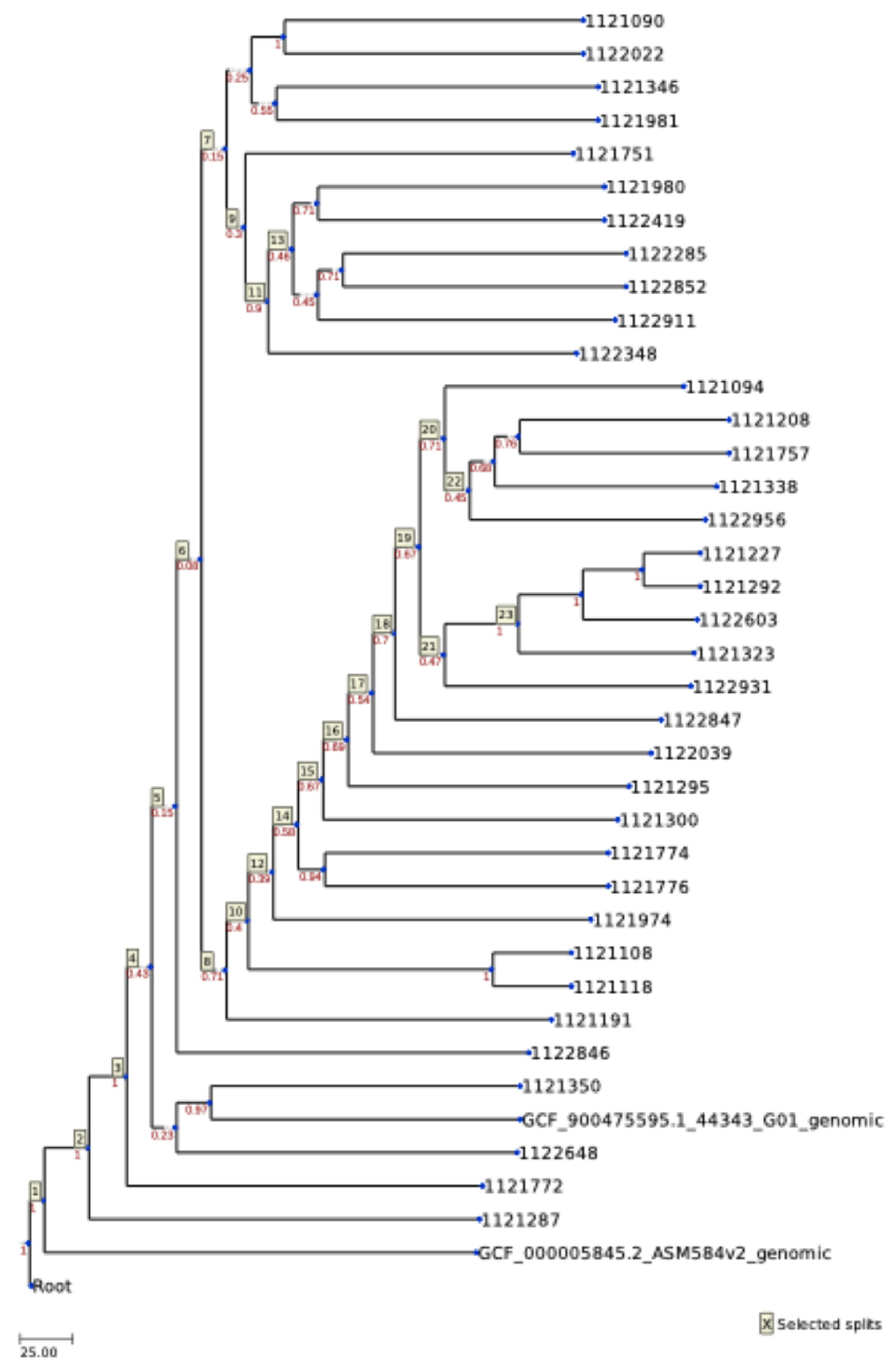
A wgMLST tree was also constructed (Figure 2). The topology of the tree was divided into two major groups: one containing the E. coli reference strain, and the other comprising all the S. uberis strains, including the S. uberis reference strain. Within the S. uberis branch, the reference strain clustered in the third divergence event, forming a group with strains 1121350 and 1122648, both isolated in the province of Lugo. The second branch from this divergence included all remaining isolates, except for strain 1121772 (from the province of A Coruña) and strain 111287 (from the province of Lugo), which diverged earlier. The fifth split divided the strains into two main groups, but this division did not correlate with their geographic origin. Interestingly, the two strains from the province of Pontevedra fell into separate groups: strain 1121757 clustered with one branch, while strain 1122419 clustered with the other. Figure 3 presents the heatmap of these 101 highly discriminatory loci.
To obtain the pangenome, the genomic sequences of the 36 S. uberis isolates were reanalyzed strains in the Cano-wgMLST_BacCompare platform, this time excluding the reference strains. The resulting pangenome comprised 10,122 genes: 521 (5%) core genes present in ≥95% of the isolates, 5691 (56%) accessory genes, and 3910 (39%) unique genes specific to individual strains. The corresponding Venn diagram, wgMLST tree, most discriminatory loci, and heatmap profile are provided in Supplementary File S3A–D.

2.3. Antimicrobial Profile

The phenotypic antimicrobial profiles and resistance genes of the S. uberis strains are summarized in Table 1. Twelve of the 36 strains (33.3%) showed phenotypic resistance to one or more of the following antibiotics: tetracycline (TET), erythromycin (ERY), clindamycin (CLI), and ceftriaxone (CRO). Although no strain was resistant to all four antibiotics simultaneously, all resistant strains (12/36) exhibited resistance to CLI. Three strains showed phenotypic resistance to TET, ERY, and CLI, while only one strain showed resistance to CRO. Four strains did not display any phenotypic antimicrobial resistance.
The resistance genes are also listed in Table 1 and classified by frequency of occurrence in Table 2. The lnu gene was the most common, with 27 occurrences out of 102 total hits, demonstrating concordance between phenotypic and genotypic resistance. Only three strains carried two lnu genes simultaneously: 1121772, 1122852, and 1122911. The mph14 gene was the least frequent (3/102). The predicted antimicrobial resistance phenotypes for all S. uberis strains are provided in Supplementary File S4.

2.4. Plasmids and Virulence

Plasmid content was assessed using the PlasmidFinder tool with default thresholds (95% identity and 60% coverage). Only 10 strains showed alignment to a plasmid sequence, and all matched a single plasmid replicon: repUS43 (Supplementary File S5A). This replicon initiates plasmid DNA replication and has been associated with resistance to beta-lactamases, aminoglycosides, and tetracycline. The repUS43 plasmid is also found in mobilizable plasmids, which are capable of being transferred to other bacteria via conjugation [35]. The query/HSP length was identical: 1206/1206, with Rep_trans identified as the targeted locus.
Virulence factors (VF), defined as gene products (e.g., toxins, cell surface attachment proteins, enzymes) that contribute to the pathogenicity, were identified using the VFDB. Table 3 lists the VF detected in the studied strains, grouped by major functional classes. Among the categories identified were adherence, enzymes, immune evasion, manganese uptake, proteases, cell surface components, and serum resistance. Additional details, including VF coverage, functional roles, and mechanisms in each S. uberis strain, are provided in Supplementary File S5B.
Finally, Supplementary File S5C summarizes the predicted pathogenic potential of the isolates toward human hosts, using the PathogenFinder tool. In silico analysis predicted all S. uberis isolates to have human pathogenic potential.

3. Discussion

This study aimed to identify the genetic diversity among 36 strains of S. uberis previously isolated from bovine mastitis in northwestern Spain. S. uberis is one of the main pathogens responsible for bovine mastitis [36], causing estimated annual global losses of up to 125 billion Euros [8,36]. Although several strains have been typed by MLST, only 187 S. uberis genomes are currently available in the NCBI database. Consequently, prophylactic vaccine development remains under investigation, as knowledge about the bacterium is still limited [36,37].
The genome sizes of the isolates ranged from 1.9 to 2.3 Mbp, and GC content varied between 36.3 and 36.8%, consistent with previously reported values [36,38]. The NCBI database also shows comparable ranges (1.6–2.5 Mbp, GC content 36.0–37.0%).
Different gene expression patterns enable S. uberis to adapt to changing environmental and stress conditions [6]. Accurate strain identification is therefore essential, not only for understanding these adaptations but also for guiding epidemiological surveillance and public health interventions [39]. MLST, based on the sequencing of internal fragments of 6–7 housekeeping genes (approximately 450–500 bp each), provides a robust method for characterizing bacterial isolates [40]. Although this represents only a fraction of the genome, it is considered representative of overall genomic diversity [39,41,42].
In our study, a high proportion of novel alleles (32/36 strains) were detected, indicating considerable genetic diversity that likely reflects environmental conditions rather than clonal spread. Alleles were identified across 7 loci (arcC, ddl, gki, recP, tdk, tpi, and yqiL). The loci with the highest allele counts were ddl and tdk (33/36 strains), while arcC was the least represented (19 strains). The tdk gene exhibited the highest variability (12 variants), followed by ddl and gki with 9 variants each. These findings align with Fenske et al. [36], who also identified tdk as highly variable; the pubMLST database listed a high number of records for this gene (175). The ddl and gki genes also have a high number of records or variants in the database: 96 and 110, respectively. Interestingly, the pubMLST database shows a high number of records for the gene yqiL (111), in contrast with its low variability in the present work. These findings suggest that these housekeeping genes are not as stable as initially thought. Fenske et al. [36] reported similar conclusions.
Despite high allelic diversity, only 4 strains matched 3 known sequence types (STs): ST6, ST184, and ST307. Strain 1121751 was related to ST386 but showed alleles with less than 100% identity despite full coverage, suggesting polymorphic variants. Strain 1121774 was classified as ST815, with multiple perfect allele matches. Overall, 32/36 strains carried novel alleles, reflecting a high level of diversity similar to findings from Germany (17/24 novel) and Australia (27/27 novel) [36,38]. Tomita et al. [43] also observed wide diversity, identifying 33 STs among 46 isolates, with ST60 and ST155 being the most common. Despite the small size of the sample, our study revealed a great diversity of allelic profiles or STs, indicating that the spread of S. uberis in northwestern Spain is more heterogeneous than clonal [15,36], with no clear correlation between STs and geographic location (i.e., farms in the interior vs. farms closer to the seacoast).
Single-genome sequencing provides limited insight into the genetic variability that drives pathogenicity and vaccine target identification [44,45,46]. Like Streptococcus agalactiae, S. uberis exhibits an open pangenome, consisting of a core genome (genes present in all strains) and a dispensable genome (genes absent from one or more strains—or accessory—, and genes specific to each strain). Our dataset contained 10,122 genes: 5.1% core, 56.2% accessory, and 38.6% unique. This indicates high intraspecies variability and global diversity, consistent with other studies [36,47]. Fenske et al. [36] reported smaller pangenomes (2508 genes) but larger core genomes (1611 genes). These values differ from those reported by other authors [37,38,48]. Some authors have noted that new unique genes are detected as more strains are sequenced [44].
The observed diversity may also be influenced by sequencing technology. Although High Accuracy Calling (HAC) has been used for basecalling, and assembly correction has been performed with the Medaka tool, sequencing errors inherent to Nanopore sequencing technology could affect allele identification. New flow cells, advanced pore design, and AI-driven neural networks, such as Guppy, dramatically improve the consensus accuracy of ONT up to ≥99.9% after polishing [49]. The detection of novel alleles in 32 isolates strongly suggests substantial natural diversity. Environmental factors may also play a role, given the large geographic area covered by the sampling sites across Lugo, A Coruña, and Pontevedra provinces, which encompass distinct ecological conditions.
The most common method for pangenome estimation is to include only genes present in 100% of isolates, but this approach has several limitations [50]. Any set of isolates represents only a subset of the population; if this subset lacks genetic diversity, the estimated number of core genes will be higher than in a more diverse dataset. In the present study, we defined core genes as those present in ≥95% of isolates. Using this threshold, only 5% of the pangenome was classified as core, while a larger proportion (56%) consisted of accessory genes, suggesting strong adaptive potential. Recent studies in Streptococcus pneumoniae have shown that core genes can exhibit higher recombination rates than accessory genes at the population level [51]. While core genes encode essential functions shared across strains, accessory genes often provide niche-specific adaptations. In our dataset, the abundance of accessory genes points to enhanced adaptability to the udder environment, where microbial communities interact and compete during mastitis [52].
Notably, novel alleles were identified in 32 of the 36 isolates, underscoring the high genetic diversity of these strains. Sequencing methodology is also a source of variability among studies. The workflow with R9.4.1 flow cells and the Rapid Barcoding Sequencing Kit enabled rapid library preparation and multiplexing of isolates, producing long-read data. In addition, the wgMLST is a whole-genome multilocus sequence typing that extends MLST to the genome level, and it has been successfully applied to several species of bacteria [53]. Homologous recombination analyses have shown that S. pneumoniae and other species (Escherichia coli, Shigella flexneri, Neisseria meningitidis, and C. jejuni) display relatively high recombination rates in core genes but low mutational divergence, with mutations driving the genetic differentiation underlying speciation [51,54,55]. Similarly, high mutation rates have been reported in other complex environments, such as mouse gut commensals [56]. Microbial evolution of Escherichia coli demonstrates that beneficial mutations can coexist with slightly deleterious ones, maintaining long-term intraspecies diversity.
Bacteria live in rich and dynamic ecosystems—such as the udder or gut—where multiple interspecies interactions shape evolution. However, little is known about how bacterial evolution unfolds in natural environments [56,57]. In this context, it is important to note the wide geographical range of the sampled provinces—Lugo (9856 km2), A Coruña (7950 km2), and Pontevedra (4495 km2)—which may significantly contribute to the observed variability among isolates.
S. uberis is a significant public health concern, as it may serve as a reservoir of antimicrobial resistance genes that can be transferred to other bacteria, including pathogens and commensals [58,59]. Antimicrobial treatment of bovine mastitis has reduced the susceptibility of this microorganism to penicillin, macrolides, aminoglycosides, and clindamycin [60]. Although antibiotics remain the most effective therapy for mastitis, monitoring the antimicrobial susceptibility of S. uberis is essential for effective veterinary treatment. Moreover, the antimicrobial resistance phenomenon in dairy cattle poses a potential risk to human health [59,61]. Importantly, S. uberis is also the most frequently isolated pathogen in sheep and goat milk [62], highlighting the importance of its surveillance in antimicrobial stewardship programs within veterinary practice.
Over the last decade, Category B antibiotics (fluoroquinolones and third- and fourth-generation cephalosporins) were among the most commonly used treatments in Spain. However, the implementation of the National Plan Against Resistance to Antibiotics (PRAN) and the adoption of electronic prescriptions have led to a marked decline in their use. At present, Category B antibiotics are reserved exclusively for cases of hyperacute mastitis caused by Gram-negative bacteria, while penicillins and sulfonamides are now the most frequently administered antimicrobials [63]. Our results show only one out of 36 strains with a phenotypic antimicrobial profile for ceftriaxone (beta-lactams and cephalosporines of third generation); these results could indicate that the PRAN has been applied in the veterinary clinical practice, reducing the number of resistant strains to this group of antibiotics.
Boireau et al. [64] conducted a ten-year study in France and reported that antibiotic resistance levels of S. uberis were low for gentamycin (approximately 2%), but higher for tetracycline, lincomycin, and erythromycin (20%). They also observed no seasonal variation in antimicrobial resistance across the study period. Regarding multidrug resistance, 14.5% of the S. uberis isolates met this criterion, while 33% were resistant to one or two antibiotic classes. In our study, 9 strains exhibited a phenotypic antimicrobial profile resistant to 2 or 3 antibiotics, specifically tetracycline, erythromycin, and clindamycin, while one strain showed resistance to tetracycline, erythromycin, and ceftriaxone. Two additional strains displayed resistance to only one antibiotic, clindamycin. In relation to other animals, Rosa et al. [65] investigated the antimicrobial susceptibility profiles of S. uberis isolated from sheep milk and reported that 97% of the isolates (120/124) were resistant to at least one of the 14 antibiotics tested, including erythromycin (genes ermB and ermC) and tetracycline (genes tetM, tetO, tetK, and tetS). Interestingly, genes such as lnu, ant, lsa, aph, and mph were not detected in their study, which contrasts with our findings.
The lnu gene was the most frequently detected, accounting for 27 out of 112 resistance genes identified. Resistance to lincosamides arises from inactivation by lincosamide nucleotidyl-transferase enzymes encoded by lnu genes. Six variants of the lnu gene have been described in the literature—lnu(A), lnu(B), lnu(C), lnu(D), lnu(E), and lnu(F). Among the strains analyzed in this study, three variants were detected: lnu(B), lnu(C), and lnu(D).
A total of 19 VFs and their associated genes were identified (Table 3). Five genes—fbp54, “undetermined”, psaA, cppA, and htrA/degP—were present in all the strains. The “undetermined” gene was linked to the capsule protein within the immune evasion VF class. An additional undetermined gene, associated with the agglutinin receptor protein and classified under the adherence VF class, was detected in 5 strains (15.15%). The least frequently detected genes were srtC (strain 1121300; related to the rlrA islet protein, adherence VF class) and galE and wbtP (strains 1121757 and 1122603; associated with the exopolysaccharide Hemophylus protein and the LPS Francisella protein, respectively).
Although S. uberis is rarely associated with human infections, several cases have been reported. Eight cases involved direct contact with cows, milk, and/or dairy products [66,67]. In contrast, 9 cases were not linked to cattle or milk [11,12,68,69,70,71,72,73], although 2 of them may have been indirectly related: a 9-year-old boy kicked by a horse in a field recently grazed by cattle and sheep [12], and a 75-year-old man who frequently hiked barefoot across pastures densely populated with cows [11]. Some authors have suggested that environmental factors may enhance the pathogenic potential of S. uberis in humans, as has been observed for Streptococcus agalactiae [74]. Overall, the reported cases suggest that infection with S. uberis may result not only from direct contact with the microorganism but also from exposure to cattle feces and/or indirect contact with contaminated pastures or farm soil.

4. Materials and Methods

4.1. Isolates and Strain Identification

Thirty-six S. uberis isolates were included in the study. All the strains were isolated from 36 different clinical-bovine mastitis cases detected in 42 different dairy farms in Galicia (NW Spain; ETRS89/UTM Zone 29) from the 9th to the 18th of July of 2022. Milk samples were transported under refrigeration conditions to the Galician Professional Laboratory for Milk Analyses (Laboratorio Interprofesional Gallego de Análisis de Leche—LIGAL, 15318 Abegondo, Coruña, Spain) for analysis. The MALDI-TOF (matrix-assisted laser desorption/ionization time-of-flight) mass spectrometry methodology with VITEK®MS (Biomérieux, Madrid, Spain) was used for strain identification.

4.2. Susceptibility to Antibiotics

The isolates were studied for their susceptibility to 9 common antibiotics (penicillin G, ampicillin, tetracycline, erythromycin, clindamycin, cefotaxime, ceftriaxone, levofloxacin, and moxifloxacin; Merck-Millipore, Darmstadt, Germany) via the VITEK®2 (Biomérieux, Madrid, Spain). The susceptibility testing (AST) cards used for the analyses are compliant with EUCAST/CLSI guidelines.

4.3. DNA Isolation

The isolates were transferred to our laboratory in cryovials and stored at −20 °C. For DNA isolation, a cryoball from frozen stocks was transferred to a flask containing 10 mL of BHI (Merck-Millipore, Darmstadt, Germany) and incubated at 37 °C for 18 h at 150 rpm. Then, 100 μL of the growth media was transferred to a new flask containing 10 mL of BHI and incubated under the same conditions described previously for 12 h. After that, 1 mL was transferred to a 1.5 mL microtube and centrifuged at 16,000× g for 2 min. The supernatant was discarded, and the DNA was isolated from the pellet following the manufacturer’s Gram-Positive Cell Lysate protocol for the Purelink Genomic DNA Mini Kit (InvitrogenTM, Thermo Fisher Scientific, Waltham, MA, USA). In the final step, the DNA was eluted in 50 μL of elution buffer. The DNA concentration was determined with a QubitTM fluorometer (Invitrogen, Thermo Fisher, Waltham, MA, USA) via the Invitrogen™ Qubit™ dsDNA BR Assay Kit (Invitrogen, Thermo Fisher). The DNA was stored at −20 °C until use.

4.4. Whole-Genome Sequencing

A concentration of 200 ng of DNA was used for sequencing with the Rapid Barcoding Sequencing Kit (SQK-RBK004, Oxford Nanopore Technologies Ltd., ONT, Oxford, UK) in combination with Flow cells R9.4.1 (ONT). MinION Mk1C software 2024.0 with integrated and preinstalled base calling and analysis software was used for sequencing. The time of sequencing was set at 24 h. Oxford Nanopore preloaded basecalling software Guppy in high accuracy (HAC) mode was used with default parameters for basecalling. Data quality control was performed as follows (see below for details): (i) a recommended coverage of 30× for bacterial genomes using NanoPlot tool; (ii) assembly and polishing with Medaka tool; and (iii) assembly with Raven tool, which performs two-step polishing with Racon tool.

4.5. Bioinformatic Analysis

4.5.1. Assembly

De novo assembly was performed on the Galaxy platform through its European server (https://usegalaxy.eu; accessed on 1 February 2024) [75]. FASTQ files from every strain were uploaded to the Galaxy server and transformed into FASTA files via the “FASTQ to FASTA converter” tool (Galaxy Version 1.1.5) [76]. To merge all the obtained FASTA files from every strain into a unique file, the “Merge.files Merge data” tool (Galaxy Version 1.39.5.0) [77] was used. The “NanoPlot—plotting suite for Oxford Nanopore sequencing data and alignments” tool (Galaxy Version 1.41.0+galaxy0) [78] was used with the FASTQ files to create a statistical summary of the sequencing reads and to calculate the next-generation sequencing (NGS) coverage. FASTA files were used for the assembly via the “Raven—de novo assembly of Oxford Nanopore Technologies data” tool (Galaxy Version 1.8.0+galaxy0) [79]. All the tools were used with default parameters.
FASTA files were also introduced in Geneious bioinformatics software version 2024.0 (http://www.geneious.com; accessed on 1 February 2024), which was used to obtain the number of sequences, the percentage of GCs, and the sequence length. The sequencing coverage c describes the average number of reads that align to known bases; at higher levels of coverage, each base is covered by a greater number of aligned sequence reads. To estimate and achieve the desired sequencing coverage level, the Lander/Waterman equation [80,81,82] was used, that is, c = (L × N)/G, where c is the coverage, L × N is the number of bases sequenced (L is the read length in bp and N is the number of reads), and G is the haploid genome length in bp.

4.5.2. Polishing and Annotation

The tool “medaka consensus pipeline—Assembly polishing via neural networks” (Galaxy Version 1.7.2+galaxy0) [83] was used to polish the final assembly obtained from Raven tool FASTA files. The obtained draft genome was used for subsequent analysis. The “Prokka—Prokaryotic genome annotation” (Galaxy Version 1.14.6+galaxy1) tool [84,85] was used for genome annotation. Both tools were used with default parameters.

4.5.3. Typing, Antimicrobial Resistance and Virulence

The tool “ABRicate—Mass screening of contigs for antimicrobial and virulence genes” (Galaxy Version 1.0.1) [86] was used to identify the presence of antimicrobial resistance genes. The Center for Genomic Epidemiology (CGE; http://www.genomicepidemiology.org/; accessed on 1 March 2024) is entirely noncommercial and operates several free online bioinformatics services and tools. MLST 2.0.9 [87] and PlasmidFinder 2.0.1 [88] tools were used for multilocus sequence typing (MLST) and identification of plasmids, respectively. Four prerequisites have been considered for their selection for MLST: (i) the alignment length (also known as the high-scoring segment pair or HSP) must equal the allele length; (ii) the identity must be 100%; (iii) the coverage must be 100%; and (iv) the number of gaps in the HSP must be zero. The length of the alignment between the best-matching MLST allele in the database and the corresponding sequence in the genome of the S. uberis strains studied is referred to as the alignment length. The length of the best-matching MLST allele in the database is called the allele length. The percentage of identity is the percentage of nucleotides that are identical in the best-matching MLST allele in the database and the corresponding sequence in the genome of S. uberis strains.
The Cano-wgMLST_BacCompare platform (https://github.com/cenesis/cano-wgMLST; accessed on 1 March 2024) was used for epidemiological investigations and comparative genomic analysis [89]. The Virulence Factor Database (VFDB; http://www.mgc.ac.cn/VFs/main.htm; accessed on 1 March 2024) was used for the prediction of virulence factors (VFs) and related genes for each S. uberis strain [90].
The FASTA files of the isolates were used with CGE’s PathogenFinder 1.1 [91] tool for predicting the pathogen action of the studied strains toward human cells.

5. Conclusions

This study aimed to identify the genetic differences and similarities among S. uberis strains previously isolated from bovine mastitis in northwestern Spain, a region ranked among the top ten European milk producers. Current knowledge of the genomic characteristics of this bacterium is limited, underscoring the need for further research.
Different gene expression patterns allow S. uberis to adapt to different environmental conditions and stress situations. Understanding these adaptations is essential for the design of public health control strategies. Our study revealed a great diversity of allelic profiles or STs, indicating that the spread of S. uberis in NW Spain is more heterogeneous than contagious. On the basis of graphical analysis, no correlations were found between the known sequencing types of this study and the geographical characteristics of the area.
The findings highlight that S. uberis exhibits diverse gene expression patterns, allowing adaptation to a range of environmental conditions and stress factors. Understanding these adaptations is essential for developing effective public health control strategies. Our results revealed a high diversity of allelic profiles and sequence types, suggesting that the spread of S. uberis in northwestern Spain is more heterogeneous than clonal. Graphical analysis showed no correlation between the identified sequence types and the geographical characteristics of the sampling areas.
The isolates analyzed in this study showed considerable intraspecies variability and global diversity, consistent with observations in other regions. The relatively large proportion of accessory genes detected indicates an increased adaptive capacity, particularly to the challenging udder environment, which harbors diverse microbial communities competing for resources.
Of particular concern is the role of S. uberis as a potential reservoir of antimicrobial resistance genes, which may be transmitted to other bacteria, pathogens, and commensals. Monitoring the antimicrobial susceptibility of S. uberis is crucial for guiding veterinary treatment of bovine mastitis and for supporting the One Health approach. The antimicrobial resistance detected in several strains raises concerns regarding the effectiveness of current treatment strategies in dairy farming.
Although S. uberis is rarely associated with human infections, reported cases do exist. Importantly, all the strains analyzed in this study were predicted to have pathogenic potential in humans, emphasizing the relevance of continued surveillance and characterization of this bacterium.

Supplementary Materials

The following supporting information can be downloaded at: https://www.mdpi.com/article/10.3390/antibiotics14111059/s1, Supplementary File S1: Sequencing statistics obtained from the Nanoplot tool and/or Geneious software; Supplementary File S2A: MLST loci against which the input sequence has been aligned; Supplementary File S2B: Sequence types (STs) and nearest STs that the MLST server associated with the submitted data; Supplementary File S2C: Most discriminatory loci (416 with ˃95% strain occurrence) from wgMLST; Supplementary File S2D: Venn diagram output with ellipses from wgMLST (36 S. uberis strains plus 2 reference strains); Supplementary File S3A: Venn diagram output with ellipses from wgMLST (36 S. uberis strains without reference strains); Supplementary File S3B: Genetic relatedness tree from wgMLST; Supplementary File S3C: Most discriminatory loci (402 with ˃95% strain occurrence) from wgMLST; Supplementary File S3D: Genetic relatedness tree from wgMLST (36 strains); Supplementary File S4: Predicted resistance phenotype to antibiotics from the S. uberis strains studied; Supplementary File S5A: Plasmids results from PlasmidFinder tool; Supplementary File S5B: Additional information related to the virulence factors that contribute to the pathogenicity of S. uberis. Different open reading frames (orf) are shown for each gene; Supplementary File S5C: Prediction of possible human pathogenicity using PathogenFinder tool.

Author Contributions

Conceptualization: E.J.Q., C.M.F. and B.I.V.; Methodology: C.M.F.; Formal Analysis: E.J.Q., A.P. and G.L.-L.; Investigation: E.J.Q. and B.I.V.; Resources: P.R.d.R., B.d.M.S. and C.M.F.; Writing—Original Draft Preparation: E.J.Q. and B.I.V.; Writing—Review and Editing: E.J.Q., B.I.V. and C.M.F.; Supervision: C.M.F.; Funding Acquisition: P.R.d.R., B.d.M.S. and C.M.F. All authors have read and agreed to the published version of the manuscript.

Funding

This research received no external funding.

Institutional Review Board Statement

Not applicable.

Informed Consent Statement

Not applicable.

Data Availability Statement

The raw data supporting the conclusions of this article were deposited in the NCBI GenBank upon the Bioproject PRJNA1321121.

Acknowledgments

The authors acknowledge the support of the Freiburg Galaxy Team: Person X and Björn Grüning, Bioinformatics, University of Freiburg (Germany) funded by the German Federal Ministry of Education and Research BMBF grant 031 A538A de. NBI-RBC and the Ministry of Science, Research and the Arts Baden-Württemberg (MWK) within the framework of LIBIS/de. NBI Freiburg. In addition, the authors would like to thank LIGAL (Laboratorio Interprofesional Gallego de Análisis de Leche, 15318 Abegondo, A Coruña, Spain) for its invaluable collaboration in obtaining the isolates.

Conflicts of Interest

The authors declare no conflicts of interest.

References

  1. Gibbs, E.P.J. The Evolution of One Health: A Decade of Progress and Challenges for the Future. Vet. Rec. 2014, 174, 85–91. [Google Scholar] [CrossRef]
  2. FAO; OIE; WHO; UNICEF; UNSIC; World Bank. Contributing to One World, One Health—A Strategic Framework for Reducing Risks of Infectious Diseases at the Animal–Human–Ecosystems Interface 2008, pp. 1–68. Available online: https://www.fao.org/4/aj137e/aj137e00.htm (accessed on 1 March 2024).
  3. FAO. Dairy Market Review: Emerging Trends and Outlook 2022; FAO: Rome, Italy, 2022. [Google Scholar]
  4. Fundación Juana de Vega. Informe Do Sector Lácteo 2025. Available online: https://www.campogalego.gal/wp-content/uploads/2025/04/InformedoSectorLacteo2024_FundacionJuanadeVega.pdf (accessed on 1 March 2024).
  5. OECD/FAO Dairy and Dairy Products. In OECD-FAO Agricultural Outlook 2022–2031; OECE, FAO, Eds.; OECD: Paris, France; FAO: Rome, Italy, 2022; pp. 212–223. ISBN 978-92-5-136313-3. [Google Scholar]
  6. Ward, P.N.; Holden, M.T.G.; Leigh, J.A.; Lennard, N.; Bignell, A.; Barron, A.; Clark, L.; Quail, M.A.; Woodward, J.; Barrell, B.G.; et al. Evidence for Niche Adaptation in the Genome of the Bovine Pathogen Streptococcus uberis. BMC Genom. 2009, 10, 54. [Google Scholar] [CrossRef] [PubMed]
  7. Di Domenico, E.G.; Toma, L.; Prignano, G.; Pelagalli, L.; Police, A.; Cavallotti, C.; Torelli, R.; Sanguinetti, M.; Ensoli, F. Misidentification of Streptococcus uberis as a Human Pathogen: A Case Report and Literature Review. Int. J. Infect. Dis. 2015, 33, 79–81. [Google Scholar] [CrossRef] [PubMed]
  8. Kabelitz, T.; Aubry, E.; van Vorst, K.; Amon, T.; Fulde, M. The Role of Streptococcus spp. in Bovine Mastitis. Microorganisms 2021, 9, 1497. [Google Scholar] [CrossRef]
  9. Bisharat, N.; Crook, D.W.; Leigh, J.; Harding, R.M.; Ward, P.N.; Coffey, T.J.; Maiden, M.C.; Peto, T.; Jones, N. Hyperinvasive Neonatal Group B Streptococcus Has Arisen from a Bovine Ancestor. J. Clin. Microbiol. 2004, 42, 2161–2167. [Google Scholar] [CrossRef]
  10. Gottschalk, M.; Segura, M.; Xu, J. Streptococcus Suis Infections in Humans: The Chinese Experience and the Situation in North America. Anim. Health Res. Rev. 2007, 8, 29–45. [Google Scholar] [CrossRef] [PubMed]
  11. Valentiny, C.; Dirschmid, H.; Lhotta, K. Streptococcus uberis and Staphylococcus aureus Forefoot and Blood Stream Co-Infection in a Hemodialysis Patient: A Case Report. BMC Nephrol. 2015, 16, 73. [Google Scholar] [CrossRef]
  12. Murdoch, D.R.; Seaward, L.M.; MacFarlane, M.R. Human Wound Infection with Streptococcus uberis. Clin. Microbiol. Newsl. 1997, 19, 174–175. [Google Scholar] [CrossRef]
  13. Tamilselvam, B.; Almeida, R.A.; Dunlap, J.R.; Oliver, S.P. Streptococcus Uberis Internalizes and Persists in Bovine Mammary Epithelial Cells. Microb. Pathog. 2006, 40, 279–285. [Google Scholar] [CrossRef]
  14. De Jong, A.; El Garch, F.; Simjee, S.; Moyaert, H.; Rose, M.; Youala, M.; Siegwart, E. Monitoring of Antimicrobial Susceptibility of Udder Pathogens Recovered from Cases of Clinical Mastitis in Dairy Cows across Europe: VetPath Results. Vet. Microbiol. 2018, 213, 73–81. [Google Scholar] [CrossRef]
  15. Käppeli, N.; Morach, M.; Zurfluh, K.; Corti, S.; Nüesch-Inderbinen, M.; Stephan, R. Sequence Types and Antimicrobial Resistance Profiles of Streptococcus uberis Isolated from Bovine Mastitis. Front. Vet. Sci. 2019, 6, 234. [Google Scholar] [CrossRef]
  16. Reyes, J.; Rodriguez-Lecompte, J.C.; Blanchard, A.; McClure, J.T.; Sánchez, J. Molecular Variability of Streptococcus uberis Isolates from Intramammary Infections in Canadian Dairy Farms from the Maritime Region. Can. J. Vet. Res. 2019, 83, 168–176. [Google Scholar]
  17. Saini, V.; McClure, J.T.; Léger, D.; Dufour, S.; Sheldon, A.G.; Scholl, D.T.; Barkema, H.W. Antimicrobial Use on Canadian Dairy Farms. J. Dairy Sci. 2012, 95, 1209–1221. [Google Scholar] [CrossRef]
  18. Tomazi, T.; Freu, G.; Alves, B.G.; de Souza Filho, A.F.; Heinemann, M.B.; dos Santos, M.V. Genotyping and Antimicrobial Resistance of Streptococcus uberis Isolated from Bovine Clinical Mastitis. PLoS ONE 2019, 14, e0223719. [Google Scholar] [CrossRef]
  19. Pol, M.; Ruegg, P.L. Treatment Practices and Quantification of Antimicrobial Drug Usage in Conventional and Organic Dairy Farms in Wisconsin. J. Dairy Sci. 2007, 90, 249–261. [Google Scholar] [CrossRef]
  20. Fessia, A.S.; Odierno, L.M. Potential Factors Involved in the Early Pathogenesis of Streptococcus uberis Mastitis: A Review. Folia Microbiol. 2021, 66, 509–523. [Google Scholar] [CrossRef]
  21. Zadoks, R.N.; Tikofsky, L.L.; Boor, K.J. Ribotyping of Streptococcus Uberis from a Dairy’s Environment, Bovine Feces and Milk. Vet. Microbiol. 2005, 109, 257–265. [Google Scholar] [CrossRef]
  22. Lopez-Benavides, M.G.; Williamson, J.H.; Pullinger, G.D.; Lacy-Hulbert, S.J.; Cursons, R.T.; Leigh, J.A. Field Observations on the Variation of Streptococcus uberis Populations in a Pasture-Based Dairy Farm. J. Dairy Sci. 2007, 90, 5558–5566. [Google Scholar] [CrossRef] [PubMed]
  23. Ericsson Unnerstad, H.; Lindberg, A.; Persson Waller, K.; Ekman, T.; Artursson, K.; Nilsson-Öst, M.; Bengtsson, B. Microbial Etiology of Acute Clinical Mastitis and Agent-Specific Risk Factors. Vet. Microbiol. 2009, 137, 90–97. [Google Scholar] [CrossRef] [PubMed]
  24. Monistero, V.; Barberio, A.; Cremonesi, P.; Castiglioni, B.; Morandi, S.; Lassen, D.C.K.; Astrup, L.B.; Locatelli, C.; Piccinini, R.; Filippa Addis, M.; et al. Genotyping and Antimicrobial Susceptibility Profiling of Streptococcus uberis Isolated from a Clinical Bovine Mastitis Outbreak in a Dairy Farm. Antibiotics 2021, 10, 644. [Google Scholar] [CrossRef] [PubMed]
  25. Zadoks, R.N.; Gillespie, B.E.; Barkema, H.W.; Sampimon, O.C.; Oliver, S.P.; Schukken, Y.H. Clinical, Epidemiological and Molecular Characteristics of Streptococcus uberis Infections in Dairy Herds. Epidemiol. Infect. 2003, 130, 335–349. [Google Scholar] [CrossRef]
  26. Davies, P.L.; Leigh, J.A.; Bradley, A.J.; Archer, S.C.; Emes, R.D.; Green, M.J. Molecular Epidemiology of Streptococcus uberis Clinical Mastitis in Dairy Herds: Strain Heterogeneity and Transmission. J. Clin. Microbiol. 2016, 54, 68–74. [Google Scholar] [CrossRef] [PubMed]
  27. Leelahapongsathon, K.; Schukken, Y.H.; Pinyopummintr, T.; Suriyasathaporn, W. Comparison of Transmission Dynamics between Streptococcus uberis and Streptococcus agalactiae Intramammary Infections. J. Dairy Sci. 2016, 99, 1418–1426. [Google Scholar] [CrossRef] [PubMed]
  28. Wente, N.; Klocke, D.; Paduch, J.H.; Zhang, Y.; Seeth, M.T.; Zoche-Golob, V.; Reinecke, F.; Mohr, E.; Krömker, V. Associations between Streptococcus uberis Strains from the Animal Environment and Clinical Bovine Mastitis Cases. J. Dairy Sci. 2019, 102, 9360–9369. [Google Scholar] [CrossRef]
  29. Wald, R.; Baumgartner, M.; Gutschireiter, J.; Bazzanella, B.; Lichtmannsperger, K.; Wagner, M.; Wittek, T.; Stessl, B. Comparison of the Population Structure of Streptococcus uberis Mastitis Isolates from Austrian Small-Scale Dairy Farms and a Slovakian Large-Scale Farm. J. Dairy Sci. 2020, 103, 1820–1830. [Google Scholar] [CrossRef]
  30. Díaz-Cao, J.M.; Barreal, M.L.; Pombo, B.; Prieto, A.; Alonso, J.M.; Iglesias, A.; Lorenzana, R.; López-Novo, C.; Díez-Baños, P.; Fernández, G. Evaluation and Cluster Analysis of Inflammatory Reactions of Dairy Cattle Mastitis Pathogens in Milk Samples Submitted for Microbiological Examination. Span. J. Agric. Res. 2019, 17, e0505. [Google Scholar] [CrossRef]
  31. Vélez, J.R.; Cameron, M.; Rodríguez-Lecompte, J.C.; Xia, F.; Heider, L.C.; Saab, M.; Trenton McClure, J.; Sánchez, J. Whole-Genome Sequence Analysis of Antimicrobial Resistance Genes in Streptococcus uberis and Streptococcus dysgalactiae Isolates from Canadian Dairy Herds. Front. Vet. Sci. 2017, 4, 63. [Google Scholar] [CrossRef] [PubMed]
  32. Silva, N.C.C.; Yang, Y.; Rodrigues, M.X.; Tomazi, T.; Bicalho, R.C. Whole-Genome Sequencing Reveals High Genetic Diversity of Streptococcus uberis Isolated from Cows with Mastitis. BMC Vet. Res. 2021, 17, 321. [Google Scholar] [CrossRef]
  33. Galician Institute of Statistics Registro de Ganado Bovino. 2025. Available online: https://www.ige.gal/igebdt/igeapi/datos/2917/0:2022,1:4,2:1,3:11 (accessed on 1 June 2025).
  34. Geurts, P.; Ernst, D.; Wehenkel, L. Extremely Randomized Trees. Mach. Learn. 2006, 63, 3–42. [Google Scholar] [CrossRef]
  35. Huang, J.; Li, T.; Zhu, Y.; Li, Q.; Kuo, C.; Guo, X.; Wei, B.; Ni, P.; Dong, K. Molecular Characterization and Potential Host-switching of Swine Farm associated Clostridioides difficile ST11. Vet. Microbiol. 2024, 294, 110129. [Google Scholar] [CrossRef]
  36. Fenske, L.; Noll, I.; Blom, J.; Ewers, C.; Semmler, T.; Fawzy, A.; Eisenberg, T. A Dominant Clonal Lineage of Streptococcus uberis in Cattle in Germany. Antonie Leeuwenhoek Int. J. Gen. Mol. Microbiol. 2022, 115, 857–870. [Google Scholar] [CrossRef]
  37. Hossain, M.; Egan, S.A.; Coffey, T.; Ward, P.N.; Wilson, R.; Leigh, J.A.; Emes, R.D. Virulence Related Sequences; Insights Provided by Comparative Genomics of Streptococcus uberis of Differing Virulence. BMC Genom. 2015, 16, 334. [Google Scholar] [CrossRef]
  38. Vezina, B.; Al-harbi, H.; Ramay, H.R.; Soust, M.; Moore, R.J.; Olchowy, T.W.J.; Alawneh, J.I. Sequence Characterization and Novel Insights into Bovine Mastitis-Associated Streptococcus uberis in Dairy Herds. Sci. Rep. 2021, 11, 3046. [Google Scholar] [CrossRef]
  39. Pérez-Losada, M.; Cabezas, P.; Castro-Nallar, E.; Crandall, K.A. Pathogen Typing in the Genomics Era: MLST and the Future of Molecular Epidemiology. Infect. Genet. Evol. 2013, 16, 38–53. [Google Scholar] [CrossRef] [PubMed]
  40. Maiden, M.C.J.; Bygraves, J.A.; Feil, E.; Morelli, G.; Russell, J.E.; Urwin, R.; Zhang, Q.; Zhou, J.; Zurth, K.; Caugant, D.A.; et al. Multilocus Sequence Typing: A Portable Approach to the Identification of Clones within Populations of Pathogenic Microorganisms. Proc. Natl. Acad. Sci. USA 1998, 95, 3140–3145. [Google Scholar] [CrossRef] [PubMed]
  41. Didelot, X.; Maiden, M.C.J. Impact of Recombination on Bacterial Evolution. Trends Microbiol. 2010, 18, 315–322. [Google Scholar] [CrossRef] [PubMed]
  42. Joshi, C.J.; Ke, W.; Drangowska-Way, A.; O’Rourke, E.J.; Lewis, N.E. What Are Housekeeping Genes? PLoS Comput. Biol. 2022, 18, e1010295. [Google Scholar] [CrossRef]
  43. Tomita, T.; Meehan, B.; Wongkattiya, N.; Malmo, J.; Pullinger, G.; Leigh, J.; Deighton, M. Identification of Streptococcus uberis Multilocus Sequence Types Highly Associated with Mastitis. Appl. Environ. Microbiol. 2008, 74, 114–124. [Google Scholar] [CrossRef]
  44. Tettelin, H.; Masignani, V.; Cieslewicz, M.J.; Donati, C.; Medini, D.; Ward, N.L.; Angiuoli, S.V.; Crabtree, J.; Jones, A.L.; Durkin, A.S.; et al. Genome Analysis of Multiple Pathogenic Isolates of Streptococcus agalactiae: Implications for the Microbial “Pan-Genome”. Proc. Natl. Acad. Sci. USA 2005, 102, 13950–13955. [Google Scholar] [CrossRef]
  45. Maione, D.; Margarit, I.; Rinaudo, C.D.; Masignani, V.; Mora, M.; Scarselli, M.; Tettelin, H.; Brettoni, C.; Iacobini, E.T.; Rosini, R.; et al. Identification of a Universal Group B Streptococcus Vaccine by Multiple Genome Screen. Science 2005, 309, 148–150. [Google Scholar] [CrossRef]
  46. Lauer, P.; Rinaudo, C.D.; Soriani, M.; Margarit, I.; Maione, D.; Rosini, R.; Taddei, A.R.; Mora, M.; Rappuoli, R.; Grandi, G.; et al. Genome Analysis Reveals Pili in Group B Streptococcus. Science 2005, 309, 105. [Google Scholar] [CrossRef]
  47. Morneau, D. Pan-Genomes: Moving beyond the Reference. Nature Milestones, 10 February 2021; p. S19. [Google Scholar]
  48. Lang, P.; Lefébure, T.; Wang, W.; Zadoks, R.N.; Schukken, Y.; Stanhope, M.J. Gene Content Differences across Strains of Streptococcus uberis Identified Using Oligonucleotide Microarray Comparative Genomic Hybridization. Infect. Genet. Evol. 2009, 9, 179–188. [Google Scholar] [CrossRef] [PubMed]
  49. Ni, Y.; Liu, X.; Simeneh, Z.M.; Yang, M.; Li, R. Benchmarking of Nanopore R10.4 and R9.4.1 Flow Cells in Single-Cell Whole-Genome Amplification and Whole-Genome Shotgun Sequencing. Comput. Struct. Biotechnol. J. 2023, 21, 2352–2364. [Google Scholar] [CrossRef] [PubMed]
  50. van Tonder, A.J.; Mistry, S.; Bray, J.E.; Hill, D.M.C.; Cody, A.J.; Farmer, C.L.; Klugman, K.P.; von Gottberg, A.; Bentley, S.D.; Parkhill, J.; et al. Defining the Estimated Core Genome of Bacterial Populations Using a Bayesian Decision Model. PLoS Comput. Biol. 2014, 10, e1003788. [Google Scholar] [CrossRef]
  51. Preska Steinberg, A.; Lin, M.; Kussell, E. Core Genes Can Have Higher Recombination Rates than Accessory Genes within Global Microbial Populations. Elife 2022, 11, e78533. [Google Scholar] [CrossRef]
  52. Panchal, J.; Patel, A.; Patel, S.; Goswami, D. Understanding Mastitis: Microbiome, Control Strategies, and Prevalence—A Comprehensive Review. Microb. Pathog. 2024, 187, 106533. [Google Scholar] [CrossRef]
  53. Kluytmans-van den Bergh, M.F.Q.; Rossen, J.W.A.; Bruijnin-Verhagen, P.C.J.; Bonten, M.J.M.; Friedrich, A.W.; Vandenbroucke-Grauls, C.M.J.E.; Willems, R.J.L.; Kluytmans, J.A.J.W. Whole-Genome Multilocus Sequence Typing of Extended-Spectrum-Beta-Lactamase-Producing Enterobacteriaceae. J. Clin. Microbiol. 2016, 12, 2919–2927. [Google Scholar] [CrossRef] [PubMed]
  54. Vulić, M.; Lenski, R.E.; Radman, M. Mutation, Recombination, and Incipient Speciation of Bacteria in the Laboratory. Proc. Natl. Acad. Sci. USA 1999, 96, 7348–7351. [Google Scholar] [CrossRef]
  55. Horton, J.S.; Taylor, T.B. Mutation Bias and Adaptation in Bacteria. Microbiology 2023, 169, 1404. [Google Scholar] [CrossRef]
  56. Ramiro, R.S.; Durão, P.; Bank, C.; Gordo, I. Low Mutational Load and High Mutation Rate Variation in Gut Commensal Bacteria. PLoS Biol. 2020, 18, e3000617. [Google Scholar] [CrossRef]
  57. Derakhshani, H.; Plaizier, J.C.; De Buck, J.; Barkema, H.W.; Khafipour, E. Composition and Co-Occurrence Patterns of the Microbiota of Different Niches of the Bovine Mammary Gland: Potential Associations with Mastitis Susceptibility, Udder Inflammation, and Teat-End Hyperkeratosis. Anim. Microbiome 2020, 2, 11. [Google Scholar] [CrossRef]
  58. Barlow, J. Mastitis Therapy and Antimicrobial Susceptibility: A Multispecies Review with a Focus on Antibiotic Treatment of Mastitis in Dairy Cattle. J. Mammary Gland. Biol. Neoplasia 2011, 16, 383–407. [Google Scholar] [CrossRef] [PubMed]
  59. Zhang, T.; Tao, L.; Boonyayatra, S.; Niu, G. Antimicrobial Resistance of Streptococcus uberis Isolated from Bovine Mastitis: A Review. Indian J. Anim. Res. 2021, 56, 1435–1441. [Google Scholar] [CrossRef]
  60. Overesch, G.; Stephan, R.; Perreten, V. Antimicrobial Susceptibility of Gram-Positive Udder Pathogens from Bovine Mastitis Milk in Switzerland. Schweiz. Arch. Tierheilkd. 2013, 155, 339–350. [Google Scholar] [CrossRef]
  61. Molineri, A.I.; Camussone, C.; Zbrun, M.V.; Suárez Archilla, G.; Cristiani, M.; Neder, V.; Calvinho, L.; Signorini, M. Antimicrobial Resistance of Staphylococcus aureus Isolated from Bovine Mastitis: Systematic Review and Meta-Analysis. Prev. Vet. Med. 2021, 188, 105261. [Google Scholar] [CrossRef]
  62. Rosa, N.M.; Agnoletti, F.; Lollai, S.; Tola, S. Comparison of PCR-RFLP, API® 20 Strep and MALDI-TOF MS for Identification of Streptococcus Spp. Collected from Sheep and Goat Milk Samples. Small Rumin. Res. 2019, 180, 35–40. [Google Scholar] [CrossRef]
  63. Preine, F.; Herrera, D.; Scherpenzeel, C.; Kalmus, P.; McCoy, F.; Smulski, S.; Rajala-Schultz, P.; Schmenger, A.; Moroni, P.; Krömker, V. Different European Perspectives on the Treatment of Clinical Mastitis in Lactation. Antibiotics 2022, 11, 1107. [Google Scholar] [CrossRef] [PubMed]
  64. Boireau, C.; Cazeau, G.; Jarrige, N.; Calavas, D.; Madec, J.-Y.; Leblond, A.; Haenni, M.; Gay, É. Antimicrobial Resistance in Bacteria Isolated from Mastitis in Dairy Cattle in France, 2006–2016. J. Dairy Sci. 2018, 101, 9451–9462. [Google Scholar] [CrossRef] [PubMed]
  65. Rosa, N.M.; Duprè, I.; Azara, E.; Longheu, C.M.; Tola, S. Molecular Typing and Antimicrobial Susceptibility Profiles of Streptococcus uberis Isolated from Sheep Milk. Pathogens 2021, 10, 1489. [Google Scholar] [CrossRef]
  66. Gulen, D.; Kaya, A.D.; Aydin, M.; Tanriverdi, Y. Urinary Tract Infections Caused by Streptococcus uberis: A Pathogen of Bovine Mastitis-Report of Seven Cases. Afr. J. Microbiol. Res. 2013, 7, 3908–3912. [Google Scholar]
  67. Sánchez, J.; Moreno, J.J.; Roldán, A.; Ildefonso, J.A.; Florensa, J. Hepatic Abscess Caused by Streptococcus uberis. Enferm. Infecc. Microbiol. Clin. 1991, 9, 189–190. [Google Scholar]
  68. Sarkar, T.K.; Murarka, R.S.; Gilardi, G.L. Primary Streptococcus viridans Pneumonia. Chest 1989, 96, 831–834. [Google Scholar] [CrossRef]
  69. Bouskraoui, M.; Benbachir, M.; Abid, A. Endocardite a Streptococcus uberis Chez Un Nourrisson Atteint d’un Canal Atrioventriculaire. Arch. Pediatr. 1999, 6, 481. [Google Scholar] [CrossRef]
  70. Łazińska, B.; Ciszek, M.; Rokosz, A.; Sawicka-Grzelak, A.; Pa̧czek, L.; Łuczak, M. Bacteriological Urinalysis in Patients after Renal Transplantation. Pol. J. Microbiol. 2005, 54, 317–321. [Google Scholar]
  71. Huang, W.T.; Chang, L.Y.; Hsueh, P.R.; Lu, C.Y.; Shao, P.L.; Huang, F.Y.; Lee, P.I.; Chen, C.M.; Lee, C.Y.; Huang, L.M. Clinical Features and Complications of Viridans Streptococci Bloodstream Infection in Pediatric Hemato-Oncology Patients. J. Microbiol. Immunol. Infect. 2007, 40, 349–354. [Google Scholar]
  72. Kessel, S.; Wittenberg, C.E. Joint Infection in a Young Patient Caused by Streptococcus uberis, a Pathogen of Bovine Mastitis: A Case Report. Z. Orthop. Unfallchirurgie 2008, 146, 507–509. [Google Scholar] [CrossRef]
  73. Velez-Montoya, R.; Rascón-Vargas, D.; Mieler, W.F.; Fromow-Guerra, J.; Morales-Cantón, V. Intravitreal Ampicillin Sodium for Antibiotic-Resistant Endophthalmitis: Streptococcus uberis First Human Intraocular Infection Report. J. Ophthalmol. 2010, 2010, 169739. [Google Scholar] [CrossRef]
  74. Zadoks, R.N.; Middleton, J.R.; McDougall, S.; Katholm, J.; Schukken, Y.H. Molecular Epidemiology of Mastitis Pathogens of Dairy Cattle and Comparative Relevance to Humans. J. Mammary Gland Biol. Neoplasia 2011, 16, 357–372. [Google Scholar] [CrossRef] [PubMed]
  75. Abueg, L.A.L.; Afgan, E.; Allart, O.; Awan, A.H.; Bacon, W.A.; Baker, D.; Bassetti, M.; Batut, B.; Bernt, M.; Blankenberg, D.; et al. The Galaxy Platform for Accessible, Reproducible, and Collaborative Data Analyses: 2024 Update. Nucleic Acids Res. 2024, 52, W83–W94. [Google Scholar] [CrossRef] [PubMed]
  76. Blankenberg, D.; Gordon, A.; Von Kuster, G.; Coraor, N.; Taylor, J.; Nekrutenko, A.; Team, G. Manipulation of FASTQ Data with Galaxy. Bioinform. Appl. Note 2010, 26, 1783–1785. [Google Scholar] [CrossRef] [PubMed]
  77. Schloss, P.D.; Westcott, S.L.; Ryabin, T.; Hall, J.R.; Hartmann, M.; Hollister, E.B.; Lesniewski, R.A.; Oakley, B.B.; Parks, D.H.; Robinson, C.J.; et al. Introducing Mothur: Open-Source, Platform-Independent, Community-Supported Software for Describing and Comparing Microbial Communities. Appl. Environ. Microbiol. 2009, 75, 7537–7541. [Google Scholar] [CrossRef]
  78. De Coster, W.; Rademakers, R. NanoPack2: Population-Scale Evaluation of Long-Read Sequencing Data. Bioinformatics 2023, 39, btad311. [Google Scholar] [CrossRef]
  79. Vaser, R.; Šikić, M. Time- and Memory-Efficient Genome Assembly with Raven. Nat. Comput. Sci. 2021, 1, 332–336. [Google Scholar] [CrossRef] [PubMed]
  80. Lander, E.S.; Waterman, M.S. Genomic Mapping by Fingerprinting Random Clones: A Mathematical Analysis. Genomics 1988, 2, 231–239. [Google Scholar] [CrossRef] [PubMed]
  81. Arratia, R.; Lander, E.S.; Tavaré, S.; Waterman, M.S. Genomic Mapping by Anchoring Random Clones: A Mathematical Analysis. Genomics 1991, 11, 806–827. [Google Scholar] [CrossRef]
  82. Port, E.; Sun, F.; Martin, D.; Waterman, M.S. Genomic Mapping by End-Characterized Random Clones: A Mathematical Analysis. Genomics 1995, 26, 84–100. [Google Scholar] [CrossRef]
  83. Oxford Nanopore Technologies Ltd. Medaka. GitHub. 2018. Available online: https://github.com/nanoporetech/medaka (accessed on 1 March 2024).
  84. Cuccuru, G.; Orsini, M.; Pinna, A.; Sbardellati, A.; Soranzo, N.; Travaglione, A.; Uva, P.; Zanetti, G.; Fotia, G. Orione, a Web-Based Framework for NGS Analysis in Microbiology. Bioinformatics 2014, 30, 1928–1929. [Google Scholar] [CrossRef] [PubMed]
  85. Seemann, T. Prokka: Rapid Prokaryotic Genome Annotation. Bioinformatics 2014, 30, 2068–2069. [Google Scholar] [CrossRef]
  86. Seemann, T. ABRicate. GitHub. 2016. Available online: https://github.com/tseemann/abricate (accessed on 1 March 2024).
  87. Larsen, M.V.; Cosentino, S.; Rasmussen, S.; Friis, C.; Hasman, H.; Marvig, R.L.; Jelsbak, L.; Sicheritz-Pontén, T.; Ussery, D.W.; Aarestrup, F.M.; et al. Multilocus Sequence Typing of Total-Genome-Sequenced Bacteria. J. Clin. Microbiol. 2012, 50, 1355–1361. [Google Scholar] [CrossRef]
  88. Carattoli, A.; Zankari, E.; García-Fernández, A.; Voldby Larsen, M.; Lund, O.; Villa, L.; Møller Aarestrup, F.; Hasman, H. In Silico Detection and Typing of Plasmids Using PlasmidFinder and Plasmid Multilocus Sequence Typing. Antimicrob. Agents Chemother. 2014, 58, 3895–3903. [Google Scholar] [CrossRef]
  89. Liu, Y.Y.; Lin, J.W.; Chen, C.C. Cano-WgMLST_BacCompare: A Bacterial Genome Analysis Platform for Epidemiological Investigation and Comparative Genomic Analysis. Front. Microbiol. 2019, 10, 1687. [Google Scholar] [CrossRef] [PubMed]
  90. Liu, B.; Zheng, D.; Zhou, S.; Chen, L.; Yang, J. VFDB 2022: A General Classification Scheme for Bacterial Virulence Factors. Nucleic Acids Res. 2022, 50, D912–D917. [Google Scholar] [CrossRef] [PubMed]
  91. Cosentino, S.; Voldby Larsen, M.; Møller Aarestrup, F.; Lund, O. PathogenFinder—Distinguishing Friend from Foe Using Bacterial Whole Genome Sequence Data. PLoS ONE 2013, 8, e77302. [Google Scholar] [CrossRef]
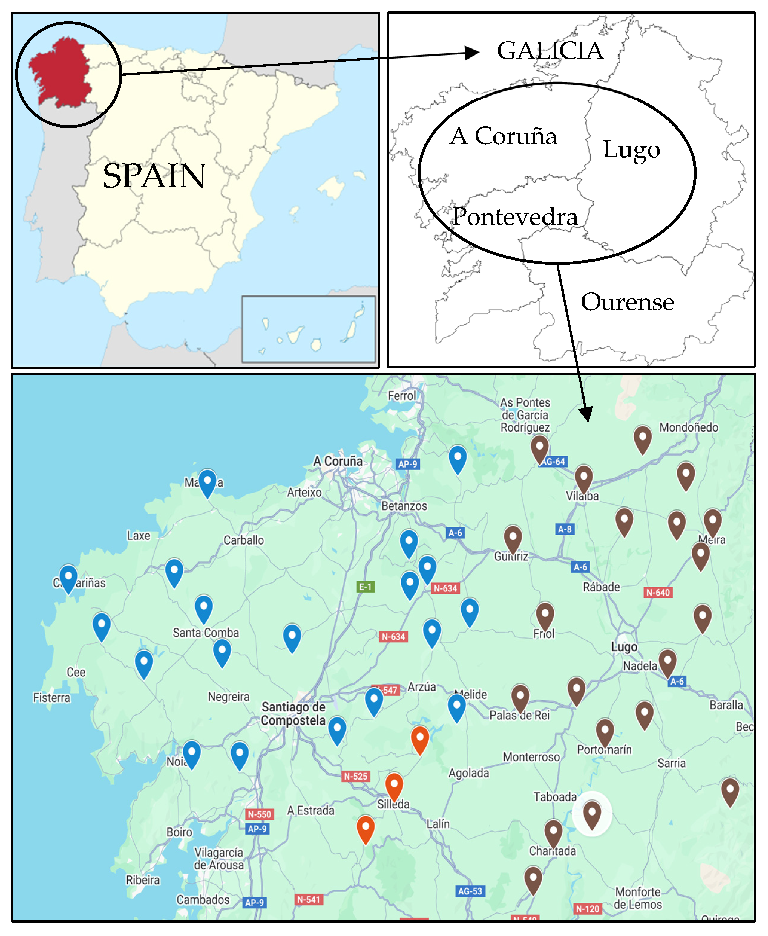
Figure 1. Geographical locations of the sampled dairy farms. Brown markers: Lugo province; blue markers: A Coruña province; and red markers: Pontevedra province.
Antibiotics 14 01059 g001
Figure 2. The wgMLST tree. It illustrates the genetic relatedness among the analyzed isolates based on allelic differences across the whole genome. Each node represents an individual isolate, and branch lengths correspond to the number of allele differences between isolates. Isolates closely clustered share a high proportion of identical alleles, indicating close genetic relationships and potential epidemiological linkage.
Antibiotics 14 01059 g002
Figure 3. Heatmap from wgMLST (101 highly discriminatory loci; see Supplementary File S2C). Each cell represents the number of differing alleles between two genomes, with color intensity indicating the degree of genetic similarity (lighter colors represent closely related isolates, i.e., fewer allele differences; conversely, darker colors indicate greater allelic divergence).
Antibiotics 14 01059 g003
Table 1. Phenotypic antimicrobial profile and genomic resistance to antibiotics from the S. uberis strains studied. The number of virulence factors (VF) has been included. TET, tetracycline; ERY, erythromycin; CLI, clindamycin; CRO, ceftriaxone.
Strains
Code
Phenotypic
Antimicrobial
Profile
Resistance GenesNumber
of VF
1121090TET, ERY, CLIant(6)-Ia_2; lnu(B)_2; lsa(E)_18
1121094-erm(B)_1811
1121108-lnu(C)_110
1121118-lnu(C)_111
1121191 lnu(C)_113
1121208 mph(B)_111
1121227 tet(L)_2; tet(M)_5; ant(6)-Ia_2; lnu(B)_2; lsa(E)_114
1121287-erm(B)_18-
1121292TET, CLIlsa(E)_1; lnu(B)_2; ant(6)-Ia_2; tet(L)_2; tet(M)_513
1121295-erm(B)_18; lnu(C)_110
1121300--12
1121323TET, CLIlsa(E)_1; lnu(B)_2; ant(6)-Ia_2; tet(M)_5; tet(L)_211
1121338--10
1121346 erm(B)_18;aph(3′)-III_1;ant(6)-Ia_1;tet(O)_3-
1121350 lnu(C)_1;erm(B)_18;aph(3′)-III_1;ant(6)-Ia_1;tet(O)_311
1121751CLIlsa(E)_1; lnu(B)_2; ant(6)-Ia_26
1121757TET, CLIlsa(E)_1; lnu(B)_2; ant(6)-Ia_2; mph(B)_1; tet(M)_5; tet(L)_213
1121772 tet(L)_2; tet(M)_5; ant(6)-Ia_2; lnu(B)_2; lsa(E)_1; lnu(C)_110
1121774 lsa(E)_1;lnu(B)_2;ant(6)-Ia_2-
1121776 lnu(B)_2; lsa(E)_1; ant(6)-Ia_2; erm(B)_1811
1121974 tet(O)_3;erm(B)_18;ant(6)-Ia_313
1121980TET, ERY, CLItet(O)_3;ant(6)-Ia_1;aph(3′)-III_1;erm(B)_1813
1121981 -10
1122022 erm(B)_18; aph(3′)-III_1; ant(6)-Ia_1; lnu(B)_2; lsa(E)_19
1122039 ant(6)-Ia_2; lnu(B)_2; lsa(E)_1; tet(S)_311
1122285TET, ERY, CLIlsa(E)_1; lnu(B)_2; ant(6)-Ia_2; erm(B)_18; aph(3′)-III_1; ant(6)-Ia_1; tet(O)_39
1122348TET, ERY, CLIant(6)-Ia_3;erm(B)_18;tet(O)_3;tet(L)_2;tet(M)_5;10
1122419 ant(6)-Ia_2; lnu(B)_2; lsa(E)_110
1122603TET, CLItet(L)_2; tet(M)_5; ant(6)-Ia_2; lnu(B)_2; lsa(E)_114
1122648ERY, CLIerm(B)_188
1122846--9
1122847CLIlsa(E)_1; lnu(B)_2; ant(6)-Ia_211
1122852TET, CLI, CROlsa(E)_1; lnu(B)_2; ant(6)-Ia_2; tet(L)_2; tet(M)_5; lnu(C)_19
1122911 tet(L)_2;tet(M)_5; ant(6)-Ia_2; lnu(B)_2;lsa(E)_1;lnu(D)_1;mph(B)_110
1122931 tet(M)_5; lsa(E)_1; lnu(B)_2; ant(6)-Ia_210
1122956 erm(B)_18;lnu(D)_110
Table 2. Resistance gene classification according to their frequency of appearance and type (left side of the table). Virulence factors (VF) major classes from each strain and their relationship to their related genes (right side of the table).
Strainslnuant(6)-Ialsa(E)_1teterm(B)_18aph(3′)-III_1mph(B)_1StrainsIIIIIIIVVVIVIIVIIIIXXXIXIIXIIIXIVXVXVIXVIIXVIIIXIX
1121090B2+ 1121090
1121094 + 1121094
1121108C 1121108
1121118C 1121118
1121191C 1121191
1121208 +1121208
1121227B2+M, L 1121227
1121287 + 1121287
1121292B2+M, L 1121292
1121295C + 1121295
1121300 1121300
1121323B2+M, L 1121323
1121338 1121338
1121346 1 O++ 1121346
1121350C1 O++ 1121350
1121751B2+ 1121751
1121757B2+M, L +1121757
1121772B, C2+M, L 1121772
1121774B2+ 1121774
1121776B2+ + 1121776
1121974 3 O+ 1121974
1121980 1 O++ 1121980
1121981 1121981
1122022B1+ ++ 1122022
1122039B2+S 1122039
1122285B2, 1+O++ 1122285
1122348 3 M, L, O+ 1122348
1122419B2+ 1122419
1122603B2+M, L 1122603
1122648 + 1122648
1122846 1122846
1122847B2+ 1122847
1122852B, C2+M, L 1122852
1122911B, D2+M, L +1122911
1122931B2+M 1122931
1122956D + 1122956
Types of genes ordered from most to least frequent: Gene lnu: B, gene lnu(B)_2 (18/36 strains); C, gene lnu(C)_1 (7/36); D, gene lnu(D)_1 (2/36). Gene ant(6)-Ia: “2”, gene ant(6)-Ia_2 (17/36); “1”, gene ant(6)-Ia_1 (4/36); “3”, gene ant(6)-Ia_3 (2/36). Gene lsa(E)_1 (18/36). Gene tet: M, gene tet(M)_5 (10/36); L, tet(L)_2 (9/36); O, tet(O)_3 (6/36); S, tet(S)_3 (1/36). Gene erm(B)_18 (13/36). Gene aph(3′)-III_1 (5/36). Gene mph(B)_1 (3/36). Virulence factors (VFs): I: Agglutinin receptor. II: Fibronectin-binding proteins. III: Laminin-binding proteins. IV: Streptococcal lipoprotein rotamase A. V: Streptococcal plasmid receptor/GAPDH. VI: rlrA islet. VII: Hyaluronidase. VIII: Streptococcal enolase. IX: Capsule. X: polysaccharide capsule (Bacillus). XI: Exopolysaccharide (Haemophilus). XII: Rib. XIII: Pneumococcal surface antigen A/Metal binding protein SloC. XIV: C3-degrading protease. XV: C5a peptidase. XVI: Serine protease. XVII: Trigger factor. XVIII: Trehalose-recycling ABC transporter (Mycobacterium). XIX: LPS (Francisella).
Table 3. Number and percentage of strains with detected virulence factors (VF).
VF ClassVFRelated GenesStrains (%) 1
AdherenceAgglutinin receptorUndetermined5 (15.15)
Fibronectin-binding proteinsfbp5433 (100)
Laminin-binding proteinslmb30 (90.91)
Streptococcal lipoprotein rotamase A-/slrA13 (39.39)
Streptococcal plasmid receptor/GAPDHplr/gapA21 (63.64)
rlrA isletsrtC1 (3.03)
EnzymeHyaluronidasehyIB12 (36.36)
Streptococcal enolaseeno31 (93.94)
Immune evasionCapsuleUndetermined33 (100)
Polysaccharide capsule (Bacillus)/galE11 (33.33)
Exopolysaccharide (Haemophilus)galE2 (6.06)
Immunoreactive antigenRibrib4 (12.12)
Manganese uptakePneumococcal surface antigen A/Metal binding protein SloCpsaA33 (100)
ProteaseC3-degrading proteasecppA33 (100)
C5a peptidasescpA/scpB24 (72.73)
Serine proteasehtrA/degP33 (100)
Trigger factortig/ropA21 (63.64)
Cell surface componentsTrehalose-recycling ABC transporter (Mycobacterium)sugC10 (30.30)
Serum resistance and immune evasionLPS (Francisella)wbtP2 (6.06)
1 From a total of 33 strains.
Disclaimer/Publisher’s Note: The statements, opinions and data contained in all publications are solely those of the individual author(s) and contributor(s) and not of MDPI and/or the editor(s). MDPI and/or the editor(s) disclaim responsibility for any injury to people or property resulting from any ideas, methods, instructions or products referred to in the content.