Article

Synthesis and Structure–Activity Relationship Study of 2-(amino)quinazolin-4(3H)-one Derivatives as Potential Inhibitors of Methicillin-Resistant Staphylococcus aureus (MRSA)

1
Infectious Diseases Therapeutic Research Center, Korea Research Institute of Chemical Technology, 141 Gajeong-ro, Yuseong-gu, Daejeon 34114, Republic of Korea
2
Research Institute of Pharmaceutical Sciences, College of Pharmacy, Seoul National University, Seoul 08826, Republic of Korea
3
Antibacterial Resistance Laboratory, Institut Pasteur Korea, Seongam-si 13488, Republic of Korea
4
Data Convergence Drug Research Center, Korea Research Institute of Chemical Technology, Daejeon 34114, Republic of Korea
5
Medicinal Chemistry and Pharmacology, Korea University of Science and Technology (UST), 141 Gajeong-ro, Yuseong-gu, Daejeon 34114, Republic of Korea
6
Department of Biological Systems Engineering, University of Nebraska-Lincoln, Lincoln, NE 68588, USA
7
Department of Food Science and Technology, Nebraska Food for Health Center, University of Nebraska-Lincoln, Lincoln, NE 68588, USA
*
Authors to whom correspondence should be addressed.
These authors contributed equally to this work.
Antibiotics 2025, 14(10), 967; https://doi.org/10.3390/antibiotics14100967 (registering DOI)
Submission received: 20 August 2025 / Revised: 18 September 2025 / Accepted: 19 September 2025 / Published: 25 September 2025

Abstract

Background/Objectives: The rise in methicillin-resistant Staphylococcus aureus (MRSA) demands new therapeutic strategies. In this study, a series of 2-(amino)quinazolin-4(3H)-one derivatives were synthesized and evaluated for antistaphylococcal activity. Methods/Results: Through screening against S. aureus ATCC25923 and USA300 JE2, several submicromolar inhibitors were identified. Among them, compound 6l, which contains a 7-chloro substituent on the key parental scaffold, exhibited strong overall antibacterial activity (MIC50: 1.0 µM, ATCC25923; 0.6 µM, JE2) and served as a lead for further structural optimization. Structure–activity relationship analysis showed that substitution at the 2-position was critical, with its optimized analog 6y (3,4-difluorobenzylamine) exhibiting the highest potency (MIC50: 0.36 µM, ATCC25923; 0.02 µM, JE2). Cytotoxicity assays in HepG2 cells revealed six compounds with IC50 values above 20 µM, yielding efficacy windows greater than 10. Compound 6y exhibited an exceptional index (~885). Consistently, in an H460 lung epithelial infection model mimicking MRSA pneumonia, 6y significantly reduced intracellular bacterial loads with minimal host cell damage, outperforming comparator compounds. Conclusions: These findings highlight 2-(amino)quinazolin-4(3H)-one derivatives, particularly 6y, as promising leads for the development of new antistaphylococcal agents.

1. Introduction

Staphylococcus aureus infections, particularly those caused by methicillin-resistant strains (MRSA), remain a major global health challenge [1,2]. MRSA is a significant public health concern in the context of hospital-acquired infections and is associated with the highest age-standardized mortality rate, especially among hospitalized and immunocompromised patients [3,4,5,6,7]. Although MRSA infections are generally treated with vancomycin, daptomycin, clindamycin, or linezolid, the rapid emergence of resistance to these agents threatens their long-term efficacy [8,9]. This escalating resistance crisis highlights the urgent need for novel antibacterial agents with new mechanisms of action and durable therapeutic effectiveness [1,2,10]. In our previous studies, we identified 3-acyl-2-anilino-1,4-dihydroquinolin-4-one derivatives as promising inhibitors of Gram-positive bacteria, including MRSA [11,12,13]. Building upon this work, further screening efforts led to the discovery of 5,8-dichloro-2-((3,5-dichlorophenyl)amino)quinazolin-4(3H)-one (compound 1) as a hit compound, with an MIC50 of 3.9 µM against S. aureus ATCC25923 (Figure 1). Quinazolin-4(3H)-one scaffolds are structurally versatile and have been widely studied as epidermal growth factor receptor (EGFR) tyrosine inhibitors [14], anti-viral agents [15], and inhibitors of rat lens aldose reductase [16]. Here, we report the design, synthesis, and biological evaluation of a series of 2-(amino)quinazolin-4(3H)-one derivatives. Their antistaphylococcal activities were systematically investigated against MRSA strains, and structure–activity relationship (SAR) analyses were performed to identify key determinants of antibacterial potency. The outcomes of this study provide important insights into the development of new quinazolinone-based agents as potential therapeutics against MRSA.

2. Results and Discussion

2.1. Synthesis of 2-(amino)quinazolin-4(3H)-one Derivatives

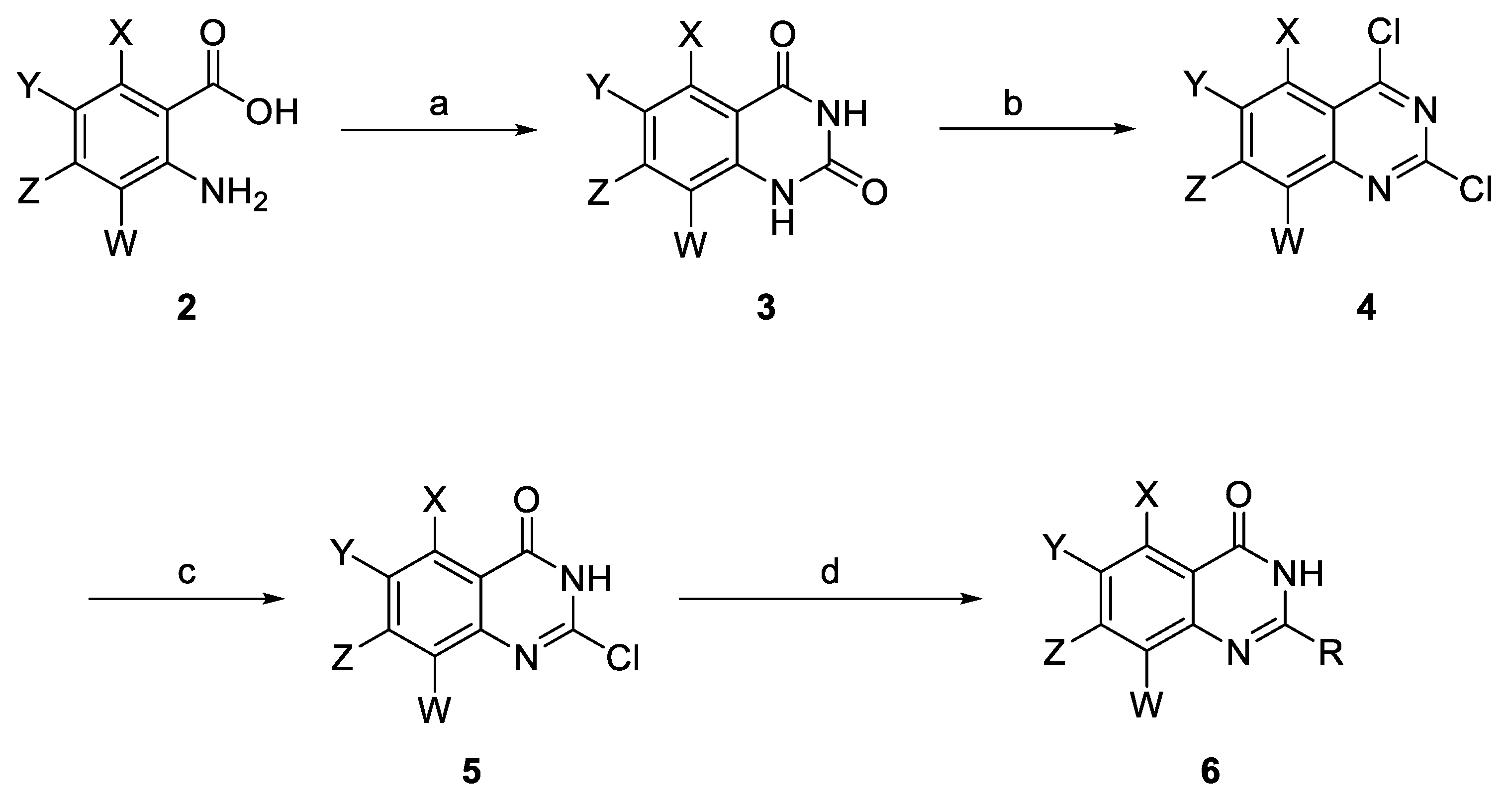
The synthetic methods used to synthesize 2-(amino)quinazolin-4(3H)-one are described in Scheme 1 [15,16]. Anthranilic acids (2) underwent urea-mediated cyclocondensation at 160 °C for 20 h to afford quinazolinediones (3). Chlorination of quinazolinediones (3) with POCl3 in the presence of triethylamine under reflux conditions (120 °C, 17 h) furnished dichloroquinazolines (4). Subsequent base-promoted hydrolysis at C4 (2 N NaOH, rt, 20 h) provided 2-chloro-4(3H)-quinazolinones (5). Finally, nucleophilic substitution at C2 with substituted anilines or amines in DMF at 85 °C for 16 h afforded the target 2-(amino)quinazolin-4(3H)-ones (6).

2.2. Antistaphylococcal Activities Screening and SAR Studies

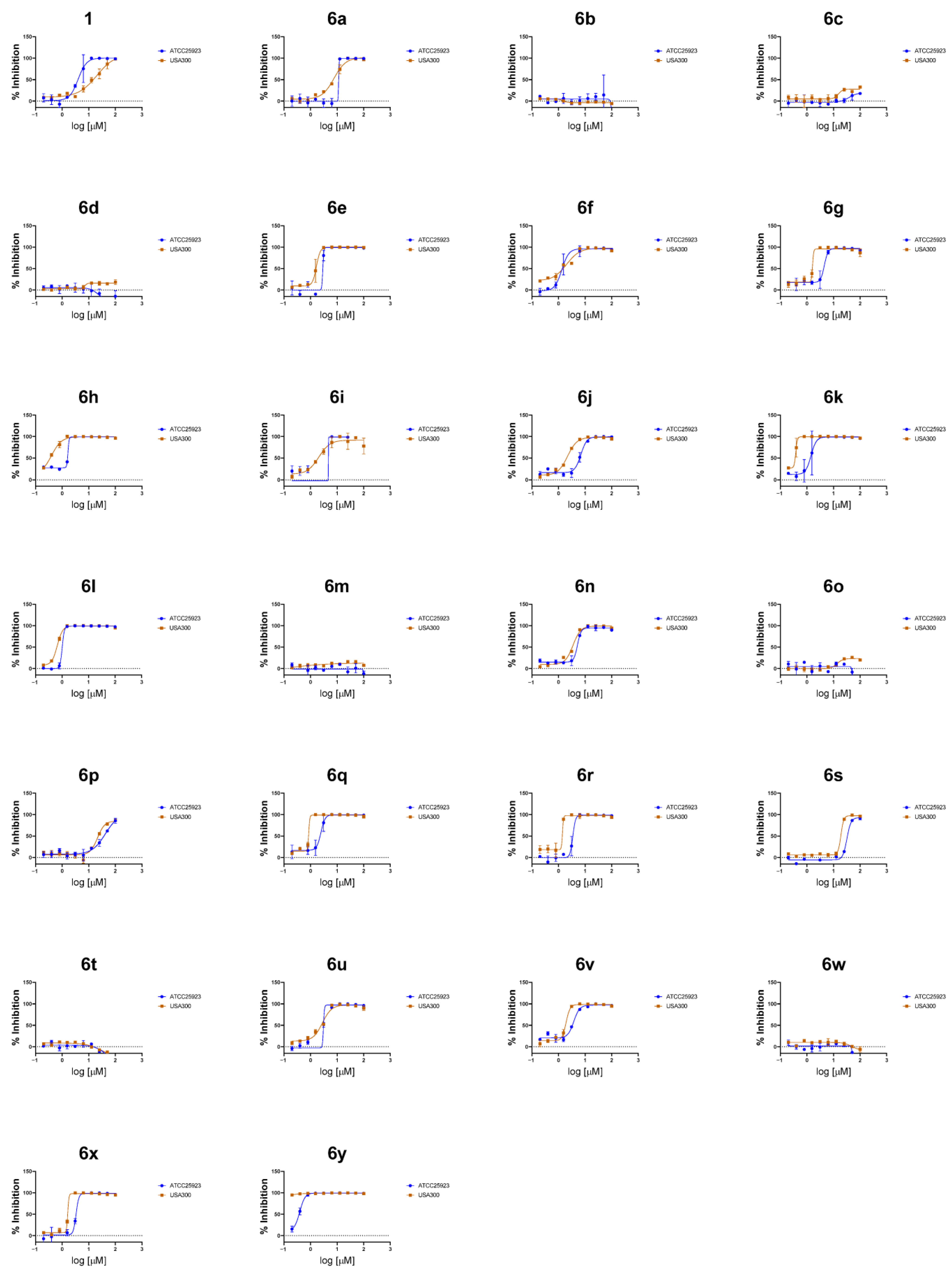
The antistaphylococcal activities (MIC50) of the synthesized compounds were examined via a previously described broth microdilution method against two S. aureus strains: ATCC25923, a methicillin-sensitive reference strain, and JE2, a methicillin-resistant strain of the USA300 lineage (Figure 2, Table 1) [17].
Initially, the effects of substituents at positions 5 through 8 in quinazolin-4(3H)-one of compound 1, having been fixed with 3,5-dichloroaniline at position 2, were investigated (6al). The compound with 7,8-dichloro groups (6a) exhibited decreased antibacterial activity against ATCC25923 (MIC50 = 10.8 μM) but increased activity against JE2 (MIC50 = 6.9 μM) compared to compound 1. A morpholine substitution at position 7 (6b) and an electron-donating group of 6,7-dimethoxy (6c) or a 7-methoxy group (6d) resulted in the complete abolition of antibacterial activity. Compounds with 7-nitro groups (6e), 6,8-dibromo groups (6f), 6,7-difluoro groups (6g), 6,7-dichlro groups (6h), and 6,8-dichloro groups (6i), as well as a compound with no substituents (6j), showed at least 6.5-fold better antibacterial activities (MIC50 = 0.3–1.9 μM) against JE2 with similar activities (MIC50 = 3.0–6.0 μM) against ATCC25923, compared to compound 1. Interestingly, 7-trifluoromethyl (6k) and 7-chloro (6l) substituents resulted in increased antibacterial activity against both strains, showing lower MIC50 values (MIC50 = 1.3 and 1.0 μM for ATCC25923, MIC50 = 0.3 and 0.6 μM for JE2, respectively).
Since the 7-chloro (6l) substituent showed the overall best activity, subsequent investigation was conducted to examine the effect of substituents at position 2 in compound 6l (6my). Decreased antibacterial activities were observed with substituents of 3-methoxyaniline (6p) as well as cyclohexylmethylamine (6s) at position 2, and the complete loss of activity occurred with substituents of aniline (6m), 2,4-difluoroaniline (6o), 2-chloroaniline (6t), and 3,5-difluorobenzylamine (6w) at position 2. Compounds with 3-fluoroaniline (6n), 3,5-difluoroaniline (6q), 3-trifluoromethylaniline (6r), 3-chloroaniline (6u), 4-chloroaniline (6v), and 2,4-dichlorobenzylamine (6x) exhibited less antibacterial activity than compound 61 against both strains (MIC50 = 2.1–4.5 μM for ATCC25923, MIC50 = 0.9–3.0 μM for JE2). Among all derivatives, compound 6y, containing a 3,4-difluorobenzylamine moiety at the 2-position, exhibited the highest antibacterial activity, with MIC50 values of 0.36 µM for ATCC25923 and 0.02 µM for JE2.
Antistaphylococcal activity tests suggested that the synthesized derivatives were improved for inhibitory effects against MRSA USA300 compared to Compound 1, except those with no activity for both strains, ATCC25923 and USA300. Compound 6y demonstrated nanomolar MIC50 values against USA300, highlighting its potential as a lead candidate for MRSA treatment.

2.3. In Vitro Cytotoxicity

The in vitro cytotoxicity of the compounds was evaluated by measuring the viability of HepG2 cells, a human liver cell line, following treatment in a dose–response manner, as previously described [12]. Fifteen compounds with MIC50 values below 5 µM against JE2 were selected for the cytotoxicity testing (Table 1). Among these, six compounds exhibited IC50 values exceeding 20 µM, with the highest IC50 value observed for compound 6l (62 µM). The efficacy window was calculated as the ratio of IC50 in HepG2 cells to MIC50 against JE2. All compounds with IC50 values above 20 µM demonstrated efficacy windows greater than 10, indicating a favorable safety profile. Notably, compound 6y displayed the most promising efficacy window, with an MIC50 USA300 of 0.02 µM, approximately 885 times lower than its IC50 HepG2 value (20.2 µM).

2.4. Antibacterial Activity in a Cell Infection Model

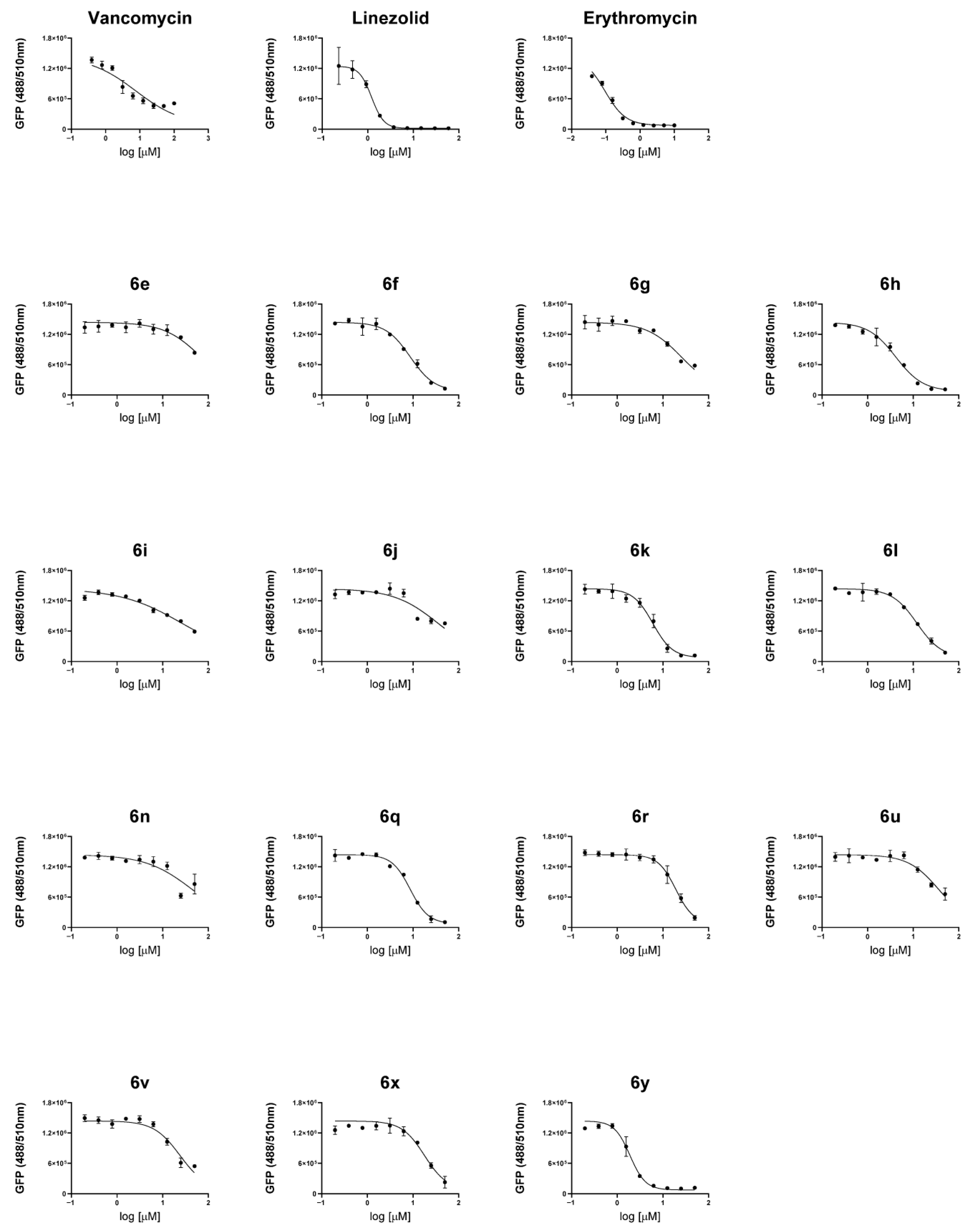
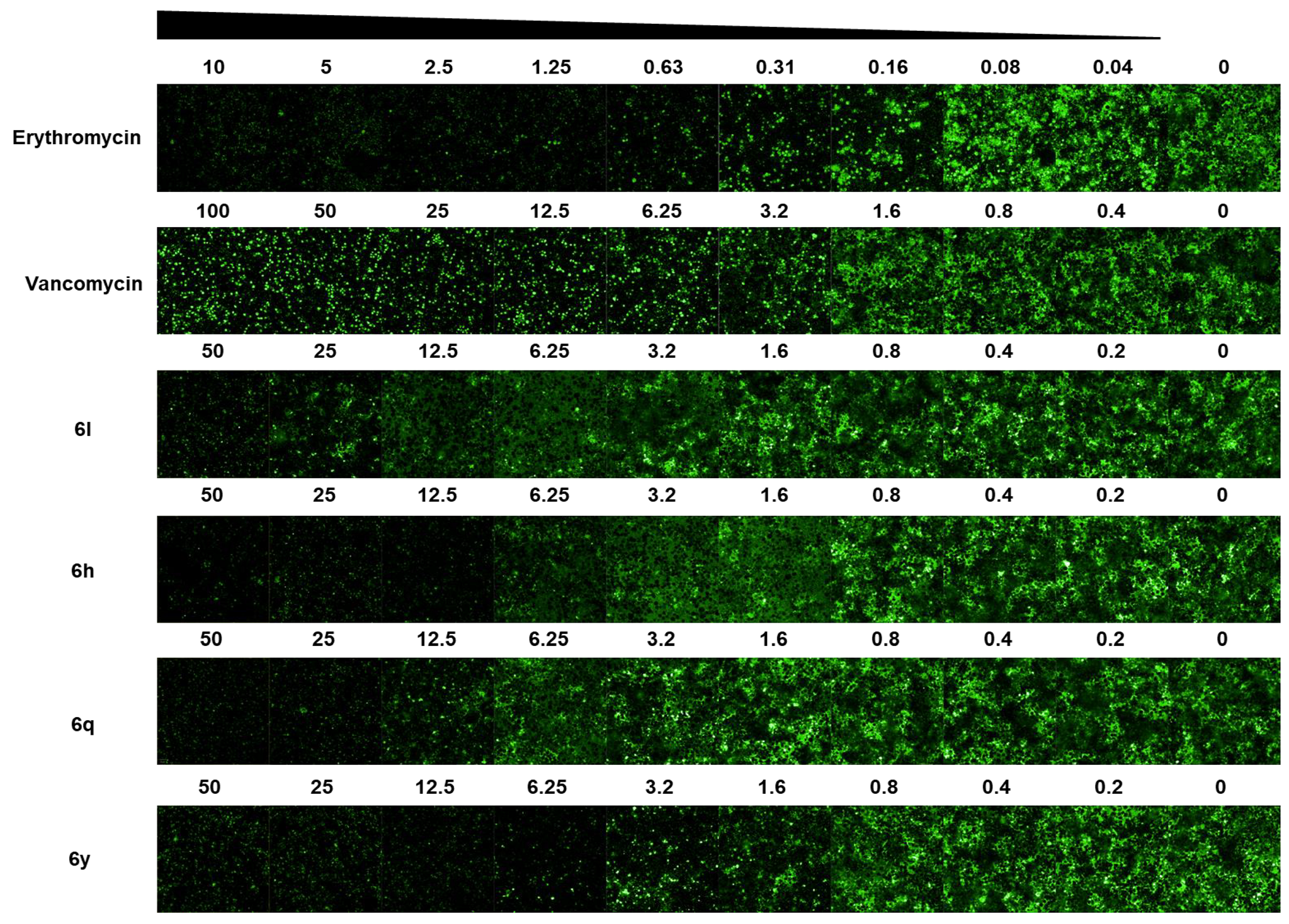
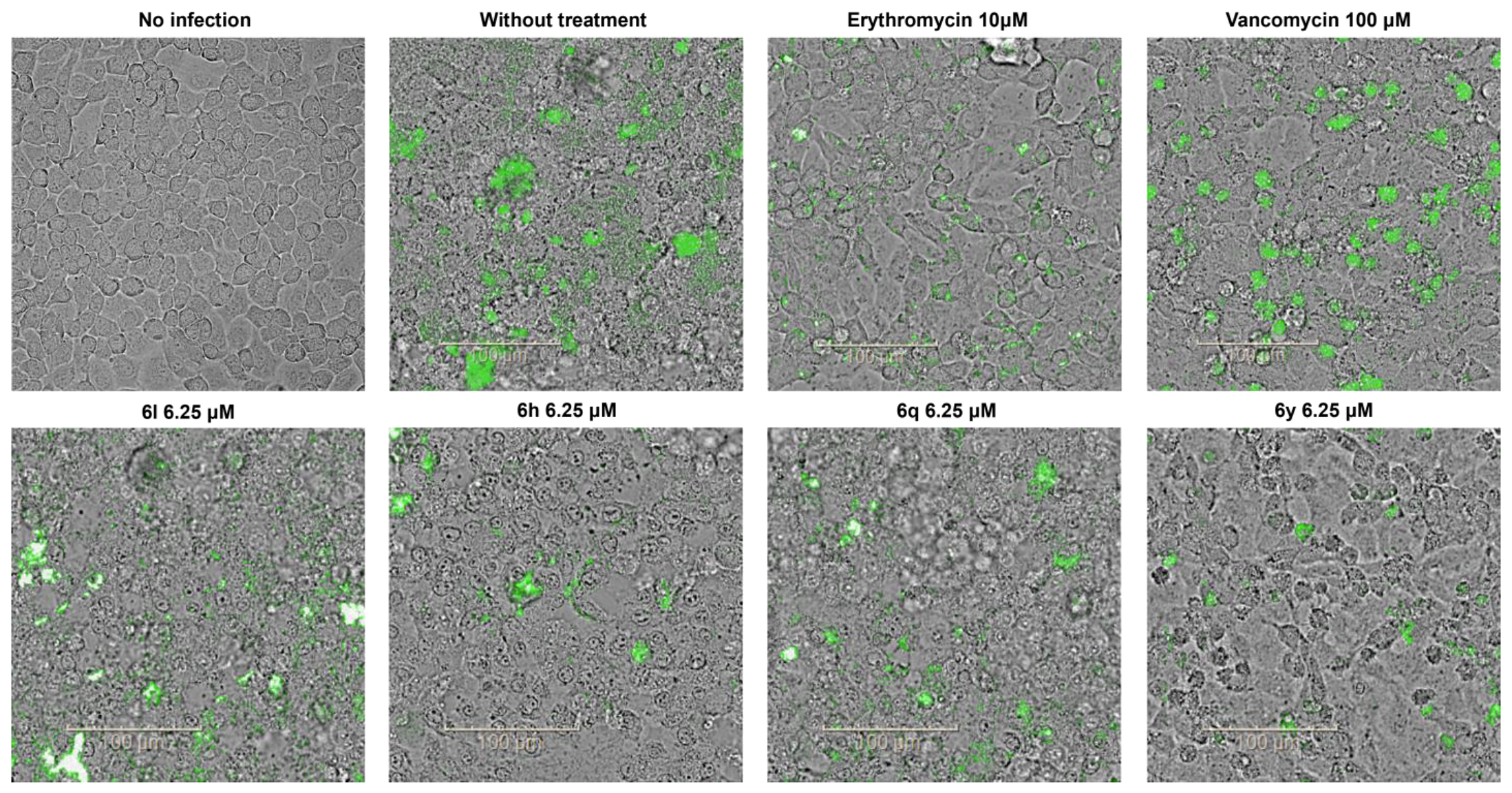
Staphylococcus aureus, including MRSA, can invade host cells and replicate intracellularly, which complicates treatment with current therapies [18,19,20,21,22,23]. To evaluate the intracellular antibacterial activities of the selected compounds, the fifteen compounds tested for cytotoxicity were further assessed using a cell infection model based on H460 human lung epithelial cells, which mimic MRSA-mediated pneumonia. H460 cells were infected with the USA300 JE2 expressing green fluorescent protein (GFP) and subsequently treated with the compounds in a dose–response manner, along with three reference antibiotics: vancomycin, linezolid, and erythromycin. After 24 h of incubation, bacterial loads were quantified by measuring GFP fluorescence in each well. MIC50 values determined in the H460 infection model (MIC50 ex vivo) were generally higher than those obtained from the in vitro tests, likely due to either limited compound penetration into host cells or reduced antibacterial effect of the compounds against intracellular bacteria (Figure 3, Table 2). Despite this reduced efficacy in the H460 infection model, four compounds—6h, 6l, 6q, and 6y—showed at least a two-fold efficacy window (IC50 HepG2/MIC50 ex vivo) (Table 2, Figure 4). Notably, compound 6y, which showed the most potency in vitro, exhibited an MIC50 ex vivo value of 1.9 µM, which was superior to vancomycin (MIC50 ex vivo = 7.7 µM) and similar to linezolid (MIC50 ex vivo = 1.2 µM). The largest efficacy window was also observed for compound 6y (10.6-fold), which caused relatively minimal distortion of H460 cell morphology while markedly reducing the bacterial load compared with the other compounds at the same concentration of 6.25 µM (Table 2, Figure 5). These findings underscore the potential of compound 6y as a promising lead for further development as a novel antistaphylococcal agent.

3. Materials and Methods

3.1. Materials

All reagents and solvents were obtained from commercial suppliers (Sigma-Aldrich, Burlington, MA, USA; Alfa Aesar, Ward Hill, MA, USA; TCI, Tokyo, Japan; Combi-Blocks, San Diego, CA, USA; Angene, Nanjing, China) and used as received unless otherwise noted. Organic solvents were removed under reduced pressure using a rotary evaporator (Rotavapor R-300, Büchi, Flawil, Switzerland). Reaction progress was monitored by thin-layer chromatography (TLC) on a silica gel 60 F254 plate (Merck, Darmstadt, Germany). Crude products were purified by flash column chromatography on silica gel (230–400 mesh, Merck).
Liquid chromatography was performed on a UPLC (Acquity H class, Waters, Milford, MA, USA) using a C18 column (Waters, Acquity UPLC BCH C18, 1.7 μM, 2.1 × 50 mm) maintained at 40 °C. Mobile phase A was water with 0.1% formic acid; mobile phase B was acetonitrile with 0.1% formic acid. The gradient program was as follows: 0–1 min, 5% B; 1–3 min, 5%→95% B (linear); 3–4 min, 95% B (hold); 4–5 min, 95%→5% B; 5–7 min, 5% B (re-equilibration). The flow rate was 0.3 mL/min, and the injection volume was 1 μL. UV detection was carried out with a DAD at 254 nm. Mass spectrometry was performed on an SQ detector 2 (Waters) equipped with an ESI source operating in positive or negative ion mode. Acquisition was carried out in full scan, m/z 100–1000. Key source parameters were as follows: capillary voltage 1.0 kV; desolvation temperature 350 °C; drying gas 650 L/h; nebulizer 35 psi. Data were processed using MassLynx V4.2. HRMS analysis in EI mode, 70eV, and resolution 5000 was conducted using a JEOL JMS-700 High-Resolution Mass Spectrometer (JEOL Ltd., Tokyo, Japan).
NMR spectra were recorded at 300 MHz, 400 MHz, 500 MHz (1H), and 100 MHz, 125 MHz (13C), and chemical shifts are reported in δ values downfield from TMS or relative to residual chloroform (7.26 ppm, 77.16 ppm), with residual DMSO (2.50 ppm, 39.52 ppm) as an internal standard. Data are reported in the following manner: chemical shift, multiplicity, coupling constant (J) in hertz (Hz), and integrated intensity and assignment (when possible). Multiplicities are reported using the following abbreviations: s, singlet; d, doublet; dd, doublet of doublets; t, triplet; q, quadruplet; m, multiplet; br s, broad signal; app, apparent.

3.2. Chemistry

3.2.1. 5,8-Dichloro-2-((3,5-dichlorophenyl)amino)quinazolin-4(3H)-one (1)

Following the procedure of compound 6l. The compound 1 (71% chemical yield) was obtained as a white solid. 1H NMR (300 MHz, (CD3)2SO) δ 11.36 (s, 1H), 9.32 (s, 1H), 8.27 (d, J = 1.9 Hz, 1H), 8.07 (d, J = 1.9 Hz, 2H), 7.80 (d, J = 3.5 Hz, 1H), 7.26 (t, J = 1.8 Hz, 1H) ppm; 13C NMR (100 MHz, (CD3)2SO) δ 159.31, 148.19, 147.44, 140.82, 134.03, 133.84, 131.54, 127.91, 125.77, 121.68, 117.36, 116.69 ppm; HRMS (EI) m/z: [M]+ calcd for C14H7Cl4N3O: 372.9343; found: 372.9343.

3.2.2. 7-Chloroquinazoline-2,4(1H,3H)-dione (3l)

4-Chloroanthranilic acid (2l, 5 g, 29.1 mmoL) and urea (30 g, 582 mmoL) were poured into a 100 mL round-bottom flask. Then, the reaction mixture was heated at 150 °C for 22 h. After the reaction was completed, the mixture was cooled down to room temperature. Then, 50 mL of water was poured into the reaction mixture and heated at 110 °C for 1 h. The reaction mixture was cooled down using an ice bath, and the white solid was precipitated. The white solid was filtered and washed with water and hexane. The white solid (3l, 4.95 g, 87%) was dried in vacuo and used in the next step without further purification. 1H NMR (400 MHz, (CD3)2SO) δ 11.32 (s, 2H), 7.87 (d, J = 8.4 Hz, 1H), 7.33–7.06 (m, 2H).

3.2.3. 2,4,7-Trichloroquinazoline (4l)

The compound 3l (5.6 g, 28.5 mmoL) was dissolved in triethylamine (8 mL, 57 mmoL). Then, POCl3 (24 mL, 256.4 mmoL) was slowly added to the stirred reaction mixture. The reaction mixture was heated at 115 °C for 15 h. After the reaction was completed, the solvents were evaporated with toluene several times. The residue was diluted with water and extracted with ethyl acetate several times. The extracted organic layer was dried over MgSO4 and filtered, concentrated in vacuo. The residue 4l (6.63 g, 99.6%) was used in the next step without further purification. 1H NMR (300 MHz, CDCl3) δ 8.22–8.19 (d, J = 9.0 Hz, 1H), 8.00 (s, 1H), 7.71–7.68 (d, J = 9 Hz, 1H).; LC/MS (ESI) m/z: [M + H]+ calcd for C8H3Cl3N2: 232.9; found: 232.9.

3.2.4. 2,7-Dichloroquinazolin-4(3H)-one (5l)

The compound 4l (2 g, 8.6 mmoL) was dissolved in 2 N NaOH solution (13 mL, 26 mmoL) and stirred at room temperature for 12 h. After the reaction was completed, the acetic acid (1.5 mL, 26 mmoL) was added to the reaction mixture. The aqueous phase was extracted with ethyl acetate several times. The extracted organic layer was dried over MgSO4 and filtered, concentrated in vacuo. The residue 5l (1.8 g, 99%) was used in next step without further purification. 1H NMR (300 MHz, (CD3)2CO) δ 8.14 (d, J = 8.5 Hz, 1H), 7.63 (d, J = 2.1 Hz, 1H), 7.56 (dd, J = 8.5, 2.1 Hz, 1H).; LC/MS (ESI) m/z: [M + H]+ calcd for C8H4Cl2N2O: 215.0; found: 215.1.

3.2.5. 7,8-Dichloro-2-((3,5-dichlorophenyl)amino)quinazolin-4(3H)-one (6a)

Following the procedure of compound 6l. Compound 6a (10% chemical yield) was obtained as a white solid. mp > 300 °C; 1H NMR (500 MHz, DMSO) δ 11.05 (s, 1H), 9.07 (s, 1H), 8.00 (d, J = 1.9 Hz, 2H), 7.93 (d, J = 8.5 Hz, 1H), 7.43 (d, J = 8.5 Hz, 1H), 7.19 (d, J = 2.2 Hz, 1H).; 13C NMR (125 MHz, DMSO) δ 160.26, 147.73, 140.53, 137.25, 133.71, 124.90, 123.90, 121.49, 118.17, 117.43.; HRMS (EI) m/z: [M]+ calcd for C14H7Cl4N3O: 372.9343; found: 372.9340.

3.2.6. 2-((3,5-Dichlorophenyl)amino)-7-morpholinoquinazolin-4(3H)-one (6b)

The 7-Chloro-2-((3,5-dichlorophenyl)amino)quinazolin-4(3H)-one (6l, 50 mg, 0.15 mmol) was dissolved in morpholine (1 mL). The test tube was sealed with a cap and heated at 130 °C for 22 h. After the reaction was completed, ice water was poured into the reaction mixture, and a precipitate formed. The precipitate was filtered and washed with water and hexane thoroughly. The solid was dried in a vacuum, and 6b (11 mg, 19% yield) was obtained as a white solid. 1H NMR (300 MHz, (CD3)2SO) δ 9.02 (s, 1H), 7.82 (s, 3H), 7.78 (s, 1H), 7.21 (t, J = 1.9 Hz, 1H), 6.97 (d, J = 9.2 Hz, 1H), 6.73 (d, J = 2.4 Hz, 1H), 3.73 (d, J = 4.9 Hz, 4H), 3.30 (d, J = 6.5 Hz, 4H).; LC/MS (ESI) m/z: [M + H]+ calcd for C18H16Cl2N4O2: 391.1; found: 391.4.

3.2.7. 2-((3,5-Dichlorophenyl)amino)-6,7-dimethoxyquinazolin-4(3H)-one (6c)

Following the procedure of compound 6l. Compound 6c (90% chemical yield) was obtained as a gray solid. mp 277–279 °C; 1H NMR (400 MHz, (CD3)2SO) δ 11.52 (s, 1H), 11.01 (s, 1H), 7.87 (d, J = 1.9 Hz, 2H), 7.34 (s, 1H), 7.16 (t, J = 1.9 Hz, 1H), 6.91 (s, 1H), 3.90 (s, 3H), 3.82 (s, 3H).; 13C NMR (100 MHz, (CD3)2SO) δ 154.82, 146.45, 142.12, 134.09, 120.70, 116.19, 111.05, 105.60, 55.88, 55.62.; HRMS (EI) m/z: [M]+ calcd for C16H13Cl2N3O3: 365.0334; found: 365.0334.

3.2.8. 2-((3,5-Dichlorophenyl)amino)-7-methoxyquinazolin-4(3H)-one (6d)

Following the procedure of compound 6l. Compound 6d (91% chemical yield) was obtained as a white solid. 1H NMR (400 MHz, (CD3)2SO) δ 10.89 (s, 1H), 8.98 (s, 1H), 7.90–7.86 (m, 1H), 7.84–7.79 (m, 2H), 7.23 (t, J = 1.9 Hz, 1H), 6.88–6.85 (m, 2H), 3.88 (s, 3H) ppm; LC/MS (ESI) m/z: [M + H]+ calcd for C15H11Cl2N3O2: 336.0; found: 336.3.

3.2.9. 2-((3,5-Dichlorophenyl)amino)-7-nitroquinazolin-4(3H)-one (6e)

Following the procedure of compound 6l. Compound 6e (40% chemical yield) was obtained as a yellow solid. 1H NMR (400 MHz, (CD3)2SO) δ 11.50 (s, 1H), 9.24 (s, 1H), 8.18 (d, J = 8.7 Hz, 1H), 8.10 (d, J = 2.3 Hz, 1H), 7.98 (dd, J = 8.6, 2.3 Hz, 1H), 7.83 (s, 2H), 7.27 (t, J = 1.8 Hz, 1H).; 13C NMR (100 MHz, DMSO) δ 162.33, 151.41, 140.96, 134.10, 128.08, 122.10, 120.22, 117.97, 117.12.; HRMS (EI) m/z: [M]+ calcd for C14H8Cl2N4O3: 349.9973; found: 349.9951.

3.2.10. 6,8-Dibromo-2-((3,5-dichlorophenyl)amino)quinazolin-4(3H)-one (6f)

Following the procedure of compound 6l. Compound 6f (10% chemical yield) was obtained as a brown solid. 1H NMR (400 MHz, (CD3)2SO) δ 11.64 (s, 1H), 9.57 (s, 1H), 8.18 (d, J = 2.2 Hz, 1H), 8.06 (d, J = 1.9 Hz, 2H), 8.01 (d, J = 2.3 Hz, 1H), 7.21 (d, J = 2.1 Hz, 1H).; 13C NMR (100 MHz, (CD3)2SO) δ 160.73, 148.02, 146.49, 141.06, 139.15, 134.05, 127.75, 121.58, 121.06, 120.89, 117.42, 114.88.; HRMS (EI) m/z: [M]+ calcd for C14H7Br2Cl2N3O: 460.8333; found: 460.8310.

3.2.11. 2-((3,5-Dichlorophenyl)amino)-6,7-difluoroquinazolin-4(3H)-one (6g)

Following the procedure of compound 6l. Compound 6g (11% chemical yield) was obtained as a brown solid.; mp > 300 °C; 1H NMR (400 MHz, (CD3)2SO) δ 10.57 (s, 1H), 8.33 (s, 1H), 7.65 (d, J = 1.9 Hz, 2H), 7.36–7.21 (m, 2H).; 13C NMR (100 MHz, (CD3)2SO) δ 162.78, 160.44, 141.24, 140.46, 134.72, 134.22, 122.93, 122.53, 117.37, 115.47.; HRMS (EI) m/z: [M]+ calcd for C14H7Cl2F2N3O: 340.9934; found: 340.9922.

3.2.12. 6,7-Dichloro-2-((3,5-dichlorophenyl)amino)quinazolin-4(3H)-one (6h)

Following the procedure of compound 6l. Compound 6h (7% chemical yield) was obtained as a white solid. 1H NMR (300 MHz, (CD3)2SO) δ 11.38 (s, 1H), 9.21 (s, 1H), 8.05 (s, 1H), 7.79 (s, 2H), 7.69 (s, 1H), 7.25 (s, 1H).; LC/MS (ESI) m/z: [M + H]+ calcd for C14H7Cl4N3O: 373.9; found: 374.4.

3.2.13. 6,8-Dichloro-2-((3,5-dichlorophenyl)amino)quinazolin-4(3H)-one (6i)

Following the procedure of compound 6l. Compound 6i (12% chemical yield) was obtained as a white solid. mp > 300 °C; 1H NMR (400 MHz, (CD3)2SO) δ 11.41 (s, 1H), 9.43 (s, 1H), 7.99 (s, 2H), 7.97–7.89 (m, 1H), 7.89–7.78 (m, 1H), 7.21 (s, 1H).; 13C NMR (100 MHz, (CD3)2SO) δ 160.62, 148.05, 145.51, 141.47, 134.53, 134.31, 130.39, 127.51, 124.59, 122.16, 121.26, 117.81.; HRMS (EI) m/z: [M]+ calcd for C14H7Cl4N3O: 372.9343; found: 372.9337.

3.2.14. 2-((3,5-Dichlorophenyl)amino)quinazolin-4(3H)-one (6j)

Following the procedure of compound 6l. Compound 6j (80% chemical yield) was obtained as a white solid.; mp > 300 °C; 1H NMR (400 MHz, (CD3)2SO) δ 11.08 (s, 1H), 9.04 (s, 1H), 7.98 (dd, J = 7.9, 1.6 Hz, 1H), 7.81 (d, J = 2.0 Hz, 2H), 7.67 (ddd, J = 8.5, 7.1, 1.6 Hz, 1H), 7.42 (d, J = 8.1 Hz, 1H), 7.30–7.25 (m, 1H), 7.19 (t, J = 1.9 Hz, 1H).; 13C NMR (100 MHz, (CD3)2SO) δ 162.17, 147.44, 142.03, 135.02, 134.48, 126.43, 125.76, 124.21, 121.88, 119.07, 117.87.; HRMS (EI) m/z: [M]+ calcd for C14H9Cl2N3O: 305.0123; found: 305.0127.

3.2.15. 2-((3,5-Dichlorophenyl)amino)-7-(trifluoromethyl)quinazolin-4(3H)-one (6k)

Following the procedure of compound 6l. Compound 6k (33% chemical yield) was obtained as a pink solid.; mp > 300 °C; 1H NMR (400 MHz, (CD3)2SO) δ 11.29 (s, 1H), 9.10 (s, 1H), 8.12 (d, J = 8.2 Hz, 1H), 7.78 (s, 2H), 7.66 (s, 1H), 7.54–7.48 (m, 1H), 7.20 (d, J = 2.0 Hz, 1H).; 13C NMR (100 MHz, (CD3)2SO) δ 160.81, 149.50, 148.01, 141.03, 134.03, 127.57, 125.00, 122.29, 121.87, 121.42, 119.18, 117.71; HRMS (EI) m/z: [M]+ calcd for C15H8Cl2F3N3O: 372.9997; found: 372.9996.

3.2.16. 7-Chloro-2-((3,5-dichlorophenyl)amino)quinazolin-4(3H)-one (6l)

To a stirred solution of compound 5l (200 mg, 0.93 mmoL) in DMF (3.1 mL), 3,5-dichloroaniline (7l, 452 mg, 2.8 mmoL) was slowly added. The reaction mixture was heated at 85 °C for 19 h. The reaction mixture was cooled down to room temperature, and precipitates were formed. The precipitates were filtered and washed with water and hexane thoroughly. The white solid (285 mg, 90%) was dried in vacuo. If the precipitates were not formed, the reaction mixture was purified by prep HPLC (Shim-pack PREP-ODS, H2O:CH3CN = 90:10 to H2O:CH3CN = 10:90, flow rate = 12 mL/min, 40 °C, λ = 254 nm, retention time: 30 min).; mp > 300 °C; 1H NMR (400 MHz, (CD3)2SO) δ 11.09 (s, 1H), 9.02 (s, 1H), 7.93 (d, J = 8.4 Hz, 1H), 7.76 (s, 2H), 7.41 (s, 1H), 7.32–7.05 (m, 2H); 13C NMR (100 MHz, (CD3)2SO) δ 160.88, 150.47, 147.85, 141.09, 139.13, 134.00, 127.82, 124.47, 123.77, 121.71, 117.58; HRMS (FAB) m/z: [M + H]+ calcd for C14H8Cl3N3O: 339.9733; found: 339.9813.

3.2.17. 7-Chloro-2-(phenylamino)quinazolin-4(3H)-one (6m)

Following the procedure of compound 6l. Compound 6m (94% chemical yield) was obtained as a white solid. mp 258–261 °C; 1H NMR (400 MHz, (CD3)2SO) δ 10.90 (s, 1H), 8.75 (s, 1H), 7.94 (d, J = 8.4 Hz, 1H), 7.76–7.68 (m, 2H), 7.43 (d, J = 2.0 Hz, 1H), 7.38–7.32 (m, 2H), 7.23 (dd, J = 8.4, 2.1 Hz, 1H), 7.09–7.02 (m, 1H).; 13C NMR (100 MHz, (CD3)2SO) δ 160.99, 151.31, 148.39, 139.09, 138.63, 128.87, 127.88, 124.39, 123.15, 122.84, 119.62, 117.17.; HRMS (EI) m/z: [M]+ calcd for C14H10ClN3O: 271.0512; found: 271.0531.

3.2.18. 7-Chloro-2-((3-fluorophenyl)amino)quinazolin-4(3H)-one (6n)

Following the procedure of compound 6l. Compound 6n (93% chemical yield) was obtained as a white solid. mp 271–273 °C; 1H NMR (400 MHz, (CD3)2SO) δ 10.95 (s, 1H), 8.96 (s, 1H), 7.94 (d, J = 8.4 Hz, 1H), 7.91–7.81 (m, 1H), 7.48 (d, J = 2.0 Hz, 1H), 7.39–7.29 (m, 2H), 7.25 (dd, J = 8.5, 2.1 Hz, 1H), 6.86 (td, J = 8.7, 2.3 Hz, 1H).; 13C NMR (100 MHz, (CD3)2SO) δ 163.51, 161.01 (d, J(C-F) = 21.0 Hz), 150.93, 148.11, 140.45 (d, J(C-F) = 11.4 Hz), 139.16, 130.34 (d, J(C-F) = 9.8 Hz), 127.86, 124.55, 123.52, 117.32, 115.23, 109.09 (d, J(C-F) = 21.2 Hz), 106.32 (d, J(C-F) = 27.0 Hz).; HRMS (EI) m/z: [M]+ calcd for C14H9ClFN3O: 289.0418; found: 289.0408.

3.2.19. 7-Chloro-2-((2,4-difluorophenyl)amino)quinazolin-4(3H)-one (6o)

Following the procedure of compound 6l. Compound 6o (99% chemical yield) was obtained as a white solid. mp 288–290 °C; 1H NMR (400 MHz, (CD3)2SO) δ 11.20 (s, 1H), 8.59 (s, 1H), 8.45 (td, J = 9.2, 5.9 Hz, 1H), 7.92 (d, J = 8.5 Hz, 1H), 7.39–7.31 (m, 2H), 7.22 (dd, J = 8.5, 2.1 Hz, 1H), 7.08 (tdd, J = 8.4, 3.0, 1.5 Hz, 1H).; 13C NMR (100 MHz, (CD3)2SO) δ 160.87, 158.76 (d, J(C-F) = 12.0 Hz), 156.34 (d, J(C-F) = 11.7 Hz), 154.30 (d, J(C-F) = 12.4 Hz), 151.85 (d, J(C-F) = 12.5 Hz), 150.95, 148.49, 139.10, 127.88, 124.38, 123.68 (d, J(C-F) = 9.1 Hz), 123.37, 117.23, 111.10 (dd, J(C-F) = 21.6, 3.6 Hz), 104.74–102.78 (m). HRMS (EI) m/z: [M]+ calcd for C14H8ClF2N3O: 307.0324; found: 307.0327.

3.2.20. 7-Chloro-2-((3-methoxyphenyl)amino)quinazolin-4(3H)-one (6p)

Following the procedure of compound 6l. Compound 6p (99% chemical yield) was obtained as a white solid. mp 242–245 °C; 1H NMR (400 MHz, (CD3)2SO) δ 10.87 (s, 1H), 8.76 (s, 1H), 7.94 (d, J = 8.4 Hz, 1H), 7.54 (d, J = 2.3 Hz, 1H), 7.43 (d, J = 2.0 Hz, 1H), 7.27–7.19 (m, 2H), 7.17–7.09 (m, 1H), 6.63 (dd, J = 8.1, 2.5 Hz, 1H), 3.78 (s, 3H).; 13C NMR (100 MHz, (CD3)2SO) δ 160.94, 159.67, 151.20, 148.29, 139.80, 139.12, 129.61, 127.87, 124.42, 123.25, 117.19, 111.76, 108.31, 105.29, 55.05.; HRMS (EI) m/z: [M]+ calcd for C15H12ClN3O2: 301.0618; found: 301.0603.

3.2.21. 7-Chloro-2-((3,5-difluorophenyl)amino)quinazolin-4(3H)-one (6q)

Following the procedure of compound 6l. C 6q (88% chemical yield) was obtained as a yellow solid. mp > 300 °C; 1H NMR (400 MHz, (CD3)2SO) δ 11.04 (s, 1H), 9.10 (s, 1H), 7.93 (d, J = 8.4 Hz, 1H), 7.56–7.36 (m, 3H), 7.25 (dd, J = 8.5, 2.2 Hz, 1H), 6.84 (tt, J = 9.3, 2.4 Hz, 1H).; 13C NMR (100 MHz, (CD3)2SO) δ 163.70 (d, J(C-F) = 15.6 Hz), 161.29 (d, J(C-F) = 15.5 Hz), 160.90, 150.59, 147.89, 141.31 (t, J(C-F) = 14.0 Hz), 139.18, 127.82, 124.66, 123.81, 117.44, 102.27 (d, J(C-F) = 29.6 Hz), 97.60 (t, J(C-F) = 26.2 Hz).; HRMS (EI) m/z: [M]+ calcd for C14H8ClF2N3O: 307.0324; found: 307.0328.

3.2.22. 7-Chloro-2-((3-(trifluoromethyl)phenyl)amino)quinazolin-4(3H)-one (6r)

Following the procedure of compound 6l. Compound 6r (85% chemical yield) was obtained as a white solid. mp 255–257 °C; 1H NMR (400 MHz, (CD3)2SO) δ 11.09 (s, 1H), 9.09 (s, 1H), 8.24 (s, 1H), 7.96 (d, J = 8.4 Hz, 1H), 7.92–7.86 (m, 1H), 7.57 (t, J = 8.0 Hz, 1H), 7.42–7.35 (m, 2H), 7.27 (dd, J = 8.5, 2.0 Hz, 1H).; 13C NMR (100 MHz, (CD3)2SO) δ 161.00, 150.82, 148.26, 139.52, 139.17, 129.98, 129.69, 129.37, 127.94, 125.53, 124.39, 123.61, 123.30, 122.82, 119.03, 117.41, 115.70 (d, J(C-F) = 3.9 Hz).; HRMS (EI) m/z: [M]+ calcd for C15H9ClFN3O: 339.0386; found: 339.0384.

3.2.23. 7-Chloro-2-((cyclohexylmethyl)amino)quinazolin-4(3H)-one (6s)

Following the procedure of compound 6l. Compound 6s (18% chemical yield) was obtained as a white solid. 1H NMR (400 MHz, (CD3)2SO) δ 10.84 (s, 1H), 7.84 (d, J = 8.4 Hz, 1H), 7.24 (d, J = 2.1 Hz, 1H), 7.08 (dd, J = 8.4, 2.1 Hz, 1H), 6.45 (s, 1H), 3.17 (t, J = 6.2 Hz, 2H), 1.69 (ddd, J = 13.1, 8.5, 4.6 Hz, 4H), 1.62 (t, J = 5.2 Hz, 1H), 1.51 (ddt, J = 11.1, 7.2, 3.6 Hz, 1H), 1.22–1.12 (m, 3H), 0.95 (td, J = 11.7, 3.0 Hz, 2H).; 13C NMR (100 MHz, (CD3)2SO) δ 161.27, 160.93, 151.63, 138.83, 127.94, 123.52, 121.56, 116.09, 46.25, 37.16, 30.29, 26.04, 25.37.; HRMS (EI) m/z: [M]+ calcd for C15H18ClN3O2: 291.1138; found: 291.1118.

3.2.24. 7-Chloro-2-((2-chlorophenyl)amino)quinazolin-4(3H)-one (6t)

Following the procedure of compound 6l. Compound 6t (94% chemical yield) was obtained as a white solid. mp > 300 °C; 1H NMR (400 MHz, (CD3)2SO) δ 11.65 (s, 1H), 8.52 (d, J = 8.3 Hz, 1H), 8.39 (s, 1H), 7.96 (d, J = 8.4 Hz, 1H), 7.52 (dd, J = 8.0, 1.5 Hz, 1H), 7.44 (d, J = 2.1 Hz, 1H), 7.38 (ddd, J = 8.5, 7.4, 1.6 Hz, 1H), 7.27 (dd, J = 8.5, 2.1 Hz, 1H), 7.14 (td, J = 7.7, 1.6 Hz, 1H).; 13C NMR (100 MHz, (CD3)2SO) δ 161.01, 150.88, 148.44, 139.10, 134.98, 129.34, 127.88, 127.66, 124.49, 123.55, 123.46, 123.15, 117.41.; HRMS (EI) m/z: [M]+ calcd for C14H9Cl2N3O: 305.0123; found: 305.0121.

3.2.25. 7-Chloro-2-((3-chlorophenyl)amino)quinazolin-4(3H)-one (6u)

Following the procedure of compound 6l. Compound 6u (99% chemical yield) was obtained as a white solid. mp 284–287 °C; 1H NMR (400 MHz, (CD3)2SO) δ 10.99 (s, 1H), 8.93 (s, 1H), 7.97 (t, J = 2.2 Hz, 1H), 7.94 (d, J = 8.4 Hz, 1H), 7.53 (dd, J = 8.2, 2.2 Hz, 1H), 7.44 (d, J = 2.0 Hz, 1H), 7.35 (t, J = 8.1 Hz, 1H), 7.25 (dd, J = 8.4, 2.1 Hz, 1H), 7.10 (dd, J = 8.0, 2.1 Hz, 1H).; 13C NMR (100 MHz, (CD3)2SO) δ 160.94, 160.01, 150.89, 148.15, 140.17, 139.14, 133.20, 130.62, 130.42, 127.90, 124.45, 123.53, 123.37, 122.43, 118.97, 118.70, 118.06, 117.54, 117.33.; HRMS (EI) m/z: [M]+ calcd for C14H9Cl2N3O: 305.0123; found: 305.0107.

3.2.26. 7-Chloro-2-((4-chlorophenyl)amino)quinazolin-4(3H)-one (6v)

Following the procedure of compound 6l. Compound 6v (89% chemical yield) was obtained as a white solid. mp > 300 °C; 1H NMR (400 MHz, (CD3)2SO) δ 10.95 (s, 1H), 8.88 (s, 1H), 7.94 (d, J = 8.4 Hz, 1H), 7.75 (d, J = 8.7 Hz, 2H), 7.43 (d, J = 2.1 Hz, 1H), 7.40–7.35 (m, 2H), 7.24 (dd, J = 8.4, 2.1 Hz, 1H).; 13C NMR (100 MHz, (CD3)2SO) δ 160.96, 151.06, 148.24, 139.10, 137.64, 128.67, 127.92, 126.40, 124.41, 123.37, 121.24, 117.25.; HRMS (EI) m/z: [M]+ calcd for C14H9Cl2N3O: 305.0123; found: 305.0146.

3.2.27. 7-Chloro-2-((3,5-difluorobenzyl)amino)quinazolin-4(3H)-one (6w)

Following the procedure of compound 6l. Compound 6w (49% chemical yield) was obtained as a white solid. mp 253–255 °C; 1H NMR (400 MHz, (CD3)2SO) δ 7.88 (d, J = 8.5 Hz, 1H), 7.27 (d, J = 2.0 Hz, 1H), 7.15 (dd, J = 8.5, 2.1 Hz, 1H), 7.11 (td, J = 7.5, 2.9 Hz, 3H), 4.59 (d, J = 5.2 Hz, 2H).; 13C NMR (100 MHz, (CD3)2SO) δ 163.62 (d, J(C-F) = 13.1 Hz), 161.44, 161.18 (d, J(C-F) = 13.2 Hz), 151.43, 144.09 (t, J(C-F) = 8.9 Hz), 138.96, 128.06, 122.33, 116.33, 110.79–109.58 (m), 102.35 (t, J(C-F) = 25.8 Hz), 42.99. HRMS (EI) m/z: [M]+ calcd for C15H10ClF2N3O: 321.0480; found: 321.0480.

3.2.28. 7-Chloro-2-((2,4-dichlorobenzyl)amino)quinazolin-4(3H)-one (6x)

Following the procedure of compound 6l. Compound 6x (10% chemical yield) was obtained as a white solid. 1H NMR (300 MHz, (CD3)2SO) δ 11.23 (s, 1H), 7.88 (d, J = 8.5 Hz, 1H), 7.64 (d, J = 2.0 Hz, 1H), 7.49–7.39 (m, 2H), 7.26 (d, J = 2.0 Hz, 1H), 7.14 (dd, J = 8.4, 2.1 Hz, 1H), 7.01 (s, 1H), 4.60 (d, J = 5.9 Hz, 2H).; LC/MS (ESI) m/z: [M + H]+ calcd for C15H10Cl3N3O: 354.0; found: 353.3.

3.2.29. 7-Chloro-2-((3,4-difluorobenzyl)amino)quinazolin-4(3H)-one (6y)

Following the procedure of compound 6l. Compound 6y (69% chemical yield) was obtained as a yellow solid. 1H NMR (400 MHz, (CD3)2SO) δ 7.89 (d, J = 8.4 Hz, 1H), 7.50–7.44 (m, 1H), 7.45–7.33 (m, 2H), 7.31 (d, J = 2.0 Hz, 1H), 7.26–7.21 (m, 1H), 7.18 (dd, J = 8.5, 2.1 Hz, 1H), 4.56 (d, J = 4.4 Hz, 2H).; 13C NMR (100 MHz, (CD3)2SO) δ 161.24, 151.32, 150.52 (d, J(C-F) = 12.8 Hz), 149.76 (d, J(C-F) = 12.5 Hz), 148.08 (d, J(C-F) = 12.7 Hz), 147.33 (d, J(C-F) = 12.6 Hz), 139.09, 136.64, 128.14, 124.19 (dd, J(C-F) = 6.5, 3.4 Hz), 122.57, 117.37 (d, J(C-F) = 17.0 Hz), 116.49 (d, J(C-F) = 17.3 Hz), 116.12, 42.83.; HRMS (EI) m/z: [M]+ calcd for C15H10ClF2N3O: 321.0480; found: 321.0476.

3.3. Biological Studies

3.3.1. Strains and Culture Conditions

Staphylococcus aureus USA300, a methicillin-resistant strain, was obtained from the Nebraska Transposon Mutant Library. Staphylococcus aureus ATCC25923, a pan-drug-susceptible strain, was purchased from the American Type Culture Collection (ATCC) (Manassas, VA, USA). S. aureus strains were grown in Cation-Adjusted Mueller–Hinton broth (CAMHB) (Difco) at 37 °C. Bacterial strain stocks were stored in vials with 25% glycerol at −80 °C. For each experiment, overnight cultures were prepared by inoculating a frozen bacterial strain stock in 10 mL of CAMHB medium and incubating at 37 °C in a shaking incubator (180 rpm). For the preliminary screen and dose–response experiments, 100 µL of overnight culture was inoculated into 10 mL of fresh CAMHB and incubated for 3 h at 37 °C and 180 rpm.

3.3.2. Dose–Response Curve Assay

Antibiotic susceptibility assay was tested according to modified Clinical and Laboratory Standards Institute (CLSI) laboratory guidelines for broth microdilution assays. S. aureus was cultured in CAMHB, and bacterial cells were prepared with an OD600 of 0.005. DRC plates were prepared by adding 40 µL of bacterial suspension to each well, where 10 µL of each compound had been dispensed for 17 points of two-fold serial dilutions starting at 40 µM. After incubation of the plates at 37 °C for 18–24 h with 5% CO2, the growth of S. aureus was detected by measuring the OD600nm using an Ensight (Revvity) spectrometer (Revvity, Waltham, MA, USA).

3.3.3. Cytotoxicity Test of Compounds

To test cytotoxicity with compounds, the human liver cell line HepG2 (ATCC #HB-8065) was used for the assay. HepG2 was cultured in DMEM with 10% fetal bovine serum (FBS) in a 96-well microplate at 37 °C and 5% CO2. HepG2 cells were seeded at a final density of 5 × 10⁴ cells/well, and DMEM-containing diluted compounds were added and incubated on the plates at 37 °C in 5% CO2 for 24 h. After that, resazurin (final concentration, 0.1 mg/mL) was added to each well and incubated on the plates at 37 °C in 5% CO2 for 2 h. To detect the fluorescence at 530 nm excitation and 590 nm emission, an Ensight multimode plate reader was used (Revvity, Waltham, MA, USA).

3.3.4. Effect of Compounds on a Cell Infection Model of S. aureus

To see the ex vivo antimicrobial activity of the compound, the human lung carcinoma cell line H460 (ATCC #HTB-177) was used. The H460 cells were cultured in RPMI 1640 medium with 10% fetal bovine serum (FBS) and in a 96-well microplate at 37 °C and 5% CO2. Cell lines were cultured for 2 days, until they reached full confluence in the well. S. aureus (USA300-containing and pCM29-encoding GFP) was cultured in CAMHB with 10 µg/mL of chloramphenicol, and bacterial cells were harvested. After washing the bacterial cells with phosphate-buffered saline (PBS) and suspending them in RPMI, infected H460 cells with S. aureus (1 × 10⁷ CFU) and incubated them for 2.5 h. Samples were washed three times with PBS. Then, cells were treated with gentamicin (50 µg/mL of gentamicin in RPMI) and incubated for 1 h. Samples were washed with PBS, RPMI-containing diluted compounds were added, and then they were incubated at 37 °C in 5% CO2 for 24 h. To detect the Green fluorescent protein (GFP) signal, an Ensight multimode plate reader and Opera Phenix Plus (Revvity, Waltham, MA, USA) were used.

3.3.5. Confocal Images

Ex vivo infection model images were generated using the Opera Phenix Plus high-content imaging system. Images were obtained by a confocal microscope with a 20× water objective lens. To detect the bacteria fluorescence signal (GFP), the Alexa 488 channel (488 nm laser/500–550 filter, Thermo Fisher Scientific, Waltham, MA, USA) was used, and the brightfield channel was used for H460 cell images.

4. Conclusions

This study identified and optimized 2-(amino)quinazolin-4(3H)-one derivatives as promising antistaphylococcal agents. Compound 6l was identified as a leading compound with submicromolar activity, and structural optimization yielded 6y, which exhibited the highest potency (MIC50: 0.36 µM for ATCC25923; 0.02 µM for USA300 JE2) and the greatest efficacy window (~885). In an H460 lung epithelial infection model mimicking MRSA pneumonia, 6y markedly reduced intracellular bacterial loads with minimal host cell damage, demonstrating superior activity to vancomycin and efficacy comparable to linezolid.
Given their structural similarity to 3-acyl-2-phenylamino-1,4-dihydroquinolin-4-one (APDQ) derivatives, which have been shown to disrupt peptidoglycan biosynthesis (11), it is plausible that these compounds may also interfere with bacterial cell wall synthesis. However, the precise antibacterial mechanism of the 2-(amino)quinazolin-4(3H)-one derivatives, including 6l and 6y, remains under investigation, and ongoing mechanistic studies are expected to provide further insight.
Collectively, these findings establish 6l as a key parental compound and 6y as a lead candidate for further development toward novel therapeutics targeting MRSA infections.

Supplementary Materials

The following supporting information can be downloaded at https://www.mdpi.com/article/10.3390/antibiotics14100967/s1: Figure S1: The structures of 2-(amino)quinazolin-4(3H)-one derivatives; Figure S2: 1H-NMR Spectrum (400 MHz, (CD3)2SO) and 13C-NMR Spectrum (100 MHz, (CD3)2SO) of compound 6f; Figure S3: 1H-NMR Spectrum (400 MHz, (CD3)2SO) and 13C-NMR Spectrum (100 MHz, (CD3)2SO) of compound 6h; Figure S4: 1H-NMR Spectrum (400 MHz, (CD3)2SO) and 13C-NMR Spectrum (100 MHz, (CD3)2SO) of compound 6j.; Figure S5: 1H-NMR Spectrum (400 MHz, (CD3)2SO) and 13C-NMR Spectrum (100 MHz, (CD3)2SO) of compound 6k; Figure S6: 1H-NMR Spectrum (400 MHz, (CD3)2SO) and 13C-NMR Spectrum (100 MHz, (CD3)2SO) of compound 6l; Figure S7: 1H-NMR Spectrum (400 MHz, (CD3)2SO) and 13C-NMR Spectrum (100 MHz, (CD3)2SO) of compound 6p; Figure S8: 1H-NMR Spectrum (400 MHz, (CD3)2SO) and 13C-NMR Spectrum (100 MHz, (CD3)2SO) of compound 6q; Figure S9: 1H-NMR Spectrum (400 MHz, (CD3)2SO) and 13C-NMR Spectrum (100 MHz, (CD3)2SO) of compound 6t; Figure S10: 1H-NMR Spectrum (400 MHz, (CD3)2SO) and 13C-NMR Spectrum (100 MHz, (CD3)2SO) of compound 6w; Figure S11: 1H-NMR Spectrum (400 MHz, (CD3)2SO) and 13C-NMR Spectrum (100 MHz, (CD3)2SO) of compound 6y; Figure S12. The UPLC-MS spectrum of compound 6f; Figure S13. The UPLC-MS spectrum of compound 6h; Table S1. Results of hERG binding assay and cytotoxicity of 6l and 6y; Table S2. Result of microsomal stability, plasma protein binding rate, and CYP inhibition of 6l and 6y; Table S3. Rat pharmacokinetic study of 6l and 6y.

Author Contributions

Conceptualization: C.M.P. and S.J.; methodology: J.Y.L., H.L. and C.J.L.; software: H.-S.S.; validation: S.K., J.G. and Y.L.; formal analysis: C.J.L., H.-S.S. and S.J.; investigation: J.Y.L., H.L. and Y.L.; Writing—original draft: J.Y.L., H.L., S.J. and C.M.P.; Writing—review and editing: H.-g.P., S.J. and C.M.P.; supervision: H.-g.P.; project administration: S.J.; funding acquisition: C.M.P. and S.J. All authors have read and agreed to the published version of the manuscript.

Funding

This research was supported by the Korea Health Technology R&D Project through the Korea Health Industry Development Institute (KHDI), funded by the Ministry of Health and Welfare, Republic of Korea (grant number: RS-2023-00265209) and the National Research Foundation of Korea (NRF), funded by the Ministry of Science and ICT, Republic of Korea (grant number: RS-2023-00228746, RS-2024-00398073). This study was also supported by the Korea Research Institute of Chemical Technology (grant number KK2532-10).

Institutional Review Board Statement

All procedures were performed in accordance with national and international guidelines, and were approved by the Institut Pasteur Korea Institutional Biosafety Committee (approval number 15-RDM-23).

Informed Consent Statement

Not applicable.

Data Availability Statement

The original contributions presented in this study are included in the article/Supplementary Materials. Further inquiries can be directed to the corresponding author(s).

Acknowledgments

We would like to thank Hyeong Rae Kim and Jong Hwan Song (KRICT) for their helpful discussions and valuable suggestions throughout this study.

Conflicts of Interest

The authors declare that they have no known competing financial interests or personal relationships that could have appeared to influence the work reported in this paper.

References

  1. Gupta, B.B.; Soman, K.C.; Bhoir, L.; Gadahire, M.; Patel, B.; Ahdal, J. The Burden of Methicillin Resistant Staphylococcus aureus in Surgical Site Infections: A Review. J. Clin. Diagn. Res. 2021, 15, E01–E05. [Google Scholar] [CrossRef]
  2. Hasanpour, A.H.; Sepidarkish, M.; Mollalo, A.; Ardekani, A.; Almukhtar, M.; Mechaal, A.; Hosseini, S.R.; Bayani, M.; Javanian, M.; Rostami, A. The global prevalence of methicillin-resistant Staphylococcus aureus colonization in residents of elderly care centers: A systematic review and meta-analysis. Antimicrob. Resist. Infect. Control. 2023, 12, 4. [Google Scholar] [CrossRef] [PubMed]
  3. Foster, T.J. Colonization and infection of the human host by staphylococci: Adhesion, survival and immune evasion. Vet. Dermatol. 2009, 20, 456–470. [Google Scholar] [CrossRef]
  4. Iwatsuki, K.; Yamasaki, O.; Morizane, S.; Oono, T. Staphylococcal cutaneous infections: Invasion, evasion and aggression. J. Dermatol. Sci. 2006, 42, 203–214. [Google Scholar] [CrossRef]
  5. Tong, S.Y.C.; Davis, J.S.; Eichenberger, E.; Holland, T.L.; Fowler, V.G., Jr. Staphylococcus aureus infections: Epidemiology, pathophysiology, clinical manifestations, and management. Clin. Microbiol. Rev. 2015, 28, 603–661. [Google Scholar] [CrossRef]
  6. Mattos-Graner, R.O.; Duncan, M.J. Two-component signal transduction systems in oral bacteria. J. Oral Microbiol. 2017, 9, 1400858. [Google Scholar] [CrossRef]
  7. Shoaib, M.; Aqib, A.I.; Muzammil, I.; Majeed, N.; Bhutta, Z.A.; Kulyar, M.F.-E.-A.; Fatima, M.; Zaheer, C.-N.F.; Muneer, A.; Murtaza, M.; et al. MRSA compendium of epidemiology, transmission, pathophysiology, treatment, and prevention within one health framework. Front. Microbiol. 2023, 13, 1067284. [Google Scholar] [CrossRef]
  8. Mahjabeen, F.; Saha, U.; Mostafa, M.N.; Siddique, F.; Ahsan, E.; Fathma, S.; Tasnim, A.; Rahman, T.; Faruq, R.; Sakibuzzaman, M.; et al. An Update on Treatment Options for Methicillin-Resistant Staphylococcus aureus (MRSA) Bacteremia: A Systematic Review. Cureus 2022, 14, e31486. [Google Scholar] [CrossRef]
  9. Tuon, F.F.; Suss, P.H.; Telles, J.P.; Dantas, L.R.; Borges, N.H.; Ribeiro, V.S.T. Antimicrobial treatment of Staphylococcus aureus biofilms. Antibiotics 2023, 12, 87. [Google Scholar] [CrossRef]
  10. Rossolini, G.M.; Arena, F.; Pecile, P.; Pollini, S. Update on the antibiotic resistance crisis. Curr. Opin. Pharmacol. 2014, 18, 56–60. [Google Scholar] [CrossRef]
  11. Kim, H.J.; Kim, N.; Shum, D.; Huddar, S.; Park, C.M.; Jang, S. Identification of antipneumococcal molecules effective against different Streptococcus pnueumoniae serotypes using a resazurin-based high-throughput screen. Assay Drug Dev. Technol. 2017, 15, 198–209. [Google Scholar] [CrossRef]
  12. Lee, S.-Y.; Lee, H.; Yun, S.H.; Jun, S.; Lee, Y.; Kim, W.; Park, E.C.; Baek, J.; Kwak, Y.; Noh, S.; et al. Application of multiomics technology for the elucidation of anti-pneumococcal activity of 3-acyl-2-phenylamino-1,4-dihydroquinolin-4-one (ADPQ) derivatives against Streptococcus pneumoniae. Sci. Rep. 2020, 10, 20685. [Google Scholar] [CrossRef]
  13. Lee, J.Y.; Kim, S.; Gim, J.; Lee, Y.; Lee, H.; Lim, C.J.; Song, H.-S.; Park, H.-G.; Jang, S.; Park, C.M. Discovery of 3-acyl-2-anilino-1,4-dihydroquinolin-4-one derivatives as potential inhibitors of methicillin-resistant Staphylococcus aureus. Bioorg. Med. Chem. Lett. 2025, 118, 130084. [Google Scholar] [CrossRef]
  14. Nematpour, M.; Rezaee, E.; Nazari, M.; Hosseini, O.; Tabatabai, S.A. Targeting EGFR tyrosine kinase: Design, synthesis and biological evaluation of novel quinazolinone derivatives. Iran J. Pharm. Res. 2022, 21, e123826. [Google Scholar] [CrossRef] [PubMed]
  15. Lee, J.Y.; Shin, Y.S.; Jeon, S.; Lee, S.I.; Noh, S.; Cho, J.-E.; Jang, M.S.; Kim, S.; Song, J.H.; Kim, H.R.; et al. Design, synthesis and biological evaluation of 2-aminoquinazolin-4(3H)-one derivatives as potential SARS-CoV-2 and MERS-CoV treatments. Bioorg. Med. Chem. Lett. 2021, 39, 127885. [Google Scholar] [CrossRef] [PubMed]
  16. DeRuiter, J.; Brubaker, A.N.; Millen, J.; Riley, T.N. Design and synthesis of 2-(arylamino)-4(3H)-quinazolinones as novel inhibitors of rat lens aldose reductase. J. Med. Chem. 1986, 29, 627–629. [Google Scholar] [CrossRef] [PubMed]
  17. Lee, J.Y.; Shin, Y.S.; Jeon, S.; Lee, S.I.; Cho, J.-E.; Myung, S.; Jang, M.S.; Kim, S.; Song, J.H.; Kim, H.R.; et al. Synthesis and biological evaluation of 2-benzylaminoquinazolin-4(3H)-one derivatives as a potential treatment for SARS-CoV-2. Bull. Korean Chem. Soc. 2022, 43, 412. [Google Scholar] [CrossRef]
  18. French, G.L. Methods for screening for methicillin-resistant Staphylococcus aureus carriage. Clin. Microbiol. Infect. 2009, 15, 10. [Google Scholar] [CrossRef]
  19. Lowy, F.D. Is Staphylococcus aureus an intracellular pathogen? Trends Microbiol. 2000, 8, 341–343. [Google Scholar] [CrossRef]
  20. Sendi, P.; Proctor, R.A. Staphylococcus aureus as an intracellular pathogen: The role of small colony variants. Trends Microbiol. 2009, 17, 54–58. [Google Scholar] [CrossRef]
  21. Garzoni, C.; Kelley, W.L. Staphylococcus aureus: New evidence for intracellular persistence. Trends Microbiol. 2009, 17, 59–65. [Google Scholar] [CrossRef]
  22. Fraunholz, M.; Sinha, B. Intracellular Staphylococcus aureus: Live-in and let die. Front. Cell. Infect. Microbiol. 2012, 2, 43. [Google Scholar] [CrossRef]
  23. Dombach, J.L.; Quintana, J.L.J.; Detweiler, C.S. Staphylococcal bacterial persister cells, biofilms, and intracellular infection are disrupted by JD1, a Membrane-damaging small molecule. mBio 2021, 12, e0180121. [Google Scholar] [CrossRef]
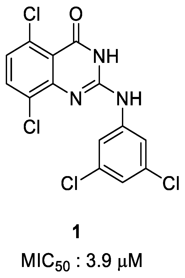
Figure 1. Hit compound against S. aureus ATCC25923.
Antibiotics 14 00967 g001
Scheme 1. Synthesis of 2-(amino)quinazolin-4(3H)-ones derivatives. Reagents and conditions: (a) urea, 160 °C, 20 h, 87%; (b) POCl3, TEA, 120 °C, 17 h, 99%; (c) 2 N NaOH, rt, 20 h, 99%; (d) amines, DMF, 85 °C, 16 h, 7–99%.
Antibiotics 14 00967 sch001
Figure 2. In vitro antibacterial activities of 2-(amino)quinazolin-4(3H)-one derivatives. S. aureus ATCC25923 (blue) and USA300 JE (orange) strains were incubated with serial dilutions of 2-(amino)quinazolin-4(3H)-one derivatives (0.19–100 µM) for 18–24 h. The dose–responses were observed by measuring OD600nm. The plots were drawn from each group of strains with nonlinear curve fits.
Antibiotics 14 00967 g002
Figure 3. Antibacterial activities of 2-(amino)quinazolin-4(3H)-one derivatives in the H460 infection model. H460 cells were infected with S. aureus USA300 JE strain expressing GFP, followed by treatment with serial dilutions of 2-(amino)quinazolin-4(3H)-one derivatives. After incubation at 37 °C for 24 h with 5% CO2, fluorescence signals of the bacteria were detected at 488 nm/510 nm using a multilabel plate reader.
Antibiotics 14 00967 g003
Figure 4. Dose–response images of 2-(amino)quinazolin-4(3H)-one derivatives in the H460 infection model. H460 cells were infected with S. aureus USA300 JE strain expressing GFP, followed by treatment with serial dilutions of 2-(amino)quinazolin-4(3H)-one derivatives 6l, 6h, 6q, and 6y. After incubation at 37 °C for 24 h with 5% CO2, images were obtained by a confocal microscope with a 20× water objective lens detecting fluorescence signals of the bacteria (green) at 488 nm/500–550 nm.
Antibiotics 14 00967 g004
Figure 5. Images of infected H460 cells with treatment using selected 2-(amino)quinazolin-4(3H)-one derivatives. H460 cells were infected with S. aureus USA300 JE strain expressing GFP, followed by treatment with serial dilutions of 2-(amino)quinazolin-4(3H)-one derivatives. After incubation at 37 °C for 24 h with 5% CO2, brightfield images of H460 cells, as well as confocal fluorescence images of USA300 (green), were obtained and merged using an automated microlens-enhanced spinning disk confocal microscope.
Antibiotics 14 00967 g005
Table 1. Antibiotic effects of 2-(amino)quinazolin-4(3H)-one derivatives.
Antibiotics 14 00967 i001
NoCompoundXYZWRATCC25923
MIC50 (μM) a
USA300
MIC50 (μM) a
HepG2
IC50 (μM) b
Efficacy Window
(IC50 HepG2/MIC50 USA300)
11ClHHCl3,5-Cl2-PhNH-3.912.9--
2 6aHHClCl3,5-Cl2-PhNH-10.86.9--
36bHHMorpholineH3,5-Cl2-PhNH->100>100--
46cHOMeOMeH3,5-Cl2-PhNH->100>100--
56dHHOMeH3,5-Cl2-PhNH->100>100--
66eHHNO2H3,5-Cl2-PhNH-3.01.69.35.9
76fHBrHBr3,5-Cl2-PhNH-6.01.29.68.3
86gHFFH3,5-Cl2-PhNH-4.01.412.98.9
96hHClClH3,5-Cl2-PhNH-3.70.310.230.3
106iHClHCl3,5-Cl2-PhNH-5.71.839.521.8
116jHHHH3,5-Cl2-PhNH-5.91.929.715.7
126kHHCF3H3,5-Cl2-PhNH-1.30.310.539.0
136lHHClH3,5-Cl2-PhNH-1.00.662.2101.3
146mHHClHPhNH->100>100--
156nHHClH3-F-PhNH-4.53.019.76.6
166oHHClH2,4-F2-PhNH->100>100--
176pHHClH3-MeO-PhNH-35.124.9--
186qHHClH3,5-F2-PhNH-2.10.935.539.2
196rHHClH3-CF3-PhNH-3.12.215.16.9
206sHHClHCyclohexymethylNH-35.518.1--
216tHHClH2-Cl-PhNH->100>100--
226uHHClH3-Cl-PhNH-3.51.14.34.0
236vHHClH4-Cl-PhNH-2.71.725.515.0
246wHHClH3,5-F2-PhCH2NH->100>100--
256xHHClH2,4-Cl2-PhCH2NH-3.41.715.38.8
266yHHClH3,4-F2-PhCH2NH-0.360.0220.2885.2
Erythromycin0.70.49>100-
Vancomycin0.30.35>100-
a MIC50 values for the synthesized compounds were derived from the results of at least two independent experiments against ATCC25923 or USA300 JE2 strains. b IC50 values for the synthesized compounds were derived from the results of at least two independent experiments using HepG2 cells.
Table 2. Antibiotic effects of 2-(amino)quinazolin-4(3H)-one derivatives in the cell infection model.
CompoundH460 Assay
MIC50 (μM) a
Efficacy Window
(IC50 HepG2/MIC50 USA300)
6e68.80.1
6f8.71.1
6g25.30.5
6h4.12.5
6i26.51.5
6j35.40.8
6k6.01.7
6l12.15.2
6n43.80.4
6q8.74.1
6r35.80.4
6x17.80.9
6u19.20.2
6v23.91.1
6y1.910.6
Vancomycin7.7
Erythromycin0.1
a MIC50 values for the synthesized compounds were derived from the results of at least two independent experiments in the H460 infection model using the USA300 JE2 strain.
Disclaimer/Publisher’s Note: The statements, opinions and data contained in all publications are solely those of the individual author(s) and contributor(s) and not of MDPI and/or the editor(s). MDPI and/or the editor(s) disclaim responsibility for any injury to people or property resulting from any ideas, methods, instructions or products referred to in the content.