Article

Genomic Characterization of Methicillin-Resistant and Methicillin-Susceptible Staphylococcus aureus Implicated in Bloodstream Infections, KwaZulu-Natal, South Africa: A Pilot Study

1
Antimicrobial Research Unit, College of Health Sciences, University of KwaZulu-Natal, Durban 4000, South Africa
2
School of Pharmacy and Pharmaceutical Sciences, University of Cape Coast, PMB, Cape Coast, Ghana
3
Department of Applied Biology & Biochemistry, National University of Science and Technology, Corner Cecil Avenue & Gwanda Road, Bulawayo 263, Zimbabwe
4
Sequencing Core Facility, National Institute for Communicable Diseases, Division of the National Health Laboratory Service, Johannesburg 2193, South Africa
5
Department of Biochemistry and Microbiology, Faculty of Science, Engineering and Agriculture, University of Venda, Thohoyandou 0950, South Africa
6
Department of Pathobiology, University of Guelph, Guelph, ON N1G 2W1, Canada
7
School of Pharmacy, University of Jordan, Amman 11942, Jordan
*
Author to whom correspondence should be addressed.
Antibiotics 2024, 13(9), 796; https://doi.org/10.3390/antibiotics13090796
Submission received: 13 July 2024 / Revised: 19 August 2024 / Accepted: 21 August 2024 / Published: 23 August 2024

Abstract

:
Staphylococcus aureus is an opportunistic pathogen and a leading cause of bloodstream infections, with its capacity to acquire antibiotic resistance genes posing significant treatment challenges. This pilot study characterizes the genomic profiles of S. aureus isolates from patients with bloodstream infections in KwaZulu-Natal, South Africa, to gain insights into their resistance mechanisms, virulence factors, and clonal and phylogenetic relationships. Six multidrug-resistant (MDR) S. aureus isolates, comprising three methicillin-resistant S. aureus (MRSA) and three methicillin-susceptible S. aureus (MSSA), underwent whole genome sequencing and bioinformatics analysis. These isolates carried a range of resistance genes, including blaZ, aac(6′)-aph(2″), ant(9)-Ia, ant(6)-Ia, and fosB. The mecA gene, which confers methicillin resistance, was detected only in MRSA strains. The isolates exhibited six distinct spa types (t9475, t355, t045, t1265, t1257, and t7888) and varied in virulence gene profiles. Panton–Valentine leukocidin (Luk-PV) was found in one MSSA isolate. Two SCCmec types, IVd(2B) and I(1B), were identified, and the isolates were classified into four multilocus sequence types (MLSTs), with ST5 (n = 3) being the most common. These sequence types clustered into two clonal complexes, CC5 and CC8. Notably, two MRSA clones were identified: ST5-CC5-t045-SCCmec_I(1B) and the human-associated endemic clone ST612-CC8-t1257-SCCmec_IVd(2B). Phylogenomic analysis revealed clustering by MLST, indicating strong genetic relationships within clonal complexes. These findings highlight the value of genomic surveillance in guiding targeted interventions to reduce treatment failures and mortality.

1. Introduction

Staphylococcus aureus is a Gram-positive bacterium inhabiting healthy individuals’ nostrils and skin. However, it has become an important opportunistic pathogen in communities and hospitals [1]. It causes severe skin infections, pneumonia, endocarditis, and bloodstream infections (BSIs) [2]. BSIs caused by S. aureus infections have high morbidity and mortality if not treated timeously [3]. The most significant risk factors for S. aureus BSIs are intravascular devices, surgical procedures, and a debilitated immune system [4].
Methicillin-resistant S. aureus (MRSA) has become a significant cause of BSIs. MRSA poses a major public health threat because of multidrug resistance to different antibiotic classes that limit treatment options [5]. Methicillin resistance in MRSA strains is mediated by the mecA gene, found on a mobile genetic element (MGE) known as the staphylococcal cassette chromosome mec (SCCmec) [6]. Methicillin-susceptible S. aureus (MSSA) is also emerging as a causative agent of BSIs [7] and has been reported to display high virulence and multidrug resistance [8].
The pathogenicity of S. aureus depends on its ability to produce a wide array of virulence factors involved in adhesion, invasion of host tissues, immune system evasion, and biofilm formation [9,10]. Also, S. aureus produces metallophores that enable bacteria to scavenge metal ions such as iron and zinc essential for bacterial metabolism and pathogenicity [11]. Virulence factors and multiple resistance genes can be transmitted by horizontal gene transfer (HGT) [12] on diverse MGEs, amongst which plasmids are reported as the primary sources for dissemination [4].
The epidemiology of S. aureus strains indicates that its molecular characteristics continually change over time, resulting in new clones, which vary by region. In a study in the United States, ST5 and ST8 were the most prevalent sequence types [13]. In South Africa, ST612 is dominant in the hospital environment [14]. The ST612-IV [2B], belonging to spa type t1257, was identified as a typical clone in clinical settings [15] and sporadically in poultry settings [16]. The ST5 and ST8 clones are commonly associated with BSIs and the pandemic lineages of S. aureus, such as the clonal complex CC8 and CC5 [17]. Notably, the sequence types ST612, ST5, ST8, and ST72 have displayed high resistance to most antibiotic drug classes and are challenging to treat [17].
Multidrug-resistant (MDR) S. aureus infections pose a serious clinical concern. A high incidence of pathogenic MDR MRSA has been reported, and the data suggest that its prevalence is increasing in Africa [18]. A recent South African study investigating the genetic relatedness of hospital-acquired-associated MRSA isolates in two hospitals revealed that all isolates were resistant to aminoglycosides and β-lactams. All the isolates carried the aacA-aphD and mecA-resistant genes and clusters of virulence genes [19]. This pilot study aimed to comprehensively characterize the genomic profiles, resistance mechanisms, virulence factors, pathogenicity, phylogenomic relationships, and clonal diversity of Staphylococcus aureus clinical strains implicated in BSIs at a regional hospital in KwaZulu-Natal, South Africa.

2. Results

2.1. Patient Demographics and Characteristics

The 6 isolates investigated in this study were obtained from patients who visited a regional hospital in the uMgungundlovu District in the KwaZulu-Natal Province. Three of the six isolates were recovered from the neonatal ICU (n = 3, 50%), two from surgical wards, and one isolate from the pediatric ward. Four patients were male, while two were female. The age distribution of patients ranged from 0 to 33 years old, and the mean age was 8.83 years (Table 1). The demographic details of the source participants of the isolates that were selected for WGS are shown in Supplementary Table S1.

2.2. Antibiotic Susceptibility Test Results

The isolates displayed varying phenotypic resistance profiles, with most being resistant to penicillin G (n = 6), tetracycline (n = 5), doxycycline (n = 5), clindamycin (n = 5), moxifloxacin (n = 5), rifampicin (n = 4), and erythromycin (n = 3). The lowest resistance was against nitrofurantoin, tigecycline, and chloramphenicol (n = 1) (Table 1).

2.3. Phenotypic and Genotypic Identification of MRSA Isolates

MRSA isolates were confirmed by phenotypic resistance to cefoxitin (Table 1) and the detection of the mecA gene using polymerase chain reaction (PCR).

2.4. Genomic Features

The genome size of our draft genomes ranged from 2.7 Mb to 2.9 Mb. The genomic characteristics of the sequences in relation to G + C content (%), number of RNAs, number of coding sequences, size, N50, L50, and coverage are shown in Table S2.

2.5. In Silico ARGs Analysis

Isolates harbored various permutations and combinations of ARGs, which included ARGs against β-lactams [blaZ], aminoglycosides [aac(6′)-aph(2″), aad(6’), ant(9)-la, ant(6)-Ia, aph(2″)-Ia, aph(3′)-IIa, sat-4], trimethoprim [dfrG, dfrC], macrolides [erm(C), erm(A)], tetracycline [tet(K), tet(M), mepR, mepA], flouroquinolones [parE, parC, grlA, gyrA, norA, norC (multidrug efflux pumps)], rifampicin [rpoB] and fosfomycin [fosB, murA] (Table 2). Only the MRSA isolates harbored the mecA gene. There was good concordance between ARGs and phenotypic profiles for ARGs in all MRSA and MSSA isolates.

2.5.1. MLST, spa Typing, and Clonal Complex

MLST revealed total four sequence types, i.e., ST5 (CC5, n = 3), ST152 (n = 1), ST612 (CC8, n = 1), and ST8 (CC8, n = 1). Two MRSA strains belonged to CC8 and one to CC5. Methicillin-susceptible (MSSA) isolates were identified as ST5 (n = 2) or ST152 (n = 1). The genetic diversity of the isolates was confirmed by spa typing, which revealed six different spa types: t9475, t1265, t355, t045, t1257, and t7888 (Table 2). CC and spa type combinations were CC8-t9475, CC8-t1257, and CC5-t045 among MRSA isolates, with CC5-t1265 belonging to one MSSA isolate. There was no association observed between STs, spa type, and CC. The grouping of the STs and spa-types yielded six genotypes, i.e., ST8-t9475, ST152-t355, ST5-t045, ST5-t1265, ST612-1257, and ST5-t7888, indicating that isolates were not clonally related.
The SCCmecFinder analysis identified two SCCmec types, i.e., IVd (2B) and I (1B), among the MRSA isolates (Table 2). One MRSA isolate was non-typeable (NT) for SCCmec. The combination of MLST, CC, spa, and SCCmec yielded the ST612-CC8-t1257-SCCmec_IVd (2B) and ST5-CC5-t045-SCCmec_I (1B), clones both of which have been reported in South Africa.

2.5.2. Mobilome (Plasmids, Insertion Sequences, Intact Prophages, and SCCmec Elements)

Analysis of the six isolate genomes identified various MGEs, including plasmid replicons, IS’s, prophages, and SCCmec elements. A total of eight different plasmid replicons were detected, of which rep20 (n = 3) was the most prevalent (Table 2). There were no associations between plasmid replicons and STs. However, the rep7c was found in CC8 isolates in addition to other plasmid replicons, while rep16 and rep5a were found in isolates with the non-typeable CC. The rep20 plasmid replicon was associated with CC5 and CC8 isolates. The rep10 was carried in CC8 and CC5 isolates, while the re7a and rep21 were carried in CC8 and CC5 isolates, respectively. IS6 and IS256 were identified in three isolates, and their occurrence was not associated with any STs or CC (Table 2). The ddistribution of ISs and plasmid replicons detected among the isolates are shown in Supplementary Table S4. A total of six intact prophages were detected, of which the most identified were PHAGE_Staphy_phi2958PVL (n = 2) and PHAGE_Staphy_P282 (n = 2) (Table S5). PHAGE_Staphy_phiJB was associated with the dfrG gene.

2.5.3. Virulome and Pathogenicity of S. aureus Strains

A total of 82 virulence genes were detected across the isolates (Table S3). The virulence genes belonged to the five main virulence determinant classes of S. aureus: adherence factors, immune evasion, enzymes (exoenzymes), toxins, and the secretion system. It is noteworthy that the most prevalent toxins were hemolysins, i.e., gamma (hlg), delta (hld), alpha (hly/hla), staphylococcal enterotoxins (se, set, sel) genes, and leucocidin genes (lukD/E), while lukS-PV and lukF-PV genes were detected in two isolates (S24 and S29). The prediction of isolates pathogenicity towards humans yielded a high average probability score (Pscore ≈ 0.980).

2.6. Genetic Environment of the ARGs and Virulence Genes

The co-carriage of ARGs and virulence genes was evident across the isolates. Using NCBI annotation, we identified blaZ genes on five isolates in parallel with cacD, virulence genes, and the type 1 toxin–antitoxin system. Across the isolates, most blaZ genes were associated with regulator genes blaR and blaI and frequently found with either a recombinase, integrase, cadmium resistance (cadD) gene, or type I toxin–antitoxin system (Table 3). A similar genetic context was detected in the S13 isolate, where blaZ, blaR, and blaI were flanked by IS6, cadD, a type I toxin–antitoxin system, on a contig with the closest nucleotide homology to a plasmid from S. aureus pER10678.3A.1 (CP051928.1), suggesting that ARGs, heavy metal resistance genes (HMRGs), and virulence genes may be mobilized by plasmids (Table 3). It is noteworthy that IS1182 was associated with the mecA, mecI, and mecR1 genes together with recombinases, while IS6 bracketed the mecA gene and its regulatory genes (mecI and mecR) in three MRSA isolates (Table 4). Most ARGs, including erm(A), ant(9)-Ia, dfrG, and tet(M), were associated with a recombinase and integrase. One isolate was found harboring the dfrG gene bracketed by ISL3 and recombinases (Table 4).

Regulatory Genes

The accessory gene regulator system (agr) involved in the regulation and expression of toxins, exoenzymes, and biofilm was detected in all isolates. Isolates carried agr type I and II. The distribution of the agr group in MRSA was: agr I (n = 1), agr II (n = 2), while in MSSA agr I (n = 2), and agr II (n = 1).

2.7. Phylogenomics

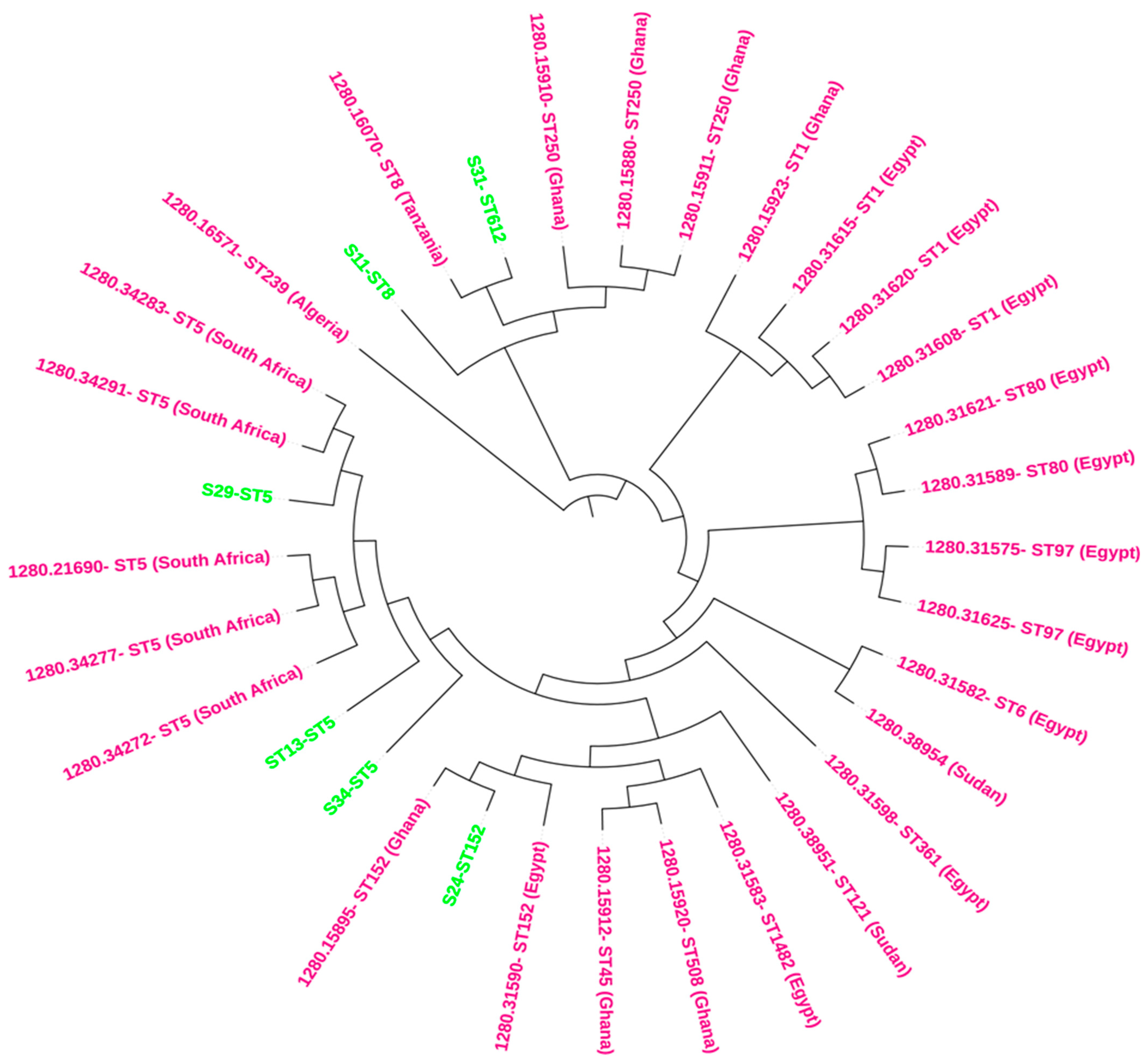
The phylogenetic analysis, integrated with metadata, reveals clear clustering patterns based on Multilocus Sequence Typing (MLST) and geographic origin. Isolates sharing the same MLST type generally clustered together, with additional grouping observed by country of isolation (Figure 1). For instance, the ST5 isolates from this study (S11-ST5, S34-ST5, and S29-ST5) are closely aligned with other South African isolates, indicating minimal genetic divergence within this MLST type in the region. This suggests a strong regional lineage for ST5 in South Africa. Additionally, this study isolates S24-ST152 clusters with other ST152 isolates from various African countries, including Kenya and Ghana, highlighting the broader geographic distribution and potential mobility of this MLST type across the continent (Figure 1). Notably, the tree analysis also reveals that ST8 and ST612 isolates are clustered together, suggesting close genetic relatedness despite being distinct MLST types (Figure 1). This finding could indicate a shared ancestry or recent genetic exchange between these groups, pointing to complex evolutionary dynamics within these populations.
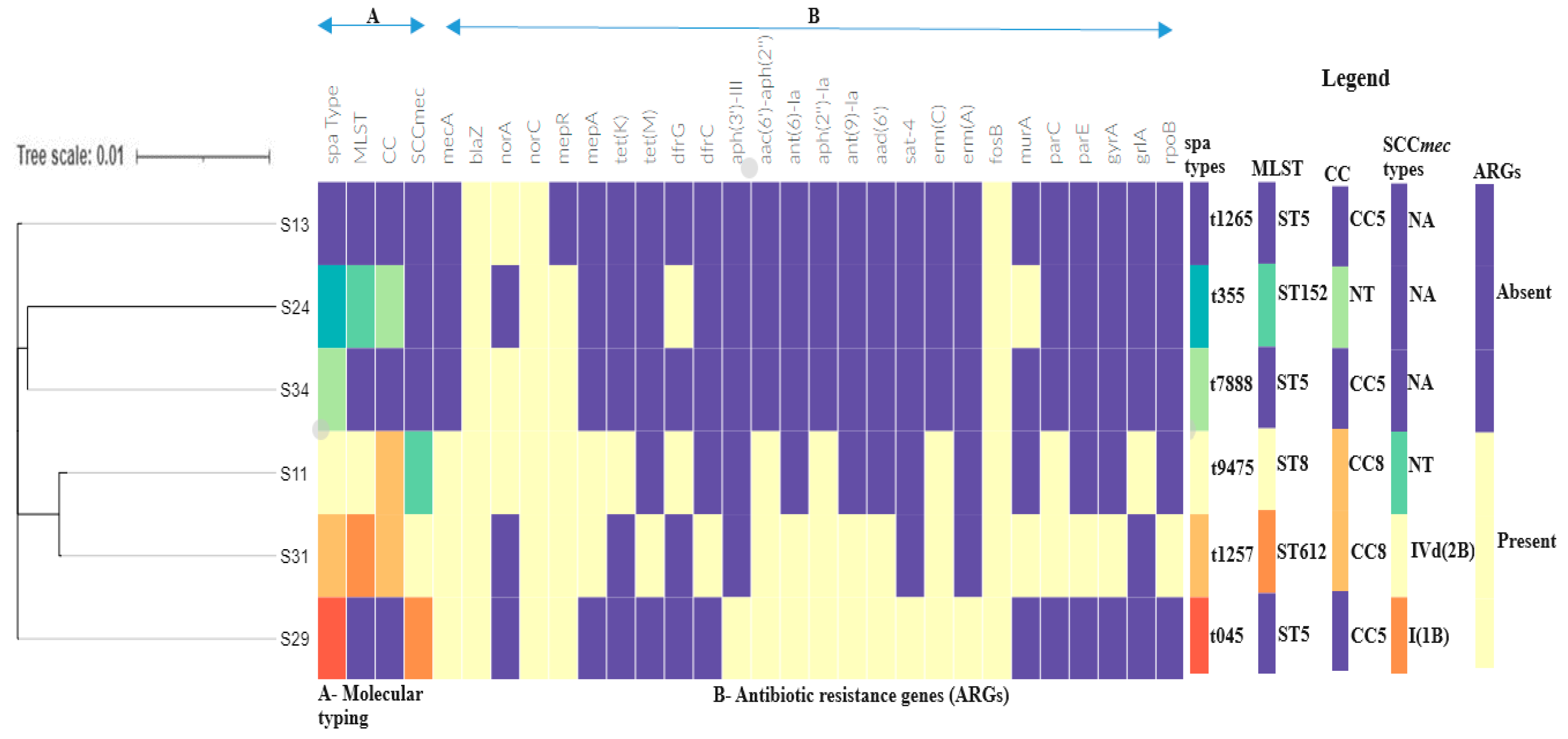
The linkage between the mecA gene, SCCmec types, and clonal complexes is particularly notable, as it highlights the genetic mechanisms underlying methicillin resistance and the clustering of MRSA strains (Figure 2). The distinct clustering patterns observed for different isolates underscore the complex interplay between genetic background, resistance gene acquisition, and selective pressures in the evolution of these pathogens.
Table 3. Genetic context of virulence genes in S. aureus isolates.
Strain (MLST)StrainContigSynteny of Virulence Genes and MGEsPlasmid/Chromosomal Sequence with Closest Nucleotide
Homology (Accession Number)
S11 (ST8)MRSA4pmtC:pmtB:pmtA:eap::scn::sak:::sph::lukG::lukH::intergrase:::agrBS. aureus strain Laus385 chromosome (CP071350.1)
6icaR::icaD:icaB:icaC:vraD:vraE:vraH::IS30:vraH::recombinase:IS6S. aureus strain TF3198 chromosome, complete genome (CP023561.1)
10lukE:lukD::splA::epiE::splA:splB:splC:splD:splE:splF::pepA1:transposaseS. aureus strain 82 chromosome, complete genome (CP031661.1)
S29 (ST5)MRSA53type I toxin–antitoxin system:IS6:cadDS. aureus strain MIN-175 chromosome (CP086121.1)
40clfA:vwb:empS. aureus strain ER02693.3 chromosome, complete genome (CP030605.1)
S31 (ST612)MRSA11pmtD:pmtC:pmtB:pmtA::eap:scn::sakS. aureus strain 2395 USA500, complete genome (CP007499.1)
15lukE:lukD::::splA:splB:splC:splF::type I restriction-modification systemS. aureus strain NRL 02/947 chromosome, complete genome (CP103850.1)
19lukG:lukH:pathogenicity island:intergrase::phenol-soluble modulin:agrBS. aureus strain 2395 USA500, complete genome (CP007499.1)
22seq:sek:integrase::::emp:clfAS. aureus strain 2395 USA500, complete genome (CP007499.1)
33recombinase::universal stress protein:::cadD::seq:sek:integrase:::emp:clfAS. aureus plasmid SAP017A, complete sequence (GQ900382.1)
64sea:putative holin-like toxinS. aureus strain R50 chromosome, complete genome (CP039167.1)
S13 (ST5)MSSA4sbi:hlgA:hlgC:hlgBS. aureus strain AR462 chromosome, complete genome (CP029086.1)
5scpA:::eap::scn:sak::::::intergrase:sph:lukH:sbi:hlgA:hlgC:hlgBS. aureus strain pt239 chromosome, complete genome (CP049467.1)
15IS6::cadD:::sed:sej:ser::recombinases:cpA::eap::scn:sak::integrase:sph:lukHS. aureus strain ER10678.3 plasmid pER10678.3A.1 (CP051928.1)
S24 (ST152)MSSA8arsB::crcB::scn:sak:::recombinase::type II toxin–antitoxin system toxin:intergraseS. aureus strain UMCG579 chromosome, complete genome (CP091066.1)
21cadD:type toxin–antitoxin::integraseS. aureus strain GHA13 chromosome (CP043911.1)
11BrxA/BrxB:::msrA:msrB:::norD::cspA:cvfBS. aureus strain NGA84b chromosome, complete genome (CP051165.2)
S34 (ST5)MSSA7eap/map::scn:sak::::sea:::type II toxin–antitoxin:integrase:sph:lukG:lukHS. aureus strain HPV107 chromosome, complete genome (CP026074.1)
8clfA:vwb:emp::thermonuclease protein:::sek:seq::pathogenicity island S. aureus strain B4-59C chromosome, complete genome (CP042153.1)
12sem:sei:seu:sen:seg:::lukE:lukD::splA:splB:splC:splD:splFS. aureus strain ER03588.3 chromosome, complete genome (CP030595.1)
14isdB:isdA:isdC:isdD:isdE:isdF::isdG::ecb::efb:scbS. aureus strain B3-17D chromosome, complete genome (CP042157.1)
20SSL13:SSL12:hylS. aureus strain NAS_AN_239 chromosome, complete genome (CP062409.1)
Virulence gene(s) in bold.
Table 4. Genetic environment of antibiotic resistance genes in S. aureus isolates.
Isolate ID (MLST)StrainContigSynteny of Resistance Genes and MGEsPlasmid/Chromosomal Sequence with Closest Nucleotide
Homology (Accession Number)
S11 (ST8)MRSA4blaI:blaR1:blaZ::recombinase/integraseS. aureus strain ER02826.3 chromosome (CP030661.1)
7recombinase::dfrG:insertionelement:::ISL3:::recombinase:S. aureus strain UP_403 chromosome (CP047849.1)
59IS6::mecA:MecR1:IS6::S. aureus strain ER03868.3 chromosome (CP030403.1)
123Plasmid recombination:tet(K)S. epidermidis isolate BPH0662 genome assembly, plasmid: 1 (LT614820.1)
S29 (ST5)MRSA8erm(A):ant(9)-Ia:transposase:recombinase:integraseS. aureus strain 628 chromosome (CP022905.1)
11gyrB:gyrA:::ligaseS. aureus strain MIN-175 chromosome (CP086121.1)
38recombinase:IS1182::mecR1:mecA:::IS6S. aureus subsp. aureus strain FDAARGOS_5 chromosome (CP007539.3)
51 *recombinase:blaI:blaR1:blaZ:cadC:cadAS. aureus plasmid pSK57, partial sequence (GQ900493.1)
56ant(6)-Ia:sat4:aph(3′)-IIIaS. pseudintermedius strain MAD627 chromosome (CP039743.1)
64qacA/B:qacRS. aureus strain MIN-175 chromosome (CP086121.1)
67ermCL:erm(C)S. epidermidis strain TMDU-137 plasmid p5, complete sequence (CP093178.1)
S31 (ST612)MRSA23mecA:mecR1::IS1182::recombinaseS. aureus strain 2395 USA500 (CP007499.1)
17integrase::::::tet(M):::IS256S. aureus strain NRS120 chromosome, complete genome (CP026072.1)
S13 (ST5) MSSA15 *IS6 IS6::cadD:::type I toxin–antitoxin::recombinase::blaI:blaR1:blaZS. aureus strain ER10678.3 plasmid pER10678.3A.1 (CP051928.1)
S24 (ST152)MSSA21cadD:typetoxinantitoxin::recombinase:blaI:blaR1:blaZ:recombinaseS. aureus strain GHA13 chromosome (CP043911.1)
S34 (ST5)MSSA19recombinase:blaZ:blaR1:blaI:recombinase:integraseS. aureus strain UP_678 plasmid unnamed (CP047840.1)
* Co-occurrence of a heavy metal resistance gene (HMRG) and antibiotic resistance genes (ARGs).

3. Discussion

We studied the genomic characteristics of six MDR S. aureus isolates implicated in BSIs. This study analyzed the resistome, virulome, mobilome, phylogeny, and genetic environment of the resistance genes using WGS and bioinformatics. The genomes analyzed herein were predominantly recovered from ≤1-year-old patients.
There was a diversity of ARGs encoding resistance to different antibiotics and good concordance between the observed phenotypic and genotypic resistance. The incidence of ARG’s encoding resistance to β-lactams, aminoglycosides, macrolides, fosfomycin, trimethoprim, tetracycline, and genes coding multidrug resistance (MDR) efflux pumps (norA, mepR, and mgrA) was not dependent on the clonal type. The erm(C) and erm(A) genes that are commonly found in macrolide–lincosamide–streptogramin B (MLSB)-resistant S. aureus were found in erythromycin and clindamycin-resistant isolates (Table 2), which was expected since resistance to erythromycin co-selects resistance to other antibiotics, such as streptogramin B (MLSB) and lincosamides [20]. The ermC gene is among the primary erm types that facilitate ribosome methylation of the 23S rRNA, triggering conformational changes resulting in drug binding inhibition [21], and has been reported in clinical S. aureus isolates from South Africa [22]. In this study, the ermC encoding macrolide resistance was carried on a plasmid, on a contig that had the closest nucleotide homology to plasmids from S. epidermidis strain TMDU-137 plasmid p5, complete sequence (CP093178.1), implying the likelihood of horizontal transfer of ermC genes in clinical S. aureus isolates. The ermC are often plasmid-mediated, resulting in high resistance to macrolides in S. aureus [23].
The blaZ gene, which inactivates penicillin through hydrolysis of the beta-lactam ring, was observed in all six isolates that were phenotypically resistant to penicillin. The blaZ genes have also been isolated in clinical isolates of Staphylococci in South Africa [24]. In this study, the blaZ genes were found on contigs with closest homology to either chromosomes or plasmids. This agrees with a study conducted in Spain that analyzed ARGs presence in chromosomes and plasmids from the genomes of S. aureus. WGS analysis of S. aureus revealed that blaZ (n = 2) was located on chromosomic contigs, while blaZ was found in plasmid contigs in three isolates [25]. It is important to note that most blaZ and associated MGEs from isolates belonging to ST5 (S13, S34) isolated from the intensive care unit (ICU) and pediatric ward (S29) were located on contigs that had the closest homology to plasmids, implying that plasmids play a crucial role in mobilizing the blaZ gene in clinical S. aureus isolates. The S29 isolate, belonging to the t045-CC5 lineage, carried an assortment of ARGs encoding resistance to different antibiotics (Table 4). Similar ARGs in MRSA lineage t045-CC5-MRSA were also reported in a study conducted in South Africa, where t045-CC5 MRSA lineages obtained from different clinical samples from South Africa and Nigeria reported that t045 lineages were MDR, suggesting that this lineage is hospital-associated and their multidrug resistance nature may compromise treatment [26].
Also, the blaZ genes, heavy metal genes, and associated MGEs were carried on either plasmid or chromosome. The blaZ and cadAC genes were found on the genetic element recombinase blaI:blaR1:blaZ:cadC:cadA for isolates S24 (MSSA) that was from the ICU and S29 (MRSA) from the pediatric ward, suggesting co-selection of heavy metal resistance dissemination and adaptation in different wards. The cadA gene confers a high resistance to cadmium and other heavy metals like zinc and lead in S. aureus isolates [27]. The cadA was associated with a plasmid, similar to the findings of a study that was conducted by Al-Trad et al. (2023) in Malaysia, who used WGS to analyze the plasmid content of clinical MRSA isolates and reported that heavy metal resistance plasmids harbored cadmium resistance genes, with the majority being cadAC [28]. The HMRGs have been reported to trigger a co-selection mechanism with antibiotics, which may complicate treatment [29]. This may pose a challenge, especially among patients in the ICU, where broad-spectrum antibiotics are often used.
Tetracycline resistance genes (tetK and tetM) were observed in two isolates. Isolate S11 carried tet(K) associated with the following genetic context: plasmid recombination tet(K) that had a high similarity to S. epidermidis BPH0662, and plasmid 1 (LT614820.1), which could be significant in mobilizing TET-resistant genes. Also, the tet(M) was bracketed by integrase and IS256 in isolate S31. The IS256 is a retrotransposon that can mobilize the resistance genes through a copy-and-paste mechanism and has been shown to confer a robust genomic plasticity in MRSA strains [30].
We found that ARGs and virulence genes were associated with MGEs, which may enable their transfer within and between plasmids and chromosomes [31]. In this study, the mecA gene was located on IS1182 in two MRSA isolates, surrounded by recombinase in the genetic context mecA:mecR1::IS1182::recombinase. The insertion sequence IS1182 was present in 2/3 MRSA strains that contained mecA. IS1182 has been shown to occur close to the SCCmec element and increase resistance through inactivating the lytH gene encoding a putative lytic enzyme in pathogenic MRSA isolates [32].
MLST, clonal complex typing, spa typing, and SCCmec typing were used to analyze the molecular characteristics of the S. aureus isolates. Four ST types and two clonal clusters (CCs) were found among the six clinical isolates in this study, with ST5 as the most predominant complex clonal CC5 and CC8. Generally, clonal lineages ST5, ST8, ST152, and ST612 are among the most commonly reported in hospital environments, along with other sequence types of S. aureus [33]. S. aureus ST5, belonging to CC5, was predominant in this study and was previously reported among patients with bloodstream infections at Ruijin Hospital in Shanghai [3]. The detection of clonal complexes CC5 and CC8 agrees with a study by Smith et al. [17], which also found CC8 and CC5 were predominant in a study that analyzed the genomic epidemiology of MRSA and MSSA from bloodstream infections in the USA. Their results revealed that the MDR phenotype observed in strains belonging to CC5 and CC8 was responsible for the occurrence of multidrug and methicillin resistance in the S. aureus population. MRSA strains belonging to CC8 and CC5 are frequently associated with global outbreaks and have been identified in Africa [34].
The spa typing revealed six different spa types, suggesting a non-clonal MRSA and MSSA distribution. The detection of spa types t1257, t045, and t355 agrees with a study conducted in South Africa, which analyzed the diversity of SCCmec elements and spa types in S. aureus isolates from blood culture in the Gauteng, KwaZulu-Natal, Free State, and Western Cape provinces [15], in which t037 and t1257 were the most common and predominated throughout the seven-year study period. In this study, some antibiotic resistance genes were associated with specific MRSA clones belonging to spa types t1257, t045, and t9475. Shittu et al. (2021) found the spa types t045 and t1257 to be the most prevalent and associated with genes conferring resistance to aminoglycosides, trimethoprim, macrolides, and tetracycline in clinical isolates of S. aureus from South Africa and Nigeria [26].
The analysis of SCCmec types revealed the presence of SCCmec type IVd (2B) and SCCmec type I (B) carrying the mecA gene, which occurred in tandem with mecR1 in both MRSA isolates. However, one MRSA (S11) isolate had a non-typeable SCCmec element cassette due to the missing cassette chromosome recombinase (ccr) gene complex [35]. The ccr gene complex is an essential component required to facilitate the integration or excision of the SCCmec element in the staphylococcal chromosome, and their loss has also been reported [36]. The SCCmec IV detected in our study is associated with the spa type t1257, previously reported in South Africa in S. aureus obtained from poultry isolates [16], implying its possible transfer between humans and animals.
We found different MRSA genotypes, ST612-t1257-CC8, ST8-t9475-CC8, and ST5-t045-CC5, suggesting that MRSA isolates were not clonally and epidemiologically related. The ST612-t1257-CC8 identified in this study is an endemic MRSA clone that has been reported in animal and clinical settings [15,16]. The ST5-I-MRSA, known as the pandemic British EMRSA-3 clone, was detected in the pediatric ward. This is similar to a study conducted in South Africa, where the t045-MRSA strain occurred in pediatric patients [19]. The isolation of the t045-ST5-MRSA strain could confirm its successful persistence in the hospital and its capacity to cause infections in neonatal and pediatric wards [37].
Several virulence factors, including adherence, immune invasion, toxins, and exoenzymes associated with invasive infections, were detected in our isolates. The virulence genes encoding clumping factor proteins (clfA and clfB) are involved in the pathogenesis of S. aureus, including bacteremia [9]. Consistent with pathogenic S. aureus strains isolated in various environments globally, our isolates were characterized by the icaADBC operon and sdrC, sdrD, and sdrE involved in biofilm-forming genes [38]. Most strains harbored genes, including the alpha and gamma-hemolysin genes (hlgA, hlgB, hlgC, hly/hla, and hlb), and the ica operon associated with pathogenicity and adhesion. Additionally, our isolates were characterized by various toxins, including lukE/D genes and Panton–Valentine leukocidin (PVL) lukS-PV/lukF-PV genes in one MSSA and MRSA strain. The expression of these PVL toxin genes in S. aureus isolates lyses host cells and promotes virulence of the bacteria [39], which might worsen the outcomes of S. aureus infection. Consistent with clinical S. aureus strains, our isolates were characterized by a capsular polysaccharide (CP) serotype 8, which shields the bacterial pathogen from host immune defense mechanisms associated with increased virulence in BSIs [40].
Most virulence genes, including those encoding SEs, sak, hlg, luk, scn, clfA, sbi, and associated MGEs, were carried on chromosomes in the majority of isolates. The ica gene operon and vra genes were found to be associated with ISs (IS30, IS6) and recombinase for S11 (ST8) isolate from the surgical ward. The ica genes vraDEH genes have been shown to play an important role in biofilm formation [41] and daptomycin resistance in S. aureus [42], which could enhance antibiotic resistance traits and chronic infection. The occurrence of ST8-t9475 MRSA strains co-harboring ica genes and genes encoding daptomycin resistance in ST8 MRSA could be advantageous to the ST8-t9475 colonization, invasion, and survival in the surgical ward. The virulence genes encoding SEs, eap, scn, sak, sph, lukH, and cadA, were found on a contig that had high sequence similarity to S. aureus strain ER10678.3 plasmid pER10678.3A.1 (CP051928.1), implying that they are mobilized by plasmids. Virulence genes, including those encoding hla/hld, toxin production, and biofilm formation, are plasmid-mediated [43], thus could easily facilitate their transfer, resulting in highly pathogenic strains that may be difficult to treat.
Phylogenomic analyses revealed that the clinical isolates in this study clustered mainly with clinical isolates from hospital patients (Figure 2). ST5 study isolates were closely related to clinical isolates from South Africa, suggesting possible dissemination of ST5 strains and adaptation in hospital environments. Furthermore, the ST152 isolate was closely related to ST152 strains from Egypt and Ghana, implying a possible spread and epidemiological linkage between these isolates. ST152-PVL-producing S. aureus isolates are particularly frequent and widespread in West and Central Africa [44] and livestock [45]. The ST152-PVL-positive MSSA has also been reported from cutaneous abscesses among mine workers at a gold mine in Gauteng, South Africa [46]. Identifying ST152 in livestock and humans suggests animal–human transmission, which requires further investigation. ST8 and ST612 isolates were closely related to ST8 isolated from Tanzania, indicating that ST612 is a double-locus variant of ST8. ST8 and ST612 isolates are potentially multidrug-resistant and highly virulent strains associated with hospital outbreaks [47]. The integration of phylogenetic data with resistance profiles offers valuable insights into the epidemiology and evolutionary dynamics of these isolates, highlighting potential patterns of transmission and resistance development [48].
In light of the increasing resistance observed in S. aureus strains, innovative strategies are being explored to combat antimicrobial resistance. One promising approach focuses on targeting microbial metallophores, molecules that bacteria use to scavenge essential metals from their environment. By inhibiting metallophore function, it is possible to disrupt bacterial metabolism and enhance the effectiveness of existing antibiotics [49]. Additionally, alternative strategies, such as the use of bacteriophages, antimicrobial peptides, and immune system modulation, offer potential avenues for treating resistant infections [50]. Finally, combination therapy, which involves using multiple antibiotics or combining antibiotics with adjuvants that inhibit resistance mechanisms, is gaining attention as a way to overcome multi-drug resistance and reduce the likelihood of treatment failure [51]. These emerging strategies represent critical avenues for future research and clinical application, aiming to curb the growing threat of antimicrobial resistance.
This study analyzed a limited number of Staphylococcus aureus isolates (n = 6), which may not fully represent the epidemiology of MRSA/MSSA in South Africa or bloodstream infections more broadly. The small sample size limits the generalizability of the findings to the wider population. Therefore, while this study offers valuable insights into the genomic characteristics and resistance profiles of these isolates, it should be viewed as a pilot study that lays the groundwork for larger, more comprehensive investigations. The small sample size also limits the ability to validate the findings, particularly concerning virulence factors and other genomic features. Future studies with larger sample sizes and experimental validation are necessary to confirm and extend these observations.

4. Materials and Methods

4.1. Ethical Consideration

The ethical approval for this study was issued by the Biomedical Research Ethics Committee of the University of KwaZulu-Natal under the following reference number: BCA444/16.

4.2. Sample Collection and Bacterial Identification

A total of forty-five presumptive Staphylococcus isolates from blood cultures sourced from patients with BSIs at two hospitals in the uMgungundlovu district in the KwaZulu-Natal province from November 2017 to December 2018. All isolates were confirmed as S. aureus using the automated VITEK 2 system (BioMérieux, MarcyL’Etoile, France). We selected a subset of 10 MDR isolates for WGS based on their antibiotic-resistant profiles/patterns, but 4 isolates were excluded during the quality control process.

4.3. Antimicrobial Susceptibility and MRSA Detection

Isolates were tested for antibiotic susceptibility by disk-diffusion method on Mueller–Hinton agar as recommended by the European Committee on Antimicrobial Susceptibility Testing (EUCAST) [52] or Clinical and Laboratory Standards Institute (CLSI) [53]. The antibiotics tested and interpreted according to the EUCAST breakpoints (EUCAST, 2017) were penicillin G (10 µg), ampicillin (10 µg), cefoxitin (30 µg), tigecycline (15 µg), and nitrofurantoin (300 µg). The CLSI guidelines (CLSI, 2017) were used for the following antibiotics: ciprofloxacin (5 µg), levofloxacin (5 µg), moxifloxacin (5 µg), erythromycin (15 µg), gentamicin (10 µg), amikacin (30 µg), chloramphenicol (30 µg), tetracycline (30 µg), doxycycline (30 µg), sulphamethoxazole/trimethoprim (1.25 µg + 23.75 µg), teicoplanin (30 µg), linezolid (30 µg), clindamycin (2 µg), and rifampicin (5 µg). MRSA isolates were identified using a cefoxitin disk (30 μg). The antibiotic disks were obtained from Oxoid (Oxoid, Basingstoke, UK). S. aureus ATCC 29213, was used as the quality control strain. Multidrug resistance (MDR) was defined as resistance to three or more antibiotic classes [54].

4.4. Whole-Genome Sequencing (WGS) and Bioinformatic Analysis

Genomic DNA extraction was performed using the GenElute Bacterial Genomic DNA kit (Sigma Aldrich, St. Louis, MO, USA) following the manufacturer’s instructions. The quality of the DNA was assessed using NanoDrop 8000 (Thermo Fisher Scientific Waltham, MA, USA). Genome libraries were constructed using the Nextera XT DNA Library Preparation Kit (Illumina, San Diego, CA, USA) and sequenced on the Illumina NextSeq Machine (Illumina, San Diego, CA, USA). The raw reads were trimmed using Sickle v1.33 (https://github.com/najoshi/sickle accessed on 15 August 2020) and assembled using the SPAdes v3.6.2 assembler (https://cab.spbu.ru/software/spades/ accessed on 15 August 2020). Assembled genome sequences were submitted to Genebank and assigned accession numbers under the BioProject number PRJNA400143.

4.5. Genomic Analysis

Genotyping of the assembled genomes was performed using the MLST 2.0, spa Typer 1.0, and SCCmecFinder 1.2 available at the Centre for Genomic Epidemiology (CGE) (https://www.genomicepidemiology.org/services/ accessed on 1 October 2023). The detection of antibiotic resistance genes (ARGs) was examined by ResFinder 4.1 (https://cge.cbs.dtu.dk/servic es/ResFinder/ accessed on 1 October 2023) and the comprehensive antibiotic resistance database (https://card.mcmaster.ca/analyze/rgi accessed on 1 October 2023). Virulence determinants were identified with default settings using the virulence factor database (VFDB: http://www.mgc.ac.cn/VFs/main.htm accessed on 15 December 2023) and VirulenceFinder 2.0 https://cge.food.dtu.dk/services/VirulenceFinder/ accessed on 1 October 2023). Pathogenicity of isolates was found out using PathogenFinder 1.1 (https://cge.food.dtu.dk/services/PathogenFinder/ accessed on 1 October 2023). PHASTER was used to identify prophage elements (https://phaster.ca/ accessed on 1 October 2023). Mobile genetic elements (MGEs) in relation to ARGs, virulent factors, and plasmid replicons were identified using MobileElementFinder (https://cge.food.dtu.dk/services/MobileElementFinder/ accessed on 1 October 2023) and Plasmid Finder 2.1 (https://cge.cbs.dtu.dk/services/PlasmidFinder/ accessed on 1 October 2023). The genetic environment of ARGs, virulence factors, and associated MGEs was examined using GenBank’s general feature format (GFF3) files and imported into Geneious Prime 2020.2 (https://www.geneious.com accessed on 10 December 2023) for analysis [55]. The accessory gene regulator (agr) typing was performed by employing nucleotide BLAST, and the following GenBank accession numbers: AFS50129.1, AFS50128.1, AFS50130.1, and AFS50131.1 were used as reference sequences for agr types I–IV [56].

4.6. Phylogenomic Analysis

For phylogeny analysis, whole-genome sequences of S. aureus isolates from blood culture were selected from Northern Africa (Egypt, Algeria, and Sudan), Western Africa (Ghana), and Eastern Africa (Tanzania) and downloaded from the bacterial and viral bioinformatics resource center’s (BV-BRC) online platform (https://www.bv-brc.org/ accessed on 15 May 2024) and used together with our study’s isolates. We constructed a phylogenomic tree using the online Phylogenetic Tree Building tool available on the BV-BRC website (https://www.bv-brc.org/ accessed on 15 May 2024). The generated phylogenetic tree was visualized, annotated, and edited using iTOL (https://itol.embl.de/ accessed on 15 May 2024) and Figtree (http://tree.bio.ed.ac.uk/software/figtree/ accessed on 15 May 2024).

4.7. Nucleotide Sequence Accession Number

The nucleotide sequences of MRSA (S29, S11, S31) and MSSA (S13, S24, S34) isolates were submitted to the NCBI GenBank database under the following accession numbers: JADQTH000000000, JADIXB000000000, JADIXC000000000, JADIXA000000000, JADIXE000000000, and JADIXD000000000.

5. Conclusions

This study presents an insight into ARGs, virulence genes, MGEs, and genetic diversity of S. aureus collected from a public hospital in uMgungundlovu. We observed high diversity of spa types, STs, and a predominance of CC8 and CC5, indicating the genetic variability of S. aureus in hospital settings. The occurrence of pathogenic and MDR strains in the hospital setting, especially in the ICU, can pose a serious threat that limits the therapeutic options available. Here, we demonstrate that while MRSA displayed multidrug resistance, MSSA reflected potentially increasing resistance to the antibiotics used for treatment. Continuous surveillance and monitoring of MRSA and MSSA strains circulating in hospital environments is needed.

Supplementary Materials

The following supporting information can be downloaded at: https://www.mdpi.com/article/10.3390/antibiotics13090796/s1. Table S1: Patient demographics. Table S2: Genomic characteristics of S. aureus strains. Table S3: Virulence genes identified in MSSA and MRSA isolates in this study. Table S4: Distribution of insertion sequences and plasmid replicon among the Staphylococcus aureus strains. Table S5: Distribution of intact prophage region among the Staphylococcus aureus strains.

Author Contributions

Conceptualization, B.A.H., D.G.A. and S.Y.E.; methodology, B.A.H., A.I. and A.L.K.A.; formal analysis, B.A.H., D.G.A., J.M., J.A. and A.L.K.A.; investigation, B.A.H.; resources, A.I., D.G.A. and S.Y.E.; writing—original draft preparation, B.A.H.; writing—review and editing, B.A.H., J.A., J.M., A.I., A.L.K.A., D.G.A. and S.Y.E.; supervision, S.Y.E., A.L.K.A. and D.G.A. All authors have read and agreed to the published version of the manuscript.

Funding

This work was supported by the South African Research Chairs Initiative of the Department of Science and Technology and the National Research Foundation of South Africa (Grant No. 98342), the SAMRC and UK MRC Newton Fund, the SAMRC Self-Initiated Research Grant, and the College of Health Sciences University of KwaZulu-Natal, South Africa.

Institutional Review Board Statement

Ethical approval for this study was obtained from the Biomedical Research Ethics Committee of the University of KwaZulu-Natal under the following reference number BCA444/16. The study isolates were part of a larger surveillance study using the Global Antimicrobial Resistance and Use Surveillance System (GLASS) guidelines.

Informed Consent Statement

Not applicable.

Data Availability Statement

The data presented in this study are openly available in GenBank and assigned accession numbers under the BioProject PRJNA400143. [NCBI Genebank] [https://www.ncbi.nlm.nih.gov/bioproject/?term=PRJNA400143 accessed on 15 May 2024] [PRJNA400143].

Acknowledgments

We are grateful to Sumayya Haffejee of the National Health Laboratory Services for her assistance during sample collection and obtaining demographic data.

Conflicts of Interest

S.Y.E. is a chairperson of the Global Respiratory Infection Partnership and member of the Global Hygiene Council, both funded by unrestricted educational grants from Reckitt and Benckiser (Pty.), UK. The remaining authors declare that research was conducted in the absence of any commercial or financial relationships that could be construed as a potential conflict of interest.

References

  1. Asadollahi, P.; Farahani, N.N.; Mirzaii, M.; Khoramrooz, S.S.; van Belkum, A.; Asadollahi, K.; Dadashi, M.; Darban-Sarokhalil, D. Distribution of the Most Prevalent Spa Types among Clinical Isolates of Methicillin-Resistant and-Susceptible Staphylococcus Aureus Around the World: A Review. Front. Microbiol. 2018, 9, 163. [Google Scholar] [CrossRef] [PubMed]
  2. Liang, Y.; Tu, C.; Tan, C.; El-Sayed Ahmed, M.A.E.G.; Dai, M.; Xia, Y.; Liu, Y.; Zhong, L.L.; Shen, C.; Chen, G.; et al. Antimicrobial Resistance, Virulence Genes Profiling and Molecular Relatedness of Methicillin-Resistant Staphylococcus Aureus Strains Isolated from Hospitalized Patients in Guangdong Province, China. Infect. Drug Resist. 2019, 12, 447–459. [Google Scholar] [CrossRef] [PubMed]
  3. Gu, F.; He, W.; Xiao, S.; Wang, S.; Li, X.; Zeng, Q.; Ni, Y.; Han, L. Antimicrobial Resistance and Molecular Epidemiology of Staphylococcus Aureus Causing Bloodstream Infections at Ruijin Hospital in Shanghai from 2013 to 2018. Sci. Rep. 2020, 10, 6019. [Google Scholar] [CrossRef]
  4. Diekema, D.J.; Hsueh, P.R.; Mendes, R.E.; Pfaller, M.A.; Rolston, K.V.; Sader, H.S.; Jones, R.N. The Microbiology of Bloodstream Infection: 20-Year Trends from The SENTRY Antimicrobial Surveillance Program. Antimicrob. Agents Chemother. 2019, 63, e00355-19. [Google Scholar] [CrossRef]
  5. Turner, N.A.; Sharma-Kuinkel, B.K.; Maskarinec, S.A.; Eichenberger, E.M.; Shah, P.P.; Carugati, M.; Holland, T.L.; Fowler, V.G. Methicillin-Resistant Staphylococcus Aureus: An Overview of Basic and Clinical Research. Nat. Rev. Microbiol. 2019, 17, 203–218. [Google Scholar] [CrossRef]
  6. Hadyeh, E.; Azmi, K.; Seir, R.A.; Abdellatief, I.; Abdeen, Z. Molecular Characterization of Methicillin Resistant Staphylococcus Aureus in West Bank-Palestine. Front. Public Health 2019, 7, 130. [Google Scholar] [CrossRef]
  7. Jin, Y.; Zhou, W.; Zhan, Q.; Chen, Y.; Luo, Q.; Shen, P.; Xiao, Y. Genomic Epidemiology and Characterisation of Penicillin-Sensitive Staphylococcus Aureus Isolates from Invasive Bloodstream Infections in China: An Increasing Prevalence and Higher Diversity in Genetic Typing Be Revealed. Emerg. Microbes Infect. 2022, 11, 326–336. [Google Scholar] [CrossRef] [PubMed]
  8. Yuan, W.; Liu, J.; Zhan, Y.; Wang, L.; Jiang, Y.; Zhang, Y.; Sun, N.; Hou, N. Molecular Typing Revealed the Emergence of PVL-Positive Sequence Type 22 Methicillin-Susceptible Staphylococcus Aureus in Urumqi, North Western China. Infect. Drug Resist. 2019, 12, 1719–1728. [Google Scholar] [CrossRef]
  9. Foster, T.J. The MSCRAMM Family of Cell-Wall-Anchored Surface Proteins of Gram-Positive Cocci. Trends Microbiol. 2019, 27, 927–941. [Google Scholar] [CrossRef]
  10. Foster, T.J.; Geoghegan, J.A.; Ganesh, V.K.; Hook, M. Adhesion, Invasion and Evasion: The Many Functions of the Surface Proteins of Staphylococcus Aureus. Nat. Rev. Microbiol. 2014, 12, 46–62. [Google Scholar] [CrossRef]
  11. Ghssein, G.; Ezzeddine, Z. The Key Element Role of Metallophores in the Pathogenicity and Virulence of Staphylococcus Aureus: A Review. Biology 2022, 11, 1525. [Google Scholar] [CrossRef] [PubMed]
  12. Warnes, S.L.; Highmore, C.J.; Keevil, C.W. Horizontal Transfer of Antibiotic Resistance Genes on Abiotic Touch Surfaces: Implications for Public Health. MBio 2012, 3, e00489-12. [Google Scholar] [CrossRef] [PubMed]
  13. Park, K.H.; Greenwood-Quaintance, K.E.; Uhl, J.R.; Cunningham, S.A.; Chia, N.; Jeraldo, P.R.; Sampathkumar, P.; Nelson, H.; Patel, R. Molecular Epidemiology of Staphylococcus Aureus Bacteremia in a Single Large Minnesota Medical Center in 2015 As Assessed Using MLST, Core Genome MLST and Spa Typing. PLoS ONE 2017, 12, e0179003. [Google Scholar] [CrossRef] [PubMed]
  14. Perovic, O.; Iyaloo, S.; Kularatne, R.; Lowman, W.; Bosman, N.; Wadula, J.; Seetharam, S.; Duse, A.; Mbelle, N.; Bamford, C.; et al. Prevalence and Trends of Staphylococcus Aureus Bacteraemia in Hospitalized Patients in South Africa, 2010 to 2012: Laboratory-Based Surveillance Mapping of Antimicrobial Resistance and Molecular Epidemiology. PLoS ONE 2015, 10, e0145429. [Google Scholar] [CrossRef] [PubMed]
  15. Singh-Moodley, A.; Lowe, M.; Mogokotleng, R.; Perovic, O. Diversity of SCCmec Elements and Spa Types in South African Staphylococcus Aureus MecA-Positive Blood Culture Isolates. BMC Infect. Dis. 2020, 20, 816. [Google Scholar] [CrossRef]
  16. Amoako, D.G.; Somboro, A.M.; Abia, A.L.K.; Allam, M.; Ismail, A.; Bester, L.; Essack, S.Y. Genomic Analysis of Methicillin-Resistant Staphylococcus Aureus Isolated from Poultry and Occupational Farm Workers in Umgungundlovu District, South Africa. Sci. Total Environ. 2019, 670, 704–716. [Google Scholar] [CrossRef] [PubMed]
  17. Smith, J.T.; Eckhardt, E.M.; Hansel, N.B.; Eliato, T.R.; Martin, I.W.; Andam, C.P. Genomic Epidemiology of Methicillin-Resistant and-Susceptible Staphylococcus Aureus from Bloodstream Infections. BMC Infect. Dis. 2021, 21, 589. [Google Scholar] [CrossRef] [PubMed]
  18. Schaumburg, F.; Alabi, A.S.; Peters, G.; Becker, K. New Epidemiology of Staphylococcus Aureus Infection in Africa. Clin. Microbiol. Infect. 2014, 20, 589–596. [Google Scholar] [CrossRef] [PubMed]
  19. Strasheim, W.; Perovic, O.; Singh-Moodley, A.; Kwanda, S.; Ismail, A.; Lowe, M. Ward-Specific Clustering of Methicillin-Resistant Staphylococcus Aureus Spa-Type T037 and T045 in Two Hospitals in South Africa: 2013 to 2017. PLoS ONE 2021, 16, e0253883. [Google Scholar] [CrossRef]
  20. Silva, V.; Hermenegildo, S.; Ferreira, C.; Manaia, C.M.; Capita, R.; Alonso-Calleja, C.; Carvalho, I.; Pereira, J.E.; Maltez, L.; Capelo, J.L.; et al. Genetic Characterization of Methicillin-Resistant Staphylococcus Aureus Isolates from Human Bloodstream Infections: Detection of MLSB Resistance. Antibiotics 2020, 9, 375. [Google Scholar] [CrossRef]
  21. Asante, J.; Govinden, U.; Owusu-Ofori, A.; Bester, L.A.; Essack, S.Y. Molecular Characterization of Methicillin-Resistant Staphylococcus Aureus Isolates from a Hospital in Ghana. African J. Clin. Exp. Microbiol. 2019, 20, 164. [Google Scholar] [CrossRef]
  22. Mkhize, S.; Amoako, D.G.; Shobo, C.O.; Zishiri, O.T.; Bester, L.A. Genotypic and Phenotypic Characterizations of Methicillin-Resistant Staphylococcus Aureus (MRSA) on Frequently Touched Sites from Public Hospitals in South Africa. Int. J. Microbiol. 2021, 2021, 6011045. [Google Scholar] [CrossRef] [PubMed]
  23. McCarthy, A.J.; Lindsay, J.A. The Distribution of Plasmids That Carry Virulence and Resistance Genes in Staphylococcus Aureus Is Lineage Associated. BMC Microbiol. 2012, 12, 104. [Google Scholar] [CrossRef] [PubMed]
  24. Ocloo, R.; Newton-Foot, M.; Ziebuhr, W.; Whitelaw, A.C. Molecular Epidemiology and Antibiotic Resistance of Staphylococci Other than Staphylococcus Aureus in Children in Cape Town, South Africa. Front. Microbiol. 2023, 14, 1239666. [Google Scholar] [CrossRef] [PubMed]
  25. Pennone, V.; Prieto, M.; Avelino, Á.; Cobo-diaz, J.F. Antimicrobial Resistance Genes Analysis of Publicly Available Staphylococcus Aureus Genomes. Antibiotics 2022, 11, 1632. [Google Scholar] [CrossRef] [PubMed]
  26. Shittu, A.O.; Adesoji, T.; Udo, E.E. DNA Microarray Analysis of Staphylococcus Aureus from Nigeria and South Africa. PLoS ONE 2021, 16, e0237124. [Google Scholar] [CrossRef]
  27. Parsons, C.; Lee, S.; Kathariou, S. Dissemination and Conservation of Cadmium and Arsenic Resistance Determinants in Listeria and Other Gram-Positive Bacteria. Mol. Microbiol. 2020, 113, 560–569. [Google Scholar] [CrossRef]
  28. Al-Trad, E.I.; Chew, C.H.; Che Hamzah, A.M.; Suhaili, Z.; Rahman, N.I.A.; Ismail, S.; Puah, S.M.; Chua, K.H.; Kwong, S.M.; Yeo, C.C. The Plasmidomic Landscape of Clinical Methicillin-Resistant Staphylococcus Aureus Isolates from Malaysia. Antibiotics 2023, 12, 733. [Google Scholar] [CrossRef]
  29. Zhong, Q.; Cruz-Paredes, C.; Zhang, S.; Rousk, J. Can Heavy Metal Pollution Induce Bacterial Resistance to Heavy Metals and Antibiotics in Soils from An Ancient Land-Mine? J. Hazard. Mater. 2021, 411, 124962. [Google Scholar] [CrossRef]
  30. Kleinert, F.; Kallies, R.; Hort, M.; Zweynert, A.; Szekat, C.; Nagel, M.; Bierbaum, G. Influence of IS256 on Genome Variability and Formation of Small-Colony Variants in Staphylococcus Aureus. Antimicrob. Agents Chemother. 2017, 61, e00144-17. [Google Scholar] [CrossRef]
  31. Mbelle, N.M.; Feldman, C.; Osei Sekyere, J.; Maningi, N.E.; Modipane, L.; Essack, S.Y. Publisher Correction: The Resistome, Mobilome, Virulome and Phylogenomics of Multidrug-Resistant Escherichia Coli Clinical Isolates from Pretoria, South Africa. Sci. Rep. 2020, 10, 1270. [Google Scholar] [CrossRef]
  32. Wang, W.; Baker, M.; Hu, Y.; Xu, J.; Yang, D.; Maciel-Guerra, A.; Xue, N.; Li, H.; Yan, S.; Li, M.; et al. Whole-Genome Sequencing and Machine Learning Analysis of Staphylococcus Aureus from Multiple Heterogeneous Sources in China Reveals Common Genetic Traits of Antimicrobial Resistance. mSystems 2021, 6, e0118520. [Google Scholar] [CrossRef]
  33. Sands, K.; Carvalho, M.J.; Spiller, O.B.; Portal, E.A.R.; Thomson, K.; Watkins, W.J.; Mathias, J.; Dyer, C.; Akpulu, C.; Andrews, R.; et al. Characterisation of Staphylococci Species from Neonatal Blood Cultures in Low- and Middle-Income Countries. BMC Infect. Dis. 2022, 22, 593. [Google Scholar] [CrossRef]
  34. Lawal, O.U.; Ayobami, O.; Abouelfetouh, A.; Mourabit, N.; Kaba, M.; Egyir, B.; Abdulgader, S.M.; Shittu, A.O. A 6-Year Update on the Diversity of Methicillin-Resistant Staphylococcus Aureus Clones in Africa: A Systematic Review. Front. Microbiol. 2022, 13, 860436. [Google Scholar] [CrossRef] [PubMed]
  35. Elshabrawy, W.; Elsayed Zaki, M.; Farag Kamel, M. Genetic and Phenotypic Study of Methicillin-Resistant Staphylococcus Aureus Among Patients and Health Care Workers in Mansoura University Hospital, Egypt. Iran. J. Microbiol. 2017, 9, 82–88. [Google Scholar] [PubMed]
  36. Soliman, M.S.; Soliman, N.S.; El-Manakhly, A.R.; Elbanna, S.A.; Aziz, R.K.; El-Kholy, A.A. Genomic Characterization of Methicillin-Resistant Staphylococcus Aureus (MRSA) by High-Throughput Sequencing in A Tertiary Care Hospital. Genes 2020, 11, 1219. [Google Scholar] [CrossRef]
  37. Abdulgader, S.M.; van Rijswijk, A.; Whitelaw, A.; Newton-Foot, M. The Association Between Pathogen Factors and Clinical Outcomes in Patients With Staphylococcus Aureus Bacteraemia in a Tertiary Hospital, Cape Town. Int. J. Infect. Dis. 2020, 91, 111–118. [Google Scholar] [CrossRef]
  38. Peng, Q.; Tang, X.; Dong, W.; Sun, N.; Yuan, W. A Review of Biofilm Formation of Staphylococcus Aureus and Its Regulation Mechanism. Antibiotics 2023, 12, 12. [Google Scholar] [CrossRef]
  39. Vasquez, M.T.; Lubkin, A.; Reyes-Robles, T.; Day, C.J.; Lacey, K.A.; Jennings, M.P.; Torres, V.J. Identification of a Domain Critical for Staphylococcus Aureus LuKED Receptor Targeting and Lysis of Erythrocytes. J. Biol. Chem. 2020, 295, 17241–17250. [Google Scholar] [CrossRef] [PubMed]
  40. Mohamed, N.; Timofeyeva, Y.; Jamrozy, D.; Rojas, E.; Hao, L.; Silmon de Monerri, N.C.; Hawkins, J.; Singh, G.; Cai, B.; Liberator, P.; et al. Molecular Epidemiology and Expression of Capsular Polysaccharides in Staphylococcus Aureus Clinical Isolates in the United States. PLoS ONE 2019, 14, e0208356. [Google Scholar] [CrossRef]
  41. Singh, S.; Singh, S.K.; Chowdhury, I.; Singh, R. Understanding the Mechanism of Bacterial Biofilms Resistance to Antimicrobial Agents. Open Microbiol. J. 2017, 11, 53–62. [Google Scholar] [CrossRef]
  42. Popella, P.; Krauss, S.; Ebner, P.; Nega, M.; Deibert, J.; Götz, F. VraH Is the Third Component of the Staphylococcus Aureus VraDEH System Involved in Gallidermin and Daptomycin Resistance and Pathogenicity. Antimicrob. Agents Chemother. 2016, 60, 2391–2401. [Google Scholar] [CrossRef] [PubMed]
  43. Bukowski, M.; Piwowarczyk, R.; Madry, A.; Zagorski-Przybylo, R.; Hydzik, M.; Wladyka, B. Prevalence of Antibiotic and Heavy Metal Resistance Determinants and Virulence-Related Genetic Elements in Plasmids of Staphylococcus Aureus. Front. Microbiol. 2019, 10, 805. [Google Scholar] [CrossRef] [PubMed]
  44. Abdulgader, S.M.; Shittu, A.O.; Nicol, M.P.; Kaba, M. Molecular Epidemiology of Methicillin-Resistant Staphylococcus Aureus in Africa: A Systematic Review. Front. Microbiol. 2015, 6, 348. [Google Scholar] [CrossRef] [PubMed]
  45. Agabou, A.; Ouchenane, Z.; Essebe, C.N.; Khemissi, S.; Chehboub, M.T.E.; Chehboub, I.B.; Sotto, A.; Dunyach-Remy, C.; Lavigne, J.P. Emergence of Nasal Carriage of ST80 and ST152 PVL+ Staphylococcus Aureus Isolates from Livestock in Algeria. Toxins 2017, 9, 303. [Google Scholar] [CrossRef] [PubMed]
  46. Ismail, H.; Govender, N.P.; Singh-Moodley, A.; Van Schalkwyk, E.; Shuping, L.; Moema, I.; Feller, G.; Mogokotleng, R.; Strasheim, W.; Lowe, M.; et al. An Outbreak of Cutaneous Abscesses Caused by Panton-Valentine Leukocidin-Producing Methicillin-Susceptible Staphylococcus Aureus Among Gold Mine Workers, South Africa, November 2017 to March 2018. BMC Infect. Dis. 2020, 20, 621. [Google Scholar] [CrossRef] [PubMed]
  47. Wang, X.; Zhao, H.; Wang, B.; Zhou, Y.; Xu, Y.; Rao, L.; Ai, W.; Guo, Y.; Wu, X.; Yu, J.; et al. Identification of Methicillin-Resistant Staphylococcus Aureus ST8 Isolates in China with Potential High Virulence. Emerg. Microbes Infect. 2022, 11, 507–518. [Google Scholar] [CrossRef]
  48. Agyepong, N.; Govinden, U.; Owusu-Ofori, A.; Amoako, D.G.; Allam, M.; Janice, J.; Pedersen, T.; Sundsfjord, A.; Essack, S. Genomic characterization of multidrug-resistant ESBL-producing Klebsiella pneumoniae isolated from a Ghanaian teaching hospital. Int. J. Infect. Dis. 2019, 85, 117–123. [Google Scholar] [CrossRef]
  49. Ezzeddine, Z.; Ghssein, G. Towards new antibiotics classes targeting bacterial metallophores. Microb. Pathog. 2023, 29, 06221. [Google Scholar] [CrossRef]
  50. Murugaiyan, J.; Kumar, P.A.; Rao, G.S.; Iskandar, K.; Hawser, S.; Hays, J.P.; Mohsen, Y.; Adukkadukkam, S.; Awuah, W.A.; Jose, R.A.M.; et al. Progress in Alternative Strategies to Combat Antimicrobial Resistance: Focus on Antibiotics. Antibiotics 2022, 11, 200. [Google Scholar] [CrossRef]
  51. Fischbach, M.A. Combination Therapies for Combating Antimicrobial Resistance. Curr. Opin. Microbiol. 2011, 14, 519–523. [Google Scholar] [CrossRef] [PubMed]
  52. EUCAST European Committee on Antimicrobial Susceptibility Testing, Breakpoint Tables for Interpretation of MICs and Zone Diameters. Version 8 2017. Available online: www.eucast.org/clinical_breakpoints (accessed on 12 November 2022).
  53. CLSI Document M100-S27; Performance Standards for Antimicrobial Susceptibility Testing; Twenty-Seventh Informational Supplement. CLSI: Malvern, PA, USA, 2017; ISBN 1562387855.
  54. Magiorakos, A.P.; Srinivasan, A.; Carey, R.B.; Carmeli, Y.; Falagas, M.E.; Giske, C.G.; Harbarth, S.; Hindler, J.F.; Kahlmeter, G.; Olsson-Liljequist, B.; et al. Multidrug-Resistant, Extensively Drug-Resistant and Pandrug-Resistant Bacteria: An International Expert Proposal for Interim Standard Definitions for Acquired Resistance. Clin. Microbiol. Infect. 2012, 18, 268–281. [Google Scholar] [CrossRef] [PubMed]
  55. Mbanga, J.; Amoako, D.G.; Abia, A.L.K.; Allam, M.; Ismail, A.; Essack, S.Y. Genomic Insights of Multidrug-Resistant Escherichia Coli From Wastewater Sources and Their Association With Clinical Pathogens in South Africa. Front. Vet. Sci. 2021, 8, 636715. [Google Scholar] [CrossRef]
  56. Francois, P.; Koessler, T.; Huyghe, A.; Harbarth, S.; Bento, M.; Lew, D.; Pittet, D.; Schrenzel, J. Rapid Staphylococcus Aureus Agr Type Determination by a Novel Multiplex Real-Time Quantitative PCR Assay. J. Clin. Microbiol. 2006, 44, 1892–1895. [Google Scholar] [CrossRef] [PubMed]
Figure 1. The circular phylogenetic tree provides a visual representation of the genetic relationships between Staphylococcus aureus isolates from this study (highlighted in green) and various African blood culture isolates (highlighted in purple). This tree illustrates how isolates cluster based on their Multilocus Sequence Typing (MLST) and geographic origin.
Antibiotics 13 00796 g001
Figure 2. The phylogenetic branch and metadata, A—Molecular typing [spa type, sequence type (ST), clonal complex, SCCmec types], and B—Antibiotic resistance genes [ARGs], visualized using Phandango (https://github.com/jameshadfield/phandango/wiki accessed on 15 August 2024) in S. aureus isolates. The heat map in the middle indicates the presence (yellow) and absence (purple) of antibiotic resistance genes. NT indicates non-typeable. Isolates S13, S24, and S34 are MSSA and therefore do not harbor SCCmec types, which is indicated as “None (NA)” to reflect their SCCmec-negative status.
Antibiotics 13 00796 g002
Table 1. Antibiotic susceptibility profiles, age, and demographic characteristics of patients with BSIs attributed to S. aureus.
Isolate IDSpeciesSexWardAgeAntibiotics
PENAMPFOXCIPMXFLEVGENAMKERYCLITETDOXTGCCHLNITSXTVANRIFLZDTEC
S11MRSAFSurgical ward17 yearsRRRRRRRRRRRRRISRSRRR
S29MRSAMPediatric ward<1 yearRRRRRRRRIRRRSISRSIRR
S31MRSAFSurgical ward3 yearsRRRRRRRRRRRRSRSRSRRR
S24MSSAMICU33 yearsRSSRRRSSRRRISIRRSRII
S13MSSAMICU<1 yearRSSRRRIRIRIRSSSISSIR
S34MSSAMNICU<1 yearRSSRRRIRIRRRSISSSISS
Key: PEN, penicillin; AMP, ampicillin; FOX, cefoxitin; CIP, ciprofloxacin; MXF, moxifloxacin; LEV, levofloxacin; GEN, gentamicin; AMK, amikacin; ERY, erythromycin; CLI, clindamycin; TET, tetracycline; DOX, doxycycline; TGC, tigecycline; CHL, chloramphenicol; NIT, nitrofurantoin; SXT, trimethoprim-sulfamethoxazole; VAN, vancomycin; RIF, rifampicin; LZD, linezolid; TEC, teicoplanin. R, resistant; I, intermediate; S, susceptible; M, male; F, female; NICU, neonatal intensive care unit; ICU, intensive care unit.
Table 2. Genotypic characteristics of S. aureus implicated in BSIs.
Isolate
ID
MRSA/MSSAMLSTspa TypeResistomePlasmid Replicon TypeInsertion SequencesConfirmed CRISPRs (CAS)Clonal
Complex
* SCCmec Typeagr Type Pathogenicity Score
S11MRSAST8t9475blaZ, mecA, aac(6′)-aph(2), parC, dfrG, erm(C), grlA, tetK, mepR, mepA, norA, norC, fosBrep10, rep7a, rep7c-6 (0)CC8NTType I0.982 (882)
S29MRSAST5t045blaZ, mecA, aph(3′)-III, aac(6′)-aph(2), ant(6)-Ia, ant(9)-Ia, aad(6′), erm(C), erm(A), qacA, mepR, fosB, norA, norC, sat-4rep10, rep21IS6, IS25612 (0)CC5SCCmec type I(1B)Type II0.98 (914)
S31MRSAST612t1257blaZ, mecA, aac(6′)-aph(2), aph(2)-Ia, aad(6′), ant(6)-Ia, ant(9)-Ia, tet(M), mepR, mepA, dfrC, parC, erm(C), parE, gyrA, rpoB, fosB, norA, norC, murArep7c, rep20IS256, IS67 (0)CC8SCCmec type IVd(2B)Type I0.976 (978)
S24MSSAST152t355blaZ, dfrG, mepR, norC, murArep16, rep5a-8 (0)-NAType IV0.975(225)
S13MSSAST5t1265blaZ, norA, norC, fosBrep20-9 (0)CC5NAType II0.985 (844)
S34MSSAST5t7888blaZ, norA, norC, mepR, fosBrep19, rep16, rep20, rep5a 1S67 (0)CC5NAType II0.983 (871)
* SCCmec typing was predicted with the SCCmecFinder, MSSA—Methicillin-susceptible Staphylococcus aureus, MRSA—Methicillin-resistant Staphylococcus aureus—non-typeable (NT). The MSSA do not harbor SCCmec types, which is indicated as “None (NA)” to reflect their SCCmec-negative status.
Disclaimer/Publisher’s Note: The statements, opinions and data contained in all publications are solely those of the individual author(s) and contributor(s) and not of MDPI and/or the editor(s). MDPI and/or the editor(s) disclaim responsibility for any injury to people or property resulting from any ideas, methods, instructions or products referred to in the content.