Article

CRISPR Interference-Mediated Silencing of the mmpL3 Gene in Mycobacterium smegmatis and Its Impact on Antimicrobial Susceptibility

by
Yonita Yuliani
1,
Azizah Fitriana Nurul Ilmi
1,
Suthidee Petsong
2,
Ajcharaporn Sawatpanich
2,
Sunisa Chirakul
2,3,
Tanittha Chatsuwan
2,3,
Tanapat Palaga
4 and
Suwatchareeporn Rotcheewaphan
2,3,*
1
Medical Sciences, Faculty of Medicine, Chulalongkorn University, Bangkok 10330, Thailand
2
Department of Microbiology, Faculty of Medicine, Chulalongkorn University, Bangkok 10330, Thailand
3
Center of Excellence in Antimicrobial Stewardship, Chulalongkorn University, Bangkok 10330, Thailand
4
Department of Microbiology, Faculty of Science, Chulalongkorn University, Bangkok 10330, Thailand
*
Author to whom correspondence should be addressed.
Antibiotics 2024, 13(6), 483; https://doi.org/10.3390/antibiotics13060483
Submission received: 28 April 2024 / Revised: 17 May 2024 / Accepted: 22 May 2024 / Published: 24 May 2024
(This article belongs to the Section Antibiotic Therapy in Infectious Diseases)

Abstract

:
Background: The discovery of novel therapeutic agents, especially those targeting mycobacterial membrane protein large 3 (mmpL3), has shown promise. In this study, the CRISPR interference-Streptococcus thermophilus nuclease-deactivated Cas9 (CRISPRi-dCas9Sth1) system was utilized to suppress mmpL3 expression in Mycobacterium smegmatis, and its impacts on susceptibility to antimicrobial agents were evaluated. Methods: The repression of the mmpL3 gene was confirmed by RT-qPCR. The essentiality, growth curve, viability, and antimicrobial susceptibility of the mmpL3 knockdown strain were investigated. Results: mmpL3 silencing was achieved by utilizing 0.5 and 1 ng/mL anhydrotetracycline (ATc), resulting in reductions in the expression of 60.4% and 74.4%, respectively. mmpL3 silencing led to a significant decrease in bacterial viability when combined with one-half of the minimal inhibitory concentrations (MICs) of rifampicin, rifabutin, ceftriaxone, or isoniazid, along with 0.1 or 0.5 ng/mL ATc (p < 0.05). However, no significant difference was observed for clarithromycin or amikacin. Conclusions: The downregulation of the mmpL3 gene in mycobacteria was achieved through the use of CRISPRi-dCas9Sth1, resulting in growth deficiencies and resensitization to certain antimicrobial agents. The impact was dependent upon the level of gene expression.

1. Introduction

Mycobacterial infections have emerged as a significant global health problem, primarily due to the increase in the prevalence of drug-resistant Mycobacterium tuberculosis and drug-resistant nontuberculous mycobacteria (NTM) in various regions. These drug-resistant strains present a substantial challenge, as they diminish the efficacy of commonly used antimicrobial agents in treatment regimens. To address this challenge, the discovery of new therapeutic agents and targets for effective pathogen elimination is important. An example of an essential gene of interest is mycobacterial membrane protein large 3 (mmpL3), responsible for transporting trehalose monomycolate, which serves as a precursor for producing trehalose dimycolate and mycolate-bound arabinogalactan within the cell [1,2]. The efficacy of mmpL3 inhibitors against M. tuberculosis, such as SQ109 [3,4], NITD-304, NITD-349, AU1235, and AU36 [5], and NTM, such as PIPD1 [6], has been established, demonstrating their effectiveness as antimycobacterial agents.
Combination drug regimens are recommended for mycobacterial infections, including the use of rifampicin, isoniazid, ethambutol and pyrazinamide for tuberculosis [7], as well as macrolides, aminoglycosides, ethambutol, and rifampicin for NTM infections [8]. Nevertheless, mycobacteria exhibit intrinsic resistance to diverse classes of antimicrobial drugs, such as rifampicin in M. smegmatis and β-lactams in both M. smegmatis and M. tuberculosis [9,10,11,12]. Furthermore, acquired resistance to these antimicrobial drugs further limits treatment options. Therefore, enhancing the efficacy of existing antimicrobial drugs that are currently ineffective is imperative for managing and eradicating mycobacterial infections. Previously, the synergistic effects of MmpL3 inhibitors [4,5,13,14] or mmpL3 knockdown [15,16,17] and certain antimicrobial agents have been investigated. However, these studies demonstrated different susceptibility results depending on the experimental conditions.
One promising tool for studying mycobacterial genes is the clustered regularly interspaced short palindromic repeats interference (CRISPRi) system. This system has been developed to facilitate the manipulation and regulation of gene silencing in Mycobacterium. Specifically, the CRISPRi system utilizes Streptococcus thermophilus nuclease-deactivated Cas9 (dCas9Sth1) to effectively suppress the expression of endogenous mycobacterial genes [18]. The CRISPRi-dCas9Sth1 system demonstrates both specificity and efficiency in the gene knockdown of essential and nonessential mycobacterial genes, including those associated with drug resistance [19,20]. In this study, we investigated the impact of silencing the mycobacterial mmpL3 gene via the CRISPRi-dCas9Sth1 system on susceptibility to different classes of non-MmpL3-dependent antimicrobial agents, including rifampicin, rifabutin, isoniazid, amikacin, clarithromycin, and β-lactam drug. A comprehensive understanding of how a reduction in mmpL3 expression influences antimycobacterial susceptibility is crucial for the advancement of therapeutic strategies.

2. Results

2.1. CRISPRi Targeting of the mmpL3 Gene Impacts M. smegmatis Growth

The mmpL3 knockdown M. smegmatis (mmpL3_KD) strain was constructed to verify the essentiality of the mmpL3 gene using the CRISPRi-dCas9Sth1 system which interferes the transcription elongation from binding of dCas9– single guide RNA (sgRNA) complex to mmpL3 gene [18]. The specificity of sgRNA targeting mmpL3 gene [21] was verified using BLAST tool (NCBI) against the genome of M. smegmatis MC2155. No complementarity of the mmpL3 sgRNA sequence was identified in the other sites of the genome of M. smegmatis. The sgRNAs targeting the mmpL3 gene effectively suppressed the growth of the mmpL3_KD strain in a dose-dependent manner at anhydrotetracycline (ATc) concentrations ranging from 0.5 to 50 ng/mL, while the pLJR962 control strain exhibited normal growth (Figure 1 and Supplementary Figure S1). Mycobacterial growth of mmpL3_KD was completely inhibited at ATc concentrations of 5, 10, and 50 ng/mL. There was no apparent impairment of mycobacterial growth in the presence of 0.1 ng/mL ATc. Furthermore, a few ATc-resistant strains were observed on 7H10 agar plates containing high concentrations of ATc (5, 10, or 50 ng/mL). These ATc-resistant colonies were capable of growing upon subculture on agar containing ATc. Moreover, induction with ATc did not result in toxicity, as evidenced by the normal growth of the pLJR962 control strain (Supplementary Figure S1).

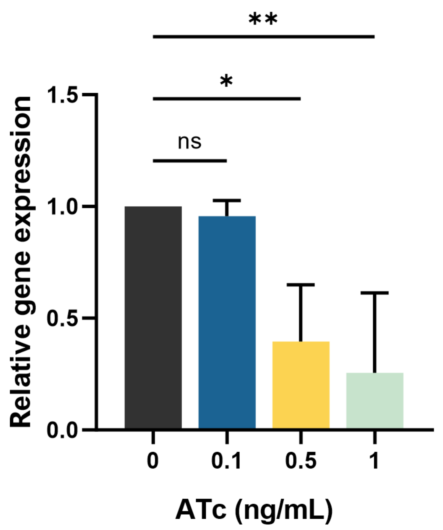
2.2. mmpL3 Gene Expression in the Presence and Absence of ATc

The impact of inducing the transcription response on mmpL3_KD expression in the M. smegmatis strain was assessed using RT-qPCR. mmpL3 expression in mmpL3_KD decreased upon induction with 0.1, 0.5, and 1 ng/mL ATc, resulting in reductions in the expression of 4.4% ± 7.1% (mean ± standard deviation (SD)), 60.4% ± 25.5%, and 74.4% ± 35.7%, respectively. Compared to that in the absence of ATc, mmpL3 expression was significantly lower in the presence of 0.5 and 1 ng/mL ATc, with p < 0.05 and p < 0.01, respectively (Figure 2).

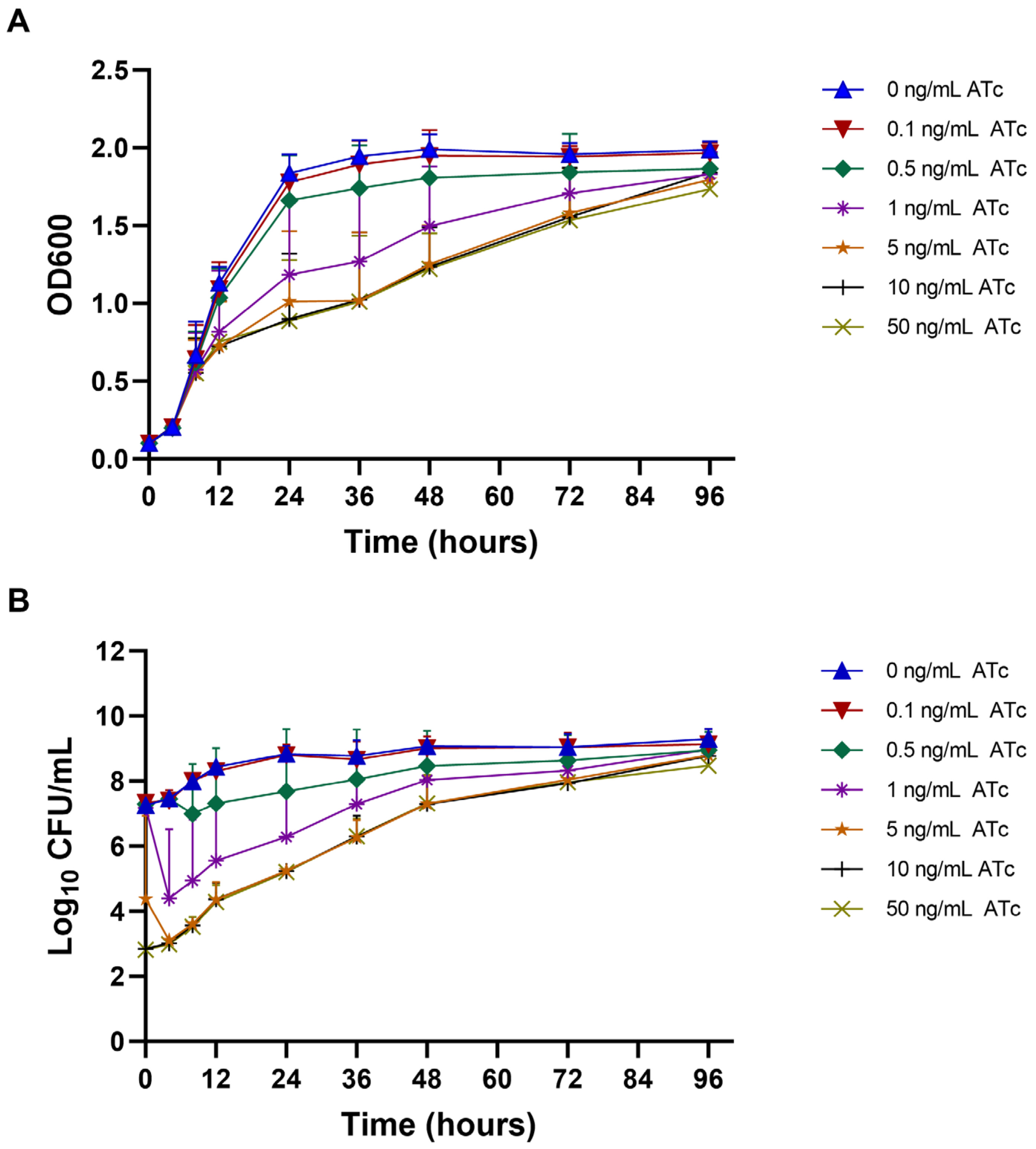
2.3. Growth Curve and Viability of mmpL3_KD M. smegmatis at Different ATc Concentrations

The impact of mmpL3 transcriptional repression on the growth curve and viability (colony forming unit (CFU)/mL) of the mmpL3_KD strain was examined in liquid media supplemented with ATc at concentrations ranging from 0 to 50 ng/mL. A defect in the growth curve and viability of the mmpL3_KD strain was evident at ATc concentrations ≥0.5 ng/mL. Moreover, a significant reduction in the optical density of 600 nm (OD600) of the culture was observed at 24, 36, and 48 h of induction with 1, 5, 10, and 50 ng/mL ATc (p < 0.05) compared to conditions without ATc (Figure 3A). In addition, the CFU/mL of mmpL3_KD rapidly decreased upon exposure to 1, 5, 10, and 50 ng/mL ATc (p < 0.05) within 4 h (Figure 3B). These significant reductions in CFU/mL corresponded to 3.65 ± 0.78-log10 CFU/mL.
Furthermore, the OD600 of the mmpL3_KD culture gradually increased after 24 h of ATc induction and was not significantly different from that in the absence of ATc at 72 and 96 h (Figure 3A). Similarly, the viability (CFU/mL) slowly increased and showed nonsignificant differences at 48, 72, and 96 h of ATc induction (Figure 3B). Additionally, the growth curve and viability of the pLJR962 control strain were comparable to those of the mmpL3_KD strain under conditions without ATc induction or with 0.1 ng/mL ATc (Supplementary Figure S2A,B). Moreover, the biofilm formation of mmpL3_KD was comparable to the pLJR962 control (p > 0.05). For pellicle formation, no pellicle was observed at high ATc concentrations (≥1 ng/mL), which could be due to defects in mycobacterial growth (Supplementary Figures S3 and S4).

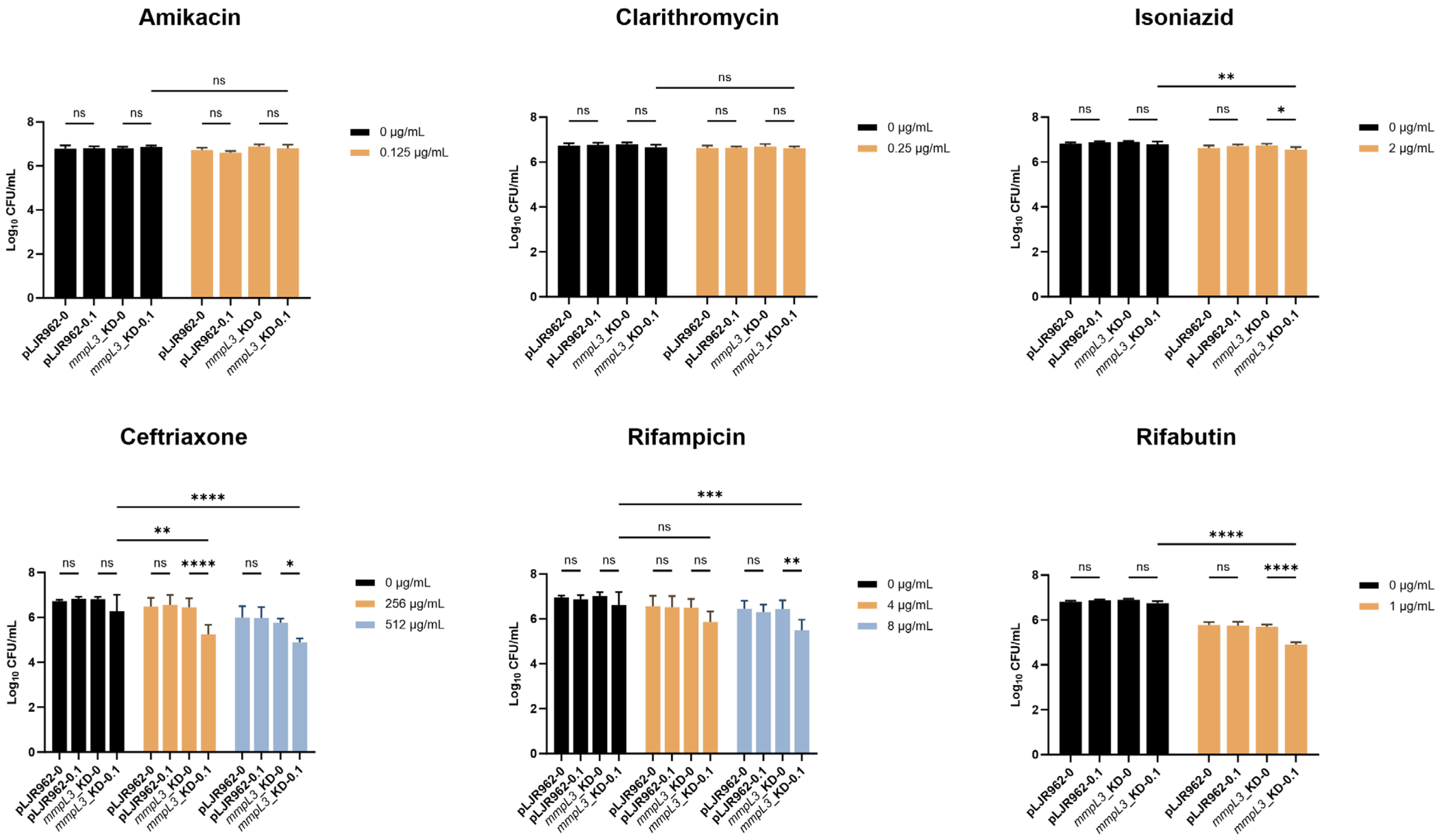
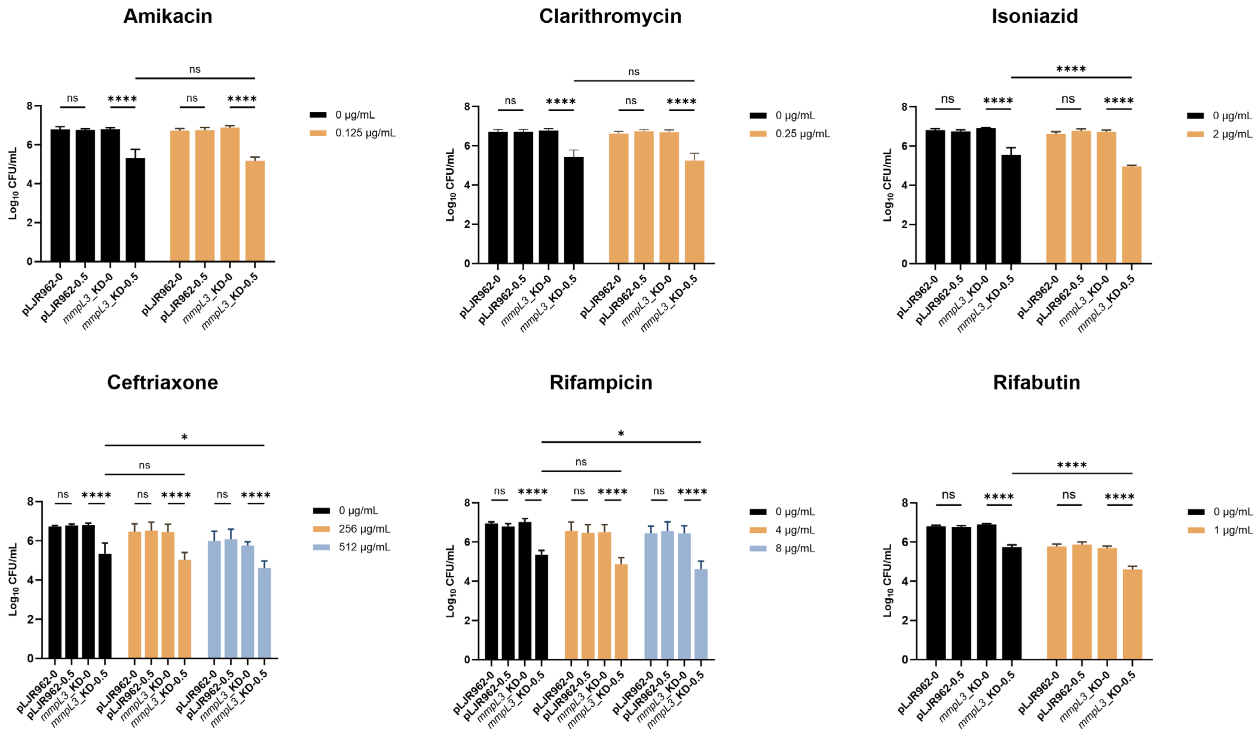
2.4. Resensitization of the mmpL3_KD M. smegmatis Strain to Antimicrobial Agents

To evaluate whether mmpL3 repression affects the susceptibility of the mmpL3_KD strain to non-MmpL3-dependent antimicrobial agents, the minimal inhibitory concentrations (MICs) of various antimicrobial agents against the mmpL3_KD strain were initially assessed. The MICs were determined as follows: 0.25 µg/mL for amikacin, 0.5 µg/mL for clarithromycin, 4 µg/mL for isoniazid, >512 µg/mL for ceftriaxone, >8 µg/mL for rifampicin, and 2 µg/mL for rifabutin. In the presence of 0.1 ng/mL ATc, the MICs of mmpL3_KD were not affected. However, the MICs decreased by one- to two-fold dilutions in the presence of 0.5 ng/mL ATc, which could be due to a growth defect resulting from mmpL3 repression.
Therefore, the impact on the viability (CFU/mL) of the mmpL3_KD strain was assessed in the presence of 0.1 ng/mL ATc, which suppressed mmpL3 expression but did not affect the growth of mycobacterial cells, and one-half of the MICs of the antimicrobial agents, which were 0.125 µg/mL for amikacin, 0.25 µg/mL for clarithromycin, 2 µg/mL for isoniazid, 256 µg/mL and 512 µg/mL for ceftriaxone, 4 µg/mL and 8 µg/mL for rifampicin, and 1 µg/mL for rifabutin. For rifampicin and ceftriaxone, two concentrations were tested because the MIC values exceeded the highest concentration tested in the experiment. One-half of the MIC of each antimicrobial agent reduced the CFU/mL of mmpL3_KD by approximately 0.17-log10 for isoniazid, 0.36-log10 for 256 µg/mL of ceftriaxone, 1.04-log10 for 512 µg/mL of ceftriaxone, 0.52-log10 for 4 µg/mL of rifampicin, 0.57-log10 for 8 µg/mL of rifampicin, and 1.20-log10 for rifabutin, compared to the no-drug control in the absence of ATc. No significant reduction in CFU/mL was observed with amikacin or clarithromycin.
Although the suppression of mmpL3 with 0.1 ng/mL ATc did not alter the MIC, the combination of this ATc level and one-half of the MIC of certain antimicrobial agents resulted in a significant decrease in mmpL3_KD viability. Specifically, the viability of mmpL3_KD with 0.1 ng/mL ATc with isoniazid, ceftriaxone, rifampicin (8 µg/mL), and rifabutin was significantly reduced for 0.77 ± 0.35-log10 CFU/mL) compared with antimicrobial agents alone (p < 0.05) (Figure 4). However, clarithromycin and amikacin exhibited reductions of only 0.07 and 0.08-log10 CFU/mL, respectively (Figure 4). To assess whether greater mmpL3 suppression can more effectively reduce mycobacterial viability, 0.5 ng/mL ATc was added to one-half of the MIC plates. As expected, the viability of the mmpL3_KD strain significantly decreased in the presence of 0.5 ng/mL ATc (p < 0.0001), showing a decrease of 1.47 ± 0.23-log10 CFU/mL compared to that in the absence of ATc, regardless of the antimicrobial agent concentration (0 µg/mL, one-half MIC) (Figure 5). Conversely, the addition of one-half MIC of isoniazid, rifabutin, ceftriaxone (512 µg/mL), or rifampicin (8 µg/mL) to mmpL3_KD with 0.1 or 0.5 ng/mL ATc significantly enhanced the decrease in bacterial viability (p < 0.05) compared to the condition with ATc alone (Figure 4 and Figure 5).
Additionally, induction of dCas9Sth1 without a sgRNA in the pLJR962 control did not increase the defects of growth or viability of M. smegmatis compared to treatment with antimicrobial agents alone (p > 0.05) (Figure 4 and Figure 5).

3. Discussion

MmpL3 functions as a transporter of trehalose monomycolate, a precursor of trehalose dimycolate [1,2]. mmpL3 in M. smegmatis consists of 3042 base pairs encoding 1013 amino acids, serving as an ortholog of rv0206c in M. tuberculosis. Extensive studies have underscored the importance of mmpL3 as an essential gene in M. tuberculosis [15,22], M. smegmatis [23], and M. abscessus [24]. Therefore, mmpL3 has been identified as a promising target for developing new therapeutic agents against M. tuberculosis and NTM [3,4,5,6,25,26]. Previously, the synergistic effects of MmpL3 inhibitors [4,5,13,14] or mmpL3 knockdown using various genetic tools [15,16,17] and certain antimicrobial agents have been investigated. However, these studies have demonstrated varying susceptibilities to non-MmpL3-dependent antimicrobial agents, which may be explained by differences in the level of mmpL3 inhibition or repression achieved under different experimental conditions and in different organisms which can differ substantially in many aspects, limiting the direct application of findings from one to another.
The CRISPRi system is easily constructed and requires only a single transformation, enabling the rapid generation of transcriptional knockdown strains of mycobacteria. In mycobacteria, CRISPRi has demonstrated effectiveness in suppressing drug resistance genes, particularly those associated with β-lactams, by targeting essential peptidoglycan synthesis genes such as pbpB and cwlM [20]. Furthermore, it has been utilized to target the rifampicin resistance gene ADP-ribosyltransferase (arr) [19]. This versatility makes CRISPRi a valuable tool for investigating resistance mechanisms and devising strategies to combat drug-resistant mycobacteria. In our study, the CRISPR-dCas9Sth1 system was used to repress mmpL3 expression, and its impacts on M. smegmatis viability and susceptibility to non-MmpL3-dependent antimicrobial agents were evaluated. The induction of dCas9Sth1 with a high concentration of ATc did not adversely affect the growth or viability of M. smegmatis wild-type or vector control strains, as similarly demonstrated in a previous study [18]. Similar to a previous study [17], the depletion of MmpL3, which is below the levels required for the in vitro growth of mycobacteria, was rapidly achieved with low concentrations of ATc (0.5 ng/mL) in an ATc dose-dependent manner. In our study, this is attributed to the permissive PAM sequences utilized (NNAGAAA), which achieved a gene repression of up to 158.1-fold [18], and the sgRNA targeting the coding region of the mmpL3 gene. Additionally, the gradual increase in growth was observed after 24 h of ATc induction. This phenomenon could be attributed to the degradation of ATc or the growth of the mmpL3_KD M. smegmatis escape mutants. Consequently, their potential influence on the results and subsequent statistical analysis cannot be disregarded. The emergence of strains unresponsive to the ATc inducer is consistent with findings from a previous study [17] and with other tetracycline-regulated promoters utilized in mycobacteria [27].
Importantly, this study investigated the effects of mmpL3 repression on susceptibility to antimicrobial agents. Although a reduction in MIC and CFU/mL was not observed with an approximately 4% to 10% decrease in mmpL3 expression in response to 0.1 ng/mL ATc, a significant reduction (p < 0.05) in CFU/mL was noted when this decreased expression was combined with half of the MICs of several classes of antibiotics, including rifamycin, β-lactam, and isoniazid, demonstrating a similar pattern. However, this reduction in viability did not meet the criteria for synergy, defined as a ≥2 log10 reduction in CFU compared to the antimicrobial agent alone [20]. Several hypotheses could explain these findings. For instance, the ATc concentration might be too low, resulting in insufficient mmpL3 repression. Additionally, the emergence of nonresponsive escape mutants resistant to ATc could have contributed to these results. The significant decrease in CFU/mL observed in the rifamycin group, regardless of rifampicin or rifabutin, can be attributed to disruption of the mycolic acid in the cell wall, resulting in alterations in hydrophobicity [16]. This explanation could also apply to isoniazid, where mmpL3 knockdown may enhance the inhibition of mycolic acid synthesis. In contrast, repression of mmpL3 in M. tuberculosis using the CRISPRi system did not change the MICs of rifampin, isoniazid, or linezolid [17]. Additionally, the synergistic effect of SQ109, an MmpL3 inhibitor, against M. tuberculosis has been demonstrated with isoniazid and rifampicin but not with ethambutol and pyrazinamide in vitro [13]. With respect to ceftriaxone, mmpL3_KD was observed to result in improved susceptibility due to enhanced penetration of β-lactams facilitated by alterations in outer membrane assembly. However, no study has explored the effect of clarithromycin on mmpL3 knockdown. One study demonstrated that specific single nucleotide polymorphisms (SNPs) within the mmpL3 gene of M. smegmatis resulted in growth defects and susceptibility to ampicillin, rifampicin, and erythromycin, but not to chloramphenicol or kanamycin. This susceptibility arose because the depletion of mmpL3 disrupts cell wall formation in mycobacteria [16]. These findings suggest that the synergistic mechanism of mmpL3 repression or inhibition with antimycobacterial agents relies on optimal conditions, such as the level of repression and classes or concentration of antimicrobial agents used. Therefore, further studies are needed to elucidate the true mechanism involved.
In conclusion, these findings emphasize the utility of the CRISPRi-dCas9Sth1 system for selectively targeting and suppressing genes associated with different classes of antimicrobial resistance. Moreover, this approach has the potential to enhance studies focused on understanding of gene functions and resistance to antimicrobial agents. Additionally, the repression of the mmpL3 gene in mycobacteria represents a promising strategy for treating mycobacterial infections, particularly those caused by drug-resistant strains.

4. Materials and Methods

4.1. Bacterial Growth and Culture Conditions

M. smegmatis MC2155 and its derivative strains were grown either in Middlebrook 7H9 (7H9) broth or Middlebrook 7H10 (7H10) solid media (BD Difco, Sparks, MD, USA) supplemented with 0.2% v/v glycerol (HiMedia, Maharashtra, India) and 10% ADC (5% BSA, 2% dextrose, 0.003% catalase) at 37 °C. The broth was further supplemented with 0.05% Tween 80 (Ajax Finechem, NSW, Australia) and incubated with shaking at 200 rpm. For cloning experiments, Escherichia coli DH5α was grown in Luria Bertani (LB) broth and agar at 37 °C. Kanamycin (GoldBio, St. Loius, MO, USA) was added as needed at concentrations of 50 µg/mL for E. coli and 25 µg/mL for M. smegmatis. Anhydrotetracycline (ATc, Sigma-Aldrich, St. Louis, MO, USA) was added at different concentrations as required for specific experiments.

4.2. Construction of sgRNA Expression Plasmids and mmpL3_KD Strains

mmpL3 (msmeg_0250)-knockdown M. smegmatis (mmpL3_KD) was constructed using the CRISPRi system with the pLJR962 plasmid (Addgene no. 115162). This plasmid expresses both the targeting sgRNA and Sth1 dCas9 from a Tet repressor (TetR)-regulated promoter induced by ATc and contains a kanamycin selection marker. This plasmid integrates into the L5 attB site of mycobacterial chromosome when transformed into mycobacteria [18,21]. Briefly, the dCas9Sth1 protospacer adjacent motif (PAM) sequence against the reference M. smegmatis MC2155 gene was identified using a design tool (https://pebble.rockefeller.edu/tools/sgrna-design/, accessed on 1 November 2021). Then, 23 nucleotide sgRNA targeting sequences upstream of the PAM, 5′ NNAGAAA 3′, were extracted. The 5′ GGGA 3′ sequence was appended to the 5′ sequence of the sgRNA targeting sequence for cloning the sgRNA [21]. The sgRNA oligos targeting the mmpL3 gene were mmpL3_T: 5′ GGGAGCGACAGACTGGCTGCCCTCGTC 3′ and mmpL3_B: 5′AAACGACGAGGGCAGCCAGTCTGTCGC 3′, which were previously designed to target the nontemplate strand of mmpL3 [21]. The sequence of sgRNA is specific to mmpL3 of M. smegmatis MC2155 without complementarity to other sites in the genome.
These sgRNA oligos were annealed, ligated into a BsmBI-digested CRISPRi backbone (pLJR962), and transformed into E. coli DH5α, which was selected on LB agar supplemented with kanamycin. Purified sgRNA-pLJR962 plasmids from E. coli were verified using Sanger sequencing with the pLJR962-965-SS primer: 5′ TTCCTGTGAAGAGCCATTGATAATG 3′. Next, sequence-verified plasmids (100 ng) and pLJR962 plasmids without sgRNA (empty vector control) were electroporated at 25 kV and 25 µF with 1000 W resistance into electrocompetent M. smegmatis MC2155 strains, which were prepared as previously described [21]. The transformants were selected on 7H10 agar supplemented with 25 µg/mL kanamycin in the presence or absence of 50 ng/mL ATc to determine the bacterial viability of the mmpL3_KD strains. Subsequently, the presence of the CRISPRi-dCas9Sth1 construct in M. smegmatis was confirmed by PCR.

4.3. Determination of the Essentiality of the mmpL3 Gene in M. smegmatis

Mid-log phase cultures of both mmpL3_KD and pLJR962 control M. smegmatis strains grown in 7H9 broth supplemented with 0.2% glycerol, 0.05% Tween 80, 10% ADC, and 25 µg/mL kanamycin were diluted to an OD600 of 0.1. Subsequently, these cultures were serially diluted 10-fold (10−1–10−5). Five microliters of the diluted cultures were then spotted on 7H10 agar plates containing different concentrations of ATc (0, 0.1, 0.5, 1, 5, 10, and 50 ng/mL) and incubated at 37 °C for 3 days.

4.4. Bacterial Growth Curve and Viability Count

The mid-log phase cultures of the mmpL3_KD and pLJR962 control M. smegmatis strains were prepared at an OD600 of 0.1 and induced with various concentrations of ATc: 0, 0.1, 0.5, 1, 5, 10, and 50 ng/mL. The cultures were then incubated, and the OD600 was measured at different time points from 0 to 96 h. For viability assessment, the CFU/mL of cultures were determined by collecting cultures both with and without ATc at various time points ranging from 0 to 96 h. Each culture condition was serially diluted (10−2–10−8), and 20 µL of the diluted samples was plated and incubated at 37 °C for 3 days or until visible colonies were observed to determine the CFU/mL. All experiments were conducted in biological and technical triplicates.

4.5. Determination of the Minimal Inhibitory Concentration and Mycobacterial Viability

The M. smegmatis mmpL3_KD and pLJR962 control strains were cultured on 7H10 agar plates and incubated at 37 °C until visible colonies were observed (2–3 days). Drug susceptibility tests were conducted using a broth microdilution method following the Clinical & Laboratory Standards Institute (CLSI) recommendations to determine MIC values [28]. Briefly, 100 µL of bacterial suspensions (approximately 5 × 10⁵ CFU) were added to each well of sterile flat bottom 96-well plates (Jet Biofil, Guangzhou, China) that contained drugs at 2-fold serial dilutions. The antimicrobial agents tested included ceftriaxone (1–512 µg/mL), rifampicin (0–8 µg/mL), amikacin (1–6 µg/mL), rifabutin (0.25–8 µg/mL), isoniazid (0.25–8 µg/mL), and clarithromycin (0.06–16 µg/mL) with the presence or absence of ATc. Then, the plates were incubated at 30 °C for 2–3 days until a sufficient positive control (no antimicrobial agent) was obtained. Mycobacterium peregrinum ATCC700686 served as the quality control strain.
The impact of the ATc inducer and antimicrobial agents on the viability of the mmpL3_KD and pLJR962 control M. smegmatis strains was evaluated. Five microliters of 10-fold serial dilutions (10−1–10−6) of mid-log phase cultures were plated on 7H10 agar containing one-half of the MIC of the respective antimicrobial agents in the presence or absence of 0.1 and 0.5 ng/mL ATc. The plates were then incubated at 37 °C until visible colonies were observed to determine the CFU/mL. All experiments were conducted with biological and technical replicates.

4.6. RNA Extraction, cDNA Synthesis, and RT-qPCR

To investigate the strength of the transcriptional repression, mid-log phase cultures of the mmpL3_KD and pLJR962 control M. smegmatis strains were initiated at an OD600 of 0.1 in 7H9 broth supplemented with ADC, 0.05% Tween 80, and 25 µg/mL kanamycin. These cultures were then induced with varying concentrations of ATc (0, 0.1, 0.5, and 1 ng/mL) for 12 h. RNA extraction was conducted as previously described [29]. Briefly, bacterial cells harvested at 0 and 12 h after ATc induction were collected via centrifugation at 2000× g at 4 °C for 5 min and then resuspended in 1 mL of TRIzol solution (Invitrogen, Waltham, MA, USA) on ice. Then, the cells were disrupted using acid-washed glass beads (150–212 µm) (Sigma-Aldrich, St. Louis, MO, USA) for three cycles of 30 s at a speed of 6.5 m/s using a FastPrep-24 instrument (MP Biomedicals, Santa Ana, CA, USA).
RNA was purified using 300 µL of chloroform: isoamyl alcohol (24:1) followed by precipitation using 270 µL of isopropanol and 270 µL of 1.2 M NaCl at 4 °C for 3–4 h. The RNA pellet was then washed twice with 75% ethanol. Furthermore, the RNA samples were treated with a Turbo-DNA-free Kit (Invitrogen, Waltham, MA, USA) according to the manufacturer’s instructions. cDNA was synthesized using iScript™ Reverse Transcription Supermix (Bio-Rad Laboratories, Hercules, CA, USA) following the manufacturer’s instructions. qPCR was conducted to quantify the expression of mmpL3 and sigA using Luna® Universal qPCR Master Mix (New England Biolabs, Ipswich, MA, USA) with 50 ng of cDNA and 0.12 µL of sigA primers (msmeg_sigA_F: 5′ GACGACGACATCGACGAG 3′ and msmeg_sigA_R: 5′ GTCAGCTCGGCGTCTTTG 3′) and mmpL3 primers (msmeg_0250_F: 5′ TCGATCAGGTGGTCAAGGA 3′ and msmeg_0250_R: 5′ GCAGATCCTGCGTCTTCAT 3′), respectively, in a final volume of 20 µL per reaction.
The qPCRs were run on a QuantStudio 5 Real-Time PCR system (Applied Biosystems, Waltham, MA, USA) with the following cycling conditions: an initial denaturation step at 95 °C for 60 s, followed by 45 cycles of denaturation at 95 °C for 15 s and annealing and extension at 60 °C for 30 s. To normalize the expression levels of mmpL3_KD, the mRNA levels of the reference sigA gene were used. The relative gene expression levels were analyzed relative to the expression of the pLJR962 control. Additionally, melting curve analysis was performed to verify the specificity of the qPCR amplification. All experiments were conducted in biological and technical triplicates.

4.7. Statistical Analysis

The statistical significance of differences in viability and gene expression at different ATc concentrations or time points was assessed using one-way or two-way ANOVA, as appropriate. A p value less than 0.05 was considered to indicate statistical significance. Statistical analysis and graph were generated using GraphPad Prism version 10.0.0 for Windows.

Supplementary Materials

The following supporting information can be downloaded at: https://www.mdpi.com/article/10.3390/antibiotics13060483/s1, Supplementary Materials and Methods: Biofilm and pellicle formation; Figure S1: Essentiality of the mmpL3 gene in the mmpL3_KD and pLJR962 control M. smegmatis strains; Figure S2: Growth curve and viability of the mmpL3_KD and pLJR962 control M. smegmatis strains at different time points; Figure S3: Biofilm formation of the mmpL3_KD and pLJR962 control M. smegmatis strains assessed by a quantitative crystal violet assay; Figure S4: Pellicle formation of the mmpL3_KD and pLJR962 control M. smegmatis strains observed on day 3 of incubation. Reference [30] is cited in the Supplementary Materials.

Author Contributions

Conceptualization, S.R. and T.P.; methodology, S.R. and T.P.; validation, S.R.; formal analysis, S.R. and Y.Y.; investigation, Y.Y., A.F.N.I., S.P. and A.S.; resources, T.C. and S.C.; data curation, S.R. and Y.Y.; writing—original draft preparation, S.R. and Y.Y.; writing—review and editing, S.R., Y.Y., A.F.N.I., S.C., T.C. and T.P.; supervision, T.C. and T.P.; funding acquisition, S.R. All authors have read and agreed to the published version of the manuscript.

Funding

This work (Grant No. RGNS 63-008) was supported by the Office of the Permanent Secretary, Ministry of Higher Education, Science, Research and Innovation (OPS MHESI), Thailand Science Research and Innovation (TSRI) and Chulalongkorn University.

Institutional Review Board Statement

Not applicable.

Informed Consent Statement

Not applicable.

Data Availability Statement

No datasets were generated during the study. All the data analyzed in this study are included in this published article.

Conflicts of Interest

The authors declare no conflicts of interest.

References

  1. Xu, Z.; Meshcheryakov, V.A.; Poce, G.; Chng, S.-S. MmpL3 is the flippase for mycolic acids in mycobacteria. Proc. Natl. Acad. Sci. USA 2017, 114, 7993–7998. [Google Scholar] [CrossRef]
  2. Domenech, P.; Reed, M.B.; Barry, C.E. Contribution of the Mycobacterium tuberculosis MmpL Protein Family to Virulence and Drug Resistance. Infect. Immun. 2005, 73, 3492–3501. [Google Scholar] [CrossRef]
  3. Sacksteder, K.A.; Protopopova, M.; Barry, C.E.; Andries, K.; Nacy, C.A. Discovery and development of SQ109: A new antitubercular drug with a novel mechanism of action. Future Microbiol. 2012, 7, 823–837. [Google Scholar] [CrossRef]
  4. Nikonenko, B.V.; Protopopova, M.; Samala, R.; Einck, L.; Nacy, C.A. Drug Therapy of Experimental Tuberculosis (TB): Improved Outcome by Combining SQ109, a New Diamine Antibiotic, with Existing TB Drugs. Antimicrob. Agents Chemother. 2007, 51, 1563–1565. [Google Scholar] [CrossRef]
  5. Li, W.; Sanchez-Hidalgo, A.; Jones, V.; de Moura, V.C.; North, E.J.; Jackson, M. Synergistic Interactions of MmpL3 Inhibitors with Antitubercular Compounds In Vitro. Antimicrob. Agents Chemother. 2017, 61, e02399-16. [Google Scholar] [CrossRef]
  6. Dupont, C.; Viljoen, A.; Dubar, F.; Blaise, M.; Bernut, A.; Pawlik, A.; Bouchier, C.; Brosch, R.; Guérardel, Y.; Lelièvre, J.; et al. A new piperidinol derivative targeting mycolic acid transport in Mycobacterium abscessus. Mol. Microbiol. 2016, 101, 515–529. [Google Scholar] [CrossRef]
  7. WHO. WHO Consolidated Guidelines on Tuberculosis: Module 4: Treatment-Drug-Susceptible Tuberculosis Treatment; World Health Organization: Geneva, Switzerland, 2022. [Google Scholar]
  8. Daley, C.L.; Iaccarino, J.M.; Lange, C.; Cambau, E.; Wallace, R.J., Jr.; Andrejak, C.; Böttger, E.C.; Brozek, J.; Griffith, D.E.; Guglielmetti, L.; et al. Treatment of Nontuberculous Mycobacterial Pulmonary Disease: An Official ATS/ERS/ESCMID/IDSA Clinical Practice Guideline. Clin. Infect. Dis. 2020, 71, e1–e36. [Google Scholar] [CrossRef]
  9. Quan, S.; Imai, T.; Mikami, Y.; Yazawa, K.; Dabbs, E.R.; Morisaki, N.; Iwasaki, S.; Hashimoto, Y.; Furihata, K. ADP-Ribosylation as an intermediate step in inactivation of rifampin by a mycobacterial gene. Antimicrob. Agents Chemother. 1999, 43, 181–184. [Google Scholar] [CrossRef]
  10. Quan, S.; Venter, H.; Dabbs, E.R. Ribosylative inactivation of rifampin by Mycobacterium smegmatis is a principal contributor to its low susceptibility to this antibiotic. Antimicrob. Agents Chemother. 1997, 41, 2456–2460. [Google Scholar] [CrossRef]
  11. Hetherington, S.V.; Watson, A.S.; Patrick, C.C. Sequence and analysis of the rpoB gene of Mycobacterium smegmatis. Antimicrob. Agents Chemother. 1995, 39, 2164–2166. [Google Scholar] [CrossRef]
  12. Flores, A.R.; Parsons, L.M.; Pavelka, M.S. Genetic analysis of the β-lactamases of Mycobacterium tuberculosis and Mycobacterium smegmatis and susceptibility to β-lactam antibiotics. Microbiology 2005, 151, 521–532. [Google Scholar] [CrossRef]
  13. Chen, P.; Gearhart, J.; Protopopova, M.; Einck, L.; Nacy, C.A. Synergistic interactions of SQ109, a new ethylene diamine, with front-line antitubercular drugs in vitro. J. Antimicrob. Chemother. 2006, 58, 332–337. [Google Scholar] [CrossRef]
  14. Stec, J.; Onajole, O.K.; Lun, S.; Guo, H.; Merenbloom, B.; Vistoli, G.; Bishai, W.R.; Kozikowski, A.P. Indole-2-carboxamide-based MmpL3 Inhibitors Show Exceptional Antitubercular Activity in an Animal Model of Tuberculosis Infection. J. Med. Chem. 2016, 59, 6232–6247. [Google Scholar] [CrossRef]
  15. Li, W.; Obregón-Henao, A.; Wallach, J.B.; North, E.J.; Lee, R.E.; Gonzalez-Juarrero, M.; Schnappinger, D.; Jackson, M. Therapeutic Potential of the Mycobacterium tuberculosis mycolic acid transporter, MmpL3. Antimicrob. Agents Chemother. 2016, 60, 5198–5207. [Google Scholar] [CrossRef]
  16. McNeil, M.B.; Dennison, D.; Parish, T. Mutations in MmpL3 alter membrane potential, hydrophobicity and antibiotic susceptibility in Mycobacterium smegmatis. Microbiology 2017, 163, 1065–1070. [Google Scholar] [CrossRef]
  17. McNeil, M.B.; Cook, G.M. Utilization of CRISPR interference to validate MmpL3 as a drug target in Mycobacterium tuberculosis. Antimicrob. Agents Chemother. 2019, 63, e00629-19. [Google Scholar] [CrossRef]
  18. Rock, J.M.; Hopkins, F.F.; Chavez, A.; Diallo, M.; Chase, M.R.; Gerrick, E.R.; Pritchard, J.R.; Church, G.M.; Rubin, E.J.; Sassetti, C.M.; et al. Programmable transcriptional repression in mycobacteria using an orthogonal CRISPR interference platform. Nat. Microbiol. 2017, 2, 16274. [Google Scholar] [CrossRef]
  19. Faulkner, V.; Cox, A.A.; Goh, S.; van Bohemen, A.; Gibson, A.J.; Liebster, O.; Wren, B.W.; Willcocks, S.; Kendall, S.L. Re-sensitization of Mycobacterium smegmatis to rifampicin using CRISPR interference demonstrates its utility for the study of non-essential drug resistance traits. Front. Microbiol. 2020, 11, 619427. [Google Scholar] [CrossRef]
  20. Kurepina, N.; Chen, L.; Composto, K.; Rifat, D.; Nuermberger, E.L.; Kreiswirth, B.N. CRISPR inhibition of essential of peptidoglycan biosynthesis genes in Mycobacterium abscessus and its impact on β-lactam susceptibility. Antimicrob. Agents Chemother. 2022, 66, e0009322. [Google Scholar] [CrossRef]
  21. Wong, A.I.; Rock, J.M. CRISPR Interference (CRISPRi) for Targeted Gene Silencing in Mycobacteria. In Mycobacteria Protocols; Parish, T., Kumar, A., Eds.; Springer: New York, NY, USA, 2021; pp. 343–364. [Google Scholar]
  22. Degiacomi, G.; Benjak, A.; Madacki, J.; Boldrin, F.; Provvedi, R.; Palù, G.; Kordulakova, J.; Cole, S.T.; Manganelli, R. Essentiality of mmpL3 and impact of its silencing on Mycobacterium tuberculosis gene expression. Sci. Rep. 2017, 7, 43495. [Google Scholar] [CrossRef]
  23. Varela, C.; Rittmann, D.; Singh, A.; Krumbach, K.; Bhatt, K.; Eggeling, L.; Besra, G.S.; Bhatt, A. MmpL genes are associated with mycolic acid metabolism in mycobacteria and corynebacteria. Chem. Biol. 2012, 19, 498–506. [Google Scholar] [CrossRef] [PubMed]
  24. Boudehen, Y.-M.; Tasrini, Y.; Aguilera-Correa, J.J.; Alcaraz, M.; Kremer, L. Silencing essential gene expression in Mycobacterium abscessus during infection. Microbiol. Spectr. 2023, 11, e0283623. [Google Scholar] [CrossRef] [PubMed]
  25. Tahlan, K.; Wilson, R.; Kastrinsky, D.B.; Arora, K.; Nair, V.; Fischer, E.; Barnes, S.W.; Walker, J.R.; Alland, D.; Barry, C.E.; et al. SQ109 Targets MmpL3, a Membrane Transporter of Trehalose Monomycolate Involved in Mycolic Acid Donation to the Cell Wall Core of Mycobacterium tuberculosis. Antimicrob. Agents Chemother. 2012, 56, 1797–1809. [Google Scholar] [CrossRef] [PubMed]
  26. Lun, S.; Guo, H.; Onajole, O.K.; Pieroni, M.; Gunosewoyo, H.; Chen, G.; Tipparaju, S.K.; Ammerman, N.C.; Kozikowski, A.P.; Bishai, W.R. Indoleamides are active against drug-resistant Mycobacterium tuberculosis. Nat. Commun. 2013, 4, 2907. [Google Scholar] [CrossRef] [PubMed]
  27. Evans, J.C.; Mizrahi, V. The application of tetracyclineregulated gene expression systems in the validation of novel drug targets in Mycobacterium tuberculosis. Front. Microbiol. 2015, 6, 812. [Google Scholar] [CrossRef]
  28. CLSI. Susceptibility Testing of Mycobacteria, Nocardia spp., and Other Aerobic Actinomycetes, 3rd ed.; CLSI Standard M24; Clinical and Laboratory Standads Institute: Wayne, PA, USA, 2018. [Google Scholar]
  29. Rustad, T.R.; Roberts, D.M.; Liao, R.P.; Sherman, D.R. Isolation of mycobacterial RNA. In Mycobacteria Protocols, 2nd ed.; Parish, T., Brown, A.C., Eds.; Humana Press: Totowa, NJ, USA, 2009; pp. 13–22. [Google Scholar]
  30. Di Somma, A.; Caterino, M.; Soni, V.; Agarwal, M.; di Pasquale, P.; Zanetti, S.; Molicotti, P.; Cannas, S.; Nandicoori, V.K.; Duilio, A. The bifunctional protein GlmU is a key factor in biofilm formation induced by alkylating stress in Mycobacterium smegmatis. Res. Microbiol. 2019, 170, 171–181. [Google Scholar] [CrossRef]
Figure 1. Essentiality of the mmpL3 gene in the mmpL3_KD M. smegmatis strain. Serial dilutions (10−1 to 10−5) of log-phase cultures of the mmpL3_KD M. smegmatis strain were plated on 7H10 agar in both the absence and presence of varying concentrations of ATc (0.1, 0.5, 1, or 5 ng/mL).
Antibiotics 13 00483 g001
Figure 2. mmpL3 gene expression in the mmpL3_KD M. smegmatis strain in the presence and absence of ATc (ng/mL). The experiments were conducted in biological and technical triplicates. Statistical significance is indicated as * p < 0.05 and ** p < 0.01, and “ns” indicates not statistically significant. The error bars represent the SDs of the means.
Antibiotics 13 00483 g002
Figure 3. Growth curve and viability of the mmpL3_KD M. smegmatis strain at different time points. (A) OD600 of the mmpL3_KD M. smegmatis strain at different ATc concentrations (ng/mL). (B) The viability (log10 CFU/mL) of the mmpL3_KD M. smegmatis strain at different ATc concentrations (ng/mL). The experiments were conducted in biological and technical triplicates. The error bars represent the SDs of the means.
Antibiotics 13 00483 g003
Figure 4. Repression of mmpL3 with 0.1 ng/mL ATc resulted in resensitization to isoniazid, ceftriaxone, rifampicin, and rifabutin. The viability (log10 CFU/mL) of the mmpL3_KD and pLJR962 control M. smegmatis strains treated with antimicrobial agents in the presence or absence of 0.1 ng/mL ATc was determined in biological and technical triplicates. Statistical significance is indicated as * p < 0.05, ** p < 0.01, *** p < 0.001, and **** p < 0.0001, and “ns” indicates not statistically significant. The error bars represent the SDs of the means.
Antibiotics 13 00483 g004
Figure 5. Repression of mmpL3 with 0.5 ng/mL ATc significantly impacted M. smegmatis viability and antimicrobial susceptibility. The viability (log10 CFU/mL) of the mmpL3_KD and pLJR962 control M. smegmatis strains treated with antimicrobial agents in the presence or absence of 0.5 ng/mL ATc was determined in biological and technical triplicates. Statistical significance is indicated as * p < 0.05, **** p < 0.0001, and “ns” indicates not statistically significant. The error bars represent the SDs of the means.
Antibiotics 13 00483 g005
Disclaimer/Publisher’s Note: The statements, opinions and data contained in all publications are solely those of the individual author(s) and contributor(s) and not of MDPI and/or the editor(s). MDPI and/or the editor(s) disclaim responsibility for any injury to people or property resulting from any ideas, methods, instructions or products referred to in the content.

