Article

Whole Genome Sequencing Reveals Antimicrobial Resistance and Virulence Genes of Both Pathogenic and Non-Pathogenic B. cereus Group Isolates from Foodstuffs in Thailand

by
Phornphan Sornchuer
1,2,*,
Kritsakorn Saninjuk
3,†,
Sumet Amonyingcharoen
4,
Jittiporn Ruangtong
2,
Nattaya Thongsepee
1,2,
Pongsakorn Martviset
1,2,
Pathanin Chantree
1,2 and
Kant Sangpairoj
1,2
1
Department of Preclinical Science, Faculty of Medicine, Thammasat University, Pathum Thani 12120, Thailand
2
Thammasat University Research Unit in Nutraceuticals and Food Safety, Faculty of Medicine, Thammasat University, Pathum Thani 12120, Thailand
3
Porcinotec Co., Ltd., Nonthaburi 11000, Thailand
4
Medical Life Sciences Institute, Department of Medical Sciences, Ministry of Public Health, Nonthaburi 11000, Thailand
*
Author to whom correspondence should be addressed.
Current address: School of Science, Mae Fah Luang University, Chiang Rai 57100, Thailand.
Antibiotics 2024, 13(3), 245; https://doi.org/10.3390/antibiotics13030245
Submission received: 20 January 2024 / Revised: 1 March 2024 / Accepted: 5 March 2024 / Published: 7 March 2024
(This article belongs to the Special Issue Antimicrobial Resistance: What Can We Learn from Genomics?)

Abstract

:
Members of the Bacillus cereus group are spore-forming Gram-positive bacilli that are commonly associated with diarrheal or emetic food poisoning. They are widespread in nature and frequently present in both raw and processed food products. Here, we genetically characterized 24 B. cereus group isolates from foodstuffs. Whole-genome sequencing (WGS) revealed that most of the isolates were closely related to B. cereus sensu stricto (12 isolates), followed by B. pacificus (5 isolates), B. paranthracis (5 isolates), B. tropicus (1 isolate), and “B. bingmayongensis” (1 isolate). The most detected virulence genes were BAS_RS06430, followed by bacillibactin biosynthesis genes (dhbA, dhbB, dhbC, dhbE, and dhbF), genes encoding the three-component non-hemolytic enterotoxin (nheA, nheB, and nheC), a gene encoding an iron-regulated leucine-rich surface protein (ilsA), and a gene encoding a metalloprotease (inhA). Various biofilm-associated genes were found, with high prevalences of tasA and sipW genes (matrix protein-encoding genes); purA, purC, and purL genes (eDNA synthesis genes); lytR and ugd genes (matrix polysaccharide synthesis genes); and abrB, codY, nprR, plcR, sinR, and spo0A genes (biofilm transcription regulator genes). Genes related to fosfomycin and beta-lactam resistance were identified in most of the isolates. We therefore demonstrated that WGS analysis represents a useful tool for rapidly identifying and characterizing B. cereus group strains. Determining the genetic epidemiology, the presence of virulence and antimicrobial resistance genes, and the pathogenic potential of each strain is crucial for improving the risk assessment of foodborne B. cereus group strains.

1. Introduction

Bacillus species, which are spore-forming Gram-positive bacteria, are widespread in nature as spores and vegetative cells. They easily spread to both raw and processed food such as rice, meat, seafood, vegetables, and dairy products. As the spores are resistant to heat, freezing, drying, and radiation, there is a risk of their transmission in heat-treated and processed food products.
The B. cereus group, also known as B. cereus sensu lato (s.l.), consists of three main species: B. cereus sensu stricto (s.s.), which is an opportunistic pathogen associated with foodborne infections as well as extraintestinal infections; B. anthracis, the etiological agent of anthrax in humans and animals; and B. thuringiensis, an entomopathogen that is well-known for its use as a biopesticide [1,2]. The B. cereus group also includes B. mycoides and B. pseudomycoides, which are characterized by rhizoidal colonies on culture media [3]; B. weihenstephanensis and B. wiedmannii, which are psychrotolerant bacteria [4,5,6]; and B. cytotoxicus, which is a thermotolerant bacterium [7]. Other bacteria that are classified as B. cereus group species are B. albus, B. bingmayongensis, B. fungorum, B. gaemokensis, B. luti, B. manliponensis, B. mobilis, B. nitratireducens, B. pacificus, B. paramycoides, B. paranthracis, B. proteolyticus, B. toyonensis, and B. tropicus [8,9,10,11]. Additionally, there are more species recently confirmed to be members of this group according to the List of Prokaryotic Names with Standing in Nomenclature (LPSN; http://www.bacterio.net, accessed on 10 February 2024).
Some members of the B. cereus group are causative agents of gastrointestinal syndrome (emetic type or diarrheal type) and non-gastrointestinal tract infections, including septicemia, central nervous system infection, respiratory tract infection, and severe ocular infection, especially in immunocompromised individuals [12,13]. The pathogenicity of B. cereus, whether intestinal or non-intestinal, is associated with the production of tissue-destructive exotoxins (hemolysins, phospholipases, pore-forming enterotoxins) and emesis-inducing toxin [14,15]. The notable pore-forming enterotoxins comprise hemolysin BL (HBL), non-hemolytic enterotoxin (NHE), and cytotoxin K (CytK) [16,17,18]. The enterotoxins are produced by vegetative cells (which are ingested as viable cells or spores) in the small intestine, which causes abdominal pain and watery diarrhea [15]. The emesis-inducing toxin cereulide, a plasmid-encoded cyclic peptide, can be synthesized in contaminated food products such as rice, milk, and pasta. Other virulence factors include hemolysin II (encoded by hlyII), enterotoxin FM (encoded by entFM), and another putative enterotoxin (encoded by entABC) [19].
Biofilm formation can be associated with chronic and/or severe bacterial infections in humans as it can protect bacteria against antimicrobials and/or the host immune system. Biofilms are also important in the food industry, especially for the B. cereus group where biofilms can contaminate food products. Biofilm is considered an extremely strong extracellular matrix, and it facilitates bacterial attachment to both abiotic and biotic surfaces [20]. Moreover, biofilm formation mediates the development and transmission of antimicrobial resistance (AMR) genes by facilitating bacterial interactions within the biofilm [21,22]. Therefore, biofilm formation is considered to be an important virulence determinant of various pathogenic bacteria including Bacillus species [23].
Although most B. cereus group infections may not need antimicrobial treatment, it is still crucial to prepare for some circumstances, especially those that warrant critical care [24]. Foodborne illness associated with B. cereus group strains do not need to be treated with antibiotics. However, B. cereus bacteremia, which can cause severe life-threatening systemic infections especially in immunocompromised patients, need to be treated with appropriate antibiotic agents. Extensive antimicrobial use can lead to the emergence of AMR bacterial strains, which can cause the failure of routine treatments. Determining the AMR profile of Bacillus species is important not only for treatment selection but also for providing basic knowledge on the transmissible AMR genes in the food chain.
The number of whole-genome sequencing (WGS) studies of bacteria has increased as the cost of second-generation, short-read sequencing platforms (e.g., Illumina) has steadily decreased [25]. WGS is an effective strategy for rapidly characterizing B. cereus group strains, in terms of their genetic epidemiology and the presence of virulence and AMR genes. The use of genetic loci as markers to discriminate between pathogenic and nonpathogenic B. cereus group strains is more effective than phenotypic and biochemical methods in some circumstances [26,27]. The pantoate-beta-alanine ligase (panC) gene is used to classify B. cereus isolates (e.g., from the environment and cases of bacteremia in humans) into seven phylogenetic clades (I to VII) [10,28].
In this study, 24 B. cereus group isolates were characterized by WGS using Illumina sequencing. The presence of genes related to virulence and AMR was evaluated in all isolates. The study aimed to characterize these foodborne bacteria, which may cause health hazards if not controlled.

2. Results

2.1. Phylogenetic Analysis of B. cereus Group Isolates

The sources and genomic information of the 24 B. cereus group isolates investigated in this study are summarized in Table 1. A phylogenetic tree of the 24 B. cereus group isolates and other type strains of B. cereus group genome sequences was generated using GtoTree, a genome-based phylogenomics workflow (Figure 1A,B).
Based on this phylogenetic tree, the 24 B. cereus group isolates were classified as B. cereus s.s. (12 isolates), B. pacificus (5 isolates), B. paranthracis (5 isolates), B. tropicus (1 isolate), and “B. bingmayongensis” (1 isolate). In detail, Bacillus sp. B08, Bacillus sp. B14, Bacillus sp. B21, Bacillus sp. B54, Bacillus sp. B66, Bacillus sp. B68, Bacillus sp. B69, Bacillus sp. B102, Bacillus sp. B112, Bacillus sp. B123, Bacillus sp. B124, and Bacillus sp. B125 were closely related to B. cereus s.s.; Bacillus sp. B18, Bacillus sp. B62, Bacillus sp. B92, Bacillus sp. B106, and Bacillus sp. B122 were closely related to B. pacificus; Bacillus sp. B81, Bacillus sp. B84, Bacillus sp. B85, Bacillus sp. B94, and Bacillus sp. B98 were closely related to B. paranthracis; Bacillus sp. B134 was closely related to B. tropicus; and Bacillus sp. B103 was closely related to “B. bingmayongensis”.
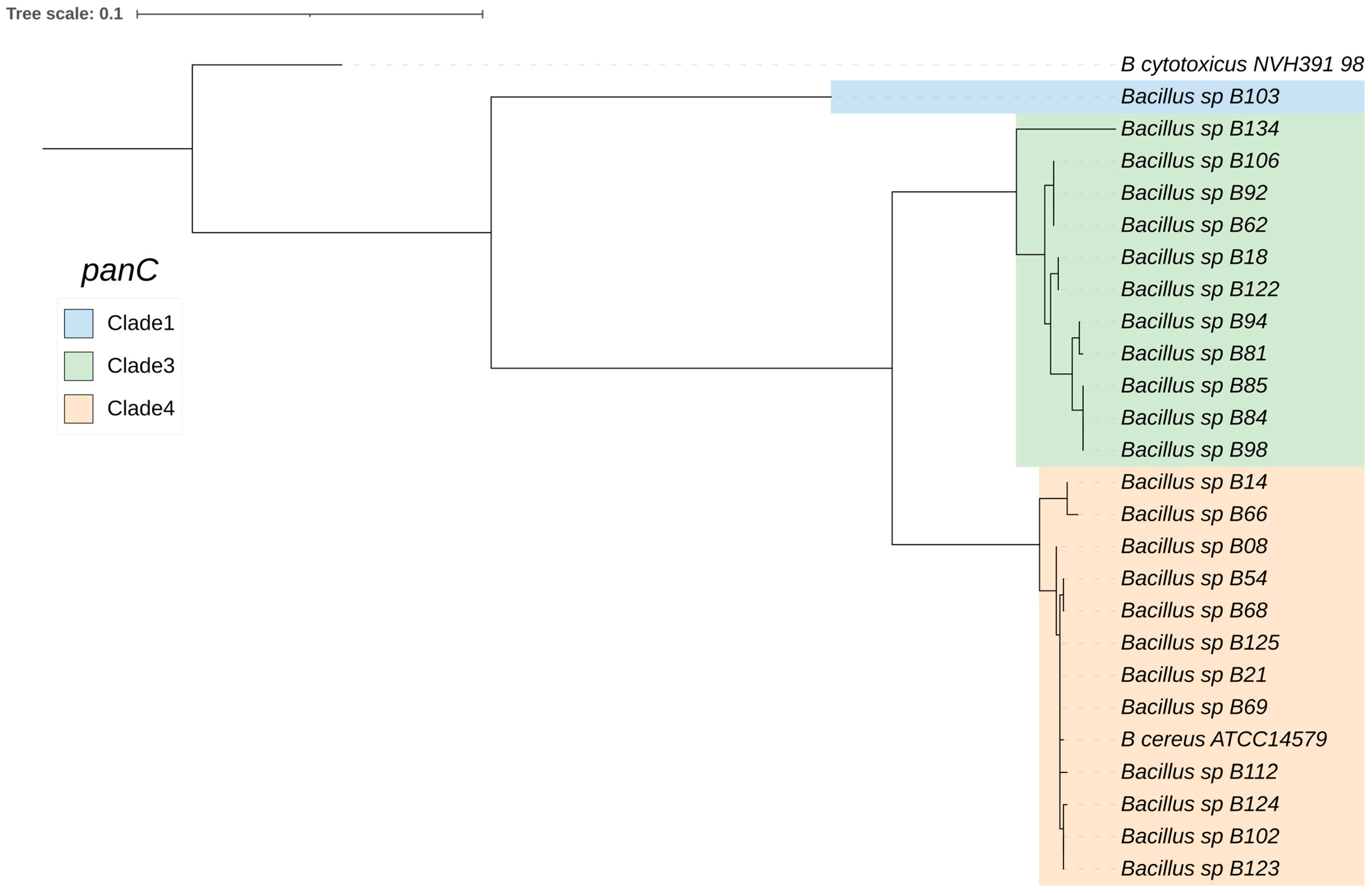
Next, the genomic sequences of the 24 B. cereus group isolates were used to generate a core-gene phylogenetic tree based on panC (Figure 2). This revealed the following three B. cereus group clades (designated according to previously proposed phylogenetic groups based on panC sequence types [28]): clade III (11 isolates), clade IV (12 isolates, which is the same clade as B. cereus s.s.), and clade I (1 isolate, which was Bacillus sp. B103, closely related to “B. bingmayongensis”).

2.2. Subsystem Categorization

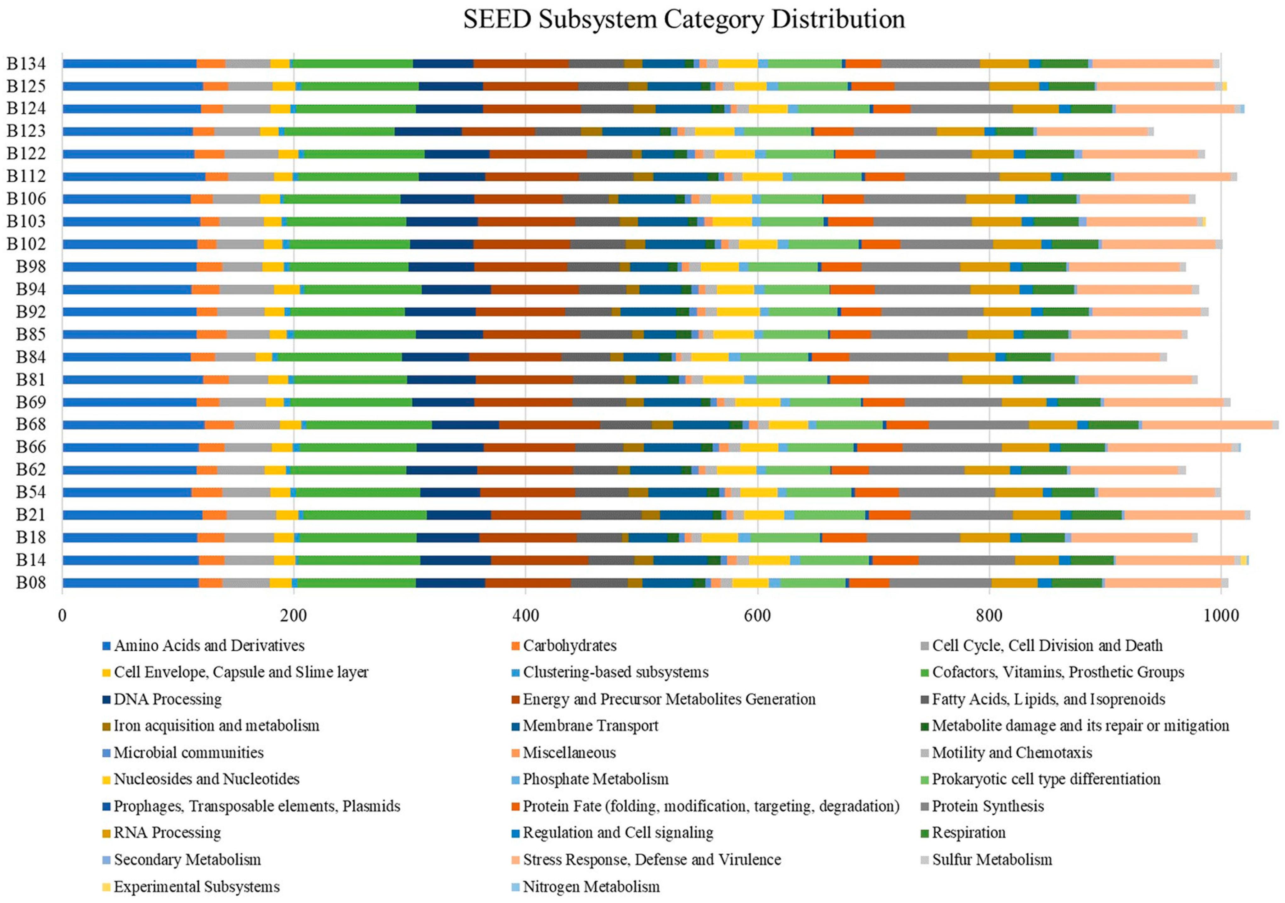
The SEED subsystem categorization based on RASTtk annotation is illustrated in Figure 3. The analysis predicted 27–29 categories among the 24 B. cereus group genomes. The most abundant systems were “Amino Acids and Derivatives”, followed by “Cofactors, Vitamins, Prosthetic Groups” and “Stress Response, Defense, and Virulence”. In contrast, the genomes had a remarkably low number of “Prophages, Transposable Elements, Plasmids” and “Secondary Metabolism”. Several of the isolates exhibited “Experimental Subsystems” and “Nitrogen Metabolism” in their genomes.

2.3. AMR Gene Analysis

AMR genes in the B. cereus group isolate genomes were identified (Figure 4). AMR genes that are associated with resistance to one or more antimicrobials were predicted based on the ARG-ANNOT, CARD, NCBI, and ResFinder databases. The data from ARG-ANNOT, CARD, and ResFinder databases all showed that fosB1 (95.8%) was the most prevalent gene among the six observed AMR genes. The other five AMR genes were blaZ (16.7%), fosB (12.5%), mef(A) (8.3%), msr(D) (8.3%), and tet(45) (16.7%). The NCBI database showed that there were 13 AMR genes, with bcII (95.8%) and bla1 (95.8%) being the most prevalent, followed by fosB_gen (91.7%), satA_Ba (encoding streptothricin-N-acetyltransferase) (91.7%), vanZ-F (79.2%), tet(45) (16.7%), fosB-38141535 (12.5%), mef(A) (8.3%), msr(D) (8.3%), vanR-A (8.3%), vanS-Pt (8.3%), rphC (encoding rifamycin-inactivating phosphotransferase) (4.2%), and fosBx1 (4.2%).

2.4. Virulence Factor Gene Analysis

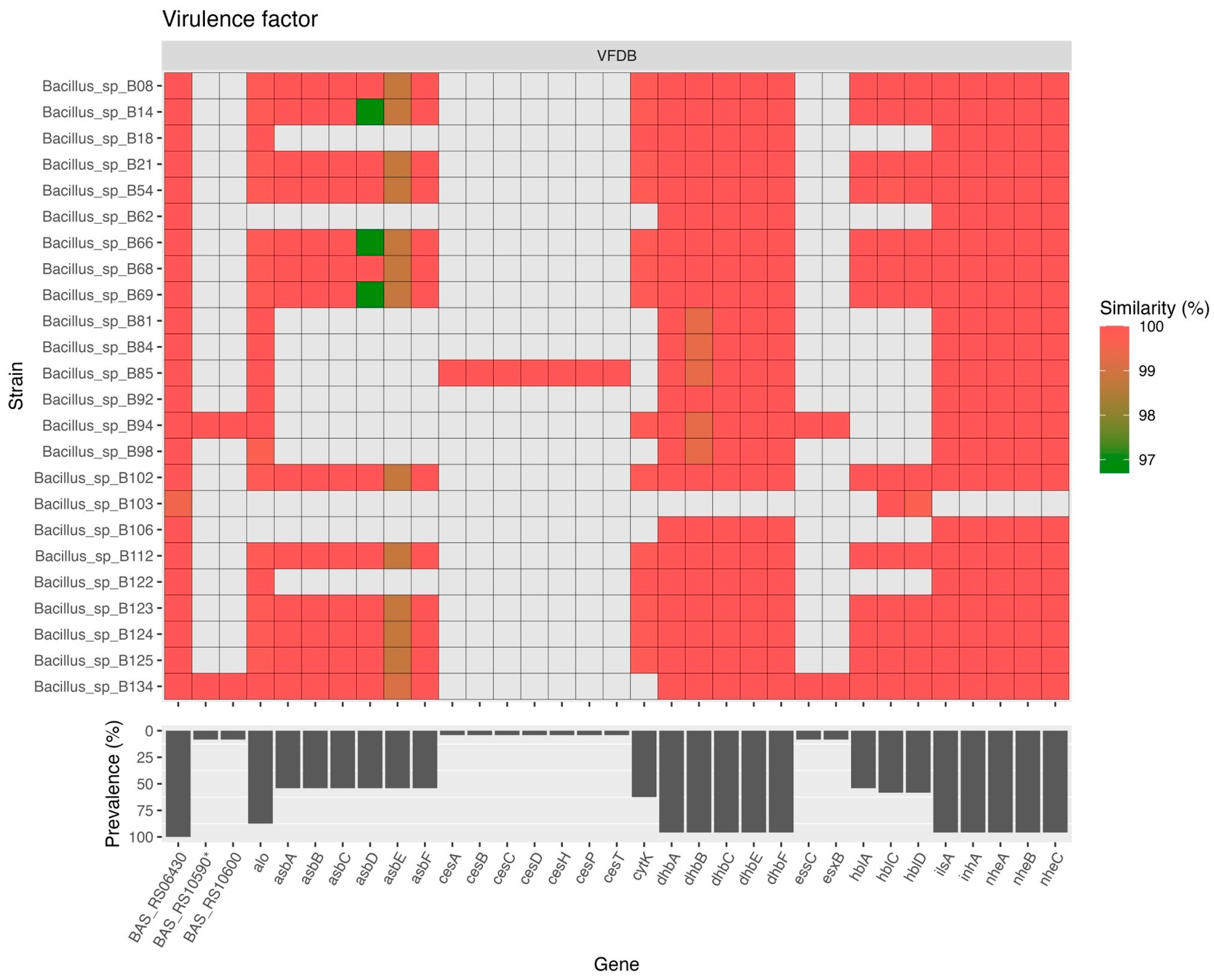
There were 33 genes in the B. cereus group isolate genomes that were classified as virulence factors according to the VFDB database (Figure 5). Among these genes, the most prevalent gene (harbored by all 24 isolates) was BAS_RS06430 (inhA1 in Bacillus anthracis str. Sterne; complete sequence available at https://www.genome.jp/dbget-bin/www_bget?refseq+NC_005945, unpublished data, accessed on 12 January 2024). Additionally, 23 (95.8%) of the isolates harbored bacillibactin biosynthesis genes (dhbA, dhbB, dhbC, dhbE, and dhbF), genes encoding the three-component non-hemolytic enterotoxin (nheA, nheB, and nheC), a gene encoding an iron-regulated leucine-rich surface protein (ilsA), and a gene encoding a metalloprotease (inhA). Moreover, 21 (87.5%) and 15 (62.5%) of the isolates harbored genes encoding cytotoxin anthrolysin O (alo), which is also known as a cholesterol-dependent cytolysin secreted by B. anthracis, and the single-component enterotoxin cytotoxin K (cytK), respectively. Furthermore, 13–14 (54.2–58.3%) of the isolates harbored genes encoding the three-component enterotoxin hemolysin BL (hblA, hblC and hblD), which was similar to the prevalence of genes (13 [54.2%]) in the B. anthracis siderophore biosynthesis (asb) operon (asbA, asbB, asbC, asbD, asbE, and asbF). Only two (8.3%) isolates harbored BAS_RS10590*, BAS_RS10600, type VII secretion gene (essC), and WXG100 gene (esxB). Only one (4.2%) isolate harbored a cereulide synthetase (ces) gene cluster (cesA, cesB, cesC, cesD, cesH, cesP, and cesT).

2.5. Biofilm-Associated Gene Analysis

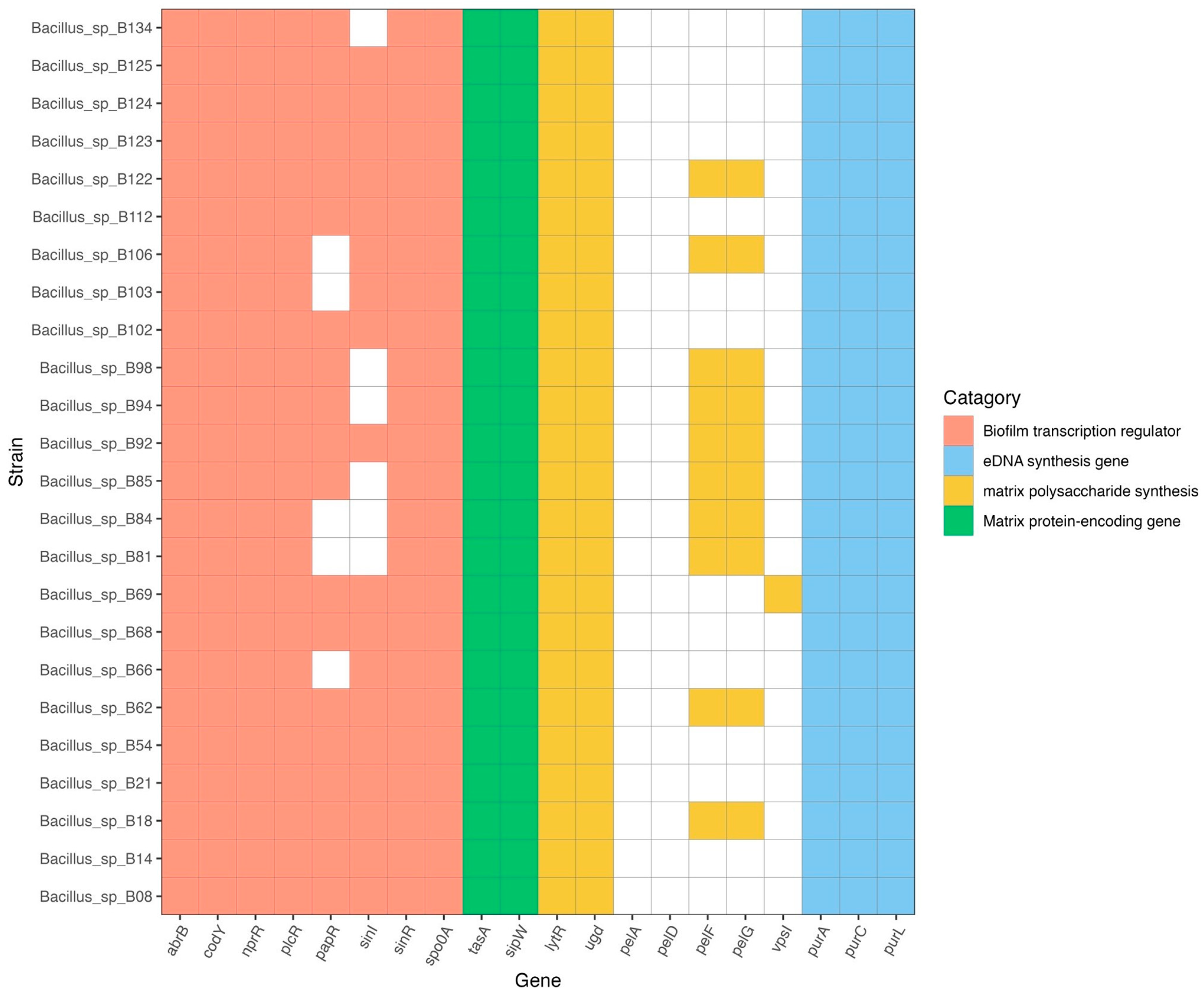
The biofilm-associated genes in the B. cereus group genomes were identified using BlastKOALA (https://www.kegg.jp/blastkoala/, accessed on 3 November 2023) (Figure 6). The crucial genes for biofilm formation comprise matrix protein-encoding genes, matrix polysaccharide synthesis genes, eDNA synthesis genes, and biofilm transcription regulator genes. All isolates harbored tasA and sipW genes (matrix protein-encoding genes); purA, purC, and purL genes (eDNA synthesis genes); lytR and ugd genes (matrix polysaccharide synthesis genes); and abrB, codY, nprR, plcR, sinR, and spo0A genes (biofilm transcription regulator genes). Additionally, 19 (79%) isolates harbored papR (biofilm transcription regulator gene), while 18 (75%) isolates harbored sinI (another biofilm transcription regulator gene). Moreover, 10 (41.7%) isolates harbored pelF and pelG of the pel operon (which encodes matrix polysaccharide synthesis genes), but none harbored pelA or pelD. Only one (4.2%) isolate (Bacillus sp. B69) harbored vpsI (exopolysaccharide synthesis gene, to produce the main component of the biofilm matrix).

3. Discussion

The Bacillus cereus group, also known as B. cereus s.l., is composed of several species, some of which can cause gastrointestinal or non-gastrointestinal tract infections. Members of the B. cereus group are classified into seven phylogenetic clades based on panC: clade I, B. pseudomycoides; clade II, B. wiedmannii; clade III, B. anthracis; clade IV, B. cereus s.s. and B. thuringiensis; clade V, B. toyonensis; clade VI, B. weihenstephanensis and B. mycoides; and clade VII, B. cytotoxicus [28,29,30]. In this study, 24 B. cereus group isolates from foodstuffs were characterized using WGS and bioinformatic tools. Most isolates were phylogenetically classified as B. cereus s.s. (clade IV, 12 isolates) followed by B. pacificus (clade III, 5 isolates), B. paranthracis (clade III, 5 isolate), B. tropicus (clade III, 1 isolate), and “B. bingmayongensis” (clade I, 1 isolate).
B. cereus s.s. is a serious human pathogen that has caused foodborne outbreaks in several countries including Thailand. Most cases involved self-limiting diarrhea, nausea, or vomiting due to bacterial enterotoxins, especially hemolysin BL (HBL), the three-component non-hemolytic enterotoxin (NHE), and cytotoxin K (CytK), as well as cereulide [31]. In this study, 12 isolates were closely related to B. cereus s.s. Furthermore, 23 (95.8%) of the 24 B. cereus group isolates (all isolates except for Bacillus sp. B103, which was closely related to “B. bingmayongensis”) harbored nhe genes. However, the presence of toxin genes is not always consistent with the pathogenic potential of a B. cereus group isolate [29]. Moreover, BTyper may detect the presence of a toxin gene, while the PCR result may be negative [29]. A negative PCR result may be due to variant sequences causing poor binding of the published primers to the DNA sequence [32]. Therefore, cytotoxicity assays of B. cereus group isolates, along with their virulence factor gene profiles, may offer more information about potential harm to human health.
B. pacificus was isolated from the sediment of the Pacific Ocean [9], and, recently, the B. pacificus strain CR121 was isolated from the intestine of the Indian major carp species rohu (Labeo rohita) [33]. The CR121 strain has been proposed to be a candidate fish probiotic as it possesses in vivo disease prevention efficacy in L. rohita and Oreochromis niloticus [33]. Moreover, a B. pacificus strain was isolated from a spicy mussel salad in Pathum Thani province, Thailand, which was concerning as it exhibited strong biofilm formation and tetracycline resistance phenotypes [27]. In this study, five isolates were closely related to B. pacificus, comprising Bacillus sp. B18 (isolated from stir-fried mixed vegetables), Bacillus sp. B62 (isolated from rice with spicy shrimp paste dip and fried mackerel), Bacillus sp. B92 (isolated from sushi), Bacillus sp. B106 (isolated from stir-fried lotus stem and straw mushroom with pork), and Bacillus sp. B122 (isolated from stir-fried pickled turnip with egg). It is suggested that B. pacificus can be isolated from foodstuffs that do not contain seafood. In addition, contamination may occur during or after-cooking processing, which should be considered in more depth.
B. paranthracis, which was first described in 2017 [9], has been identified as group III B. cereus according to microbiologic methods and panC phylogenetic group assignment [34]. In 2020, B. paranthracis was isolated from blood of a fatal Ebola virus disease case in Liberia and was identified by whole genome sequencing [35]. B. paranthracis is considered as a pathogenic bacterium both in human and animal hosts since it could be isolated from the milk of cows diagnosed with mastitis [36]. It is also known as emetic B. cereus due to the ability to produce cereulide, an emetic toxin. In 2016, B. paranthracis was linked to a foodborne outbreak, possibly caused by the consumption of refried beans, in upstate New York [37]. Five isolates in this study were closely related to B. paranthracis, comprising Bacillus sp. B81 (isolated from spicy Vietnamese pork sausage salad), Bacillus sp. B84 (isolated from spicy tremella mushroom salad), Bacillus sp. B85 (isolated from spicy pork meatball salad), Bacillus sp. B94 (isolated from spicy fermented fish dip), and Bacillus sp. B98 (isolated from stir-fried glass noodles). Most of the contaminated food samples were spicy salad. The ingredients of this type of food might be further determined.
B. bingmayongensis strain FJAT-13831 was originally isolated from the pit soil of Emperor Qin’s terracotta warriors in China [38]. The colonies on nutrient agar were flat, greyish white, undulate in margin. Its closest phylogenetic neighbor was B. pseudomycoides. In 2022, B. bingmayongensis KNUAS006 was isolated from fermented Korean food samples and classified as a probiotic strain [39]. Currently, little is known about the pathogenic potential of both B. bingmayongensis and B. pseudomycoides. Literally, B. pseudomycoides, characterized by rhizoidal colonies on culture media [3], is not recognized as a pathogen, but its toxigenic potential remains uncertain. A previous study reported that all B. pseudomycoides isolates harbor hblA [40], another study reported that 30% and 70% of B. pseudomycoides isolates harbor nheA and hblA, respectively [41], and a third study reported that a B. pseudomycoides isolate (isolated from Shepherd’s purse in South Korea) harbored all the genes of the HBL enterotoxin complex (hblA, hblC, and hblD) [42]. The presence of enterotoxin genes seems to vary among isolates in various studies. In this study, we found that Bacillus sp. B103 (isolated from salad roll with crab stick and exhibiting rhizoidal colonies) was closely related to B. bingmayongensis. The isolate harbored three virulence genes comprising BAS_RS06430, hblC, and hblD genes. Therefore, a safety assessment of B. bingmayongensis needs to be performed. Moreover, the colony morphology of Bacillus sp. B103 exhibited rhizoidal appearance while it was classified as B. bingmayongensis. However, the ANI value of Bacillus sp. B103 was only 90.05%, which is below the 95% cutoff value for the delimitation of bacterial species. Therefore, species classification within the B. cereus group is still challenging.
B. tropicus has been isolated from various environmental samples, including a soil sample from a marine duck farm of Beibu Gulf in Guangxi, China [43]; a soil sample from a city center rubbish dump in Durgapur, India [44]; and kawal (a fermented condiment obtained by natural and alkaline fermentation of Senna obtusifolia leaves) from the markets of Abéché, Bokoro, Mandelia, and N’Djamena in Tchad [45]. In this study, one isolate (Bacillus sp. B134, isolated from stewed pork leg on rice) was closely related to B. tropicus. The isolate harbored several virulence genes including nhe and hbl but not cytK. In addition, essC and esxB were present. Esat-6 protein secretion systems (ESX or Ess) are required for the virulence of several human pathogens including Mycobacterium tuberculosis and Staphylococcus aureus as well as B. subtilis [46]. However, the molecular mechanism of this system in relation to the virulence of B. tropicus needs to be further characterized.
Determining the prevalence of antimicrobial resistance among B. cereus group isolates can provide information for proper antibiotic use and improve treatment outcomes [24]. Although most B. cereus group infections tend to be self-limiting and may not need antimicrobial treatment, severe infections, particularly in immunocompromised patients, need to be treated with appropriate antibiotic agents. The most suitable drug of choice for B. cereus bacteremia is vancomycin [47]. Moreover, carbapenem antibiotics are also considered to be an effective treatment [15]. B. cereus s.s. is typically resistant to beta-lactam antibiotics [48]. In addition, acquired resistance to commonly used antibiotics such as ciprofloxacin, cloxacillin, erythromycin, tetracycline, and streptomycin has been literally reported [48,49]. The AMR mechanisms of B. cereus group strains may vary between strains [50]. In this study, all isolates except for Bacillus sp. B103 harbored bcII and bla1 (related to resistance to beta-lactams) and fosB1 (related to the inactivation of fosfomycin antibiotic). Minimum inhibitory concentration (MIC) experiments of B. cereus isolates (from indoor air) that harbored fosB, bcI, and bcII have demonstrated their ampicillin and fosfomycin resistance phenotypes [50]. Other AMR genes were observed in some of our isolates, including those related to resistance to tetracycline and vancomycin. The results of AMR gene analysis in this study were consistent with antimicrobial susceptibility testing in the previous study [51]. The isolates that harbored genes responsible for beta-lactam resistance showed the resistance phenotypes to ampicillin, amoxicillin-clavulanic acid, and penicillin. Two isolates (Bacillus sp. B18 and Bacillus sp. B122) which harbored genes responsible for macrolide resistance (mef(A) and msr(D)) demonstrated intermediate resistance to erythromycin. However, discrepancies between antimicrobial resistance phenotypes and the prevalence of antimicrobial resistance genes were observed in this study. The isolates that carried the tet(45) gene exhibited a susceptible phenotype to tetracycline. It has been reported that Bacillus isolates showed an antibiotic phenotype that was sensitive to tetracycline while the tetB detection rate was 100% [52]. This might be due to the presence of varied antimicrobial resistance mechanisms and many genetic factors required for the resistance phenotypes [53]. Moreover, the AMR phenotypes to the other antimicrobial agents (e.g., fosfomycin) need to be further determined in vitro. In addition, the transferable mobile genetic elements (such as transposon or insertion sequences) should be characterized based on long-read genome sequencing in order to verify the risk of horizontal transfer of AMR genes among bacterial species [54].
Among the virulence genes observed in this study, the most prevalent gene (harbored by all 24 isolates) was BAS_RS06430 (inhA1 in Bacillus anthracis str. Sterne). It has been reported that inhA1 is produced only by pathogenic members of the Bacillus genus and associated with the cleavage of host proteins during infection [55]. Moreover, 95.8% of the isolates harbored a gene encoding a metalloprotease (inhA). Therefore, the pathogenic effects of these genes should be further characterized. Additionally, most isolates (95.8%) harbored bacillibactin biosynthesis genes (dhbA, dhbB, dhbC, dhbE, and dhbF), genes encoding the three-component non-hemolytic enterotoxin (nheA, nheB, and nheC), and a gene encoding an iron-regulated leucine-rich surface protein (ilsA). Under iron-limited conditions, B. anthracis produces two catecholate siderophores which are petrobactin (encoded by asb operon) and bacillibactin (encoded by bac operon) [56,57]. However, only the asb locus is essential for growth in iron-depleted media and for virulence in mice [56]. Daou et al. [58] reported that IlsA-like proteins are restricted to B. cereus group bacteria. IlsA is an important adaptation factor required for the development of B. cereus in susceptible hosts including mammals and insects [58]. The isolates that harbored these virulence genes are of interest since they may cause harmful effects to the human host in some circumstances.
Biofilm formation is a potential virulence determinant of various pathogens including B. cereus group species as it allows the bacteria to resist disinfectants and antimicrobials, as well as to escape the host immune system [59,60]. The important genes for biofilm formation in B. cereus group species include biofilm transcriptional regulator genes (abrB, codY, nprR, plcR, papR, sinI, sinR, and spo0A), matrix protein-encoding genes (tasA and sipW), matrix polysaccharide synthesis genes (lytR, ugd, vpsI, and pelDEADAFG operon), eDNA synthesis genes (purA, purC, and purL), and cyclic-di-GMP metabolism genes [27,61]. All isolates in this study harbored the above-mentioned biofilm-associated genes except for papR, sinI, and the pelDEADAFG operon, which were present in some isolates. Moreover, vpsI (exopolysaccharide synthesis gene) was found in one isolate (Bacillus sp. 69). In Vibrio cholerae, the biofilm matrix is mainly composed of exopolysaccharides (VPS), whose synthesis is encoded by two vps operons (vpsI and vpsII) [62]. The role of vpsI in biofilm formation in Bacillus species is still unclear. It has been proposed that the pelDEADAFG operon might play an important role in the biofilm-formation capacity of Bacillus species, as the lack of this operon reduced the biofilm-formation capacity of the tested strain [27,63]. Comprehensive in vitro biofilm formation assays need to be performed to confirm the potential roles of these genes in biofilm formation in Bacillus species.
In conclusion, we employed WGS to characterize 24 B. cereus group isolates from foodstuffs. We identified the closest reference strains to the isolates based on a phylogenetic tree analysis and whole genome-based average nucleotide identity. The most prevalent species were B. cereus s.s. (12 isolates) followed by B. pacificus (5 isolates). The remaining three species were B. paranthracis, B. tropicus, and B. bingmayongensis. We also observed the prevalence of AMR and virulence genes in the genomes of the isolates. Most isolates harbored genes related to resistance to fosfomycin and beta-lactams. Moreover, additional AMR genes were identified in some isolates, including those related to resistance to erythromycin and tetracycline. However, the risk of horizontal transfer of AMR genes among B. cereus group species and other bacterial species needs to be further clarified. Most of the isolates harbored virulence genes (especially those related to metalloprotease production, bacillibactin biosynthesis, and enterotoxin production). Altogether, our findings suggest that WGS combined with appropriate bioinformatic tools can be used to evaluate the pathogenic potential of B. cereus group isolates from foodstuffs. Determining the genetic epidemiology, the presence of virulence and AMR genes, and the potential hazards of foodborne B. cereus group species help to raise awareness of the underestimated foodborne diseases caused by these species.

4. Materials and Methods

4.1. Bacterial Strains

Twenty-four B. cereus group isolates from foodstuffs collected in 2018 [51] in Pathum Thani Province, Thailand, were characterized in this study. The sampling procedure and microbiological analysis of the isolates has been previously described [51]. Briefly, all isolates were identified using both conventional microbiological techniques and API 50 CHB test. Glycerol stocks of the bacteria (stored at −80 °C) were cultured on nutrient agar plates and incubated overnight at 35 ± 2 °C.

4.2. DNA Extraction and WGS

The genomic DNA was extracted using Monarch HMW DNA Extraction Kits (NEB, Ipswich, MA, USA) according to the manufacturer’s instructions. The purified genomic DNA was qualitatively assessed using a fluorometer. Paired-end sequencing (2 × 250-bp reads) was performed using V2 chemistry and a MiSeq platform (Illumina, San Diego, CA, USA). The reads were processed using Trimmomatic v0.39 to remove adapter and barcode sequences, correct mismatched bases in overlaps, and filter out low-quality reads (<Q30) [64]. FastQC v0.12.1 was used to determine the quality of the reads.

4.3. Genome Assembly and Annotation

Assembly of the short-read data sets was performed using Unicycler v0.5.0 with default parameters [65]. QUAST v5.2.0 was used to evaluate the genome assembly quality [66]. FastANI v1.33 was used to calculate the genome-wide average nucleotide identity (ANI) between genomes [67]. Gene prediction and annotation were performed using Prokka v1.14.6 [68] and Bacterial and Viral Bioinformatics Resource Center (BV-BRC) (https://www.bv-brc.org/, accessed on 12 September 2023) [69]. Functional annotation of the predicted proteins was conducted using BlastKOALA of Kyoto Encyclopedia of Genes and Genomes (KEGG) (https://www.kegg.jp/blastkoala/, accessed on 3 November 2023) [70].
The presence of AMR genes in the draft genomes of the strains was determined using ABRicate v0.8 (https://github.com/tseemann/ABRicate, accessed on 15 September 2023), which included several predownloaded databases (ResFinder [71], CARD [72], NCBI [73], ARG-ANNOT [74], and NDARO [75]) with default settings [76]. Putative virulence factors were predicted by conducting BLAST searches against the Virulence Factor Database (VFDB) (http://www.mgc.ac.cn/VFs/main.htm, accessed on 15 September 2023) [77]. BTyper3 v3.4.0 was used to assign each genome, determine the multilocus sequence typing (MLST) profiles, and identify the panC-based phylogenetic groups [78].

4.4. Phylogenetic Trees

A maximum likelihood phylogenetic tree of the 24 B. cereus group isolates and other type strains of B. cereus group genome sequences obtained from NCBI was constructed. This involved using GToTree v1.8.3 with the hidden Markov model (HMM) source bacteria and default parameters [79].
Next, the panC nucleotide sequences of the 24 B. cereus group isolates were extracted from the BTyper3 tool [78] and then aligned using the multiple sequence alignment algorithm MUSCLE [80]. A maximum likelihood core-gene phylogenetic tree was constructed using RAxML with the GTRGAMMA model and 1000 bootstrap replicates. The phylogenetic tree was visualized using iTOL v6 (https://itol.embl.de/, accessed on 17 October 2023).

4.5. Subsystem Categorization

The 24 B. cereus group genomes were submitted to Rapid Annotations using Subsystems Technology (RASTtk), available in The Pathosystems Resource Integration Center (PATRIC) (https://www.bv-brc.org/ accessed on 12 September 2023), for annotation, subsystem categorization, and comparison purposes [81].

4.6. Nucleotide Sequence Accession Numbers

The draft genomes of the 24 B. cereus group isolates were deposited in the NCBI database (https://submit.ncbi.nlm.nih.gov/subs/, accessed on 18 October 2023) under the accession numbers shown in Table 1. The GenBank BioProject ID is PRJNA1030778.

Author Contributions

Conceptualization, P.S.; Methodology, P.S., K.S. (Kritsakorn Saninjuk) and J.R.; Software, K.S. (Kritsakorn Saninjuk), S.A., N.T., P.M., P.C. and K.S. (Kant Sangpairoj); Validation, P.S. and K.S. (Kritsakorn Saninjuk); Formal analysis, P.S. and K.S. (Kritsakorn Saninjuk); Resources, P.S., N.T., P.M., P.C. and K.S. (Kant Sangpairoj); Writing—original draft preparation, P.S.; Writing—review and editing, P.S. and K.S. (Kritsakorn Saninjuk); Visualization, K.S. (Kritsakorn Saninjuk); Supervision, P.S.; Funding acquisition, P.S. All authors have read and agreed to the published version of the manuscript.

Funding

This research was funded by the Thailand Science Research and Innovation Fundamental Fund fiscal year 2023, grant number TUFF 55/2566, the Research Group in Multidrug Resistant Bacteria and the Antimicrobial Herbal Extracts, Faculty of Medicine, Thammasat University, and Thammasat University Research Unit in Nutraceuticals and Food Safety.

Institutional Review Board Statement

Not applicable.

Informed Consent Statement

Not applicable.

Data Availability Statement

Data are contained within the article.

Conflicts of Interest

Kritsakorn Saninjuk has been involved as a Research and Development manager in Porcinotec Co., Ltd. The rest of the authors declare no conflicts of interest.

References

  1. Kolstø, A.B.; Tourasse, N.J.; Økstad, O.A. What sets Bacillus anthracis apart from other Bacillus species? Annu. Rev. Microbiol. 2009, 63, 451–476. [Google Scholar] [CrossRef]
  2. Ehling-Schulz, M.; Lereclus, D.; Koehler, T.M. The Bacillus cereus Group: Bacillus Species with Pathogenic Potential. Microbiol. Spectr. 2019, 7, 10–1128. [Google Scholar] [CrossRef]
  3. Nakamura, L.K. Bacillus pseudomycoides sp. nov. Int. J. Syst. Bacteriol. 1998, 48 Pt 3, 1031–1035. [Google Scholar] [CrossRef]
  4. Lechner, S.; Mayr, R.; Francis, K.P.; Prüss, B.M.; Kaplan, T.; Wiessner-Gunkel, E.; Stewart, G.S.; Scherer, S. Bacillus weihenstephanensis sp. nov. is a new psychrotolerant species of the Bacillus cereus group. Int. J. Syst. Bacteriol. 1998, 48 Pt 4, 1373–1382. [Google Scholar] [CrossRef]
  5. Hoton, F.M.; Fornelos, N.; N’Guessan, E.; Hu, X.; Swiecicka, I.; Dierick, K.; Jääskeläinen, E.; Salkinoja-Salonen, M.; Mahillon, J. Family portrait of Bacillus cereus and Bacillus weihenstephanensis cereulide-producing strains. Environ. Microbiol. Rep. 2009, 1, 177–183. [Google Scholar] [CrossRef]
  6. Miller, R.A.; Beno, S.M.; Kent, D.J.; Carroll, L.M.; Martin, N.H.; Boor, K.J.; Kovac, J. Bacillus wiedmannii sp. nov., a psychrotolerant and cytotoxic Bacillus cereus group species isolated from dairy foods and dairy environments. Int. J. Syst. Evol. Microbiol. 2016, 66, 4744–4753. [Google Scholar] [CrossRef] [PubMed]
  7. Guinebretiere, M.H.; Auger, S.; Galleron, N.; Contzen, M.; De Sarrau, B.; De Buyser, M.L.; Lamberet, G.; Fagerlund, A.; Granum, P.E.; Lereclus, D.; et al. Bacillus cytotoxicus sp. nov. is a novel thermotolerant species of the Bacillus cereus Group occasionally associated with food poisoning. Int. J. Syst. Evol. Microbiol. 2013, 63, 31–40. [Google Scholar] [CrossRef] [PubMed]
  8. Liu, Y.; Lai, Q.; Göker, M.; Meier-Kolthoff, J.P.; Wang, M.; Sun, Y.; Wang, L.; Shao, Z. Genomic insights into the taxonomic status of the Bacillus cereus group. Sci. Rep. 2015, 5, 14082. [Google Scholar] [CrossRef]
  9. Liu, Y.; Du, J.; Lai, Q.; Zeng, R.; Ye, D.; Xu, J.; Shao, Z. Proposal of nine novel species of the Bacillus cereus group. Int. J. Syst. Evol. Microbiol. 2017, 67, 2499–2508. [Google Scholar] [CrossRef]
  10. Bianco, A.; Capozzi, L.; Monno, M.R.; Del Sambro, L.; Manzulli, V.; Pesole, G.; Loconsole, D.; Parisi, A. Characterization of Bacillus cereus Group Isolates From Human Bacteremia by Whole-Genome Sequencing. Front. Microbiol. 2020, 11, 599524. [Google Scholar] [CrossRef]
  11. Liu, X.; Wang, L.; Han, M.; Xue, Q.H.; Zhang, G.S.; Gao, J.; Sun, X. Bacillus fungorum sp. nov., a bacterium isolated from spent mushroom substrate. Int. J. Syst. Evol. Microbiol. 2020, 70, 1457–1462. [Google Scholar] [CrossRef] [PubMed]
  12. Goldstein, B.; Abrutyn, E. Pseudo-outbreak of Bacillus species: Related to fibreoptic bronchoscopy. J. Hosp. Infect. 1985, 6, 194–200. [Google Scholar] [CrossRef]
  13. Bryce, E.A.; Smith, J.A.; Tweeddale, M.; Andruschak, B.J.; Maxwell, M.R. Dissemination of Bacillus cereus in an intensive care unit. Infect. Control Hosp. Epidemiol. 1993, 14, 459–462. [Google Scholar] [CrossRef] [PubMed]
  14. Granum, P.E. Bacillus cereus and its toxins. Soc. Appl. Bacteriol. Symp. Ser. 1994, 23, 61s–66s. [Google Scholar] [CrossRef]
  15. Bottone, E.J. Bacillus cereus, a volatile human pathogen. Clin. Microbiol. Rev. 2010, 23, 382–398. [Google Scholar] [CrossRef]
  16. Lund, T.; Granum, P.E. Comparison of biological effect of the two different enterotoxin complexes isolated from three different strains of Bacillus cereus. Microbiology 1997, 143 Pt 10, 3329–3336. [Google Scholar] [CrossRef]
  17. Lund, T.; De Buyser, M.L.; Granum, P.E. A new cytotoxin from Bacillus cereus that may cause necrotic enteritis. Mol. Microbiol. 2000, 38, 254–261. [Google Scholar] [CrossRef]
  18. Sakai, C.; Iuchi, T.; Ishii, A.; Kumagai, K.; Takagi, T. Bacillus cereus brain abscesses occurring in a severely neutropenic patient: Successful treatment with antimicrobial agents, granulocyte colony-stimulating factor and surgical drainage. Intern. Med. 2001, 40, 654–657. [Google Scholar] [CrossRef]
  19. Clair, G.; Roussi, S.; Armengaud, J.; Duport, C. Expanding the known repertoire of virulence factors produced by Bacillus cereus through early secretome profiling in three redox conditions. Mol. Cell. Proteom. 2010, 9, 1486–1498. [Google Scholar] [CrossRef]
  20. Caro-Astorga, J.; Pérez-García, A.; de Vicente, A.; Romero, D. A genomic region involved in the formation of adhesin fibers in Bacillus cereus biofilms. Front. Microbiol. 2015, 5, 745. [Google Scholar] [CrossRef]
  21. McCarthy, H.; Rudkin, J.K.; Black, N.S.; Gallagher, L.; O’Neill, E.; O’Gara, J.P. Methicillin resistance and the biofilm phenotype in Staphylococcus aureus. Front. Cell. Infect. Microbiol. 2015, 5, 1. [Google Scholar] [CrossRef] [PubMed]
  22. Gomes, F.; Saavedra, M.J.; Henriques, M. Bovine mastitis disease/pathogenicity: Evidence of the potential role of microbial biofilms. Pathog. Dis. 2016, 74, ftw006. [Google Scholar] [CrossRef]
  23. Rutherford, S.T.; Bassler, B.L. Bacterial quorum sensing: Its role in virulence and possibilities for its control. Cold Spring Harb. Perspect. Med. 2012, 2, a012427. [Google Scholar] [CrossRef] [PubMed]
  24. Mills, E.; Sullivan, E.; Kovac, J. Comparative Analysis of Bacillus cereus Group Isolates’ Resistance Using Disk Diffusion and Broth Microdilution and the Correlation between Antimicrobial Resistance Phenotypes and Genotypes. Appl. Environ. Microbiol. 2022, 88, e0230221. [Google Scholar] [CrossRef]
  25. Boolchandani, M.; D’Souza, A.W.; Dantas, G. Sequencing-based methods and resources to study antimicrobial resistance. Nat. Rev. Genet. 2019, 20, 356–370. [Google Scholar] [CrossRef] [PubMed]
  26. Yan, F.; Yu, Y.; Gozzi, K.; Chen, Y.; Guo, J.H.; Chai, Y. Genome-Wide Investigation of Biofilm Formation in Bacillus cereus. Appl. Environ. Microbiol. 2017, 83, e00561-17. [Google Scholar] [CrossRef]
  27. Sornchuer, P.; Saninjuk, K.; Prathaphan, P.; Tiengtip, R.; Wattanaphansak, S. Antimicrobial Susceptibility Profile and Whole-Genome Analysis of a Strong Biofilm-Forming Bacillus sp. B87 Strain Isolated from Food. Microorganisms 2022, 10, 252. [Google Scholar] [CrossRef]
  28. Guinebretiere, M.H.; Thompson, F.L.; Sorokin, A.; Normand, P.; Dawyndt, P.; Ehling-Schulz, M.; Svensson, B.; Sanchis, V.; Nguyen-The, C.; Heyndrickx, M.; et al. Ecological diversification in the Bacillus cereus Group. Environ. Microbiol. 2008, 10, 851–865. [Google Scholar] [CrossRef]
  29. Miller, R.A.; Jian, J.; Beno, S.M.; Wiedmann, M.; Kovac, J. Intraclade Variability in Toxin Production and Cytotoxicity of Bacillus cereus Group Type Strains and Dairy-Associated Isolates. Appl. Environ. Microbiol. 2018, 84, e02479-17. [Google Scholar] [CrossRef]
  30. Carroll, L.M.; Cheng, R.A.; Wiedmann, M.; Kovac, J. Keeping up with the Bacillus cereus group: Taxonomy through the genomics era and beyond. Crit. Rev. Food Sci. Nutr. 2022, 62, 7677–7702. [Google Scholar] [CrossRef]
  31. Messelhäußer, U.; Ehling-Schulz, M. Bacillus cereus—A Multifaceted Opportunistic Pathogen. Curr. Clin. Microbiol. Rep. 2018, 5, 120–125. [Google Scholar] [CrossRef]
  32. Gardes, J.; Croce, O.; Christen, R. In silico analyses of primers used to detect the pathogenicity genes of Vibrio cholerae. Microbes Environ. 2012, 27, 250–256. [Google Scholar] [CrossRef]
  33. Paul, S.I.; Khan, S.U.; Sarkar, M.K.; Foysal, M.J.; Salam, M.A.; Rahman, M.M. Whole-Genome Sequence of Bacillus pacificus CR121, a Fish Probiotic Candidate. Microbiol. Resour. Announc. 2023, 12, e0120622. [Google Scholar] [CrossRef] [PubMed]
  34. Carroll, L.M.; Matle, I.; Kovac, J.; Cheng, R.A.; Wiedmann, M. Laboratory Misidentifications Resulting from Taxonomic Changes to Bacillus cereus Group Species, 2018–2022. Emerg. Infect. Dis. 2022, 28, 1877–1881. [Google Scholar] [CrossRef]
  35. Matson, M.J.; Anzick, S.L.; Feldmann, F.; Martens, C.A.; Drake, S.K.; Feldmann, H.; Massaquoi, M.; Chertow, D.S.; Munster, V.J. Bacillus paranthracis Isolate from Blood of Fatal Ebola Virus Disease Case. Pathogens 2020, 9, 475. [Google Scholar] [CrossRef]
  36. Sokolov, S.; Brovko, F.; Solonin, A.; Nikanova, D.; Fursova, K.; Artyemieva, O.; Kolodina, E.; Sorokin, A.; Shchannikova, M.; Dzhelyadin, T.; et al. Genomic analysis and assessment of pathogenic (toxicogenic) potential of Staphylococcus haemolyticus and Bacillus paranthracis consortia isolated from bovine mastitis in Russia. Sci. Rep. 2023, 13, 18646. [Google Scholar] [CrossRef]
  37. Carroll, L.M.; Wiedmann, M.; Mukherjee, M.; Nicholas, D.C.; Mingle, L.A.; Dumas, N.B.; Cole, J.A.; Kovac, J. Characterization of Emetic and Diarrheal Bacillus cereus Strains From a 2016 Foodborne Outbreak Using Whole-Genome Sequencing: Addressing the Microbiological, Epidemiological, and Bioinformatic Challenges. Front. Microbiol. 2019, 10, 144. [Google Scholar] [CrossRef]
  38. Liu, B.; Liu, G.-H.; Hu, G.-P.; Cetin, S.; Lin, N.-Q.; Tang, J.-Y.; Tang, W.-Q.; Lin, Y.-Z. Bacillus bingmayongensis sp. nov., isolated from the pit soil of Emperor Qin’s Terra-cotta warriors in China. Antonie Van Leeuwenhoek 2014, 105, 501–510. [Google Scholar] [CrossRef]
  39. Sathiyaseelan, A.; Saravanakumar, K.; Han, K.; Naveen, K.V.; Wang, M.-H. Antioxidant and Antibacterial Effects of Potential Probiotics Isolated from Korean Fermented Foods. Int. J. Mol. Sci. 2022, 23, 10062. [Google Scholar] [CrossRef]
  40. Pruss, B.M.; Dietrich, R.; Nibler, B.; Martlbauer, E.; Scherer, S. The hemolytic enterotoxin HBL is broadly distributed among species of the Bacillus cereus group. Appl. Environ. Microbiol. 1999, 65, 5436–5442. [Google Scholar] [CrossRef] [PubMed]
  41. Bartoszewicz, M.; Hansen, B.M.; Swiecicka, I. The members of the Bacillus cereus group are commonly present contaminants of fresh and heat-treated milk. Food Microbiol. 2008, 25, 588–596. [Google Scholar] [CrossRef] [PubMed]
  42. Lee, W.J.; Kim, H.B.; Kim, K.S. Isolation and Characterization of Spore-Forming Bacilli (SFB) from Shepherd’s Purse (Capsella bursa-pastoris). J. Food Sci. 2016, 81, M684–M691. [Google Scholar] [CrossRef]
  43. Shen, N.; Yang, M.; Xie, C.; Pan, J.; Pang, K.; Zhang, H.; Wang, Y.; Jiang, M. Isolation and identification of a feather degrading Bacillus tropicus strain Gxun-17 from marine environment and its enzyme characteristics. BMC Biotechnol. 2022, 22, 11. [Google Scholar] [CrossRef]
  44. Samanta, S.; Datta, D.; Halder, G. Biodegradation efficacy of soil inherent novel sp. Bacillus tropicus (MK318648) onto low density polyethylene matrix. J. Polym. Res. 2020, 27, 324. [Google Scholar] [CrossRef]
  45. Waongo, B.; Pupin, M.; Duban, M.; Chataigne, G.; Zongo, O.; Cisse, H.; Savadogo, A.; Leclere, V. Kawal: A fermented food as a source of Bacillus strain producing antimicrobial peptides. Sci. Afr. 2023, 20, e01714. [Google Scholar] [CrossRef]
  46. Huppert, L.A.; Ramsdell, T.L.; Chase, M.R.; Sarracino, D.A.; Fortune, S.M.; Burton, B.M. The ESX system in Bacillus subtilis mediates protein secretion. PLoS ONE 2014, 9, e96267. [Google Scholar] [CrossRef] [PubMed]
  47. Aygun, F.D.; Aygun, F.; Cam, H. Successful Treatment of Bacillus cereus Bacteremia in a Patient with Propionic Acidemia. Case Rep. Pediatr. 2016, 2016, 6380929. [Google Scholar] [CrossRef]
  48. Citron, D.M.; Appleman, M.D. In vitro activities of daptomycin, ciprofloxacin, and other antimicrobial agents against the cells and spores of clinical isolates of Bacillus species. J. Clin. Microbiol. 2006, 44, 3814–3818. [Google Scholar] [CrossRef]
  49. Jensen, L.B.; Baloda, S.; Boye, M.; Aarestrup, F.M. Antimicrobial resistance among Pseudomonas spp. and the Bacillus cereus group isolated from Danish agricultural soil. Environ. Int. 2001, 26, 581–587. [Google Scholar] [CrossRef]
  50. Premkrishnan, B.N.V.; Heinle, C.E.; Uchida, A.; Purbojati, R.W.; Kushwaha, K.K.; Putra, A.; Santhi, P.S.; Khoo, B.W.Y.; Wong, A.; Vettath, V.K.; et al. The genomic characterisation and comparison of Bacillus cereus strains isolated from indoor air. Gut Pathog. 2021, 13, 6. [Google Scholar] [CrossRef] [PubMed]
  51. Sornchuer, P.; Tiengtip, R. Prevalence, virulence genes, and antimicrobial resistance of Bacillus cereus isolated from foodstuffs in Pathum Thani Province, Thailand. Pharm. Sci. Asia 2021, 48, 194–203. [Google Scholar] [CrossRef]
  52. Yu, Y.; Zhang, Y.; Wang, Y.; Liao, M.; Li, B.; Rong, X.; Wang, C.; Ge, J.; Wang, J.; Zhang, Z. The Genetic and Phenotypic Diversity of Bacillus spp. from the Mariculture System in China and Their Potential Function against Pathogenic Vibrio. Mar. Drugs 2023, 21, 228. [Google Scholar] [CrossRef]
  53. Van, C.N.; Zhang, L.; Thanh, T.V.T.; Son, H.P.H.; Ngoc, T.T.; Huang, Q.; Zhou, R. Association between the Phenotypes and Genotypes of Antimicrobial Resistance in Haemophilus parasuis Isolates from Swine in Quang Binh and Thua Thien Hue Provinces, Vietnam. Engineering 2020, 6, 40–48. [Google Scholar] [CrossRef]
  54. Susanti, D.; Volland, A.; Tawari, N.; Baxter, N.; Gangaiah, D.; Plata, G.; Nagireddy, A.; Hawkins, T.; Mane, S.P.; Kumar, A. Multi-Omics Characterization of Host-Derived Bacillus spp. Probiotics for Improved Growth Performance in Poultry. Front. Microbiol. 2021, 12, 747845. [Google Scholar] [CrossRef]
  55. Pflughoeft, K.J.; Swick, M.C.; Engler, D.A.; Yeo, H.J.; Koehler, T.M. Modulation of the Bacillus anthracis secretome by the immune inhibitor A1 protease. J. Bacteriol. 2014, 196, 424–435. [Google Scholar] [CrossRef]
  56. Cendrowski, S.; MacArthur, W.; Hanna, P. Bacillus anthracis requires siderophore biosynthesis for growth in macrophages and mouse virulence. Mol. Microbiol. 2004, 51, 407–417. [Google Scholar] [CrossRef]
  57. Lee, J.Y.; Passalacqua, K.D.; Hanna, P.C.; Sherman, D.H. Regulation of petrobactin and bacillibactin biosynthesis in Bacillus anthracis under iron and oxygen variation. PLoS ONE 2011, 6, e20777. [Google Scholar] [CrossRef]
  58. Daou, N.; Buisson, C.; Gohar, M.; Vidic, J.; Bierne, H.; Kallassy, M.; Lereclus, D.; Nielsen-LeRoux, C. IlsA, a unique surface protein of Bacillus cereus required for iron acquisition from heme, hemoglobin and ferritin. PLoS Pathog. 2009, 5, e1000675. [Google Scholar] [CrossRef] [PubMed]
  59. Ryu, J.-H.; Beuchat, L.R. Biofilm Formation and Sporulation by Bacillus cereus on a Stainless Steel Surface and Subsequent Resistance of Vegetative Cells and Spores to Chlorine, Chlorine Dioxide, and a Peroxyacetic Acid–Based Sanitizer. J. Food Prot. 2005, 68, 2614–2622. [Google Scholar] [CrossRef]
  60. Caro-Astorga, J.; Frenzel, E.; Perkins, J.R.; Alvarez-Mena, A.; de Vicente, A.; Ranea, J.A.G.; Kuipers, O.P.; Romero, D. Biofilm formation displays intrinsic offensive and defensive features of Bacillus cereus. NPJ Biofilms Microbiomes 2020, 6, 3. [Google Scholar] [CrossRef]
  61. Ikram, S.; Heikal, A.; Finke, S.; Hofgaard, A.; Rehman, Y.; Sabri, A.N.; Okstad, O.A. Bacillus cereus biofilm formation on central venous catheters of hospitalised cardiac patients. Biofouling 2019, 35, 204–216. [Google Scholar] [CrossRef]
  62. Giacomucci, S.; Cros, C.D.; Perron, X.; Mathieu-Denoncourt, A.; Duperthuy, M. Flagella-dependent inhibition of biofilm formation by sub-inhibitory concentration of polymyxin B in Vibrio cholerae. PLoS ONE 2019, 14, e0221431. [Google Scholar] [CrossRef] [PubMed]
  63. Karunakaran, E.; Biggs, C.A. Mechanisms of Bacillus cereus biofilm formation: An investigation of the physicochemical characteristics of cell surfaces and extracellular proteins. Appl. Microbiol. Biotechnol. 2011, 89, 1161–1175. [Google Scholar] [CrossRef]
  64. Bolger, A.M.; Lohse, M.; Usadel, B. Trimmomatic: A flexible trimmer for Illumina sequence data. Bioinformatics 2014, 30, 2114–2120. [Google Scholar] [CrossRef] [PubMed]
  65. Wick, R.R.; Judd, L.M.; Gorrie, C.L.; Holt, K.E. Unicycler: Resolving bacterial genome assemblies from short and long sequencing reads. PLoS Comput. Biol. 2017, 13, e1005595. [Google Scholar] [CrossRef]
  66. Gurevich, A.; Saveliev, V.; Vyahhi, N.; Tesler, G. QUAST: Quality assessment tool for genome assemblies. Bioinformatics 2013, 29, 1072–1075. [Google Scholar] [CrossRef]
  67. Jain, C.; Rodriguez, R.L.; Phillippy, A.M.; Konstantinidis, K.T.; Aluru, S. High throughput ANI analysis of 90K prokaryotic genomes reveals clear species boundaries. Nat. Commun. 2018, 9, 5114. [Google Scholar] [CrossRef]
  68. Seemann, T. Prokka: Rapid prokaryotic genome annotation. Bioinformatics 2014, 30, 2068–2069. [Google Scholar] [CrossRef]
  69. Olson, R.D.; Assaf, R.; Brettin, T.; Conrad, N.; Cucinell, C.; Davis, J.J.; Dempsey, D.M.; Dickerman, A.; Dietrich, E.M.; Kenyon, R.W.; et al. Introducing the Bacterial and Viral Bioinformatics Resource Center (BV-BRC): A resource combining PATRIC, IRD and ViPR. Nucleic Acids Res. 2023, 51, D678–D689. [Google Scholar] [CrossRef]
  70. Kanehisa, M.; Sato, Y.; Kawashima, M.; Furumichi, M.; Tanabe, M. KEGG as a reference resource for gene and protein annotation. Nucleic Acids Res. 2016, 44, D457–D462. [Google Scholar] [CrossRef]
  71. Zankari, E.; Hasman, H.; Cosentino, S.; Vestergaard, M.; Rasmussen, S.; Lund, O.; Aarestrup, F.M.; Larsen, M.V. Identification of acquired antimicrobial resistance genes. J. Antimicrob. Chemother. 2012, 67, 2640–2644. [Google Scholar] [CrossRef]
  72. Jia, B.; Raphenya, A.R.; Alcock, B.; Waglechner, N.; Guo, P.; Tsang, K.K.; Lago, B.A.; Dave, B.M.; Pereira, S.; Sharma, A.N.; et al. CARD 2017: Expansion and model-centric curation of the comprehensive antibiotic resistance database. Nucleic Acids Res. 2017, 45, D566–D573. [Google Scholar] [CrossRef]
  73. Feldgarden, M.; Brover, V.; Haft, D.H.; Prasad, A.B.; Slotta, D.J.; Tolstoy, I.; Tyson, G.H.; Zhao, S.; Hsu, C.H.; McDermott, P.F.; et al. Validating the AMRFinder Tool and Resistance Gene Database by Using Antimicrobial Resistance Genotype-Phenotype Correlations in a Collection of Isolates. Antimicrob. Agents Chemother. 2019, 63, 10–1128. [Google Scholar] [CrossRef] [PubMed]
  74. Gupta, S.K.; Padmanabhan, B.R.; Diene, S.M.; Lopez-Rojas, R.; Kempf, M.; Landraud, L.; Rolain, J.M. ARG-ANNOT, a new bioinformatic tool to discover antibiotic resistance genes in bacterial genomes. Antimicrob. Agents Chemother. 2014, 58, 212–220. [Google Scholar] [CrossRef]
  75. Feldgarden, M.; Brover, V.; Gonzalez-Escalona, N.; Frye, J.G.; Haendiges, J.; Haft, D.H.; Hoffmann, M.; Pettengill, J.B.; Prasad, A.B.; Tillman, G.E.; et al. AMRFinderPlus and the Reference Gene Catalog facilitate examination of the genomic links among antimicrobial resistance, stress response, and virulence. Sci. Rep. 2021, 11, 12728. [Google Scholar] [CrossRef]
  76. Bortolaia, V.; Kaas, R.S.; Ruppe, E.; Roberts, M.C.; Schwarz, S.; Cattoir, V.; Philippon, A.; Allesoe, R.L.; Rebelo, A.R.; Florensa, A.F.; et al. ResFinder 4.0 for predictions of phenotypes from genotypes. J. Antimicrob. Chemother. 2020, 75, 3491–3500. [Google Scholar] [CrossRef]
  77. Chen, L.; Yang, J.; Yu, J.; Yao, Z.; Sun, L.; Shen, Y.; Jin, Q. VFDB: A reference database for bacterial virulence factors. Nucleic Acids Res. 2005, 33, D325–D328. [Google Scholar] [CrossRef]
  78. Carroll, L.M.; Cheng, R.A.; Kovac, J. No Assembly Required: Using BTyper3 to Assess the Congruency of a Proposed Taxonomic Framework for the Bacillus cereus Group With Historical Typing Methods. Front Microbiol. 2020, 11, 580691. [Google Scholar] [CrossRef]
  79. Lee, M.D. Applications and Considerations of GToTree: A User-Friendly Workflow for Phylogenomics. Evol. Bioinform. 2019, 15, 1176934319862245. [Google Scholar] [CrossRef] [PubMed]
  80. Edgar, R.C. MUSCLE: Multiple sequence alignment with high accuracy and high throughput. Nucleic Acids Res. 2004, 32, 1792–1797. [Google Scholar] [CrossRef] [PubMed]
  81. Aziz, R.K.; Bartels, D.; Best, A.A.; DeJongh, M.; Disz, T.; Edwards, R.A.; Formsma, K.; Gerdes, S.; Glass, E.M.; Kubal, M.; et al. The RAST Server: Rapid annotations using subsystems technology. BMC Genom. 2008, 9, 75. [Google Scholar] [CrossRef] [PubMed]
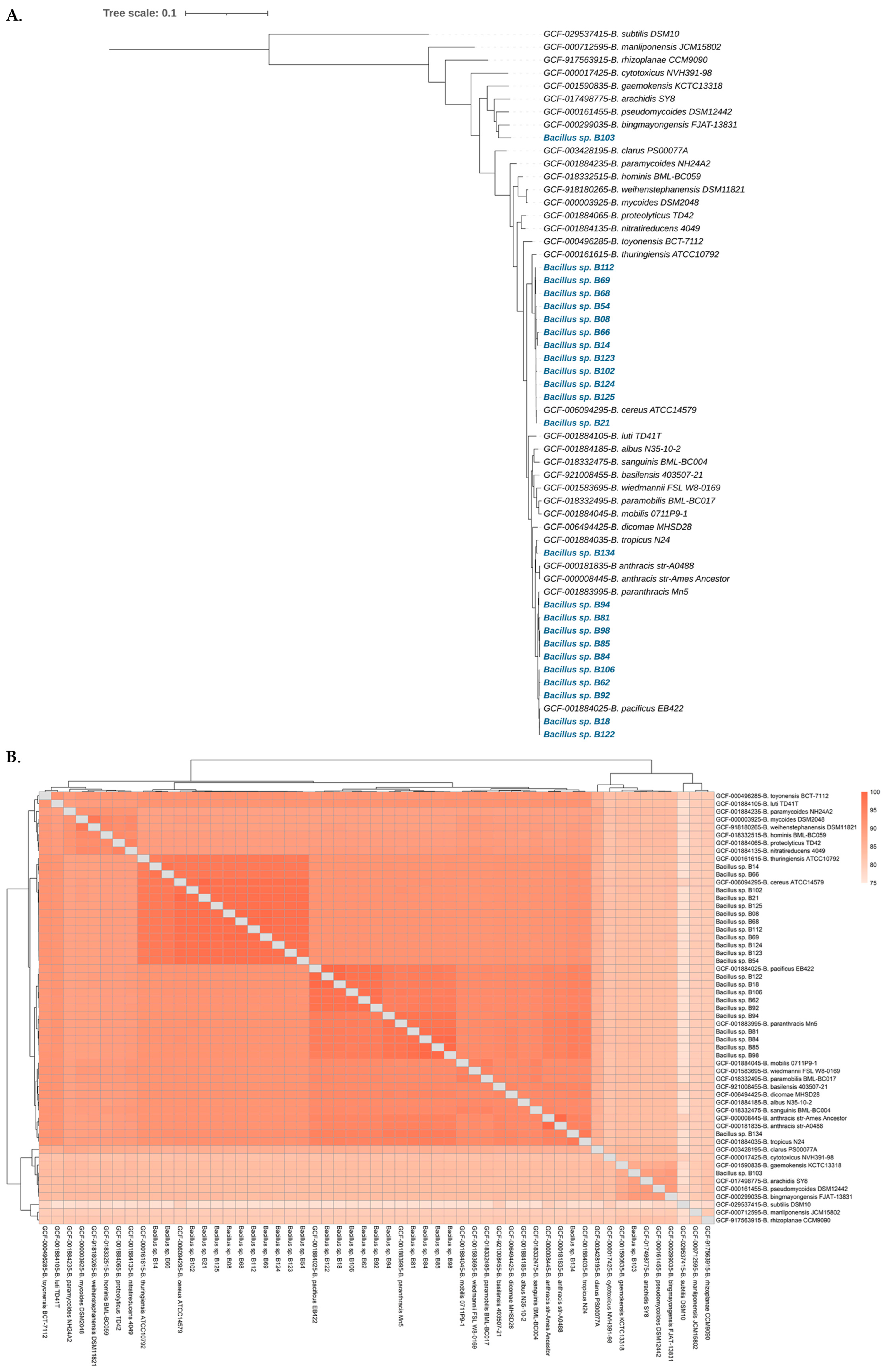
Figure 1. (A) Maximum likelihood phylogenetic tree of the 24 B. cereus group isolates (in blue font) and other type strains of B. cereus group genome sequences, generated using the GToTree v1.8.3 workflow and visualized using the web-based tool iTOL v6. (B) Heatmap of whole genome-based average nucleotide identity (ANI) of the 24 B. cereus group isolates and other type strains of B. cereus group genome sequences, constructed using FastANI v1.33.
Antibiotics 13 00245 g001
Figure 2. Maximum likelihood core-gene phylogenetic tree of the 24 B. cereus group isolates. The tree was constructed using panC sequences extracted from the BTyper3 tool in RAxML, with the GTRGAMMA model and 1000 bootstrap replicates. The clades were designated according to previously proposed phylogenetic groups based on panC sequence types.
Antibiotics 13 00245 g002
Figure 3. Comparison of functional categories in the B. cereus isolate genomes based on SEED subsystem categorization. Functional categorization was based on the roles of annotated and assigned genes. Each colored bar denotes the number of genes assigned to each category.
Antibiotics 13 00245 g003
Figure 4. Distribution of AMR genes among the B. cereus group isolates. The presence of AMR genes in the draft genomes of the isolates was determined using ABRicate v0.8, which involved the ARG-ANNOT, CARD, NCBI, and ResFinder databases. The prevalence of each gene is shown as a percentage. blaZ: beta-lactamase III gene, fosB1: fosfomycin resistance gene, fosB: fosfomycin resistance gene, mef(A): macrolide efflux gene, msr(D): macrolide resistance gene, tet(45): tetracycline resistance gene, BcII: beta-lactamase gene, bla1: beta-lactam resistance gene, rphC: rifampicin resistance gene, satA_Ba: streptothricin resistance gene, vanR-A: glycopeptide resistance gene, vanS-Pt: glycopeptide resistance gene, vanZ-F: glycopeptide resistance gene.
Antibiotics 13 00245 g004
Figure 5. Distribution of virulence genes among the B. cereus group isolates. The putative virulence factors were predicted by a BLAST search against the Virulence Factor Database. The prevalence of each gene is shown as a percentage. alo: thiol-activated cytolysin, asb: B. anthracis siderophore biosynthesis, ces: cereulide synthetase, cytK: cytolytic pore-forming protein cytotoxin K, dhb: bacillibactin biosynthesis, essC: type VII secretion protein, esxB: WXG100 proteins, hblA: hemolysin BL binding component precursor, hblC: hemolysin BL lytic component L2, hblD: hypothetical protein, ilsA: iron-regulated leucine rich surface protein, inhA: immune inhibitor A, metalloprotease, nheA: non-hemolytic enterotoxin A, nheB: non-hemolytic enterotoxin lytic component L1, nheC: enterotoxin C.
Antibiotics 13 00245 g005
Figure 6. Distribution of biofilm-associated genes among the 24 B. cereus group isolates. Genes relevant to biofilm formation (biofilm transcription regulator genes, eDNA synthesis genes, matrix polysaccharide synthesis genes, and matrix protein-encoding genes) were identified using BlastKOALA.
Antibiotics 13 00245 g006
Table 1. The genomes of Bacillus spp. used in this study.
Strain NamesSourceBioSample
Accession
Closest species (ANI%)CoverageContig (s)Size (bp)GC%CDS
B08spicy green papaya salad with bamboo shootSAMN37915793B. cereus (98.84%)78.2×395,344,84435.065371
B14clear soup with tofu and minced porkSAMN37915794B. cereus (96.86%)57.8×2586,378,09534.666524
B18stir-fried mixed vegetablesSAMN37915795B. pacificus (99.92%)74.1×555,453,84535.275571
B21stir-fried pumpkinSAMN37915796B. cereus (99.85%)81.8×305,262,39135.045292
B54stir-fried noodle with yentafo sauceSAMN37915797B. cereus (98.67%)68.4×345,880,55134.805970
B62rice with spicy shrimp paste dip and fried mackerelSAMN37915798B. pacificus (98.12%)72.5×1455,542,77935.175703
B66stir-fried noodleSAMN37915799B. cereus (96.98%)61.4×1046,152,93534.766074
B68stir-fried mixed vegetablesSAMN37915800B. cereus (98.75%)73.1×655,951,70735.016082
B69spicy mixed vegetable soupSAMN37915801B. cereus (98.90%)71.7×435,378,13335.015376
B81spicy Vietnamese pork sausage saladSAMN37915802B. paranthracis (97.80%)73.4×445,223,90035.355287
B84spicy tremella mushroom saladSAMN37915803B. paranthracis (97.68%)80.9×525,126,33635.345169
B85spicy pork meatball saladSAMN37915804B. paranthracis (97.54%)77.9×785,473,50035.255546
B92sushiSAMN37915805B. pacificus (98.14%)70.9×1005,528,81235.315660
B94spicy fermented fish dipSAMN37915806B. paranthracis (98.52%)66.2×605,435,64235.155496
B98stir-fried glass noodlesSAMN37915807B. paranthracis (97.53%)157.5×975,426,06235.175512
B102sandwichSAMN37915808B. cereus (98.47%)188.0×375,221,58435.085237
B103salad roll with crab stickSAMN37915809B. bingmayongensis” (90.05%)181.6×645,437,99535.505436
B106stir-fried lotus stem and straw mushroom with porkSAMN37915810B. pacificus (98.18%)76.8×705,401,24035.185541
B112stir-fried tofu with bean sproutsSAMN37915811B. cereus (98.87%)122.2×1275,712,86635.055773
B122stir-fried pickled turnip with eggSAMN37915812B. pacificus (99.95%)64.5×615,431,86035.275487
B123stir-fried zucchini with eggSAMN37915813B. cereus (98.85%)66.2×475,481,27334.875454
B124egg noodles with roasted red pork and wontonSAMN37915814B. cereus (98.89%)89.7×585,856,37134.765803
B125Thai rice noodle salad rollSAMN37915815B. cereus (98.85%)84.3×275,631,59534.925647
B134stewed pork leg on riceSAMN37915816B. tropicus (96.87%)47.7×385,208,04535.245239
ANI: Average Nucleotide Identity, GC: Guanine and Cytosine, CDS: Coding Sequence.
Disclaimer/Publisher’s Note: The statements, opinions and data contained in all publications are solely those of the individual author(s) and contributor(s) and not of MDPI and/or the editor(s). MDPI and/or the editor(s) disclaim responsibility for any injury to people or property resulting from any ideas, methods, instructions or products referred to in the content.