Article

A FtsZ Inhibitor That Can Utilize Siderophore-Ferric Iron Uptake Transporter Systems for Activity against Gram-Negative Bacterial Pathogens

1
Department of Pharmacology, Rutgers Robert Wood Johnson Medical School, Piscataway, NJ 08854, USA
2
Department of Molecular Design and Synthesis, Rutgers University Biomedical Innovation Cores, Piscataway, NJ 08854, USA
3
Department of Medicinal Chemistry, Ernest Mario School of Pharmacy, Rutgers, The State University of New Jersey, Piscataway, NJ 08854, USA
*
Author to whom correspondence should be addressed.
Antibiotics 2024, 13(3), 209; https://doi.org/10.3390/antibiotics13030209
Submission received: 2 February 2024 / Revised: 16 February 2024 / Accepted: 19 February 2024 / Published: 22 February 2024
(This article belongs to the Special Issue Design and Synthesis of Novel Antibiotics)

Abstract

:
The global threat of multidrug-resistant Gram-negative bacterial pathogens necessitates the development of new and effective antibiotics. FtsZ is an essential and highly conserved cytoskeletal protein that is an appealing antibacterial target for new antimicrobial therapeutics. However, the effectiveness of FtsZ inhibitors against Gram-negative species has been limited due in part to poor intracellular accumulation. To address this limitation, we have designed a FtsZ inhibitor (RUP4) that incorporates a chlorocatechol siderophore functionality that can chelate ferric iron (Fe3+) and utilizes endogenous siderophore uptake pathways to facilitate entry into Gram-negative pathogens. We show that RUP4 is active against both Klebsiella pneumoniae and Acinetobacter baumannii, with this activity being dependent on direct Fe3+ chelation and enhanced under Fe3+-limiting conditions. Genetic deletion studies in K. pneumoniae reveal that RUP4 gains entry through the FepA and CirA outer membrane transporters and the FhuBC inner membrane transporter. We also show that RUP4 exhibits bactericidal synergy against K. pneumoniae when combined with select antibiotics, with the strongest synergy observed with PBP2-targeting β-lactams or MreB inhibitors. In the aggregate, our studies indicate that incorporation of Fe3+-chelating moieties into FtsZ inhibitors is an appealing design strategy for enhancing activity against Gram-negative pathogens of global clinical significance.

1. Introduction

The global spread of multidrug-resistant (MDR) bacterial pathogens represents a public health crisis [1,2,3]. Patients diagnosed with MDR infections suffer much worse prognoses, including extended hospital stays and higher mortality rates, than patients with drug-susceptible infections [1]. Globally, 1.3 million deaths per year are directly attributable to resistant bacterial infections [4], with this figure predicted to rise to 10 million annual deaths by 2050 in the absence of intervention [5]. Of particular concern are ESKAPE pathogens [6], bacterial strains that are typically associated with high rates of clinical resistance [7,8]. Two of the more resistant ESKAPE pathogens are the Gram-negative species Klebsiella pneumoniae and Acinetobacter baumannii, each of which is responsible for more than 100,000 deaths annually [2,4]. Current clinical antibiotics, including carbapenems, third generation cephalosporins, and fluoroquinolones, are becoming increasingly ineffective against these two pathogens, leaving clinicians with few treatment options [4,9,10].
The threat posed by highly resistant Gram-negative pathogens emphasizes the need to develop new and effective antibiotics with novel bacterial targets. One promising new antibacterial drug target is FtsZ, an essential and highly conserved bacterial divisome protein that is not targeted by any current clinical drug [11,12]. FtsZ is a GTPase that polymerizes into a structure known as the Z-ring at the midcell of a dividing bacterium [13]. The FtsZ Z-ring is a major factor in driving bacterial cytokinesis, acting as a scaffold for recruitment of essential cell division components, which include the cell wall-forming penicillin-binding proteins (PBPs) [11,14]. In addition, FtsZ acts in concert with the cell wall synthesis coordinator, MreB, in both septal cell wall synthesis and lateral cell wall synthesis at the poles of rod-shaped bacteria [15,16].
FtsZ is a highly druggable target, and benzamide-based FtsZ inhibitors (including PC190723, TXA707, and TXH9179, as well as their corresponding prodrugs TXY541, TXA709, and TXH1033, respectively) with potent bactericidal activity against clinically significant Gram-positive pathogens, such as methicillin- and vancomycin-resistant S. aureus (MRSA and VRSA, respectively), have been previously described by us and others [17,18,19,20,21,22,23,24,25,26,27,28,29,30,31]. However, these FtsZ-targeting drug candidates are typically associated with poor activity against Gram-negative bacteria [20,32]. One of the principal resistance mechanisms in Gram-negative bacteria is their outer membrane, a hydrophobic barrier that limits the intracellular accumulation of most antibiotics, including benzamide FtsZ inhibitors [33,34,35]. This barrier must be overcome to improve the intracellular uptake of FtsZ inhibitors by Gram-negative pathogens as well as their resultant antibacterial efficacy.
One appealing approach for enhancing antibiotic uptake by Gram-negative bacteria is the design of agents that incorporate iron-chelating siderophore functionalities into their structures [36]. Ferric iron (Fe3+) is an essential nutrient for bacterial growth and survival, providing a critical redox factor for many important metabolic processes, including respiration, oxygen transport, and DNA synthesis [37,38]. However, Fe3+ availability is typically limited at sites of infection, due to restriction by the innate immune system in host environments [37,38,39,40]. To facilitate the scavenging of Fe3+ from the environment, bacteria synthesize and export molecules, termed siderophores, which chelate Fe3+ and are then internalized by the bacteria via dedicated uptake pathways [41,42]. Many clinically important Gram-negative pathogens, including K. pneumoniae and A. baumannii, rely on siderophores for Fe3+ acquisition and virulence [43,44]. Incorporation of Fe3+-chelating siderophore functionalities in antibacterial drug structures offers the potential for drug uptake by endogenous siderophore uptake pathways. In support of this design approach, cefiderocol, a PBP-targeting cephalosporin analog containing an Fe3+-chelating chlorocatechol moiety, has been recently approved for the treatment of complicated urinary tract infections caused by highly resistant Gram-negative pathogens [45,46,47].
In this study, we describe a novel oxazole-benzamide FtsZ inhibitor (RUP4) that, like cefiderocol, incorporates a Fe3+-chelating chlorocatechol moiety. This chlorocatechol moiety contains hydroxyl groups at the 3- and 4-positions (Figure 1), which are designed to chelate Fe3+. Significantly, RUP4 is active against both K. pneumoniae and A. baumannii, in marked contrast to a corresponding negative control compound (RUP5) that lacks the hydroxyl group at the 3-position of the catechol moiety (Figure 1). We show that the antibacterial activity of RUP4 against K. pneumoniae is due to its ability to chelate Fe3+ and that the compound utilizes endogenous K. pneumoniae siderophore-Fe3+ uptake transporters for entry into the bacteria. We also show that RUP4 acts synergistically against K. pneumoniae when combined with select β-lactam antibiotics as well as a MreB inhibitor, with this bactericidal synergy being greatest upon combination with the PBP2-targeting antibiotic mecillinam and the MreB inhibitor TXH11106. Viewed as a whole, our results highlight RUP4 as an appealing proof-of-concept agent for the design of next-generation benzamide FtsZ inhibitors with enhanced activity against Gram-negative bacterial pathogens.

2. Results

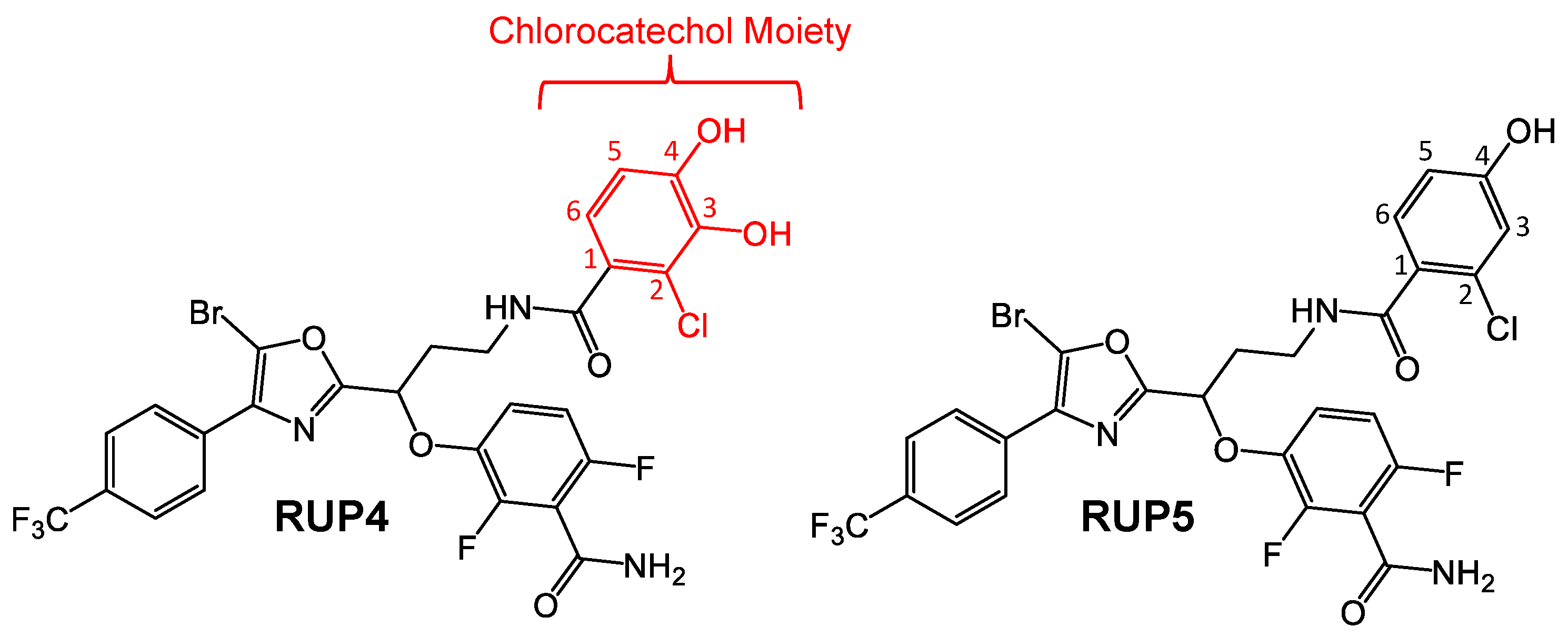
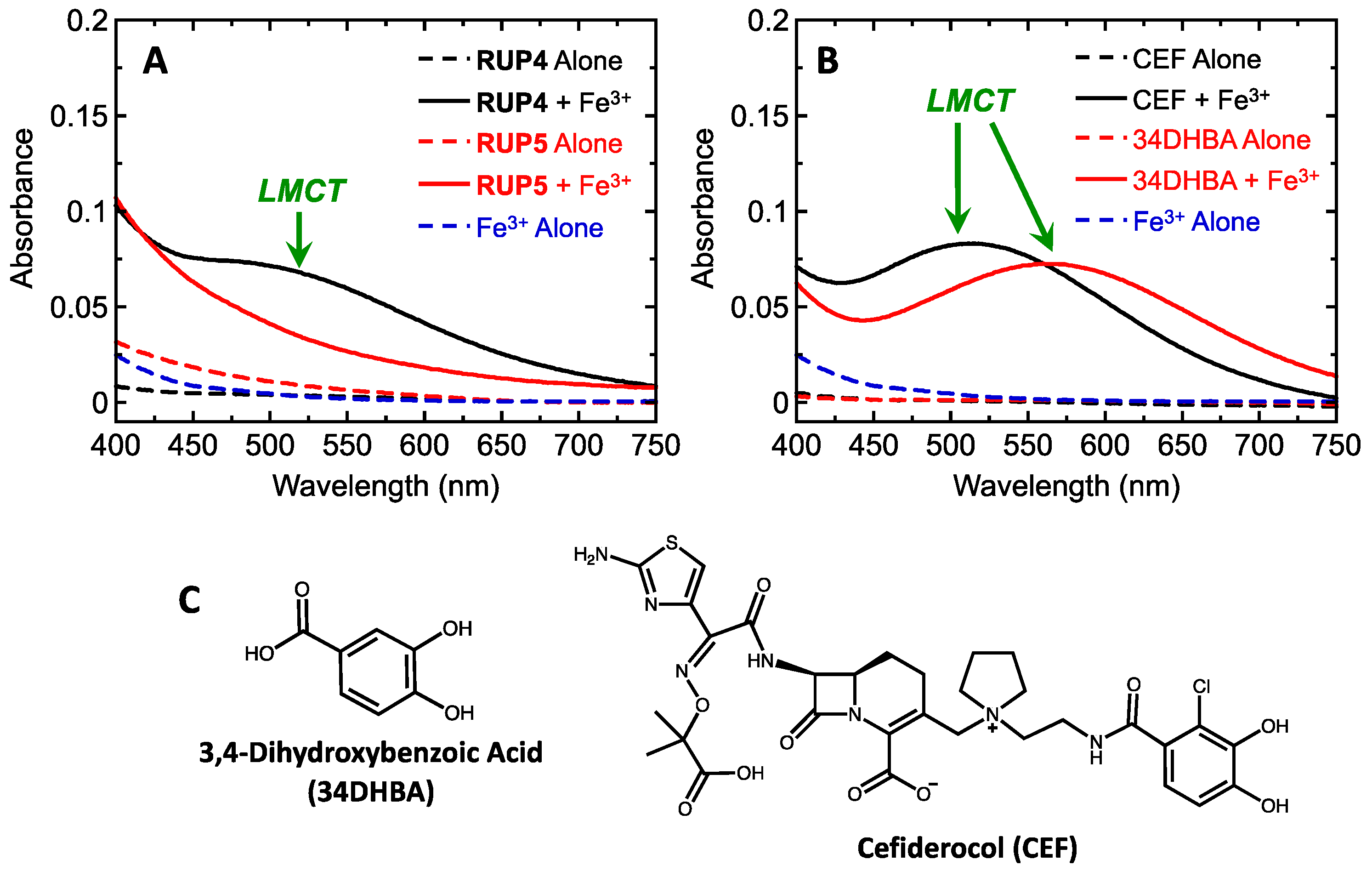
2.1. Evaluation of RUP4 and RUP5 for Their Ability to Chelate Fe3+

We first sought to determine the potential for RUP4 and RUP5 to chelate Fe3+. Nolan and co-workers have shown that catechol chelation of Fe3+ is associated with a ligand-to-metal charge transfer (LMCT) reaction that is manifested by the induction of a peak in the absorption spectrum of the catechol in the wavelength region of 400–750 nm [48]. In this connection, we measured the absorption spectra of 50 µM RUP4 and RUP5 in the absence or presence of an equimolar concentration of Fe3+. The addition of Fe3+ to RUP4 resulted in the induction of a broad absorbance peak in the region between 400 and 750 nm that was absent from the spectrum of either RUP4 or Fe3+ alone (Figure 2A). This induced absorption peak is indicative of an LMCT reaction (denoted by the green arrow in Figure 2A). We observed the induction of similar LMCT peaks in the absorption spectra of our positive comparator control agents 3,4-dihydroxybenzoic acid (34DHBA) and cefiderocol (CEF) upon addition of Fe3+ (see spectra in Figure 2B and chemical structures of 34DHBA and CEF in Figure 2C). Note that the LMCT peak in the spectrum of RUP5 in the presence of Fe3+ is markedly reduced in magnitude compared with the corresponding LMCT peak in the spectrum of RUP4 (Figure 2A).

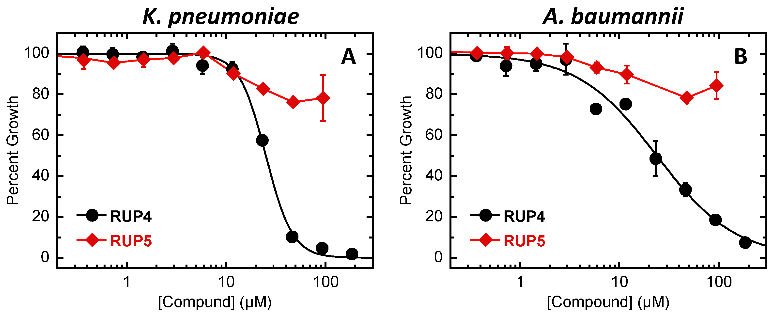
2.2. Evaluation of RUP4 and RUP5 for Antibacterial Activity against K. pneumoniae and A. baumannii

We characterized the antibacterial activity of RUP4 and RUP5 against K. pneumoniae (ATCC 10031) and A. baumannii (ATCC 19606) in Fe3+-limiting M9 media. In these studies, both compounds were assessed at concentrations up to their limits of solubility, with the resulting growth profiles being shown in Figure 3. RUP4 inhibits the growth of both K. pneumoniae (Figure 3A) and A. baumannii (Figure 3B) with similar IC50 values of 25 ± 1 and 24 ± 2 µM, respectively, as well as corresponding minimal inhibitory concentration (MIC) values of 46 µM against K. pneumoniae and 185 µM against A. baumannii (Table 1). We also determined the minimal bactericidal concentration (MBC) of RUP4 against K. pneumoniae to be 92 µM, a value twice the corresponding MIC of 46 µM, with the MBC of RUP4 against A. baumannii being greater than the solubility limit of the compound (>185 µM). In marked contrast to RUP4, RUP5 did not inhibit the growth of K. pneumoniae or A. baumannii to any significant degree (Figure 3), with associated IC50, MIC, and MBC values all being greater than the solubility limit of the compound (>95 µM) (Table 1).

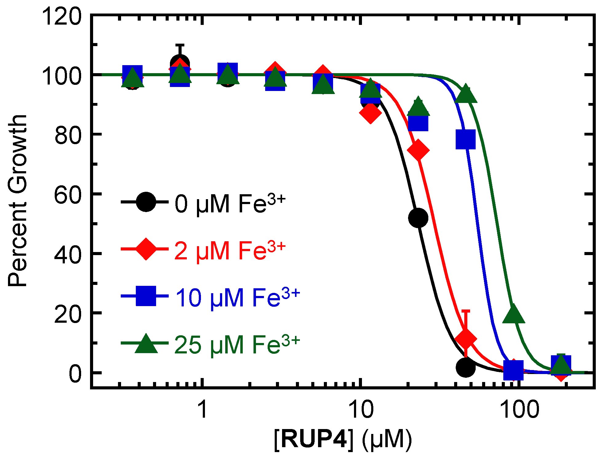
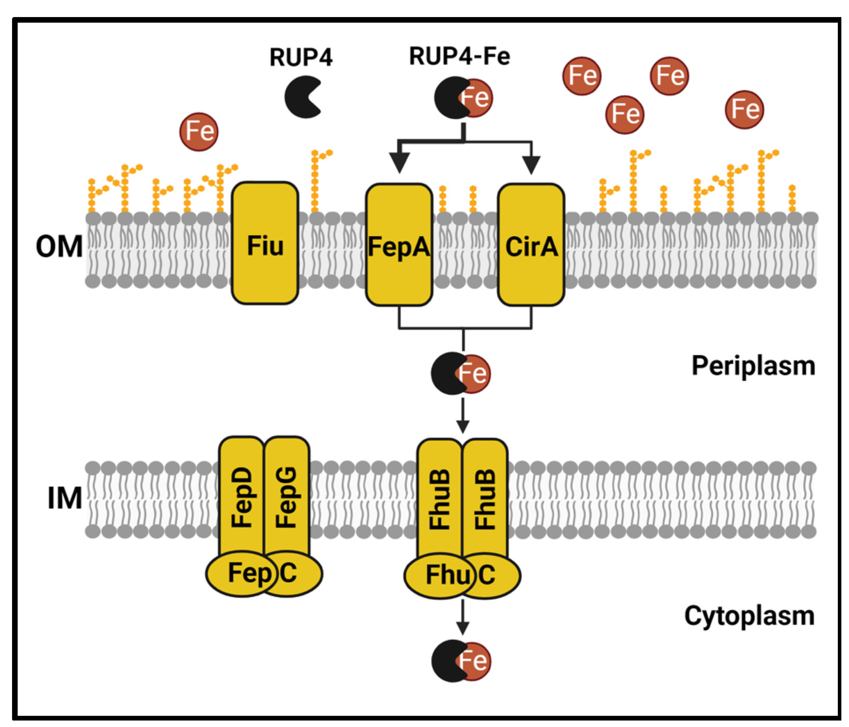
2.3. Impact of Added Exogenous Fe3+ on the Antibacterial Activity of RUP4 against K. pneumoniae

We sought to determine whether the observed activity of RUP4 against K. pneumoniae is sensitive to the concentration of exogenous Fe3+. To this end, we explored the impact of adding 0, 2, 10, or 25 µM exogenous Fe3+ on the activity of RUP4 against K. pneumoniae in modified M9 media. The resulting growth profiles are shown in Figure 4. Significantly, the addition of exogenous Fe3+ markedly reduced the activity of RUP4, as reflected by IC50 values that increased with increasing concentrations of added exogenous Fe3+ [with these IC50 values being 23 ± 1 µM with 0 µM added Fe3+, 29 ± 1 µM with 2 µM added Fe3+, 55 ± 6 µM with 10 µM added Fe3+, and 73 ± 4 µM with 25 µM added Fe3+] (Table 2).

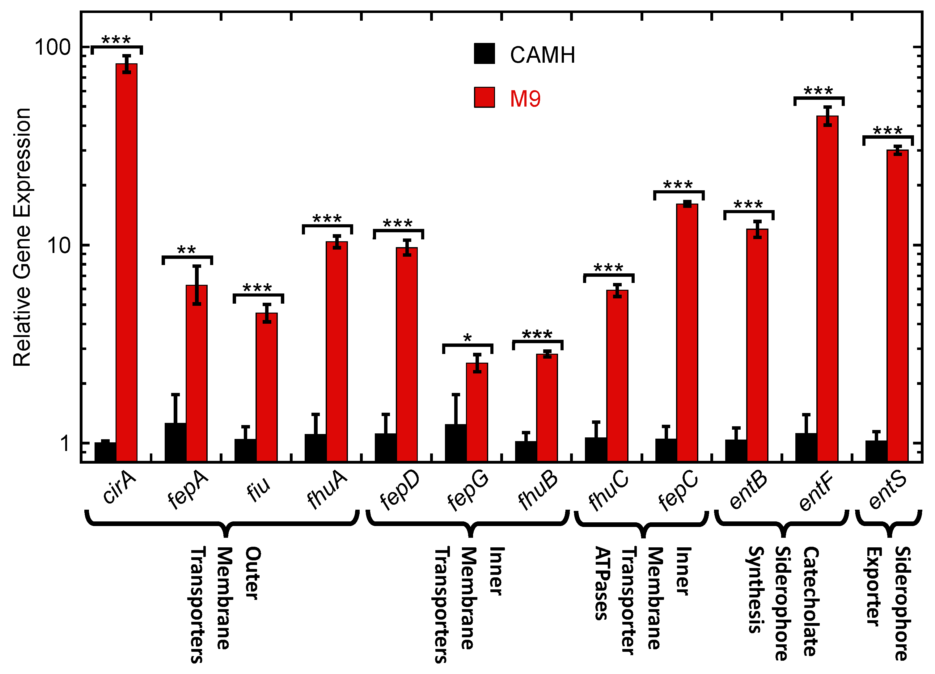
2.4. Expression of Siderophore Biosynthesis, Exporter, and Uptake Transporter Genes by K. pneumoniae in Nutrient-Rich CAMH Media Versus Fe3+-Limiting M9 Media

As an initial step toward determining whether RUP4 can utilize the native siderophore-Fe3+ uptake machinery of target bacteria, we first explored how Fe3+ restriction affects the expression of native siderophore uptake, biosynthesis, and exporter genes in K. pneumoniae. To this end, we used RT-qPCR to compare the expression of key siderophore uptake, biosynthesis, and exporter genes in nutrient-rich CAMH media versus Fe3+-limiting M9 media. For siderophore uptake transporter genes, we evaluated the expression of the enterobactin outer membrane transporter fepA, the catecholate outer membrane transporters cirA and fiu, the ferrichrome outer membrane transporter fhuA, the enterobactin ABC inner membrane transporter components fepD and fepG (channel components) as well as fepC (ATPase), and the ferrichrome ABC inner membrane transporter components fhuB (channel) and fhuC (ATPase). For siderophore biosynthesis genes, we evaluated the expression of the catecholate synthesis pathway component entB and the enterobactin synthesis pathway component entF. For siderophore exporter genes, we evaluated expression of the enterobactin exporter entS. The expression of the bacterial transcription factor rho was used as an endogenous control for all comparisons.
Expression of all siderophore genes was significantly upregulated in M9 media relative to CAMH media (Figure 5). Among the outer membrane uptake transporters, cirA was upregulated to the greatest extent (82.1-fold), followed by fhuA, fepA, and fiu at 10.4-, 6.3-, and 4.5-fold, respectively. Among the inner membrane uptake transporters, fepD was upregulated to the greatest extent (9.7-fold), followed by fhuB and fepG at 2.8- and 2.5-fold, respectively. The inner membrane uptake transporter ATPases fepC and fhuC were upregulated by 16.1- and 5.9-fold, respectively. Finally, the catecholate siderophore synthesis genes entF and entB as well as the siderophore exporter gene entS were upregulated by 44.8-, 12.0-, and 30.1-fold, respectively.

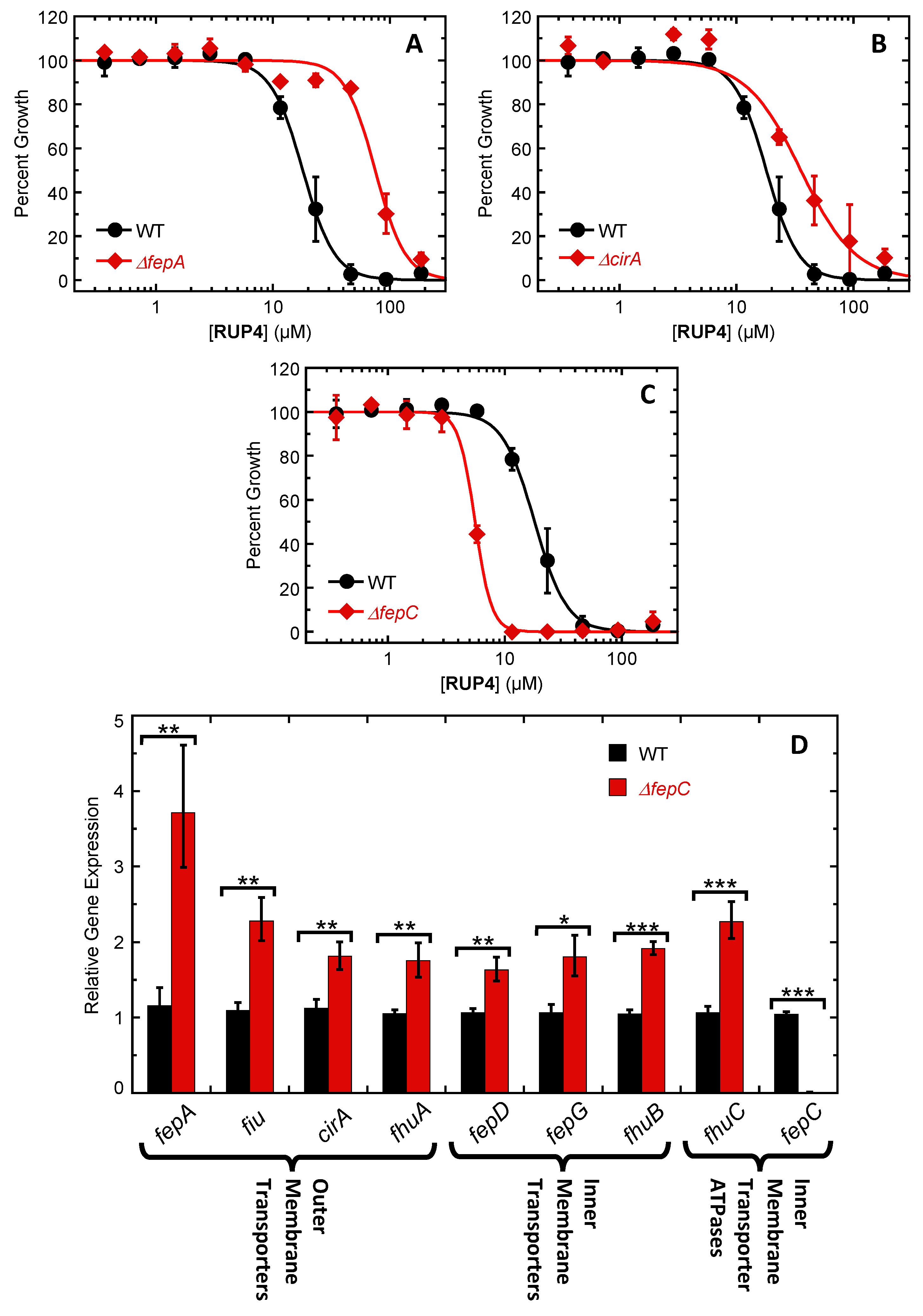
2.5. Impact of Deleting Select Siderophore-Fe3+ Uptake Transporter Genes on RUP4 Activity against K. pneumoniae

Towards the goal of elucidating the specific uptake pathway of RUP4, we generated mutant strains of K. pneumoniae containing a deletion of the siderophore-Fe3+ outer membrane transporter fepA, fiu, or cirA. We then compared the activity of RUP4 against each of these deletion mutant strains (∆fepA, ∆fiu, or ∆cirA) relative to the activity against the wild-type (WT) K. pneumoniae strain, with the corresponding growth profiles comparing ∆fepA and ∆cirA versus WT being shown in Figure 6A. The activity of RUP4 was attenuated by approximately 4-fold against the ∆fepA mutant and 2-fold against the ∆cirA mutant, as reflected by IC50 values of 75 ± 5 µM for the ∆fepA strain and 35 ± 5 µM for the ∆cirA strain relative to 18 ± 1 µM for WT (Table 3). By contrast, the activity of RUP4 against the ∆fiu strain (IC50 = 22 ± 4 µM) was comparable to the activity versus WT (Table 3).
We also generated a mutant strain of K. pneumoniae containing a deletion of the siderophore-Fe3+ inner membrane transporter ATPase fepC, with the corresponding growth profiles comparing ΔfepC versus WT being shown in Figure 6C. Unlike the attenuated activity exhibited by RUP4 versus the ΔfepA and ΔcirA mutant strains relative to WT, the corresponding activity of the compound versus the ΔfepC mutant strain was enhanced by approximately 3-fold (IC50 = 5.6 ± 0.1 µM) relative to WT (Figure 6C, Table 3). To further explore the basis for the increased activity of RUP4 against the ΔfepC mutant, we used RT-qPCR to compare the expression of the siderophore outer membrane uptake genes fepA, fiu, cirA, and fhuA as well as the inner membrane uptake genes fepD, fepG, fhuB, and fhuC in the ΔfepC mutant strain relative to WT. The expression of all the tested genes was significantly upregulated (Figure 6D). Among the outer membrane uptake genes, expression of fepA was upregulated to the greatest extent (3.7-fold), followed by fiu, cirA, and fhuA at 2.3-, 1.8-, and 1.7-fold, respectively. Expression of the inner membrane uptake genes fepD, fepG, fhuB, and fhuC was upregulated by 1.6-, 1.8-, 1.9-, and 2.3-fold, respectively.

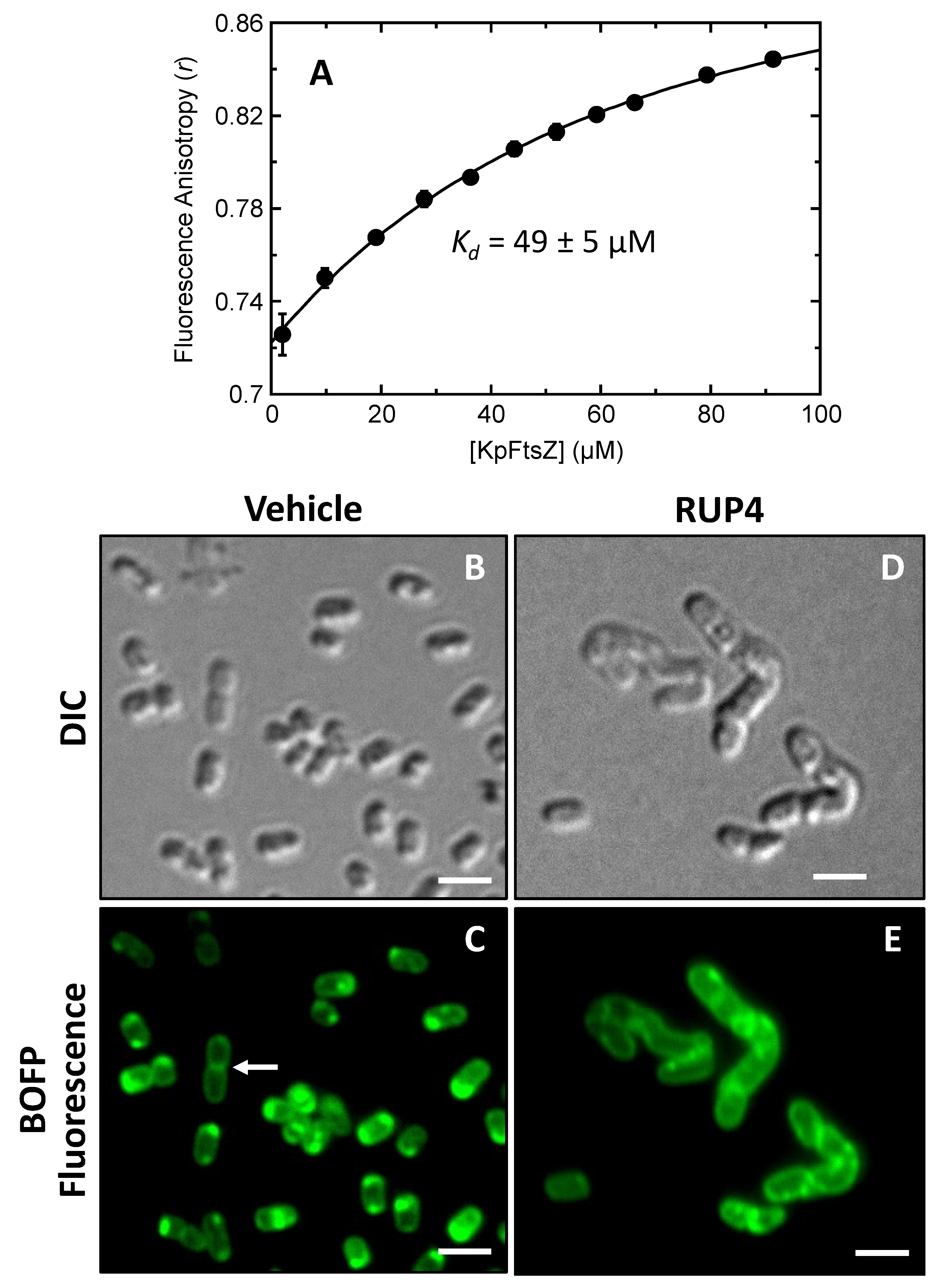
2.6. Direct Binding of RUP4 to Purified K. pneumoniae FtsZ (KpFtsZ)

As an initial step towards the validation of FtsZ as the antibacterial target of RUP4, we sought to demonstrate that RUP4 can directly interact with purified KpFtsZ. To this end, we leveraged the intrinsic fluorescence properties of RUP4, whose excitation and emission spectra are shown in Figure S1. Specifically, we monitored the impact of added KpFtsZ on the fluorescence anisotropy of RUP4 at 25 °C. The addition of KpFtsZ induced an increase in fluorescence anisotropy with increasing protein concentration (Figure 7A), indicative of a direct binding reaction. The resulting binding profile was fit with Equation (2) to yield a Kd value of 49 ± 5 µM.

2.7. Impact of RUP4 on Cell Division and FtsZ Localization in K. pneumoniae

To further establish FtsZ as the antibacterial target of RUP4, we explored the impact of RUP4 treatment on cell morphology and FtsZ localization in K. pneumoniae grown in Fe3+-limiting M9 media. In these studies, K. pneumoniae cells were treated for 3 h with either DMSO vehicle or RUP4 at 185.3 µM (4× MIC) and then labeled with 0.1 µM BOFP. Differential interference contrast (DIC) and fluorescence micrographs of the treated cells are shown in Figure 7B–E. Treatment with vehicle results in normal cell morphology and size, with approximately 3–8% of cells actively undergoing cell division. An actively dividing cell with FtsZ localized to the septum at the midcell is highlighted by the white arrow in Figure 7C. In non-dividing vehicle-treated cells, FtsZ often appears to localize in punctate regions at the cell poles (Figure 7C). In contrast to vehicle treatment, RUP4 treatment results in enlarged and elongated cells with no defined septa and FtsZ more diffusely localized to foci throughout the cell as well as along the cell periphery (Figure 7D,E). This pattern of behavior is similar to that previously observed in rod-shaped bacteria that have been treated with a FtsZ inhibitor or in which FtsZ has been depleted [20,49,50].

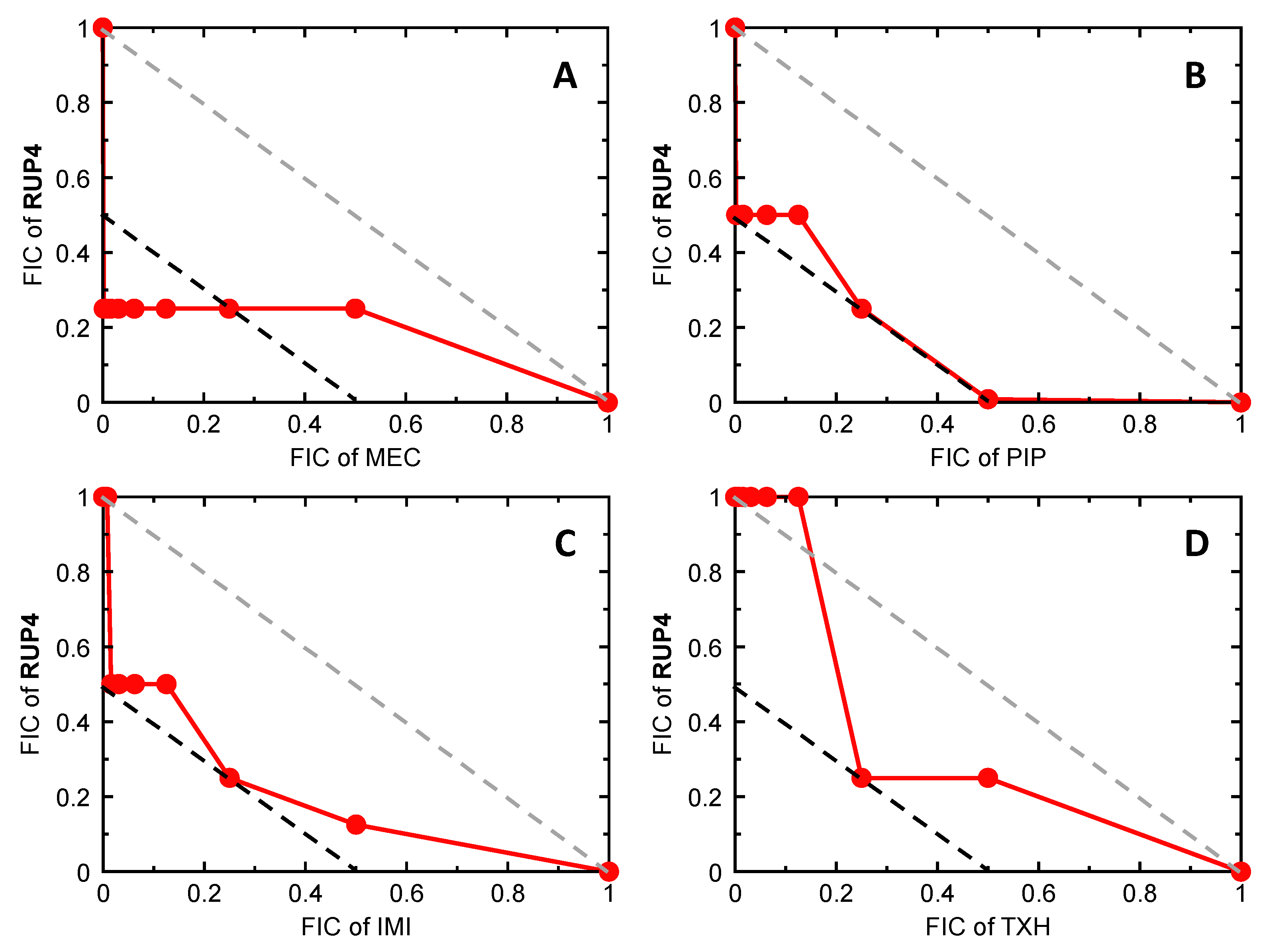
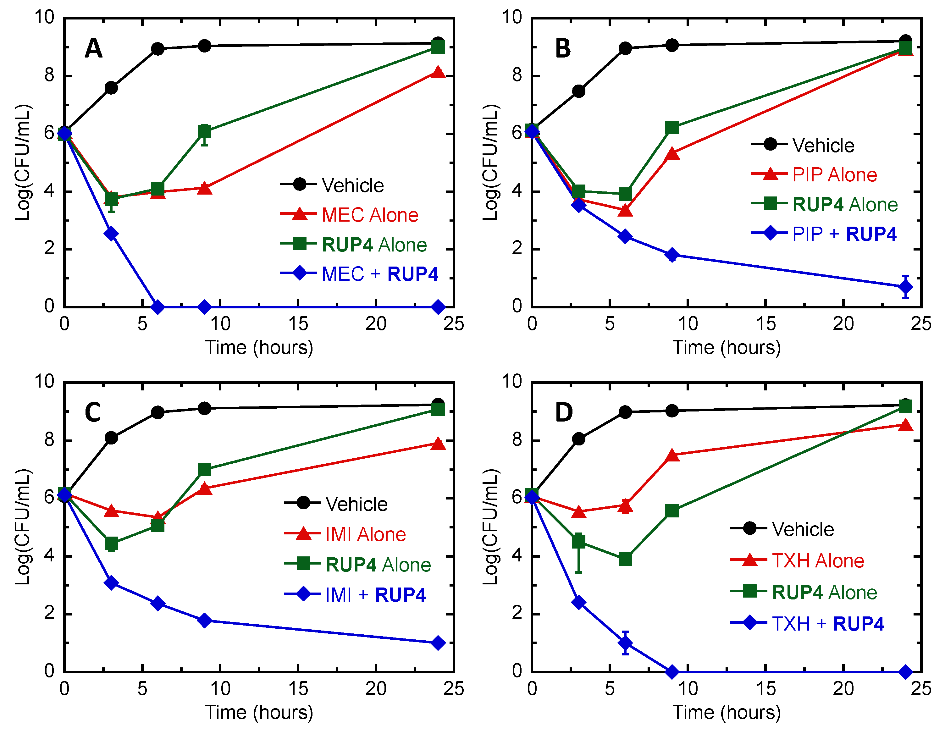
2.8. Bactericidal Synergy of RUP4 in Combination with Select PBP-Targeting β-Lactam Antibiotics and an MreB-Targeting Agent against K. pneumoniae

We assessed the potential of RUP4 to act synergistically in combination with various PBP-targeting β-lactam antibiotics [mecillinam (MEC), amoxicillin (AMX), piperacillin (PIP), cefazolin (CFZ), imipenem (IMI), and meropenem (MER)] as well as an MreB-targeting agent TXH11106 (TXH) we have previously developed [51]. In this connection, we evaluated the intrinsic activities of the test agents against K. pneumoniae, with the resulting MIC values being listed in Table S1. We then used a checkerboard assay to define the fractional inhibitory concentration (FIC) of each test agent and RUP4 when used in combination against K. pneumoniae. For each combination, synergy is indicated when the FIC values for both agents are ≤0.25. Figure 8 shows the isobolograms plotting the FICs for RUP4 as a function of the FICs for MEC (Figure 8A), PIP (Figure 8B), IMI (Figure 8C), or TXH (Figure 8D). All four test agents act synergistically with RUP4, as indicated by the presence of combination FICs in the lower left quadrant of the isobolograms (on or to the left of the black dashed line in each plot). The corresponding isobolograms for the combinations of RUP4 and CFZ, AMX, or MER are plotted in Figure S2, with these plots revealing additive behavior (with combination FICs falling to the right of the black dashed line and on or to the left of the gray dashed line).
Another indication of synergistic versus additive activity is provided by the FIC index (FICI), defined as the sum of the FICs of the two agents being tested in combination. A FICI ≤ 0.5 is indicative of synergy, while a FICI > 0.5 but ≤1 is reflective of additivity. The synergistic actions of RUP4 in combination with MEC are reflected by a FICI of 0.251 (Table 4), with those of RUP4 in combination with PIP, IMI, or TXH being reflected by FICI values of 0.5. The additive behavior of RUP4 in combination with CFZ, AMX, or MER is reflected by FICI values of 0.508, 0.531, and 0.531, respectively (Table 4).
In addition to checkerboard assays for synergy, we also performed time–kill assays of RUP4 in combination with MEC, PIP, IMI, or TXH against K. pneumoniae. In these assays, cells were treated with DMSO vehicle, 0.5× MIC test agent alone, 0.5× MIC RUP4 alone, or a combination of 0.5× MIC test agent and 0.5× MIC RUP4, and the number of colony-forming units (CFUs) was assessed after 0, 3, 6, 9, and 24 h of treatment. The resulting time–kill curves are shown in Figure 9. Treatment with either the test agent alone or RUP4 alone resulted in growth at 24 h comparable to that of the vehicle. By contrast, the combination of each test agent with RUP4 resulted in bactericidal behavior. In this connection, complete kill was observed after 6 h of treatment with the combination of MEC and RUP4, and after 9 h of treatment with the combination of TXH and RUP4 (Figure 9A,D). Combination of RUP4 and IMI or PIP resulted in 5-logs of kill within 24 h of treatment (Figure 9B,C).

3. Discussion

Gram-negative bacterial pathogens are often more difficult to treat than Gram-positive pathogens due to the presence of an outer membrane barrier that limits the intracellular accumulation of many antibiotics [33,35]. In this connection, benzamide-based FtsZ inhibitors have been associated with potent activity against Gram-positive pathogens like S. aureus while exhibiting poor or no activity against Gram-negative pathogens [17,18,19,20,21,22,23,24,25,26,27,28,29,30,31,34]. Here, we describe a strategy for overcoming this limitation by developing an oxazole-benzamide FtsZ inhibitor (RUP4) that incorporates a chlorocatechol siderophore moiety (Figure 1) and can utilize endogenous siderophore-Fe3+ uptake pathways to facilitate entry into the target bacterial cells. Significantly, RUP4 is active against both K. pneumoniae and A. baumannii (Figure 3, Table 1), two Gram-negative pathogens of acute clinical importance. As indicated by MIC, the observed activity of RUP4 is approximately 4-fold greater against K. pneumoniae (MIC = 46.3 µM) than against A. baumannii (MIC = 185.3 µM), though RUP4 exhibited a similar IC50 value of approximately 25 µM against both pathogens (Table 1).

3.1. Importance of the Catechol Siderophore Functionality for the Antibacterial Activity of RUP4

Our design strategy is premised on the hypothesis that the catechol siderophore moiety plays an important role in promoting the observed antibacterial activity of RUP4. To this end, we first sought to establish that the two hydroxyl groups at positions 3 and 4 of the RUP4 catechol functionality (Figure 1) can chelate Fe3+. Catechol chelation of Fe3+ is associated with a ligand-to-metal charge transfer (LMCT) reaction that is measurable by the induction of an absorption peak between 400 and 750 nm [48]. Consistent with such an Fe3+ chelation reaction, RUP4 exhibits a robust LMCT peak in the presence of Fe3+ that is absent in the absorption spectra of either RUP4 or Fe3+ alone (Figure 2A). The Fe3+-chelating capability of RUP4 is further supported by the corresponding spectra of the positive control molecules cefiderocol and 34DHBA, which exhibit similar LMCT peaks in the presence of Fe3+ (Figure 2B). By contrast, the spectrum of the negative control compound RUP5, which lacks the 3-hydroxyl on the catechol ring, has a markedly reduced LMCT peak (Figure 2A), indicative of an attenuated ability to chelate Fe3+.
Armed with the knowledge that RUP4 chelates Fe3+ to a significantly greater extent than RUP5, we determined if this differential capability manifested in a corresponding difference in antibacterial activity. Significantly, in striking contrast to RUP4, RUP5 exerts a minimal impact on the growth of either K. pneumoniae or A. baumannii at concentrations up to its solubility limit of approximately 95 µM (Figure 3, Table 1). The absence of significant RUP5 activity implies that the antibacterial activity of RUP4 is linked to its ability to chelate Fe3+. Consistent with this notion, we show that the activity of RUP4 against K. pneumoniae is systematically reduced in the presence of increasing concentrations of added exogenous Fe3+ (Figure 4), with IC50 values ranging from 23 µM in the absence of added Fe3+ to 73 µM in the presence of 25 µM added Fe3+ (Table 2). In addition, the expression of siderophore-Fe3+ uptake transporter, catecholate siderophore biosynthesis, and siderophore exporter genes in K. pneumoniae is significantly upregulated under Fe3+-limiting conditions (Figure 5). These collective results are indicative of RUP4 utilizing endogenous siderophore-Fe3+ uptake pathways for its activity against K. pneumoniae.
We next sought to determine the specific siderophore-Fe3+ uptake transporters that RUP4 can utilize for entry into K. pneumoniae. To this end, we generated deletion mutant strains of three different outer membrane transporters (ΔfepA, ΔcirA, and Δfiu) in K. pneumoniae 10031 and compared the activity of RUP4 against each deletion mutant strain relative to WT. The activity of RUP4 was reduced against both the ΔfepA and ΔcirA strains compared to WT (Figure 6A,B), with the extent of this reduction being approximately 4-fold for the ΔfepA strain and 2-fold for the ΔcirA strain, as reflected by IC50 values (Table 3). By contrast, RUP4 exhibited similar activity against the Δfiu strain relative to WT (Table 3). These results suggest that RUP4 crosses the outer membrane through both the FepA and CirA transporters, with FepA being the predominant transporter (schematically depicted in Figure 10).
We also generated a deletion mutant strain of the inner membrane transporter ATPase fepC. Surprisingly, the activity of RUP4 against the ΔfepC strain was improved by approximately 3-fold compared to WT (Figure 6C, Table 3), suggesting that the enterobactin inner membrane transporter FepCDG is not the principal transporter through which RUP4 crosses the inner membrane. To better understand the enhanced activity of RUP4 against the ΔfepC strain, we measured the expression of all siderophore-Fe3+ uptake transporter genes in the ΔfepC strain relative to WT. Significantly, expression of the outer membrane transporter genes fepA and fiu, as well as the ferrichrome inner membrane transporter genes fhuB and fhuC, was among the most upregulated (Figure 6D), with fepA expression being upregulated to the greatest extent of all (approximately 3.7-fold). As previously noted, FepA appears to be the principle outer membrane transporter utilized by RUP4. Furthermore, aside from FepCDG, FhuBC is the only other inner membrane uptake transporter present in K. pneumoniae 10031. These collective results suggest that RUP4 utilizes the FhuBC transporter for crossing the inner membrane, and the increased activity of RUP4 against the ΔfepC strain relative to WT is primarily due to the increased expression of genes encoding the FepA and FhuBC transporters. Viewed as a whole, our genetic studies in K. pneumoniae are consistent with a putative uptake pathway (depicted in Figure 10) in which RUP4 utilizes predominantly FepA but also CirA for crossing the outer membrane while utilizing FhuBC for crossing the inner membrane.

3.2. Validation of FtsZ as the Antibacterial Target of RUP4

As important as establishing the importance of the catechol siderophore moiety for the antibacterial activity of RUP4 is verifying that incorporation of the catechol functionality does not hinder the ability of the compound to target FtsZ. In this connection, we first sought to confirm that RUP4 can directly interact with FtsZ. To assay RUP4 binding, we monitored changes in the fluorescence anisotropy of RUP4 as a function of added KpFtsZ. We found that KpFtsZ increased the fluorescence anisotropy of RUP4 in a concentration-dependent manner (Figure 7A), indicative of a direct binding reaction. Analysis of the resulting binding profile revealed that the binding interaction is associated with a Kd of 49 µM, a value similar in magnitude to the observed IC50 (25 µM) and MIC (46 µM) of RUP4 against wild-type K. pneumoniae (Table 1).
To further establish FtsZ as the antibacterial target of RUP4, we explored the impact of RUP4 on morphology and FtsZ localization in live K. pneumoniae cells. Previous studies have shown that FtsZ inhibition or depletion in rod-shaped Gram-negative bacteria results in elongated cells that lack septa, with FtsZ being more diffusely localized throughout the elongated cell [49,50]. Consistent with this behavior, treatment of K. pneumoniae with RUP4 results in elongated cells that lack septa and exhibit a pattern of dispersed FtsZ localization (Figure 7D,E). This behavior markedly contrasts that observed in vehicle-treated cells, which exhibit normal size and shape as well as FtsZ localization either to the poles of non-dividing cells or to the septa of actively dividing cells (Figure 7B,C).
A third observation consistent with FtsZ being the antibacterial target of RUP4 is the bactericidal action of the compound, as revealed by an MBC/MIC ratio of 2 against K. pneumoniae (Table 1). This type of behavior agrees with numerous previous studies demonstrating that FtsZ inhibitors act in a bactericidal fashion [17,18,19,20,21,22,25,27,29]. In the aggregate, the collective studies described above are fully consistent with RUP4 acting as a FtsZ inhibitor.

3.3. Synergistic Drug Combinations with RUP4

Synergistic antibiotic combinations offer the potential for enhancing potency, reducing the potential for both toxicity and resistance, and repurposing existing drugs [52]. We therefore explored if the activity of RUP4 against K. pneumoniae could be enhanced by a synergistic combination with other antibiotics. To this end, we probed for potential synergy when combining RUP4 with select β-lactam antibiotics as well as a MreB inhibitor. RUP4 exhibited bactericidal synergy in combination with the PBP2-targeting antibiotic mecillinam, the PBP3-targeting antibiotic piperacillin, the carbapenem antibiotic imipenem (which primarily targets PBP2 and PBP4), and the MreB-targeting agent TXH11106 (Figure 8 and Figure 9, Table 4). The strongest bactericidal synergy was observed with mecillinam and TXH11106, with complete bacterial kill observed after 6–9 h of combination treatment (Figure 9A,D). Both PBP2 and MreB are critical components of the bacterial elongasome complex that synthesizes the lateral cell wall in rod-shaped bacteria and are known to transiently localize to the early divisome at the septum [15,53,54]. These results suggest that combining FtsZ inhibitors with agents that target the bacterial elongasome complex, including PBP2 and MreB, is an especially appealing strategy for enhancing the activity of FtsZ inhibitors against Gram-negative pathogens. In addition, mecillinam is associated with very poor intrinsic activity against K. pneumoniae 10031, with an associated MIC of 3147 µM (Table S3). However, combination with RUP4 strongly activates mecillinam, as reflected by an FIC of 0.0005 (Table 4). Thus, combination with FtsZ inhibitors can also repurpose β-lactam antibiotics for use against Gram-negative pathogens that were previously resistant to those drugs. Future studies will be directed at further optimizing the antibacterial activity of RUP4 both through chemical modification and in combination with other antibiotics.

4. Materials and Methods

4.1. Bacterial Strains, Growth Media, and Reagents

K. pneumoniae ATCC 10031 and A. baumannii ATCC 19606 were acquired from the American Type Culture Collection (ATCC, Manassas, VA, USA). Strains of K. pneumoniae 10031 containing the ΔfepA, Δfiu, ΔcirA, and ΔfepC deletions were generated as described in the Supplementary Materials (Section 2). Unless otherwise noted, bacterial strains were grown in modified M9 minimal growth media (6.8 g/L Na2HPO4, 3 g/L KH2PO4, 0.5 g/L NaCl, 1 g/L NH4Cl) supplemented with 0.4% glucose, 2 mM MgSO4, 0.1 mM CaCl2, 0.2% low-iron casein amino acids, and 16.5 µg/mL thiamine hydrochloride [48]. Agar, casein amino acids, cation-adjusted Mueller-Hinton (CAMH) media, Luria-Bertani (LB) media, and trypticase soy agar (TSA) were obtained from Becton Dickinson (Franklin Lakes, NJ, USA). Phosphate-buffered saline (PBS) was obtained from Lonza (Walkersville, MD, USA), and Tris-acetate-EDTA (TAE) buffer was obtained from Thermo Fisher Scientific (Waltham, MA, USA. Imipenem was obtained from LKT Labs (St. Paul, MN, USA), mecillinam was obtained from RPI (Mount Prospect, IL, USA), and meropenem was obtained from Toku-E (Bellingham, WA, USA). Amoxicillin, piperacillin, cefazolin, pentamidine isethionate, D-(+)-glucose, and thiamine hydrochloride were obtained from Sigma-Aldrich (St. Louis, MO, USA).

4.2. Compound Synthesis and K. pneumoniae FtsZ Protein Expression and Purification

RUP4 and RUP5 were synthesized as detailed in the Supplementary Materials. BOFP was synthesized as described previously [49] and is also available from Millipore-Sigma (#SCT090). TXH11106 was synthesized as described previously [51]. K. pneumoniae FtsZ (KpFtsZ) was expressed and purified as described previously [49].

4.3. Antibacterial Assays

Two-fold serial dilutions of RUP4 and RUP5 were prepared in microtiter plates containing M9 media following Clinical and Laboratory Standards Institute (CLSI) protocols for broth microdilution assays [55]. The final volume in each well was 100 µL, and each test concentration was present in triplicate. Log-phase bacteria were added to the microtiter plates at a final concentration of 5 × 10⁵ CFU/mL. All plates were incubated with shaking at 37 °C for 18 h. Bacterial growth in each well was determined by measuring the optical density at 600 nm (OD600) using a VersaMax plate reader (Molecular Devices, San Jose, CA, USA). The percent growth of bacteria in each well corresponding to the associated OD600 value was then determined by normalization with the OD600 value associated with the bacterial growth in the absence of the test compound (100% growth) and the background value in M9 media alone (0% growth). These percent growth values were then plotted versus compound concentration (C), and IC50 values were derived from non-linear least squares fits of the resulting plots with the following relationship:
P e r c e n t   G r o w t h = 100 1 + C I C 50 m
In this relationship, m is the Hill slope.
Values of MBC for RUP4 were determined by plating (in triplicate) on TSA plates 50 µL of solution from the wells corresponding to 1×, 2×, and 4× MIC. These plates were incubated at 37 °C for 24 h and counts of CFU/mL were then determined. MBC values were defined as the lowest compound concentration that yielded ≥3 logs of kill relative to the initial inoculum of 5 × 10⁵ CFU/mL.
For assays determining the effect of added exogenous Fe3+ on RUP4 activity, separate tubes containing 5 mL of M9 media were supplemented with 0, 2, 10, or 25 µM FeCl3. The tubes were then inoculated with K. pneumoniae 10031 and incubated with shaking at 37 °C overnight. The overnight cultures were then diluted 1:10 in M9 media supplemented with the appropriate concentration of FeCl3 and incubated with shaking at 37 °C to mid-log phase. Microtiter plates containing two-fold serial dilutions of RUP4 were prepared as described above. The M9 media in each plate was supplemented with 0, 2, 10, or 25 µM FeCl3. Each microtiter plate was then inoculated with 5 × 10⁵ CFU/mL of log-phase bacteria grown in the corresponding concentration of FeCl3, and the microtiter plates were then incubated with shaking at 37 °C for 18 h. Bacterial growth in each well was determined as described above.

4.4. Absorption Spectroscopy

All absorption experiments were conducted at 25 °C using an AVIV model 14DS spectrophotometer (Aviv Biomedical, Lakewood, NJ, USA) equipped with thermoelectric temperature control. Solutions containing 50 µM of either RUP4, RUP5, 3,4-dihydroxybenzoic acid (34DHBA), or cefiderocol (CEF) were prepared in 75 mM Tris-HCl (pH 8.0), and an absorbance spectrum of each solution was acquired from 750 to 400 nm in 1-nm increments (with an averaging time of 0.5 s for each reading). Furthermore, 50 µM of FeCl3 was added directly to each compound solution, and an absorbance spectrum was obtained as described above. Corresponding absorbance spectra of buffer alone and buffer with 50 µM FeCl3 were acquired as background controls.

4.5. Fluorescence Spectroscopy

Fluorescence spectroscopy experiments were conducted at 25 °C using an AVIV Model ATF105 spectrofluorometer (Aviv Biomedical, Lakewood, NJ, USA), with bandwidths set to 5 nm in both the excitation and emission directions. Excitation and emission spectra of 20 µM RUP4 (in buffer consisting of 50 mM Tris-HCl, pH 7.6, and 50 mM KCl) were acquired in 1-nm increments with an averaging time of 1 s for each reading. The excitation spectrum was acquired with the emission wavelength set at 508 nm, and the emission spectrum was acquired with the excitation wavelength set at 319 nm. For these experiments, a quartz cell (Hellma, Plainview, NY, USA) was used with a path length of 1 cm in both the excitation and emission directions.
For the fluorescence anisotropy measurements, the excitation and emission wavelengths were set to 319 and 508 nm, respectively. KpFtsZ was titrated into a solution containing 20 µM RUP4 in the same buffer used for the acquisition of the excitation and emission spectra, with the final concentrations of KpFtsZ ranging from 2–91 µM. After each protein addition, the samples were equilibrated for 5 min, and the fluorescence anisotropy (r) was recorded, with each r value being the average of 5 readings. For these experiments, a quartz ultra-micro cell (Hellma, Plainview, NY, USA) was used with a 2 mm × 5 mm aperture and a 15 mm center height. The path lengths in the excitation and emission directions were 1 and 0.2 cm, respectively.
Values of r were plotted as a function of KpFtsZ concentration, and the Kd value for the binding reaction was derived from the non-linear least squares fit of the resulting plot with the following 1:1 binding formalism:
r = r 0 + r r 0 2 [ P ] t o t × [ C ] t o t + [ P ] t o t + K d ( [ C ] t o t + [ P ] t o t   + K d ) 2 4 [ C ] t o t [ P ] t o t
In this relationship, r0 is the fluorescence anisotropy in the absence of KpFtsZ, r is the fluorescence anisotropy in the presence of an infinite concentration of KpFtsZ, [C]tot is the total concentration of RUP4, and [P]tot is the total concentration of KpFtsZ.

4.6. Differential Interference Contrast (DIC) and Fluorescence Microscopy

Log-phase K. pneumoniae 10031 bacteria were diluted to 0.1 OD600 and treated (with shaking) for 3 h at 37 °C with RUP4 at 4× MIC (185 µM) or an equal volume of DMSO. The cells were then washed, labeled with BOFP, and imaged by DIC and fluorescence microscopy as described previously [49].

4.7. RNA Extraction and Reverse Transcription-Quantitative Polymerase Chain Reaction (RT-qPCR) Assays

RT-qPCR assays comparing expression levels of siderophore-Fe3+ uptake genes (fepA, cirA, fiu, cirA, fhuA, fepD, fepG, fhuB, fepC, and fhuC), enterobactin synthesis genes (entB and entF), and an enterobactin exporter gene (entS) in K. pneumoniae 10031 cells grown in either CAMH or M9 media were performed using a Luna Universal One-Step RT-qPCR Kit (New England Biolabs, Ipswich, MA, USA). The bacterial transcription termination factor gene rho was used as an endogenous control for all assays. Briefly, RNA was extracted from five different replicates of log-phase K. pneumoniae 10031 cells grown in either CAMH or M9 media using a RNeasy Mini Kit (Qiagen, Germantown, MD, USA), and contaminating DNA was removed using a TURBO DNA-free Kit (Thermo Fisher Scientific, Waltham, MA, USA). Samples containing 1× Luna Universal One-Step Reaction Mix, 1× Luna WarmStart RT Enzyme Mix, 0.4 µM forward primer, 0.4 µM reverse primer, and 25 ng of total RNA were prepared in nuclease-free water. The primers used for each gene are listed in Table S2. The samples were then transferred to a MicroAmp Optical 96-Well Reaction Plate (Applied Biosystems, Waltham, MA, USA). Each sample was added in triplicate, and the final volume in each reaction well was 20 µL. The plate was then affixed with a MicroAmp Optical Adhesive Film (Applied Biosystems, Waltham, MA, USA) and centrifuged at maximum speed in a VWR plate centrifuge for 2 min. The reaction plate was then inserted into a QuantStudio 5 real-time PCR system (Applied Biosystems, Waltham, MA, USA) affixed with a 96-well 0.2 mL block. A comparative CT (ΔΔCT) experiment was run with the SYBR Green Reagents chemistry setting, utilizing the standard Luna Universal One-Step RT-qPCR thermocycling protocol. The protocol consisted of: (a) reverse transcription, 1 cycle of 55 °C for 1 min; (b) initial denaturation, 1 cycle of 95 °C for 1 min; (c) 40 cycles of denaturation at 95 °C for 10 s followed by extension at 60 °C for 30 s with plate read; and (d) 1 cycle of a three-step melt curve consisting of (i) 95 °C for 15 s, (ii) 60 °C for 1 min, and (iii) 95 °C for 15 s. The resulting amplification plots were visualized and analyzed using the QuantStudio Design and Analysis Software (v1.5.2). The statistical significance of differences in relative gene expression was analyzed with a Student’s t-test. Assays comparing the gene expression levels of fepA, cirA, fiu, fhuA, fepD, fepG, fhuB, fepC, and fhuC in WT versus ΔfepC K. pneumoniae 10031 cells were performed as described above.

4.8. Antibacterial Synergy Assays

4.8.1. Checkerboard Assays

The checkerboard assay [56] was used to evaluate the synergy between RUP4 and the test agents mecillinam, piperacillin, imipenem, TXH11106, amoxicillin, meropenem, and cefazolin against K. pneumoniae 10031. These assays were performed as described previously [57]. The fractional inhibitory concentration for each test agent (FICTA) was determined from the ratio of the MIC of the test agent in combination with RUP4 to the MIC of the test agent alone. The fractional inhibitory concentration for RUP4 (FICRUP4) was determined from the ratio of the MIC of RUP4 in combination with each test agent to the MIC of RUP4 alone. The values of FICTA and FICRUP4 were then used to determine the corresponding FIC index (FICI) using the relationship FICI = FICTA + FICRUP4. A FICI ≤ 0.5 is indicative of a synergistic RUP4-test agent combination, while a FICI > 0.5 but ≤1.0 is indicative of an additive combination.

4.8.2. Time–Kill Assays

Overnight cultures of K. pneumoniae 10031 were diluted to 106 CFU/mL in four separate culture tubes containing 5 mL of M9 media. DMSO vehicle was added to the first tube, RUP4 at 0.5× MIC (23 µM) alone was added to the second tube, test agent alone at 0.5× MIC (1574 µM mecillinam, 7.4 µM piperacillin, 0.8 µM imipenem, or 16 µM TXH11106) was added to the third tube, and a combination of 0.5× MIC RUP4 and 0.5× MIC test agent was added to the final tube. Time-dependent kill assays were then conducted at 37 °C over a period of 24 h, as described previously [57].

5. Conclusions

Here we describe RUP4 as a novel oxazole-benzamide FtsZ inhibitor that incorporates a chlorocatechol siderophore moiety and exhibits antibacterial activity against the clinically important Gram-negative pathogens K. pneumoniae and A. baumannii. Fe3+ chelation and addback studies establish that the catechol siderophore functionality plays a key role in the activity of RUP4. In this connection, RUP4 is able to utilize endogenous siderophore uptake transporters for entry into K. pneumoniae, crossing the bacterial outer membrane through the FepA and CirA transporters and the inner membrane through the FhuBC transporter system. We validate FtsZ as the antibacterial target of RUP4 by demonstrating the direct interaction of the compound with KpFtsZ as well as the induction of changes in morphology and FtsZ localization in treated K. pneumoniae cells consistent with FtsZ inhibition. Finally, synergy studies reveal that combinations of RUP4 with select PBP- and MreB-targeting agents are associated with bactericidal synergy against K. pneumoniae, with combinations involving agents that target PBP2 and MreB being the most synergistic. Viewed as a whole, our results highlight the incorporation of Fe3+-chelating siderophore moieties into FtsZ inhibitors as a promising design strategy for enhancing activity against Gram-negative pathogens of acute clinical significance.

Supplementary Materials

The following supporting information can be downloaded at: https://www.mdpi.com/article/10.3390/antibiotics13030209/s1. Figure S1: Excitation and emission spectra of RUP4 at 25 °C; Figure S2: Isobolograms for K. pneumoniae treated with a combination of RUP4 and either cefazolin (CFZ), amoxicillin (AMX), or meropenem (MER); Table S1: Intrinsic activities of test β-lactam antibiotics and a MreB-targeting agent against K. pneumoniae; Table S2: Sequences of the oligonucleotide primers used in the qPCR studies of the K. pneumoniae wild-type and deletion mutant strains; Table S3: Sequences of the oligonucleotide primers used for deleting and sequencing ferric iron transporter genes in K. pneumoniae; Supplementary Methods: Compound synthesis and generation of ΔfepA, Δfiu, ΔcirA, and ΔfepC mutant strains of K. pneumoniae. References [24,58,59] are cited in the Supplementary Materials.

Author Contributions

E.J.B. contributed to the conceptualization and performance of the experiments, interpretation of the results, and writing and editing of the paper. Q.Q. contributed to the conceptualization and performance of the experiments and the interpretation of the results. Y.W. contributed to the conceptualization of the experiments and the interpretation of the results. J.Y.R. contributed to the conceptualization of the experiments, interpretation of the results, and writing and editing of the paper. E.J.L. contributed to the conceptualization of the experiments, interpretation of the results, and editing of the paper. D.S.P. contributed to the conceptualization of the experiments, interpretation of the results, writing and editing of the paper, and funding acquisition. All authors have read and agreed to the published version of the manuscript.

Funding

This research was funded by grant # PC 154-23 from the New Jersey Health Foundation (to D.S.P.), a Busch Biomedical Grant from Rutgers University (to D.S.P.), and a Rutgers University Core Facilities Utilization Grant (to D.S.P.).

Institutional Review Board Statement

Not applicable.

Informed Consent Statement

Not applicable.

Data Availability Statement

Data are contained within the article and Supplementary Materials.

Conflicts of Interest

The authors declare no conflicts of interest.

References

  1. World Health Organization. Global Action Plan on Antimicrobial Resistance; World Health Organization: Geneva, Switzerland, 2015.
  2. Centers for Disease Control and Prevention. Antibiotic Resistance Threats in the United States, 2019; Centers for Disease Control and Prevention: Atlanta, GA, USA, 2019.
  3. Ventola, C.L. The Antibiotic Resistance Crisis: Part 1: Causes and Threats. Pharm. Ther. 2015, 40, 277–283. [Google Scholar]
  4. Murray, C.J.L.; Ikuta, K.S.; Sharara, F.; Swetschinski, L.; Robles Aguilar, G.; Gray, A.; Han, C.; Bisignano, C.; Rao, P.; Wool, E.; et al. Global Burden of Bacterial Antimicrobial Resistance in 2019: A Systematic Analysis. Lancet 2022, 399, 629–655. [Google Scholar] [CrossRef] [PubMed]
  5. O-Neill, J. Antimicrobial Resistance: Tackling a Crisis for the Health and Wealth of Nations; Wellcome Collection: London, UK, 2014. [Google Scholar]
  6. Oliveira, D.M.P.D.; Forde, B.M.; Kidd, T.J.; Harris, P.N.A.; Schembri, M.A.; Beatson, S.A.; Paterson, D.L.; Walker, M.J. Antimicrobial Resistance in ESKAPE Pathogens. Clin. Microbiol. Rev. 2020, 33, e00181-19. [Google Scholar] [CrossRef] [PubMed]
  7. Breijyeh, Z.; Jubeh, B.; Karaman, R. Resistance of Gram-Negative Bacteria to Current Antibacterial Agents and Approaches to Resolve It. Molecules 2020, 25, 1340. [Google Scholar] [CrossRef]
  8. Morris, S.; Cerceo, E. Trends, Epidemiology, and Management of Multi-Drug Resistant Gram-Negative Bacterial Infections in the Hospitalized Setting. Antibiotics 2020, 9, 196. [Google Scholar] [CrossRef]
  9. Kyriakidis, I.; Vasileiou, E.; Pana, Z.D.; Tragiannidis, A. Acinetobacter baumannii Antibiotic Resistance Mechanisms. Pathogens 2021, 10, 373. [Google Scholar] [CrossRef] [PubMed]
  10. Navon-Venezia, S.; Kondratyeva, K.; Carattoli, A. Klebsiella pneumoniae: A Major Worldwide Source and Shuttle for Antibiotic Resistance. FEMS Microbiol. Rev. 2017, 41, 252–275. [Google Scholar] [CrossRef]
  11. Margolin, W. FtsZ and the Division of Prokaryotic Cells and Organelles. Nat. Rev. Mol. Cell Biol. 2005, 6, 862–871. [Google Scholar] [CrossRef] [PubMed]
  12. Silber, N.; Opitz, C.L.M.d.; Mayer, C.; Sass, P. Cell Division Protein FtsZ: From Structure and Mechanism to Antibiotic Target. Future Microbiol. 2020, 15, 801–831. [Google Scholar] [CrossRef]
  13. Adams, D.W.; Errington, J. Bacterial Cell Division: Assembly, Maintenance and Disassembly of the Z Ring. Nat. Rev. Microbiol. 2009, 7, 642–653. [Google Scholar] [CrossRef]
  14. Erickson, H.P.; Anderson, D.E.; Osawa, M. FtsZ in Bacterial Cytokinesis: Cytoskeleton and Force Generator All in One. Microbiol. Mol. Biol. Rev. 2010, 74, 504–528. [Google Scholar] [CrossRef]
  15. Fenton, A.K.; Gerdes, K. Direct Interaction of FtsZ and MreB is Required for Septum Synthesis and Cell Division in Escherichia coli. Embo J. 2013, 32, 1953–1965. [Google Scholar] [CrossRef]
  16. Varma, A.; Pedro, M.A.d.; Young, K.D. FtsZ Directs a Second Mode of Peptidoglycan Synthesis in Escherichia coli. J. Bacteriol. 2007, 189, 5692–5704. [Google Scholar] [CrossRef]
  17. Bryan, E.; Ferrer-González, E.; Sagong, H.Y.; Fujita, J.; Mark, L.; Kaul, M.; LaVoie, E.J.; Matsumura, H.; Pilch, D.S. Structural and Antibacterial Characterization of a New Benzamide FtsZ Inhibitor with Superior Bactericidal Activity and In Vivo Efficacy against Multidrug-Resistant Staphylococcus aureus. ACS Chem. Biol. 2023, 18, 629–642. [Google Scholar] [CrossRef]
  18. Chiodini, G.; Pallavicini, M.; Zanotto, C.; Bissa, M.; Radaelli, A.; Straniero, V.; Bolchi, C.; Fumagalli, L.; Ruggeri, P.; De Giuli Morghen, C.; et al. Benzodioxane–Benzamides as New Bacterial Cell Division Inhibitors. Eur. J. Med. Chem. 2015, 89, 252–265. [Google Scholar] [CrossRef]
  19. Deng, J.; Zhang, T.; Li, B.; Xu, M.; Wang, Y. Design, Synthesis and Biological Evaluation of Biphenyl-Benzamides as Potent FtsZ Inhibitors. Eur. J. Med. Chem. 2022, 239, 114553. [Google Scholar] [CrossRef]
  20. Haydon, D.J.; Stokes, N.R.; Ure, R.; Galbraith, G.; Bennett, J.M.; Brown, D.R.; Baker, P.J.; Barynin, V.V.; Rice, D.W.; Sedelnikova, S.E.; et al. An Inhibitor of FtsZ with Potent and Selective Anti-Staphylococcal Activity. Science 2008, 321, 1673–1675. [Google Scholar] [CrossRef] [PubMed]
  21. Kaul, M.; Mark, L.; Zhang, Y.; Parhi, A.K.; LaVoie, E.J.; Pilch, D.S. An FtsZ-Targeting Prodrug with Oral Antistaphylococcal Efficacy In Vivo. Antimicrob. Agents Chemother. 2013, 57, 5860–5869. [Google Scholar] [CrossRef] [PubMed]
  22. Kaul, M.; Mark, L.; Zhang, Y.; Parhi, A.K.; Lyu, Y.L.; Pawlak, J.; Saravolatz, S.; Saravolatz, L.D.; Weinstein, M.P.; LaVoie, E.J.; et al. TXA709, an FtsZ-Targeting Benzamide Prodrug with Improved Pharmacokinetics and Enhanced In Vivo Efficacy against Methicillin-Resistant Staphylococcus aureus. Antimicrob. Agents Chemother. 2015, 59, 4845–4855. [Google Scholar] [CrossRef] [PubMed]
  23. Stokes, N.R.; Baker, N.; Bennett, J.M.; Berry, J.; Collins, I.; Czaplewski, L.G.; Logan, A.; Macdonald, R.; Macleod, L.; Peasley, H.; et al. An Improved Small-Molecule Inhibitor of FtsZ with Superior In Vitro Potency, Drug-Like Properties, and In Vivo Efficacy. Antimicrob. Agents Chemother. 2013, 57, 317–325. [Google Scholar] [CrossRef] [PubMed]
  24. Stokes, N.R.; Baker, N.; Bennett, J.M.; Chauhan, P.K.; Collins, I.; Davies, D.T.; Gavade, M.; Kumar, D.; Lancett, P.; Macdonald, R.; et al. Design, Synthesis and Structure–Activity Relationships of Substituted Oxazole–Benzamide Antibacterial Inhibitors of FtsZ. Bioorg. Med. Chem. Lett. 2014, 24, 353–359. [Google Scholar] [CrossRef] [PubMed]
  25. Straniero, V.; Pallavicini, M.; Chiodini, G.; Zanotto, C.; Volontè, L.; Radaelli, A.; Bolchi, C.; Fumagalli, L.; Sanguinetti, M.; Menchinelli, G.; et al. 3-(Benzodioxan-2-ylmethoxy)-2,6-Difluorobenzamides Bearing Hydrophobic Substituents at the 7-Position of the Benzodioxane Nucleus Potently Inhibit Methicillin-Resistant Sa and Mtb Cell Division. Eur. J. Med. Chem. 2016, 120, 227–243. [Google Scholar] [CrossRef] [PubMed]
  26. Fujita, J.; Maeda, Y.; Mizohata, E.; Inoue, T.; Kaul, M.; Parhi, A.K.; LaVoie, E.J.; Pilch, D.S.; Matsumura, H. Structural Flexibility of an Inhibitor Overcomes Drug Resistance Mutations in Staphylococcus aureus FtsZ. ACS Chem. Biol. 2017, 12, 1947–1955. [Google Scholar] [CrossRef] [PubMed]
  27. Kaul, M.; Mark, L.; Zhang, Y.; Parhi, A.K.; LaVoie, E.J.; Pilch, D.S. Pharmacokinetics and In Vivo Antistaphylococcal Efficacy of TXY541, a 1-Methylpiperidine-4-Carboxamide Prodrug of PC190723. Biochem. Pharmacol. 2013, 86, 1699–1707. [Google Scholar] [CrossRef]
  28. Tan, C.M.; Therien, A.G.; Lu, J.; Lee, S.H.; Caron, A.; Gill, C.J.; Lebeau-Jacob, C.; Benton-Perdomo, L.; Monteiro, J.M.; Pereira, P.M.; et al. Restoring Methicillin-Resistant Staphylococcus aureus Susceptibility to β-Lactam Antibiotics. Sci. Transl. Med. 2012, 4, 126ra135. [Google Scholar] [CrossRef] [PubMed]
  29. Song, D.; Bi, F.; Zhang, N.; Qin, Y.; Liu, X.; Teng, Y.; Ma, S. Design, Synthesis of Novel 4,5-Dihydroisoxazole-Containing Benzamide Derivatives as Highly Potent FtsZ Inhibitors Capable of Killing a Variety of MDR Staphylococcus aureus. Bioorg. Med. Chem. 2020, 28, 115729. [Google Scholar] [CrossRef] [PubMed]
  30. Bi, F.; Song, D.; Qin, Y.; Liu, X.; Teng, Y.; Zhang, N.; Zhang, P.; Zhang, N.; Ma, S. Discovery of 1,3,4-Oxadiazol-2-One-Containing Benzamide Derivatives Targeting FtsZ as Highly Potent Agents of Killing a Variety of MDR Bacteria Strains. Bioorg. Med. Chem. 2019, 27, 3179–3193. [Google Scholar] [CrossRef]
  31. Suigo, L.; Margolin, W.; Ulzurrun, E.; Hrast Rambaher, M.; Zanotto, C.; Sebastián-Pérez, V.; Campillo, N.E.; Straniero, V.; Valoti, E. Benzodioxane–Benzamides as FtsZ Inhibitors: Effects of Linker’s Functionalization on Gram-Positive Antimicrobial Activity. Antibiotics 2023, 12, 1712. [Google Scholar] [CrossRef]
  32. Kaul, M.; Zhang, Y.; Parhi, A.K.; LaVoie, E.J.; Pilch, D.S. Inhibition of RND-Type Efflux Pumps Confers the FtsZ-Directed Prodrug TXY436 With Activity against Gram-Negative Bacteria. Biochem. Pharmacol. 2014, 89, 321–328. [Google Scholar] [CrossRef]
  33. Delcour, A.H. Outer Membrane Permeability and Antibiotic Resistance. Biochim. Biophys. Acta 2009, 1794, 808–816. [Google Scholar] [CrossRef]
  34. Khare, S.; Hsin, J.; Sorto, N.A.; Nepomuceno, G.M.; Shaw, J.T.; Shi, H.; Huang, K.C. FtsZ-Independent Mechanism of Division Inhibition by the Small Molecule PC190723 in Escherichia coli. Adv. Biosyst. 2019, 3, 1900021. [Google Scholar] [CrossRef] [PubMed]
  35. Miller, S.I. Antibiotic Resistance and Regulation of the Gram-Negative Bacterial Outer Membrane Barrier by Host Innate Immune Molecules. mBio 2016, 7, e01541-16. [Google Scholar] [CrossRef] [PubMed]
  36. Negash, K.H.; Norris, J.K.S.; Hodgkinson, J.T. Siderophore–Antibiotic Conjugate Design: New Drugs for Bad Bugs? Molecules 2019, 24, 3314. [Google Scholar] [CrossRef]
  37. Andrews, S.; Norton, I.; Salunkhe, A.S.; Goodluck, H.; Aly, W.S.M.; Mourad-Agha, H.; Cornelis, P. Control of Iron Metabolism in Bacteria. In Metallomics and the Cell; Banci, L., Ed.; Springer: Dordrecht, The Netherlands, 2013; pp. 203–239. [Google Scholar]
  38. Andrews, S.C.; Robinson, A.K.; Rodríguez-Quiñones, F. Bacterial Iron Homeostasis. FEMS Microbiol. Rev. 2003, 27, 215–237. [Google Scholar] [CrossRef]
  39. Finkelstein, R.A.; Sciortino, C.V.; McIntosh, M.A. Role of Iron in Microbe-Host Interactions. Rev. Infect. Dis. 1983, 5, S759–S777. [Google Scholar] [CrossRef]
  40. Schaible, U.E.; Kaufmann, S.H.E. Iron and Microbial Infection. Nat. Rev. Microbiol. 2004, 2, 946–953. [Google Scholar] [CrossRef] [PubMed]
  41. Chu, B.C.; Garcia-Herrero, A.; Johanson, T.H.; Krewulak, K.D.; Lau, C.K.; Peacock, R.S.; Slavinskaya, Z.; Vogel, H.J. Siderophore Uptake in Bacteria and the Battle for Iron with the Host; A Bird’s Eye View. BioMetals 2010, 23, 601–611. [Google Scholar] [CrossRef]
  42. Miethke, M.; Marahiel, M.A. Siderophore-Based Iron Acquisition and Pathogen Control. Microbiol. Mol. Biol. Rev. 2007, 71, 413–451. [Google Scholar] [CrossRef]
  43. Paczosa, M.K.; Mecsas, J. Klebsiella pneumoniae: Going on the Offense with a Strong Defense. Microbiol. Mol. Biol. Rev. 2016, 80, 629–661. [Google Scholar] [CrossRef]
  44. Sheldon, J.R.; Skaar, E.P. Acinetobacter baumannii Can Use Multiple Siderophores for Iron Acquisition, but Only Acinetobactin is Required for Virulence. PLoS Pathog. 2020, 16, e1008995. [Google Scholar] [CrossRef]
  45. Hackel, M.A.; Tsuji, M.; Yamano, Y.; Echols, R.; Karlowsky, J.A.; Sahm, D.F. In Vitro Activity of the Siderophore Cephalosporin, Cefiderocol, against Carbapenem-Nonsusceptible and Multidrug-Resistant Isolates of Gram-Negative Bacilli Collected Worldwide in 2014 to 2016. Antimicrob. Agents Chemother. 2018, 62, e01968-17. [Google Scholar] [CrossRef] [PubMed]
  46. Shortridge, D.; Streit, J.M.; Mendes, R.; Castanheira, M. In Vitro Activity of Cefiderocol against U.S. and European Gram-Negative Clinical Isolates Collected in 2020 as Part of the SENTRY Antimicrobial Surveillance Program. Microbiol. Spectr. 2022, 10, e0271221. [Google Scholar] [CrossRef] [PubMed]
  47. Yamano, Y. In Vitro Activity of Cefiderocol against a Broad Range of Clinically Important Gram-Negative Bacteria. Clin. Infect. Dis. 2019, 69, S544–S551. [Google Scholar] [CrossRef] [PubMed]
  48. Neumann, W.; Sassone-Corsi, M.; Raffatellu, M.; Nolan, E.M. Esterase-Catalyzed Siderophore Hydrolysis Activates an Enterobactin–Ciprofloxacin Conjugate and Confers Targeted Antibacterial Activity. J. Am. Chem. Soc. 2018, 140, 5193–5201. [Google Scholar] [CrossRef] [PubMed]
  49. Ferrer-González, E.; Fujita, J.; Yoshizawa, T.; Nelson, J.M.; Pilch, A.J.; Hillman, E.; Ozawa, M.; Kuroda, N.; Al-Tameemi, H.M.; Boyd, J.M.; et al. Structure-Guided Design of a Fluorescent Probe for the Visualization of FtsZ in Clinically Important Gram-Positive and Gram-Negative Bacterial Pathogens. Sci. Rep. 2019, 9, 20092. [Google Scholar] [CrossRef] [PubMed]
  50. Sánchez-Gorostiaga, A.; Palacios, P.; Martínez-Arteaga, R.; Sánchez, M.; Casanova, M.; Vicente, M. Life without Division: Physiology of Escherichia coli FtsZ-Deprived Filaments. mBio 2016, 7, e01620-16. [Google Scholar] [CrossRef]
  51. Bryan, E.J.; Sagong, H.Y.; Parhi, A.K.; Grier, M.C.; Roberge, J.Y.; LaVoie, E.J.; Pilch, D.S. TXH11106: A Third-Generation MreB Inhibitor with Enhanced Activity against a Broad Range of Gram-Negative Bacterial Pathogens. Antibiotics 2022, 11, 693. [Google Scholar] [CrossRef]
  52. CLSI. Methods for Dilution Antimicrobial Susceptibility Tests for Bacteria That Grow Aerobically, 11th ed.; Clinical and Laboratory Standards Institute: Wayne, PA, USA, 2018. [Google Scholar]
  53. Orhan, G.; Bayram, A.; Zer, Y.; Balci, I. Synergy Tests by E test and Checkerboard Methods of Antimicrobial Combinations against Brucella melitensis. J. Clin. Microbiol. 2005, 43, 140–143. [Google Scholar] [CrossRef]
  54. Kaul, M.; Mark, L.; Parhi, A.K.; LaVoie, E.J.; Pilch, D.S. Combining the FtsZ-Targeting Prodrug TXA709 and the Cephalosporin Cefdinir Confers Synergy and Reduces the Frequency of Resistance in Methicillin-Resistant Staphylococcus aureus. Antimicrob. Agents Chemother. 2016, 60, 4290–4296. [Google Scholar] [CrossRef]
  55. Roemer, T.; Boone, C. Systems-Level Antimicrobial Drug and Drug Synergy Discovery. Nat. Chem. Biol. 2013, 9, 222–231. [Google Scholar] [CrossRef]
  56. Figge, R.M.; Divakaruni, A.V.; Gober, J.W. MreB, the Cell Shape-Determining Bacterial Actin Homologue, Co-ordinates Cell Wall Morphogenesis in Caulobacter crescentus. Mol. Microbiol. 2004, 51, 1321–1332. [Google Scholar] [CrossRef] [PubMed]
  57. Den Blaauwen, T.; De Pedro, M.A.; Nguyen-Distèche, M.; Ayala, J.A. Morphogenesis of Rod-Shaped Sacculi. FEMS Microbiol. Rev. 2008, 32, 321–344. [Google Scholar] [CrossRef] [PubMed]
  58. Gao, C.; Fisher, Z.B.; Edgar, K.J. Azide Reduction by DTT or Thioacetic Acid Provides Access to Amino and Amido Polysaccharides. Cellulose 2019, 26, 445–462. [Google Scholar] [CrossRef]
  59. Huang, T.-W.; Lam, I.; Chang, H.-Y.; Tsai, S.-F.; Palsson, B.O.; Charusanti, P. Capsule Deletion Via a λ-Red Knockout System Perturbs Biofilm Formation and Fimbriae Expression in Klebsiella pneumoniae MGH 78578. BMC Res. Notes 2014, 7, 13. [Google Scholar] [CrossRef] [PubMed]
Figure 1. Chemical structures of RUP4 and RUP5. The chlorocatechol siderophore moiety in RUP4 is shown in red. The atomic numbering for the chlorocatechol moiety of RUP4 and the equivalent ring of RUP5 is also indicated.
Antibiotics 13 00209 g001
Figure 2. Absorption spectra of RUP4 and RUP5 (A) or 3,4-dihydroxybenzoic acid (34DHBA) and cefiderocol (CEF) (B) in the absence or presence of an equimolar concentration of added Fe3+. All compounds were used at a concentration of 50 µM, and all absorbance spectra were acquired in 75 mM Tris-HCl (pH 8.0). The green arrows in (A) and (B) highlight absorption peaks representative of ligand-to-metal charge transfer (LMCT) reactions associated with compound chelation of Fe3+. The chemical structures of 34DHBA and CEF are shown in (C).
Antibiotics 13 00209 g002
Figure 3. Impact of increasing concentrations of RUP4 or RUP5 on the percent growth of K. pneumoniae 10031 (A) or A. baumannii 19606 (B) in modified M9 media. Each experimental data point represents the average of three replicates, and the error bars reflect the standard deviation from the mean. The black curves represent nonlinear least squares fits of the experimental data points for RUP4 using Equation (1).
Antibiotics 13 00209 g003
Figure 4. Impact of increasing concentrations of added exogenous Fe3+ on the antibacterial activity of RUP4 against K. pneumoniae 10031 in modified M9 media. The media was supplemented with 0, 2, 10, or 25 µM Fe3+. Each experimental data point represents the average of three replicates, and the error bars reflect the standard deviation from the mean. The solid curves represent nonlinear least squares fits of the experimental data points using Equation (1).
Antibiotics 13 00209 g004
Figure 5. Relative expression of Fe3+ uptake outer membrane transporter, inner membrane transporter, inner membrane transporter ATPase, catecholate siderophore synthesis, and siderophore exporter genes in K. pneumoniae 10031 cells grown in either cation-adjusted Mueller Hinton (CAMH) or modified M9 media. Each bar represents an average of five replicates, and the error bars reflect the standard error from the mean. The statistical differences between gene expression levels in CAMH and M9 were determined by a Student’s t-test. ***, p ≤ 0.001; **, 0.01 ≥ p > 0.001; *, 0.05 ≥ p > 0.01.
Antibiotics 13 00209 g005
Figure 6. (AC) Impact of deleting the fepA (A), cirA (B), or fepC (C) gene on the antibacterial activity of RUP4 against K. pneumoniae 10031 in modified M9 media. Each experimental data point represents the average of five replicates, and the error bars reflect the standard error from the mean. The solid curves represent nonlinear least squares fits of the experimental data points using Equation (1). (D) Relative expression of Fe3+ uptake outer membrane transporter, inner membrane transporter, and inner membrane transporter ATPase genes in wild-type (WT) versus ΔfepC K. pneumoniae 10031 cells grown in modified M9 media. Each bar represents an average of five replicates, and the error bars reflect the standard error from the mean. The statistical differences between relative gene expression levels in WT versus ΔfepC cells were determined by a Student’s t-test. ***, p ≤ 0.001; **, 0.01 ≥ p > 0.001; *, 0.05 ≥ p > 0.01.
Antibiotics 13 00209 g006
Figure 7. (A) Fluorescence anisotropy (r) of 20 µM RUP4 as a function of increasing concentrations of K. pneumoniae FtsZ (KpFtsZ). The titration experiment was conducted at 25 °C in a solution containing 50 mM Tris-HCl (pH 7.6) and 50 mM KCl. The solid curve represents a nonlinear least squares fit of the experimental data points using Equation (2), with the indicated dissociation constant (Kd) value being derived from this fit. (BE) Differential interference contrast (DIC) and fluorescence micrographs of K. pneumoniae 10031 cells treated for 3 h with either DMSO vehicle (B,C) or 185.3 µM (4× MIC) RUP4 (D,E). The fluorescence micrographs in (C,E) depict cells labeled with BOFP just prior to visualization. The white arrow in (C) highlights an actively dividing cell with FtsZ localized to the septum at midcell. Scale bars reflect 1 µm.
Antibiotics 13 00209 g007
Figure 8. Isobolograms for K. pneumoniae 10031 treated with a combination of RUP4 and either mecillinam (MEC) (A), piperacillin (PIP) (B), imipenem (IMI) (C), or TXH11106 (TXH) (D). In each plot, the black dashed line indicates the upper boundary for a synergistic combination, while the gray dashed line indicates the upper boundary for an additive combination. FIC denotes the fractional inhibitory concentration, as defined in Materials and Methods (Section 4.8.1.).
Antibiotics 13 00209 g008
Figure 9. Time–kill curves for K. pneumoniae 10031 showing bactericidal synergy between RUP4 and mecillinam (MEC) (A), piperacillin (PIP) (B), imipenem (IMI) (C), or TXH11106 (TXH) (D). Bacteria were treated with DMSO vehicle (black), test agent alone at 0.5× MIC (red), RUP4 alone at 0.5× MIC (green), or a combination of test agent at 0.5× MIC and RUP4 at 0.5× MIC (blue). Each experimental data point represents the average of two replicates, with the error bars reflecting the standard deviation from the mean.
Antibiotics 13 00209 g009
Figure 10. Putative mechanism for the uptake of RUP4 into K. pneumoniae by endogenous outer membrane (OM) and inner membrane (IM) siderophore-Fe3+ uptake transporters.
Antibiotics 13 00209 g010
Table 1. Antibacterial activities of RUP4 and RUP5 against K. pneumoniae and A. baumannii.
StrainMIC (µM)MBC (µM)IC50 (µM)
RUP4RUP5RUP4RUP4 *RUP5
K. pneumoniae 1003146>959225 ± 1>95
A. baumannii 19606185>95>18524 ± 2>95
MIC and MBC values were determined as described in Materials and Methods Section 4.3. * IC50 values for RUP4 were determined from fits of the growth profiles shown in Figure 3 using Equation (1), with the indicated uncertainties reflecting the standard deviation of the fitted curves from the experimental data.
Table 2. Impact of added exogenous Fe3+ on the activity of RUP4 against K. pneumoniae 10031.
Added
[Fe3+] (µM)
IC50 (µM)
023 ± 1
229 ± 1
1055 ± 6
2573 ± 4
IC50 values were determined from fits of the growth profiles shown in Figure 4 using Equation (1), with the indicated uncertainties reflecting the standard deviation of the fitted curves from the experimental data.
Table 3. Impact of deleting specific siderophore-Fe3+ uptake transporters on the activity of RUP4 against K. pneumoniae 10031.
StrainIC50 (µM)
WT18 ± 1
ΔfepA75 ± 5
ΔcirA35 ± 5
Δfiu22 ± 4
ΔfepC5.6 ± 0.1
IC50 values were determined from fits of growth profiles represented by those shown in Figure 6 using Equation (1), with the indicated uncertainties reflecting the standard deviation of the fitted curves from the experimental data.
Table 4. Combinatorial activities RUP4 and select PBP-targeting β-lactam antibiotics or an MreB-targeting agent against K. pneumoniae 10031.
Test Agent (TA)FICTAFICRUP4FICI
MEC0.00050.250.2505
PIP0.250.250.5
IMI0.250.250.5
TXH0.250.250.5
CFZ0.0080.50.508
AMX0.0310.50.531
MER0.0310.50.531
MEC = Mecillinam; PIP = Piperacillin; IMI = Imipenem; TXH = TXH11106; CFZ = Cefazolin; AMX = Amoxicillin; MER = Meropenem. FICTA and FICRUP4 reflect the fractional inhibitory concentrations of the indicated antibacterial test agent and RUP4, respectively. The fractional inhibitory concentration index (FICI) reflects the sum of FICTA + FICRUP4. Synergistic combinations are indicated by a FICI ≤ 0.5 (denoted in red), while additive combinations are indicated by a FICI > 0.5 but ≤1.
Disclaimer/Publisher’s Note: The statements, opinions and data contained in all publications are solely those of the individual author(s) and contributor(s) and not of MDPI and/or the editor(s). MDPI and/or the editor(s) disclaim responsibility for any injury to people or property resulting from any ideas, methods, instructions or products referred to in the content.

