Systematic Review

Molecular Epidemiology of Pseudomonas aeruginosa in Brazil: A Systematic Review and Meta-Analysis

by
Yan Corrêa Rodrigues
1,2,*,†,
Marcos Jessé Abrahão Silva
1,3,*,†,
Herald Souza dos Reis
1,
Pabllo Antonny Silva dos Santos
1,3,
Daniele Melo Sardinha
1,
Maria Isabel Montoril Gouveia
1,2,
Carolynne Silva dos Santos
1,3,
Davi Josué Marcon
1,3,
Caio Augusto Martins Aires
4,
Cintya de Oliveira Souza
1,
Ana Judith Pires Garcia Quaresma
1,3,
Luana Nepomuceno Gondim Costa Lima
1,2,3,
Danielle Murici Brasiliense
1,2,3 and
Karla Valéria Batista Lima
1,2,3,*
1
Bacteriology and Mycology Section, Evandro Chagas Institute (SABMI/IEC), Ministry of Health, Ananindeua 67030-000, PA, Brazil
2
Program in Epidemiology and Health Surveillance (PPGEVS), Evandro Chagas Institute (IEC), Ministry of Health, Ananindeua 67030-000, PA, Brazil
3
Program in Parasitic Biology in the Amazon Region (PPGBPA), State University of Pará (UEPA), Tv. Perebebuí, 2623-Marco, Belém 66087-662, PA, Brazil
4
Department of Health Sciences (DCS), Federal Rural University of the Semi-Arid Region (UFERSA), Av. Francisco Mota, 572-Bairro Costa e Silva, Mossoró 59625-900, RN, Brazil
*
Authors to whom correspondence should be addressed.
These authors contributed equally to this work.
Antibiotics 2024, 13(10), 983; https://doi.org/10.3390/antibiotics13100983
Submission received: 7 September 2024 / Revised: 7 October 2024 / Accepted: 11 October 2024 / Published: 17 October 2024

Abstract

:
Background: Globally, Pseudomonas aeruginosa is a high-priority opportunistic pathogen which displays several intrinsic and acquired antimicrobial resistance (AMR) mechanisms, leading to challenging treatments and mortality of patients. Moreover, its wide virulence arsenal, particularly the type III secretion system (T3SS) exoU+ virulotype, plays a crucial role in pathogenicity and poor outcome of infections. In depth insights into the molecular epidemiology of P. aeruginosa, especially the prevalence of high-risk clones (HRCs), are crucial for the comprehension of virulence and AMR features and their dissemination among distinct strains. This study aims to evaluate the prevalence and distribution of HRCs and non-HRCs among Brazilian isolates of P. aeruginosa. Methods: A systematic review and meta-analysis were conducted on studies published between 2011 and 2023, focusing on the prevalence of P. aeruginosa clones determined by multilocus sequence typing (MLST) in Brazil. Data were extracted from retrospective cross-sectional and case-control studies, encompassing clinical and non-clinical samples. The analysis included calculating the prevalence rates of various sequence types (STs) and assessing the regional variability in the distribution of HRCs and non-HRCs. Results: A total of 872 samples were analyzed within all studies, of which 298 (34.17%) were MLST typed, identifying 78 unique STs. HRCs accounted for 48.90% of the MLST-typed isolates, with ST277 being the most prevalent (100/298—33.55%), followed by ST244 (29/298—9.73%), ST235 (13/298—4.36%), ST111 (2/298—0.67%), and ST357 (2/298—0.67%). Significant regional variability was observed, with the Southeast region showing a high prevalence of ST277, while the North region shows a high prevalence of MLST-typed samples and HRCs. Conclusions: Finally, this systematic review and meta-analysis highlight the role of P. aeruginosa clones in critical issue of AMR in P. aeruginosa in Brazil and the need of integration of comprehensive data from individual studies.

1. Introduction

Due to the globally increasing threat of antimicrobial resistance (AMR) in the human, animal, and environmental interfaces, the World Health Organization (WHO) pooled several bacterial pathogens urgently needing epidemiology studies, enhanced surveillance, and novel antimicrobial drugs discovery and development, in which Pseudomonas aeruginosa appears as a ‘high-priority’ pathogen, mainly due to the dissemination of carbapenem-resistant strains (CR) exhibiting difficult-to-treat resistance (DTR), extensively drug-resistance (XDR), and even pan-drug-resistance (PDR) phenotypes [1,2,3,4,5].
In Brazil, the overall resistance rates of clinically relevant bacterial species, including P. aeruginosa, indicate that the AMR situation is similar to European countries, such as Turkey and Greece, but opposite to that reported in Spain and Sweden [6]. In four countries in the Latin America—Bolivia, Brazil, Paraguay, and Peru—a significant increasing trend in non-susceptibility to carbapenems was identified, while in these other three countries—Argentina, Guatemala, and Panama—decreasing trends were noticed since 2014 [7]. As a reflection of this scenario, national surveillance data on AMR recently reported P. aeruginosa as the third mostly isolated bacterial pathogen in Brazilian intensive care units (ICUs) in 2021, after the peak of the COVID-19 pandemic period. Indeed, during the pandemic, P. aeruginosa was among the most frequently causative agents of ventilator-associated pneumonia (VAP) [8,9]. CR and MDR P. aeruginosa isolates were responsible for up to 34.4% of primary bloodstream infections (BSI) and up to 43.0% of urinary tract infections (UTI). Additionally, as a troubling consequence, the increased use of last-line antimicrobials, such as polymyxin and colistin, in clinical practice has led to resistance rates as high as 25.0% among these isolates [10].
Among the several intrinsic and acquired AMR-mediating mechanisms of P. aeruginosa, (I) the activity/overexpression of efflux pumps of the resistance-nodulation-division (RND) family, mainly the MexAB-OprM efflux pump; (II) chromosomally encoded AmpC cephalosporinases overexpression; (III) intrinsic loss or decrease in the porin protein OprD, and (IV) acquisition of carbapenemases, especially metallo-β-lactamases, are the most commonly described among CR and MDR/XDR strains [11,12,13]. Moreover, the secretion of multiple virulence factors by P. aeruginosa strains plays a critical role in bacterial survival and host damage, and often negatively impact hospitalized patients’ prognosis [14,15]. Considered the most clinically relevant and extensively characterized virulence determinant, the type III secretion system (T3SS), particularly cytotoxic exoU+ strains, have been linked to fluoroquinolones and carbapenems resistance, polymicrobial infections, mortality of hospitalized patients, and intermittent infection and early clinical alterations among cystic fibrosis (CF) patients [16,17,18,19,20,21,22].
Along with the emergence and detection of highly resistant strains combining distinct resistance and virulence features, it has been crucial to apply typing methods for analysis of clonal relationships, epidemiological surveillance, population structure studies, outbreaks identification, and environmental reservoirs of P. aeruginosa strains [23,24,25]. The multilocus sequence typing (MLST) scheme for P. aeruginosa genotyping developed by Curran et al. [26] remains as the reference/gold standard method for the identification of clones based on the sequence type (ST) definition with more than 5000 STs described so far (Accessed on 10 August 2024) [27].
Single and unrelated STs, with local endemicity and importance, mainly compose the polyclonal population structure of observed P. aeruginosa strains. Nevertheless, widespread clones selected at high antibiotic pressure settings (e.g., healthcare institutions) are referred to as international and epidemic high-risk clones (HRCs), which are responsible for most of the MDR/XDR strains prevalent in hospital-acquired infections (HAIs) cases, mortality of patients, and restrictions on antimicrobial therapy regimens. Importantly, the P. aeruginosa HRCs ST235, ST111, ST233, ST244, ST357, ST308, ST175, ST277, ST654, and ST298 have been demonstrated as a major concern [28,29,30,31]. Indeed, several molecular epidemiology-based studies evaluating clinical and non-clinical Brazilian samples demonstrated the circulation of different HRCs; however, the HRC ST277 has been described as endemic and responsible to the remarkable spread of the blaSPM-1 (São Paulo Metallo-β-lactamase-1) in Brazil [29,32,33].
Considering the importance of comprehensive data on the molecular epidemiology based on MLST, as well as the surveillance of HRCs and their role in the dissemination of AMR and virulence mechanisms on human, animal, and environment settings, the present study aims to provide a condensed summary on the genetic diversity and prevalence of P. aeruginosa clones reported by MLST genotyping in Brazilian territory.

2. Material and Methods

2.1. Study Design

This work consists of a systematic review and meta-analysis to assemble succinct and up to date information on the molecular epidemiology based on the MLST of P. aeruginosa strains reported in Brazil. The present study followed the recommendations of the Preferred Report Items of a Systematic Review and Meta-Analyses (PRISMA) Statement and was registered with the International Prospective Register of Systematic Reviews (PROSPERO) under registration number CRD42024588801 [34].
A ratio-type meta-analysis was performed in this study for the correlated investigations: (I) to verify the prevalence of MLST genotypes among P. aeruginosa isolates in Brazil, (II) to determine and compare the prevalence of HRCs and non-HRCs, and (III) to determine the prevalence of the ST277 in comparison to other HRCs within the analyzed studies. The POT strategy was used, and it consisted of the following question: “What is the prevalence of MLST genotypes (STs) among P. aeruginosa isolates in Brazil?” The anagram for its formation was composed of “P” for problem (P. aeruginosa STs), “O” for outcome (frequency/prevalence of P. aeruginosa STs in Brazil), and “T” for study type (original studies). The search terms that were used for the search in the title, abstract, and keywords fields based on medical subject headings (MeSH) on the PubMed database were as follows: “Humans” OR “Animals” AND “Pseudomonas aeruginosa/classification” OR “Pseudomonas aeruginosa/genetics” OR “Pseudomonas aeruginosa/pathogenicity” AND “Brazil”. On LILACS and SciELO databases, the health sciences descriptors (DeCS) applied were the following: “Pseudomonas aeruginosa” AND “Brazil”. Additionally, reference lists of the selected articles were reviewed to verify further relevant articles.

2.2. Eligibility Criteria and Definitions

Cross-sectional studies, case-control studies, and cohort studies were all considered as having available, full open access in Portuguese, English, or Spanish between 2010 and July 2024 on PubMed, LILACS, and SciELO. This time frame was used to condense updates from the previous 14 years. Articles published before 2010, short reports (communications), genome notes, duplicates, abstract-only availability, inaccessibility to key information in the article, and subjects unrelated to the research query were among the exclusion criteria. This led to the creation of the end sample, which reflected all phases, inclusions, and exclusions.
Also, studies included in the present meta-analysis consisted of reports assessing the genetic diversity of P. aeruginosa isolates from human, animal, and environment settings, describing the presence of at least two isolates genotyped by MLST (with defined ST results). Reported STs and allelic profiles from each study were double checked to match the available data at the PubMLST database (http://pubmlst.org/paeruginosa, accessed on 10 August 2024). PHYLOViZ 2.0 and Bionumerics v6.6 platforms were used for data management and the analysis of clonal complexes (CCs), which were defined by related ST clusters exhibiting variation in a single locus (single locus variants—SLV), and the construction of the minimum spanning tree (MST) was based on the goeBURST Full algorithm [35]. Finally, HRCs and emerging HRCs were considered according to those previously listed [29,31].

2.3. Data Extraction

Data collection, extraction, and analysis were performed independently by two researchers (HSR and PASS), which organized the data in tabular form with the aid of Microsoft Office Excel 365. In case of disagreement, discussions were resolved by two other researchers (YCR and MJAS). All data were collected and reviewed in June and July 2024 and included the following: (1) title; (2) year; (3) database; (4) type of study; (5) sample/isolates period; (6) region; (7) setting; (8) number of samples/isolates; (9) number of MLST-genotyped samples/isolates; (10) MLST genotypes; (11) resistance phenotype; (12) results of resistance markers; and (13) results of virulence markers.

2.4. Methodological Quality and Risk of Bias Assessment

For the analysis of methodological quality, the 13 included papers were evaluated using Joanna Briggs Institute (JBI) checklists, consisting of a series of questions organized according to the study design. Each checklist is specific to a type of research, allowing reviewers to determine whether the methods used were appropriate and whether the results are reliable. The answers to these questions can be “Yes”, “No”, “Uncertain”, or “Not Applicable” which helps in assessing the robustness of the reviewed studies [36].
The accuracy of the scores was only considered if the answers were “Yes” to the questions on the items in each checklist. In this sense, two checklists were used in this review, the JBI Checklist for Analytical Cross-Sectional Studies (scores ranging from 0 to 8) and the JBI Checklist for Case-Control Studies (scores ranging from 0 to 10) [37]. If the score of each article scored more than or equal to 60% of the total points, it was approved for inclusion in this item [38].
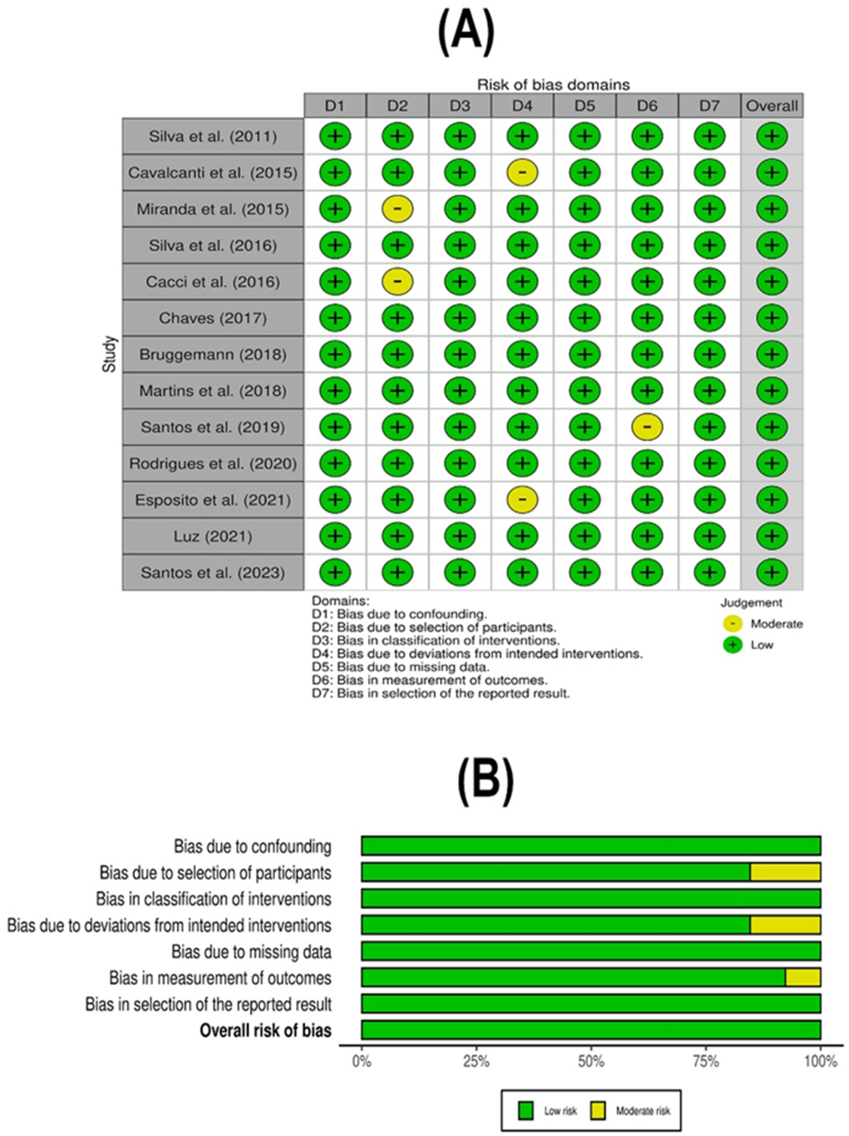
To evaluate the risk of bias in non-randomized controlled studies, the Non-Randomized Studies of Interventions (ROBINS-I) instrument was used [39]. The recovered articles were subjected to both independent risk of bias assessments and methodological evaluation by YCR and HSR. Any disagreement was settled through discussion by a third author (MJAS).

2.5. Statistical Analysis

Using the fixed effects model, the summary event rate and associated 95% confidence intervals (CIs) were calculated. Subgroup analyses were performed based on the geographical regions of Brazil. Heterogeneity between studies was assessed using the I2 statistic and the chi-square test, with a significance threshold of p ≤ 0.05. A meta-regression was performed to determine the predictors of the HRC status, with the year of publication and number of samples included as predictor variables. Potential publication bias was evaluated using funnel plots and Begg’s test. Funnel plots were constructed to visualize the distribution of effect sizes (prevalence rates) against their standard errors. The Comprehensive Meta-Analyses—CMA program, version 2.2 (Biostat, Englewood, NJ, USA) was used on a computer to perform the statistical analyzes of this meta-analysis.

3. Results

3.1. Literature Search

By applying the selection criteria, a total of 425 articles were retrieved from the PUBMED, SciELO, and LILACS databases on the topic for reading. However, 392 articles were letters to the editor, duplicates, short communications, studies unavailable in full version, outside the intended languages, or contained information not relevant to the research question, and were therefore excluded. By applying the eligibility criteria, 33 studies were read in full, independently and in pairs, which resulted in the final sample of the 13 studies included (Figure 1).

3.2. Characterization of Included Studies and Molecular Epidemiology Data

A total of 13 studies were included in the final meta-analysis, all of which were presented in English, published between 2011–2023, and sourced from PubMED and LILACS databases. These studies were conducted by authors affiliated with Brazilian teaching and research institutions: nine (69.22%) from the Southeast region, two (15.42%) from the North, and two (15.42%) from the Northeast regions. Most of the studies (92.31%) were retrospective cross-sectional studies, with one retrospective case-control study (7.69%), including samples from clinical/hospital settings, as well as environmental (non-clinical) samples (water and animal sources) obtained between 1994–2021 (Table 1).
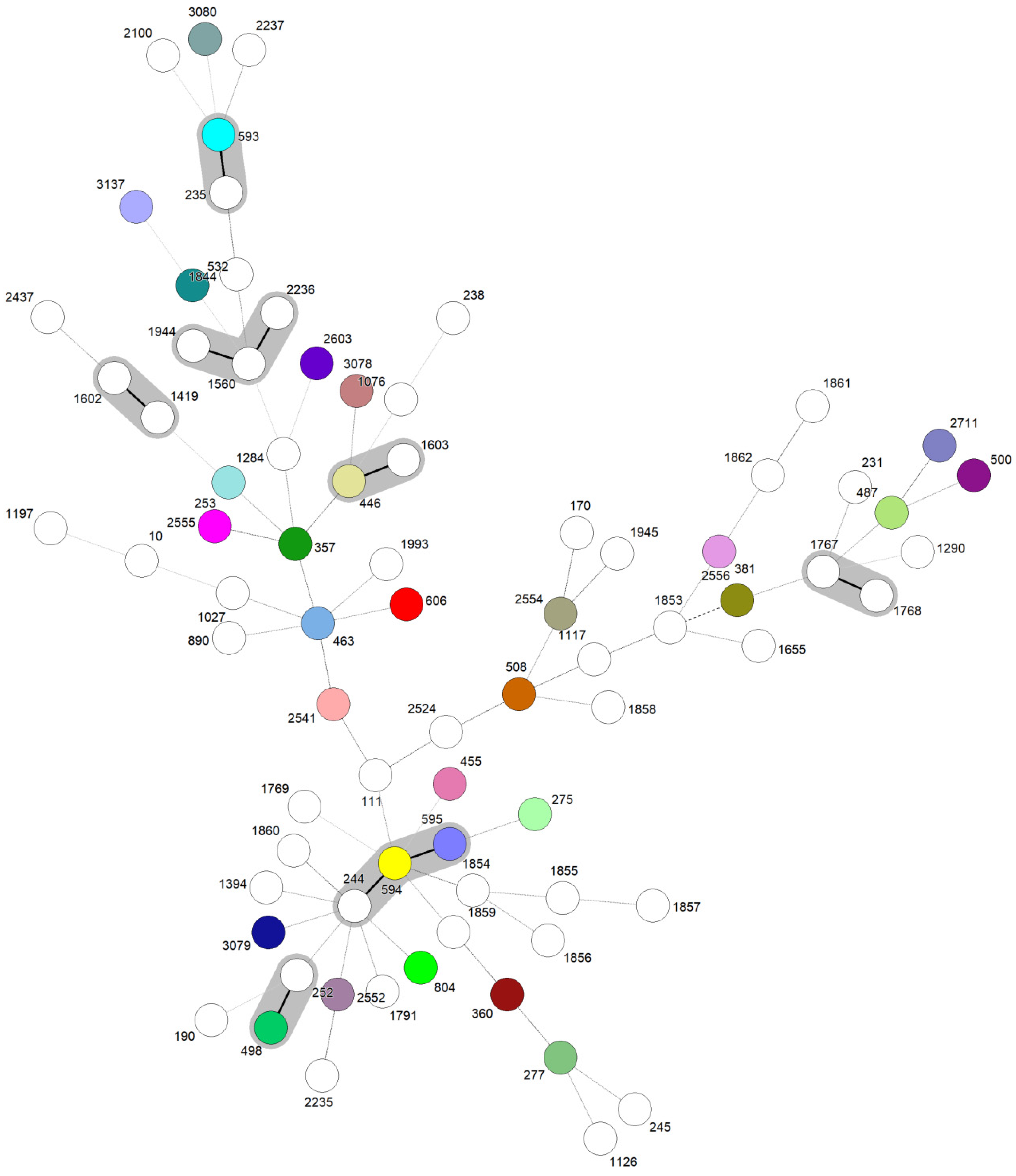
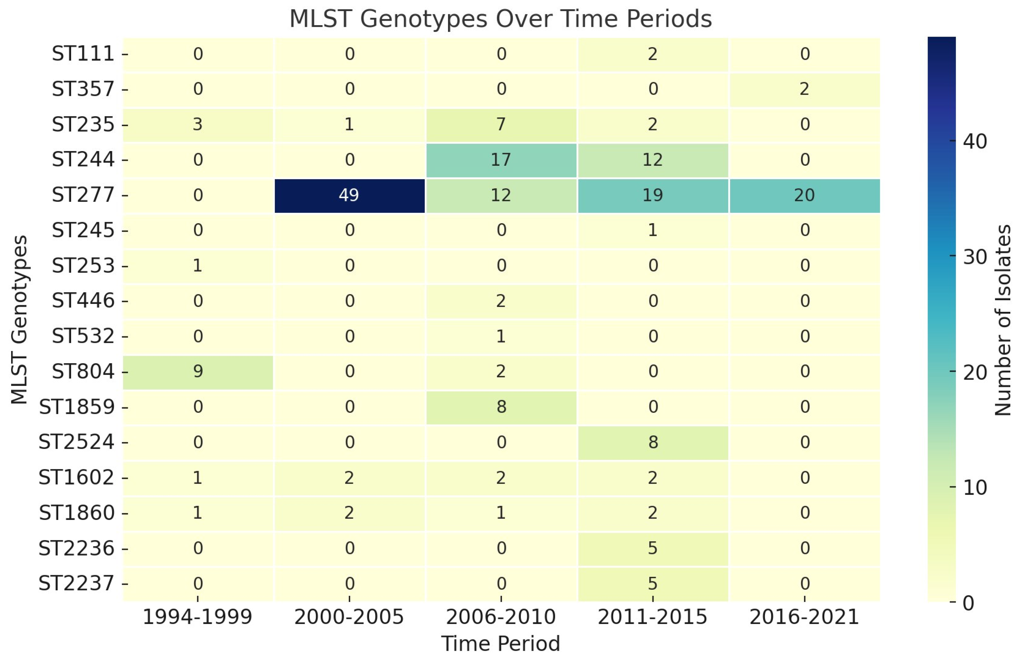
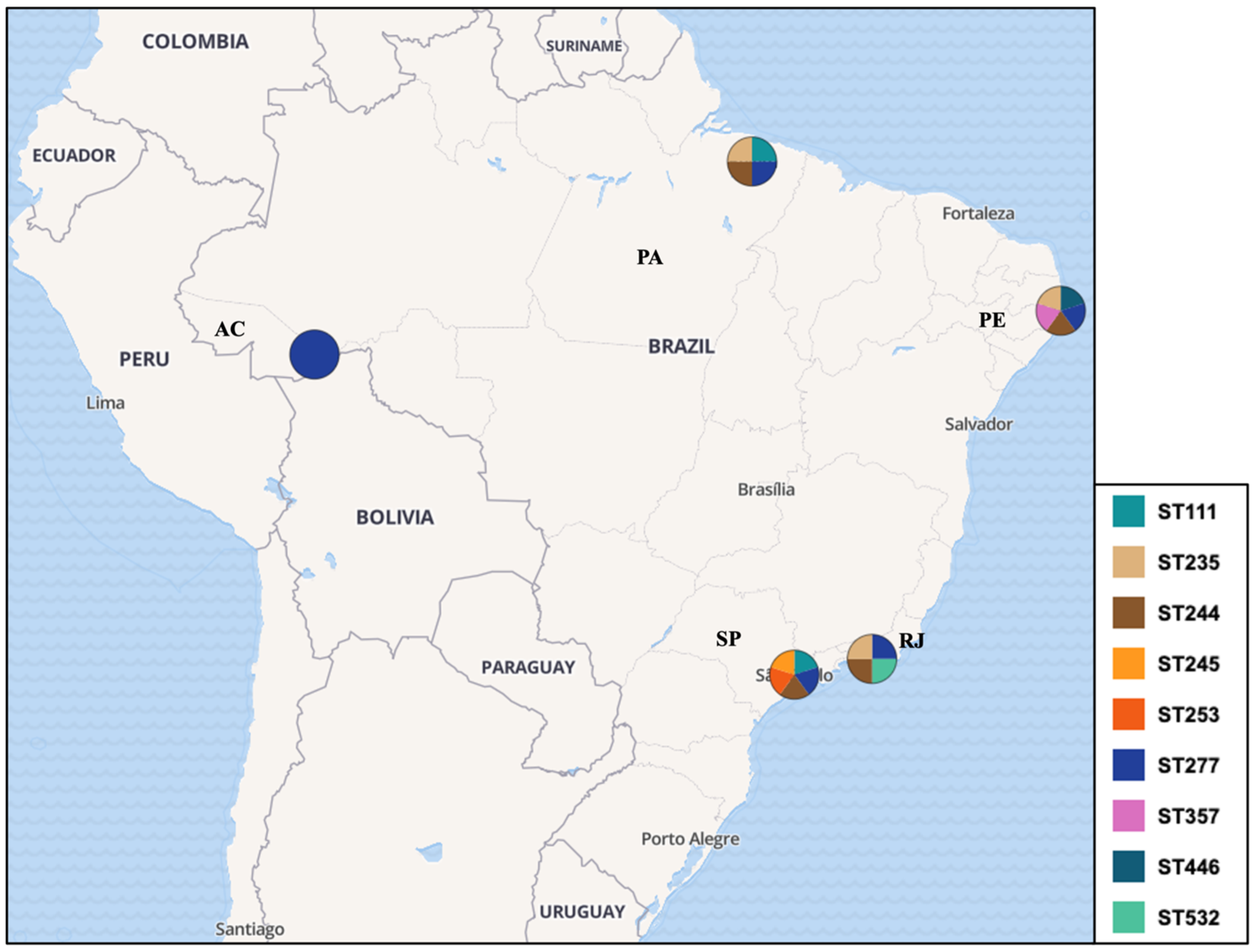
A total of 872 P. aeruginosa isolates were evaluated within studies, with only 298/872 (34.17%) genotyped by the MLST, revealing 78 unique STs (Figure 2). Specifically, 48.90% (146/298) of the isolates were identified as HRCs, including ST277 (100/298—33.55%), ST244 (29/298—9.73%), ST235 (13—4.36%), ST111 (2/298—0.67%), and ST357 (2/298—0.67%). In addition, HRCs were detected over different time periods in Brazil since 1994, as presented in Figure 3. The remaining isolates (152/298—51.10%) were related to non-HRCs, distributed among 73 different STs, of which ST804 (11/298—3.69%), ST1859 (8/298—2.68%), ST2524 (8/298—2.68%), ST1602 (5/298—1.67%), ST1860 (5/298—1.67%), ST2236 (5/298—1.67%), and ST2237 (5/298—1.67%) were the most detected (Table 1, Figure 2 and Figure 3). Suggested emerging HRCs were identified, ST245, ST253, ST446, and ST532, but for analysis were considered as non-HRCs (Figure 2, Figure 3 and Figure 4). Furthermore, clonal complex (CC) relationships were evidenced, including the following: CC244 (ST244, ST595, and ST594), CC235 (ST235 and ST593), CC1560 (ST1560, ST1944, and ST2236), CC498 (ST498 and ST252), CC1602 (ST1602 and ST1419), CC1603 (ST1603 and ST446), and CC1767 (ST1767 and ST1768) (Figure 2).
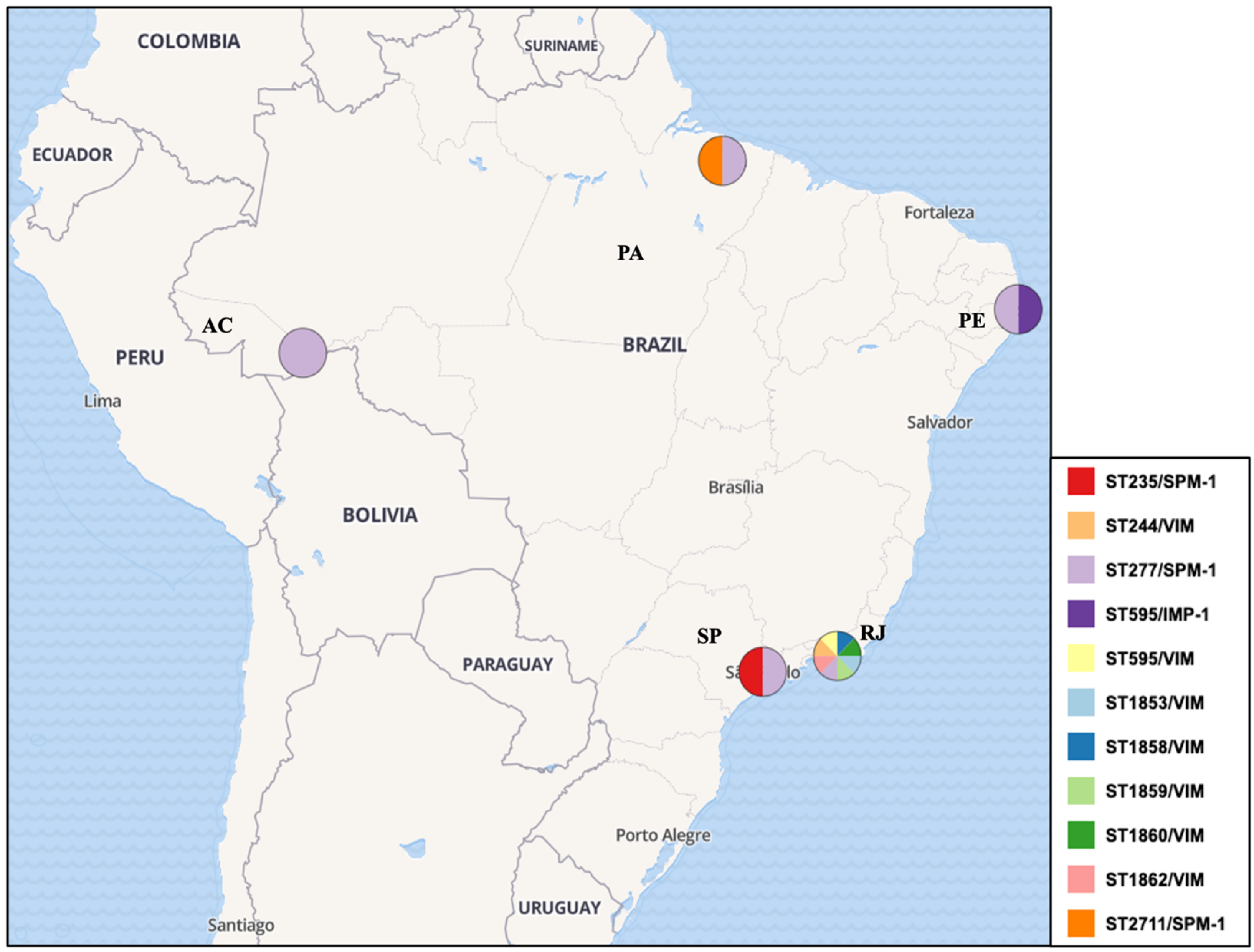
All studies included CR isolates, predominantly associated with MDR and XDR phenotypes. The blaSPM-1 gene emerged as the most frequently detected resistance marker, followed by blaKPC, aminoglycoside-modifying enzymes (AMEs), and efflux pump overexpression. Regarding virulence, only six studies investigated virulence-related genes, with a particular focus on the T3SS, where notably, the cytotoxic (exoU+) and invasive (exoS+/exoU) virulotypes were identified in 41 and 117 isolates, respectively (Supplementary Table S1). Particularly, several HRCs and non-HRCs were detected harboring MβL genes in Brazil (Figure 5).
The methodological quality assessment revealed a high level of quality, and the risk of bias analysis showed a low risk of bias among studies for the included studies (Figure 6).

3.3. Results and Publication Bias of Meta-Analysis of Proportion of P. aeruginosa HRCs and Non-HRCs Strains

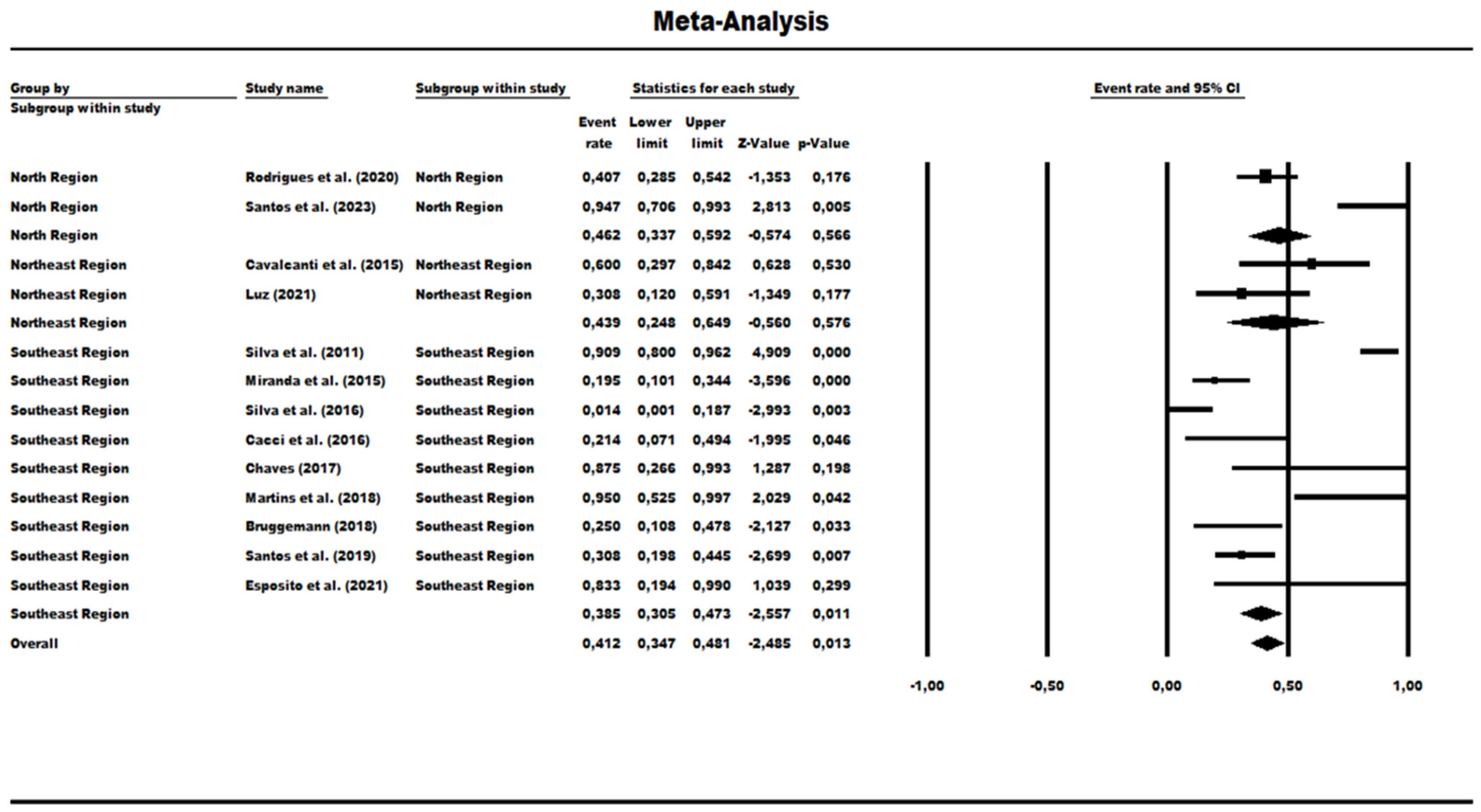
A total of 13 studies were evaluated to determine the prevalence rate for HRCs and non-HRCs among the MLST-genotyped isolates, revealing the combined prevalence rate of 41.20% (0.412) (95% CI = 0.347–0.481) (Figure 7). The statistical analysis showed significant differences among the studies (χ2 = 74.41; df = 12; p < 0.001; I2 = 83.87%), indicating high heterogeneity. A subgroup group analysis was performed based on Brazilian geographic regions: In the North region, the prevalence rate of MLST-genotyped samples was 46.20% (0.462) (95% CI = 0.337–0.592), but analysis also showed significant variability among the studies within this region (χ2 = 9.41; df = 1; p-value = 0.002; I2 = 89.37%). The Northeast region had a prevalence rate of 43.90% (95% CI = 0.248–0.649), and it was observed as a low level of heterogeneity with no statistical significance (χ2 = 1.90; df = 1; p-value = 0.168; I2 = 47.43%). In the Southeast region, the prevalence rate was much lower at 38.50% (95% CI = 0.305 to 0.473), and with a mean difference in heterogeneity among the studies within this region (χ2 = 62.09; df = 8; p-value < 0.001; I2 = 87.11%) (Figure 7).
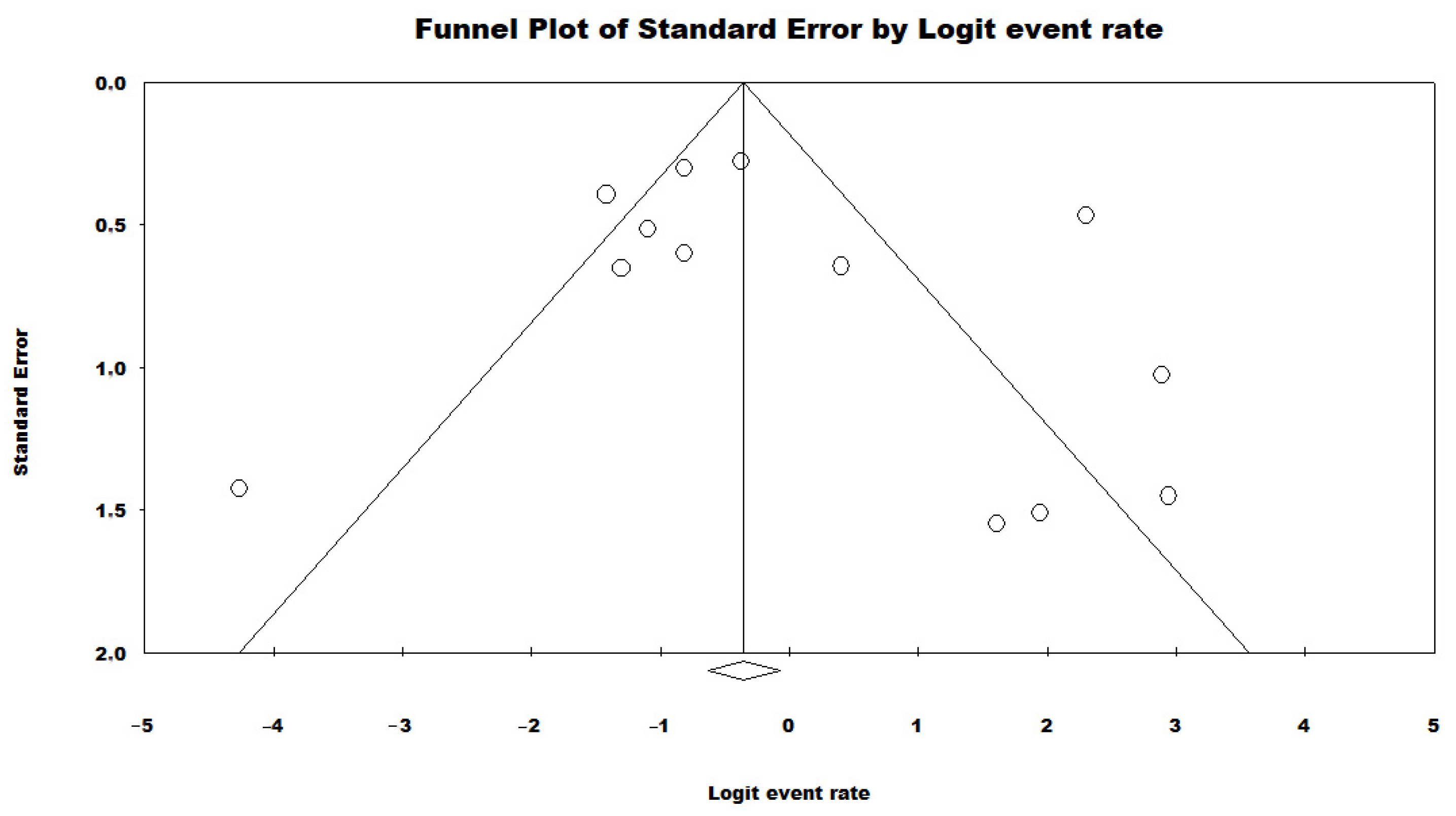
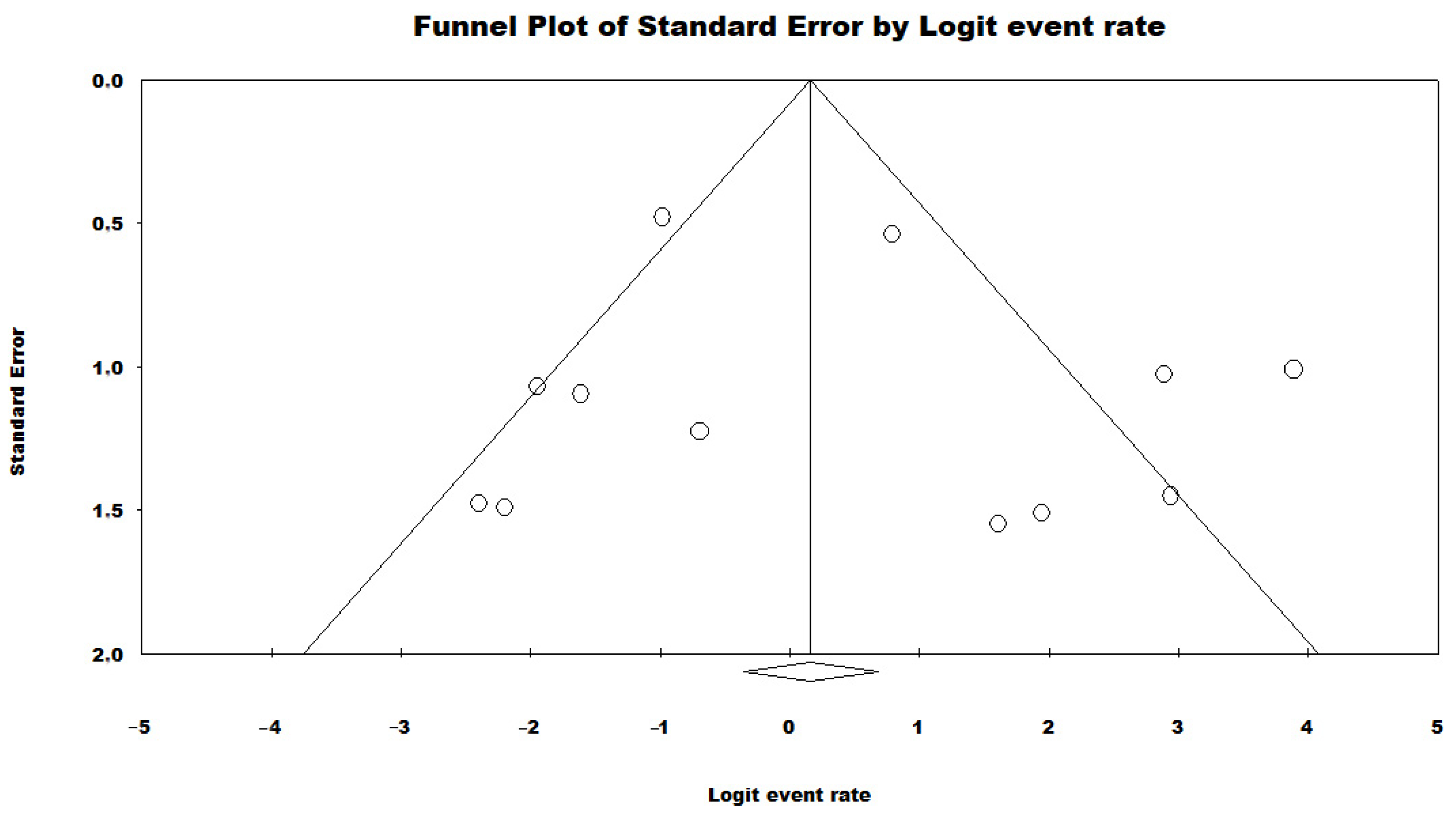
To evaluate the potential publication bias in the meta-analysis of the proportion of HRCs and non-HRCs, a funnel plot was constructed, and Begg’s test was conducted. The funnel plot displays the distribution of effect sizes (prevalence rates) against their standard errors (Figure 8). The results of the Begg’s test revealed a significant correlation with a Kendall’s tau coefficient of 0.23 and a p-value of 0.27, indicating no publication bias.
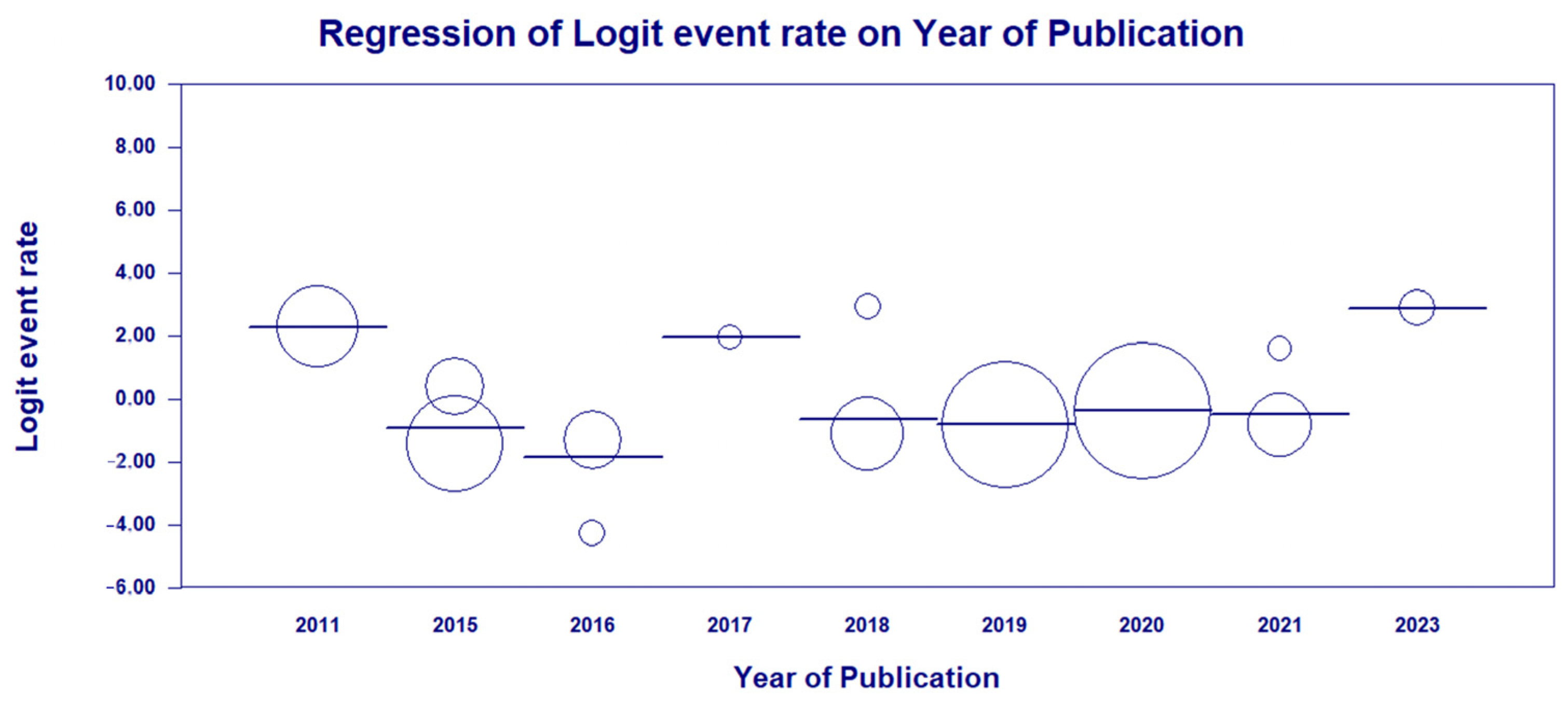
As high heterogeneity was found among the studies investigated in this first analysis, a meta-regression was conducted in terms of the year of publication of each study in fixed model effects, indicating a statistical significance and suggesting the potential bias source (χ2 = 56.01; df = 8; p-value < 0.001) (Figure 9).

3.4. Results and Publication Bias of Meta-Analysis of ST277 within the HRCs Genotypes

A proportion analysis was also performed from the perspective of the MLST-genotyped data from the samples from these studies. In this sense, the prevalence of ST277 in relation to other HRC STs found in these studies was determined. Only one study of the 13 evaluated in the previous analysis was now ineligible as no HRCs were detected (Silva et al. [43]). The analysis revealed a prevalence rate of 54% (95% CI = 0.412–0.663). In relation to geographic subgroups, the North region presented a prevalence of 42.8% (95% CI = 0.242–0.636). The Northeast had a low frequency rate of 14% (95% CI = 0.02–0.47). Regarding the Southeast region, it presented the highest rate of 69% or 0.690 (95% CI = 0.523–0.818) (Figure 10). Regarding the heterogeneity between studies, the Northeast region was the only one that did not present a significant difference (χ2 = 0.101; df = 1; p-value = 0.75; I2 = 0%), while the other analyses for both subgroups and overall revealed high heterogeneity in the data (overall [χ2 = 46.175; df = 11; p-value < 0.001; I2 = 0%]; North [χ2 = 11.665; df = 1; p-value < 0.001; I2 = 91.42%]; Southeast [χ2 = 25.171; df = 7; p-value < 0.001; I2 = 72.19%]) (Figure 10).
A funnel plot was created to assess the publication bias in the meta-analysis of the MLST genotypes (Figure 11). The funnel plot illustrates the distribution of prevalence rates against their standard errors. The Begg’s test was performed to quantitatively assess publication bias, yielding a Kendall’s tau coefficient of 0.03 and a p-value of 0.89. These results indicate no significant correlation, suggesting that there is no evidence of publication bias in this meta-analysis.
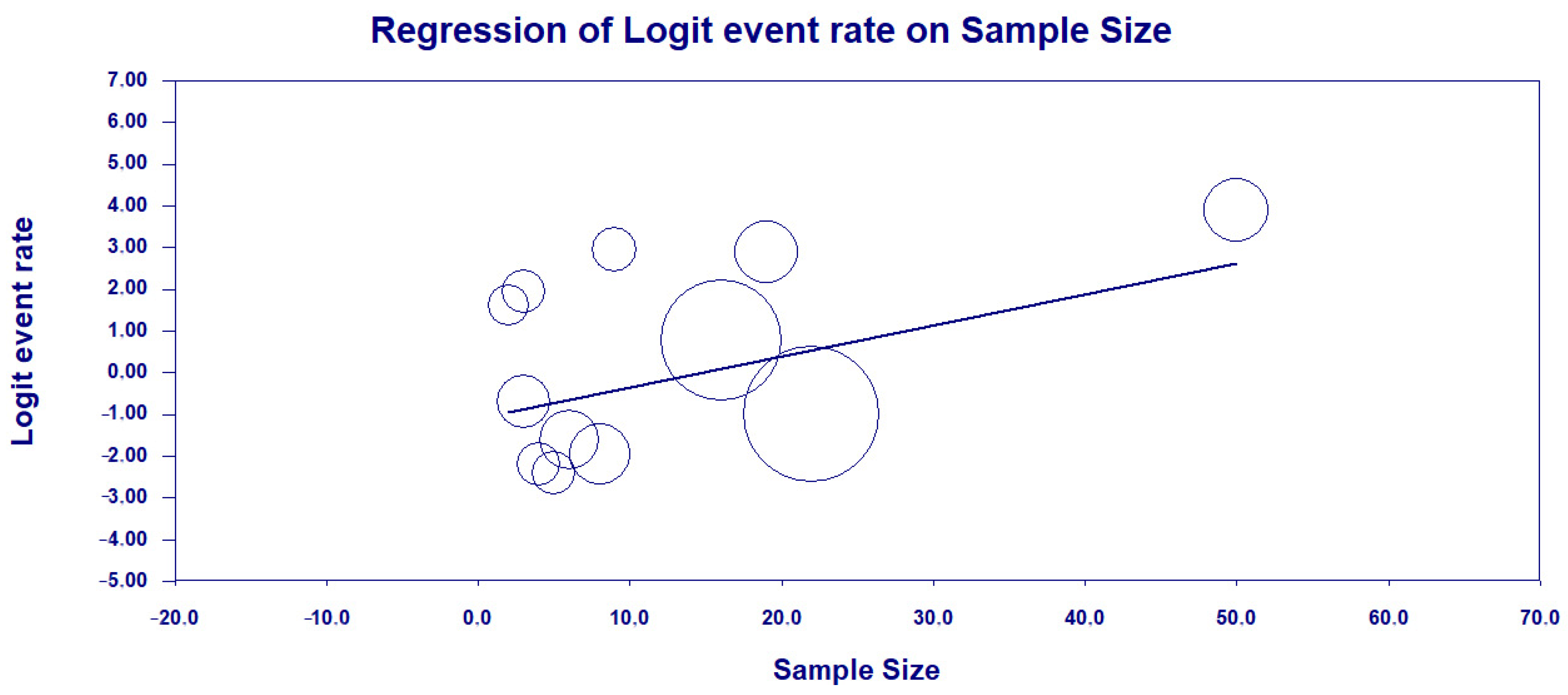
As high heterogeneity was found among the studies investigated, a meta-regression was performed in terms of the sample size of each study in fixed model effects, indicating a statistical significance (a positive association between sample size and HRC classification) and suggesting the potential bias source (coefficients = −1.0983; 95% CI = −2.02–−0.16; p-value = 0.0014) (Figure 12).

4. Discussion

This systematic review and meta-analysis provide a comprehensive overview on the molecular epidemiology of P. aeruginosa in Brazil, emphasizing the prevalence and distribution of HRCs and non-HRCs identified through MLST genotyping. In this study, 78 unique STs were identified among 298 isolates, reflecting considerable genetic diversity (Figure 2). Our findings also reveal a significant prevalence and diversity of HRCs (ST111, ST244, ST235, ST277, and ST357), along with the remarkable dominance of ST277. Non-HRCs made up 51.10% of the MLST-typed isolates, underscoring the presence of several STs with potential epidemiological significance. Moreover, strains were observed in clonal expansion, as six CCs were detected including at least two distinct STs (CC244, CC235, CC498, CC1560, CC1602, CC1603, and CC1767). This genetic prospect is in sense with data from various studies on P. aeruginosa molecular epidemiology, which consistently show extensive clonal diversity and most isolates being represented by unique and distinct genotypes, reflecting a non-clonal epidemic population structure [25,32].
ST277 is a particularly problematic P. aeruginosa HRC recognized for its extensive distribution and critical impact on the AMR scenario in Brazil, heightening public health concerns nationwide. Since its first detection in an oncology patient in São Paulo (Southeast region), this clone has played a major role in the dissemination of the blaSPM-1 gene across various clinical settings in the country [33,40,46]. Indeed, there are a few reports of ST277 outside of Brazil, as it has been documented in Japan and China related to IMP-producing strains, and in the UK, related to an XDR (colistin-susceptible only) isolate [53,54,55]. Even though ST277 is endemic to Brazil, 87 isolates deposited in the PubMLST database belonging to this clone (last accessed 10 August 2024) have been reported in Austria, Australia, Brazil, Central African Republic, China, France, and Ghana [27]. Oppositely, this dominance is well documented in the Brazilian territory, with a prevalence rate of 54% (95% CI = 0.412–0.663) and reports of its presence in both clinical and environmental samples in states from different regions, including São Paulo, Rio de Janeiro, Pernambuco, Acre, and Pará states. Such a high prevalence further highlights the clone’s adaptability and ability to thrive in various environments subjected to intense antibiotic pressure, such as hospitals, where it exacerbates the difficulty of managing infections caused by the MDR/XDR P. aeruginosa.
Our data showed that ST277 was the dominant HRC in the Southeast region, identified in up to 89.0% of isolates in some studies, particularly in clinical settings, as deeply explored by Silva et al. [40], which was one of the first studies providing MLST and other genotyping (PFGE and ribotyping) data for the SPM-1-producing P. aeruginosa in Brazil, and De Oliveira Santos et al. [48], which documented a significant presence of ST277 in clinical isolates from Rio de Janeiro over a 21-year period. Alarmingly, only in the Southeast region, studies evaluating non-clinical isolates (environmental samples) were conducted; Esposito et al. [50] raises concerns about impacted urban river environments potentially serving as reservoirs and sources of re-infection, especially in densely populated urban areas with significant hospital infrastructure; likewise, Martins et al. [46] identified migratory birds in Brazil harboring the ST277 strains in their microbiota, implying that these birds may have played a crucial role in the broad dissemination of this clone within the territory, along with the blaSPM-1 gene. This presence indicates a potential cycle of transmission between hospitals and the surrounding environment, further complicating efforts to control its spread. In the North region, ST277 among clinical isolates was identified in two studies conducted by our research group; Rodrigues et al. [49], which firstly provided data on MLST genotyping in the region, and Dos Santos et al. [52], describing a possible outbreak of ST277 in healthcare institutions in the North region. In the Northeast, ST277 presence was reported by Cavalcanti et al. [41] and related to one CR-PA isolate. This data scenario reflects the clone’s substantial, yet variable, impact across the country.
Curiously, P. aeruginosa ST277 exhibits a unique and highly conserved set of AMR and virulence genes. Key resistance markers commonly identified in ST277 strains include aac(6′)-Ib’, aadA7, aph(3′)-IIb, blaOXA-56, blaPDC-374, blaSPM-1, catB7, cmx, crpP, fosA, and rmtD1, while for virulence, aprA, lasA, lasB, toxA, exo S, exoT, and exoY genes are identified, as well as the O2 serotype group. These genes are largely chromosomally encoded, contributing to the stability and persistence of this clone in nosocomial infections [56,57]. The integration of these AMR genes into genomic islands like PAGI-15 and PAGI-25 further underscores the adaptive success of ST277 within Brazilian healthcare settings, where it poses a significant challenge due to its extensive drug resistance. In addition, the resistome and virulome of ST277 is not only extensive but also largely restricted to Brazilian strains, highlighting a strong geographical and evolutionary link. The study by Dos Santos et al. [52] recently revealed that ST277 containing this resistome and virulome likely played a crucial role in its persistence and spread during the COVID-19 pandemic in the North region This has further complicated treatment options and strongly corroborates previous findings regarding the clone’s impact on AMR in Brazil [50,56,58].
CC/ST244 is a globally distributed P. aeruginosa HRC that is frequently associated with MDR and even non-MDR infections, suggesting a prevalence not fully related to AMR [30,31,32,59]. Its presence has been recorded across various regions in Asia, Europe, North America, and Oceania, mainly due to its involvement in hospital outbreaks and association with multiple AMR genes, including those conferring resistance to aminoglycosides, carbapenems, and macrolides, as well as exoS+ and O2 serogroup virulence features [27,60,61,62]. This HRC has been widely reported in China, where it has been implicated in IMP- and VIM-producing isolates causing CR P. aeruginosa infections among burn and ICU patients [30,62,63,64,65]. Furthermore, in-depth genetic analysis of ST244 strains from this country demonstrated a significant relation to those from North America, suggesting either a shared evolutionary ancestor or frequent strain exchanges between these regions [63]. In Spain, the ST244 isolates were found in multiple regions, indicating interregional dissemination, particularly associated with the production of VIM-2 carbapenemase, and even co-producing both VIM-2 and KPC-2 carbapenemases, further complicating treatment options due to the dual resistance mechanisms [66]. ST244 is not limited to clinical environments; it has also been detected in various other sources across different continents. Likewise in Spain and China, this clone has been detected on children’s fecal samples and on animal-derived foods, indicating the adaptability of ST244, its potential role as a community reservoir, and its ability to colonize diverse ecological niches, which could contribute to its spread within both healthcare and community settings [67,68].
In Brazil, CC/ST244 has been identified as the second most prevalent significant P. aeruginosa HRC, particularly in the Southeast and North regions. According to the data presented in the systematic review, ST244 has been detected in both clinical and environmental samples among 29 (9.73%) of the MLST-typed isolates in seven studies, demonstrating its widespread presence. Data from Miranda et al. [42] show that in the Southeast region of Brazil (RJ state), this clone was mainly detected in non-clinical settings, particularly in hospital wastewater treatment plants, even across various stages of the treatment process. Such isolates exhibited MDR/XDR and non-resistant phenotypes, associated with critical resistance genes such as blaVIM and blaKPC. In the North region, our research group reported ST244 was the most prevalent HRC among the tested strains from 2010–2013, being predominantly found in adult ICUs and harboring the blaCTX-M (five isolates) and exoU+ genes (two isolates) [49]. In the Northeast region, this clone was found only among clinical samples and related to the spread of the blaKPC gene and harbors in the CRISPR–Cas adaptive immune systems [41,51]. Finally, this data draw attention to the significant role of ST244 in the dissemination of blaVIM and blaKPC variants in Brazil, mirroring its impact on a global scale [31,32].
CC/ST235 is recognized as the most remarkable and widely disseminated P. aeruginosa HRC worldwide contributing to the prevalence of XDR/DTR strains in healthcare settings across all continents, mainly in Europe and Asia [38,59,69,70,71,72]. ST235 has remarkable genomic adaptability, which includes a heightened ability to acquire and integrate foreign resistance elements through homologous recombination, that further distinguishes it from other HRCs. The DprA determinant, specifically identified in ST235, enhances this adaptability, contributing to the clone’s resilience and ability to persist in diverse environments [31,32]. This success of ST235 is also closely linked to its association with over 60 β-lactamase variants, worryingly those strains harboring blaKPC, blaGES, blaFIM, blaIMP, blaVIM, and blaNDM variants, which further amplifies the clinical challenges associated with this clone [29,30,73]. Beyond its resistance features, ST235 is characterized by enhanced virulence due to the presence of the ExoU cytotoxin delivered via the T3SS and O11 serotype, key factors driving the severe clinical outcomes associated with ST235 infections [16,17,28]. Interestingly, in non-human and non-clinical samples, P. aeruginosa ST235 has been recently linked to infection among companion animals (dogs and cats) in Japan and Thailand, and in VIM-producing isolates recovered from hospital wastewater in Brazil [71,74,75].
Across various regions of Brazil, ST235 was identified in four studies on clinical samples, accounting for 4.36% (13/298) of the MLST-typed P. aeruginosa isolates. This presence is notable nationwide, even though it is less dominant compared to other HRCs like ST277 and ST244. In the Southeast region, Brüggemann et al. [47] conducted a genomic analysis of 20 P. aeruginosa strains isolated over two decades in São Paulo. Among these, three strains were identified as ST235, which clustered closely with other Brazilian and Argentinean isolates, suggesting regional relatedness. These ST235 strains carried multiple resistance determinants, including aac(6′)-Ib-cr, which confers resistance to both aminoglycosides and fluoroquinolones, and the exoU gene. In Rio de Janeiro, Cacci et al. [44] described this clone as related to both MDR and non-resistant isolates, but did not clearly define AMR markers related to this clone. In the Northeast region, Cavalcanti et al. [41] identified ST235 among clinical isolates collected in Pernambuco and highlighted two P. aeruginosa isolates of this clone co-harboring both blaGES and blaKPC, which might have posed a significant treatment challenge in the region. Rodrigues et al. [49] identified ST235 in five of MLST-typed P. aeruginosa isolates from a referral hospital in Pará, highlighting its presence in the North region of Brazil since 2010. All those ST235 isolates were classified as MDR/XDR; three of these isolates were clonally related and dispersed across different ICUs harboring the blaCTX-M-1 gene, and four of these also presented the cytotoxic virulotype (exoU+). Collectively, these factors indicate the critical importance of P. aeruginosa ST235 as a relevant lineage in the national and global context of AMR, also necessitating ongoing surveillance and strategic infection control measures.
The HRCs ST111 and ST357 highlight their significant role in the adaptability and persistence of P. aeruginosa isolates across healthcare settings. Both clones have been detected in diverse regions, showing a strong presence in Europe, Asia, Africa, and South America (Poland, Singapore, Malaysia, Sweden, Croatia, Austria, Spain, Norway, France Hungary, Senegal, Nigeria, Egypt, Ghana, and Brazil) [76,77,78]. The resistance profiles of ST111 and ST357 pose substantial challenges to clinical treatment. ST111 is strongly associated with the production of VIM-2 in Europe, along with other β-lactamases such as GES and PSE, which confer resistance to a broad spectrum of β-lactam antibiotics [13,30,79,80,81,82]. Similarly, ST357 has usually been linked to IMP-producing isolates, with recent reports of NDM-producing isolates, with both mechanisms contributing to resistance to carbapenems [80,83,84,85,86,87]. As for virulence, ST111 typically lacks the ExoU cytotoxin, exhibiting considerable virulence through the exoS+ invasive virulotype and is often associated with the O12 serotype. In contrast, P. aeruginosa ST357 strains usually produce the potent ExoU cytotoxin and are related to the O11 serotype, similarly to ST235 [28,31,32].
Both ST111 and ST357, even though identified in smaller proportions according to this study (4/298—1.34%), represent significant concerns due to their high resistance and virulence profiles. Recent findings from [49,51] emphasize their restricted spread in the Brazilian territory and the importance of surveillance for such clones, as both are significant contributors to HAIs. ST111 was found only in the State of Pará (North region) harboring the blaCTX-M gene in the adult ICU at a referral hospital, while in the State of Pernambuco (Northeast region), ST357 was suggested as an emerging threat in the clinical environment of ICUs, especially considering its association with the CRISPR/Cas systems, which could confer advantages in resisting bacteriophage attacks and maintaining resistance genes.
According to del Barrio-Tofiño et al. [30], other STs have been suggested as emerging HRCs of P. aeruginosa, which have been detected in at least three countries and are often associated with MDR/XDR phenotypes. These clones frequently harbor intrinsic horizontally acquired β-lactamases, contributing to their enhanced resistance profiles and making them a significant concern for global public health. Among them, ST245, ST253, ST446, and ST532 were detected among Brazilian isolates in the Southeast and Northeast regions. STs 245 and 253 were related to one isolate each, detected co-harboring the blaOXA-50 and blaPAO genes, and were associated to invasive and cytotoxic virulotypes, respectively [47]. A genome note also described the presence of a polymyxin-resistant isolate assigned to ST245 causing VAP in São Paulo [88]. In the city of Recife, ST446 was related to one isolate presenting an overexpression of the MexAB-OprM system, and in Rio de Janeiro, ST532 was detected on a susceptible clinical isolate [41,44]. Globally, ST245 has been reported in China among hospital inpatients and in fecal samples from healthy individuals in Spain [63,89]. The PA14 strain (ST253) has been widely detected in several countries associated with the exoU+ virulotype [90,91]. In the United Kingdon (UK) and Germany, ST466 was associated with a SIM-producing isolate and was colonizing patients at a pulmonary clinic [92,93]. Finally, ST532 was related to an isolate harboring a ciprofloxacin-modifying enzyme (CrpP) in South Africa and among patients with bronchiectasis in Spain [94,95].
Non-HRCs constituted 51.10% of the MLST-typed isolates, with 73 different STs identified. Among these, ST804, ST1602, ST1859, ST1860, ST2524, ST2236, and ST2237 were the most frequently detected. Although these STs are not classified as HRCs, they still play a major role in the genetic diversity and epidemiology of P. aeruginosa as their presence in distinct environments may contribute to the dissemination of resistance and virulence factors. Interestingly, most of the predominant non-HRCs (ST1602, ST1859, ST1860, ST2236, and ST2237) were firstly detected in Rio de Janeiro, Southeast Brazil. De Oliveira Santos [48] is one of the few studies reporting the presence of ST804 among clinical samples, which was predominant among resistant isolates between 1995–1999 and 2006–2010. Outside Brazil, an Australian study described its presence in sputum samples from CF patients [96]. Similarly, ST1602 was first detected in Rio de Janeiro in 2008, but retrospective data demonstrate its presence in clinical and environmental samples as early as 1995 [44,48]. STs 1859 and 1860 were identified in hospital effluent samples in 2010 by Miranda et al. [42]. Additionally, ST2236 and ST2237 were associated with invasive and cytotoxic virulotypes, respectively, among burn patients in a public hospital [43]. Lastly, the only non-HRC detected outside Rio de Janeiro, ST2524, was found to be clonally related to various types of infections among patients in adult, pediatric, and neonatal ICUs at a referral hospital in Pará State [49].
The present systematic review also identified several key mechanisms in P. aeruginosa that significantly contribute to the high levels of resistance phenotypes. The association of specific resistance genes and mutations with both HRCs and non-HRCs highlights the complexity of AMR in this pathogen. Except in the study by De Oliveira Luz et al. [51], all other studies include data on detection of β-lactamases, particularly as CR is further exacerbated by the acquisition of carbapenemases, including metallo-β-lactamases (MβLs) and serin-β-lactamases. For example, the blaSPM-1 gene, which encodes one such enzyme, has been widely reported across Brazilian studies as deeply investigated by our research group [33]. Other notable carbapenemases include blaKPC, blaIMP, and blaVIM, which have been detected at varying frequencies in different studies. For example, blaKPC was found in 34.1% of isolates in the study by Miranda et al. [42] and 5.68% in the study by De Oliveira Santos et al. [48], reflecting the complex resistance profiles these isolates can exhibit. Even though not reported in the included studies, the blaNDM and blaKPC genes have been increasingly reported in Brazil since 2018, as a possible impact of COVID-19 [10,97]. Additionally, the overexpression of chromosomally encoded AmpC cephalosporinases has been identified as a significant resistance mechanism in studies by Cacci et al. [44] and Cavalcanti et al. [41], which showed that up to 30% of the isolates exhibited this overexpression.
Another primary mechanism contributing to resistance in P. aeruginosa is the overexpression of resistance-nodulation-division (RND) family efflux pumps, such as MexAB-OprM and MexXY-OprM. These efflux pumps play a crucial role in both intrinsic and acquired resistance, as noted in several studies [41,44,46,48]. These studies reported the overexpression of these pumps in a significant proportion of the isolates, emphasizing their contribution to the multidrug resistance observed in clinical settings in the Southeast and Northeast regions. Moreover, the loss or decreased expression of the porin protein OprD, which is essential for carbapenem uptake, has been extensively documented. For instance, mutations and deletions in the oprD gene were reported with some studies indicating that these mutations were present in all CR isolates [41,44,48]. Aminoglycoside resistance is also a significant concern and is frequently mediated by AMEs and 16S rRNA methylases. For instance, the aac(6′)-Ib gene, which modifies aminoglycosides and contributes to resistance, was identified in 50% of isolates by Silva et al. [41] and in 100% of isolates by Martins et al. [46]. The rmtD gene, which confers resistance to aminoglycosides by methylating the 16S rRNA, was identified in 100% of the tested isolates [46,52]. The complexity of resistance in P. aeruginosa is further demonstrated by the variety of other resistance mechanisms identified, such as fosA, catB7, cmx, and sul1, which have been detected in multiple studies.
Regarding virulence, the studies conducted on P. aeruginosa isolates from Brazil have highlighted the critical role of the T3SS in bacterial pathogenicity. The T3SS is a key virulence mechanism that enables P. aeruginosa to inject effector proteins directly into host cells, thereby modulating host cell processes and contributing to disease severity [15]. Among the six studies that specifically investigated virulence-related genes, a significant focus was placed on the detection of the exoS, exoU, exoT, and exoY genes, which encode T3SS effector proteins. The exoS gene encodes a bifunctional toxin that disrupts host cell signaling and contributes to cytotoxicity and immune evasion [98,99]. This gene was the most frequently detected T3SS-related gene among the studied isolates. In contrast, the exoU gene encodes a potent cytotoxin associated with more severe clinical outcomes, including rapid tissue damage and increased mortality rates [17,100]. The lower prevalence of exoU compared to exoS suggests variability in virulence strategies among different P. aeruginosa strains. Additionally, the exoT and exoY genes were identified in most of the isolates. ExoT has similar activities to exoS, affecting cytoskeleton integrity and promoting apoptosis, while exoY disrupts endothelial barriers and contributes to immune modulation [15,100,101]. Overall, these findings emphasize the need for the ongoing surveillance of virulence factors in P. aeruginosa to understand their distribution, impact on clinical outcomes, and potential role in guiding treatment decisions.
Despite these insights, significant heterogeneity was observed among the included studies, particularly from the Southeast region. This heterogeneity could be attributed to the diverse study designs, varying sample sizes, and methodological approaches employed across the studies. The meta-analysis results indicate that such variability is statistically significant, as reflected in the high I2 values for both HRC and ST277 analyses. The Southeast region’s data demonstrated a wide range of results, contributing to its significant heterogeneity (I2 = 87.11%). This variability might reflect regional differences in healthcare practices, antibiotic use, and environmental factors influencing the spread and prevalence of HRCs, particularly ST277. The higher prevalence rate of ST277 in this region could be indicative of localized outbreaks or adaptations, stressing the need for targeted public health interventions.
The Northern region exhibited the highest prevalence of HRCs, suggesting a unique epidemiological scenario possibly influenced by local environmental factors and hospital infection control practices. The substantial heterogeneity observed in the North (I2 = 89.37%) might also be a result of differing study designs and sampling periods. The presence of MDR clones is often associated with outbreaks in hospitals, where transmission between patients and environmental contamination can occur. In this sense, this finding in the Northern region of Brazil may be associated with the high rate of infections associated with this bacterium in healthcare institutions in the region [33,102]. Furthermore, environmental conditions, such as the contamination of surface waters due to its aquatic bacterial profile and the variation between rainy and dry periods may influence the presence of P. aeruginosa in waters of North regions [103]. Indeed, BIM-1, a novel acquired MBL belonging to subclass B1, was identified in an MDR Pseudomonas #2 strain recovered from a river in the state of Pará, demonstrating the possible spread of AMR markers in this context [104].
The Northeast region showed more uniformity in study findings, with low heterogeneity and a moderate prevalence of HRCs, likely due to smaller, more consistent study samples. Worryingly, a lack of data from the Central-West and South regions of Brazil was evidenced, which presents a significant limitation in this meta-analysis. The absence of studies from these regions means that the findings may not fully represent the national genetic landscape of P. aeruginosa. Future research should aim to address these gaps by conducting more recent and region-specific studies.
The studies included in this meta-analysis evaluated samples spanning from a wide range of years (1994–2021), and changes in molecular epidemiology and resistance patterns over time may not be fully captured, adding to the complexity of drawing uniform conclusions. The evolving nature of AMR necessitates continuous monitoring and updated research efforts. Overall, the meta-analysis highlights the importance of considering regional and temporal factors when assessing the molecular epidemiology of P. aeruginosa. The absence of publication bias suggests that the observed heterogeneity is due to true variations in study conditions and regional epidemiology rather than selective reporting. Future research should focus on recent data and standardized methodologies to better capture the evolving landscape of P. aeruginosa resistance and spread in Brazil.

5. Conclusions

The significance of HRCs such as ST235, ST244, ST111, ST357, and particularly ST277, along with non-HRCs, in the molecular epidemiology of P. aeruginosa in Brazil cannot be overstated. The persistence and spread of these strains across diverse regions and environments—from clinical settings to broader ecological reservoirs—highlight the critical need for ongoing surveillance and vigilant monitoring. This study’s findings reveal significant disparities in the detection of these strains, emphasizing the importance of comprehensive tracking efforts. Notably, several other non-HRCs were reported for the first time in Brazil, suggesting that these STs may have evolved locally and are potentially endemic to the region. This evidenced their contribution to the unique genetic landscape of P. aeruginosa in the country and reinforces the need for targeted public health strategies to manage their impact.
Finally, this systematic review and meta-analysis also highlight the critical issue of AMR in P. aeruginosa in Brazil and the need for targeted infection control measures, effective antimicrobial stewardship, and regional strategies tailored to local epidemiological patterns. Continued research and investment in novel therapeutic options are essential to combat the growing threat of AMR in this clinically important pathogen. The integration of detailed data from individual studies enhances our understanding of the epidemiological landscape and underscores the importance of addressing regional variations in AMR management.

Supplementary Materials

The following supporting information can be downloaded at https://www.mdpi.com/article/10.3390/antibiotics13100983/s1; Table S1: Summary of Studies Analyzing the Molecular Epidemiology of Pseudomonas aeruginosa Isolates from Brazil.

Author Contributions

Conceptualization, Y.C.R. and M.J.A.S.; Validation, Y.C.R., M.J.A.S., H.S.d.R. and P.A.S.d.S.; Formal Analysis, Y.C.R. and M.J.A.S.; Investigation, Y.C.R., M.J.A.S., H.S.d.R., P.A.S.d.S., D.M.S., M.I.M.G., C.S.d.S., D.J.M. and C.A.M.A.; Resources, C.A.M.A., C.d.O.S., A.J.P.G.Q., L.N.G.C.L., D.M.B. and K.V.B.L.; Writing—Original Draft Preparation, Y.C.R., M.J.A.S. and H.S.d.R.; Writing—Review & Editing, Y.C.R., M.J.A.S., H.S.d.R., P.A.S.d.S., D.M.S., M.I.M.G., C.S.d.S., D.J.M., C.A.M.A., C.d.O.S., A.J.P.G.Q., L.N.G.C.L., D.M.B. and K.V.B.L.; Visualization, Y.C.R. and M.J.A.S.; Supervision, Y.C.R. and K.V.B.L.; Project Administration, Y.C.R., M.J.A.S. and K.V.B.L.; Funding Acquisition, L.N.G.C.L. and K.V.B.L. All authors have read and agreed to the published version of the manuscript.

Funding

The APC was funded by Evandro Chagas Institute (IEC/PA). Yan Corrêa Rodrigues scholarship is funded by PDPG—Pós-Doutorado Estratégico (PDPG-POSDOC), Coordenação de Aperfeiçoamento de Pessoal de Nível Superior (CAPES/Edital 16/2022—File Nº 88887.759036/2022-00). Marcos Jessé Abrahão Silva scholarship is funded by Coordenação de Aperfeiçoamento de Pessoal de Nível Superior (CAPES/File Nº 88887.940087/2024-00). Pabllo Antonny Silva dos Santos scholarship is funded by Coordenação de Aperfeiçoamento de Pessoal de Nível Superior (CAPES/File Nº 88887.940090/2024-00). Maria Isabel Montoril Gouveia scholarship is funded by Coordenação de Aperfeiçoamento de Pessoal de Nível Superior (CAPES/File Nº 88887.9292272/2023-00). Carolynne Silva dos Santos scholarship is funded by National Council for Scientific and Technological Development (CNPq/Edital 69/2022—File Nº 130122/2024-6). Davi Josué Marcon is funded by Coordenação de Aperfeiçoamento de Pessoal de Nível Superior (CAPES/File Nº 88887.825814/2023-00).

Data Availability Statement

The original contributions presented in the study are included in the article/Supplementary Material, further inquiries can be directed to the corresponding authors.

Conflicts of Interest

The authors declare no conflicts of interest.

References

  1. Kadri, S.S.; Adjemian, J.; Lai, Y.L.; Spaulding, A.B.; Ricotta, E.; Prevots, D.R.; Palmore, T.N.; Rhee, C.; Klompas, M.; Dekker, J.P. Difficult-to-Treat Resistance in Gram-Negative Bacteremia at 173 US Hospitals: Retrospective Cohort Analysis of Prevalence, Predictors, and Outcome of Resistance to All First-Line Agents. Clin. Infect. Dis. 2018, 67, 1803–1814. [Google Scholar] [CrossRef] [PubMed]
  2. Tacconelli, E.; Carrara, E.; Savoldi, A.; Harbarth, S.; Mendelson, M.; Monnet, D.L.; Pulcini, C.; Kahlmeter, G.; Kluytmans, J.; Carmeli, Y. Discovery, Research, and Development of New Antibiotics: The WHO Priority List of Antibiotic-Resistant Bacteria and Tuberculosis. Lancet Infect. Dis. 2018, 18, 318–327. [Google Scholar] [CrossRef] [PubMed]
  3. Bertagnolio, S.; Dobreva, Z.; Centner, C.M.; Olaru, I.D.; Donà, D.; Burzo, S.; Huttner, B.D.; Chaillon, A.; Gebreselassie, N.; Wi, T. WHO Global Research Priorities for Antimicrobial Resistance in Human Health. Lancet Microbe 2024, 100902. [Google Scholar] [CrossRef]
  4. Coque, T.; Graham, D.W.; Pruden, A.; So, A.; Topp, E. Bracing for Superbugs: Strengthening Environmental Action in the One Health Response to Antimicrobial Resistance; United Nations Environment Programme: Geneva, Switzerland, 2023. [Google Scholar]
  5. Mendelson, M.; Lewnard, J.A.; Sharland, M.; Cook, A.; Pouwels, K.B.; Alimi, Y.; Mpundu, M.; Wesangula, E.; Weese, J.S.; Røttingen, J.-A. Ensuring Progress on Sustainable Access to Effective Antibiotics at the 2024 UN General Assembly: A Target-Based Approach. Lancet 2024, 403, 2551–2564. [Google Scholar] [CrossRef] [PubMed]
  6. Pillonetto, M.; Jordão, R.T.d.S.; Andraus, G.S.; Bergamo, R.; Rocha, F.B.; Onishi, M.C.; Almeida, B.M.M.d.; Nogueira, K.d.S.; Dal Lin, A.; Dias, V.M.d.C.H. The Experience of Implementing a National Antimicrobial Resistance Surveillance System in Brazil. Front. Public Health 2021, 8, 575536. [Google Scholar] [CrossRef]
  7. Pan American Health Organization Magnitude and Trends of Antimicrobial Resistance in Latin America. ReLAVRA 2014, 2015, 2016. Summary Report—PAHO/WHO | Pan American Health Organization; PAHO Antimicrobial Resistance Special Program: Washington, DC, USA, 2020. [Google Scholar]
  8. Wicky, P.-H.; Dupuis, C.; Cerf, C.; Siami, S.; Cohen, Y.; Laurent, V.; Mourvillier, B.; Reignier, J.; Goldgran-Toledano, D.; Schwebel, C. Ventilator-Associated Pneumonia in COVID-19 Patients Admitted in Intensive Care Units: Relapse, Therapeutic Failure and Attributable Mortality—A Multicentric Observational Study from the Outcomerea Network. J. Clin. Med. 2023, 12, 1298. [Google Scholar] [CrossRef]
  9. Velasquez-Garcia, L.; Mejia-Sanjuanelo, A.; Viasus, D.; Carratala, J. Causative Agents of Ventilator-Associated Pneumonia and Resistance to Antibiotics in COVID-19 Patients: A Systematic Review. Biomedicines 2022, 10, 1226. [Google Scholar] [CrossRef] [PubMed]
  10. ANVISA Boletim Segurança do Paciente e Qualidade em Serviços de Saúde no 30—Avaliação Nacional dos Indicadores de IRAS e RM—2022; Agência Nacional de Vigilância Sanitária: Brasília, Brazil, 2022; p. 11.
  11. Behzadi, P.; Baráth, Z.; Gajdács, M. It’s Not Easy Being Green: A Narrative Review on the Microbiology, Virulence and Therapeutic Prospects of Multidrug-Resistant Pseudomonas aeruginosa. Antibiotics 2021, 10, 42. [Google Scholar] [CrossRef]
  12. García-Betancur, J.C.; Appel, T.M.; Esparza, G.; Gales, A.C.; Levy-Hara, G.; Cornistein, W.; Vega, S.; Nuñez, D.; Cuellar, L.; Bavestrello, L. Update on the Epidemiology of Carbapenemases in Latin America and the Caribbean. Expert Rev. Anti Infect. Ther. 2021, 19, 197–213. [Google Scholar] [CrossRef]
  13. Halat, D.H.; Moubareck, C.A. The Intriguing Carbapenemases of Pseudomonas aeruginosa: Current Status, Genetic Profile, and Global Epidemiology. Yale J. Biol. Med. 2022, 95, 507–515. [Google Scholar]
  14. Sawa, T.; Shimizu, M.; Moriyama, K.; Wiener-Kronish, J.P. Association between Pseudomonas aeruginosa Type III Secretion, Antibiotic Resistance, and Clinical Outcome: A Review. Crit. Care 2014, 18, 668. [Google Scholar] [CrossRef] [PubMed]
  15. Qin, S.; Xiao, W.; Zhou, C.; Pu, Q.; Deng, X.; Lan, L.; Liang, H.; Song, X.; Wu, M. Pseudomonas aeruginosa: Pathogenesis, Virulence Factors, Antibiotic Resistance, Interaction with Host, Technology Advances and Emerging Therapeutics. Signal Transduct. Target. Ther. 2022, 7, 199. [Google Scholar] [CrossRef] [PubMed]
  16. Cho, H.H.; Kwon, K.C.; Kim, S.; Koo, S.H. Correlation between Virulence Genotype and Fluoroquinolone Resistance in Carbapenem-Resistant Pseudomonas aeruginosa. Ann. Lab. Med. 2014, 34, 286. [Google Scholar] [CrossRef]
  17. Peña, C.; Cabot, G.; Gómez-Zorrilla, S.; Zamorano, L.; Ocampo-Sosa, A.; Murillas, J.; Almirante, B.; Pomar, V.; Aguilar, M.; Granados, A. Influence of Virulence Genotype and Resistance Profile in the Mortality of Pseudomonas aeruginosa Bloodstream Infections. Clin. Infect. Dis. 2015, 60, 539–548. [Google Scholar] [CrossRef]
  18. Sánchez-Diener, I.; Zamorano, L.; López-Causapé, C.; Cabot, G.; Mulet, X.; Peña, C.; Del Campo, R.; Cantón, R.; Doménech-Sánchez, A.; Martínez-Martínez, L. Interplay among Resistance Profiles, High-Risk Clones, and Virulence in the Caenorhabditis Elegans Pseudomonas aeruginosa Infection Model. Antimicrob. Agents Chemother. 2017, 61, 5806–5816. [Google Scholar] [CrossRef]
  19. Recio, R.; Mancheño, M.; Viedma, E.; Villa, J.; Orellana, M.Á.; Lora-Tamayo, J.; Chaves, F. Predictors of Mortality in Bloodstream Infections Caused by Pseudomonas aeruginosa and Impact of Antimicrobial Resistance and Bacterial Virulence. Antimicrob. Agents Chemother. 2020, 64, e01759-19. [Google Scholar] [CrossRef] [PubMed]
  20. Horna, G.; Amaro, C.; Palacios, A.; Guerra, H.; Ruiz, J. High Frequency of the exoU+/exoS+ Genotype Associated with Multidrug-Resistant “High-Risk Clones” of Pseudomonas aeruginosa Clinical Isolates from Peruvian Hospitals. Sci. Rep. 2019, 9, 10874. [Google Scholar] [CrossRef]
  21. Sarges, E.d.S.N.F.; Rodrigues, Y.C.; Furlaneto, I.P.; de Melo, M.V.H.; Brabo, G.L.d.C.; Lopes, K.C.M.; Quaresma, A.J.P.G.; Lima, L.N.G.C.; Lima, K.V.B. Pseudomonas aeruginosa Type III Secretion System Virulotypes and Their Association with Clinical Features of Cystic Fibrosis Patients. Infect. Drug Resist. 2020, 13, 3771–3781. [Google Scholar] [CrossRef]
  22. Cepas, V.; Soto, S.M. Relationship between Virulence and Resistance among Gram-Negative Bacteria. Antibiotics 2020, 9, 719. [Google Scholar] [CrossRef]
  23. Achtman, M. Evolution, Population Structure, and Phylogeography of Genetically Monomorphic Bacterial Pathogens. Annu. Rev. Microbiol. 2008, 62, 53–70. [Google Scholar] [CrossRef]
  24. Woodford, N.; Turton, J.F.; Livermore, D.M. Multiresistant Gram-Negative Bacteria: The Role of High-Risk Clones in the Dissemination of Antibiotic Resistance. FEMS Microbiol. Rev. 2011, 35, 736–755. [Google Scholar] [CrossRef]
  25. Sawa, T.; Momiyama, K.; Mihara, T.; Kainuma, A.; Kinoshita, M.; Moriyama, K. Molecular Epidemiology of Clinically High-risk Pseudomonas aeruginosa Strains: Practical Overview. Microbiol. Immunol. 2020, 64, 331–344. [Google Scholar] [CrossRef] [PubMed]
  26. Curran, B.; Jonas, D.; Grundmann, H.; Pitt, T.; Dowson, C.G. Development of a Multilocus Sequence Typing Scheme for the Opportunistic Pathogen Pseudomonas aeruginosa. J. Clin. Microbiol. 2004, 42, 5644–5649. [Google Scholar] [CrossRef] [PubMed]
  27. Jolley, K.A.; Bray, J.E.; Maiden, M.C. Open-Access Bacterial Population Genomics: BIGSdb Software, the PubMLST. Org Website and Their Applications. Wellcome Open Res. 2018, 3, 124. [Google Scholar] [CrossRef] [PubMed]
  28. Mulet, X.; Cabot, G.; Ocampo-Sosa, A.A.; Domínguez, M.A.; Zamorano, L.; Juan, C.; Tubau, F.; Rodríguez, C.; Moyà, B.; Peña, C. Biological Markers of Pseudomonas aeruginosa Epidemic High-Risk Clones. Antimicrob. Agents Chemother. 2013, 57, 5527–5535. [Google Scholar] [CrossRef] [PubMed]
  29. Kocsis, B.; Gulyás, D.; Szabó, D. Diversity and Distribution of Resistance Markers in Pseudomonas aeruginosa International High-Risk Clones. Microorganisms 2021, 9, 359. [Google Scholar] [CrossRef]
  30. del Barrio-Tofiño, E.; López-Causapé, C.; Oliver, A. Pseudomonas aeruginosa Epidemic High-Risk Clones and Their Association with Horizontally-Acquired β-Lactamases: 2020 Update. Int. J. Antimicrob. Agents 2020, 56, 106196. [Google Scholar] [CrossRef]
  31. Oliver, A.; Rojo-Molinero, E.; Arca-Suarez, J.; Beşli, Y.; Bogaerts, P.; Cantón, R.; Cimen, C.; Croughs, P.D.; Denis, O.; Giske, C.G. Pseudomonas aeruginosa Antimicrobial Susceptibility Profiles, Resistance Mechanisms and International Clonal Lineages: Update from ESGARS-ESCMID/ISARPAE Group. Clin. Microbiol. Infect. 2024, 30, 469–480. [Google Scholar] [CrossRef]
  32. Oliver, A.; Mulet, X.; López-Causapé, C.; Juan, C. The Increasing Threat of Pseudomonas aeruginosa High-Risk Clones. Drug Resist. Updat. 2015, 21, 41–59. [Google Scholar] [CrossRef]
  33. Dos Santos, P.A.S.; Silva, M.J.A.; Gouveia, M.I.M.; Lima, L.N.G.C.; Quaresma, A.J.P.G.; De Lima, P.D.L.; Brasiliense, D.M.; Lima, K.V.B.; Rodrigues, Y.C. The Prevalence of Metallo-Beta-Lactamese-(MβL)-Producing Pseudomonas aeruginosa Isolates in Brazil: A Systematic Review and Meta-Analysis. Microorganisms 2023, 11, 2366. [Google Scholar] [CrossRef]
  34. Page, M.J.; McKenzie, J.E.; Bossuyt, P.M.; Boutron, I.; Hoffmann, T.C.; Mulrow, C.D.; Shamseer, L.; Tetzlaff, J.M.; Akl, E.A.; Brennan, S.E.; et al. The PRISMA 2020 Statement: An Updated Guideline for Reporting Systematic Reviews. BMJ 2021, 372, n71. [Google Scholar] [CrossRef] [PubMed]
  35. Francisco, A.P.; Vaz, C.; Monteiro, P.T.; Melo-Cristino, J.; Ramirez, M.; Carriço, J.A. PHYLOViZ: Phylogenetic Inference and Data Visualization for Sequence Based Typing Methods. BMC Bioinform. 2012, 13, 87. [Google Scholar] [CrossRef] [PubMed]
  36. Peters, M.D.J.; Godfrey, C.M.; Khalil, H.; McInerney, P.; Parker, D.; Soares, C.B. Guidance for Conducting Systematic Scoping Reviews. Int. J. Evid. Based Healthc. 2015, 13, 141–146. [Google Scholar] [CrossRef]
  37. Munn, Z.; Aromataris, E.; Tufanaru, C.; Stern, C.; Porritt, K.; Farrow, J.; Lockwood, C.; Stephenson, M.; Moola, S.; Lizarondo, L.; et al. The Development of Software to Support Multiple Systematic Review Types: The Joanna Briggs Institute System for the Unified Management, Assessment and Review of Information (JBI SUMARI). Int. J. Evid. Based Healthc. 2019, 17, 36–43. [Google Scholar] [CrossRef] [PubMed]
  38. Silva, M.J.A.; Silva, C.S.; Da Silva Vieira, M.C.; Dos Santos, P.A.S.; Frota, C.C.; Lima, K.V.B.; Lima, L.N.G.C. The Relationship between TLR3 Rs3775291 Polymorphism and Infectious Diseases: A Meta-Analysis of Case-Control Studies. Genes 2023, 14, 1311. [Google Scholar] [CrossRef]
  39. Sterne, J.A.; Hernán, M.A.; Reeves, B.C.; Savović, J.; Berkman, N.D.; Viswanathan, M.; Henry, D.; Altman, D.G.; Ansari, M.T.; Boutron, I.; et al. ROBINS-I: A Tool for Assessing Risk of Bias in Non-Randomised Studies of Interventions. BMJ 2016, 355, i4919. [Google Scholar] [CrossRef]
  40. Silva, F.M.; Carmo, M.S.; Silbert, S.; Gales, A.C. SPM-1-Producing Pseudomonas aeruginosa: Analysis of the Ancestor Relationship Using Multilocus Sequence Typing, Pulsed-Field Gel Electrophoresis, and Automated Ribotyping. Microb. Drug Resist. 2011, 17, 215–220. [Google Scholar] [CrossRef]
  41. Cavalcanti, F.L.D.S.; Mirones, C.R.; Paucar, E.R.; Montes, L.Á.; Leal-Balbino, T.C.; Morais, M.M.C.D.; Martínez-Martínez, L.; Ocampo-Sosa, A.A. Mutational and Acquired Carbapenem Resistance Mechanisms in Multidrug Resistant Pseudomonas aeruginosa Clinical Isolates from Recife, Brazil. Mem. Inst. Oswaldo Cruz 2015, 110, 1003–1009. [Google Scholar] [CrossRef]
  42. Miranda, C.C.; De Filippis, I.; Pinto, L.H.; Coelho-Souza, T.; Bianco, K.; Cacci, L.C.; Picão, R.C.; Clementino, M.M. Genotypic Characteristics of Multidrug-Resistant Pseudomonas aeruginosa from Hospital Wastewater Treatment Plant in Rio de Janeiro, Brazil. J. Appl. Microbiol. 2015, 118, 1276–1286. [Google Scholar] [CrossRef]
  43. De Almeida Silva, K.D.C.F.; Calomino, M.A.; Deutsch, G.; De Castilho, S.R.; De Paula, G.R.; Esper, L.M.R.; Teixeira, L.A. Molecular Characterization of Multidrug-Resistant (MDR) Pseudomonas aeruginosa Isolated in a Burn Center. Burns 2017, 43, 137–143. [Google Scholar] [CrossRef]
  44. Cacci, L.C.; Chuster, S.G.; Martins, N.; Carmo, P.R.D.; Girão, V.B.D.C.; Nouér, S.A.; Freitas, W.V.D.; Matos, J.A.D.; Magalhães, A.C.D.G.; Ferreira, A.L.P.; et al. Mechanisms of Carbapenem Resistance in Endemic Pseudomonas aeruginosa Isolates after an SPM-1 Metallo-β-Lactamase Producing Strain Subsided in an Intensive Care Unit of a Teaching Hospital in Brazil. Mem. Inst. Oswaldo Cruz 2016, 111, 551–558. [Google Scholar] [CrossRef] [PubMed]
  45. Chaves, L.; Tomich, L.M.; Salomão, M.; Leite, G.C.; Ramos, J.; Martins, R.R.; Rizek, C.; Neves, P.; Batista, M.V.; Amigo, U.; et al. High Mortality of Bloodstream Infection Outbreak Caused by Carbapenem-Resistant P. aeruginosa Producing SPM-1 in a Bone Marrow Transplant Unit. J. Med. Microbiol. 2017, 66, 1722–1729. [Google Scholar] [CrossRef] [PubMed]
  46. Martins, W.M.B.S.; Narciso, A.C.; Cayô, R.; Santos, S.V.; Fehlberg, L.C.C.; Ramos, P.L.; Da Cruz, J.B.; Gales, A.C. SPM-1-Producing Pseudomonas aeruginosa ST277 Clone Recovered from Microbiota of Migratory Birds. Diagn. Microbiol. Infect. Dis. 2018, 90, 221–227. [Google Scholar] [CrossRef]
  47. Brüggemann, H.; Migliorini, L.B.; Sales, R.O.D.; Koga, P.C.M.; Souza, A.V.D.; Jensen, A.; Poehlein, A.; Brzuszkiewicz, E.; Doi, A.M.; Pasternak, J.; et al. Comparative Genomics of Nonoutbreak Pseudomonas aeruginosa Strains Underlines Genome Plasticity and Geographic Relatedness of the Global Clone ST235. Genome Biol. Evol. 2018, 10, 1852–1857. [Google Scholar] [CrossRef]
  48. De Oliveira Santos, I.C.; Pereira De Andrade, N.F.; Da Conceição Neto, O.C.; Da Costa, B.S.; De Andrade Marques, E.; Rocha-de-Souza, C.M.; Asensi, M.D.; D’Alincourt Carvalho-Assef, A.P. Epidemiology and Antibiotic Resistance Trends in Clinical Isolates of Pseudomonas aeruginosa from Rio de Janeiro—Brazil: Importance of Mutational Mechanisms over the Years (1995–2015). Infect. Genet. Evol. 2019, 73, 411–415. [Google Scholar] [CrossRef]
  49. Rodrigues, Y.C.; Furlaneto, I.P.; Maciel, A.H.P.; Quaresma, A.J.P.G.; De Matos, E.C.O.; Conceição, M.L.; Vieira, M.C.D.S.; Brabo, G.L.D.C.; Sarges, E.D.S.N.F.; Lima, L.N.G.C.; et al. High Prevalence of Atypical Virulotype and Genetically Diverse Background among Pseudomonas aeruginosa Isolates from a Referral Hospital in the Brazilian Amazon. PLoS ONE 2020, 15, e0238741. [Google Scholar] [CrossRef]
  50. Esposito, F.; Cardoso, B.; Fontana, H.; Fuga, B.; Cardenas-Arias, A.; Moura, Q.; Fuentes-Castillo, D.; Lincopan, N. Genomic Analysis of Carbapenem-Resistant Pseudomonas aeruginosa Isolated From Urban Rivers Confirms Spread of Clone Sequence Type 277 Carrying Broad Resistome and Virulome Beyond the Hospital. Front. Microbiol. 2021, 12, 701921. [Google Scholar] [CrossRef]
  51. De Oliveira Luz, A.C.; Da Silva Junior, W.J.; Do Nascimento Junior, J.B.; Da Silva, J.M.A.; De Queiroz Balbino, V.; Leal-Balbino, T.C. Genetic Characteristics and Phylogenetic Analysis of Brazilian Clinical Strains of Pseudomonas aeruginosa Harboring CRISPR/Cas Systems. Curr. Genet. 2021, 67, 663–672. [Google Scholar] [CrossRef] [PubMed]
  52. Dos Santos, P.A.S.; Rodrigues, Y.C.; Marcon, D.J.; Lobato, A.R.F.; Cazuza, T.B.; Gouveia, M.I.M.; Silva, M.J.A.; Souza, A.B.; Lima, L.N.G.C.; Quaresma, A.J.P.G.; et al. Endemic High-Risk Clone ST277 Is Related to the Spread of SPM-1-Producing Pseudomonas aeruginosa during the COVID-19 Pandemic Period in Northern Brazil. Microorganisms 2023, 11, 2069. [Google Scholar] [CrossRef]
  53. Mano, Y.; Saga, T.; Ishii, Y.; Yoshizumi, A.; Bonomo, R.A.; Yamaguchi, K.; Tateda, K. Molecular Analysis of the Integrons of Metallo-β-Lactamase-Producing Pseudomonas aeruginosa Isolates Collected by Nationwide Surveillance Programs across Japan. BMC Microbiol. 2015, 15, 41. [Google Scholar] [CrossRef]
  54. Chen, Y.; Sun, M.; Wang, M.; Lu, Y.; Yan, Z. Dissemination of IMP-6-Producing Pseudomonas aeruginosa ST244 in Multiple Cities in China. Eur. J. Clin. Microbiol. Infect. Dis. 2014, 33, 1181–1187. [Google Scholar] [CrossRef] [PubMed]
  55. Hopkins, K.L.; Meunier, D.; Findlay, J.; Mustafa, N.; Parsons, H.; Pike, R.; Wright, L.; Woodford, N. SPM-1 Metallo-β-Lactamase-Producing Pseudomonas aeruginosa ST277 in the UK. J. Med. Microbiol. 2016, 65, 696–697. [Google Scholar] [CrossRef] [PubMed]
  56. Galetti, R.; Andrade, L.N.; Varani, A.M.; Darini, A.L.C. SPM-1-Producing Pseudomonas aeruginosa ST277 Carries a Chromosomal Pack of Acquired Resistance Genes: An Example of High-Risk Clone Associated with ‘Intrinsic Resistome’. J. Glob. Antimicrob. Resist. 2019, 16, 183–186. [Google Scholar] [CrossRef] [PubMed]
  57. Do Nascimento, A.P.B.; Medeiros Filho, F.; Pauer, H.; Antunes, L.C.M.; Sousa, H.; Senger, H.; Albano, R.M.; Trindade Dos Santos, M.; Carvalho-Assef, A.P.D.; Da Silva, F.A.B. Characterization of a SPM-1 Metallo-Beta-Lactamase-Producing Pseudomonas aeruginosa by Comparative Genomics and Phenotypic Analysis. Sci. Rep. 2020, 10, 13192. [Google Scholar] [CrossRef]
  58. Silveira, M.C.; Rocha-de-Souza, C.M.; Albano, R.M.; De Oliveira Santos, I.C.; Carvalho-Assef, A.P.D. Exploring the Success of Brazilian Endemic Clone Pseudomonas aeruginosa ST277 and Its Association with the CRISPR-Cas System Type I-C. BMC Genom. 2020, 21, 255. [Google Scholar] [CrossRef]
  59. Zhao, X.; Qin, J.; Chen, G.; Yang, C.; Wei, J.; Li, W.; Jia, W. Whole-Genome Sequencing, Multilocus Sequence Typing, and Resistance Mechanism of the Carbapenem-Resistant Pseudomonas aeruginosa in China. Microb. Pathog. 2024, 192, 106720. [Google Scholar] [CrossRef] [PubMed]
  60. Merradi, M.; Kassah-Laouar, A.; Ayachi, A.; Heleili, N.; Menasria, T.; Hocquet, D.; Cholley, P.; Sauget, M. Occurrence of VIM-4 Metallo-β-Lactamase-Producing Pseudomonas aeruginosa in an Algerian Hospital. J. Infect. Dev. Ctries. 2019, 13, 284–290. [Google Scholar] [CrossRef]
  61. Sadek, M.; Le Guern, R.; Kipnis, E.; Gosset, P.; Poirel, L.; Dessein, R.; Nordmann, P. Progressive in Vivo Development of Resistance to Cefiderocol in Pseudomonas aeruginosa. Eur. J. Clin. Microbiol. Infect. Dis. 2023, 42, 61–66. [Google Scholar] [CrossRef]
  62. Fan, X.; Wu, Y.; Xiao, M.; Xu, Z.-P.; Kudinha, T.; Bazaj, A.; Kong, F.; Xu, Y.-C. Diverse Genetic Background of Multidrug-Resistant Pseudomonas aeruginosa from Mainland China and Emergence of an Extensively Drug-Resistant ST292 Clone in Kunming. Sci. Rep. 2016, 6, 26522. [Google Scholar] [CrossRef]
  63. Zhao, Y.; Chen, D.; Chen, K.; Xie, M.; Guo, J.; Chan, E.W.C.; Xie, L.; Wang, J.; Chen, E.; Chen, S.; et al. Epidemiological and Genetic Characteristics of Clinical Carbapenem-Resistant Pseudomonas aeruginosa Strains in Guangdong Province, China. Microbiol. Spectr. 2023, 11, e04261-22. [Google Scholar] [CrossRef]
  64. Fang, Y.; Baloch, Z.; Zhang, W.; Hu, Y.; Zheng, R.; Song, Y.; Tai, W.; Xia, X. Emergence of Carbapenem-Resistant ST244, ST292, and ST2446 Pseudomonas aeruginosa Clones in Burn Patients in Yunnan Province. Infect. Drug Resist. 2022, 15, 1103–1114. [Google Scholar] [CrossRef]
  65. Hu, Y.; Liu, C.; Wang, Q.; Zeng, Y.; Sun, Q.; Shu, L.; Lu, J.; Cai, J.; Wang, S.; Zhang, R.; et al. Emergence and Expansion of a Carbapenem-Resistant Pseudomonas aeruginosa Clone Are Associated with Plasmid-Borne Bla KPC-2 and Virulence-Related Genes. mSystems 2021, 6, e00154-21. [Google Scholar] [CrossRef] [PubMed]
  66. Pérez-Vázquez, M.; Sola-Campoy, P.J.; Zurita, Á.M.; Ávila, A.; Gómez-Bertomeu, F.; Solís, S.; López-Urrutia, L.; Gónzalez-Barberá, E.M.; Cercenado, E.; Bautista, V.; et al. Carbapenemase-Producing Pseudomonas aeruginosa in Spain: Interregional Dissemination of the High-Risk Clones ST175 and ST244 Carrying blaVIM-2, blaVIM-1, blaIMP-8, blaVIM-20 and blaKPC-2. Int. J. Antimicrob. Agents 2020, 56, 106026. [Google Scholar] [CrossRef] [PubMed]
  67. Ruiz-Roldán, L.; Bellés, A.; Bueno, J.; Azcona-Gutiérrez, J.M.; Rojo-Bezares, B.; Torres, C.; Castillo, F.J.; Sáenz, Y.; Seral, C. Pseudomonas aeruginosa Isolates from Spanish Children: Occurrence in Faecal Samples, Antimicrobial Resistance, Virulence, and Molecular Typing. BioMed Res. Int. 2018, 2018, 8060178. [Google Scholar] [CrossRef] [PubMed]
  68. Wu, X.; Yang, L.; Wu, Y.; Li, H.; Shao, B. Spread of Multidrug-Resistant Pseudomonas aeruginosa in Animal-Derived Foods in Beijing, China. Int. J. Food Microbiol. 2023, 403, 110296. [Google Scholar] [CrossRef] [PubMed]
  69. Sastre-Femenia, M.À.; Fernández-Muñoz, A.; Gomis-Font, M.A.; Taltavull, B.; López-Causapé, C.; Arca-Suárez, J.; Martínez-Martínez, L.; Cantón, R.; Larrosa, N.; Oteo-Iglesias, J.; et al. Pseudomonas aeruginosa Antibiotic Susceptibility Profiles, Genomic Epidemiology and Resistance Mechanisms: A Nation-Wide Five-Year Time Lapse Analysis. Lancet Reg. Health Eur. 2023, 34, 100736. [Google Scholar] [CrossRef]
  70. Yano, H.; Hayashi, W.; Kawakami, S.; Aoki, S.; Anzai, E.; Zuo, H.; Kitamura, N.; Hirabayashi, A.; Kajihara, T.; Kayama, S.; et al. Nationwide Genome Surveillance of Carbapenem-Resistant Pseudomonas aeruginosa in Japan. Antimicrob. Agents Chemother. 2024, 68, e01669-23. [Google Scholar] [CrossRef]
  71. Zagui, G.S.; De Almeida, O.G.G.; Moreira, N.C.; Silva, N.G.A.; Meschede, M.S.C.; Darini, A.L.C.; Andrade, L.N.; Segura-Muñoz, S.I. Hospital Wastewater as Source of Human Pathogenic Bacteria: A Phenotypic and Genomic Analysis of International High-Risk Clone VIM-2-Producing Pseudomonas aeruginosa ST235/O11. Environ. Res. 2024, 255, 119166. [Google Scholar] [CrossRef]
  72. Talat, A.; Khan, F.; Khan, A.U. Genome Analyses of Colistin-Resistant High-Risk blaNDM-5 Producing Klebsiella Pneumoniae ST147 and Pseudomonas aeruginosa ST235 and ST357 in Clinical Settings. BMC Microbiol. 2024, 24, 174. [Google Scholar] [CrossRef]
  73. Antonelli, A.; Coppi, M.; Bonaiuto, C.; Giovacchini, N.; Vaggelli, G.; Farese, A.; Pollini, S.; Rossolini, G.M. Novel Resistance ICEs Carrying the Bla FIM-1 Metallo-β-Lactamase Gene from an ST235 Pseudomonas aeruginosa Sublineage. Antimicrob. Agents Chemother. 2024, 68, e01205-23. [Google Scholar] [CrossRef]
  74. Hayashi, W.; Izumi, K.; Yoshida, S.; Takizawa, S.; Sakaguchi, K.; Iyori, K.; Minoshima, K.; Takano, S.; Kitagawa, M.; Nagano, Y.; et al. Antimicrobial Resistance and Type III Secretion System Virulotypes of Pseudomonas aeruginosa Isolates from Dogs and Cats in Primary Veterinary Hospitals in Japan: Identification of the International High-Risk Clone Sequence Type 235. Microbiol. Spectr. 2021, 9, e00408-21. [Google Scholar] [CrossRef] [PubMed]
  75. Jangsangthong, A.; Lugsomya, K.; Apiratwarrasakul, S.; Phumthanakorn, N. Distribution of Sequence Types and Antimicrobial Resistance of Clinical Pseudomonas aeruginosa Isolates from Dogs and Cats Visiting a Veterinary Teaching Hospital in Thailand. BMC Vet. Res. 2024, 20, 234. [Google Scholar] [CrossRef] [PubMed]
  76. Wang, M.-G.; Liu, Z.-Y.; Liao, X.-P.; Sun, R.-Y.; Li, R.-B.; Liu, Y.; Fang, L.-X.; Sun, J.; Liu, Y.-H.; Zhang, R.-M. Retrospective Data Insight into the Global Distribution of Carbapenemase-Producing Pseudomonas aeruginosa. Antibiotics 2021, 10, 548. [Google Scholar] [CrossRef]
  77. Tickler, I.A.; Torre, J.C.G.D.L.; Alvarado, L.; Obradovich, A.E.; Tenover, F.C. Mechanisms of Carbapenemase-Mediated Resistance among High-Risk Pseudomonas aeruginosa Lineages in Peru. J. Glob. Antimicrob. Resist. 2022, 31, 135–140. [Google Scholar] [CrossRef] [PubMed]
  78. Yoon, E.-J.; Jeong, S.H. Mobile Carbapenemase Genes in Pseudomonas aeruginosa. Front. Microbiol. 2021, 12, 614058. [Google Scholar] [CrossRef] [PubMed]
  79. Papagiannitsis, C.C.; Verra, A.; Galani, V.; Xitsas, S.; Bitar, I.; Hrabak, J.; Petinaki, E. Unravelling the Features of Success of VIM-Producing ST111 and ST235 Pseudomonas aeruginosa in a Greek Hospital. Microorganisms 2020, 8, 1884. [Google Scholar] [CrossRef]
  80. Papagiannitsis, C.C.; Medvecky, M.; Chudejova, K.; Skalova, A.; Rotova, V.; Spanelova, P.; Jakubu, V.; Zemlickova, H.; Hrabak, J. Molecular Characterization of Carbapenemase-Producing Pseudomonas aeruginosa of Czech Origin and Evidence for Clonal Spread of Extensively Resistant Sequence Type 357 Expressing IMP-7 Metallo-β-Lactamase. Antimicrob. Agents Chemother. 2017, 61, e01811-17. [Google Scholar] [CrossRef]
  81. Pirzadian, J.; Persoon, M.C.; Severin, J.A.; Klaassen, C.H.W.; De Greeff, S.C.; Mennen, M.G.; Schoffelen, A.F.; Wielders, C.C.H.; Witteveen, S.; Van Santen-Verheuvel, M.; et al. National Surveillance Pilot Study Unveils a Multicenter, Clonal Outbreak of VIM-2-Producing Pseudomonas aeruginosa ST111 in the Netherlands between 2015 and 2017. Sci. Rep. 2021, 11, 21015. [Google Scholar] [CrossRef]
  82. Castanheira, M.; Deshpande, L.M.; Costello, A.; Davies, T.A.; Jones, R.N. Epidemiology and Carbapenem Resistance Mechanisms of Carbapenem-Non-Susceptible Pseudomonas aeruginosa Collected during 2009–11 in 14 European and Mediterranean Countries. J. Antimicrob. Chemother. 2014, 69, 1804–1814. [Google Scholar] [CrossRef]
  83. Miyoshi-Akiyama, T.; Tada, T.; Ohmagari, N.; Viet Hung, N.; Tharavichitkul, P.; Pokhrel, B.M.; Gniadkowski, M.; Shimojima, M.; Kirikae, T. Emergence and Spread of Epidemic Multidrug-Resistant Pseudomonas aeruginosa. Genome Biol. Evol. 2017, 9, 3238–3245. [Google Scholar] [CrossRef]
  84. Pelegrin, A.C.; Saharman, Y.R.; Griffon, A.; Palmieri, M.; Mirande, C.; Karuniawati, A.; Sedono, R.; Aditianingsih, D.; Goessens, W.H.F.; Van Belkum, A.; et al. Erratum for Pelegrin et al. “High-Risk International Clones of Carbapenem-Nonsusceptible Pseudomonas aeruginosa Endemic to Indonesian Intensive Care Units: Impact of a Multifaceted Infection Control Intervention Analyzed at the Genomic Level”. mBio 2020, 11, e01372-20. [Google Scholar] [CrossRef] [PubMed]
  85. Çekin, Z.K.; Dabos, L.; Malkoçoğlu, G.; Fortineau, N.; Bayraktar, B.; Iorga, B.I.; Naas, T.; Aktaş, E. Carbapenemase -Producing Pseudomonas aeruginosa Isolates from Turkey: First Report of P. Aeruginosa High-Risk Clones with VIM-5– and IMP-7–Type Carbapenemases in a Tertiary Hospital. Diagn. Microbiol. Infect. Dis. 2021, 99, 115174. [Google Scholar] [CrossRef] [PubMed]
  86. Rossel, C.A.J.; Hendrickx, A.P.A.; Van Alphen, L.B.; Van Der Horst, R.P.J.; Janssen, A.H.J.W.; Kooyman, C.C.; Heddema, E.R. Tracing the Origin of NDM-1-Producing and Extensively Drug-Resistant Pseudomonas aeruginosa ST357 in the Netherlands. BMC Infect. Dis. 2024, 24, 817. [Google Scholar] [CrossRef]
  87. Mondol, S.M.; Islam, M.R.; Rakhi, N.N.; Shakil, S.K.; Islam, I.; Mustary, J.F.; Amiruzzaman; Shahjalal, H.M.; Gomes, D.J.; Rahaman, M.M. Unveiling a High-Risk Epidemic Clone (ST 357) of ‘Difficult to Treat Extensively Drug-Resistant’ (DT-XDR) Pseudomonas aeruginosa from a Burn Patient in Bangladesh: A Resilient Beast Revealing Coexistence of Four Classes of Beta Lactamases. J. Glob. Antimicrob. Resist. 2024, 36, 83–95. [Google Scholar] [CrossRef] [PubMed]
  88. Orsi, T.D.; Perdigão Neto, L.V.; Martins, R.C.R.; Levin, A.S.; Costa, S.F. Polymyxin-Resistant Pseudomonas aeruginosa Assigned as ST245: First Report in an Intensive Care Unit in São Paulo, Brazil. J. Glob. Antimicrob. Resist. 2019, 16, 147–149. [Google Scholar] [CrossRef]
  89. Estepa, V.; Rojo-Bezares, B.; Torres, C.; Sáenz, Y. Faecal Carriage of Pseudomonas aeruginosa in Healthy Humans: Antimicrobial Susceptibility and Global Genetic Lineages. FEMS Microbiol. Ecol. 2014, 89, 15–19. [Google Scholar] [CrossRef]
  90. Fischer, S.; Dethlefsen, S.; Klockgether, J.; Tümmler, B. Phenotypic and Genomic Comparison of the Two Most Common ExoU-Positive Pseudomonas aeruginosa Clones, PA14 and ST235. mSystems 2020, 5, e01007-20. [Google Scholar] [CrossRef]
  91. Treepong, P.; Kos, V.N.; Guyeux, C.; Blanc, D.S.; Bertrand, X.; Valot, B.; Hocquet, D. Global Emergence of the Widespread Pseudomonas aeruginosa ST235 Clone. Clin. Microbiol. Infect. 2018, 24, 258–266. [Google Scholar] [CrossRef]
  92. Bean, D.C.; Wareham, D.W. Corrigendum to ‘Draft Genome Sequence of a Multidrug-Resistant Pseudomonas aeruginosa Producing blaSIM Metallo-β-Lactamase: London, UK’ [Journal of Global Antimicrobial Resistance 29 (2022) 222-224]. J. Glob. Antimicrob. Resist. 2022, 30, 490. [Google Scholar] [CrossRef]
  93. Rath, A.; Kieninger, B.; Fritsch, J.; Caplunik-Pratsch, A.; Blaas, S.; Ochmann, M.; Pfeifer, M.; Hartl, J.; Holzmann, T.; Schneider-Brachert, W. Whole-Genome Sequencing Reveals Two Prolonged Simultaneous Outbreaks Involving Pseudomonas aeruginosa High-Risk Strains ST111 and ST235 with Resistance to Quaternary Ammonium Compounds. J. Hosp. Infect. 2024, 145, 155–164. [Google Scholar] [CrossRef]
  94. Founou, R.C.; Founou, L.L.; Allam, M.; Ismail, A.; Essack, S.Y. First Report of a Clinical Multidrug-Resistant Pseudomonas aeruginosa ST532 Isolate Harbouring a Ciprofloxacin-Modifying Enzyme (CrpP) in South Africa. J. Glob. Antimicrob. Resist. 2020, 22, 145–146. [Google Scholar] [CrossRef] [PubMed]
  95. Cabrera, R.; Fernández-Barat, L.; Vázquez, N.; Alcaraz-Serrano, V.; Bueno-Freire, L.; Amaro, R.; López-Aladid, R.; Oscanoa, P.; Muñoz, L.; Vila, J.; et al. Resistance Mechanisms and Molecular Epidemiology of Pseudomonas aeruginosa Strains from Patients with Bronchiectasis. J. Antimicrob. Chemother. 2022, 77, 1600–1610. [Google Scholar] [CrossRef]
  96. Kidd, T.J.; Grimwood, K.; Ramsay, K.A.; Rainey, P.B.; Bell, S.C. Comparison of Three Molecular Techniques for Typing Pseudomonas aeruginosa Isolates in Sputum Samples from Patients with Cystic Fibrosis. J. Clin. Microbiol. 2011, 49, 263–268. [Google Scholar] [CrossRef] [PubMed]
  97. Kiffer, C.R.V.; Rezende, T.F.T.; Costa-Nobre, D.T.; Marinonio, A.S.S.; Shiguenaga, L.H.; Kulek, D.N.O.; Arend, L.N.V.S.; Santos, I.C.D.O.; Sued-Karam, B.R.; Rocha-de-Souza, C.M.; et al. A 7-Year Brazilian National Perspective on Plasmid-Mediated Carbapenem Resistance in Enterobacterales, Pseudomonas aeruginosa, and Acinetobacter Baumannii Complex and the Impact of the Coronavirus Disease 2019 Pandemic on Their Occurrence. Clin. Infect. Dis. 2023, 77, S29–S37. [Google Scholar] [CrossRef]
  98. Rocha, A.J.; Barsottini, M.R.D.O.; Rocha, R.R.; Laurindo, M.V.; Moraes, F.L.L.D.; Rocha, S.L.D. Pseudomonas aeruginosa: Virulence Factors and Antibiotic Resistance Genes. Braz. Arch. Biol. Technol. 2019, 62, e19180503. [Google Scholar] [CrossRef]
  99. Jurado-Martín, I.; Sainz-Mejías, M.; McClean, S. Pseudomonas aeruginosa: An Audacious Pathogen with an Adaptable Arsenal of Virulence Factors. Int. J. Mol. Sci. 2021, 22, 3128. [Google Scholar] [CrossRef]
  100. Liao, C.; Huang, X.; Wang, Q.; Yao, D.; Lu, W. Virulence Factors of Pseudomonas aeruginosa and Antivirulence Strategies to Combat Its Drug Resistance. Front. Cell. Infect. Microbiol. 2022, 12, 926758. [Google Scholar] [CrossRef]
  101. Gellatly, S.L.; Hancock, R.E.W. Pseudomonas aeruginosa: New Insights into Pathogenesis and Host Defenses. Pathog. Dis. 2013, 67, 159–173. [Google Scholar] [CrossRef] [PubMed]
  102. Matos, E.C.O.D.; Andriolo, R.B.; Rodrigues, Y.C.; Lima, P.D.L.D.; Carneiro, I.C.D.R.S.; Lima, K.V.B. Mortality in Patients with Multidrug-Resistant Pseudomonas aeruginosa Infections: A Meta-Analysis. Rev. Soc. Bras. Med. Trop. 2018, 51, 415–420. [Google Scholar] [CrossRef]
  103. Rocha, M.F.G.; Diógenes, E.M.; Carvalho, V.L.; Marmontel, M.; Da Costa, M.O.; Da Silva, V.M.F.; De Souza Amaral, R.; Gravena, W.; Do Carmo, N.A.S.; Marigo, J.; et al. One Health Implications of Antimicrobial Resistance in Bacteria from Amazon River Dolphins. EcoHealth 2021, 18, 383–396. [Google Scholar] [CrossRef]
  104. Souza, C.O.; Cayô, R.; Lima, K.V.B.; Brasiliense, D.M.; Streling, A.P.; Siqueira, A.V.; Alberto-Lei, F.; Leal, J.T.; Nodari, C.S.; Pérez-Chaparro, P.J.; et al. Genetic and Biochemical Characterization of BIM-1, a Novel Acquired Subgroup B1 MBL Found in a Pseudomonas Sp. Strain from the Brazilian Amazon Region. J. Antimicrob. Chemother. 2023, 78, 1359–1366. [Google Scholar] [CrossRef] [PubMed]
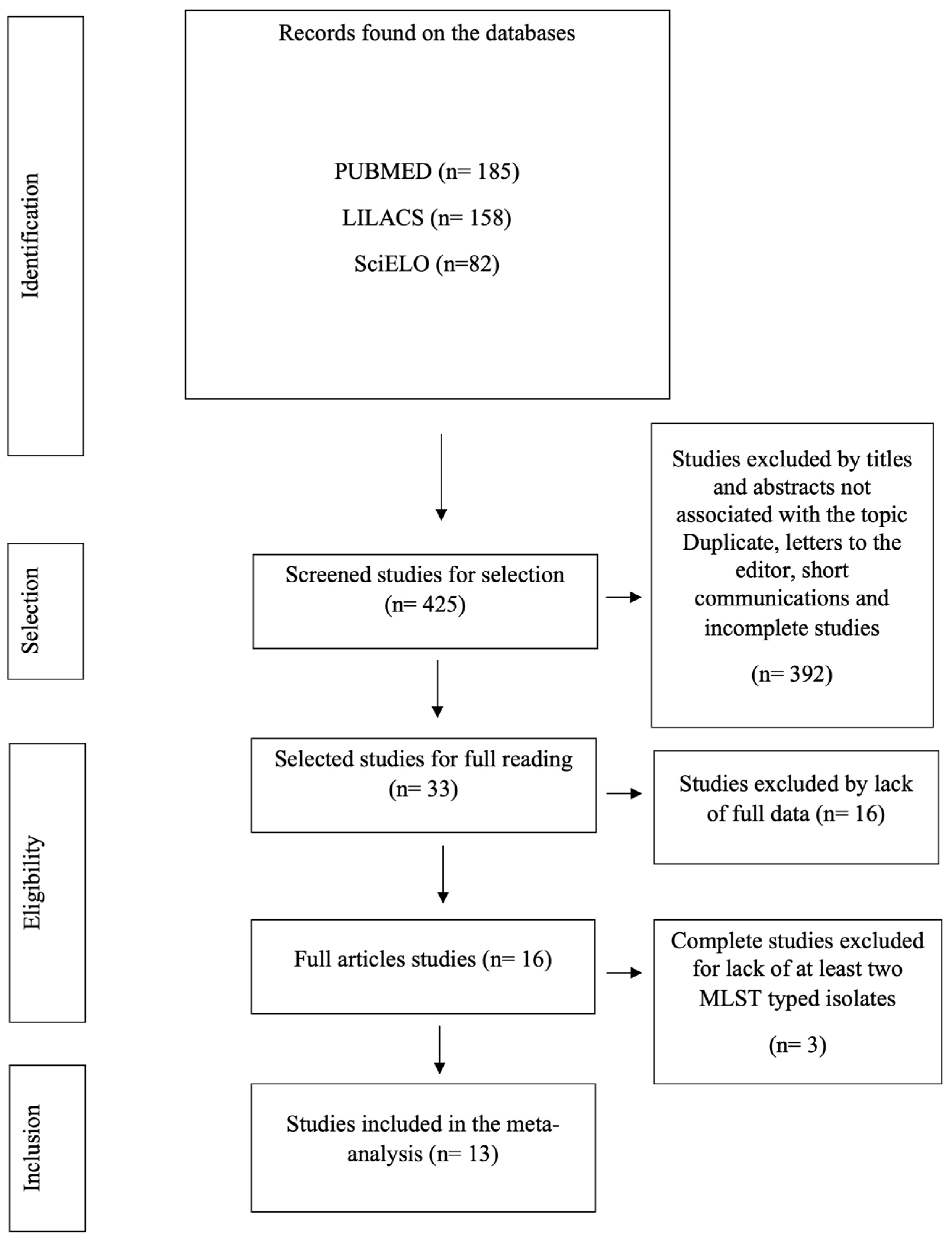
Figure 1. PRISMA flow diagram of the procedures for identifying, selecting, eligibility, and including studies for analysis. Belém, PA, Brazil, 2024.
Antibiotics 13 00983 g001
Figure 2. The goeBURST Full MST of P. aeruginosa demonstrating unique STs and clonal complexes in the Brazilian territory. Each node represents a unique ST; solid lines between STs shaded in grey represent SLVs and belong to CCs, and dashed lines between STs represent double locus variants DLVs. Colors were randomly assigned to STs.
Antibiotics 13 00983 g002
Figure 3. Distribution of the main P. aeruginosa MLST genotypes identified within studies across different time periods among isolates from 1994 to 2021.
Antibiotics 13 00983 g003
Figure 4. Distribution and regional spread of HRCs and emerging HRCs among P. aeruginosa isolates in Brazil. Legend: ST (sequence type), PA (Pará state), AC (Acre state), PE (Pernambuco state), SP (São Paulo state), RJ (Rio de Janeiro state).
Antibiotics 13 00983 g004
Figure 5. Distribution and regional spread of HRCs and non-HRCs of P. aeruginosa harboring MβL genes in Brazil. Legend: ST (sequence type), PA (Pará state), AC (Acre state), PE (Pernambuco state), SP (São Paulo state), RJ (Rio de Janeiro state).
Antibiotics 13 00983 g005
Figure 6. ROBINS-I Risk of Bias Assessment. Risk of bias assessment using the ROBINS-I tool for the observational studies included in the meta-analysis. (A) Evaluation of individual study parameters, and (B) combined data evaluation for each methodological parameter assessed [40,41,42,43,44,45,46,47,48,49,50,51,52].
Antibiotics 13 00983 g006
Figure 7. Forest plot of HRCs’ prevalence among P. aeruginosa isolates in Brazil and geographical regions [40,41,42,43,44,45,46,47,48,49,50,51,52].
Antibiotics 13 00983 g007
Figure 8. Funnel plot evaluating potential publication bias in the meta-analysis of the proportion of HRCs and non-HRCs among P. aeruginosa isolates in Brazil [40,41,42,43,44,45,46,47,48,49,50,51,52]. Each circle typically represents an individual study included in the meta-analysis. The position of the circle on the plot indicates the estimated effect size of that study (on the x-axis) and its precision (on the y-axis), often measured by standard error or sample size. Rhombuses may be used to highlight certain studies within the funnel plot. For instance, they can represent studies that are considered outliers or those that have been flagged for having methodological issues or other biases.
Antibiotics 13 00983 g008
Figure 9. Meta-regression analysis examining the relationship between the year of publication and HRCs and non-HRCs of P. aeruginosa based on the studies included in the meta-analysis [40,41,42,43,44,45,46,47,48,49,50,51,52].
Antibiotics 13 00983 g009
Figure 10. Forest plot displaying the prevalence of the ST277 genotype among HRCs of P. aeruginosa isolates in Brazil [40,41,42,44,45,46,47,48,49,50,51,52].
Antibiotics 13 00983 g010
Figure 11. Funnel plot assessing potential publication bias in the meta-analysis of the prevalence of the ST277 genotype among HRCs of P. aeruginosa in Brazil [40,41,42,44,45,46,47,48,49,50,51,52]. Each circle typically represents an individual study included in the meta-analysis. The position of the circle on the plot indicates the estimated effect size of that study (on the x-axis) and its precision (on the y-axis), often measured by standard error or sample size. Rhombuses may be used to highlight certain studies within the funnel plot. For instance, they can represent studies that are considered outliers or those that have been flagged for having methodological issues or other biases.
Antibiotics 13 00983 g011
Figure 12. Meta-regression analysis examining the influence of sample size on the prevalence of the ST277 genotype among HRCs of P. aeruginosa based on the studies included in the meta-analysis [40,41,42,44,45,46,47,48,49,50,51,52].
Antibiotics 13 00983 g012
Table 1. Characteristics of the included studies in this present meta-analysis.
Authors/YearType of StudyIsolates PeriodRegion/LocationSettingN° of Samples (872)/
N° of MLST Typed (298)
MLST GenotypesHRCJBI Score
Silva et al. (2011) #,* [40]Retrospective cross-sectional2000–2004Southeast regionClinical55/55ST277 (49/55—89.09%)
ST235 (1/55—1.82%)
ST593 (3/55—5.45%)
ST594 (1/55—1.82%)
ST595 (1/55—1.82%)
Yes—50/55 (90.9%)—ST277, ST235(7/8)
Cavalcanti et al. (2015) #,* [41]Retrospective cross-sectional2008–2010Northeast regionClinical10/10ST235 (3/10—30.0%)
ST446 (1/10—10.0%)
ST1560 (1/10—10.0%)
ST1419 (1/10—10.0%)
ST1126 (1/10—10.0%)
ST277 (1/10—10.0%)
ST244 (2/10—20.0%)
Yes—6/10 (60.0%)—ST277, ST235, ST244(8/8)
Miranda et al. (2015) #,* [42]Retrospective cross-sectional2008 and 2010Southeast regionHospital wastewater treatment plant41/36ST244 (8/36—22.22%)
ST1853 (2/36—5.56%)
ST1854 (1/36—2.78%)
ST1855 (2/36—5.56%)
ST1856 (1/36—2.78%)
ST1857 (1/36—2.78%)
ST1858 (2/36—5.56%)
ST1859 (8/36—22.22%)
ST1860 (1/36—2.78%)
ST1861 (1/36—2.78%)
ST1862 (4/36—11.11%)
ST238 (1/36—2.78%)
ST381 (1/36—2.78%)
ST595 (3/36—8.33%)
Yes—8/41 (19.51%)—ST244(6/8)
Silva et al. (2016) #,* [43]Retrospective cross-sectional2012Southeast regionBurn center: Clinical and Clinical environment35/10ST 2236 (5/10—14.28%)
ST 2237 (5/10—14.28%)
No (0.0%)(8/8)
Cacci et al. (2016) #,* [44]Retrospective cross-sectional2007–2008Southeast regionClinical and Clinical environment482/14ST1027 (1/14—7.14%)
ST1602 (2/14—14.29%)
ST1603 (1/14—7.14%)
ST1767 (1/14—7.14%)
ST1768 (1/14—7.14%)
ST1769 (1/14—7.14%)
ST1844 (1/14—7.14%)
ST235 (1/14—7.14%)
ST244 (1/14—7.14%)
ST277 (1/14—7.14%)
ST446 (1/14—7.14%)
ST532 (1/14—7.14%)
ST890 (1/14—7.14%)
Yes—(3/14—21.42%)—ST235, ST244 and ST277(8/8)
Chaves et al. (2017) #,* [45]Retrospective case-control study2011–2013Southeast regionClinical29/3ST277 (3/3—100.0%)Yes—ST277 (3/3—100.0%)(10/10)
Martins et al. (2018) #,* [46]Retrospective cross-sectional2012Southeast regionEnvironmental (Migratory birds)9/9ST277: 9/9 (100%)Yes—9/9 (100.0%)—ST277(7/8)
Bruggemann et al. (2018) #,* [47]Retrospective cross-sectional1994–2016Southeast regionClinical20/20ST235 (3/20—15.0%)
ST10 (1/20—5.0%)
ST231 (1/20—5.0%)
ST245 1/20—5.0%)
ST252 (1/20—5.0%)
ST446 (1/20—5.0%)
ST455 (1/20—5.0%)
ST498 (1/20—5.0%)
ST606 (1/20—5.0%)
ST1290 (1/20—5.0%)
ST1993 (1/20—5.0%)
ST2235 (1/20—5.0%)
ST381 (1/20—5.0%)
ST190 (1/20—5.0%)
ST244 (2/20—10.0%)
Yes—5/20 (25.0%)—ST235, ST244(8/8)
Santos et al. (2019) #,* [48]Retrospective cross-sectional1995–2015Southeast regionClinical88/52ST1117 (3/52—5.77%)
ST1560 (3/52—5.77%)
ST1945 (3/52—5.77%)
ST244 (5/52—9.62%)
ST277 (11/52—21.15%)
ST487 (4/52—7.69%)
ST1791 (3/52—5.77%)
ST1860 (4/52—7.69%)
ST804 (11/52—21.15%)
ST1602 (3/52—5.77%)
ST1944 (2/52—3.85%)
Yes—16/52 (30.76%)—ST277, ST244(7/8)
Rodrigues et al. (2020) #,* [49]Retrospective cross-sectional2010–2013North regionReferral hospital/Clinical54/54ST2524 (8/54—14.81%)
ST2541 (2/54—3.70%)
ST2552 (2/54—3.70%)
ST2554 (1/54—1.85%)
ST2555 (1/54—1.85%)
ST2556 (2/54—3.70%)
ST2603 (1/54—1.85%)
ST111 (2/54—3.70%)
ST170 (1/54—1.85%)
ST235 (5/54—9.26%)
ST244 (9/54—16.67%)
ST277 (6/54—11.11%)
ST360 (1/54—1.85%)
ST463 (1/54—1.85%)
ST500 (2/54—3.70%)
ST508 (1/54—1.85%)
ST1076 (1/54—1.85%)
ST1197 (1/54—1.85%)
ST1284 (4/54—7.41%)
ST1655 (1/54—1.85%)
ST2100 (1/54 —1.85%)
ST2437 (1/54—1.85%)
Yes—22/54 (40.7%)—ST244, ST277, ST111, ST235(8/8)
Esposito et al. (2021) # [50]Retrospective cross-sectional2016Southeast regionImpacted urban rivers (environmental)2/2ST277 (2/2—100.0%)Yes—(2/2—100.0%)
ST277
(8/8)
De Oliveira Luz et al. (2021) #,* [51]Retrospective cross-sectional2010, 2015, and 2016Northeast regionClinical13/13ST244 (2/13—15.38%)
ST1394 (1/13—7.69%)
ST252 (1/13—7.69%)
ST3079 (3/13—23.07%)
ST357 (2/13—15.38%)
ST3078 (1/13—7.69%)
ST275 (1/13—7.69%)
ST3080 (1/13—7.69%)
ST3137 (1/13—7.69%)
Yes—(4/13—30.76%)—ST244, ST357(8/8)
Santos et al. (2023) # [52]Retrospective cross-sectional2018–2021North regionClinical34/19ST277 (18/19—94.74%)
ST2711 (1/19—5.26%)
Yes—(18/19—94.74%)—ST277(8/8)
Legend: * Retrieved from PubMed database based on search strategy; # Retrieved from LILACS database based on search strategy; ST (sequence type).
Disclaimer/Publisher’s Note: The statements, opinions and data contained in all publications are solely those of the individual author(s) and contributor(s) and not of MDPI and/or the editor(s). MDPI and/or the editor(s) disclaim responsibility for any injury to people or property resulting from any ideas, methods, instructions or products referred to in the content.