Article

Effects of Dietary Supplementation of Lacticaseibacillus Chiayiensis AACE3 on Hepatic Antioxidant Capacity, Immune Factors and Gut Microbiology in Nandan Yao Chicks

1
Faculty of Life Science and Technology, Kunming University of Science and Technology, Kunming 650500, China
2
Engineering Research Center for Replacement Technology of Feed Antibiotics of Yunnan College, Kunming 650500, China
*
Author to whom correspondence should be addressed.
These authors contributed equally to this work.
Antibiotics 2023, 12(9), 1356; https://doi.org/10.3390/antibiotics12091356
Submission received: 29 June 2023 / Revised: 16 August 2023 / Accepted: 21 August 2023 / Published: 23 August 2023

Abstract

:
The growing issue of antibiotic resistance has restrained the utilization of antibiotics as growth enhancers in the poultry industry. Probiotics are candidates for replacing antibiotics in the poultry industry. However, probiotics are strain-specific and their efficacy needs to be investigated before applying them. The aim of this study was to assess the positive effects of Lacticaseibacillus chiayiensis AACE3 on the health and gut microbiota of Nandan Yao chicks. The results showed that compared with the blank control (NC) and aureomycin (PC) groups, L. chiayiensis AACE3 increased final body weight (BW), villus height and improved the ratio of villus height to crypt depth in chicken jejunal tissues. L. chiayiensis AACE3 also increased the activity of hepatic antioxidant enzymes (SOD, CAT and T-AOC) and reduced hepatic oxidative damage (MDA). Furthermore, compared to NC, L. chiayiensis AACE3, the activity of intestinal digestive enzymes (i.e., α-amylase, lipase and trypsin) was increased. L. chiayiensis AACE3 upregulated the production of IgA and IgG and downregulated the production of IL-6, IL-1β and TNF-α in chicken serum. Moreover, supplementation of L. chiayiensis AACE3 enhances the diversity of gut microbes. At the phylum level, the abundance of Actinobacteriota and Proteobacteria decreased with L. chiayiensis AACE3 supplementation, while the abundance of Verrucomicrobiota and Bacteroidetes increased. At the genus level, there was an increase in the abundance of potential probiotics Akkermansia, Romboutsia, Subdoligranulum, and Lactobacillus. This study confirms that L. chiayiensis AACE3 is an excellent feed additive as an alternative to aureomycin and offers various advantages for the healthy growth of chickens during the brooding period by positively affecting their gut microbiome.

1. Introduction

In recent years, nutritional strategies have emerged as important for improving animal health [1,2]. Feed additives promote animal digestion and health [3]. Antibiotics are frequently utilized as feed supplements in poultry farming to manage and prevent diseases, as well as to promote the growth performance of animals [4]. However, there is a worldwide effort to reduce the use of antibiotics in poultry production, as antibiotic residues in poultry products can be harmful to consumers [5]. In addition, antibiotics can disrupt the balance of animal intestinal flora and affect the health of the animal’s intestinal tract [6]. The severe negative effects of antibiotics on livestock and poultry have led to their prohibition as feed additives in Europe, the United States, and China [7]. Today, there are new approaches being used to replace antibiotics in poultry farming. For example, vaccination can prevent proliferative enteropathy (PE) and reduce the consumption of oxytetracykline used to treat PE [8]. Mannanoligosaccharides (Bio-Mos®) can be used as an alternative to antibiotic growth promoters (avilamycin) in broiler feeding [9]. Sodium butyrate can also be used as an alternative to dietary hyoscine in chickens [10]. Moreover, probiotics are a promising alternative to antibiotics in broiler farming [11]. Probiotics are living microorganisms, and consuming adequate amounts of them can provide sustained health benefits for animals [12]. For instance, chickens that received Lactobacillus plantarum LP-8 dietary supplementation displayed increased intestinal mucosal immunoglobulin IgA content and an improved antioxidant capacity [13]. Lactobacillus casei supplementation resulted in significant enhancement of the immunity of chicks, modulation of the expression of inflammation-related factors, and increased gut microbiota diversity [14]. These studies have shown that probiotics can enhance host immunity while enriching the gut flora to promote host health. A large number of studies have proved that probiotics have good effects in animal breeding, but due to the different kinds of probiotic strains, their vitality and their adaptability to different environments, leads to the different effects of probiotics [15]. Therefore, probiotics that improve host health are essential for poultry farming.
Lacticaseibacillus chiayiensis, a novel LAB closely related to the Lacticaseibacillus casei group, was first isolated from cattle manure in Chiayi City (Taiwan, China) in 2018 [16]. Kim et al. found that the L. chiayiensis genome encodes stress resistance-related (e.g., bile, acid, and oxidative stress tolerance genes), bacteriocin production and adhesion-related genes. Furthermore, in an in vitro study of probiotic properties, L. chiayiensis showed good antibacterial properties, adhesion properties and an ability to survive in adverse gastrointestinal environments [17]. Therefore, L. chiayiensis can be considered a novel potential probiotic that may have good potential for application.
Resistance to pathogen colonization and local immune responses in the gut can be enhanced in neonatal birds by adding probiotics to the diet prior to the maturation of the gut microbiota [18,19]. Nandan Yao chicken is a breed of chicken native to China. It is characterized by coarse-grained tolerance, strong foraging ability, and delicate and tasty meat [20]. Nandan Yao chicken was selected into the National Livestock Genetic Resource Breeds List in 2021, and was awarded the National Geographical Indication Protection Certification for Agricultural Products in 2022, which has high economic value [21]. Nandan Yao chicken was used as a research material to evaluate the beneficial performance of L. chiayiensis in this study, which guided poultry breeding in China to a certain extent. It also provides a new possible option for antibiotic replacement in global poultry farming. Certainly, we will also select more modern broiler strains to evaluate the generalizability of the beneficial effects of L. chiayiensis in the future. In this study, we hypothesized that supplementation with L. chiayiensis could replace aureomycin to promote microbial proliferation and persistence to improve the immunity of chicks to improve the early development of their intestinal tract. Therefore, we evaluated the intestinal morphology, liver antioxidant function, immune factor content and intestinal microbiota of Nandan Yao chickens after L. chiayiensis supplementation.

2. Results

2.1. Growth Performance

Table 1 shows the effect of the experimental group on body weight, average daily feed intake, average daily gain, and feed conversion ratio of the Nandan-Yao chickens. The chicks were weighed after 21 days, and the PC, LC6 and LC7 groups were found to be 11.28 g, 4.56 g and 14.69 g heavier than the NC group, respectively. ADFI and ADG for chicks were observed and found to be significantly higher in the PC and LC7 groups than in the NC group. However, the comparison of the FCR with the NC group does not yield a significant difference with respect to all experimental groups.

2.2. Effects of L. chiayiensis AACE3 on the Gut Morphology of Chickens

The use of L. chiayiensis AACE3 as a feed supplement significantly influenced the intestinal morphology of chickens (Figure 1). The villus height of the jejunum was significantly higher in the L. chiayiensis AACE3 group than in the NC and PC groups (p < 0.05). Regarding the VH:CD ratio, which represents the functional status of the epithelium, the LC7 group produced a significantly higher ratio than the control groups (p < 0.05), while it was increased by 5.17% relative to that with the PC group.

2.3. The Effect of L. chiayiensis AACE3 on Digestion Properties

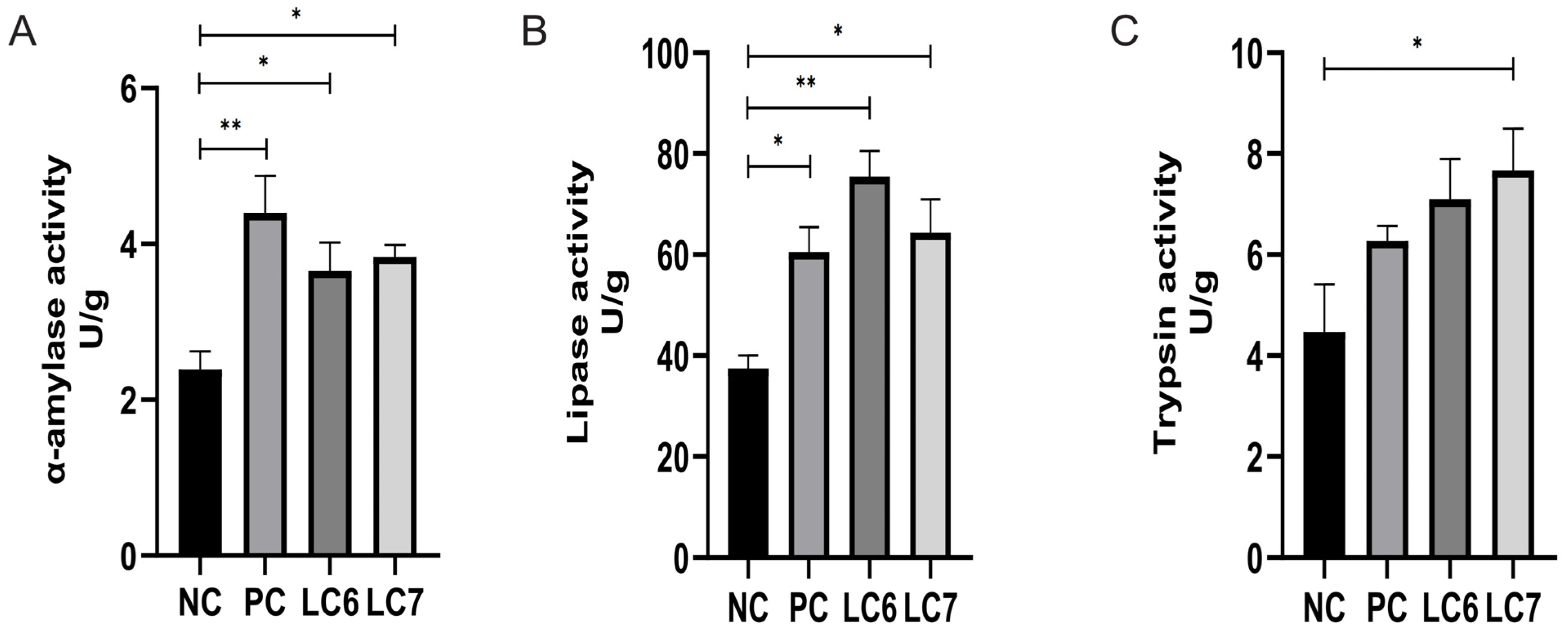
Supplementation with L. chiayiensis AACE3 led to a significant increase in α-amylase and lipase activity relative to the NC (p < 0.05). The LC7 group showed a significant improvement in trypsin activity, while the LC6 group showed 58.79% accretion. Compared with the PC group, supplementation with L. chiayiensis AACE3 caused no significant difference in α-amylase, lipase, and trypsin activity (Figure 2).

2.4. Effects of L. chiayiensis AACE3 Supplementation on Hepatic Function

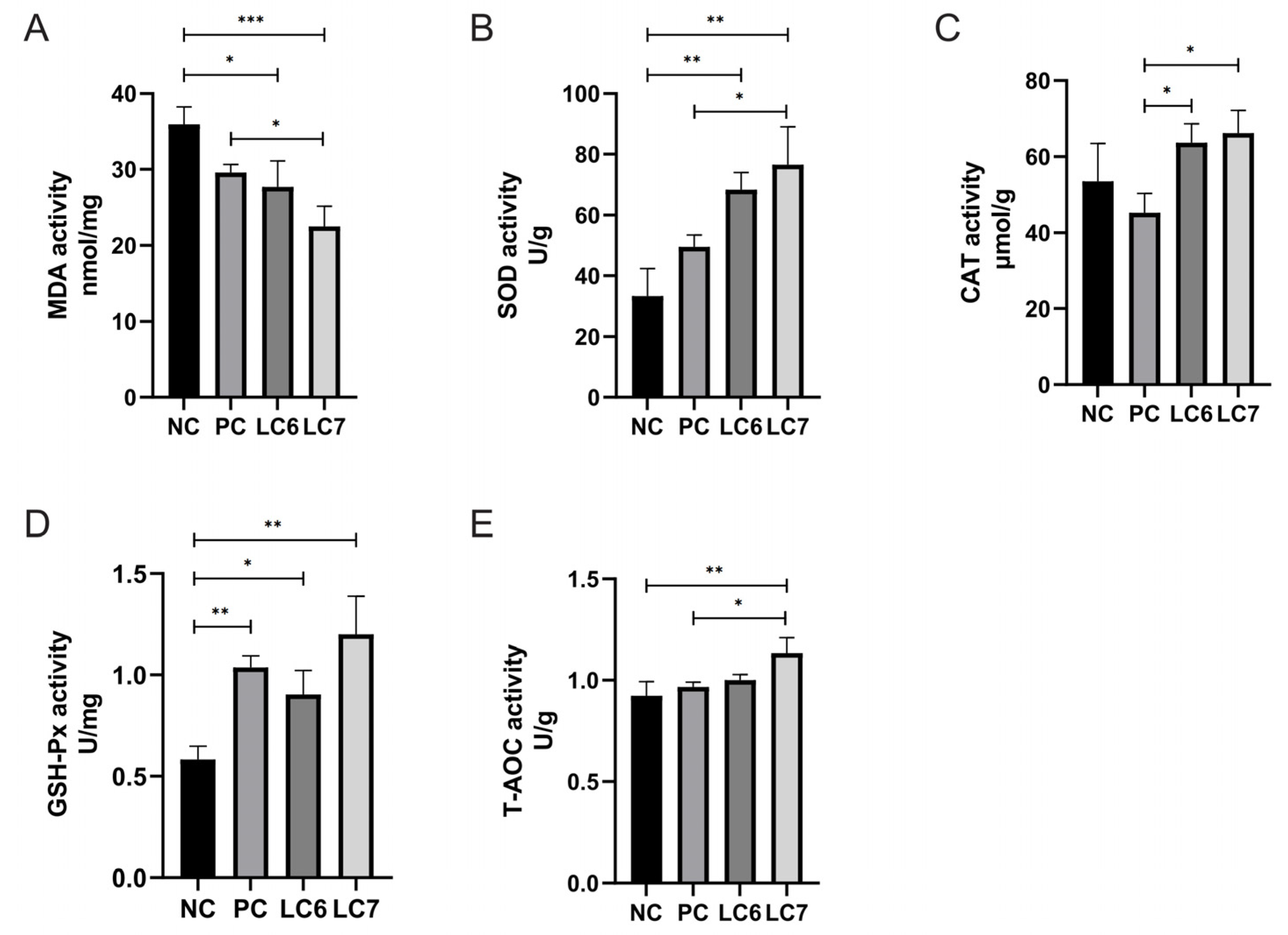
The effect of supplementation with L. chiayiensis AACE3 on the antioxidant capacity of chicken hepatic tissue is demonstrated in Figure 3. The levels of oxidative damage products (MDA) were significantly lower in chickens fed L. chiayiensis AACE3 supplementation compared to NC-treated chickens (p < 0.05). Compared to the PC group, MDA levels in the LC6 and LC7 groups decreased by 6.39% and 23.84%, respectively.
The liver SOD and GSH-Px activities were significantly increased in chickens fed L. chiayiensis AACE3 compared to the NC group. CAT and SOD activity was significantly increased in chickens fed L. chiayiensis AACE3 compared to the PC group. T-AOC activity was significantly increased in the LC7 group compared to the NC and PC groups by 22.86% and 17.27%, respectively. L. chiayiensis AACE3 effectively mitigates oxidative stress in chicken livers.

2.5. Effects of L. chiayiensis AACE3 on Immune Factors

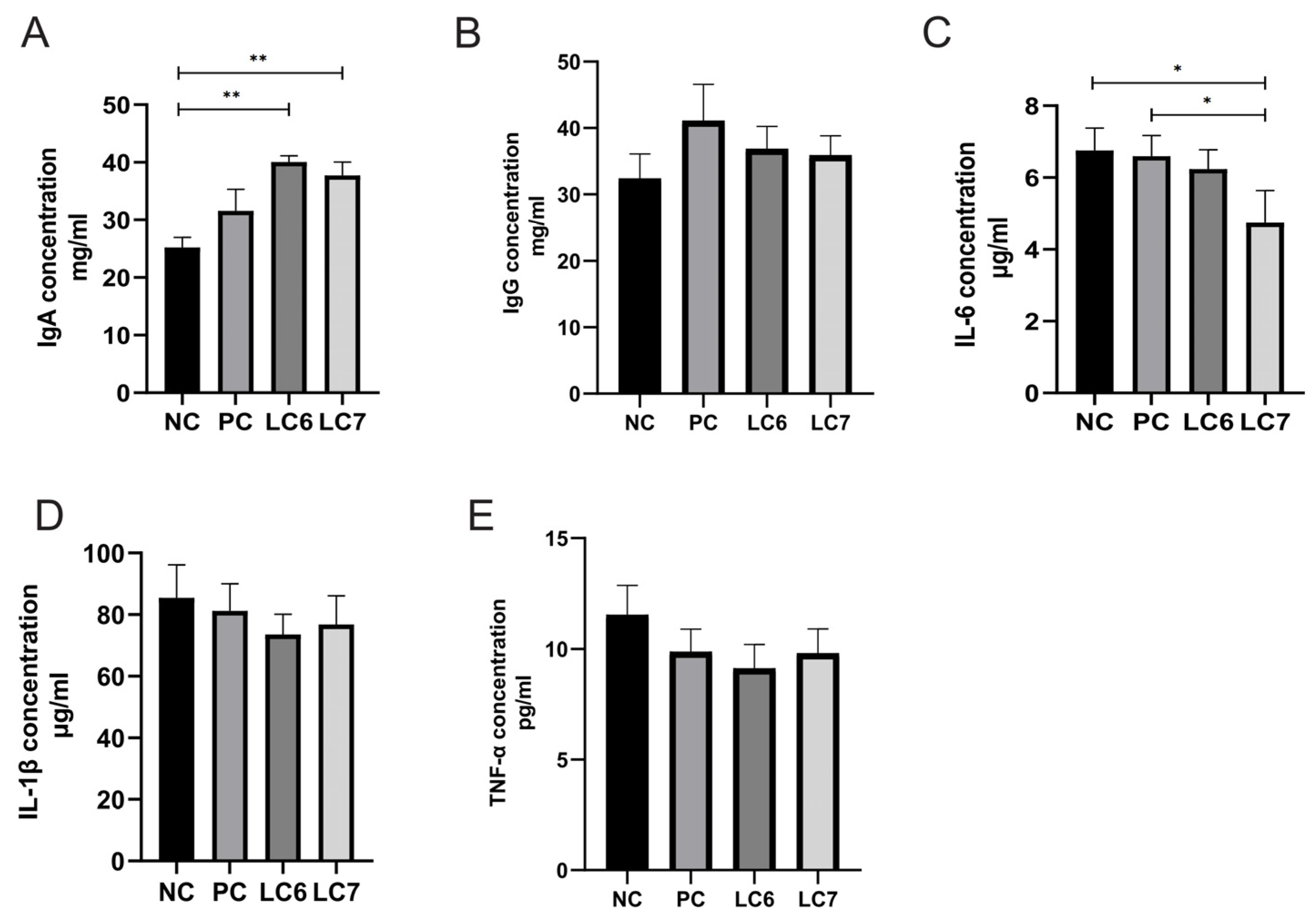
Changes in the level of immune factors have an impact on chicken health to some extent, and supplementation with L. chiayiensis AACE3 induced the production of immune factors in chickens (Figure 4). Compared to the control (NC) group, supplementation with L. chiayiensis AACE3 significantly increased the concentration of IgA (p < 0.05). In addition, the LC6 and LC7 groups increased IgG concentrations by 13.76% and 10.79%, respectively. Compared to the aureomycin group (PC), the LC6 and LC7 groups increased IgA concentrations by 26.96% and 19.56%, respectively.
The LC7 group significantly reduced IL-6 concentrations relative to those with the NC and PC groups (p < 0.05). While IL-6 levels were reduced by 5.5% in the LC6 group, there was no significant change. In addition, the LC6 and LC7 groups reduced IL-1β and TNF-α levels by 14%, and 20.98% and 10.16%, and 14.98%, respectively, relative to those with the NC group. LC6 and LC7 groups reduced IL-1β and TNF-α levels by 9.5%, and 7.71% and 5.46%, and 0.71%, respectively, relative to those with the aureomycin group (PC). Thus, the results suggest that supplementation with L. chiayiensis AACE3 altered the levels of these immune factors.

2.6. Effects of L. chiayiensis AACE3 on the Chicken Gut Microbiota

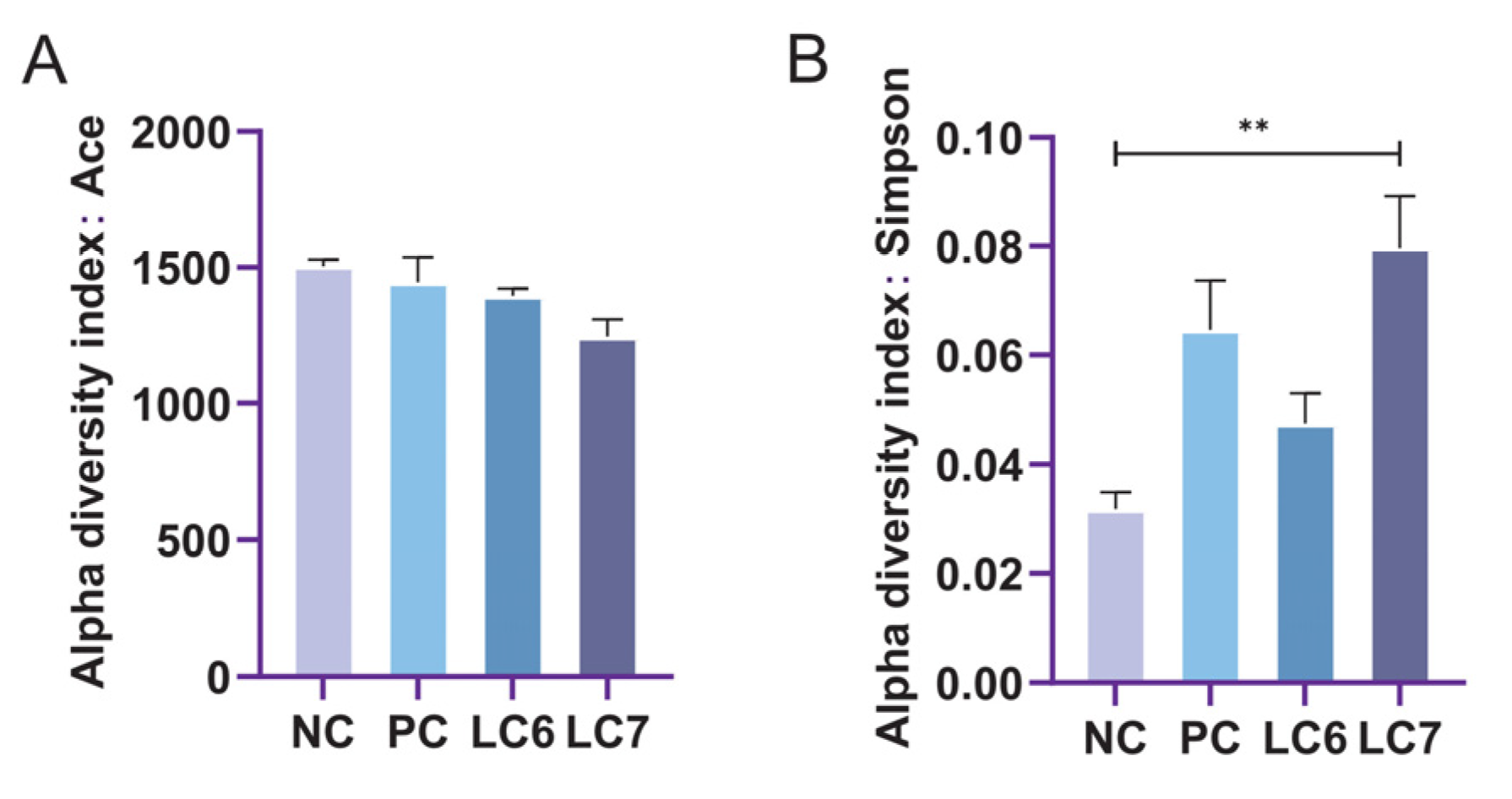
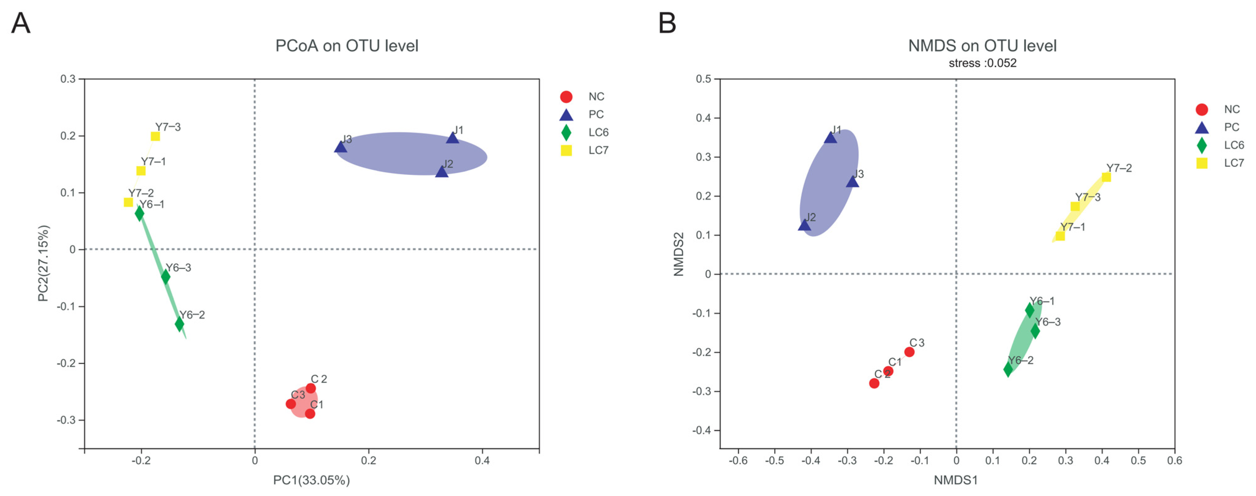
Changes in the alpha-diversity index of gut microorganisms reflect changes in microbial community abundance and diversity (Figure 5). Significant differences were found between the community diversity (Simpson) indices of the NC and LC7 groups. Principal coordinate analysis (PCoA) and non-metric multidimensional scaling (NMDS) using the Bray distance metric demonstrated that the biological communities of the NC, PC, LC6, and LC7 groups were segregated into four distinct clusters (Figure 6). The reliability of the model was evaluated based on the stress value, which was 0.052 in NMDS. The findings suggest that the administration of L. chiayiensis AACE3 leads to several alterations in gut microbial composition.
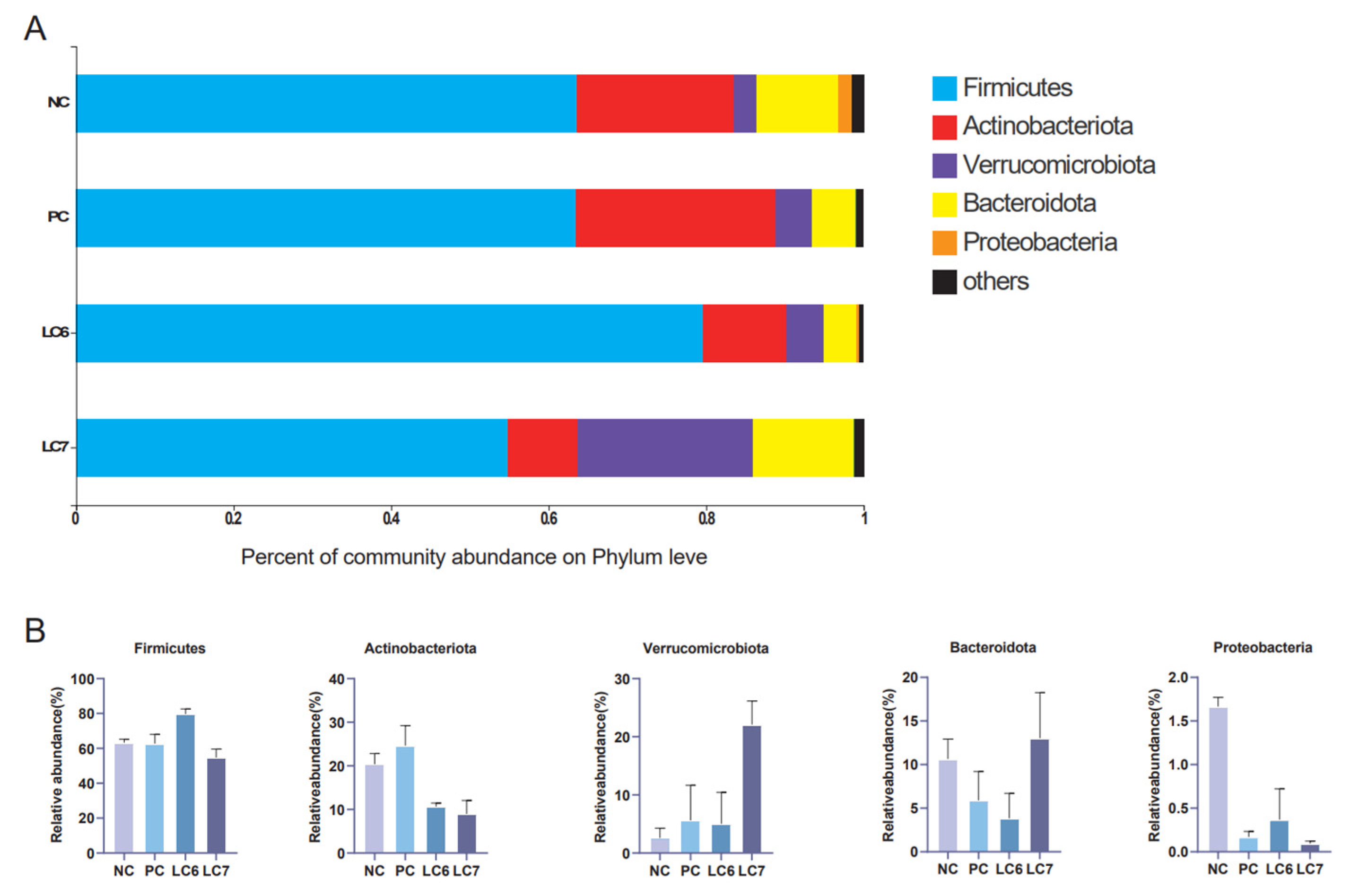
At the phylum level, the most prevalent phyla are Firmicutes, Actinobacteriota, Verrucomicrobiota, Bacteroidetes, and Proteobacteria. Firmicutes was more abundant in the LC6 group, comprising 79.62% of the phylum-level community, while Actinobacteriota was more abundant in the NC and PC groups than in the experimental group. In the LC7 group, Verrucomicrobiota and Bacteroidota are more abundant than in the other groups and accounted for 22.08% and 13.03%, respectively, of the community at the phylum level. The abundance of Proteobacteria in the control group was higher than that in the remaining groups (Figure 7).
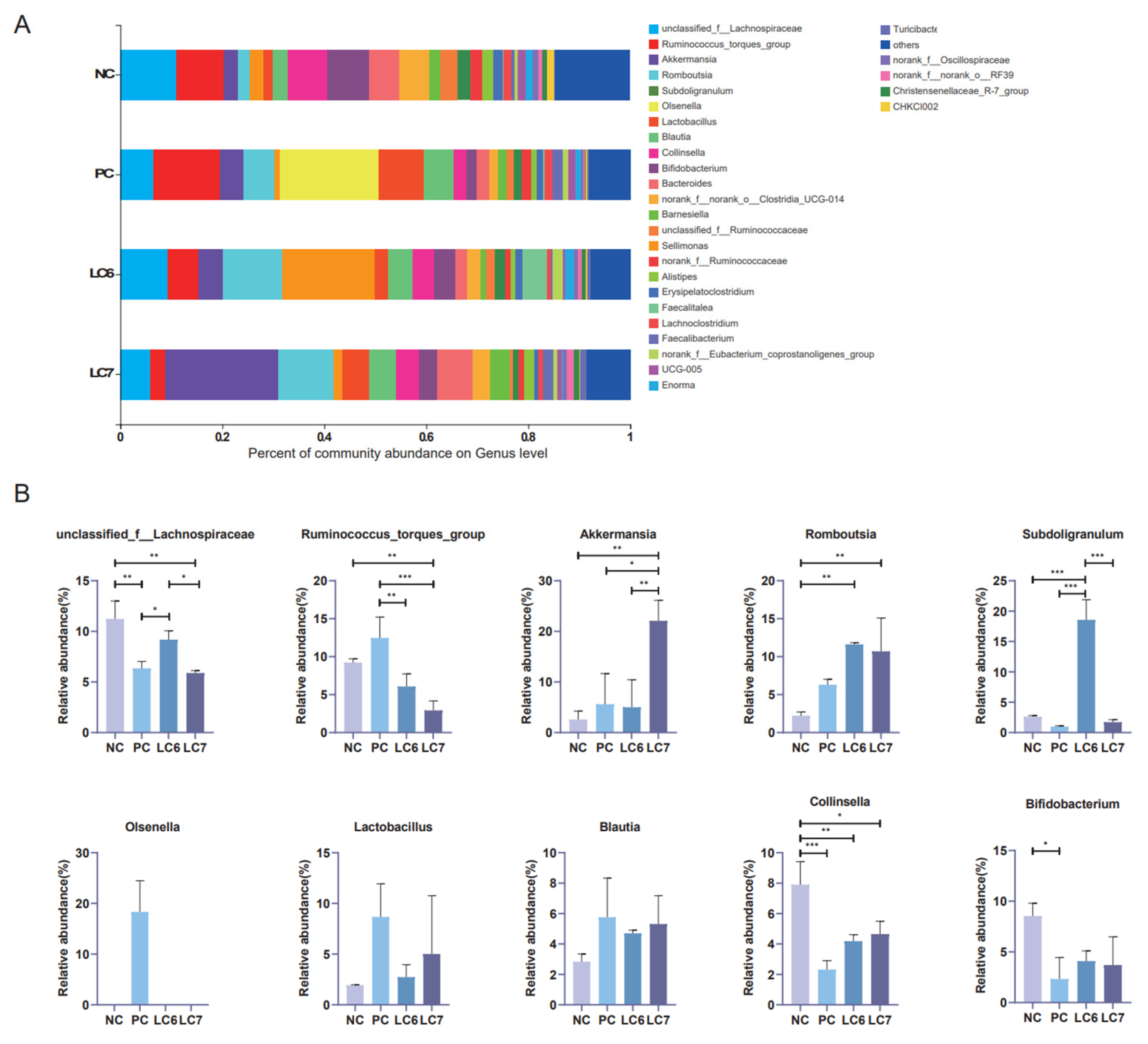
At the genus level, the top 10 dominant bacterial genera were identified (Figure 8). Among them, unclassified Lachnospiraceae was the most dominant bacterial genus, accounting for 13.25% of the community among the top 10 OTUs. Unclassified Lachnospiraceae levels were significantly higher in the LC6 group than in the PC group (p < 0.05) and in the LC7 group than in the NC group (p < 0.05). The levels of Akkermansia were significantly higher in the LC7 group (p < 0.05) than in the other groups. Romboutsia levels in the experimental groups (LC6 and LC7) were significantly higher (p < 0.05) compared to the NC and PC groups. The levels of Subdoligranulum were significantly higher in the LC6 group than in other groups (p < 0.05), while the level of Collinsella was significantly lower in the experimental LC6 and LC7 groups (p < 0.05) than in the NC group. Additionally, the levels of Olsenella and Ruminococcus torques were significantly higher in the PC group than in the other groups (p < 0.05). However, the levels of Bifidobacterium, Blautia, and Lactobacillus in the experimental group did not significantly differ from those in the control group. These results indicate that the relative abundance of some dominant bacteria in the intestinal flora changed after supplementation with L. chiayiensis AACE3.

2.7. Correlation Analysis

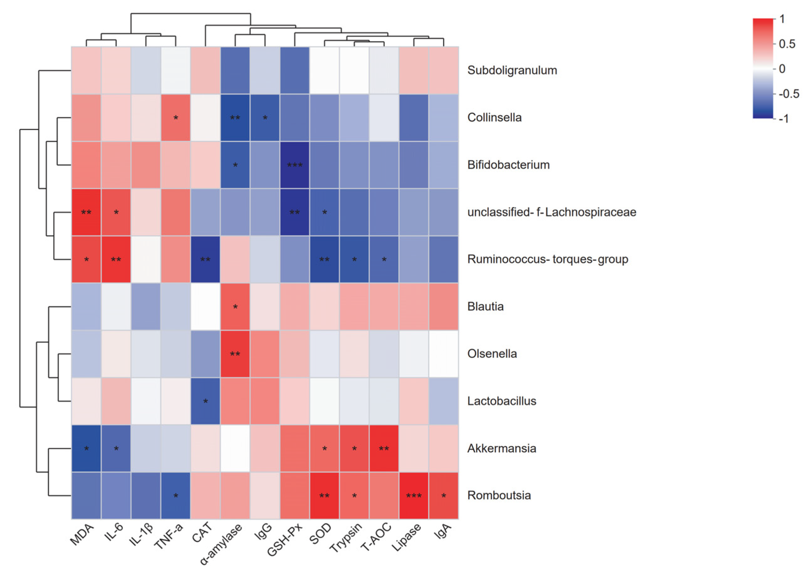
To further explore the relationship between changes in the abundance of genera in the chicken gut microbiota and changes in the chicken’s own physiology. Correlation analysis of microorganisms (10 genera with the highest relative abundance) with digestive system characteristics, antioxidant capacity and immune status was performed (Figure 9). In the correlation analysis, Ruminococcus torques showed a significant negative correlation with T-AOC, SOD, CAT, and trypsin and a significant positive correlation with IL-6 and MDA. Unclassified Lachnospiraceae showed a significant negative correlation with GSH-Px and SOD and a significant positive correlation with IL-6 and MDA. Collinsella exhibited a considerable negative correlation with IgG and α-amylase and a significant positive correlation with TNF-α. Similarly, Bifidobacterium displayed a significant negative correlation with GSH-Px and α-amylase. Olsenella was significantly and positively linked with α-amylase. Lactobacillus showed a significant negative correlation with CAT. Moreover, Romboutsia showed a significant negative correlation with TNF-α and a significant positive correlation with IgA, lipase, trypsin, and SOD. On the other hand, Akkermansia showed a noteworthy negative correlation with IL-6 and MDA and a significant positive correlation with T-AOC, trypsin, and SOD. Finally, Blautia demonstrated a significant positive correlation with α-amylase, whereas no significant correlations were found with Subdoligranulum.

3. Discussion

In the field of poultry farming, probiotics are receiving increasing attention as an alternative to antibiotics [22,23]. The discovery of new probiotics and the evaluation of their role in poultry farming is essential to meet the demand for alternative antibiotics in poultry farming. In this study, the beneficial effects of L. chiayiensis on poultry farming were comprehensively evaluated through animal feeding experiments (addition of L. chiayiensis to diets) on Nandan Yao chickens. It provides a new option for antibiotic replacement in the field of poultry farming. In this study, the use of probiotic strain L. chiayiensis AACE3 significantly increased the body weight of chickens in the LC7 group. This aroused our interest in exploring how its growth-promoting ability is realized. Through the detection of digestive enzyme activities and intestinal morphology in the intestine, it was found that L. chiayiensis mediated the upregulation of intestinal digestive enzyme activities and the alteration of intestinal morphology in 21 d Nandan scallop chickens. The height of the intestinal villi of chickens fed L. chiayiensis was significantly increased compared to the control, while the depth of crypts was not significantly changed, whereas, in previous studies, it has been shown that the higher the ratio of villus height to crypt depth, the higher the nutrient digestibility [24,25]. In addition, another study suggested that the improvement in weight gain may be related to the ability of probiotics to secrete enzymes such as amylase, protease and lipase, which target the efficient absorption of feed nutrients such as starch, fat and protein [24]. In this study, L. chiayiensis AACE3 significantly increased α-amylase, lipase, and trypsin activities relative to the control group. Thus, these findings suggest that L. chiayiensis AACE3 enhances host digestion by promoting both host digestive enzyme production and morphological changes in intestinal villi.
In addition, the antioxidant status within the host is essential to protect against pathogens and maintain homeostasis in vivo [25] as the body tends to produce reactive oxygen species (ROS) through normal cellular metabolism and in response to factors such as environmental changes and diet [26]. Additionally, the accumulation of excessive ROS can lead to host oxidative stress and oxidative damage. In this study, supplementation with L. chiayiensis AACE3 resulted in increased SOD, CAT, GSH-Px, and T-AOC levels and decreased MDA levels compared to the NC and PC groups. Among them, MDA is an important product of lipid peroxidation, and the level of MDA is a measure of the degree of oxidative damage [27]. Therefore, dietary supplementation with L. chiayiensis AACE3 significantly increased the antioxidant capacity of Nandan Yao chickens. Meanwhile, the significant reduction in MDA level also confirmed the repair of oxidative damage by the increase in antioxidant capacity induced by L. chiayiensis. Furthermore, it is important to note that the host’s ability to resist disease is more predominantly related to its own immune system [28]. Probiotics can prevent the proliferation of pathogens and thus prevent infection by improving physical mechanisms (immune organs) and chemical barriers (immune proteins, tiny molecules, etc.), contributing to host immune regulation [29]. For example, supplementation with probiotics results in higher levels of IgG and IgA, which are important markers for estimating changes in the immune function of the animal [30]. Here, supplementation with L. chiayiensis AACE3 increased the level of IgA and IgG levels in chicken serum. In addition, probiotics not only stimulate humoral and cell-mediated immunity (IgG and IgA) but also modulate the production of pro- and anti-inflammatory cytokines (IL-2 and IL-10) in the host, thereby improving host resistance to disease [31]. In this study, the levels of IL-1β, IL-6 and TNF-α were decreased by supplementation with L. chiayiensis AACE3. Overall, L. chiayiensis AACE3 improves poultry health by increasing the activity of antioxidant enzymes, stimulating humoral immunity, reducing MDA levels, and regulating the production of inflammatory factors.
There is growing evidence that gut microbes positively impact host health [32]. In this study, 16S RNA from the cecal content of chickens fed L. chiayiensis AACE3 was sequenced to investigate changes in microbial community composition. The α-diversity analysis showed altered diversity after supplementation with L. chiayiensis AACE3, which may be because L. chiayiensis AACE3 inhibits harmful bacteria in the intestine [33]. PCoA and NMDS showed different β diversity between supplementation with L. chiayiensis AACE3 chickens and the NC and PC groups, indicating that the effect of supplementation with L. chiayiensis AACE3 on the intestinal flora was different from that of the NC and PC groups. In addition, microorganisms play different roles in various hosts. Therefore, it is also important to analyze the changes in the high abundance of microorganisms in the gut microbiota that have a greater impact on the host [34]. In a phylum-level analysis, we found that the abundance of the phyla Actinobacteriota and Proteobacteria declined after feeding with L. chiayiensis AACE3. Proteobacteria consist of various zoonotic pathogens, including but not limited to Escherichia, Salmonella, and Campylobacter, as well as other highly virulent genera [35]. This study observed an improvement in Verrucomicrobiota and Bacteroidota abundance upon supplementing L. chiayiensis AACE3, which are important in energy production and metabolic processes within the gut [36]. Akkermansia is a representative species of Verrucomicrobiota, an anaerobic bacterium that degrades intestinal mucus, preserving the integrity of the intestine and avoiding the development of necrotizing small bowel inflammation (intestinal necrosis) [37,38]. In addition, the addition of L. chiayiensis AACE3 to the gut microbiota altered the composition of dominant bacterial species at the genus level. Among them, the levels of the dominant genera Akkermansia, Romboutsia and Subdoligranulum were increased significantly, and the levels of unclassified Lachnospiraceae, Ruminococcus torques group and Collinsella decreased significantly. Based on a correlation analysis, Akkermansia relative abundance was found to be positively correlated with trypsin content and T-AOC while negatively correlated with IL-6 levels in chickens. It is similar to some of the other studies on Akkermansia, namely that changes in Akkermansia levels affect host metabolic processes and reduce inflammatory responses [39]. Romboutsia was positively correlated with DAO, trypsin and lipase in the present study. Romboutsia has been studied to have a multifunctional metabolic capacity with carbohydrate utilization and single amino acid fermentation [40]. L. chiayiensis AACE3 increased the levels of Romboutsia, enhancing the activity of intestinal digestive enzymes and thus improving feed utilization in chickens. Therefore, these findings suggest that L. chiayiensis AACE3 has the potential to replace aureomycin as a more environmentally friendly feed additive in poultry farming. However, some shortcomings remain in this study, such as some regional limitations of the study materials used and an incomplete study of the molecular mechanisms of growth promotion in L. chiayiensis. In the future, we will further investigate the way in which L. chiayiensis-mediated microbial changes regulate the alteration of host physiological phenotypes through metabolomics and transcriptomics. Overall, this study provides a new possibility for antibiotic replacement in poultry farming.

4. Materials and Methods

4.1. Bacterial Strains and Culture Conditions

L. chiayiensis AACE3 (Genbank No. CP107523.1) was isolated from fermented blueberry sap. It is now stored in the Faculty of Life Science and Technology, Kunming University of Science and Technology, Kunming City, China. For the individual colonies used for routine use, the strain was grown and passaged in lactic acid bacteria selective medium (MRS) broth at 37 °C for 24 h. Probiotics were harvested via centrifugation at 6,000 × g for 10 min, and after washing twice with phosphate-buffered saline (PBS, pH 7.2), the precipitate was resuspended in sterile PBS solution. One milliliter of the bacterial suspension solution was taken, the final concentration of probiotics was adjusted using the optical density method (spectrophotometer, LAMBDA 850, Waltham, MA, USA), and the exact numbers (1 × 10⁹ CFU/mL) were determined for later feed preparation.

4.2. Birds, Diet, and Experimental Design

Nandan-Yao chickens used in this experiment were purchased from Kunming Lihua Animal Husbandry Co., Ltd. The Animal Care and Use Committee of the Kunming University of Science and Technology approved the study protocol and provided the guidelines for this study.
A total of 240 one-day-old Nandan-Yao chickens were randomly distributed into four treatment groups and fed for 21 days. Each group contained 6 replicates, with 10 chicks per replicate. The dietary groups included: (1) standard basal diet (NC group), (2) basal diet with antibiotic aureomycin (100 mg/kg of feed) (PC group), (3) basal diet with a low dose of L. chiayiensis AACE3 (1 × 10⁶ CFU/g of feed) (LC6 group), and (4) basal diet with a high dose of L. chiayiensis AACE3 (1 × 10⁷ CFU/g of feed) (LC7 group), 106 CFU/g and 107 CFU/g probiotic concentrations. It was confirmed by diluting and freeze-drying 109 CFU/mL of probiotic solution and then taking 1 gram of lyophilized powder for smear plate testing. The lyophilized powder was then added to the basal feed at 0.1%. At 0, 7, 14, and 21 days, all birds and feed were weighed, and feed consumption and body weight were recorded weekly for each group. The body weight (BW), average daily feed intake (ADFI), average daily gain (ADG), and feed conversion ratio (FCR) were calculated accordingly. The basal diet was composed of soybean meal, corn, soybean oil, vitamins, and mineral premix and made per the (National Research Council (NRC) standard) [41] (Table 2).

4.3. Sample Collection

On day 21, 6 chickens were randomly selected from each replicate and samples from the same replicate group were mixed for subsequent testing. Sampling of all chickens was performed on a sterile table in the chicken house and was performed on the same morning. All samples were placed in ice boxes during collection; the entire sample collection process took 3 hours and they were transferred to the laboratory within 20 min of completion for the next step. Blood samples were collected through the jugular vein, centrifuged at 3500× g and at 4 °C for 10 min to remove the supernatant, and then stored at −20 °C. The blood samples were collected through the jugular vein. The liver samples were collected into sterile tubes and stored at −20 °C. The duodenal digest was stripped and sonicated after tenfold dilution with PBS (pH 7.0), and the resulting digest sample was centrifuged at 15,000× g and 4 °C for 30 min. The supernatants were divided into aliquots and stored at −20 °C for enzymatic assays. From each bird, samples of the jejunum were collected, flushed with saline, and placed in a 4% formaldehyde solution for morphometric index evaluation. Cecum feces were collected in sterile plastic tubes, and samples from each chicken were combined and divided in the same proportions. The samples were stored at −20 °C until microbiota analysis.

4.4. Analysis of the Gut Morphostructure

The removed chicken jejunum was fixed in 4% paraformaldehyde and embedded in paraffin. Then, a ZEISS microscope (Shanghai, China) and ImageJ software (Version 1.8.0.345 Chinese, Kunming, China) were employed to perform histomorphometric examinations of 5 μm thick serial sections of the jejunal sample that were stained with hematoxylin and eosin (HE). Villus height (VH), crypt depth (CD), and VH:CD ratio measurements were obtained from at least 3 slides, with a minimum of 6 well-oriented indices to calculate their respective averages.

4.5. Measurement of Physiological Indicators

Precollected hepatic tissues were homogenized with 4 volumes of reagent kit extracts and centrifuged at 1000× g for 5 min. The supernatant was used for malondialdehyde (MDA, Suzhou Grace Biotechnology Co., Ltd., Suzhou, China) content, catalase (CAT, Suzhou Grace Biotechnology Co., Ltd., Suzhou, China) activity, superoxide dismutase (SOD, Suzhou Grace Biotechnology Co., Ltd., Suzhou, China) activity, total antioxidant capacity (T-AOC, Suzhou Grace Biotechnology Co., Ltd., Suzhou, China) activity, and glutathione reductase (GSH-Px, Suzhou Grace Biotechnology Co., Ltd., Suzhou, China) activity evaluation by using commercial kits. Stored sera were tested for immunoglobulin A (IgA), immunoglobulin G (IgG), interleukin-6 (IL-6), interleukin-1β (IL-1β) and tumor necrosis factor alpha (TNF-α) levels using the immune factor test kit (Quanzhou Jubang Biotechnology Co., Ltd., Quanzhou, China). Digestive fluid was collected from the duodenal intestine, and changes in amylase, trypsin and lipase levels were measured using an ELISA micro α-amylase activity assay kit, a trypsin activity assay kit and a lipase (LPS trace method) activity assay kit (Quanzhou Jubang Biotechnology Co., Ltd., Quanzhou, China).

4.6. Analyses of the Cecal Microbiota

From precollected cecum contents, a total of 12 samples were used for microbiome analysis. Microbial total genomic DNA was extracted from samples using the MagMAXTM-96 DNA multi-sample kit (Thermo Fisher Scientific, Suzhou New District, China) according to the manufacturer’s instructions. The 16S rRNA V3–V4 region was amplified and sequenced at the Majorbio Bio-Pharm Technology Co., Ltd (Shanghai, China) Genomic Analysis Platform-IBIS using Illumina MiSeq paired-end technology. Sequences were analyzed in the Ubuntu terminal using the UPARSE method [42], merging raw reads, filtering with the maximum expected error threshold at 1.0, dereplicating and mapping reads into operational taxonomic units (OTUs). Moreover, operational taxonomic units (OTUs) were clustered using Uparse based on the similarity threshold being greater than 97%. The RDP classifier algorithm was used to compare the 97% similar OTU representative sequences with the SILVA (Version 138, Shanghai, China) database for taxonomic analysis. The analyses related to the 16S data (i.e., diversity, phylum level, genus level, and correlation analysis) were performed on the Megabio online analysis platform (Shanghai, China).

4.7. Statistical Analysis

The mean ± standard error of the mean (SEM) values of all experimental data are determined from at least three independent replicates. GraphPad Prism 8.0 (GraphPad Software, Kunming, China) was used to conduct statistical analyses. The level of significance was set at * p < 0.05, ** p < 0.01, *** p < 0.001, and one-way ANOVA or non-differential/mixed comparisons were carried out to calculate statistical significance.

5. Conclusions

Reducing the overuse of antibiotics in poultry farming is one of the keys to curbing the spread of antibiotic resistance. Probiotics are a potential alternative to antibiotics in animal feed. The results of this study reveal the great potential of L. chiayiensis AACE3 as an antibiotic alternative in broiler farming. Supplementation with L. chiayiensis AACE3 enhances the health of chicks by increasing antioxidant capacity and modulating the levels of immune factors in the chicks. L. chiayiensis AACE3 also alters the gut microbial community of chicks, increasing the abundance of several bacterial species associated with inflammation, immunity and growth, such as Akkermansia, Romboutsia, and Subdoligranulum, among other beneficial microorganisms. Thus L. chiayiensis AACE3 can promote a host-friendly gut environment by changing the composition of gut microorganisms to improve health. Altogether, these findings suggest that L. chiayiensis AACE3 may be a suitable alternative to antibiotics in broiler production.

Author Contributions

X.K. conducted writing, original draft; data curation. C.-Y.L. and X.-D.L. conduct formal analysis. W.-G.X. and H.-Y.Z. provision of study materials, analysis tools. F.W. design of methodology. L.-B.L. conducted funding acquisition, project administration. All authors contributed to the article and approved the submitted version. All authors have read and agreed to the published version of the manuscript.

Funding

This study was supported by the Yunnan Major Scientific and Technological Projects (grant NO. 202202AG050008).

Institutional Review Board Statement

The study was conducted in accordance with the National Guidelines for the Care and Use of Animals approved by the Ethics Committee for Animal Experiments of Kunming University of Science and Technology, No. SYXK K2018-0008.

Informed Consent Statement

Not applicable.

Data Availability Statement

The datasets presented in this study can be found in online repositories. The names of the repository/repositories and accession number(s) can be found at: https://www.ncbi.nlm.nih.gov/ (accessed on 1 June 2023), CP107523.1, SRR23693698, SRR23693697, SRR23693694, SRR23693693, SRR23693693, SRR23693691, SRR23693690, SRR23693689, SRR23693687, SRR23693688, SRR23693696, and SRR23693695.

Conflicts of Interest

The authors declare no conflict of interest.

References

  1. Monteverde, V.; Congiu, F.; Vazzana, I.; Dara, S.; Di Pietro, S.; Piccione, G. Serum lipid profile modification related to polyunsaturated fatty acid supplementation in thoroughbred horses. J. Appl. Anim. Res. 2017, 45, 615–618. [Google Scholar] [CrossRef]
  2. Armato, L.; Gianesella, M.; Morgante, M.; Fiore, E.; Rizzo, M.; Giudice, E.; Piccione, G. Rumen volatile fatty acids × dietary supplementation with live yeast and yeast cell wall in feedlot beef cattle. Acta Agric. Scand. Sect. A-Anim. Sci. 2016, 66, 119–124. [Google Scholar] [CrossRef]
  3. Shi, Z.H.; Rothrock, M.J.; Ricke, S.C. Applications of Microbiome Analyses in Alternative Poultry Broiler Production Systems. Front. Vet. Sci. 2019, 6, 157. [Google Scholar] [CrossRef] [PubMed]
  4. Smith, J.A. Broiler production without antibiotics: United States field perspectives. Anim. Feed. Sci. Technol. 2019, 250, 93–98. [Google Scholar] [CrossRef]
  5. Chowdhury, S.; Mandal, G.P.; Patra, A.K. Different essential oils in diets of chickens: 1. Growth performance, nutrient utilisation, nitrogen excretion, carcass traits and chemical composition of meat. Anim. Feed. Sci. Technol. 2018, 236, 86–97. [Google Scholar] [CrossRef]
  6. Gao, P.; Ma, C.; Sun, Z.; Wang, L.; Huang, S.; Su, X.; Xu, J.; Zhang, H. Feed-additive probiotics accelerate yet antibiotics delay intestinal microbiota maturation in broiler chicken. Microbiome 2017, 5, 91. [Google Scholar] [CrossRef]
  7. Xu, Y.; Yu, Y.; Shen, Y.; Li, Q.; Lan, J.; Wu, Y.; Zhang, R.; Cao, G.; Yang, C. Effects of Bacillus subtilis and Bacillus licheniformis on growth performance, immunity, short chain fatty acid production, antioxidant capacity, and cecal microflora in broilers. Poult. Sci. 2021, 100, 101358. [Google Scholar] [CrossRef]
  8. Bak, H.; Rathkjen, P.H. Reduced use of antimicrobials after vaccination of pigs against porcine proliferative enteropathy in a Danish SPF herd. Acta Vet. Scand. 2009, 51, 1. [Google Scholar] [CrossRef]
  9. Eseceli, H.; Demir, E.; Degirmencioglu, N.; Bilgic, M. The Effects of Bio-Mos (R) Mannan Oligosaccharide and Antibiotic Growth Promoter Performance of Broilers. J. Anim. Vet. Adv. 2010, 9, 392–395. [Google Scholar] [CrossRef]
  10. Deepa, K.; Purushothaman, M.R.; Vasanthakumar, P.; Sivakumar, K. Effect of Sodium Butyrate as an Antibiotic Substitute on Production Performance, Carcass Characteristics and Economics in Broiler Chicken. Anim. Nutr. Feed. Technol. 2018, 18, 377–387. [Google Scholar] [CrossRef]
  11. Ahiwe, E.U.; Dos Santos, T.T.T.; Graham, H.; Iji, P.A. Can probiotic or prebiotic yeast (Saccharomyces cerevisiae) serve as alternatives to in-feed antibiotics for healthy or disease-challenged broiler chickens?: A review. J. Appl. Poult. Res. 2021, 30, 100164. [Google Scholar] [CrossRef]
  12. Hill, C.; Guarner, F.; Reid, G.; Gibson, G.R.; Merenstein, D.J.; Pot, B.; Morelli, L.; Canani, R.B.; Flint, H.J.; Salminen, S.; et al. Expert consensus document. The International Scientific Association for Probiotics and Prebiotics consensus statement on the scope and appropriate use of the term probiotic. Nat. Rev. Gastroenterol. Hepatol. 2014, 11, 506–514. [Google Scholar] [CrossRef] [PubMed]
  13. Wang, L.; Liu, C.; Chen, M.; Ya, T.; Huang, W.; Gao, P.; Zhang, H. A novel Lactobacillus plantarum strain P-8 activates beneficial immune response of broiler chickens. Int. Immunopharmacol. 2015, 29, 901–907. [Google Scholar] [CrossRef]
  14. Deng, Z.; Han, D.; Wang, Y.; Wang, Q.; Yan, X.; Wang, S.; Liu, X.; Song, W.; Ma, Y. Lactobacillus casei protects intestinal mucosa from damage in chicks caused by Salmonella pullorum via regulating immunity and the Wnt signaling pathway and maintaining the abundance of gut microbiota. Poult. Sci. 2021, 100, 101283. [Google Scholar] [CrossRef]
  15. Bahule, C.E.; Silva, T.N.S. Probiotics as a Promising Additive in Broiler Feed: Advances and Limitations. In Advances in Poultry Nutrition Research; IntechOpen: London, UK, 2021. [Google Scholar] [CrossRef]
  16. Huang, C.H.; Liou, J.S.; Lee, A.Y.; Tseng, M.; Miyashita, M.; Huang, L.; Watanabe, K. Polyphasic characterization of a novel species in the Lactobacillus casei group from cow manure of Taiwan: Description of L. chiayiensis sp nov. Syst. Appl. Microbiol. 2018, 41, 270–278. [Google Scholar] [CrossRef]
  17. Kim, E.; Yang, S.-M.; Kim, D.; Kim, H.-Y. Complete Genome Sequencing and Comparative Genomics of Three Potential Probiotic Strains, Lacticaseibacillus casei FBL6, Lacticaseibacillus chiayiensis FBL7, and Lacticaseibacillus zeae FBL8. Front. Microbiol. 2021, 12, 794315. [Google Scholar] [CrossRef] [PubMed]
  18. Almeida Paz, I.C.d.L.; de Lima Almeida, I.C.; de La Vega, L.T.; Milbradt, E.L.; Borges, M.R.; Chaves, G.H.C.; dos Ouros, C.C.; Lourenço da Silva, M.I.; Caldara, F.R.; Andreatti Filho, R.L. Productivity and Well-Being of Broiler Chickens Supplemented With Probiotic. J. Appl. Poult. Res. 2019, 28, 930–942. [Google Scholar] [CrossRef]
  19. Song, Y.; Cui, Y.; Wang, Y.; Yu, J.; Wang, B.; Wen, Q.; Zheng, X. Donor selection for fecal bacterial transplantation and its combined effects with inulin on early growth and ileal development in chicks. J. Appl. Microbiol. 2023, 134, lxad099. [Google Scholar] [CrossRef]
  20. Sun, T.; Xiao, C.; Deng, J.; Yang, Z.; Zou, L.; Du, W.; Li, S.; Huo, X.; Zeng, L.; Yang, X. Transcriptome analysis reveals key genes and pathways associated with egg production in Nandan-Yao domestic chicken. Comp. Biochem. Physiol. Part D Genom. Proteom. 2021, 40, 100889. [Google Scholar] [CrossRef]
  21. Zhang, Z.; Liao, Q.H.; Xiao, C.; Li, J.N.; Yang, X.R. Correlation analysis of crown and reproductive traits in Nandan Yao chickens (Scallopus nandans). China Poult. 2023, 45, 117–119. [Google Scholar] [CrossRef]
  22. Niewold, T.A. The nonantibiotic anti-inflammatory effect of antimicrobial growth promoters, the real mode of action? A hypothesis. Poult. Sci. 2007, 86, 605–609. [Google Scholar] [CrossRef] [PubMed]
  23. Jha, R.; Das, R.; Oak, S.; Mishra, P. Probiotics (Direct-Fed Microbials) in Poultry Nutrition and Their Effects on Nutrient Utilization, Growth and Laying Performance, and Gut Health: A Systematic Review. Animals 2020, 10, 1863. [Google Scholar] [CrossRef]
  24. Rehman, A.; Arif, M.; Sajjad, N.; Al-Ghadi, M.Q.; Alagawany, M.; Abd El-Hack, M.E.; Alhimaidi, A.R.; Elnesr, S.S.; Almutairi, B.O.; Amran, R.A.; et al. Dietary effect of probiotics and prebiotics on broiler performance, carcass, and immunity. Poult. Sci. 2020, 99, 6946–6953. [Google Scholar] [CrossRef] [PubMed]
  25. Fellenberg, M.A.; Speisky, H. Antioxidants: Their effects on broiler oxidative stress and its meat oxidative stability. Worlds Poult. Sci. 2006, 62, 53–70. [Google Scholar] [CrossRef]
  26. Sagada, G.; Gray, N.; Wang, L.; Xu, B.Y.; Zheng, L.; Zhong, Z.W.; Ullah, S.; Tegomo, A.F.; Shao, Q.J. Effect of dietary inactivated lactobacillus plantarum on growth performance, antioxidative capacity, and intestinal integrity of black sea bream (Acanthopagrus schlegelii) fingerlings. Aquaculture 2021, 535, 736370. [Google Scholar] [CrossRef]
  27. Xin, W.G.; Li, X.D.; Lin, Y.C.; Jiang, Y.H.; Xu, M.Y.; Zhang, Q.L.; Wang, F.; Lin, L.B. Whole genome analysis of host-associated Lactobacillus salivarius and the effects on hepatic antioxidant enzymes and gut microorganisms of Sinocyclocheilus grahami. Front. Microbiol. 2022, 13, 1014970. [Google Scholar] [CrossRef]
  28. Ebeid, T.A.; Al-Homidan, I.H. Organic acids and their potential role for modulating the gastrointestinal tract, antioxidative status, immune response, and performance in poultry. Worlds Poult. Sci. 2022, 78, 83–101. [Google Scholar] [CrossRef]
  29. Vieco-Saiz, N.; Belguesmia, Y.; Raspoet, R.; Auclair, E.; Gancel, F.; Kempf, I.; Drider, D. Benefits and inputs from Lactic acid bacteria and their bacteriocins as alternatives to antibiotic growth promoters during food-animal production. Front. Microbiol. 2019, 10, 57. [Google Scholar] [CrossRef]
  30. Zhang, L.; Zhang, R.; Jia, H.; Zhu, Z.; Li, H.; Ma, Y. Supplementation of probiotics in water beneficial growth performance, carcass traits, immune function, and antioxidant capacity in broiler chickens. Open Life Sci. 2021, 16, 311–322. [Google Scholar] [CrossRef]
  31. Fathi, M.M.; Ebeid, T.A.; Al-Homidan, I.; Soliman, N.K.; Abou-Emera, O.K. Influence of probiotic supplementation on immune response in broilers raised under hot climate. Br. Poult. Sci. 2017, 58, 512–516. [Google Scholar] [CrossRef]
  32. Devillard, E.; McIntosh, F.M.; Duncan, S.H.; Wallace, R.J. Metabolism of linoleic acid by human gut bacteria: Different routes for biosynthesis of conjugated linoleic acid. J. Bacteriol. 2007, 189, 2566–2570. [Google Scholar] [CrossRef] [PubMed]
  33. Abd El-Hack, M.E.; El-Saadony, M.T.; Shafi, M.E.; Qattan, S.Y.A.; Batiha, G.E.; Khafaga, A.F.; Abdel-Moneim, A.E.; Alagawany, M. Probiotics in poultry feed: A comprehensive review. J. Anim. Physiol. Anim. Nutr. 2020, 104, 1835–1850. [Google Scholar] [CrossRef] [PubMed]
  34. Cao, G.T.; Zhan, X.A.; Zhang, L.L.; Zeng, X.F.; Chen, A.G.; Yang, C.M. Modulation of broilers’ caecal microflora and metabolites in response to a potential probiotic Bacillus amyloliquefaciens. J. Anim. Physiol. Anim. Nutr. 2018, 102, e909–e917. [Google Scholar] [CrossRef] [PubMed]
  35. Clavijo, V.; Flórez, M.J.V. The gastrointestinal microbiome and its association with the control of pathogens in broiler chicken production: A review. Poult. Sci. 2018, 97, 1006–1021. [Google Scholar] [CrossRef] [PubMed]
  36. Pandit, R.J.; Hinsu, A.T.; Patel, N.V.; Koringa, P.G.; Jakhesara, S.J.; Thakkar, J.R.; Shah, T.M.; Limon, G.; Psifidi, A.; Guitian, J.; et al. Microbial diversity and community composition of caecal microbiota in commercial and indigenous Indian chickens determined using 16s rDNA amplicon sequencing. Microbiome 2018, 6, 115. [Google Scholar] [CrossRef]
  37. Derrien, M.; Vaughan, E.E.; Plugge, C.M.; de Vos, W.M. Akkermansia muciniphila gen. nov., sp. nov., a human intestinal mucin-degrading bacterium. Int. J. Syst. Evol. Microbiol. 2004, 54, 1469–1476. [Google Scholar] [CrossRef]
  38. Yang, W.-Y.; Chou, C.-H.; Wang, C. The effects of feed supplementing Akkemansia muciniphila on incidence, severity, and gut microbiota of necrotic enteritis in chickens. Poult. Sci. 2022, 101, 101751. [Google Scholar] [CrossRef]
  39. Cani, P.D.; Everard, A. Akkermansia muciniphila. A new target to control obesity, type-2 diabetes and inflammation? Med. Sci. 2014, 30, 125–127. [Google Scholar] [CrossRef]
  40. Gerritsen, J. The Genus Romboutsia: Genomic and Functional Characterization of Novel Bacteria Dedicated to Life in the Intestinal Tract. Doctoral Dissertation, Wageningen University and Research, Wageningen, The Netherlands, 2015. [Google Scholar]
  41. Council, N.R. Nutrient Requirements of Poultry: 1994; National Academies Press: Washington, DC, USA, 1994. [Google Scholar] [CrossRef]
  42. Edgar, R.C. UPARSE: Highly accurate OTU sequences from microbial amplicon reads. Nat. Methods 2013, 10, 996–998. [Google Scholar] [CrossRef]
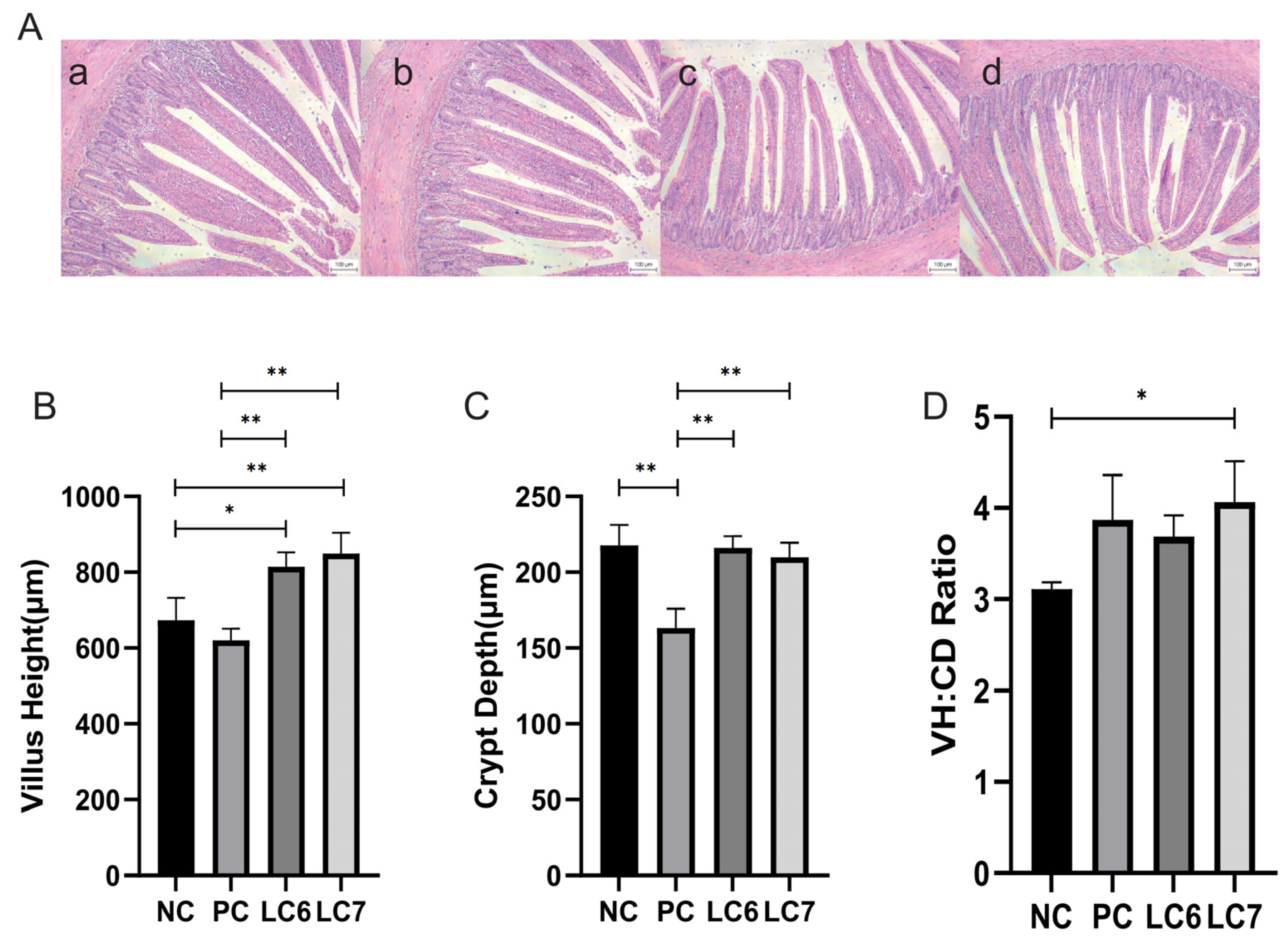
Figure 1. Effect of L. chiayiensis AACE3 group on jejunum morphology of chickens. (A) Jejunal histological samples by the groups (a): NC, (b): PC, (c): LC6, (d): LC7. (B) villus length, (C) crypt depth, (D) VH:CD ratio. Scale bars = 100 μm. * p < 0.05, ** p < 0.01.
Antibiotics 12 01356 g001
Figure 2. Effect of supplementation with L. chiayiensis AACE3 on duodenal digestive enzymes. (A) α-amylase activity. (B) Lipase activity. (C) Trypsin activity. * p < 0.05, ** p < 0.01.
Antibiotics 12 01356 g002
Figure 3. Influence of L. chiayiensis AACE3 on antioxidant enzyme activity and MDA levels in chickens. (A) MDA level content. (BE) level contents of SOD, CAT, GSH-Px, and T-AOC. * p < 0.05, ** p < 0.01, *** p < 0.001.
Antibiotics 12 01356 g003
Figure 4. Induction of serum immune factors by L. chiayiensis AACE 3. (A,B) are immunoglobulin IgA and IgG levels. (CE) are the levels of inflammatory factors IL-6, IL-1β, and TNF-α. * p < 0.05, ** p < 0.01.
Antibiotics 12 01356 g004
Figure 5. Alpha diversity of gut microbiota. (A) Ace. (B) Simpson. ** p < 0.01.
Antibiotics 12 01356 g005
Figure 6. Beta diversity of gut microbiota. (A) PCoA. (B) NMDS.
Antibiotics 12 01356 g006
Figure 7. Composition of the phylum of the gut microbiota in chickens. (A) Relative abundance of the first five phyla. (B) Comparative differences at the phylum level of the gut microbiota.
Antibiotics 12 01356 g007
Figure 8. Composition of the genus of the gut microbiota in chickens. (A) Relative abundance of the first 30 genera. (B) Comparative genus-level differences in the top 10 gut microbiota. * p < 0.05, ** p < 0.01, *** p < 0.001.
Antibiotics 12 01356 g008
Figure 9. Heat map of correlation coefficients between gut microbiota and digestive characteristics, antioxidant enzyme activity and immune factors of chickens at 21 days.” red” indicates a positive correlation (p < 0.05) and “blue” indicates a negative correlation (p < 0.05). * p < 0.05, ** p < 0.01, *** p < 0.001.
Antibiotics 12 01356 g009
Table 1. Effect of different doses of L. chiayiensis AACE 3 on growth performance of Nandan-Yao chickens from d 1 to 21 of the experiment.
VariablesNCPCLC6LC7SEM
BW (g)135.12 ± 4.86 a146.40 ± 10.32 a139.68 ± 3.14 a149.81 ± 6.37 a3.302
ADFI (g)
d 1 to d 2112.19 ± 0.16 a13.59 ± 0.60 b12.81 ± 0.27 ab13.57 ± 0.47 ab0.719
ADG (g)
d 1 to d 214.72 ± 0.07 a5.67 ± 0.09 b5.08 ± 0.15 ac5.61 ± 0.12 b0.226
FCR (g:g)
d 1 to d 212.58 ± 0.03 a2.40 ± 0.05 a2.52 ± 0.06 a2.41 ± 0.04 a0.044
The values are given as mean ± SEM. a,b,c, means in each raw with different superscripts are significantly different, means not significantly different with the same letter (p < 0.05). SEM: pooled standard error of the mean. BW: body weight; ADFI: average daily feed intake; ADG: average daily gain; FCR: feed conversion ratio between the NC group and all experimental groups.
Table 2. Ingredient composition and nutrient contents of basal diets.
Ingredients, %1–21 d
Corn55.41
Soybean meal 31.50
Palm oil5.00
Phosphorus 3.60
Calcium1.30
Salt0.34
Lysine HCL1.40
Methionine0.22
Arginine0.03
Vitamin–mineral premix 0.50
Limestone 0.60
Sodium carbonate 0.10
Metabolizable energy (MJ·kg−1)14.01
Crude protein20.0
Calcium1.00
Total phosphorus0.55
Lysine total1.41
Methionine0.50
Supplied per kilogram of diet: vitamin A (trans-retinyl acetate), 10,050 IU; vitamin D3, 2800 IU; vitamin E (DL-α-tocopheryl acetate), 50 mg; vitamin K3, 3.5 mg; thiamine, 2.5 mg; riboflavin, 7.5 mg; pantothenic acid, 15.3 mg; pyridoxine, 4.3 mg; vitamin B12 (cyanocobalamin), 0.02 mg; niacin, 35 mg; choline chloride, 1000 mg; biotin, 0.20 mg; folic acid, 1.2 mg; Mn, 100 mg; Fe, 85 mg; Zn, 60 mg; Cu, 9.6 mg; I, 0.30 mg; Co, 0.20 mg; and Se, 0.20 mg.
Disclaimer/Publisher’s Note: The statements, opinions and data contained in all publications are solely those of the individual author(s) and contributor(s) and not of MDPI and/or the editor(s). MDPI and/or the editor(s) disclaim responsibility for any injury to people or property resulting from any ideas, methods, instructions or products referred to in the content.