Article

Bioactive Naphtho-α-Pyranones from Two Endophytic Fungi of the Genus Polyphilus

1
Department of Microbial Drugs, Helmholtz Centre for Infection Research (HZI), Inhoffenstrasse 7, 38124 Braunschweig, Germany
2
Institute of Microbiology, Technische Universität Braunschweig, Spielmannstraße 7, 38106 Braunschweig, Germany
3
Department of Pharmacognosy, Faculty of Pharmacy, Ain Shams University, Cairo 11566, Egypt
4
Institute for Epidemiology and Pathogen Diagnostics, Julius Kühn Institute (JKI)—Federal Research Centre for Cultivated Plants, Messeweg 11-12, 38104 Braunschweig, Germany
5
Institute for Crop and Soil Science, Julius Kühn Institute (JKI)—Federal Research Centre for Cultivated Plants, Bundesallee 58, 38116 Braunschweig, Germany
6
Department of Organic Chemistry, University of Debrecen, P.O. Box 400, 4002 Debrecen, Hungary
*
Authors to whom correspondence should be addressed.
Antibiotics 2023, 12(8), 1273; https://doi.org/10.3390/antibiotics12081273
Submission received: 17 July 2023 / Revised: 29 July 2023 / Accepted: 31 July 2023 / Published: 2 August 2023
(This article belongs to the Section Fungi and Their Metabolites)

Abstract

:
In the course of our survey to study the metabolic potential of two species of a new helotialean genus Polyphilus, namely P. frankenii and P. sieberi, their crude extracts were obtained using different cultivation techniques, which led to the isolation and characterization of two new naphtho-α-pyranone derivatives recognized as a monomer (1) and its 6,6′-homodimer (2) together with two known diketopiperazine congeners, outovirin B (3) and (3S,6S)-3,6-dibenzylpiperazine-2,5-dione (4). The structures of isolated compounds were determined based on extensive 1D and 2D NMR and HRESIMS. The absolute configuration of new naphtho-α-pyranones was determined using a comparison of their experimental ECD spectra with those of related structural analogues. 6,6′-binaphtho-α-pyranone talaroderxine C (2) exhibited potent cytotoxic activity against different mammalian cell lines with IC50 values in the low micromolar to nanomolar range. In addition, talaroderxine C unveiled stronger antimicrobial activity against Bacillus subtilis rather than Staphylococcus aureus with MIC values of 0.52 µg mL−1 (0.83 µM) compared to 66.6 µg mL−1 (105.70 µM), respectively.

1. Introduction

Naphtho-α-pyranones comprise a unique class of fungal metabolites that have been reported as monomers, such as semiviriditoxin [1,2,3], semivioxanthin [1,2,3], and penicitor A [4] and its 7-O-methyl derivative [5]. In addition, naptho-α-pyranone dimers have been firstly reported as “mycotoxins”, such as viriditoxin [6] followed by several other symmetric dimers featuring either 6,6′-linkages, such as asteromine [7], talaroderxines [8], pigmentosins [9,10], and aschernaphtopyrone A [11], or 8,8′-linkages, such as vioxanthin [12,13], mycopyranone [14], aschernaphtopyrone B [11], and lichenocholin A [15].
A rare 5,8′-linkage was also recently reported in lulworthinone obtained from a marine-derived fungus: Lulworthia medusa [16]. Several members of the dimer class have been reported to exhibit potential antibacterial activity. In particular, viriditioxin has been reported to inhibit FtsZ, which is essential for bacterial cell division [17]. Based on the fact that antibiotic resistance is an exacerbating problem, the need for new antimicrobial agents remains an unmet demand.
During our ongoing research targeting the discovery of new fungal metabolites with potential antimicrobial activities, in this study, we investigated the root endophytic representatives of the recently described fungal genus Polyphilus, namely P. frankenii and P. sieberi [18]. The chemical exploration resulted in the isolation and identification of a monomer (1) and its symmetric 6,6′-homodimer (2) (Figure 1) together with two known metabolites identified as outovirin B (3) [19] and (3S,6S)-3,6-dibenzylpiperazine-2,5-dione (4) [20]. In this study, we report the isolation and structure elucidation of two new naphto-α-pyranone derivatives together with their antimicrobial and cytotoxic activity, which were recorded in our routine assays.

2. Results and Discussion

2.1. Isolation and Identification of Compounds (1 and 2)

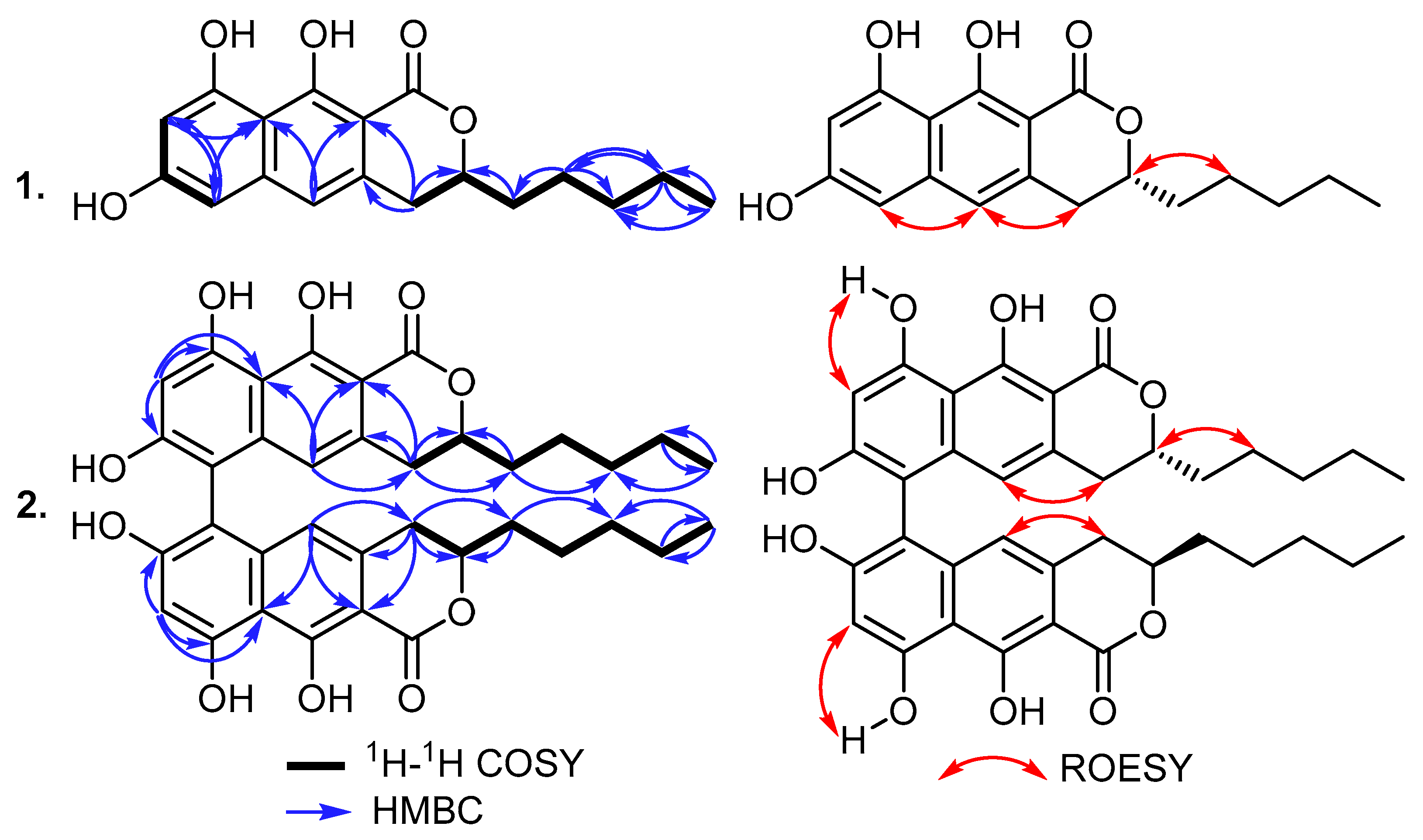
Compound 1 was purified as a white solid powder, and its molecular formula was established to be C18H20O5 based on its HRESIMS spectrum, which revealed pseudomolecular ion peaks at m/z 317.1386 [M+H]+ (calculated for 317.1384) and at m/z 339.1202 [M+Na]+ (calculated for 339.1203), indicating the existence of nine degrees of unsaturation. The 13C NMR spectral data of 1 (Table 1) displayed the presence of fifteen carbon resonances that can be differentiated into eight quaternary carbon atoms recognized as one carbonyl at δC 170.7 (C-1) and seven olefinic (two oxygenated) carbon atoms at δC 162.6 (C-10), 161.0 (C-7), 160.7 (C-9), 140.6 (C-5a), 133.9 (C-4a), 107.2 (C-9a), and 98.8 (C-10a). In addition, the 13C NMR spectrum of 1 also revealed the presence of four tertiary (one aliphatic and three olefinic) carbon atoms at δC 114.5 (C-5), 101.5 (C-8), 101.4 (C-6), and 79.3 (C-3) along with four methylenes (δC 34.0 (C-11), 32.3 (C-4), 31.0 (C-13), and 22.0 (C-14)) and one methyl carbon atom (δC 13.7 (C-15)). By comparing the obtained results with the reported literature, compound 1 was suggested to be a naphthopyrone derivative related to those reported as fungal metabolites, such as penicitor A [4,5], semiviriditoxin [1,2], and semivioxanthin [3]. Further structural features of 1 were concluded using 2D NMR spectra including 1H-1H COSY, HMBC, and HSQC. The 1H-1H COSY spectrum of 1 revealed two main spin systems, with one extending over two meta-positioned aromatic protons at δH 6.31 (H-6) and 6.47 (H-8) while the second spin system was found to begin at a methylene group at δH 2.88/δH 3.00 (H2-4) and extended to an aliphatic oxygenated methine group at δH 4.58 (H-3), thus extending over four methylenes, forming an aliphatic side chain at δH 1.68/δH 1.74 (H2-11), δH 1.41/δH 1.46 (H2-12), and δH 1.31 (H2-14 and H2-13), and ending with a terminal triplet methyl group at δH 0.88 (H3-15) to confirm the presence of the n-pentyl side chain. The HMBC spectrum of 1 (Figure 2) revealed key correlations from H2-4 to four carbon atoms ascribed to C-10a (δC 98.8), C-4a (δC 133.9), C-3 (δC 79.3), and C-11 (δC 34.0), whereas additional HMBC correlations from H2-11 to C-3 confirmed that the n-pentyl side chain was present at C-3. Further key HMBC correlations (Figure 2) were also observed in the aromatic protons from H-5, H-6, and H-8 to C-9a (δC 107.2) and from H-5 and H2-4 to C-10a, which confirmed the depicted structure of 1 as a naphthopyrone derivative. The ECD spectrum of 1 (see Supplementary Materials: Figure S9) showed negative Cotton effects (CEs) at 219 nm and 267 nm, a weaker positive one at 242 nm, and a broad positive plateau within the range of 280-400. This ECD spectrum was a near mirror image of that of (S)-7-O-methylpenicitor A [5], which differed only in the C-9 and C-14 methoxy groups, and its absolute configuration (AC) was determined using ECD calculations and single-crystal X-ray diffraction analysis. Due to the mirror image ECD curves, the AC of 1 was assigned as (R), and as a new naphthopyrone derivative, it was given the trivial name semitalaroderxine C.
Compound 2 was isolated as a white solid powder that revealed in a pseudomolecular ion peak at m/z 631.2541 [M+H]+ (calculated for 631.2538) its HRESIMS spectrum, confirming its molecular formula as C36H38O10 and indicating its inclusion of eighteen degrees of unsaturation. Intriguingly, by comparing the molecular formulas of 1 and 2, it could be obviously observed that 2 is a symmetric dimer of the two monomers of 1. This assumption was further confirmed using 13C NMR spectral data (Table 1), which revealed only eighteen carbon resonances; thus, each was assigned to two electromagnetically equivalent carbon atoms in the two monomers. The 1H NMR spectral data of 2 (Table 1) displayed an identical set of proton resonances to those of 1, except in the absence of one aromatic proton at δH 6.31 (H-6) in 1 and hence suggesting that compound 2 is a 6,6′-binaphthopyrone dimer of semitalaroderxine C (1). Based on the obtained results and by searching the reported literature, compound 2 was found to be related to the previously reported 6,6′-binaphthopyrone dimers, talaroderxines A and B, that were reported from a soil-derived fungus Talaromyces derxii [8] and pigmentosins A/B [10,21].
The major structural difference between 2 and talaroderxines A/B was the presence of n-pentyl in 2 instead of the n-propyl side chain in talaroderxines A/B. Since compound 2 is the 6,6′-linked axially chiral homodimer of 1, the (3S,3′S) absolute configuration of the central chirality elements was deduced on the basis of their common biosynthetic origin. The (aS) axial chirality of 2, arising from the hindered rotation around the C-6-C-6′ biaryl axis, was determined by comparing its experimental ECD spectrum (see Supplementary Materials Figure S18) with those of related 6,6′-linked bis-naphthopyrone pigmentosins A and B [10] and talaroderxine A [8]. Compound 2 showed an intense positive exciton-coupled couplet centered at 260 nm (268 nm (Δε: +16.58); 252 nm (Δε: −14.15)), which was a mirror image of the negative couplet of (aR)-pigmentosins A and B and (aR)-talaroderxine B [8,9,10]. Compound 2 was identified as a new 6,6′-linked bis-naphthopyrone homodimer with (aS) axial chirality, which was named talaroderxine C.

2.2. Biological Assays

Due to the limited amounts of compounds 1, 3, and 4, only talaroderxine C (2) was subjected to antimicrobial and cytotoxicity assays. Against all tested microorganisms, talaroderxine C disclosed potent antimicrobial activity against Bacillus subtilis with a minimal inhibitory concentration (MIC) of 0.52 µg/mL (0.83 µM). In the case of Staphylococcus aureus, it revealed moderate activity with an MIC of 66.6 µg/mL (105.70 µM). In addition, the obtained results of the cytotoxicity (3-(4,5-dimethylthiayol-2-yl)-2,5-diphenyltetrazolium bromide, MTT) assay (Table 2) revealed that talaroderxine C (2) exhibits pan-cytotoxic activity against all tested cell lines, with IC50 values falling within the low micromolar to nanomolar range. In particular, against breast adenocarcinoma (MCF-7) cells, talaroderxine C revealed a high potency (IC50 = 68 nM). Nevertheless, outovirin B (3) is structurally recognized as a gliovirin-like compound [22] that was reported to exhibit selective antifungal and anti-inflammatory [23] and antitrypanosomal [24] and antimycobacterial [25] activities.

3. Materials and Methods

3.1. General Experimental Procedures

UV measurements were performed using a Shimadzu UV-VIS spectrophotometer UV-2450 (Shimadzu®, Kyoto, Japan), and ECD spectra were obtained using a Jasco J-815 spectropolarimeter (JASCO®, Pfungstadt, Germany). Optical rotation values were measured using a PerkinElmer 241 polarimeter at 20 °C (Anton-Paar Opto Tec GmbH, Seelze, Germany). High-resolution electrospray ionization mass spectra (HR-ESI-MS) were acquired using an Agilent 1200 Infinity Series HPLC-UV system (Agilent Technologies®, Santa Clara, CA, USA), utilizing a C18 Acquity UPLC BEH column (2.1 × 50 mm, 1.7 µm: Waters, Milford, MA, USA); solvent A: H2O + 0.1% formic acid; solvent B: acetonitrile (MeCN) + 0.1% formic acid; gradient: 5% B for 0.5 min increasing to 100% B in 19.5 min and maintaining 100% B for 5 min at a flow rate of 0.6 mL min−1 (UV/Vis detection 190–600 nm) connected to a time-of-flight mass spectrometer (ESI-TOF-MS, Maxis, Bruker, Billerica, MA, USA) (scan range: 100–2500 m/z; rate 2: Hz; capillary voltage: 4500 V; dry temperature: 200 °C). NMR spectra were recorded using an Avance III 500 spectrometer (Bruker®, Billerica, MA, USA; 1H-NMR: 500 MHz; 13C-NMR: 125 MHz), dissolving compounds in deuterated methanol-d4 and deuterated DMSO-d6.

3.2. Fermentation, Extraction, and Isolation

The two fungal species explored in this study namely, P. frankenii strain V16 (DSM 106521) and P. sieberi strain Ref052 (DSM 106515) were recognized as endophytic fungi associated with the roots of Pinus sylvetris and Asclepias syriaca collected from Villerupt (France) and Bugac (Hungary), respectively. The fungi were maintained on a YM6.3 agar (D-glucose: 4 g L−1; malt extract: 10 g L−1: yeast extract: 4 g L−1; agar: 20 g L−1; pH adjusted to 6.3 and sterilized by autoclaving) at 23 °C. To prepare seed cultures for the following larger-scale cultivation, five mycelia plugs measuring 25 mm2 were transferred to a 500 mL Erlenmeyer shaking flask containing 200 mL of Q6/2 media (D-glucose: 2.5 g L−1; glycerine: 10 g L−1; cotton seed flour: 5 g L−1; pH: 7.2) and incubated at 23 °C and 140 min−1. After achieving a sufficient amount of biomass, the culture broth was homogenized using Ultra-Turrax (T25 easy clean digital, IKA) equipped with an S25 N-25F dispersing tool at 10,000 rpm for 10 s. This seed culture was used for all following cultivations as an inoculum for YM6.3, BRFT, and WOFT media (K2HPO4: 0.5 g L−1; sodium tartrate: 0.5 g L−1; yeast extract: 1 g L−1; 100 mL of solution was added to 28 g brown rice or whole oat and autoclaved).

3.2.1. Solid State Fermentation

Six Erlenmeyer culture flasks with BRFT and WOFT were inoculated with 6 mL of homogenized seed culture, mixed under sterile conditions, and incubated at room temperature for two, three, and four weeks in the dark. After the planned incubation time, the cultures were stopped by adding 250 mL of acetone, mixed with a spatula, and kept in an ultrasonic bath for 30 min at 40 °C for extraction. The liquid was separated from the solid phase via filtration. The extraction was repeated twice using fresh acetone. The filtrate was evaporated under a vacuum at 40 °C in a rotary evaporator, and the remaining aqueous phase was filled up to 50 mL with distilled water and extracted with EtOAc (1:1) in a separatory funnel three times. The organic phase (EtOAc) was separated, evaporated under reduced pressure, and then dissolved again in 5 mL of MeOH and extracted again with 45 mL of heptane. Extraction was performed in a separatory funnel twice with fresh heptane. Both fractions (heptane and methanol) were evaporated to dryness.

3.2.2. Liquid Fermentation

In this study, 0.5% of the homogenized inoculum was transferred into twelve 2 L Erlenmeyer culture flasks containing 400 mL of YM6.3 medium. The content of glucose was monitored using test stripes (Medi-Test Glucose, Machery-Nagel®, Düren, Germany). Incubation was terminated after five days of glucose depletion. Mycelia and supernatant were separated using a Büchner funnel and a vacuum pump. The extraction of metabolites of the mycelia followed the procedure of solid-state fermentation. The culture broth was mixed with 2% AmberLiteTM XADTM 16N polymeric absorbent and stirred for 4 h on a magnetic stirrer. The extraction of metabolites was carried out with acetone under stirring. Afterward, the purification scheme followed the same steps as described above for solid-state extraction.

3.2.3. Analytical HPLC

The extracts obtained were dissolved in Acetone:MeOH (1:1) and adjusted to a concentration of 4.5 mg mL−1. An injection volume of 2 µL was applied to an UltiMate® 3000 Series uHPLC (Thermo Fisher Scientific®, Waltman, MA, USA). Mass spectrometry was performed with a connected amaZon® speed ESI Iontrap MS (Amazon, Bruker). HRESIMS measurements were performed with sample concentrations of 1 mg mL−1 with an Agilent 1200 series HPLC-UV system in combination with an ESI-TOF-MS (Maxis, Bruker). The conditions were identical to the methods described before [26].

3.2.4. Isolation of Compounds

The mycelial extract of Polyphilus sieberi Ref052 (DSM 106515) cultivated in YM6.3 media (86 mg) was separated with a Reveleris® X2 flash chromatography system using a FlashPure ID Silica 12 g cartridge. The mobile phase consisted of three solvent mixtures supplemented with 0.1% formic acid: solvent A (heptane 100%), solvent B (heptane 58%, TBME 40%, and MeOH 2%), and solvent C (Acetone 37.5%, DCM 37.5%, and MeOH 25%); flow rate: 30 mL min−1; gradient: 3 min B at 0%, increasing to 100% B in 10 min, maintaining 100% B for 5 min, switching to solvent mixture B and C, starting from 0% C and increasing to 100% C in 10 min, and maintaining 100% C for 10 min. All the following flash chromatographic separations were performed, implementing the same conditions. The separation led to the purification of 1 (1.1 mg; tR = 8.5 min).
The second Polyphilus frankenii V16 (DSM 106521) strain was cultivated in BRFT and WOFT media. Due to the high similarity observed in ESI-MS profiles for 2–4 weeks, the extracts were combined to yield a crude extract (672 mg) and subjected to the Reveleris© X2 equipped with a FlashPure ID Silica 24 g cartridge. Further purification steps of the resulting fraction 5 (49.2 mg, tR = 13.5 min) were carried out on a Büchi Pure C-850 FlashPrep equipped with a Gemini C18 (250 × 21.2 mm, 10 µm; Phenomenex) (solvent A: H2O + 0.1% formic acid; solvent B: MeCN + 0.1% formic acid; flow rate: 20 mL min−1; gradient: 5 min maintaining 40% B, increasing B to 90% in 50 min, increasing to 100% B in 5 min, and maintaining 100% B for 10 min) led to the isolation of 2 (15.8 mg, tR = 53.4 min). The mycelial extract of the cultivation in YM6.3 media was weighed at 35 mg, and it was purified on a Gilson PLC 2250 equipped with a Gemini C18 (250 × 21.2 mm, 10 µm; Phenomenex, Torrance, CA, USA) (solvent A: H2O + 0.1% formic acid; solvent B: MeCN + 0.1% formic acid; flow rate: 20 mL min−1; gradient: 10 min maintaining 5% B, increasing B to 50% in 45 min, increasing to 100% B in 10 min, and maintaining 100% B for 10 min. This led to the isolation of known metabolites 3 (0.2 mg; tR = 17.3 min) and 4 (0.2 mg; tR = 27.5 min).
3.2.1 Semitalaroderxine C (1): white solid powder; 0.59 mg; [ α ] D 20 –7.5 (c 0.04, chloroform); UV/Vis (MeOH): λmax (log ε) = 193.5 (0.8), 200.0 (0.9), 220.5 (1.0), 261.0 (2.7), 314.5 (0.2), 369.5 (0.6) nm; ECD (c = 3.16 × 10⁻⁴ M; MeOH) λ [nm], (Δε) 373 (1.4), 310 (1.0), 268 (−3.2), 243 (1.1), 218 (−4.6), 196 (4.2); NMR data (1H NMR: 500 MHz, 13C NMR: 125 MHz in DMSO-d6) see Table 1; HR-(+)ESIMS: m/z 299.1277 [M–H2O+H]+ (calcd. 299.1278 for C18H19O4+), m/z 317.1386 [M+H]+ (calcd. 317.1384 for C18H21O5+), 339.1203 [M+Na]+ (calcd. 339.1203 for C18H20NaO5+), 655.2516 [2M+Na]+ (calcd. 655.2514 for C36H40NaO10+).
3.2.2 Talaroderxine C (2): white solid powder; 3.49 mg; [ α ] D 20 +57.5 (c 1.0, DMSO-d6); UV/Vis (MeOH): λmax (log ε) = 223.0 (0.1), 265.5 (0.1), 376.5 (0.04) nm; ECD (c = 3.17 × 10⁻⁴ M; MeOH) λ [nm], (Δε) 268 (82.9), 252 (−70.7); NMR data (1H NMR: 500 MHz, 13C NMR: 125 MHz in DMSO-d6) see Table 1; HR-(+)ESIMS: m/z 631.2541 [M+H]+ (calcd. 631.2538 for C36H39O10+).

3.3. Antimicrobial Assay

Due to the limited amounts of compounds 1, 3, and 4, only talaroderxine C (2) was assessed for their antimicrobial activity based on their minimum inhibitory concentration (MIC), following our previously reported protocol [26], against five pathogenic fungi, including Candida albicans DSM 1665, Mucor hiemails DSM 2656, Pichia anomala DSM 6766, Rhodotorula glutinis DSM 10134, and Schizosaccharomyces pombe DSM 70572; three Gram-positive bacteria, including Bacillus subtilis DSM 10, Mycobacterium smegmatis DSM ATCC 700084, and Staphylococcus aureus DSM 436; and four Gram-negative bacteria, including Chromobacterium violaceum DSM 30191, Acinetobacter baumanii DSM 30008, Escherichia coli DSM 1116, and Pseudomonas aeruginosa DSM PA14. Nystatin was used as an antifungal positive control, whereas oxytetracycline, ciprofloxacin, gentamycin, and kanamycin were used as positive controls against Gram-positive and Gram-negative bacteria.

3.4. Cytotoxicity Assay

The cytotoxicity (IC50) of Talaroderxine C (2) was also tested using the MTT (3-(4,5-dimethylthiayol-2-yl)-2,5-diphenyltetrazolium bromide) test, following the experimental procedure described by Charria-Girón et al. [27]. These compounds were tested against seven mammalian cell lines, including human endocervical adenocarcinoma KB 3.1, breast cancer MCF-7, lung cancer A549, ovary cancer SK-OV-3, prostate cancer PC-3, squamous cancer A431, and mouse fibroblasts L929. Epothilone B was used as the positive control.

4. Conclusions

This represents the first study of secondary metabolites from the fungal genus Polyphilus. In summary, semitalaroderxine C (1), talaroderxine C (2), outovirin B (3), and (3S,6S)-3,6-dibenzylpiperazine-2,5-dione (4) could be isolated and elucidated where 1 and 2 are recognized among naphtha-α-pyranone congeners that are well-known for a vast array of bioactivities. Talaroderxine C showed potent antimicrobial activity against Bacillus subtilis with an MIC of 0.52 µg/mL. In addition, it exhibited pan-cytotoxic activity against all tested cell lines, with IC50 values falling in the low micromolar to nanomolar range. Talaroderxine C (2) showed high potency (IC50 = 68 nM), particularly against breast adenocarcinoma (MCF-7) cells. Due to the limited amounts of 3 and 4, antimicrobial and cytotoxic activity assays were not determined, but the gliovirin-like compound [22], outovirin B (3), was reported to exhibit selective antifungal and anti-inflammatory [23] along with antitrypanosomal [24] and antimycobacterial [25] activities.
The strains of the genus Polyphilus arose from an attempt to find nematode antagonistic fungi that can be used as ecologically friendly alternatives to toxic chemicals and soil fumigants. Such biocontrol agents may turn out to be a valid alternative, especially as they are supposed to be host-specific and do not kill harmless organisms in the soil. However, the results of the current study, which showed that Polyphilus strains can produce rather toxic secondary metabolites, should give rise to deprioritizing them over other strains that were encountered concurrently and that were devoid of toxic metabolites. Mycopesticides, which are mostly used against insects, already have an annual market share of ca. USD 550 million [28], and this is bound to increase further in the future. Nematode antagonists will probably obtain their share as they are one of the only environmentally friendly alternatives to chemical pesticides.

Supplementary Materials

The following supporting information can be downloaded at https://www.mdpi.com/article/10.3390/antibiotics12081273/s1. Tables S1 and S2: 2D NMR data of semitalaroderxine C (1) and talaroderxine C (2); Figures S1–S4: LC-ESI-MS spectra of sole four-weeks mycelia extracts of P. sieberi or P. frankenii in different solid or liquid media; Figures S5–S14: HPLC, UV, LR-/HRESIMS, 1D/2D NMR, and ECD spectra of 1; Figures S15–S24: HPLC, UV, LR-/HRESIMS, 1D/2D NMR, and ECD spectra of 2; Figures S25–S30: HPLC and LR-/HRESIMS 1D/2D NMR spectra of 3; Figures S31–S38: HPLC and LR-/HRESIMS 1D/2D NMR spectra of 4.

Author Contributions

Conceptualization: J.-P.W., S.S.E. and M.S.; culture characterization: S.A. and W.M.; methodology and investigation: J.-P.W., E.S. and N.A.L.-L.; data curation and interpretation: J.-P.W., S.S.E. and T.K.; visualization: J.-P.W. and S.S.E.; writing—original draft preparation: J.-P.W. and S.S.E.; writing—review and editing: all authors. All authors have read and agreed to the published version of the manuscript.

Funding

This work was supported by funds from Landwirtschaftliche Rentenbank, Germany (Project “MycoNem”).

Institutional Review Board Statement

Not applicable.

Informed Consent Statement

Not applicable.

Data Availability Statement

All data are available in the manuscript or the SI.

Acknowledgments

S. Pfütze, E. Charria-Girón, and F. Surup are acknowledged for their expert advisory assistance. A. Skiba, S. Reinecke, C. Kakoschke, E. Surges, and W. Collisi are thanked for their expert technical assistance. D.G. Knapp, D. Blaudez, M. Chalot, and G.M. Kovács are acknowledged for collecting and depositing the fungal strains. T. Kurtán thanks the financial support from the National Research Development and Innovation Office (Grant number K-138672). The Georg-Forster Fellowship for Experienced Researchers stipend (Ref 3.4-1222288-EGY-GF-E) that Alexander von Humboldt (AvH) Foundation granted to S.S. Ebada is gratefully acknowledged. Financial support by a personal PhD stipend from the German Academic exchange service DAAD to N.A.L.-L. is immensely acknowledged (program ID 57552340).

Conflicts of Interest

The authors declare no conflict of interest.

References

  1. Ayer:, W.A.; Craw, P.A.; Nozawa, K. Two 1H-naphtho [2,3-c]pyran-1-one metabolites from the fungus Paecilomyces variotii. Can. J. Chem. 1991, 69, 189–191. [Google Scholar] [CrossRef]
  2. Liu, J.; Li, F.; Kim, E.L.; Hong, J.; Jung, H.J. Viriditoxin, from a jellyfish-derived fungus, is antibiotic to fish pathogens. Nat. Prod. Sci. 2013, 19, 61–65. [Google Scholar]
  3. Elix, J.A.; Wardlaw, J.H. Pigmentosin A, a new naphthopyrone from the lichen Hyptotrachyna immaculata. Aust. J. Chem. 2004, 57, 681–683. [Google Scholar] [CrossRef]
  4. Kong, F.D.; Zhou, L.M.; Ma, Q.Y.; Huang, S.Z.; Wang, P.; Dai, H.F.; Zhao, Y.X. Metabolites with Gram-negative bacteria quorum sensing inhibitory activtiy from the marine animal endogenic fungus Penicillium sp. SCS-KFDo8. Arch. Pharm. Res. 2017, 40, 25–31. [Google Scholar] [CrossRef]
  5. Luo, X.-W.; Gao, C.-H.; Han, F.-H.; Chen, X.-Q.; Lin, X.-P.; Zhou, X.-F.; Wang, J.-J.; Liu, Y.-H. A new naphthopyranone from the sponge-associated fungus Penicillium sp. XWSo2F62. Magn. Reson. Chem. 2019, 57, 982–986. [Google Scholar] [CrossRef]
  6. Suzuki, K.; Nozawa, K.; Nakajima, S.; Kawai, K. Structure revision of mycotoxin, viriditoxin, and its derivatives. Chem. Pharm. Bull. 1990, 38, 3180–3181. [Google Scholar] [CrossRef] [Green Version]
  7. Arnone, A.; Assante, G.; Montorsi, M.; Nasini, G. Asteromine, a bioactive secondary metabolite from a strain of Mycosphaerella asteroma. Phytochemistry 1995, 38, 595–597. [Google Scholar] [CrossRef]
  8. Suzuki, K.; Nozawa, K.; Nakajima, S.; Udagawa, S.; Kawai, K. Isolation and structures of antibacterial binaphtho-α-pyrones, talaroderxines A and B, from Talaromyces derxii. Chem. Pharm. Bull. 1992, 40, 1116–1119. [Google Scholar] [CrossRef] [Green Version]
  9. Tan, N.P.H.; Donner, C.D. Total synthesis and confirmationn of the absolute stereochemistry of semiviriditoxin, a naphthopyranone metabolite from the fungus Paecilomyces variotii. Tetrahedron 2009, 65, 4007–4012. [Google Scholar] [CrossRef]
  10. Helaly, S.E.; Kuephadungphan, W.; Phainuphong, P.; Ibrahim, M.A.A.; Tasanathai, K.; Mongkolsamrit, S.; Luangsa-ard, J.J.; Phongpaichit, S.; Rukachaisirikul, V.; Stadler, M. Pigmentosins from Gibellula sp. as antibiofilm agents and a new glycosylated asperfuran from Cordyceps javanica. Beilstein J. Org. Chem. 2019, 15, 2968–2981. [Google Scholar] [CrossRef] [Green Version]
  11. Isaka, M.; Yangchum, A.; Rachtawee, P.; Komwijit, S.; Lutthisungneon, A. Hopane-type triterpenes and binaphthopyrones from the scale insect pathogenic fungus Aschersonia paraphysata BCC 11964. J. Nat. Prod. 2010, 73, 688–692. [Google Scholar] [CrossRef] [PubMed]
  12. Bode, S.E.; Drochner, D.; Müller, M. Synthesis, biosynthesis and absolute configuration of vioxanthin. Angew. Chem. Int. Ed. 2007, 46, 5916–5920. [Google Scholar] [CrossRef] [PubMed]
  13. Blank, F.; Ng, A.S.; Just, G. Metabolites of pathogenic fungi. V. Isolation and tentative structures of vioxanthin and viopurpurin, two coloured metabolites from Trichophyton violaceum. Can. J. Chem. 1966, 44, 2873–2879. [Google Scholar] [CrossRef]
  14. Rivera-Chávez, J.; Caesar, L.K.; Garcia-Salazar, J.J.; Raja, M.A.; Cech, N.B.; Pearce, C.J.; Oberlies, N.H. Mycopyranone: A 8,8′-binaphthopyranone with potent anti-MRSA activity from the fungus Phialemoniopsis sp. Tetrahedron Lett. 2019, 60, 594–597. [Google Scholar] [CrossRef]
  15. He, H.; Bigelis, R.; Yang, H.Y.; Chang, L.-P.; Singh, M.P. Lichenicolins A and B, new bisnaphthopyrones from an unidentified lichenicolous fungus, strain LL-RB0668. J. Antibiot. 2005, 58, 731–736. [Google Scholar] [CrossRef] [PubMed]
  16. Jenssen, M.; Rainsford, P.; Juskewitz, E.; Andersen, J.H.; Hansen, E.H.; Isaksson, J.; Rämä, T.; Hansen, K.Ø. Lulworthinone, a new dimeric naphthopyrone from a marine fungus in the family Lulworthiaceae with antibacterial activity against clinical methicillin-resistant Staphylococcus aureus isolates. Front. Microbiol. 2021, 12, 730740. [Google Scholar] [CrossRef]
  17. Wang, J.; Galgoci, A.; Kodali, S.; Herath, K.B.; Jayasuriya, H.; Dorso, K.; Vicente, F.; González, A.; Cully, D.; Bramhill, D.; et al. Discovery of a small molecule that inhibits cell division by blocking FtsZ, a novel therapeutic target of antibiotics. J. Biol. Chem. 2003, 278, 44424–44428. [Google Scholar] [CrossRef] [Green Version]
  18. Ashrafi, S.; Knapp, D.G.; Blaudez, D.; Chalot, M.; Maciá-Vicente, J.G.; Zagyva, I.; Dababat, A.A.; Maier, W.; Kovács, G.M. Inhabiting plant roots, nematodes and truffles-Polyphilus, a new helotialean genus two globally distributed species. Mycologia 2018, 110, 286–299. [Google Scholar] [CrossRef]
  19. Kajula, M.; Ward, J.M.; Turpenien, A.; Tejesvi, M.V.; Hokkanen, J.; Tolonen, A.; Häkkänen, H.; Picart, P.; Ihalainen, J.; Sahl, H.-G.; et al. Bridged epipolythiodiketopiperazines from Penicillium raciborskii, an endophytic fungus of Rhododendron tomentosum Harmaja. J. Nat. Prod. 2016, 79, 685–690. [Google Scholar] [CrossRef]
  20. Tang, R.; Zhou, D.; Kimishima, A.; Setiawan, A.; Arai, M. Selective cytotoxicity of marine-derived fungal metabolite (3S,6S)-3,6-dibenzylpiperazine-2,5-dione against cancer cells adapted to nutrient starvation. J. Antibiot. 2020, 73, 873–875. [Google Scholar] [CrossRef]
  21. Grove, C.I.; Di Maso, M.J.; Jaipuri, F.A.; Kim, M.B.; Shaw, J.T. Synthesis of 6,6′-binaphthopyran-2-one natural products: Pigmentosin A, talaroderxines A and B. Org. Lett. 2012, 14, 4338–4341. [Google Scholar] [CrossRef] [Green Version]
  22. Yamazaki, H.; Rotinsulu, H.; Narita, R.; Takahashi, R.; Namikoshi, M. Induced production of halogenated epidithiodiketopiperazines by a marine-derived Trichoderma cf. brevicompactum with sodium halides. J. Nat. Prod. 2015, 78, 2319–2321. [Google Scholar] [CrossRef]
  23. Stipanovic, R.D.; Howell, C.R. The structure of gliovirin, a new antibiotic from Gliocladium virens. J. Antibiot. 1982, 35, 1326–1330. [Google Scholar] [CrossRef] [PubMed]
  24. Iwatsuki, M.; Otoguro, K.; Ishiyama, A.; Namatame, M.; Nishihara-Tukashima, A.; Hashida, J.; Nakashima, T.; Masuma, R.; Takahashi, Y.; Yamada, H.; et al. In vitro antitrypanosomal activity of 12-low-molecular-weight antibiotics and observations of structure/activity relationships. J. Antibiot. 2010, 63, 619–622. [Google Scholar] [CrossRef] [PubMed] [Green Version]
  25. Seephonkai, P.; Kongsaeree, S.; Prabpai, S.; Isaka, M.; Thebtaranonth, Y. Transformation of an irregularly bridged epidithiodiketopiperazine to trichodermamide A. Org. Lett. 2006, 8, 3073–3075. [Google Scholar] [CrossRef]
  26. Chepkirui, C.; Cheng, T.; Matasyoh, J.; Decock, C.; Stadler, M. An unprecedented spiro[furan-2,1’-indene]-3-one derivative and other nematicidal and antimicrobial metabolites from Sanghuangporus sp. (Hymenochaetaceae, Basidiomycota) collected in Kenya. Phytochem. Lett. 2018, 25, 141–146. [Google Scholar] [CrossRef]
  27. Charria-Girón, E.; Stchigel, A.M.; Čmoková, A.; Kolařík, M.; Surup, F.; Marin-Felix, Y. Amesia hispanica sp. nov., producer of the antifungal class of antibiotics dactylfungins. J. Fungi 2023, 9, 463. [Google Scholar] [CrossRef] [PubMed]
  28. Niego, A.G.T.; Lambert, C.; Mortimer, P.; Thongklang, N.; Rapior, S.; Grosse, M.; Schrey, H.; Charria-Girón, E.; Walker, A.; Stadler, M. The contribution of fungi to the global economy. Fungal Divers. 2023, in press. [Google Scholar] [CrossRef]
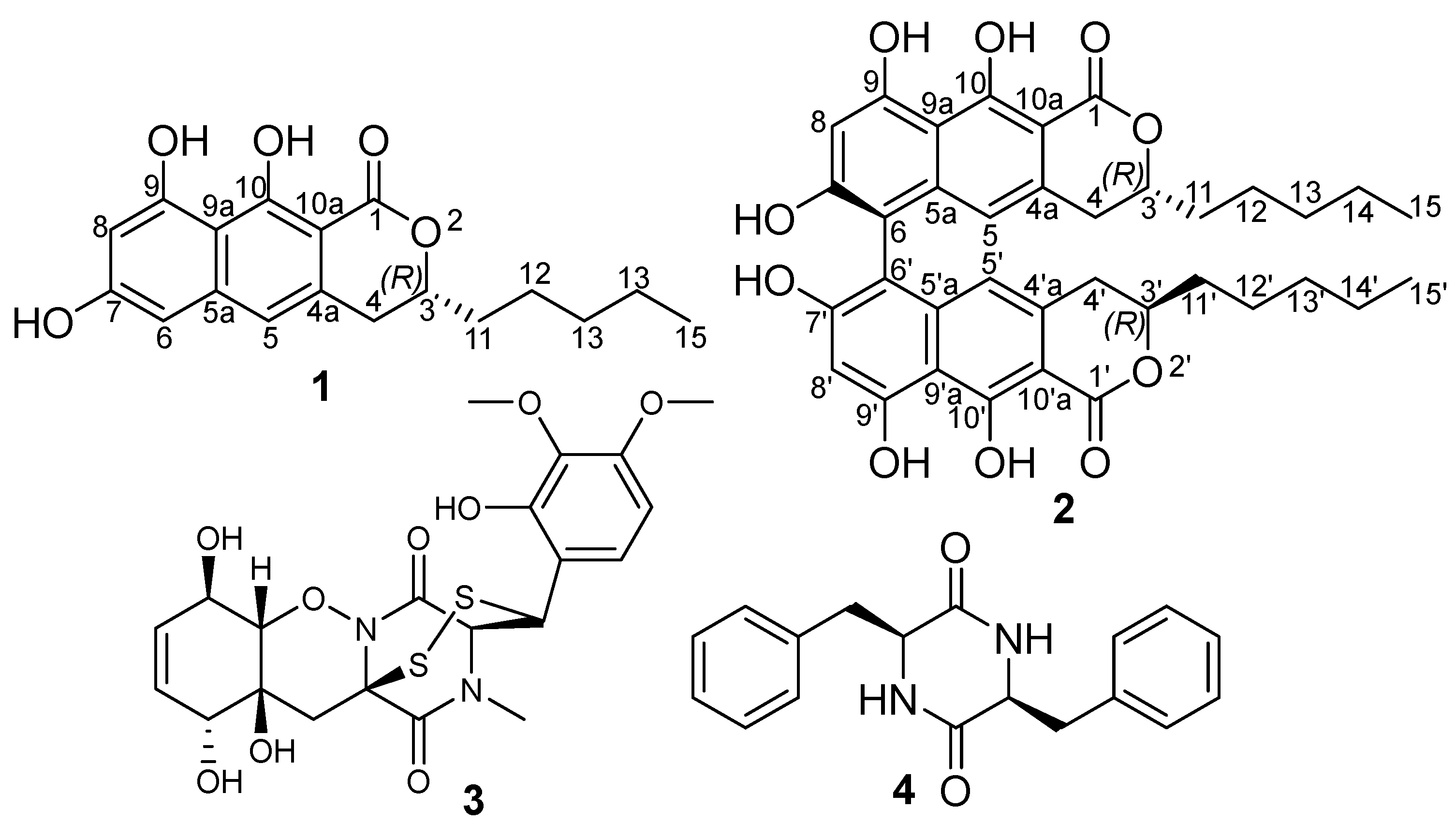
Figure 1. Chemical structures of 14.
Antibiotics 12 01273 g001
Figure 2. Key COSY, HMBC, and ROESY correlations of 1 and 2.
Antibiotics 12 01273 g002
Table 1. 1H and 13C NMR data of 1 and 2.
1 2
Pos.δH (Multi, J(Hz)) aδC, Type b,cPos.δH (Multi, J(Hz)) aδC, Type b,c
1 170.7, CO1/1′ 170.6, CO
34.58, m79.3, CH3/3′4.50, m79.4, CH
4α 2.88, dd, (15.6, 11.1)
β 3.00, br d, (15.6)
32.3, CH24/4′α 2.72, dd, (16.2, 11.0)
β 2.81, dd, (16.5, 3.1)
32.5, CH2
4a 133.9, C4a/4′a 133.6, C
56.82, s114.5, CH5/5′6.20, s112.7, CH
5a 140.6, C5a/5′a 139.9, C
66.31, s101.4, CH6/6′ 107.7, C
7 161.0, C, C7/7′ 157.8, C
86.47, s101.5, CH8/8′6.57, s101.6, CH
9 160.7, C9/9′ 158.9, C
9a 107.2, C9a/9′a 107.7, C
10 162.6, C10/10′ 163.2, C
10a 98.8, C10a/10′a 98.7, C
11α 1.68, m
β 1.74, m
34.0, CH211/11′α 1.57, ddd, (14.2, 10.8, 5.6)
β 1.66, m
34.1, CH2
12α 1.41, m; β 1.46, m24.0, CH212/12′1.36, m (2H)23.9, CH2
131.31, m, 2H31.0, CH213/13′1.23, m (2H)31.0, CH2
141.31, m, 2H22.0, CH214/14′1.25, m (2H)22.0, CH2
150.88, t, (7.0)13.9, CH315/15′0.84, t (6.8, 3H)13.9, CH3
7-OH10.11, br s 7-OH10.17, br s
9-OH9.66, br s 9-OH9.65, s
10-OH13.35, br s 10-OH13.43, br s
Measured in DMSO-d6 a at 500 MHz/b at 125 MHz. c Assigned based on HMBC and HSQC spectra.
Table 2. Cytotoxic (IC50 in µM) activity results of talaroderxine C (2).
CompoundIC50
L929KB3.1A431A549PC-3MCF-7
Talaroderxine C (2) in µM1.1910.322.382.068.730.068
Epothilone B in nM0.650.170.0650.0530.0910.075
Disclaimer/Publisher’s Note: The statements, opinions and data contained in all publications are solely those of the individual author(s) and contributor(s) and not of MDPI and/or the editor(s). MDPI and/or the editor(s) disclaim responsibility for any injury to people or property resulting from any ideas, methods, instructions or products referred to in the content.