Article

Genomic Characterization of Carbapenem-Resistant Bacteria from Beef Cattle Feedlots

1
Lethbridge Research and Development Centre, Agriculture and Agri-Food Canada, Lethbridge, AB T1J 4B1, Canada
2
Department of Chemistry and Biochemistry, University of Lethbridge, 4401 University Drive, Lethbridge, AB T1K 3M4, Canada
*
Author to whom correspondence should be addressed.
Antibiotics 2023, 12(6), 960; https://doi.org/10.3390/antibiotics12060960
Submission received: 29 April 2023 / Revised: 17 May 2023 / Accepted: 24 May 2023 / Published: 25 May 2023
(This article belongs to the Section Antibiotics in Animal Health)

Abstract

:
Carbapenems are considered a last resort for the treatment of multi-drug-resistant bacterial infections in humans. In this study, we investigated the occurrence of carbapenem-resistant bacteria in feedlots in Alberta, Canada. The presumptive carbapenem-resistant isolates (n = 116) recovered after ertapenem enrichment were subjected to antimicrobial susceptibility testing against 12 different antibiotics, including four carbapenems. Of these, 72% of the isolates (n = 84) showed resistance to ertapenem, while 27% of the isolates (n = 31) were resistant to at least one other carbapenem, with all except one isolate being resistant to at least two other drug classes. Of these 31 isolates, 90% were carbapenemase positive, while a subset of 36 ertapenem-only resistant isolates were carbapenemase negative. The positive isolates belonged to three genera; Pseudomonas, Acinetobacter, and Stenotrophomonas, with the majority being Pseudomonas aeruginosa (n = 20) as identified by 16S rRNA gene sequencing. Whole genome sequencing identified intrinsic carbapenem resistance genes, including blaOXA-50 and its variants (P. aeruginosa), blaOXA-265 (A. haemolyticus), blaOXA-648 (A. lwoffii), blaOXA-278 (A. junii), and blaL1 and blaL2 (S. maltophilia). The acquired carbapenem resistance gene (blaPST-2) was identified in P. saudiphocaensis and P. stutzeri. In a comparative genomic analysis, clinical P. aeruginosa clustered separately from those recovered from bovine feces. In conclusion, despite the use of selective enrichment methods, finding carbapenem-resistant bacteria within a feedlot environment was a rarity.

1. Introduction

Carbapenems are β-lactam antibiotics that consist of a four-membered β-lactam ring fused with a secondary five-membered thiazolidine ring through the nitrogen and adjacent tetrahedral carbon atom. Unlike other β-lactams, carbapenems have two substitutions, at position one there is a substitution of sulfur for a carbon atom and at the fourth position of the thiazolidinic moiety, a carbon is substituted for a sulfone [1,2]. So far, four carbapenems, including ertapenem, meropenem, doripenem, and imipenem, have been approved for use in the US. These members differ in their side chains, influencing their antimicrobial activity. Carbapenems inhibit cell wall synthesis by preventing the formation of cross-linkages in peptidoglycan via binding to peptidoglycan binding protein (PBP), thus leading to cell lysis and death [3]. The ability of carbapenems to bind to diverse PBPs with high affinity and their stability against extended-spectrum β-lactamases (ESBLs) and AmpC β-lactamases account for their broad-spectrum activity against both Gram-negative and Gram-positive bacteria [2,4,5]. Ertapenem binds preferentially to PBPs 2 and 3 of Escherichia coli and has a low affinity for PBP 1a, 1b, 4, and 5. Imipenem binds with high affinity to PBP2, followed by PBP1a and 1b, but binds to PBP3 with low affinity. Meropenem possesses a high affinity for PBP 2, 3, and 4. Doripenem, similar to meropenem can bind to PBPs 2 and 3 of P. aeruginosa and also has affinity for PBP2 of E. coli [2]. The increased prevalence of resistance to penicillin, cephalosporins, fluoroquinolones and aminoglycosides has resulted in a significant increase in the clinical use of carbapenems [6].
Bacteria may circumvent carbapenem hydrolysis through intrinsic or acquired resistance mechanisms including the production of β-lactamases, overexpression of efflux pumps and mutations that alter the expression or/and function of porin proteins and PBPs [7,8,9]. The β-lactamases conferring resistance against carbapenem may belong to one of the Ambler classes [10] including class A (e.g., KPC), class B (e.g., VIM, NDM), and class D (e.g., OXA-48). Carbapenem resistance was first reported in opportunistic and environmental bacteria with intrinsic resistance [11]. Since then, carbapenem-resistant bacteria have been reported in the Asia-Pacific, India, Europe, and North and Latin America [12]. Resistant isolates have been recovered from a variety of sources including human clinics [11], livestock, including dairy cattle [13,14], beef cattle [15,16,17], and swine [18], sewage water [19], and wildlife [20,21].
Consequently, carbapenem resistance in Gram-negative bacteria has become a global problem with carbapenem-resistant Enterobacteriaceae (CRE), Pseudomonas aeruginosa, and Acinetobacter baumannii listed by the World Health Organization as being on the “ESKAPE” list of pathogens for which the control and development of new antimicrobials is urgently needed. Considering the importance of carbapenem in human health, the objective of this study was to investigate if carbapenem-resistant bacteria could be recovered from beef cattle feedlots through carbapenem enrichment. Initially, our focus species was E. coli as it serves as indicator bacteria in an antimicrobial resistance surveillance program within the One-health context. Later, recovered resistant isolates belonging to species other than E. coli were also included in downstream characterization using phenotypic and genotypic approaches. Furthermore, we investigated genomic relatedness among Pseudomonas aeruginosa isolates recovered in this study to previously published P. aeruginosa genomes through comparative analysis.

2. Results

2.1. Recovery of Carbapenem-Resistant Isolates and Species Identification

A total of 116 presumptive carbapenem-resistant isolates were recovered from bovine fecal and catch basin samples. Among these 8 were E. coli, 100 were Pseudomonas spp., 5 were Acinetobacter spp., 2 were Ochrobactrum intermedium (Brucella intermedia), and 1 was identified as Stenotrophomonas maltophilia.

2.2. Phenotypic Characterization

Figure 1 shows the antimicrobial resistance profiles of 116 isolates tested using antibiotic discs. Of all tested E. coli (n = 8) isolates, one isolate was resistant to ertapenem and tetracycline, while all others exhibited intermediate resistance to ertapenem. All E. coli isolates were negative for carbapenemase production (Figure 1).
All Acinetobacter spp. isolates (n = 5) were resistant to both ertapenem and meropenem and positive for carbapenemase production. One isolate of Acinetobacter spp. was also resistant to tetracycline and trimethoprim-sulfonamide. Tobramycin resistance was also identified in two Acinetobacter spp. isolates recovered from bovine feces (Figure 1).
For Pseudomonas spp., 7% of isolates were resistant to meropenem alone, 20% were resistant to both doripenem and meropenem, and 11% were resistant to all tested carbapenems. After carbapenems, Pseudomonas spp. were most commonly resistant to trimethoprim-sulfonamides (33%) followed by chloramphenicol (30%), tetracycline (23%), ceftazidime (8%), gentamicin (6%), tobramycin (6%), piperacillin (5%), and levofloxacin (5%) (Figure 1). A total of 56 Pseudomonas spp. were tested for carbapenemase production, and 38% of these isolates were confirmed positive.
A single O. intermedium isolate was resistant to ertapenem and meropenem, with both O. intermedium isolates being negative for carbapenemase production. The single S. maltophilia isolate identified in this study was resistant to all tested antibiotics and was positive for carbapenemase production (Figure 1 and Table 1).

2.3. Genomic Characterization

The illumina short read sequencing generated reads with an average sequencing coverage of 80% per isolate (Supplementary Table S2). Hybrid assembly using Illumina short and Flye-assembled long reads generated an average of six contigs per P. aeruginosa genome, with an average genome size of 6,581,091 bp and GC% content of 66.23%. The average genome sizes for P. plecoglossicida and A. haemolyticus were 5,836,945 bp (GC content = 62.24%) and 3,327,176 bp (GC content = 39.71%). The genome sizes of other Pseudomonas spp. including P. mosselii, P. entomophila, P. putida, P. stutzeri, and P. saudiphocaensis were 5,724,129 bp (GC content = 64.56%), 5,483,085 (GC content = 62.55%), 5,784,665 bp (GC content = 61.80%), 4,059,836 bp (GC content = 63.28%), and 3,671,120 bp (GC content = 61.14%), respectively. Genomes of Acinetobacter lwoffii and A. junii had 3,648,566 bp (GC content = 40.38%) and 3,109,155 bp (43.13%), while O. intermedium and S. maltophilia genomes were 4,881,352 bp (GC content = 57.53%) and 5,264,819 bp (GC content = 66.60%). The whole genome sequence data of the 42 bacterial isolates have been deposited in GenBank under BioProject PRJNA956966.
Sequencing of the ertapenem-resistant E. coli isolate (n = 1) revealed no antimicrobial resistance genes (ARG) associated with carbapenem resistance. However, ARGs associated with β-lactams (blaEC), cephalosporins (blaCMY-2), sulfonamide (sul2), aminoglycosides (aph(3″)-Ib, aph(6)-ld), tetracycline (tetA), and fluoroquinolones (floR) were identified (Table 1). All ARGs but blaEC were mapped to a conjugative plasmid (CP025245). The virulence genes associated with type II secretion system (T2SS), type III secretion system (T3SS), pili and fimbriae synthesis, adhesion, iron import, curli biogenesis, and enterobactin synthesis were also present in the E. coli genome. (Table 2).
A total of 34 Pseudomonas spp. were sequenced including P. aeruginosa (n = 20), P. plecoglossicida (n = 09), P. entomophila (n = 1), P. mosselii (n = 1), P. putida (n = 1), P. saudiphocaensis (n = 1), and P. stutzeri (n = 1). All P. aeruginosa isolates carried blaOXA-type carbapenemase (class D) gene [blaOXA-50 (13/20, 65%), blaOXA-486 (3/20, 15%), blaOXA-494 (1/20, 5%), blaOXA-902 (1/20, 5%), blaOXA-648 (1/20, 5%)] (Table 1). Additionally, we identified ARGs associated with cephalosporin [blaPDC-197 (9/20, 45%), blaPDC-374 (3/20, 15%), blaPDC-55 (3/20, 15%), blaPDC-133 (1/20, 5%), blaPDC-66 (1/20, 5%)], aminoglycoside [aph(3′)-Iib (18/20, 90%), aph(3″)-Ia (1/20, 5%), aph(6)-Id (1/20, 5%)], chloramphenicol [catB7 (18/20, 90%)], fluoroquinolone [crpP (4/20, 20%)] and fosfomycin [fosA (18/20, 90%)] resistance in P. aeruginosa. None of these ARGs were plasmid-associated, and they were not found in clusters on the chromosome. In P. saudiphocaensis and P. stutzeri, the carbapenem-resistant gene blaPST-2 was present, but we did not find any carbapenem-resistant genes in carbapenemase negative Pseudomonas species. In P. aeruginosa, an average of 225 virulence genes were identified per isolate, whereas other Pseudomonas spp. lacked virulence genes (Table 2). Virulence genes were associated with biofilm formation, secretion system, pili, and fimbriae formation.
Five carbapenemase positive Acinetobacter spp. isolates were sequenced, with three isolates being identified as A. haemolyticus and the other two as A. lwoffii and A. junii. At least one carbapenem-resistant gene was identified in all Acinetobacter spp. including blaOXA-265 (A. haemolyticus, n = 3), blaOXA-648 (A. lwoffii, n = 1) and blaOXA-278 (A. junii, n = 1). In A. haemolyticus, the aminoglycoside (aacA-ACI) and cephalosporin (blaPDC-197) resistance genes were also identified (Table 1).
In carbapenemase positive S. maltophilia, genes associated with carbapenem (blaL1, blaL2), aminoglycosides (aph(6)-Smalt, aph(3′)-IIc), and chloramphenicol (floR2) were identified (Table 1). We also identified a multidrug efflux RND transporter operon (oqxB9, oqxA10) responsible for quinolones resistance. This multidrug efflux RND transporter was mapped twice on S. maltophilia genome along with aph(3′)-IIc. As expected, we did not identify any carbapenemase-associated ARGs in O. intermedium, as this isolate was negative for carbapenemase production. However, ARGs conferring resistance to chloramphenicol (floR), quinolones (oqxB12) and cephalosporin (blaOCH-2) were identified in this isolate.
Hybrid genome assemblies allowed complete circular genomes to be constructed for some of the isolates (Supplementary Table S1). Moreover, some ARGs, including blaOXA-265 in A. haemolyticus, and blaPST-2 in P. saudiphocaensis, were only identified after hybrid assemblies (data not shown) were generated.

2.4. Comparative Genomic Analysis of Pseudomonas aeruginosa Isolates from Bovine and Human Clinical Origin

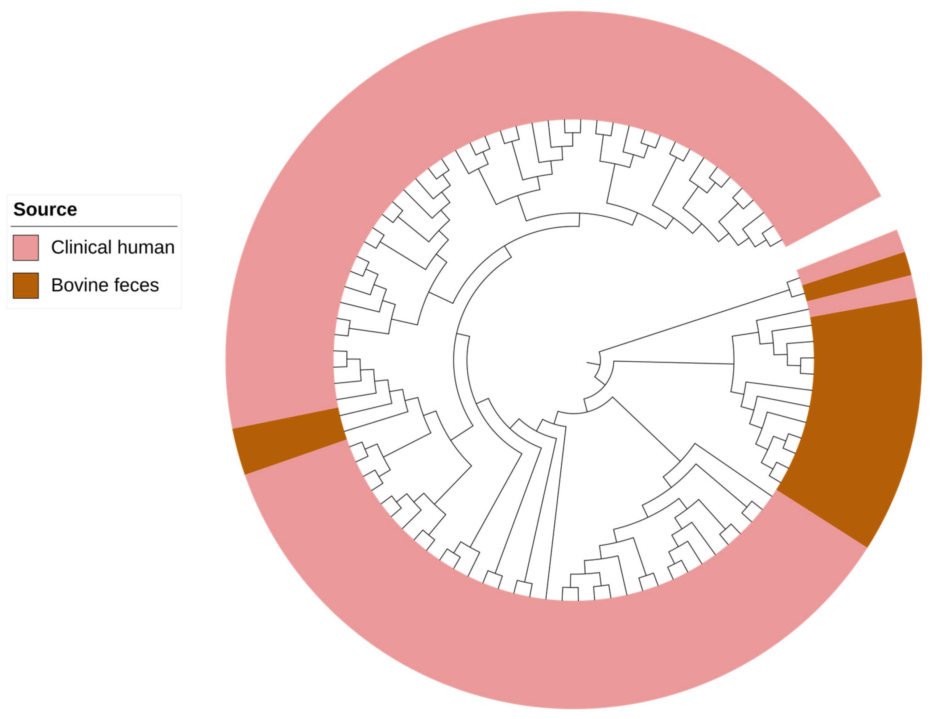
Core-genome phylogenomic analysis formed two clades where the majority of P. aeruginosa recovered from bovine sources clustered together in a paraphyletic clade (Figure 2). In human clinical isolates, collectively, we identified 20 different ARGs conferring resistance to aminoglycosides [aac(3)-Ic, aac(3)-Id, aac(3)-VIa, aac(6′)-Ib4, aac(6′)-Ib-AKT, aac(6′)-Ib-G, aac(6′)-IIa, aac(6′)-IIc, aac(6′)-Il, aacA8, aadA1, aadA5, aadA6, ant(2″)-Ia, ant(4′)-IIb, aph(3′)-Ib, aph(3″)-Ib, aph(3′)-IIb, aph(3′)-VIa, and aph(6)-Id], while in bovine P. aeruginosa only 3 genes, aph(3″)-la, aph(3′)-IIb, and aph(6)-ld were present (Supplementary Table S3).
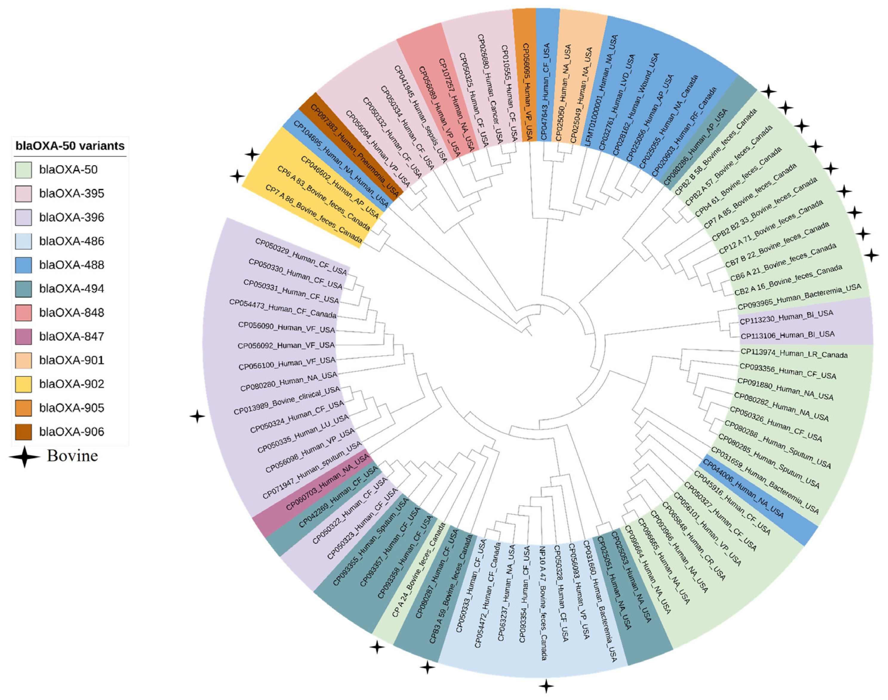
All clinical isolates carried blaOXA-50 variants (Figure 3). In addition, metallo-carbapenemase gene blaNDM-1 was found in two clinical genomes (2/75; 3%), but not in any of the bovine isolates. We also identified ARGs for cephalosporins (blaCTX-M-30, blaGES-9, blaOXA-10, blaOXA-101, blaOXA-2, blaPDC-15, blaPDC-16, blaPDC-22, blaPDC-34, blaPDC-38, blaPDC-46, blaPDC-55, blaPDC-59, blaPDC-100, blaPDC-101, blaPDC-116, blaPDC-123, blaPDC-167, blaPDC-264, blaPDC-308, blaPDC-364, blaPDC-374, blaPDC-385, and blaVEB-9), chloramphenicol (cmlA5, catB, catB7, catB, and floR2), colistin (mcr-1), quinolones (crpP and qnrVC1), fosfomycin (fosA), macrolides [mcr(E) and mph(E)], sulfonamide (sul1), tetracycline [tet(G)] and trimethoprim (dfrA1, dfrA10, and dfrB5) in human clinical P. aeruginosa genomes. Comparison of blaOXA-50 gene variants and their genomic context did not reveal any significant variation across genomes from human and bovine sources (Figure 3).

3. Discussion

Increasing carbapenem resistance has threatened the clinical utility of these drugs in human medicine, leading to the challenge of “extreme drug resistant” bacteria [22]. Therefore, the Centers for Disease Control and Prevention (CDC) has declared carbapenem-resistant Enterobacteriaceae as an urgent and serious threat to human health [23]. In this study, we investigated if carbapenem-resistant E. coli could be recovered using carbapenem enrichment from bovine feces or catch basin water samples collected in intensive feedlots. We had extremely low recovery of carbapenem-resistant E. coli in bovine feces. The majority of isolates that were presumed to be E. coli were subsequently identified as other bacterial species, primarily Pseudomonas species. This is not surprising as we used etrapenem for sample enrichment, and Pseudomonas spp. are intrinsically resistant to this antimicrobial [24,25].
In Acinetobacter spp., the carbapenem resistance genes identified were mostly intrinsic [26]. For instance, blaOXA-214, blaOXA-278, and blaOXA-648 in A. haemolyticus, A. junii, and A. lwoffii isolates, respectively, are intrinsic to these species [27,28]. A. lwoffii is a medically important Acinetobacter species, however, A. haemolyticus and A. junii have been only occasionally reported in human infections [29]. The blaL1 and blaL2 genes encoding for metallo-β-lactamase in S. maltophilia are also intrinsic and confer broad spectrum resistance against β-lactams including all carbapenems [30]. S. maltophilia is considered a “newly emerging pathogen of concern” and is frequently isolated from immunocompromised patients [31,32,33].
In P. aeruginosa, we identified four different gene variants belonging to the blaOXA-50 class (blaOXA-50, blaOXA-902, blaOXA-486, and blaOXA-494) encoding carbapenem-hydrolyzing oxacillinase (CHDLs). All of these variants were also present in clinical isolates in addition to seven other variants blaOXA-395, blaOXA-488, blaOXA-847, blaOXA-848 blaOXA-901, blaOXA-905, and blaOXA-906. It has been reported that blaOXA-50 naturally exists in all P. aeruginosa and does not appear to have been acquired based on the similar GC% content of the blaOXA-50 gene to the overall P. aeruginosa genome [24]. We did not find any insertion sequences, repeat elements, relaxases, integrases, within the genomic proximity of blaOXA-50, suggesting that this gene lacks mobility and is unlikely to be readily transferred to other bacterial species.
In the core-genome phylogenetic analysis, P. aeruginosa isolates from bovine sources clustered separately from those obtained from humans. Similar observations have been reported in Brazil, where P. aeruginosa recovered from healthy bovine urine samples segregated from human clinical urinary tract isolates [34]. These observations might be associated with a prolonged association and adaptation of these isolates within their respective environmental hosts. P. aeruginosa is known for its remarkable ability to adapt to diverse ecological niches, from soil to various living hosts [35,36]. Genes associated with metabolism and pathogenesis constitute the core-genome of P. aeruginosa, whereas genes required to adapt to various niches constitute the accessory genome. These genes were found to cluster in certain loci referred to as ‘regions of genomic plasticity’ [37,38]. In this study, P. aeruginosa recovered from the beef production system carried ARGs conferring resistant to chloramphenicol (catB7), β-lactams (blaOXA-50), fosfomycin (fosA), aminoglycosides (aph(3′)-Iib), and cephalosporins (blaPDC-55, blaPDC-374). These ARGs were also found in the genomes of human clinical isolates and were mapped on chromosomes in both bovine and human clinical isolates. In P. aeruginosa, the type IV pilus uptakes foreign DNA during transformation events [39,40]. The accessory genome can be distinguished from the core genome by its aberrant GC content, codon usage, and tetranucleotide usage [41]. The GC content of P. aeruginosa (~66.3%) is mostly higher than the foreign DNA. Over time, the acquired DNA may lose the sequence compositional difference that distinguishes it from the core genome of P. aeruginosa as it undergoes the same selective pressure as the core genome [42].
We observed variable phenotypic susceptibility profiles among P. aeruginosa isolates that harbored similar AMR profiles. Some isolates were resistant to all carbapenems, some were resistant to both meropenem and doripenem and some were only resistant to meropenem. Other than β-lactamase production, alterations or lack of porin OprD, and overexpression of resistance-nodulation-division (RND) efflux pumps (MexAB-OprM, MexCD-OprJ, MexEF-OprN, and MexXY-OprM) are also associated with carbapenem resistance in Pseudomonas species [43,44]. Not only does the combinations of these mechanisms confer reduced susceptibility to carbapenems, but the overexpression of these efflux pumps result in β-lactam, tetracycline, trimethoprim, aminoglycosides, and fluoroquinolone resistance [45,46]. It is more likely that the variation in susceptibility profiles in our isolates is associated with the varied number of MexAB-OprM operon and OprD found in each isolate [47,48]. Moreover, isolates with multiple MexAB-OprM operons also possessed a single copy of the MexR repressor gene. The presence of only one repressor gene in comparison to multiple efflux pumps as regulatory targets may disturb the molecular stoichiometry of the regulation affecting efflux pump expression, thus resulting in varied phenotypic resistance profiles among P. aeruginosa isolates.
P. stutzeri (Stutzerimonas stutzeri) is an opportunistic pathogen that rarely causes infection in humans [49]. In our study, we found the carbapenem-resistant gene, blaPST-2 in the P. stutzeri genome. The blaPST-2 encodes for a subclass B1 metallo-β-lactamase and was first identified on the chromosomes of P. stutzeri DSM 10701 [50]. From a genetic context, we did not find genes associated with mobility in proximity to this ARG. However, we suggest that blaPST-2 is not intrinsic to P. stutzeri as it was not present in all P. stutzeri genomes in the NCBI database (only 4 out of 19 P. stutzeri genomes had blaPST-2). Moreover, blaPST-2 was phylogenetically related to the previously characterized mobile subclass B1 metallo-β-lactamase families, including KHM, SIM, and IMP in P. aeruginosa and Citrobacter freundii, providing further evidence that this gene was more likely acquired [51,52,53].
Pseudomonas saudiphocaensis has only recently been classified as a new species [54] and has not been well characterized. There are only two studies where P. saudiphocaensis was recovered, first from air samples in the city environment of Makkah, Saudi Arabia, in 2012 [54] and then from a sheep dairy farm in New Zealand [55]. To the best of our knowledge, this is the first report of recovery of P. saudiphocaensis from bovine feces. With the hybrid genome assembly, we were able to construct a complete circular genome of this isolate. The genome of P. saudiphocaensis (3.6 Mbp) was small as compared to other Pseudomonas species, which ranged from 5.5 to 6.7 Mbp. These isolates also carried blaPST-2, which has not been identified in other P. saudiphocaensis genomes [54,55]. The blaPST-1 in P. stutzeri and P. saudiphocaensis showed 95% amino acid similarity. Despite limited knowledge of P. saudiphocaensis, it is suggested that it may have acquired this gene.

4. Materials and Methods

4.1. Sampling, Isolation and Identification

The study includes bovine fecal and catch-basin water samples, collected from four feedlots (pens = 301) located in Alberta, over a period of two years (August 2016 to June 2019). Briefly, 20 g of feces were collected from 20 fresh fecal pats and placed in a sterile plastic container and thoroughly mixed. Each pen housed approximately 180 feedlot cattle. For enrichment, 0.5 g of the mixed fecal sample was inoculated into 4.5 mL of Escherichia coli (EC) broth containing 2 µg/mL of ertapenem (ETP, sigma Aldrich, ref: sml1238), followed by overnight incubation at 37 °C in a shaking incubator at 250 RPM. The enriched samples were then sub-cultured on MacConkey agar supplemented with 2 µg/mL ertapenem. From each sample, a maximum of three colonies were selected and sub-cultured on nutrient agar supplemented with 0.5 µg/mL ertapenem (Dalynn Biologicals, Calgary, AB, Canada). Water samples were collected from feedlot catch basins which collected run off water from the feedlot. Briefly, 1 L of catch-basin water was collected into a polyethylene bottle attached to a telescopic pole at two different locations per site, which were combined to generate a composite sample. Water samples were transported on ice within 4 h of collection for processing. Composite catch basin water (10 mL) was filtered through a 0.45 µm pore size filter of 47 mm diameter (S-Pak® EMD Millipore Corp, Billerica, MA, USA) using a sterile vacuum-manifold filtration system (Pall Corporation, Port Washington, NY, USA). The filter was then submerged in 4.5 mL EC broth-ertapenem (0.5 mg/L) and incubated overnight at 37 °C with shaking, followed by sub-culture onto MacConkey Agar supplemented with 0.5 µg/L ertapenem at 37 °C (Dalynn Biologicals, Calgary, AB, Canada). A maximum of three colonies were selected from each sample and sub-cultured on nutrient agar supplemented with 0.5 µg/mL ertapenem at 37 °C for 24 h. For the identification of recovered isolates (n = 116), the 16S rRNA gene was amplified using universal bacterial 16S rRNA gene primers 27F (5′-AGAGTTTGATCCTGGCTCAG-3′) and 1492R (5′-GGTTACCTTGTTACGACTT-3′) followed by Sanger sequencing of the amplified PCR product [56]. Species were identified using BLAST search against the NCBI bacterial database.

4.2. Phenotypic Characterization

Isolates (n = 116) were tested against 12 different antimicrobials including all four carbapenems (ertapenem, meropenem, doripenem, and imipenem), ceftazidime, chloramphenicol, gentamicin, levofloxacin, piperacillin, trimethoprim-sulfamethoxazole, tobramycin, and tetracycline, using the disk diffusion method according to the Clinical and Laboratory Standards Institute (CLSI) guideline M02-A12 and M100-S32. Pseudomonas aeruginosa ATCC 27853 was used as a reference quality control. Zones of inhibition were recorded using the BioMic V3 imaging system (Giles Scientific, Inc., Santa Barbara, CA, USA).
Isolates were also tested for the production of carbapenemases using the chromogenic Carba NP test (RAPIDEC® CARBA NP kit, BioMérieux, St-Laurent, QC, Canada), with Klebsiella pneumoniae ATCC 700603 and Klebsiella pneumoniae OLC2685 used as negative and positive controls, respectively. A bacterial colony (10-μL loop) was picked up from overnight-incubated Mueller-Hinton agar plates and mixed into API suspension medium. The bacterial suspension was then transferred to wells in a test strip and incubated at 37 °C. Test strips were read at 30 and 120 min. A ‘positive’ test corresponded to a color change from red to yellow-orange, while no-change in color was considered ‘negative’.

4.3. Whole-Genome Sequencing, Assembly and Annotation

All Carba NP positive isolates (n = 28) and a subset (n = 14) of Carba NP negative isolates selected to be representative of identified species were subjected to whole genome sequencing using short and long reads sequencing technologies. High molecular weight genomic (HMW) DNA was extracted using a Genomic DNA preparation kit with Genomic-tip 20/G (Cat: 13323; QIAGEN, Germantown, MD, USA) according to manufacturer’s instructions. DNA quality and quantity was estimated using a Nanodrop 2000 spectrophotometer (Thermo Fisher Scientific, Mississauga, ON, Canada) and a Qubit Fluorometer with PicoGreen (Q32850, Invitrogen, Carlsbad, CA, USA), respectively. The integrity of DNA was confirmed through agarose gel electrophoresis. Short-read sequencing was performed on the Illumina MiSeq platform. A genomic library was constructed using the Illumina NexteraXT DNA sample preparation kit (Illumina Inc., San Diego, CA, USA) followed by sequencing on Illumina MiSeq platform using the MiSeq Reagent Kit V3, generating 2 × 300 base paired-end reads. Long-read sequencing was performed on PromethION platform from Oxford Nanopore technologies (ONT). Sequencing libraries were prepared using LSK-109 genomic DNA preparation kit. One hundred nanograms of HMW genomic DNA were end-repaired using a NEBNext Ultra II End prep enzyme mix [Cat: E7646AA; New England Biolabs (NEB) Ltd. Whitby, ON, Canada] and FFPE DNA repair mix [Cat: M6630L; New England Biolabs (NEB) Ltd. Whitby, ON, Canada]. End-repaired DNA from each isolate were individually barcoded using a Nanopore barcoding kit EXP-NBD196 [Oxford Nanopore Technologies (ONT), UK] and a blunt TA ligase [Cat: M0367; (NEB) Ltd. Whitby, ON, Canada], followed by chelation with 0.5 M pH 8.0 EDTA (Cat: AM9260G; Invitrogen, ON, Canada). Barcoded DNA samples were pooled in a single tube and cleaned using the Omega-bind NGS beads (M1378-01, Omegabiotek) at 0.5× volume following the manufacturer’s instructions. Beads were dried for 1 min and DNA was eluted in 32 µL of deionized sterile H2O. The DNA concentration was measured using the Invitrogen Qubit dsDNA BR assay (Q32854, Invitrogen, ON, Canada). Sequencing adapters were then ligated to 30 µL of recovered DNA using Adapter Mix II Expansion kit EXP-AMII001 (ONT) along with the 5 µL T4 DNA ligase [Cat: M0202M; New England Biolabs (NEB) Ltd. Whitby, ON, Canada] and 10 µL quick ligation buffer [Cat: B6058S; New England Biolabs (NEB) Ltd. Whitby, ON, Canada]. Adaptor-ligated DNA was bead-cleaned using a 0.4× volume of beads following the manufacturer’s instructions using the supplied polyethylene glycol (PEG) based wash buffer. The DNA from the last step (200–400 ng) was loaded onto an ONT PromethION sequencing flow cell as directed by the manufacturer. MinKNOW Core 3.1.20 and guppy 2.0.10 were used for flow cell signal processing and base calling during each run, and reads were assembled de novo using Flye version 2.9.1 [57].
Hybrid genome assemblies were generated using illumina short-reads and Flye-assembled contigs from ONT long-reads using the Unicycler assembly tool [58]. Illumina paired-end short-read sequences were used to generate an assembly graph using SPAdes followed by bridge building using Flye-assembled contigs using Miniasm (https://github.com/lh3/miniasm accessed on 29 January 2023) [59] and Racon (https://github.com/isovic/racon accessed on 29 January 2023). Multiple rounds of short-read polishing were conducted using Pilon (https://github.com/broadinstitute/pilon accessed on 29 January 2023). The contiguity and quality of each genome assembly was assessed using Quast (v.5.2.0) by computing relevant metrics, including the number of contigs, total length (bp), and GC content [60]. Contigs were then annotated using Prokka v.1.13.1 [61], and assembled contigs were screened for the presence of antimicrobial resistance and virulence genes using ABRicate v.1.0.1 (https://github.com/tseemann/ABRICATE accessed on 10 February 2023) against the NCBI Bacterial Antimicrobial Resistance Reference Gene Database (NCBI BioProject ID: PRJNA313047) and the VirulenceFinder database (PMID: 34850947) [62], respectively. To identify plasmids, Mob-recon tool v.3.0.0 (https://github.com/phac-nml/mob-suite accessed on 12 Febrary 2023) was used [63].

4.4. Comparative Genomic Analysis

Whole genome comparative genomic analysis was conducted between all the Pseudomonas aeruginosa isolates (n = 20) sequenced in this study to P. aeruginosa genomes (n = 76) (Supplementary Table S1) originating from North America, from the PathoSystems Resource Integration Center (PATRIC) (https://www.patricbrc.org; accessed on 20 February 2023). Although these same genomes were also available from the NCBI database, the metadata in the PATRIC database was more detailed. These isolates originated from human clinical sources, with the exception of a single isolate from cattle.
The core-genome phylogenomic tree was constructed using the SNVphyl pipeline v.1.2.3. The phylogenetic tree was generated by aligning paired-end reads against the P. aeruginosa POA1 reference genome (NC_002516.2) using SMALT (c.0.7.5; https://sourceforge.net/projects/smalt/ accessed on 10 March 2023). The generated read pileups were then subjected to quality filtering (minimum mean mapping quality score of 30), coverage cut-offs (15× minimum depth of coverage), and a single nucleotide variant (SNV) abundance ratio filter of 0.75 to obtain a multiple sequence alignment of SNV-containing sites. The SNV alignment, with no density filtering, was used to create a maximum likelihood phylogeny using PhyML version 3.0. The generated Newick file was visualized using the Interactive Tree Of Life (iTOL) v.6 [64].
We also compared variants of the blaOXA-50 of P. aeruginosa isolates used in this study to determine their genetic relatedness. For this, blaOXA-50 coding sequencing (CDS) was computationally extracted from each genome and aligned using MAFFT version 7.490. The resultant alignment-based tree was visualized using iTOL v.6 [64].

5. Conclusions

In conclusion, the recovery of carbapenem-resistant bacteria from beef production systems is quite rare. The majority of carbapenem-resistant bacterial species, including P. aeruginosa, A. haemolyticus, A. junii, A. lwoffii, and S. maltophilia, carried intrinsic carbapenem-resistant genes and were only recoverable following enrichment. P. aeruginosa found in this study were multidrug-resistant, a possible reflection of the ability of this species to readily acquire foreign genes via transformation through the type IV pilus. Moreover, the phylogenetic analysis showed that bovine P. aeruginosa strains formed separate clusters from human clinical strains, indicating that they may have adapted to the cattle environment, and prudent management in feedlot systems has limited the spread of these isolates outside of cattle production systems.

Supplementary Materials

The following supporting information can be downloaded at: https://www.mdpi.com/article/10.3390/antibiotics12060960/s1, Table S1 Hybrid assemblies quality statistics determined using Quast; Table S2 List of Pseudomonas aeruginosa genomes retrieved from a public database for analysis; Table S3 AMR gene profiles of Pseudomonas aeruginosa genomes retrieved from a public database for analysis.

Author Contributions

R.Z. and T.A.M., designed the study; S.-e.-Z.Z. and R.Z. analyzed sequence data; K.T. arranged for sample collection and bacterial isolation; S.A. performed short read sequencing; L.S. and T.H. performed nanopore sequencing, M.S.-E. assembled nanopore reads, S.-e.-Z.Z. and S.A. performed CarbaNP testing, S.-e.-Z.Z. performed AST, genomic DNA extraction, generated figures and analyzed overall data/results and wrote the first draft of the manuscript; and T.A.M., A.Z. and R.Z. provided funding and supervision. All authors have read and agreed to the published version of the manuscript.

Funding

The authors are grateful to the Major Innovation Fund of the Government of Alberta in conjunction with the University of Calgary AMR One Health Consortium, and the Genomics Research and Development Initiative (GRDI) of the Government of Canada for financial support.

Institutional Review Board Statement

The animal study protocol was approved by the Lethbridge Research and Development Centre Animal Care Committee as protocol (protocol #1637; 30 November 2016).” for studies involving cattle.

Data Availability Statement

The whole genome sequence data are available in GenBank under Bio Project PRJNA956966.

Acknowledgments

The authors acknowledge Gary Van Domselaar for providing the bioinformatics tools and computing environment. We thank Cheyenne Conrad for technical assistance.

Conflicts of Interest

The authors declare no conflict of interest.

References

  1. Bonfiglio, G.; Russo, G.; Nicoletti, G. Recent Developments in Carbapenems. Expert Opin. Investig. Drugs 2002, 11, 529–544. [Google Scholar] [PubMed]
  2. Nicolau, D.P. Carbapenems: A Potent Class of Antibiotics. Expert Opin. Pharmacother. 2008, 9, 23–37. [Google Scholar] [CrossRef] [PubMed]
  3. Zhanel, G.G.; Wiebe, R.; Dilay, L.; Thomson, K.; Rubinstein, E.; Hoban, D.J.; Noreddin, A.M.; Karlowsky, J.A. Comparative Review of the Carbapenems. Drugs 2007, 67, 1027–1052. [Google Scholar] [CrossRef] [PubMed]
  4. El-Gamal, M.I.; Brahim, I.; Hisham, N.; Aladdin, R.; Mohammed, H.; Bahaaeldin, A. Recent Updates of Carbapenem Antibiotics. Eur. J. Med. Chem. 2017, 131, 185–195. [Google Scholar] [CrossRef]
  5. Zhanel, G.G.; Lawrence, C.K.; Adam, H.; Schweizer, F.; Zelenitsky, S.; Zhanel, M.; Lagacé-Wiens, P.R.S.; Walkty, A.; Denisuik, A.; Golden, A.; et al. Imipenem-Relebactam and Meropenem-Vaborbactam: Two Novel Carbapenem-Β-Lactamase Inhibitor Combinations. Drugs 2018, 78, 65–98. [Google Scholar] [CrossRef]
  6. O’Donnell, J.N.; Miglis, C.M.; Lee, J.Y.; Tuvell, M.; Lertharakul, T.; Scheetz, M.H. Carbapenem Susceptibility Breakpoints, Clinical Implications with the Moving Target. Expert Rev. Anti-Infect. Ther. 2016, 14, 389–401. [Google Scholar] [CrossRef]
  7. Bush, K.; Bradford, P.A. Β-Lactams and Β-Lactamase Inhibitors: An Overview. Cold Spring Harb. Perspect. Med. 2016, 6, a025247. [Google Scholar] [CrossRef]
  8. Schweizer, H.P. Efflux as a Mechanism of Resistance to Antimicrobials in Pseudomonas aeruginosa and Related Bacteria: Unanswered Questions. Genet. Mol. Res. 2003, 2, 48–62. [Google Scholar]
  9. Meletis, G.; Exindari, M.; Vavatsi, N.; Sofianou, D.; Diza, E. Mechanisms Responsible for the Emergence of Carbapenem Resistance in Pseudomonas aeruginosa. Hippokratia 2012, 16, 303–307. [Google Scholar]
  10. Ambler, R.P.; Coulson, A.F.; Frère, J.M.; Ghuysen, J.M.; Joris, B.; Forsman, M.; Levesque, R.C.; Tiraby, G.; Waley, S.G. A standard numbering scheme for the class A beta-lactamases. Biochem. J. 1991, 276, 269–270. [Google Scholar] [CrossRef]
  11. Franco, M.R.G.; Caiaffa-Filho, H.H.; Burattini, M.N.; Rossi, F. Metallo-Beta-Lactamases among Imipenem-Resistant Pseudomonas aeruginosa in a Brazilian University Hospital. Clinics 2010, 65, 825–829. [Google Scholar] [CrossRef]
  12. Elshamy, A.A.; Khaled, M.A. A Review on Bacterial Resistance to Carbapenems: Epidemiology, Detection and Treatment Options. Future Sci. 2020, 6, FSO438. [Google Scholar] [CrossRef] [PubMed]
  13. Webb, H.E.; Bugarel, M.; den Bakker, H.C.; Nightingale, K.K.; Granier, S.A.; Scott, H.M.; Loneragan, G.H. Carbapenem-Resistant Bacteria Recovered from Faeces of Dairy Cattle in the High Plains Region of the USA. PLoS ONE 2016, 11, e0147363. [Google Scholar] [CrossRef] [PubMed]
  14. Tello, M.; Oporto, B.; Lavín, J.L.; Ocejo, M.; Hurtado, A. Characterization of a carbapenem-resistant Escherichia coli from dairy cattle harbouring blaNDM-1 in an IncC plasmid. J. Antimicrob. Chemother. 2022, 77, 843–845. [Google Scholar] [CrossRef] [PubMed]
  15. Klotz, P.; Göttig, S.; Leidner, U.; Semmler, T.; Scheufen, S.; Ewers, C. Carbapenem-resistance and pathogenicity of bovine Acinetobacter indicus-like isolates. PLoS ONE 2017, 12, e0171986. [Google Scholar] [CrossRef]
  16. Alam, M.; Rasool, M.H.; Khan, I.; Khurshid, M.; Aslam, B. Multilocus Sequence Typing of Carbapenem-Resistant Acinetobacter baumannii Isolates Harboring blaOXA-23 and blaIMP in Cattle from Punjab, Pakistan. Microb. Drug Resist. 2022, 28, 997–1002. [Google Scholar] [CrossRef]
  17. Tshitshi, L.; Manganyi, M.C.; Montso, P.K.; Mbewe, M.; Ateba, C.N. Extended Spectrum Beta-Lactamase-Resistant Determinants among Carbapenem-Resistant Enterobacteriaceae from Beef Cattle in the North West Province, South Africa: A Critical Assessment of Their Possible Public Health Implications. Antibiotics 2020, 9, 820. [Google Scholar] [CrossRef]
  18. Fischer, J.; Rodríguez, I.; Schmoger, S.; Friese, A.; Roesler, U.; Helmuth, R.; Guerra, B. Escherichia coli producing VIM-1 carbapenemase isolated on a pig farm. J. Antimicrob. Chemother. 2012, 67, 1793–1795. [Google Scholar] [CrossRef]
  19. Savin, M.; Bierbaum, G.; Mutters, N.T.; Schmithausen, R.M.; Kreyenschmidt, J.; García-Meniño, I.; Schmoger, S.; Käsbohrer, A.; Hammerl, J.A. Genetic Characterization of Carbapenem-Resistant Klebsiella spp. from Municipal and Slaughterhouse Wastewater. Antibiotics 2022, 11, 435. [Google Scholar] [CrossRef]
  20. Ahlstrom, C.A.; Frick, A.; Pongratz, C.; Spink, K.; Xavier, C.; Bonnedahl, J.; Ramey, A.M. Genomic comparison of carbapenem-resistant Enterobacteriaceae from humans and gulls in Alaska. J. Glob. Antimicrob. Resist. 2021, 25, 23–25. [Google Scholar] [CrossRef]
  21. Darwich, L.; Vidal, A.; Seminati, C.; Albamonte, A.; Casado, A.; López, F.; Molina-López, R.A.; Migura-Garcia, L. High prevalence and diversity of extended-spectrum β-lactamase and emergence of OXA-48 producing Enterobacterales in wildlife in Catalonia. PLoS ONE 2019, 14, e0210686. [Google Scholar] [CrossRef] [PubMed]
  22. Codjoe, F.S.; Donkor, E.S. Carbapenem Resistance: A Review. Med. Sci. 2017, 6, 1. [Google Scholar] [CrossRef]
  23. Centers for Disease Control and Prevention; National Center for Emerging and Zoonotic Infectious Diseases. Antibiotic/Antimicrobial Resistance. Available online: https://www.cdc.gov/drugresistance/biggest-threats.html (accessed on 15 April 2022).
  24. Girlich, D.; Naas, T.; Nordmann, P. Biochemical Characterization of the Naturally Occurring Oxacillinase Oxa-50 of Pseudomonas aeruginosa. Antimicrob. Agents Chemother. 2004, 48, 2043–2048. [Google Scholar] [CrossRef] [PubMed]
  25. Wexler, H.M. In vitro activity of ertapenem: Review of recent studies. J. Antimicrob. Chemother. 2004, 53 (Suppl. 2), ii11–ii21. [Google Scholar] [CrossRef] [PubMed]
  26. Tian, J.; Zhang, G.; Ju, Y.; Tang, N.; Li, J.; Jia, R.; Feng, J. Five novel carbapenem-hydrolysing OXA-type β-lactamase groups are intrinsic in Acinetobacter spp. J. Antimicrob. Chemother. 2018, 73, 3279–3284. [Google Scholar] [CrossRef]
  27. Figueiredo, S.; Bonnin, R.A.; Poirel, L.; Duranteau, J.; Nordmann, P. Identification of the Naturally Occurring Genes Encoding Carbapenem-Hydrolysing Oxacillinases from Acinetobacter haemolyticus, Acinetobacter johnsonii, and Acinetobacter calcoaceticus. Clin. Microbiol. Infect. 2012, 18, 907–913. [Google Scholar] [CrossRef]
  28. Figueiredo, S.; Poirel, L.; Seifert, H.; Mugnier, P.; Benhamou, D.; Nordmann, P. Oxa-134, a Naturally Occurring Carbapenem-Hydrolyzing Class D Beta-Lactamase from Acinetobacter lwoffii. Antimicrob. Agents Chemother. 2010, 54, 5372–5375. [Google Scholar] [CrossRef]
  29. Wong, D.; Nielsen, T.B.; Bonomo, R.A.; Pantapalangkoor, P.; Luna, B.; Spellberg, B. Clinical and Pathophysiological Overview of Acinetobacter Infections: A Century of Challenges. Clin. Microbiol. Rev. 2017, 30, 409–447. [Google Scholar] [CrossRef]
  30. Yang, Z.; Liu, W.; Cui, Q.; Niu, W.; Li, H.; Zhao, X.; Wei, X.; Wang, X.; Huang, S.; Dong, D.; et al. Prevalence and Detection of Stenotrophomonas maltophilia Carrying Metallo-Β-Lactamase Blal1 in Beijing, China. Front. Microbiol. 2014, 5, 692. [Google Scholar] [CrossRef]
  31. Gideskog, M.; Welander, J.; Melhus, Å. Cluster of S. maltophilia among patients with respiratory tract infections at an intensive care unit. Infect. Prev. Pract. 2020, 2, 100097. [Google Scholar] [CrossRef]
  32. Geller, M.; Nunes, C.P.; Oliveira, L.; Nigri, R.S. maltophilia pneumonia: A case report. Respir. Med. Case Rep. 2018, 24, 44–45. [Google Scholar] [CrossRef] [PubMed]
  33. Brooke, J.S. New Strategies against Stenotrophomonas maltophilia: A Serious Worldwide Intrinsically Drug-Resistant Opportunistic Pathogen. Expert Rev. Anti-Infect. Ther. 2014, 12, 1–4. [Google Scholar] [CrossRef] [PubMed]
  34. Giannattasio-Ferraz, S.; Ene, A.; Gomes, V.J.; Queiroz, C.O.; Maskeri, L.; Oliveira, A.P.; Putonti, C.; Barbosa-Stancioli, E.F. Escherichia coli and Pseudomonas aeruginosa Isolated from Urine of Healthy Bovine Have Potential as Emerging Human and Bovine Pathogens. Front. Microbiol. 2022, 13, 764760. [Google Scholar] [CrossRef]
  35. Gellatly, S.L.; Hancock, R.E. Pseudomonas aeruginosa: New insights into pathogenesis and host defenses. Pathog. Dis. 2013, 67, 159–173. [Google Scholar] [CrossRef] [PubMed]
  36. Moradali, M.F.; Ghods, S.; Rehm, B.H. Pseudomonas aeruginosa Lifestyle: A Paradigm for Adaptation, Survival, and Persistence. Front. Cell. Infect. Microbiol. 2017, 7, 39. [Google Scholar] [CrossRef]
  37. Wolfgang, M.C.; Kulasekara, B.R.; Liang, X.; Boyd, D.; Wu, K.; Yang, Q.; Miyada, C.G.; Lory, S. Conservation of genome content and virulence determinants among clinical and environmental isolates of Pseudomonas aeruginosa. Proc. Natl. Acad. Sci. USA 2003, 100, 8484–8489. [Google Scholar] [CrossRef]
  38. Mathee, K.; Narasimhan, G.; Valdes, C.; Qiu, X.; Matewish, J.M.; Koehrsen, M.; Rokas, A.; Yandava, C.N.; Engels, R.; Zeng, E.; et al. Dynamics of Pseudomonas aeruginosa genome evolution. Proc. Natl. Acad. Sci. USA 2008, 105, 3100–3105. [Google Scholar] [CrossRef]
  39. Strom, M.S.; Lory, S. Structure-function and biogenesis of the type IV pili. Annu. Rev. Microbiol. 1993, 47, 565–596. [Google Scholar] [CrossRef]
  40. Graupner, S.; Frey, V.; Hashemi, R.; Lorenz, M.G.; Brandes, G.; Wackernagel, W. Type IV pilus genes pilA and pilC of Pseudomonas stutzeri are required for natural genetic transformation, and pilA can be replaced by corresponding genes from nontransformable species. J. Bacteriol. 2000, 182, 2184–2190. [Google Scholar] [CrossRef]
  41. Zhou, H.Q.; Ning, L.W.; Zhang, H.X.; Guo, F.B. Analysis of the relationship between genomic GC Content and patterns of base usage, codon usage and amino acid usage in prokaryotes: Similar GC content adopts similar compositional frequencies regardless of the phylogenetic lineages. PLoS ONE 2014, 9, e107319. [Google Scholar] [CrossRef]
  42. Kung, V.L.; Ozer, E.A.; Hauser, A.R. The accessory genome of Pseudomonas aeruginosa. Microbiol. Mol. Biol. Rev. MMBR 2010, 74, 621–641. [Google Scholar] [CrossRef]
  43. Puzari, M.; Chetia, P. Rnd Efflux Pump Mediated Antibiotic Resistance in Gram-Negative Bacteria Escherichia coli and Pseudomonas aeruginosa: A Major Issue Worldwide. World J. Microbiol. Biotechnol. 2017, 33, 24. [Google Scholar] [CrossRef] [PubMed]
  44. Savinova, T.A.; Samchenko, A.A.; Bocharova, Y.A.; Mayansky, N.A.; Chebotar, I.V. Computer Program for Detection and Analyzing the Porin-Mediated Antibiotic Resistance of Bacteria. Sovrem. Tekhnol. Med. 2021, 13, 15–22. [Google Scholar] [CrossRef] [PubMed]
  45. Li, X.Z.; Nikaido, H.; Poole, K. Role of Mexa-Mexb-Oprm in Antibiotic Efflux in Pseudomonas aeruginosa. Antimicrob. Agents Chemother. 1995, 39, 1948–1953. [Google Scholar] [CrossRef] [PubMed]
  46. Köhler, T.; Kok, M.; Michea-Hamzehpour, M.; Plesiat, P.; Gotoh, N.; Nishino, T.; Curty, L.K.; Pechere, J.C. Multidrug Efflux in Intrinsic Resistance to Trimethoprim and Sulfamethoxazole in Pseudomonas aeruginosa. Antimicrob. Agents Chemother. 1996, 40, 2288–2290. [Google Scholar] [CrossRef]
  47. Rojo-Bezares, B.; Estepa, V.; Cebollada, R.; de Toro, M.; Somalo, S.; Seral, C.; Castillo, F.J.; Torres, C.; Sáenz, Y. Carbapenem-Resistant Pseudomonas aeruginosa Strains from a Spanish Hospital: Characterization of Metallo-Beta-Lactamases, Porin Oprd and Integrons. Int. J. Med. Microbiol. 2014, 304, 405–414. [Google Scholar] [CrossRef] [PubMed]
  48. López-García, A.; del Carmen Rocha-Gracia, R.; Bello-López, E.; Juárez-Zelocualtecalt, C.; Sáenz, Y.; Castañeda-Lucio, M.; López-Pliego, L.; González-Vázquez, M.C.; Torres, C.; Ayala-Nuñez, T.; et al. Characterization of Antimicrobial Resistance Mechanisms in Carbapenem-Resistant Pseudomonas aeruginosa Carrying Imp Variants Recovered from a Mexican Hospital. Infect. Drug Resist. 2018, 11, 1523–1536. [Google Scholar] [CrossRef]
  49. Noble, R.C.; Overman, S.B. Pseudomonas stutzeri infection. A review of hospital isolates and a review of the literature. Diagn. Microbiol. Infect. Dis. 1994, 19, 51–56. [Google Scholar] [CrossRef]
  50. Berglund, F.; Marathe, N.P.; Österlund, T.; Bengtsson-Palme, J.; Kotsakis, S.; Flach, C.F.; Larsson, D.G.J.; Kristiansson, E. Identification of 76 novel B1 metallo-β-lactamases through large-scale screening of genomic and metagenomic data. Microbiome 2017, 5, 134. [Google Scholar] [CrossRef]
  51. Fallah, F.; Borhan, R.S.; Hashemi, A. Detection of bla(IMP) and bla(VIM) metallo-β-lactamases genes among Pseudomonas aeruginosa strains. Int. J. Burn. Trauma 2013, 3, 122–124. [Google Scholar]
  52. Senda, K.; Arakawa, Y.; Nakashima, K.; Ito, H.; Ichiyama, S.; Shimokata, K.; Kato, N.; Ohta, M. Multifocal outbreaks of metallo-beta-lactamase-producing Pseudomonas aeruginosa resistant to broad-spectrum beta-lactams, including carbapenems. Antimicrob. Agents Chemother. 1996, 40, 349–353. [Google Scholar] [CrossRef] [PubMed]
  53. Sekiguchi, J.; Morita, K.; Kitao, T.; Watanabe, N.; Okazaki, M.; Miyoshi-Akiyama, T.; Kanamori, M.; Kirikae, T. KHM-1, a novel plasmid-mediated metallo-beta-lactamase from a Citrobacter freundii clinical isolate. Antimicrob. Agents Chemother. 2008, 52, 4194–4197. [Google Scholar] [CrossRef]
  54. Azhar, E.I.; Papadioti, A.; Bibi, F.; Ashshi, A.M.; Raoult, D.; Angelakis, E. Pseudomonas saudiphocaensis’ sp. nov., a new bacterial species isolated from currency notes collected during the Hajj pilgrimage in 2012 at Makkah, Saudi Arabia. New Microbes New Infect. 2016, 15, 131–133. [Google Scholar] [CrossRef] [PubMed]
  55. Gupta, T.B.; Risson, A.N.; Jauregui, R.; Maclean, P.; Brightwell, G. Draft Genome Sequence of Pseudomonas saudiphocaensis Strain AGROB56, Isolated from a Dairy Farm in New Zealand. Microbiol. Resour. Announc. 2021, 10, e01258-20. [Google Scholar] [CrossRef]
  56. Zaheer, R.; Cook, S.R.; Klima, C.L.; Stanford, K.; Alexander, T.; Topp, E.; Read, R.R.; McAllister, T.A. Effect of Subtherapeutic vs. Therapeutic Administration of Macrolides on Antimicrobial Resistance in Mannheimia haemolytica and Enterococci Isolated from Beef Cattle. Front. Microbiol. 2013, 4, 133. [Google Scholar] [CrossRef]
  57. Freire, B.; Ladra, S.; Parama, J.R. Memory-Efficient Assembly Using Flye. IEEE/ACM Trans. Comput. Biol. Bioinform. 2022, 19, 3564–3577. [Google Scholar] [CrossRef]
  58. Wick, R.R.; Judd, L.M.; Gorrie, C.L.; Holt, K.E. Unicycler: Resolving Bacterial Genome Assemblies from Short and Long Sequencing Reads. PLoS Comput. Biol. 2017, 13, e1005595. [Google Scholar] [CrossRef] [PubMed]
  59. Li, H. Minimap and Miniasm: Fast Mapping and De Novo Assembly for Noisy Long Sequences. Bioinformatics 2016, 32, 2103–2110. [Google Scholar] [CrossRef]
  60. Gurevich, A.; Saveliev, V.; Vyahhi, N.; Tesler, G. Quast: Quality Assessment Tool for Genome Assemblies. Bioinformatics 2013, 29, 1072–1075. [Google Scholar] [CrossRef] [PubMed]
  61. Seemann, T. Prokka: Rapid Prokaryotic Genome Annotation. Bioinformatics 2014, 30, 2068–2069. [Google Scholar] [CrossRef]
  62. Liu, B.; Zheng, D.; Zhou, S.; Chen, L.; Yang, J. Vfdb 2022: A General Classification Scheme for Bacterial Virulence Factors. Nucleic Acids Res. 2022, 50, D912–D917. [Google Scholar] [CrossRef] [PubMed]
  63. Robertson, J.; Nash, J.H. Mob-Suite: Software Tools for Clustering, Reconstruction and Typing of Plasmids from Draft Assemblies. Microb. Genom. 2018, 4, e000206. [Google Scholar] [CrossRef] [PubMed]
  64. Letunic, I.; Bork, P. Interactive Tree of Life (Itol) V5: An Online Tool for Phylogenetic Tree Display and Annotation. Nucleic Acids Res. 2021, 49, W293–W296. [Google Scholar] [CrossRef] [PubMed]
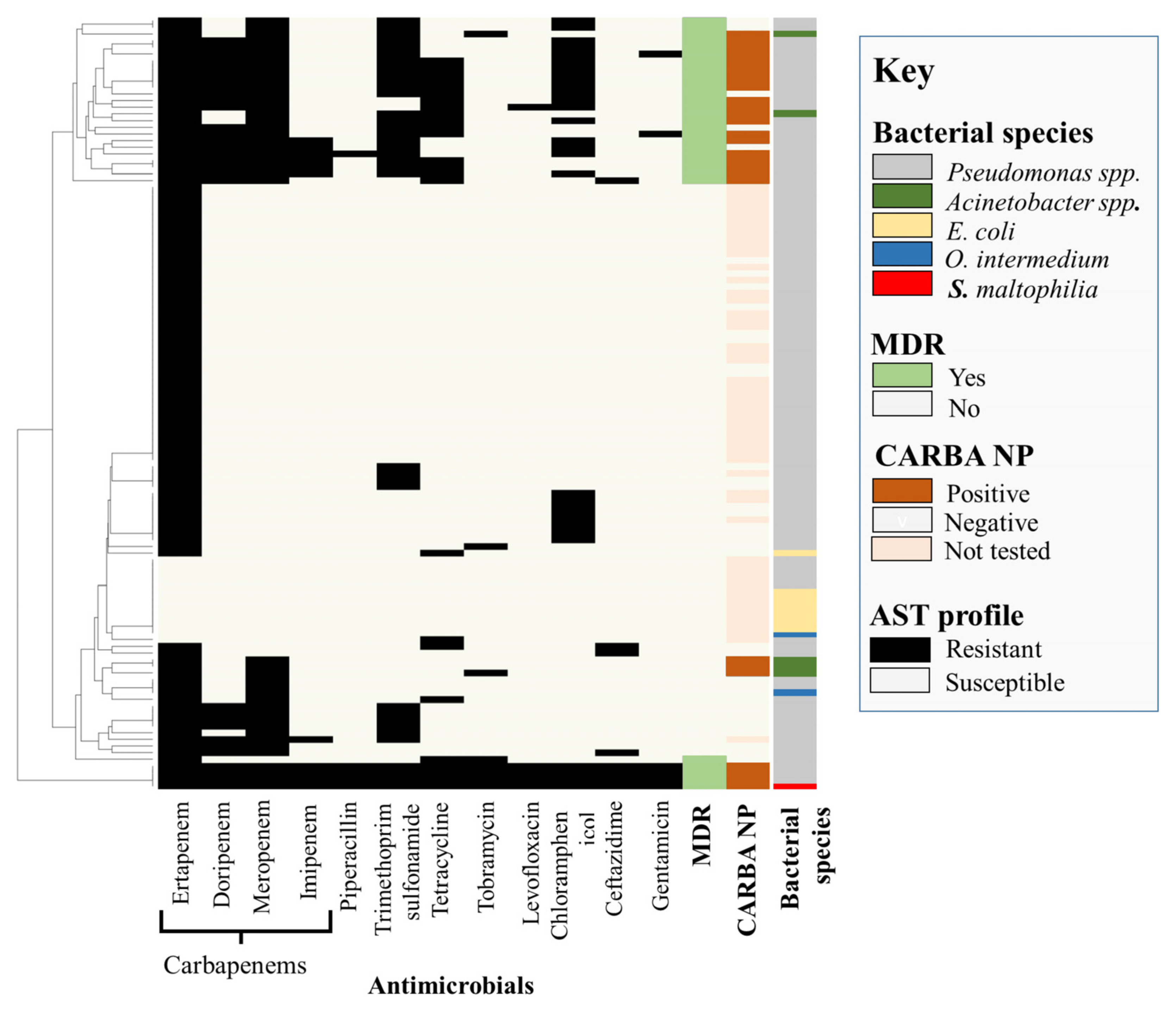
Figure 1. Antimicrobial susceptibility and carbapenemase production phenotypes of isolates recovered from cattle feedlots following carbapenem enrichment.
Antibiotics 12 00960 g001
Figure 2. Maximum likelihood core-genome phylogenetic tree of Pseudomonas aeruginosa recovered from bovine and human clinical sources. P. aeruginosa POA1 (NC_002516.2) was used as a reference genome.
Antibiotics 12 00960 g002
Figure 3. Phylogenetic tree of carbapenemase gene blaOXA-50 variants identified in Pseudomonas aeruginosa isolates from human and bovine sources, constructed using MAFFT.
Antibiotics 12 00960 g003
Table 1. Antimicrobial resistance gene profiles of whole genome sequenced isolates recovered from bovine feces and catch basin water samples.
Bacterial SpeciesAntimicrobial Resistance GenesGenotype-Based Phenotype
E. coli
(n = 1)
blaEC, blaCMY-2, aph(3″)-Ib, aph(6)-Id, sul2,
tet(A), floR
Aminoglycosides, Chloramphenicol,
Sulfisoxazole, Tetracycline
A. haemolyticus (n = 3)blaOXA-265, aacA-ACI, blaPDC-197Carbapenem
Aminoglycosides
Cephalosporin
blaOXA-265, aacA-ACI1Carbapenem
Aminoglycosides
A. lwoffii
(n = 1)
blaOXA-648Carbapenem
A. junii
(n = 1)
blaOXA -278Carbapenem
S. maltophilia
(n = 1)
blaL1,blaL2, aph(6)-Smalt, aph(3′)-IIc, oqxB9, oqxA10, floR2Carbapenem
Aminoglycosides
Phenicol
Chloramphenicol
O. intermedium
(n = 1)
floR, oqxB12, blaOCH-2Chloramphenicol
Quinolone
Cephalosporin
P. aeruginosa
(n = 20)
blaOXA-50, blaPDC-197, aph(3′)-IIb, catB7, fosACarbapenem
Cephalosporin Chloramphenicol
Aminoglycoside
Fosfomycin
blaOXA-50, blaPDC-197, aph(3′)-IIb, catB7, fosA, crpP
blaOXA-50, blaPDC-55, aph(3′)-IIb, catB7, fosA
blaOXA-50, blaPDC-66, aph(3′)-IIb, fosA, crpP, catB7
blaOXA-486, blaPDC-374, aph(3′)-IIb, fosA, crpP, catB7
blaOXA-486, blaPDC-374, aph(3′)-IIb, catB7, fosA
blaOXA-494, blaPDC-374, aph(3″)-la, aph(6)-ld, catB7 fosA, crpP
blaOXA-902, blaPDC-133, aph(3′)-IIb, catB7, fosA, crpP
blaOXA-902Carbapenem
P. entomophila
(n = 1)
blaPDC-33Cephalosporin
P. plecoglossicida
(n = 9)
No gene-
P. mosselii
(n = 1)
No gene-
P. putida
(n = 1)
No gene-
P. saudiphocaensis
(n = 1)
blaPST-2, aadA1Carbapenem,
Aminoglycoside
P. stutzeri
(n = 1)
blaPST-2Carbapenems
Table 2. Virulence gene profiles of whole genome sequenced isolates recovered from bovine feces and catch-basin water samples.
Bacterial
Species
Virulence Genes (%) 1
P. aeruginosa
(n = 20)
Biofilm and capsule synthesis: alg44 (21/24, 88%), alg8 (22/24, 92%), algA-G,I-L,P-R,U,W,X,Z (22/24, 92%), mucA-E,P (21/24, 88%)
Pili and Fimbriae: chpA-E (92%), pilA (9%), pilB (88%), pilC (34%), pilE,F (84%), pilG-K,M-X,Y1,Y2 (92%), fimT-V (84%), fleI/flag (13%), fleN (100%), fleP (13%), fleQ (100%), fleR (88%), fleS (88%), flgA (84%), flgB (88%), flgC (100%), flgD-F (84%), flgG-I (24), flgJ,K (84%), flgL (13%), flgM,N (84%), flhA,B,F (100%), fliA,C (100%), fliD (13%), fliE-L (92%), fliM,N (100%), fliO-R (88%), fliS (13%), motA-D,Y (92%)
Biosynthesis of small ferric-ion-chelating molecules: pchA-I,R (84%), fptA (84%), fpvA (21%), mbtH-like (100%), pvcA-D (84%), pvdA (84%), pvdD,E (21%), pvdF-H (88%), pvdI,J (21%), pvdL,M (100), pvdM (22), pvdN-Q (88%), pvdS (96%)
Phenazine biosynthesis: phzA1-G1,M,S (80%), phzH (30%)
Rhamnolipids: rhlA,B,I (84%), rhlC (75%)
Type VI secretion system: clpV1 (96%), dotU1 (88%), flha1 (80%), hcp1 (96%), hsiA1 (84%), hsiB1/vipA (100%), hsiC1/vipB (96%), hsiE1,F1,H1 (84%), hsiG1 (96%), hsiJ1 (92%), icmF1/tssM1 (84%), tagQ (88%), tse1-3 (84%), vgrG1a (80%), vgrG1b (84%), ppkA(84%), tagR (100%) tagS (88%), tagT (88%), pppA (84%), tagF/pppB (84%)
Type III secretion system: pscB-L, N-U (80%), popB,D,N (80%), pcr1-4,D,H,R,V (80%), exsA-E (80%), exoS,T,Y (80%), ptxR (84%)
Type I secretion system: aprA (84%)
Type II secretion system (Xcp) and exo-proteins: xcpP-Z (84%), xcpA/pilD (88%), lasA (88%), lasB (84%), plcH (84%), toxA (21%), lip1 (84%)
Quorum sensing: lasI (88%)
Lipopolysaccharide core biosynthesis: waaA,C (88%), waaF (96%), waaG,P (100%), wzy (17%), wzz (17%)
P. entomophila
(n = 1)
Biofilm and capsule synthesis: algCBU, mucD
Lipopolysaccharide core biosynthesis: waaF, wag
Type VI secretion system: clpV1, hsiG1, hcp1, hsiC1/vipB, hsiB1/vipA, tagR
Biosynthesis of small ferric-ion-chelating molecules: pvdH, pvdS, mbtH-like
Pili and Fimbriae: motC, fleNQ, flhA, fliAIGMNPQ, flgCGHI, pilH
P. mosselii
(n = 1)
Biofilm and capsule synthesis: algA-D,U,W,I (100%), alg8 (100%), mucD (100%)
Biosynthesis of small ferric-ion-chelating molecules: mbtH-like (100%), pvdH,S,M (100%)
Lipopolysaccharide core biosynthesis: waaF,G,P (100%)
Pili and Fimbriae: flgC,G-I (100%), fliA,F,G,I,M,-Q (100%), fleN,Q (100%), motA-C (100%), pilH (100%)
Type VI secretion system: tagR, dotU1 (100%), hsiB1/vipA (100%), hsiC1/vipB (100%), hcp1(100%), hsiG1 (100%), clpV1 (100%)
P. putida
(n = 2)
Biofilm and capsule synthesis: algA-D,U,I (100%),alg8 (100%), mucD (100%)
Biosynthesis of small ferric-ion-chelating molecules: mbtH-like (100%), pvdH,S (100%)
Lipopolysaccharide core biosynthesis: waaF,G (100%)
Pili and Fimbriae: flgC,G-I (100%), fliA,G,I,M,N,P,Q (100%), fleN,Q (100%), flhA (100%), motC,D (100%), pilH (100%)
P. saudiphocaensis
(n = 1)
Biofilm and capsule synthesis: algC,R,U (100%)
Pili and Fimbriae: flgC,G,I (100%), flhA (100%), fliE,G,I,M-P (100%), pilG,H,U,T (100%)
Type II secretion system (Xcp): xcpT,R (100%)
Lipopolysaccharide core biosynthesis: waaF
P. stutzeri
(n = 1)
Biofilm and capsule synthesis: algA-C,R (100%)
Pili and Fimbriae: flgG,I (100%), flhA (100%), fliA,E-G,I,M,N-R (100%), fleN,Q (100%), motA (100%), pilG,H,J,M,R,T,U (100%)
Type II secretion system (Xcp): xcpT,R (100%)
Lipopolysaccharide core biosynthesis: waaF,P (100%)
P. plecoglossicida
(n = 9)
Biofilm and capsule synthesis: algB,C,U (100%), algW (67%), mucD (100%)
Pili and Fimbriae: flgC,G,H,I (100%), flhA (100%), fliA,G,I,M,N,P,Q (100%), fleN,Q (100%), motC (100%), pilH (100%)
Lipopolysaccharide core biosynthesis: waaF (67%), waaG (100%)
Type VI secretion system: clpV1 (100%), hcp1 (100%), hsiB1/vipA (100%), hsiC1/vipB (100%), hsiG1 (100%), tagR (100%)
Biosynthesis of small ferric-ion-chelating molecules: mbtH-like (100%), pvdSH (100%)
E. coli (n = 1)Type II secretion system: gspC m (100%)
Type III secretion system: espX2 (100%), ompA (100%), espR1(100%), espR4 (100%), espR3 (100%), espL1 (100%), espY3 (100%), espY2 (100%), espX1(100%), espY4 (100%), espL4 (100%), espX4 (100%), espX5 (100%),
Curli biogenesis: csgB,D,F,G (100%)
Iron import system: shuA,S,T,W,X (100%), chuUVW (100%)
Pili and Fimbriae: fimA-H (100%), yagV/ecpE (100%), yagW/ecpD (100%), yagX/ecpC (100%), yagY/ecpB, yagZ/ecpA (100%), ykgK/ecpR (100%)
Adhesion: fdeC (100%)
Enterobactin: entA-E,F,S (100%), fepA-D,G (100%)
A. haemolyticus (n = 3)No virulence gene
A. lwoffii
(n = 1)
A. junii
(n = 1)
S. maltophilia (n = 1)
1 Bold indicates virulence genes associated with cellular component or metabolic activity of each specific bacterial species.
Disclaimer/Publisher’s Note: The statements, opinions and data contained in all publications are solely those of the individual author(s) and contributor(s) and not of MDPI and/or the editor(s). MDPI and/or the editor(s) disclaim responsibility for any injury to people or property resulting from any ideas, methods, instructions or products referred to in the content.