Communication

In Vitro Bacterial Competition of Staphylococcus aureus, Streptococcus agalactiae, and Escherichia coli against Coagulase-Negative Staphylococci from Bovine Mastitis Milk

by
Anyaphat Srithanasuwan
1,
Montira Intanon
2,3,
Wasana Chaisri
1,3 and
Witaya Suriyasathaporn
1,3,4,*
1
Department of Food Animal Clinic, Faculty of Veterinary Medicine, Chiang Mai University, Chiang Mai 50100, Thailand
2
Department of Veterinary Bioscience and Veterinary Public Health, Faculty of Veterinary Medicine, Chiang Mai University, Chiang Mai 50100, Thailand
3
Research Center of Producing and Development of Products and Innovations for Animal Health and Production, Chiang Mai University, Chiang Mai 50100, Thailand
4
Cambodia Campus, Asian Satellite Campuses Institute, Nagoya University, Nagoya 464-8601, Japan
*
Author to whom correspondence should be addressed.
Antibiotics 2023, 12(3), 600; https://doi.org/10.3390/antibiotics12030600
Submission received: 8 February 2023 / Revised: 8 March 2023 / Accepted: 14 March 2023 / Published: 16 March 2023

Abstract

:
Intramammary infection (IMI) from the environment and infected quarters can cause co-infection. The objective of this study was to determine the ability of coagulase-negative staphylococci (CNS) to survive in the same environment as Staphylococcus aureus, Streptococcus agalactiae, and Escherichia coli as major pathogens. In total, 15 and 242 CNS strains were used in Experiment I and Experiment II, respectively. Both experiments were separated into three conditions: culture with CNS 24 h before (PRIOR), after (AFTER), and at the same time (EQUAL). The lack of a clear zone, regardless of size, was determined to be the key to the survival of both. The CNS species’ percentages of survival against major pathogens were tested using Fisher’s exact test. Differences in the percentages of survival were evident among the CNS species in all conditions. For the PRIOR condition, all CNS mostly survived when living with major strains; however, S. chromogenes could degrade S. agalactiae. Although most CNS strains were degraded in the AFTER and EQUAL conditions, some strains of S. hominis and S. simulans could resist S. aureus and S. agalactiae. In conclusion, some specific strains of CNS are able to survive in an environment with major pathogens. Research into the survival strains may indicate that the concept of novel bacteria with bacteriolytic capabilities might be possible as a novel mastitis treatment.

1. Introduction

In 2020, the World Health Organization released a fact sheet indicating that antibiotic resistance is rising to dangerously high levels in all parts of the world, leading to higher medical costs, prolonged hospital stays, and increased mortality [1]. In countries without standard treatment guidelines, antimicrobials are often over-prescribed by health workers and veterinarians, leading to their over-use by the public. The world urgently needs to change the way that it prescribes and uses antimicrobials. Globally, apart from the substantial economic decrease due to disease, bovine mastitis has been associated with the increasing development and rapid emergence of antibiotic-resistant bacteria [2,3]. The recent protocol for antimicrobials used for mild clinical mastitis is recommended to be applied to quarters where the milk samples show specific bacteria growth after obtaining the results of 24 h rapid cultures (for a review, see [4]).
In the context of bovine mastitis, major mastitis pathogens, such as Staphylococcus aureus, Streptococcus uberis, Streptococcus agalactiae, and coliforms, are usually considered more virulent and damaging to the udder than minor mastitis pathogens, such as Corynebacterium spp. and coagulase-negative staphylococci (CNS). Spontaneous cure refers to the curing of a disease without any external treatment, whereby the bacteria are destroyed by the udder’s defense mechanism recruitment [5]. The spontaneous curing of mastitis, mostly referring to transient or non-persistent infection, is highly dependent on the identified bacteria. For example, S. agalactiae was found to be related to persistent infection [6], while S. uberis could be both persistent and transient based on strain [7] and virulence factors [8]. Among the mastitis pathogens, coagulase-negative staphylococci (CNS), categorized as minor pathogens that typically cause subclinical mastitis (SCM), are commonly eliminated by the udder’s immune response within a few days [9,10] without any changes in terms of milk yield and composition [11,12].
Poor mastitis control programs favor the IMI from heterogeneous bacteria, always causing mixed and co-infection [13], and CNS was the most common bacteria for mixed infection [14,15]. Bacteria in the environment and host organisms, for example, in the mammary glands, likely use networks of competitive mechanisms to survive and shape the composition and function of the diverse community [16]. The results of bacterial competition always show a single dominant mastitis pathogen in most mastitis studies [6,17]. In longitudinal mastitis studies in farms with poor mastitis control, further bacterial infections in the same quarter often occurred [7,18]. In addition, Keane et al. [19] demonstrated that the traditional culture methods mostly showed a single dominant pathogen that was the same when identified by PCR. However, the PCR showed extra mixed infections because real-time PCR can detect dead bacterial DNA 20 weeks after inactivation [20].
Evidence that appears to be beneficial for mastitis cows would be the survival of CNS or transient bacteria after co-infection with major or persistent bacteria, subsequently increasing the recruitment of white blood cells and causing a natural, spontaneous cure, as shown in the previous studies [7,18]. The events and mechanisms behind the competition between pathogens in the mammary niche are neither determined nor clearly understood [21]. Approaches to bacterial competition that rely on the culture of two or more organisms together are emerging as a powerful tool to discover and reimagine the mechanisms of competition between bacteria. Therefore, the objective of this study is to determine the abilities of CNS species when either the existing or attacking pathogen survives when living in the same environment as major mastitis pathogens, including S. aureus, S. agalactiae, and E. coli.

2. Results

After being co-cultured for 24 h, the results of Experiment I were separated into three different cultured schemes (shown in Figure 1). For the CNS incubated 24 h prior to the PRIOR condition, the sizes of the colonies formed by CNS were different, with the smallest for S. capitis (No. 14). Most CNS colonies have wrinkled surfaces, motile outgrowths, and are larger than that of the S. aureus and S. agalactiae. When cultured for 24 h prior to the addition of major pathogens (upper figure), all CNS were not lysed or had clear zones. Most E. coli, S. aureus, or S. agalactiae colonies were progressively motile and covered parts of the CNS, especially E. coli and S. aureus, but were not degraded by CNS. All colonies formed by both CNS and E. coli remained intact. When co-cultured with S. chromogenes strain No. 2, the colonies formed by S. aureus and S. agalactiae were progressively degraded, and the underlying cells were lysed. For the second scheme, all E. coli colonies were larger than CNS, and some of them were progressively motile, degrading the CNS and lysing S. warneri (No. 9), S. hominis (No. 12), and S. capitis (No. 14). The S. aureus colonies cultured for 24 h before CNS were progressively moved and degraded, with most of the CNS starting to be lysed. In this scheme, the S. agalactiae colonies were clear and intact, with no movement. However, almost all S. agalactiae were aggressively attacked, causing all CNS to be lysed, except S. epidermidis (No. 11). For example, S. epidermidis (No. 3), S. warneri (No. 8 and 9), S. cohinii (No. 13), and S. capitis (No. 14) were entirely degraded and disappeared. For the last scheme (incubation at the same time), the E. coli colonies were larger than CNS, S. aureus, and S. agalactiae. All colonies formed by CNS co-cultured with E. coli and S. aureus were intact and not degraded, while the three colonies formed by S. epidermidis (No. 3) and S. warneri (No. 8 and 9) were degraded and lysed.
Figure 2 shows the interspecies competition in the in vitro mixed intramammary infection situation between major and minor strains (Experiment II). In the PRIOR condition, all field-strain CNS survived with E. coli, and almost all field-strain CNS survived with S. aureus and S. agalactiae, except for very few S. chromogenes and S. hominis (Figure 2A). In contrast to the other CNS, these can only be survived by S. agalactiae, where colonies formed by some S. chromogenes demonstrate motile outgrowths. The S. agalactiae colonies were degraded and lysed (Figure 3A). When the major pathogens were cultured prior to CNS and for the AFTER condition, the percentages of survival among the species of CNS were significantly different for E. coli, S. aureus, and S. agalactiae (Figure 2B). S. chromogenes survived well with E. coli; however, most were lysed by S. aureus and S. agalactiae. In contrast, S. hominis had a lower survival rate than S. chromogenes and S. simulans but could survive well with S. aureus and S. agalactiae (Figure 2B). Figure 3B indicates the detached colonies formed by S. hominis that are not degraded and lysed by S. agalactiae. In the EQUAL condition, the percentages of surviving CNS show significant differences when cultured with E. coli and S. agalactiae (Figure 2C). All CNS survived when cultured at the same time with S. aureus for 24 h; however, only S. chromogenes and S. epidermidis had a 100% survival rate out of the three major pathogens (Figure 2C).

3. Discussion

In this study, the agar spot competition assays were used to evaluate the surviving CNS after being with other major bacteria, including E. coli, S. aureus, and S. agalactiae, with the latter two being contagious and persistent [6]. This might represent a unique feature of co-intramammary infections relating to CNS [13]. The designed assays demonstrated a mutual exclusion between the two species depending on the sequence of CNS inoculation at 24 h before, 24 h after, and at the same time as PRIOR, AFTER, and EQUAL, respectively. This imitates the natural IMI that might first infect a cow’s udder with either minor or major pathogens. The CNS species used in this present study were S. chromogens, S. simulans, S. epidermidis, S. hominis, and S. warneri, which are the most prevalent CNS species isolated from bovine mastitis cases in many countries [22,23].
Regarding the competition between CNS and E. coli, both laboratory strains in Experiment I (Figure 1) and all field strains in Experiment II (Figure 2) showed that 100% of the CNS at PRIOR was able to survive after being together; however, some of the S. hominis and S. warneri did not survive in AFTER and EQUAL conditions. In the PRIOR condition, although the colony formed by E. coli could phagocytose the colony formed by all CNS, it could not lyse the colony formed by CNS. Based on our literature review, this is the first report on the evidence of in vitro bacteria competition because this bacterium is more often found in monoculture [24]. Accordingly, our findings might be due to the mild interaction during early co-infection. Escherichia coli is a Gram-negative motile bacterium. Therefore, the motility of the E. coli strains might play a role in the reduction in the amount of preformed CNS biofilm, which, in turn, might result in the elimination of CNS in a longer infection [25].
Our study found there to be an aggressive degrading of most tested CNS strains when in competition with S. agalactiae in the AFTER condition in both Experiments I (Figure 1) and II (Figure 2B). However, S. epidermidis and S. hominis were able to resist. The strongest inhibitory effect for S. agalactiae found in this present study is supported by the studies by Wescombe and Tagg [26] and Karaya et al. [27], who reported that both agalacticin and nisin P. bacteriocins produced by S. agalactiae were able to inhibit Staphylococcus spp. The inhibitor acts as a self-toxin bacteriocin that kills other bacteria directly observed in major bacteria, especially S. agalactiae [28,29].
For the competition with S. aureus, most of the results using CNS laboratory strains in Experimental 1 (Figure 1) showed the degraded colony AFTER. Similarly, all CNS field strains in Experiment II (Figure 2) could not resist S. aureus AFTER, except S. hominis. The repression of S. aureus is consistent with the study by Mahmmod et al. [30], who reported the co-habitual competition between S. aureus and CNS species within the same niche. This study also supports our findings for the EQUAL condition, in which both species can survive being together.
Overall, most colonies formed by CNSs were not lysed, regardless of the presence of major pathogen species for the PRIOR and EQUAL conditions. This finding might be explained by the release of antimicrobials from the major pathogens being delayed until an achieved local quorum, ensuring the presence of sufficient cell numbers and the production of fully inhibitory antimicrobial levels in the prevailing diffusion environment. Consequently, the AFTER condition, where the population level of the major pathogens reached a critical level, is where a quorum was achieved, resulting in the production and release of antimicrobials [31]. Although most of the CNS strains were progressively degraded by the major pathogens, S. chromogenes was the only species that could evade the motile outgrowths and degraded colonies formed by S. aureus and S. agalactiae (S. chromogenes No. 2; Figure 1 and Figure 3A). Moreover, our results also reported a 100% survival rate when growing with all three major pathogens (Figure 2C). This result is consistent with the work of Braem et al. [32], who reported the strongest inhibitory effect of S. chromogenes against a wide range of mastitis-causing pathogens and other CNS. Additionally, there are also several mechanisms of S. chromogenes that inhibit the growth of major pathogens [33,34,35]. Bacteriocin production is a strain- not species-specific, trait [36]. The S. chromogenes strain has the ability to synthesize and secrete bacteriocins and 6-thioguanine (6-TG), a purine analog that suppresses S. aureus growth by inhibiting de novo purine biosynthesis. Together with an increase in SCC by S. chromogenes strains, which was shown to have elicited udder immune responses, this may offer cross-protection against other mastitis pathogens [37,38], indicating that S. chromogenes strains display the strongest inhibitory effects against major mastitis pathogens. In conclusion, both species- and strain-specific CNS are able to survive in an environment with major pathogens. All CNS cultured 24 h before could survive with all major pathogens. Following a 24 h culture, S. chromogenes with the PRIOR condition could degrade S. agalactiae colonies. When cultured for 24 h after the major pathogens were added, most S. hominis survived with S. aureus, but only some strains could survive with E. coli and S. agalactiae.

4. Conclusions

Based on the results of this study, S. chromogenes and S. hominis play a role in the natural, spontaneous curing of mastitis from S. aureus and S. agalactiae, both major and persistent pathogens, by either destroying the infection or surviving in the udder to accelerate the udder’s defense mechanism. This indicates that the concept of novel bacteria with bacteriolytic capabilities might be possible as a novel mastitis treatment. However, further studies are needed: (i) the evaluation of the ability to induce white blood cells into an infected site for pathogen clearance, (ii) the elucidation of the mode of action against major pathogens, (iii) an evaluation of the procedure’s safety regarding the cytotoxicity of the lived bacteria intramammary infection assessed by in vitro and in vivo tests, and (iv) an evaluation of the long-term effects of using novel bacteria with bacteriolytic capabilities.

5. Materials and Methods

5.1. Bacterial Strains and Study Design

Two experiments were conducted to determine the survival of minor pathogens in an environment with major pathogens. For Experiment I, 15 isolates of various CNS species from mastitis samples, ATCC, and the Microbiology Laboratory in the Faculty of Science, Chiangmai University, were used (Table 1). For Experiment II, all strains of CNS species stocked in our database, and the randomly selected Streptococcus agalactiae used in this study were obtained from frozen mastitis pathogen stock (−80 °C) from the Laboratory of Milk Quality and Mastitis, the Faculty of Veterinary Medicine, Chiangmai University. The stocked bacteria originated from milk samples of subclinical mastitis from lactating cows in smallholder dairy farms in different areas of Thailand, including northern (n = 131), northeastern (n = 75), and central (n = 36) regions. From the total of 242 stocked CNS strains, 5 staphylococci species, including S. chromogens (n = 136), S. simulans (n = 43), S. epidermidis (n = 32), S. hominis (n = 18), and S. warneri (n = 13) were used. All stocked strains were identified by conventional bacterial culture [39] and confirmed by Maldi-Tof. The three main major pathogens were Staphylococcus aureus ATCC25923, E. coli ATCC25922, and the field strain of Streptococcus agalactiae, as previously described. All strains were recovered in Tryptic Soya broth (TSB) (Himedia, Mumbai, India) and grown overnight at 37 °C. After that, the inoculums were cultured on 5% bovine blood agar (HIMEDIA, Mumbai, India) at 37 °C for 24 h.
To imitate a mixed infection situation in a mammary gland, all CNS strains were separately cultured with the selected major pathogens in one of three conditions: (1) minor pathogens were inoculated first and allowed to grow overnight before each major pathogen was inoculated nearby, which the intramammary was the first to be infected with CNS (PRIOR); (2) vice versa, where the first was infected with the major pathogens followed by the CNS (AFTER); (3) both bacteria inoculated at the same time as if the infection happened at the same time in the udder (EQUAL). The first inoculated bacteria were defined as the early colonizer, and the following inoculated bacteria were defined as the later colonizer.

5.2. Agar Spot Competition Assays

Agar spot competition assays were used in both experiments. The protocol was applied according to what was previously described by Kaspar et al. [40] with modifications. Briefly, the bacterial suspension was adjusted to a 0.5 McFarland turbidity standard to achieve 108 CFU/mL from single overnight colonies. Five µL of bacterial suspension was inoculated onto tryptic soy agar (TSA) (HIMEDIA, Mumbai, India) as the early colonizer. After overnight incubation at 37 °C, 5 µL of the competing strains from another group at the same concentration were inoculated beside the early colonizer as the later colonizer. The plates were further aerobically incubated at 37 °C overnight, then the cleared zone between colonies in three different experimental conditions was observed. This experiment was repeated twice, with two independent biological replicates in each experiment.

5.3. Statistical Analysis

For Experiment I, the data were described using pictures of the result plates. For Experiment II, the presence of a clear zone between the culture spots was used to measure the interbacterial competition between major and minor strains. Data on the presence or absence of an inhibition zone around CNS inoculation were determined as negative and positive interactions, respectively. Negative interaction was described when a zone of inhibition was present around a major strain, and no visible growth for the minor strains was found within the same area. However, if there was no inhibition zone or the resistant minor strains were able to grow within the zone of inhibition, this was interpreted as positive interactions. Percentages of survival ability of each CNS species were defined by the number of specified CNS species with the positive interaction divided by the total number of the specified CNS species. The percentage differences in the survival ability of each CNS species among the different major strains were tested using Fisher’s exact test (the SAS University Edition). The significance levels were set at p < 0.05, and tendencies were set at p < 0.1.

Author Contributions

Conceptualization, W.S., W.C. and M.I.; methodology, W.S. and A.S.; validation, W.C. and M.I.; formal analysis, W.S.; writing—original draft preparation, A.S. and W.S.; writing—review and editing, W.S.; visualization, W.S. All authors have read and agreed to the published version of the manuscript.

Funding

This research was funded by Thailand Science Research and Innovation (TSRI), project number: 167921.

Institutional Review Board Statement

Not applicable.

Informed Consent Statement

Not applicable.

Data Availability Statement

All data are shown in the manuscript.

Acknowledgments

The assistance provided by our colleagues at DVM, namely Noppason Pangprasit, Duanghathai Saipinta, Jakkrawut Srachum, and Laorat Tata, Faculty of Veterinary Medicine, Chiang Mai University, Chiang Mai, Thailand, was greatly appreciated.

Conflicts of Interest

The authors declare no conflict of interest.

References

  1. Antimicrobial Resistance. Available online: https://www.who.int/news-room/fact-sheets/detail/antimicrobial-resistance (accessed on 17 November 2021).
  2. Beyene, T.; Hayishe, H.; Gizaw, F.; Beyi, A.F.; Abunna, F.; Mammo, B.; Ayana, D.; Waktole, H.; Abdi, R.D. Prevalence and antimicrobial resistance profile of Staphylococcus in dairy farms, abattoir and humans in Addis Ababa, Ethiopia. BMC Res. Notes 2017, 10, 171. [Google Scholar] [CrossRef] [Green Version]
  3. Suriyasathaporn, W.; Chupia, V.; Sing-Lah, T.; Wongsawan, K.; Mektrirat, R.; Chaisri, W. Increases of Antibiotic Resistance in Excessive Use of Antibiotics in Smallholder Dairy Farms in Northern Thailand. Asian-Australas J. Anim. Sci. 2012, 25, 1322–1328. [Google Scholar] [CrossRef] [PubMed]
  4. Zigo, F.; Vasil, M.; Ondrašovičová, S.; Výrostková, J.; Bujok, J.; Pecka-Kielb, E. Maintaining Optimal Mammary Gland Health and Prevention of Mastitis. Front. Vet. Sci. 2021, 8, 607311. [Google Scholar] [CrossRef]
  5. Suriyasathaporn, W.; Heuer, C.; Noordhuizen-Stassen, E.N.; Schukken, Y. Hyperketonemia and the impairment of udder defense: A review. Vet. Res. 2000, 31, 397–412. [Google Scholar] [CrossRef] [PubMed] [Green Version]
  6. Leelahapongsathon, K.; Schukken, Y.H.; Pinyopummintr, T.; Suriyasathaporn, W. Comparison of transmission dynamics between Streptococcus uberis and Streptococcus agalactiae intramammary infections. J. Dairy Sci. 2016, 99, 1418–1426. [Google Scholar] [CrossRef] [PubMed] [Green Version]
  7. Leelahapongsathon, K.; Schukken, Y.; Srithanasuwan, A.; Suriyasathaporn, W. Molecular epidemiology of Streptococcus uberis intramammary infections: Persistent and transient patterns of infection in a dairy herd. J. Dairy Sci. 2020, 103, 3565–3576. [Google Scholar] [CrossRef]
  8. Srithanasuwan, A.; Pangprasit, N.; Suriyasathaporn, W. Comparison of Virulence Patterns Between Streptococcus uberis Causing Transient and Persistent Intramammary Infection. Front. Vet. Sci. 2022, 9, 806674. [Google Scholar] [CrossRef]
  9. Schukken, Y.H.; González, R.N.; Tikofsky, L.L.; Schulte, H.F.; Santisteban, C.G.; Welcome, F.L.; Bennett, G.J.; Zurakowski, M.J.; Zadoks, R.N.J.V.M. CNS mastitis: Nothing to worry about? Vet. Microbiol. 2009, 134, 9–14. [Google Scholar] [CrossRef]
  10. Zadoks, R.N.; Watts, J.L. Species identification of coagulase-negative staphylococci: Genotyping is superior to phenotyping. Vet. Microbiol. 2009, 134, 20–28. [Google Scholar] [CrossRef] [Green Version]
  11. Supré, K.; Haesebrouck, F.; Zadoks, R.; Vaneechoutte, M.; Piepers, S.; De Vliegher, S. Some coagulase-negative Staphylococcus species affect udder health more than others. J. Dairy Sci. 2011, 94, 2329–2340. [Google Scholar] [CrossRef] [Green Version]
  12. Tomazi, T.; Gonçalves, J.L.; Barreiro, J.R.; Arcari, M.A.; dos Santos, M.V. Bovine subclinical intramammary infection caused by coagulase-negative staphylococci increases somatic cell count but has no effect on milk yield or composition. J. Dairy Sci. 2015, 98, 3071–3078. [Google Scholar] [CrossRef] [Green Version]
  13. Nair, N.; Biswas, R.; Götz, F.; Biswas, L. Impact of Staphylococcus aureus on pathogenesis in polymicrobial infections. Infect. Immun. 2014, 82, 2162–2169. [Google Scholar] [CrossRef] [Green Version]
  14. Kasozi, K. High Prevalence of Subclinical Mastitis and Multidrug Resistant Staphylococcus aureus Are a Threat to Dairy Cattle Production in Kiboga District (Uganda). Open J. Vet. Med. 2014, 4, 35–43. [Google Scholar] [CrossRef] [Green Version]
  15. Vakkamäki, J.; Taponen, S.; Heikkilä, A.-M.; Pyörälä, S. Bacteriological etiology and treatment of mastitis in Finnish dairy herds. Acta Vet. Scand. 2017, 59, 33. [Google Scholar] [CrossRef] [Green Version]
  16. Stubbendieck, R.M.; Straight, P.D. Escape from Lethal Bacterial Competition through Coupled Activation of Antibiotic Resistance and a Mobilized Subpopulation. PLoS Genet. 2015, 11, e1005722. [Google Scholar] [CrossRef] [Green Version]
  17. Suriyasathaporn, W. Epidemiology of Subclinical Mastitis and Their Antibacterial Susceptibility in Smallholder Dairy Farms, Chiang Mai Province, Thailand. J. Anim. Vet. Adv. 2011, 10, 316–321. [Google Scholar] [CrossRef] [Green Version]
  18. Pangprasit, N.; Srithanasuwan, A.; Intanon, M.; Suriyasathaporn, W. Bacteriological cure rates of the primary intramammary infection due to the secondary intramammary infection of bovine mastitis. In Proceedings of the 19th AAAP (Asian-Australasian Association of Animal Production) Animal Science Congress, Jeju-do, Republic of Korea, 23–26 August 2022; p. 473. [Google Scholar]
  19. Keane, O.M.; Budd, K.E.; Flynn, J.; McCoy, F. Increased detection of mastitis pathogens by real-time PCR compared to bacterial culture. Vet. Rec. 2013, 173, 268. [Google Scholar] [CrossRef] [PubMed] [Green Version]
  20. Choe, H.; Kobayashi, I.Y.; Ike, N.; Iwamoto, H.; Ishida, N.; Yukizawa, T.; Fujimaki, H.; Tezuka, T.; Hirata, H.Y.; Saito, T. How long can dead bacterial DNA be detected by real-time PCR? In Proceedings of the 56th Annual Meeting of the Orthopaedic Research Society. Papers and Posters from the McKay Orthopaedic Laboratory, Poster No. 2144, New Orleans, LA, USA, 6–9 March 2010. [Google Scholar]
  21. Derakhshani, H.; Plaizier, J.C.; De Buck, J.; Barkema, H.W.; Khafipour, E. Composition and co-occurrence patterns of the microbiota of different niches of the bovine mammary gland: Potential associations with mastitis susceptibility, udder inflammation, and teat-end hyperkeratosis. Anim. Microbiome 2020, 2, 11. [Google Scholar] [CrossRef] [PubMed] [Green Version]
  22. Ahmadi, E.; Djeddi, A.N.; Mousavi, S.A. Prevalence of coagulase negative Staphylococcus including methicillin resistant strains in buffalo subclinical mastitis in northwest of Iran. Buffalo Bull. 2020, 39, 17–26. [Google Scholar]
  23. Klimiene, I.; Virgailis, M.; Pavilonis, A.; Siugzdiniene, R.; Mockeliunas, R.; Ruzauskas, M. Phenotypical and Genotypical Antimicrobial Resistance of Coagulase-negative staphylococci Isolated from Cow Mastitis. Pol. J. Vet. Sci. 2016, 19, 639–646. [Google Scholar] [CrossRef] [Green Version]
  24. Duse, A.; Persson-Waller, K.; Pedersen, K. Microbial Aetiology, Antibiotic Susceptibility and Pathogen-Specific Risk Factors for Udder Pathogens from Clinical Mastitis in Dairy Cows. Animals 2021, 11, 2113. [Google Scholar] [CrossRef] [PubMed]
  25. Ohn, H.M.; Mizuno, T.; Miyoshi, S.I. Inhibitory Effects of Escherichia coli on the Formation and Development of Staphylococcus epidermidis Biofilm. Biocontrol Sci. 2021, 26, 113–118. [Google Scholar] [CrossRef]
  26. Wescombe, P.A.; Tagg, J.R. Purification and Characterization of Streptin, a Type A1 Lantibiotic Produced by Streptococcus pyogenes. Appl. Environ. Microbiol. 2003, 69, 2737–2747. [Google Scholar] [CrossRef] [PubMed] [Green Version]
  27. Karaya, K.; Shimizu, T.; Taketo, A. New Gene Cluster for Lantibiotic Streptin Possibly Involved in Streptolysin S Formation. J. Biochem. 2001, 129, 769–775. [Google Scholar] [CrossRef]
  28. Vidal Amaral, J.R.; Jucá Ramos, R.T.; Almeida Araújo, F.; Bentes Kato, R.; Figueira Aburjaile, F.; de Castro Soares, S.; Góes-Neto, A.; da Costa, M.M.; Azevedo, V.; Brenig, B.; et al. Bacteriocin Producing Streptococcus agalactiae Strains Isolated from Bovine Mastitis in Brazil. Microorganisms 2022, 10, 588. [Google Scholar] [CrossRef]
  29. Vogel, V.; Spellerberg, B. Bacteriocin Production by Beta-Hemolytic Streptococci. Pathogens 2021, 10, 867. [Google Scholar] [CrossRef]
  30. Mahmmod, Y.S.; Klaas, I.C.; Svennesen, L.; Pedersen, K.; Ingmer, H. Communications of Staphylococcus aureus and non-aureus Staphylococcus species from bovine intramammary infections and teat apex colonization. J. Dairy Sci. 2018, 101, 7322–7333. [Google Scholar] [CrossRef]
  31. Hibbing, M.E.; Fuqua, C.; Parsek, M.R.; Peterson, S.B. Bacterial competition: Surviving and thriving in the microbial jungle. Nat. Rev. Microbiol. 2010, 8, 15–25. [Google Scholar] [CrossRef] [Green Version]
  32. Braem, G.; Stijlemans, B.; Van Haken, W.; De Vliegher, S.; De Vuyst, L.; Leroy, F. Antibacterial activities of coagulase-negative staphylococci from bovine teat apex skin and their inhibitory effect on mastitis-related pathogens. J. Appl. Microbiol. 2014, 116, 1084–1093. [Google Scholar] [CrossRef] [PubMed]
  33. Chin, D.; Goncheva, M.I.; Flannagan, R.S.; Deecker, S.R.; Guariglia-Oropeza, V.; Ensminger, A.W.; Heinrichs, D.E. Coagulase-negative staphylococci release a purine analog that inhibits Staphylococcus aureus virulence. Nat. Commun. 2021, 12, 1887. [Google Scholar] [CrossRef]
  34. Rahmdel, S.; Shekarforoush, S.S.; Hosseinzadeh, S.; Torriani, S.; Gatto, V. Antimicrobial spectrum activity of bacteriocinogenic Staphylococcus strains isolated from goat and sheep milk. J. Dairy Sci. 2019, 102, 2928–2940. [Google Scholar] [CrossRef] [PubMed] [Green Version]
  35. Carson, D.A.; Barkema, H.W.; Naushad, S.; Buck, J.D. Bacteriocins of Non-aureus Staphylococci Isolated from Bovine Milk. Appl. Environ. Microbiol 2017, 83, e01015–e01017. [Google Scholar] [CrossRef] [PubMed] [Green Version]
  36. Nakatsuji, T.; Chen, T.H.; Narala, S.; Chun, K.A.; Two, A.M.; Yun, T.; Shafiq, F.; Kotol, P.F.; Bouslimani, A.; Melnik, A.V. Antimicrobials from human skin commensal bacteria protect against Staphylococcus aureus and are deficient in atopic dermatitis. Sci. Transl. Med. 2017, 9, eaah4680. [Google Scholar] [CrossRef] [Green Version]
  37. Piccart, K.; Verbeke, J.; De Visscher, A.; Piepers, S.; Haesebrouck, F.; De Vliegher, S. Local host response following an intramammary challenge with Staphylococcus fleurettii and different strains of Staphylococcus chromogenes in dairy heifers. Vet. Res. 2016, 47, 56. [Google Scholar] [CrossRef] [PubMed] [Green Version]
  38. Verbeke, J.; Piccart, K.; Piepers, S.; Van Poucke, M.; Peelman, L.; De Visscher, A.; De Vliegher, S. Somatic cell count and milk neutrophil viability of dairy heifers with specific CXCR1 genotypes following experimental intramammary infection with Staphylococcus chromogenes originating from milk. Vet. J. 2015, 204, 322–326. [Google Scholar] [CrossRef]
  39. Harmon, R.; Eberhart, R.; Jasper, D.; Langlois, B.; Wilson, R.J.I.; Arlington, V.A. Microbiological Procedures for the Diagnosis of Bovine Udder Infection; National Mastitis Council: Arlington, VA, USA, 1990; p. 34. [Google Scholar] [CrossRef] [Green Version]
  40. Kaspar, J.R.; Godwin, M.J.; Velsko, I.M.; Richards, V.P.; Burne, R.A. Spontaneously Arising Streptococcus mutans Variants with Reduced Susceptibility to Chlorhexidine Display Genetic Defects and Diminished Fitness. Antimicrob. Agents Chemother. 2019, 63, 528471. [Google Scholar] [CrossRef] [Green Version]
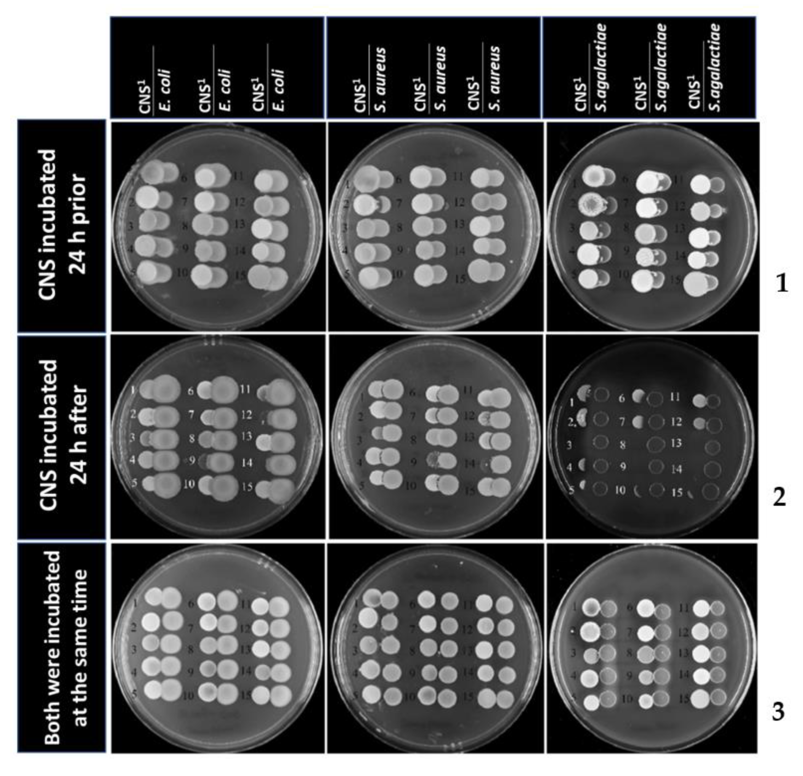
Figure 1. The inhibition zone of major strains toward minor strains in PRIOR condition. (1) 24-h prior incubation with CNS (upper), AFTER condition; (2) twenty-four-hour prior incubation with major pathogens (middle), EQUAL condition; (3) starting incubation at the same time (lower). 1 Coagulase-negative pathogens included 1., 2. S. chromogenes; 3., 4. S. epidermidis; 5., 6. S. haemolyticus; 7. S. simulans; 8., 9. S. warneri; 10. S. xylosus; 11. S. epidermidis; 12. S. hominis; 13. S. cohinii; 14. S. capitis; 15. S. sciuri.
Antibiotics 12 00600 g001
Figure 2. Percentage of the survival ability of CNS strains toward major strains in (A) PRIOR condition 1 and 24-h prior incubation with CNS; (B) AFTER condition 2 and 24-h prior incubation with major pathogens; (C) EQUAL condition 3, starting incubation at the same time. *** Indicated the significant difference in survival rates between species when incubated with the specified major strains at p < 0.05.
Antibiotics 12 00600 g002
Figure 3. Example of the result of the agar spot competition assays: (A) the spots are colonies formed by S. chromogenes, the right spots are the colonies formed by S. agalactiae (No. 31–45) when the S. chromogenes were cultured at 24 h prior (PRIOR), and many colonies formed by S. agalactiae are degraded and lysed; (B) the spots are colonies formed by S. hominis, the right spots are the colonies formed by S. agalactiae when the S. hominis (No. 1–10) and S. warneri (No. 11–15) are cultured at 24 h later (AFTER), and many colonies formed by S. hominis (No. 1–4, 6, 7, 9 and 10) are detached and are not degraded by S. agalactiae.
Antibiotics 12 00600 g003
Table 1. Source of coagulase-negative staphylococci (CNS) as minor mastitis pathogens used in Experiment I.
No.SpeciesSource
1, 2Staphylococcus chromogenesMastitis quarter
3, 4Staphylococcus epidermidisMastitis quarter
5, 6Staphylococcus haemolyticusMastitis quarter
7Staphylococcus simulansMastitis quarter
8, 9Staphylococcus warneriMastitis quarter
10Staphylococcus xylosusMastitis quarter
11Staphylococcus epidermidisATCC
12Staphylococcus hominisFaculty of Science
13Staphylococcus cohiniiFaculty of Science
14Staphylococcus capitisFaculty of Science
15Staphylococcus sciuriFaculty of Science
Disclaimer/Publisher’s Note: The statements, opinions and data contained in all publications are solely those of the individual author(s) and contributor(s) and not of MDPI and/or the editor(s). MDPI and/or the editor(s) disclaim responsibility for any injury to people or property resulting from any ideas, methods, instructions or products referred to in the content.