Abstract
Bacteriophages (phages) are antimicrobials with resurgent interest that are being investigated for the treatment of antibiotic refractory infection, including for Pseudomonas aeruginosa (Pa) lung infection in cystic fibrosis (CF). In vitro work supports the use of this therapy in planktonic and biofilm culture models; however, consistent data are lacking for efficacy across different clinical Pa strains, culture models, and in combination with antibiotics in clinical use. We first examined the efficacy of a 4-phage cocktail as an adjunct to our CF centre’s first-line systemic combination antibiotic therapy (ceftazidime + tobramycin) for 16 different clinical Pa strains and then determined subinhibitory interactions for a subset of these strains with each antibiotic in planktonic and biofilm culture. When a 4-phage cocktail (4 × 10⁸ PFU/mL) was added to a ceftazidime-tobramycin combination (ceftazidime 16 mg/mL + tobramycin 8 mg/mL), we observed a 1.7-fold and 1.3-fold reduction in biofilm biomass and cell viability, respectively. The four most antibiotic resistant strains in biofilm were very susceptible to phage treatment. When subinhibitory concentrations of antibiotics and phages were investigated, we observed additivity/synergy as well as antagonism/inhibition of effect that varied across the clinical strains and culture model. In general, more additivity was seen with the phage-ceftazidime combination than with phage-tobramycin, particularly in biofilm culture, where no instances of additivity were seen when phages were combined with tobramycin. The fact that different bacterial strains were susceptible to phage treatment when compared to standard antibiotics is promising and these results may be relevant to ongoing clinical trials exploring the use of phages, in particular in the selection of subjects for clinical trials.
1. Introduction
In cystic fibrosis (CF) lung infection, antibiotic administration has contributed to increased longevity and improved quality of life [1]. In cases of chronic infection, especially with Pseudomonas aeruginosa (Pa), this requires chronic daily suppressive therapy nebulised into the lungs [2,3]. Unfortunately, this daily nebulisation rarely clears well-established infection and the disease course of patients with CF and chronic infection is punctuated by periods of exacerbation that require further courses of oral, inhaled or intravenous antibiotics.
Within the CF lung, chronic Pa exists as aggregates that are suspended within the sputum matrix [4,5]. This biofilm lifestyle contributes to the survival of the bacteria by providing physical protection of the bacteria from immune cells and antimicrobial therapies. Additionally, the heterogeneity of the structure results in reservoirs of cells with differential access to nutrients, resulting in differing metabolic profiles and rates of cell division and this also impacts antibiotic treatment. It is known that the minimum inhibitory concentration (MIC) of planktonically grown bacteria can be many orders of magnitude higher for the same bacteria when grown in biofilms [6]; however, it is often impossible to achieve these concentrations in the airway [7,8,9].
There is an expanding body of evidence demonstrating the potential of bacteriophage (phage or phages) to combat Pa infection in CF lungs. We have previously shown increased survival and lowered inflammatory response in a model of murine Pa lung infection [10] and demonstrated phage sensitivity over time for many MDR strains (including the Liverpool Epidemic Strain) in patients at our hospital [11]. Although these and other studies provide a promising evidence base, as this therapy makes the leap from laboratory to human therapy in several ongoing human trials [12,13,14,15], there is no reason that the interactions between antibiotics, phage and bacteria should be consistent across the concentrations or type of bacterial growth encountered in the lung.
There is a body of preclinical experimentation investigating the effects of phage-antibiotic combinations in different culture systems. One study examining 17 clinical Pa strains with documented resistance to phage in a planktonic assay, demonstrated phage-replication within the biofilms of eight strains (47%) of the planktonically-resistant strains [16]. One study found that phage virion production was blocked with the administration of twice the minimum inhibitory concentration (MIC) of rifampicin in a Ralstonia solanacearum culture [17]. There are also Pa specific examples of phage-antibiotic antagonism that highlight this as a risk. One early study revealed that burst sizes (the amount of phage virion progeny released at cell lysis) of anti-Pseudomonal phage PM2 was reduced to less than 1% of that seen in control conditions with simultaneous addition of rifampicin at a concentration of 0.3 μg/mL [18]. Subsequent work with Pa decades later demonstrated that subinhibitory concentrations of rifampicin completely inhibited phage (lytic phages PAK_P3 and PAK_P4) replication [19].
There is, however, a similar body of in vitro work supporting phage additivity or synergy. Gu Liu et al. (2020) recently looked at many different antibiotic classes combined with phages against a highly drug-resistant E. coli in a checkerboard assay and found that phages could render this MDR bacteria susceptible to antibiotics at concentrations to which it had previously demonstrated resistance [20]. These experiments demonstrated synergy with phage-ceftazidime combination and additivity of effects with phage-kanamycin, an aminoglycoside antibiotic in the same class as tobramycin.
Tobramycin (TOB) is an aminoglycoside antibiotic that targets the bacterial ribosome 30S subunit, and ribosomal function is essential for virion replication and the cell lysis of bacteriophage [21]. Ceftazidime (CTZ), a ß-lactam, works at the cell wall, which is where bacteriophages adsorb to surface proteins, necessary for allowing entry of genetic material into the cell [21]. Exposing Pa to an antibiotic to which it is susceptible can, in theory, block subsequent phage infection and phage-mediated killing. Many classes of antibiotics interfere with bacterial cell physiology in ways that are crucial for phage infection, and the possibility of non-additive or even inhibitory/antagonistic results must be considered.
Here, we wished to explore the efficacy of a 4-phage cocktail that has been used in several preclinical studies and for compassionate use [22,23,24,25] against Pa isolates from CF airway samples. We used two common anti-Pseudomonal antibiotics: ceftazidime and tobramycin. In the first experiments, we use a high-concentration combination of these two agents and assess the extra biofilm reduction with the addition of phage in 16 different Pa clinical strains. In the subsequent experiments, we investigate the effects of subinhibitory concentrations of phage cocktail along with subinhibitory concentrations of ceftazidime and tobramycin individually, in both biofilm and planktonic culture for 10 different strains.
2. Results
2.1. Assessing Biofilm Eradication with Phage with an Antibiotic Mix across 16 Clinical Strains
Results of the resazurin assay (cell viability) and the crystal violet (CV) assay (biomass) correlated well with each other (Pearson coefficient 0.80, p = 0.0002). The level of planktonic sensitivity to bacteriophage treatment did not correlate with the outcomes of either biofilm assay (Pearson’s coefficient of 0.36 and 0.41 for the resazurin and CV assays, respectively). We concluded that the degree of planktonic sensitivity to phage activity did not predict biofilm eradication. These results are summarized in Figure 1.
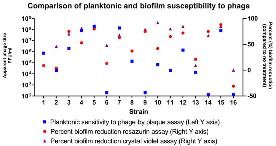
Figure 1.
Comparison of planktonic and biofilm susceptibility to phages. Planktonic sensitivity to phages as assessed by plaque assay (blue square) is shown on the logarithmic scale on the left-sided Y axis. The per cent reduction in biofilm cell viability (red circle—assessed by immunofluorescent resazurin assay) and biofilm biomass (purple triangle—assessed by crystal violet OD readings) are plotted on the right-sided Y axis.
In general, the results of the plaque assay were not predictive of the amount of biofilm reduction. The results depended on the clinical strain and also on the type of assay being used, whether assessing cell viability or biomass. Across the group of 16 strains, bacteriophage cocktail monotherapy at an apparent titre of 4 × 10⁷ PFU/mL resulted in a decrease in cell viability of 46% (95% CI 28.0–64.6%) and biomass of 59% (95% CI 43.0–74.7%) and these means they were not significantly different from one another.
The addition of phages enhanced the anti-biofilm efficacy of a fixed ceftazidime (CTZ)—tobramycin (TOB) combination across clinical strains (Figure 2).
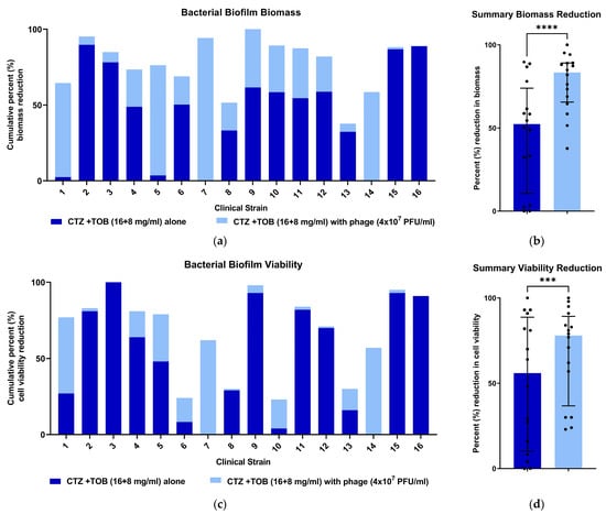
Figure 2.
The percent reduction in biofilm biomass and cell viability with antibiotic combination and phage. Dark blue shows the amount of biofilm reduction with the antibiotic combination alone, with the added light blue showing the additional reduction in killing when the bacteriophage was added. The bars, when shown, represent the median and interquartile range. (a) The percent reduction of biofilm biomass when the phage cocktail was added to a CTZ + TOB antibiotic mixture for each strain. (b) The addition of the phage cocktail resulted in a further 1.7-fold reduction in biofilm biomass across the 16 strains (****, p < 0.0001 using the Wilcoxon signed rank test) compared with the dual antibiotics alone. (c) The percent reduction in metabolic activity of biofilm cells when the phage cocktail was added to a CTZ + TOB antibiotic mixture. (d) The addition of the phage cocktail resulted in a further 1.3-fold reduction in cell viability across the 16 strains (***, p < 0.001 using the Wilcoxon signed rank test) compared with the dual antibiotics alone.
The four strains that demonstrated the greatest level of biofilm reduction with the addition of the phage cocktail (strains 1, 5, 7 and 14) were the most antibiotic resistant in biofilm culture when assessed by the CV assay (all <10% reduction in biofilm biomass).
2.2. Assessing Subinhibitory Concentrations of Phage and Antibiotics in Planktonic and Biofilm Culture
Ten (10) randomly selected strains from the initial 16, as well as laboratory strain PA01 underwent further analysis in planktonic and biofilm culture at subinhibitory phage and antibiotic combinations. Strain 16 was excluded on the basis that it was sometimes resistant to phage in plaque assay, but not at time point 1, which was when it was included in the first set of experiments. A summary of these findings is represented in Table 1.

Table 1.
Summary data for checkerboard experiments across 10 clinical strains + laboratory strain PA01.
With planktonic cultures, combination antibiotic-phage therapy resulted in a higher rate of bacterial eradication 54.5% of the time (95% CI; 34.7–73.1%), whereas in biofilm culture this was only seen 27.3% of the time (95% CI; 13.2–48.2%). For individual antibiotics, combination ceftazidime-phage therapy resulted in a statistically higher rate of bacterial eradication 59.1% of the time (95% CI; 38.7–76.7%), whereas for tobramycin this was 22.7% (95% CI; 10.1–43.4%). Notably, in biofilm, there were no strains that demonstrated significant additivity of effect with phage-tobramycin therapy, whereas 6/11 strains tested (54.5%) demonstrated additivity or synergy with ceftazidime-phage combination therapy. Strain PA01 with ceftazidime-phage in biofilm culture, coefficient of drug interaction (CDI) = 0.44 ± 0.10; Strain 7 with tobramycin-phage in planktonic culture, CDI = 0.43 ± 0.07; and Strain 11 with ceftazidime-phage in biofilm culture, CDI = 0.58 ± 0.12. Two of the 3 instances in which phage-antibiotic antagonism was seen were due to reduced efficacy of phage when combined with the antibiotic. There was one instance where combination treatment led to reduced antibiotic efficacy (Strain 5, tobramycin-phage in biofilm depicted in Figure 3d).
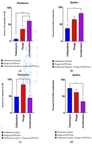
Figure 3.
Examples of all different classes of phage-antibiotic combinations: (a) synergy, (b) additivity, (c) inhibition of phage by antibiotic, (d) inhibition of antibiotic by phage, (e) trend towards efficacy, but statistical significance not met (no effect), (f) no trend seen (no effect). In (a,b), two different clinical strains demonstrate the additive effects of combining individual antibiotics and phage. (a). Strain 7 demonstrated synergy of effect in the addition of phage to tobramycin at in planktonic culture. (b). Strain 5 demonstrated the additive effects in the addition of phage with ceftazidime in biofilm culture. In (c,d), two strains demonstrated different types of inhibition when combining individual antibiotics and phage. (c). Strain 3 in planktonic culture demonstrated inhibition of the action of phage when ceftazidime was added. (d). Strain 5 in biofilm culture demonstrated antagonism of the action of tobramycin when phage was added. In (e,f), two examples where there was no significant effect of the antibiotic-phage combination. (e). Strain 14 in biofilm culture demonstrated more biofilm eradication with the combination of ceftazidime-phage, but these results did not meet statistical significance. (f). Strain 14 in biofilm culture demonstrated no effect on biomass reduction with a combination of tobramycin-phage. Results depicted are for the means of 3 biological replicates combining 6 technical replicates in each group. One-way ANOVAs correcting for multiple comparisons; * p < 0.05, ** p < 0.01, *** p < 0.001.
We also investigated whether planktonic and biofilm responses to the addition of phage correlated with one another. For the 4 conditions in Table 1 (N, A, I, S), we assigned a value of 0 for N, −1 for I, 1 for A and +2 for S. Results in either assay did not correlate with one another (Spearman correlation, p = 0.22).
3. Discussion
We have demonstrated that the addition of a lytic phage cocktail to a combination of ceftazidime and tobramycin, two anti-Pseudomonal antibiotics commonly used together in clinical practice, significantly reduced biomasses and viability of biofilm-grown clinical Pa strains from CF airway cultures. Encouragingly, the four strains demonstrating the highest level of resistance to the antibiotic combination therapy demonstrated high levels of sensitivity to the bacteriophage cocktail.
We then looked at the efficacy of subinhibitory concentrations of phage when combined with subinhibitory concentrations of ceftazidime or tobramycin in a checkerboard assay and found that there was more additivity/synergy when phage was combined with ceftazidime, as opposed to tobramycin, especially in biofilm culture. It is, worth noting that there were no instances of additivity seen in biofilm culture with tobramycin for the 10 clinical strains examined. There were instances of both synergy and inhibition for ceftazidime in both planktonic and biofilm cultures. This research is important in that it shows efficacy in an array of clinical strains found in a specific disease process alongside clinically relevant antibiotics and concentrations across different culture models (planktonic and biofilm).
Ultimately, the final message from these experiments is one of variability. Individual clinical strains interact differently with antibiotics and phages in different growth conditions and we were not able to predict responses to these combinations. To this point, we found early on that planktonic sensitivity to phage did not predict efficacy of biofilm reduction. There were instances of poor planktonic response to phages but a strong reduction in biofilm, as well as exquisite sensitivity to the phage in planktonic culture but a poor response in biofilm. Furthermore, our checkerboard experiments for individual antibiotic-phage combination therapy showed poor correlation between planktonic and biofilm culture models.
One of the explanations for additivity is that the 2 modalities target different populations, both for reasons of differential access to cells and due to the mechanism of action. For instance, in contrast to most conventional antibiotics which require actively dividing bacterial cells, bacteriophage can lyse metabolically dormant cells. A study investigating S. aureus phages demonstrated infection and lysis of metabolically dormant persister cells in addition to exopolysaccharide degradation of the biofilm matrix [26]. This finding is promising for an adjunctive therapy as it indicates that the agents would not necessarily be competing for access and killing the same cell populations, but might be broadening the spectrum of populations eradicated. With regards to cells in different geographic locations within the biofilm, it is, known that some lytic phages specific to Pa employ the use of exopolysaccharide degrading enzymes that can disperse biofilms, which may prove beneficial in chronic, biofilm infections [27]. The use of such phage-encoded exopolysaccharide depolymerases has been put forth as promising antibiotic adjuvants in their own right [28]. Thus, one hope is that by using agents that work in different ways, the success of current antimicrobial therapy would be increased and that this may translate to improved patient outcomes in CF.
There has been a large focus in the literature on conditions that inhibit the action of phages, and such studies have largely looked at the application of antibiotics prior (i.e., as pre-treatment) to the administration of phage [17,19]. There are indeed studies that suggest the order of application can significantly alter the outcome of antibiotic-phage experiments. Several studies involving both Staphylococcus aureus and Pa have shown that when phage treatment was administered prior to antibiotics, results were additive or synergistic, as opposed to when phage was administered after antibiotics when results were seen to be antagonistic to the action of phage [29,30]. Although the mechanism of this phage antagonism is unclear, some hypotheses that have been put forth are a reduction in bacterial density leading to a reduction in viral replication, as well as the possibility that antibiotics directly interfere with phage infection and/or replication within the cell. Notably, Chaudhry et al. (2017) found that a delay of 24 h between aminoglycoside administration (gentamicin or tobramycin) and phage in an in vitro Pa model of infection effectively suppressed any phage replication [30].
When designing our experimental protocol, the timing of phage and antibiotic administration had been intended to be simultaneous; however, in practice, antibiotic administration was just before the addition of phage and time lags between antibiotic and phage administration could have been as high as 10 min for individual plates. In this way, we may have been unintentionally reducing the efficacy of the phage in both planktonic and biofilm culture, giving the antibiotics time to exert effects leading to antagonism of phage. This type of antagonism (antibiotics lessening the effects of phage) was seen in 2 experimental conditions: once in planktonic culture with ceftazidime and once in biofilm culture with tobramycin; Table 1). Notably, in only 1 out of the 3 instances of antagonism seen did the combined administration of phage worsen the efficacy of antibiotics alone (this was seen in biofilm culture with tobramycin in Strain 5, Figure 3d). This example is noteworthy not only because it was the only case of antibiotic antagonism that was seen across the 10 strains, but because the results in planktonic culture were seen to be additive with tobramycin. The reasons for this antagonism are not clear, but notably, the assay used (CV) does not give an approximation of cell viability and it could have simply been more dead matter that had adhered to the well. Although the results of the CV and resazurin assays correlated well, this was not true for every strain.
An obvious criticism of any in vitro work is that it does not adequately simulate the complexity of bacterial infection in a living organism. The fact that bacteria exist in polymicrobial communities within the lung environment is one such factor [31]. This is thought to explain some of the disconnects between laboratory-based methods of antibiotic sensitivity testing (both in planktonic and in biofilm cultures) compared to clinical outcomes [32]. In one experiment using E. coli and Pa, the corresponding phage could eradicate each monoculture biofilm, whereas the two bacteriophages applied together were not effective in a mixed biofilm [33]. To address this, further in vitro work needs to be carried out in different culture models and conditions that more accurately simulate the lung environment, such as mixed species biofilms, artificial sputum media and/or small animal models of CF.
4. Materials and Methods
4.1. Selection and Storage of Pa Strains
One laboratory strain (PA01) was used in these experiments [34]. For clinical isolates, a Microsoft Access database of Pa from CF patients at the Royal Brompton Hospital (RBH) was used. Clinical Pa strains were provided by the RBH microbiology laboratory and had been identified by matrix-assisted laser desorption/ionization-time of flight (MALDI-TOF) analysis as being Pa [35]. Samples from CF patients were taken directly from sweeps of bacterial colonies grown on agar with a plastic inoculation loop. Strains were stored on glass Microbank™ beads (ProLab Diagnostics, Richmond Hill, ON, Canada) in a −80 °C freezer. Colonies were seeded on nutrient agar for overnight growth in the incubator at 37 °C.
Isolates were selected based on recorded sensitivity to both ceftazidime and tobramycin by disc diffusion assay [36].
4.2. Plaque Assay
The phages used in this study were provided by AmpliPhi Australia Pty Ltd. (Brookvale, NSW 2100, Australia). AB-PA01 is a combination of four lytic bacteriophages, Pa193 and Pa204 belonging to the Myoviridae family, and Pa222 and Pa223 belonging to the Podoviridae family. The titer for the phage mix on the company’s P. aeruginosa strains used for these experiments was 4 × 10⁸ PFU/mL. The phage titre was determined using plaque assay, as described below. None of these phage components encoded any known bacterial virulence or antibiotic resistance genes, and all phages were considered to be strictly lytic as per the company’s published procedures [25]. The phages were produced following the current good manufacturing practice standard (cGMP) and approved by the US Food and Drug Administration as an investigational new drug for an expanded access program (NCT03395743) [37]. An updated release of AB-PA01 with a higher titre has been used in preclinical studies and compassionate use cases [24,38].
We performed the standard plaque assay for bacteriophage sensitivity testing, as previously described [39]. In brief, an overnight culture of a selected Pa isolate was placed in nutrient broth and grown overnight. Overnight bacterial culture was then diluted in fresh nutrient broth to an optical density (OD) of 0.05. One hundred μL was added to 3 mL of semi-solid nutrient agar (Oxoid, Basingstoke, UK), poured over sterile nutrient agar plates (Oxoid, UK) and allowed to dry in the safety cabinet for 20–30 min. Five (5) μL spots of each phage dilution (neat phage to 10−6 dilution) were spotted onto the surface of the Pa inoculated plates and then incubated overnight (16–24 h) at 37 °C before assessing for plaques on the bacterial lawn.
All isolates were examined by plaque assay and screened for their sensitivity to at least the neat bacteriophage cocktail (4 × 10⁸ PFU/mL). Sixteen (16) different Pa isolates were chosen for the first section of experiments and 10 of these were selected for use in the antibiotic-phage checkerboard experiments.
4.3. Biofilm Assays
To assess biofilm reduction, we used 96-well plates for two different, high-throughput assays: crystal violet (CV) to assess biofilm biomass [40] and resazurin salt (Alamar Blue, IUPAC name: 7-hydroxy-10-oxiphenoxazin-10-ium-3-one) to assess cell viability [41].
Two hundred (200) μL of overnight culture shaken at 200 rpm at 37 °C in nutrient broth (Oxoid) was diluted to an OD of 0.05 and then pipetted into each well of a 96 well plate which was incubated at 37 °C for 24 h static. The plates were sealed with a membrane to reduce evaporation, but allow gas exchange (Axygen, Sigma Aldrich, Gillingham, UK). Wells were then emptied by pipetting and washed with a phosphate-buffered saline (PBS) solution before 180 µL of Mueller Hinton broth (MHB) (Sigma Aldrich) was added to all wells. Twenty (20) μL of each treatment was applied to the wells. There were three different treatment protocols:
(i) An antibiotic mixture of ceftazidime and tobramycin, both at 2× the published breakpoints for sensitivity (final concentration in wells was 16 mg/mL ceftazidime + 8 mg/mL of tobramycin);
(ii) The bacteriophage cocktail on its own (final concentration 4 × 10⁷ PFU/mL or 1 × 10⁶ PFU/phage); and
(iii) The antibiotic combination of ceftazidime and tobramycin as described above + the phage cocktail (final concentration 4 × 10⁷ PFU/mL of phage, 16 mg/mL ceftazidime and 8 mg/mL tobramycin).
Plates were then incubated overnight (a further 16–20 h) at 37 °C before final biofilm measurements were obtained.
To assess cell viability (resazurin), all wells were emptied of free-floating cells by microchannel pipette and washed with PBS before the introduction of 180 μL of MHB, then 20 μL of resazurin 0.02% w/v working solution. Plates were covered with aluminium foil and incubated at 37 °C for 3–4 h before being transferred to the plate reader for analysis using emission and excitation wavelengths of 540 and 590 nm, respectively (FI540/590).
To assess biofilm biomass (CV), plates were emptied by tipping out contents and washed twice by submersion and inversion in water before the remaining liquid was removed by inversion. One hundred eighty (180) μL of 0.1% crystal violet solution was added to each well for 15 min before 2 further inversion and washing steps in water. Two hundred (200) μL of 95% ethanol was then added to each well to solubilise the biofilms and then left for 30 min before absorption readings with the spectrophotometer (OD550).
4.4. Checkerboard Assay
The checkerboard assay has been previously described in detail, whereby serial dilutions of two different antimicrobial compounds are administered to bacterial culture in a 96-well plate [42,43].
Concentrations for ceftazidime and tobramycin ranged from 1/16th to 8× the published EUCAST minimum inhibitory concentration (MIC) values [36]. For phage, concentrations ranged from 4 × 10² to 4 × 10⁸ PFU/mL. To optimally appreciate the effects of combination therapy in planktonic and biofilm culture, for antibiotics and phages, we used the first dilution below which resulted in either >75% reduction in planktonic growth as assessed by OD or >75% reduction in biofilm biomass as assessed by CV assay. For antibiotics, this work was done in 2-fold serial dilutions and for bacteriophage, we worked with 10-fold serial dilutions. For biofilm culture, there were instances where the highest antibiotic and phage concentrations did not achieve >75% biofilm eradication. In these instances, we assessed the highest concentration tested.
For planktonic culture, 180 μL of overnight culture was diluted in Mueller Hinton broth (Sigma Aldrich) to an OD of 0.1 and then pipetted into each well of a 96-well plate. Twenty (20) μL of solution for treatment groups was administered so that final target concentrations were reached and final volume in the wells = 200 μL. For negative control wells, PBS was used as this was used for phage and antibiotic dilutions.
Results were evaluated spectrophotometrically at 18 h after treatment overnight in the incubator at 37 °C shaking at 200 rpm. Treatment groups (n = 6 technical replicates each) were compared to each other and standardized across strains to account for differences in OD measurements according to the following equation:
Percent reduction (%) in OD = ((ODcontrol/no treatment − ODtreatment)/ODcontrol/no treatment) × 100
For biofilm assays, 200 μL of overnight culture was diluted to an OD of 0.1 in Nutrient broth and then pipetted into each well of a 96-well plate. Plates were incubated at 37 °C static for 24 h to allow cells to adhere to the sides and bottom of the wells. Wells were then emptied and washed with a phosphate-buffered saline (PBS) solution before 180 μL of Mueller Hinton broth (Sigma Aldrich) was added to all wells along with different treatments so that the final volume in the wells = 200 μL. For negative control wells, PBS was used as this had been used for phage and antibiotic dilutions. Plates were then incubated at 37 °C overnight (a further 16–20 h) shaking at 200 rpm before final biofilm measurements were obtained using the crystal violet assay, as described earlier in the methods section. Treatment groups consisting of 6 technical replicates were compared to each other and standardized across strains to account for differences in biomass measurements according to the following equation:
Reduction (%) of biomass with CV assay = ((ODcontrol/no treatment − ODtreatment)/ODcontrol/no treatment) × 100
4.5. Coefficient of Drug Interaction
A coefficient of drug interaction (CDI) was developed for the assessment of the interaction between two different antimicrobial compounds and was used per previously published data [44]. It is calculated based on the following formula:
CDI = AB/(A × B)
According to the OD readings for each treatment group, AB is the ratio of the antibiotic-phage combination groups to the negative control group; A or B is the ratio of the single agent (antibiotic or phage) group to the negative control group. A CDI value of <1 indicates that the combination of A + B is synergistic. For these experiments, we have been conservative and used CDI values of <0.7 with standard deviations that do not cross 1 as synergistic.
4.6. Statistical Analysis
All statistical analysis and graphs were performed using GraphPad Prism software (Boston, MA, USA). Tests of normality were performed for all data using the Shapiro–Wilk test unless otherwise stated and data were said to be normally distributed if an alpha of 0.05 was reached. For all work in the microtiter plates, ‘robust regression followed by outlier elimination’ (ROUT) was employed to detect outliers if the data were normally distributed (false discovery rate below 1%) [45].
For the first experiments, data across all 16 clinical strains were non-parametric and the Wilkoxon signed-rank test was used for comparison between treatment groups. For the checkerboard assay, one-way ANOVAs were performed to compare outcomes between treatment groups, correcting for multiple comparisons. If the mean effects of combination treatment groups for the 3 biological replicates were statistically different using one-way ANOVAs, CDIs were calculated to determine whether the combination was synergistic (S) or simply additive (A). Experimental conditions in which the effect of combination therapy was significantly less than either single phage or antibiotic therapy were classified as inhibitory (I). Where the results of group means were not statistically different, the combination treatment was said to have no effect (N).
For comparing the relative success of combination therapy based on antibiotic choice/type of culture, classification was binomial: success (additive or synergistic) or failure (no effect or antagonistic). The modified Wald method was used to calculate confidence intervals of the proportion of successful outcomes for ceftazidime and tobramycin in combination with phage in both planktonic and biofilm cultures [46].
5. Conclusions
The 4-phage cocktail used in these experiments showed efficacy across a variety of clinical Pa strains both planktonically and within biofilm models. Notably, it was first shown to improve the eradication of pre-formed biofilms when combined with ceftazidime and tobramycin. In further experiments, subinhibitory phage-antibiotic combinations were seen to be synergistic/additive, having no statistically discernable effect, or being antagonistic/inhibitory.
Although it is difficult to draw conclusions that might affect the interpretation of clinical trials exploring phage therapy that are currently in progress, it is important that further in vitro work be carried out to investigate these interactions, similar to work that has been carried out exploring antibiotic-phage combinations in other organisms [20,47]. This is particularly valid based on our finding that the aminoglycoside tobramycin was less likely to result in a favourable outcome when combined with phages. For the clinical trials that are currently being conducted, it would be reasonable to investigate the timing of antibiotic administration in relation to phage administration or perform sub-group analyses in patients to determine if there are any clinical effects observed for patients on concurrent antimicrobial therapy—especially those on chronic suppressive therapy with aminoglycosides.
Author Contributions
Conceptualization, J.C.D. and I.M.; methodology, J.C.D. and I.M.; validation, J.C.D. and I.M.; formal analysis, I.M.; investigation, I.M.; resources, J.C.D. and I.M.; data curation, I.M.; writing—original draft preparation, J.C.D. and I.M.; writing—review and editing, J.C.D., I.M., S.M. and E.W.F.W.A.; visualization, J.C.D. and I.M.; supervision, J.C.D. and E.W.F.W.A.; project administration, J.C.D.; funding acquisition, J.C.D. All authors have read and agreed to the published version of the manuscript.
Funding
This research was partly funded by the Cystic Fibrosis Trust, UK and the Strategic Research Centre for Pseudomonas.
Institutional Review Board Statement
Not applicable.
Informed Consent Statement
Not applicable. CF patients in this study had previously consented to their clinical data being entered into the national patient registry and being used for approved research projects. Isolates and data were coded and pseudonymized for the study.
Data Availability Statement
More detailed data from this manuscript can be found by accessing the MD(Res) thesis online through the Spiral portal through Imperial College London (https://spiral.imperial.ac.uk/, accessed on 1 December 2022). The thesis is entitled “Lytic bacteriophage as an adjunct to antibiotic therapy for Pseudomonas aeruginosa lung infection in cystic fibrosis patients”.
Acknowledgments
We gratefully acknowledge funding for the Strategic Research Centre for Pseudomonas from the Cystic Fibrosis Trust, UK. The group is supported by the National Institute for Health Research through the Imperial Biomedical Research Centre, the Royal Brompton Hospital/Imperial College London Clinical Research Facility and NIHR Senior Investigator Awards (JCD, EWFWA). We thank the staff of the clinical microbiology laboratory for their help in collecting bacterial strains.
Conflicts of Interest
Sandra Morales worked at AmpliPhi Biosciences Corporation at the time of this study. The funders had no role in the design of the study; in the collection, analyses, or interpretation of data; in the writing of the manuscript, or in the decision to publish the results.
References
- Turcios, N.L. Cystic fibrosis lung disease: An overview. Respir. Care 2020, 65, 233–251. [Google Scholar] [CrossRef] [PubMed]
- Smith, S.; Rowbotham, N.J.; Charbek, E. Inhaled antibiotics for pulmonary exacerbations in cystic fibrosis. Cochrane Database Syst. Rev. 2022, 8, CD008319. [Google Scholar] [CrossRef] [PubMed]
- Smith, S.; Rowbotham, N.J.; Regan, K.H. Inhaled anti-pseudomonal antibiotics for long-term therapy in cystic fibrosis. Cochrane Database Syst. Rev. 2018, 3, CD001021. [Google Scholar] [CrossRef] [PubMed]
- Alhede, M.; Kragh, K.N.; Qvortrup, K.; Allesen-Holm, M.; van Gennip, M.; Christensen, L.D.; Jensen, P.Ø.; Nielsen, A.K.; Parsek, M.; Wozniak, D. Phenotypes of non-attached Pseudomonas aeruginosa aggregates resemble surface attached biofilm. PLoS ONE 2011, 6, e27943. [Google Scholar] [CrossRef]
- Bjarnsholt, T.; Jensen, P.Ø.; Fiandaca, M.J.; Pedersen, J.; Hansen, C.R.; Andersen, C.B.; Pressler, T.; Givskov, M.; Høiby, N. Pseudomonas aeruginosa biofilms in the respiratory tract of cystic fibrosis patients. Pediatr. Pulmonol. 2009, 44, 547–558. [Google Scholar] [CrossRef]
- Olson, M.E.; Ceri, H.; Morck, D.W.; Buret, A.G.; Read, R.R. Biofilm bacteria: Formation and comparative susceptibility to antibiotics. Can. J. Vet. Res. 2002, 66, 86–92. [Google Scholar]
- Moriarty, T.; McElnay, J.; Elborn, J.; Tunney, M. Sputum antibiotic concentrations: Implications for treatment of cystic fibrosis lung infection. Pediatr. Pulmonol. 2007, 42, 1008–1017. [Google Scholar] [CrossRef]
- Conway, S. Nebulized antibiotic therapy: The evidence. Chronic Respir. Dis. 2005, 2, 35–41. [Google Scholar] [CrossRef]
- Chmiel, J.F.; Aksamit, T.R.; Chotirmall, S.H.; Dasenbrook, E.C.; Elborn, J.S.; LiPuma, J.J.; Ranganathan, S.C.; Waters, V.J.; Ratjen, F.A. Antibiotic management of lung infections in cystic fibrosis. I. The microbiome, methicillin-resistant Staphylococcus aureus, gram-negative bacteria, and multiple infections. Ann. Am. Thorac. Soc. 2014, 11, 1120–1129. [Google Scholar] [CrossRef]
- Pabary, R.; Singh, C.; Morales, S.; Bush, A.; Alshafi, K.; Bilton, D.; Alton, E.W.; Smithyman, A.; Davies, J.C. Antipseudomonal bacteriophage reduces infective burden and inflammatory response in murine lung. Antimicrob. Agents Chemother. 2016, 60, 744–751. [Google Scholar] [CrossRef]
- Martin, I.; Kenna, D.T.; Morales, S.; Alton, E.W.; Davies, J.C. Variability in bacteriophage and antibiotic sensitivity in serial Pseudomonas aeruginosa isolates from cystic fibrosis airway cultures over 12 months. Microorganisms 2021, 9, 660. [Google Scholar] [CrossRef] [PubMed]
- Identifier: NCT05453578. A Phase 1b/2 Trial of the Safety and Microbiological Activity of Bacteriophage Therapy in Cystic Fibrosis Subjects Colonized with Pseudomonas aeruginosa. Available online: https://clinicaltrials.gov/ct2/show/NCT05453578 (accessed on 1 December 2022).
- Identifier: NCT05010577. Nebulized Bacteriophage Therapy in Cystic Fibrosis Patients with Chronic Pseudomonas aeruginosa Pulmonary Infection. Available online: https://clinicaltrials.gov/ct2/show/NCT05010577 (accessed on 1 December 2022).
- Identifier: NCT04684641. CYstic Fibrosis bacterioPHage Study at Yale (CYPHY). Available online: https://clinicaltrials.gov/ct2/show/NCT04684641 (accessed on 1 December 2022).
- Identifier: NCT04596319. Ph 1/2 Study Evaluating Safety and Tolerability of Inhaled AP-PA02 in Subjects with Chronic Pseudomonas aeruginosa Lung Infections and Cystic Fibrosis (SWARM-Pa). Available online: https://clinicaltrials.gov/ct2/show/NCT04596319 (accessed on 1 December 2022).
- Ceri, H.; Olson, M.E.; Stremick, C.; Read, R.; Morck, D.; Buret, A. The Calgary Biofilm Device: New technology for rapid determination of antibiotic susceptibilities of bacterial biofilms. J. Clin. Microbiol. 1999, 37, 1771–1776. [Google Scholar] [CrossRef] [PubMed]
- Matsui, T.; Yoshikawa, G.; Mihara, T.; Chatchawankanphanich, O.; Kawasaki, T.; Nakano, M.; Fujie, M.; Ogata, H.; Yamada, T. Replications of two closely related groups of jumbo phages show different level of dependence on host-encoded RNA polymerase. Front. Microbiol. 2017, 8, 1010. [Google Scholar] [CrossRef] [PubMed]
- Riveros-Moreno, V. Effect of rifampicin on bacteriophage PM2 biogenesis. FEBS Lett. 1975, 51, 249–252. [Google Scholar] [CrossRef]
- Blasdel, B.G.; Chevallereau, A.; Monot, M.; Lavigne, R.; Debarbieux, L. Comparative transcriptomics analyses reveal the conservation of an ancestral infectious strategy in two bacteriophage genera. ISME J. 2017, 11, 1988–1996. [Google Scholar] [CrossRef]
- Gu Liu, C.; Green, S.I.; Min, L.; Clark, J.R.; Salazar, K.C.; Terwilliger, A.L.; Kaplan, H.B.; Trautner, B.W.; Ramig, R.F.; Maresso, A.W. Phage-antibiotic synergy is driven by a unique combination of antibacterial mechanism of action and stoichiometry. MBio 2020, 11, e01462-20. [Google Scholar] [CrossRef]
- Abedon, S.T. Phage-antibiotic combination treatments: Antagonistic impacts of antibiotics on the pharmacodynamics of phage therapy? Antibiotics 2019, 8, 182. [Google Scholar] [CrossRef]
- Aslam, S.; Courtwright, A.M.; Koval, C.; Lehman, S.M.; Morales, S.; Furr, C.L.; Rosas, F.; Brownstein, M.J.; Fackler, J.R.; Sisson, B.M.; et al. Early clinical experience of bacteriophage therapy in 3 lung transplant recipients. Am. J. Transpl. 2019, 19, 2631–2639. [Google Scholar] [CrossRef]
- Fong, S.A.; Drilling, A.; Morales, S.; Cornet, M.E.; Woodworth, B.A.; Fokkens, W.J.; Psaltis, A.J.; Vreugde, S.; Wormald, P.J. Activity of Bacteriophages in Removing Biofilms of Pseudomonas aeruginosa Isolates from Chronic Rhinosinusitis Patients. Front. Cell. Infect. Microbiol. 2017, 7, 418. [Google Scholar] [CrossRef]
- Law, N.; Logan, C.; Yung, G.; Furr, C.L.; Lehman, S.M.; Morales, S.; Rosas, F.; Gaidamaka, A.; Bilinsky, I.; Grint, P.; et al. Successful adjunctive use of bacteriophage therapy for treatment of multidrug-resistant Pseudomonas aeruginosa infection in a cystic fibrosis patient. Infection 2019, 47, 665–668. [Google Scholar] [CrossRef]
- Lehman, S.M.; Mearns, G.; Rankin, D.; Cole, R.A.; Smrekar, F.; Branston, S.D.; Morales, S. Design and Preclinical Development of a Phage Product for the Treatment of Antibiotic-Resistant Staphylococcus aureus Infections. Viruses 2019, 11, 88. [Google Scholar] [CrossRef] [PubMed]
- Tkhilaishvili, T.; Lombardi, L.; Klatt, A.-B.; Trampuz, A.; Di Luca, M. Bacteriophage Sb-1 enhances antibiotic activity against biofilm, degrades exopolysaccharide matrix and targets persisters of Staphylococcus aureus. Int. J. Antimicrob. Agents 2018, 52, 842–853. [Google Scholar] [CrossRef] [PubMed]
- Glonti, T.; Chanishvili, N.; Taylor, P. Bacteriophage-derived enzyme that depolymerizes the alginic acid capsule associated with cystic fibrosis isolates of Pseudomonas aeruginosa. J. Appl. Microbiol. 2010, 108, 695–702. [Google Scholar] [CrossRef] [PubMed]
- Yan, J.; Mao, J.; Xie, J. Bacteriophage polysaccharide depolymerases and biomedical applications. BioDrugs 2014, 28, 265–274. [Google Scholar] [CrossRef] [PubMed]
- Kumaran, D.; Taha, M.; Yi, Q.; Ramirez-Arcos, S.; Diallo, J.-S.; Carli, A.; Abdelbary, H. Does treatment order matter? Investigating the ability of bacteriophage to augment antibiotic activity against Staphylococcus aureus biofilms. Front. Microbiol. 2018, 9, 127. [Google Scholar] [CrossRef]
- Chaudhry, W.N.; Concepcion-Acevedo, J.; Park, T.; Andleeb, S.; Bull, J.J.; Levin, B.R. Synergy and order effects of antibiotics and phages in killing Pseudomonas aeruginosa biofilms. PLoS ONE 2017, 12, e0168615. [Google Scholar] [CrossRef]
- Surette, M.G. The cystic fibrosis lung microbiome. Ann. Am. Thorac. Soc. 2014, 11, S61–S65. [Google Scholar] [CrossRef]
- Waters, V.J.; Kidd, T.J.; Canton, R.; Ekkelenkamp, M.B.; Johansen, H.K.; LiPuma, J.J.; Bell, S.C.; Elborn, J.S.; Flume, P.A.; VanDevanter, D.R. Reconciling antimicrobial susceptibility testing and clinical response in antimicrobial treatment of chronic cystic fibrosis lung infections. Clin. Infect. Dis. 2019, 69, 1812–1816. [Google Scholar] [CrossRef]
- Kay, M.K.; Erwin, T.C.; McLean, R.J.; Aron, G.M. Bacteriophage ecology in Escherichia coli and Pseudomonas aeruginosa mixed-biofilm communities. Appl. Environ. Microbiol. 2011, 77, 821–829. [Google Scholar] [CrossRef]
- Klockgether, J.; Munder, A.; Neugebauer, J.; Davenport, C.F.; Stanke, F.; Larbig, K.D.; Heeb, S.; Schock, U.; Pohl, T.M.; Wiehlmann, L.; et al. Genome diversity of Pseudomonas aeruginosa PAO1 laboratory strains. J. Bacteriol. 2010, 192, 1113–1121. [Google Scholar] [CrossRef]
- Pusch, W.; Kostrzewa, M. Application of MALDI-TOF mass spectrometry in screening and diagnostic research. Curr. Pharm. Des. 2005, 11, 2577–2591. [Google Scholar] [CrossRef] [PubMed]
- Matuschek, E.; Brown, D.F.; Kahlmeter, G. Development of the EUCAST disk diffusion antimicrobial susceptibility testing method and its implementation in routine microbiology laboratories. Clin. Microbiol. Infect. 2014, 20, O255–O266. [Google Scholar] [CrossRef]
- Individual Patient Expanded Access for AB-PA01, An Investigational Anti-Pseudomonas Aeruginosa Bacteriophage Therapeutic. Available online: https://clinicaltrials.gov/ct2/show/NCT03395743 (accessed on 1 December 2022).
- Maddocks, S.; Fabijan, A.P.; Ho, J.; Lin, R.C.Y.; Ben Zakour, N.L.; Dugan, C.; Kliman, I.; Branston, S.; Morales, S.; Iredell, J.R. Bacteriophage Therapy of Ventilator-associated Pneumonia and Empyema Caused by Pseudomonas aeruginosa. Am. J. Respir. Crit. Care Med. 2019, 200, 1179–1181. [Google Scholar] [CrossRef] [PubMed]
- Kropinski, A.M.; Mazzocco, A.; Waddell, T.E.; Lingohr, E.; Johnson, R.P. Enumeration of bacteriophages by double agar overlay plaque assay. In Bacteriophages; Springer: Warsaw, Poland, 2009; pp. 69–76. [Google Scholar]
- O’Toole, G.A. Microtiter dish biofilm formation assay. J. Vis. Exp. 2011, 47, e2437. [Google Scholar]
- Rampersad, S.N. Multiple applications of Alamar Blue as an indicator of metabolic function and cellular health in cell viability bioassays. Sensors 2012, 12, 12347–12360. [Google Scholar] [CrossRef]
- Orhan, G.; Bayram, A.; Zer, Y.; Balci, I. Synergy tests by E test and checkerboard methods of antimicrobial combinations against Brucella melitensis. J. Clin. Microbiol. 2005, 43, 140–143. [Google Scholar] [CrossRef]
- White, R.L.; Burgess, D.S.; Manduru, M.; Bosso, J.A. Comparison of three different in vitro methods of detecting synergy: Time-kill, checkerboard, and E test. Antimicrob. Agents Chemother. 1996, 40, 1914–1918. [Google Scholar] [CrossRef]
- Zhou, X.; Zhang, Y.; Li, Y.; Hao, X.; Liu, X.; Wang, Y. Azithromycin synergistically enhances anti-proliferative activity of vincristine in cervical and gastric cancer cells. Cancers 2012, 4, 1318–1332. [Google Scholar] [CrossRef]
- Motulsky, H.J.; Brown, R.E. Detecting outliers when fitting data with nonlinear regression–a new method based on robust nonlinear regression and the false discovery rate. BMC Bioinform. 2006, 7, 123. [Google Scholar] [CrossRef]
- Agresti, A.; Coull, B.A. Approximate is better than “exact” for interval estimation of binomial proportions. Am. Stat. 1998, 52, 119–126. [Google Scholar]
- Berryhill, B.A.; Huseby, D.L.; McCall, I.C.; Hughes, D.; Levin, B.R. Joint antibiotic and phage therapy: Addressing the limitations of a seemingly ideal phage for treating Staphylococcus aureus infections. bioRxiv 2020. [Google Scholar] [CrossRef]
Disclaimer/Publisher’s Note: The statements, opinions and data contained in all publications are solely those of the individual author(s) and contributor(s) and not of MDPI and/or the editor(s). MDPI and/or the editor(s) disclaim responsibility for any injury to people or property resulting from any ideas, methods, instructions or products referred to in the content. |
© 2023 by the authors. Licensee MDPI, Basel, Switzerland. This article is an open access article distributed under the terms and conditions of the Creative Commons Attribution (CC BY) license (https://creativecommons.org/licenses/by/4.0/).