Article

Comparative Genomics Identifies Novel Genetic Changes Associated with Oxacillin, Vancomycin and Daptomycin Susceptibility in ST100 Methicillin-Resistant Staphylococcus aureus

by
Sabrina Di Gregorio
1,2,
María Sol Haim
1,2,3,
Ángela María Rosa Famiglietti
4,
José Di Conza
1,2 and
Marta Mollerach
1,2,*
1
Instituto de Investigaciones en Bacteriología y Virología Molecular (IBaViM), Facultad de Farmacia y Bioquímica, Universidad de Buenos Aires, Ciudad Autónoma de Buenos Aires 1113, Argentina
2
Consejo Nacional de Investigaciones Científicas y Técnicas (CONICET), Ciudad Autónoma de Buenos Aires 1113, Argentina
3
Unidad Operativa Centro Nacional de Genómica y Bioinformática, ANLIS Dr. Carlos G. Malbrán, Ciudad Autónoma de Buenos Aires 1282, Argentina
4
Laboratorio de Bacteriología Clínica, Hospital de Clínicas José de San Martín, Facultad de Farmacia y Bioquímica, Universidad de Buenos Aires, Ciudad Autónoma de Buenos Aires 1113, Argentina
*
Author to whom correspondence should be addressed.
Antibiotics 2023, 12(2), 372; https://doi.org/10.3390/antibiotics12020372
Submission received: 3 January 2023 / Revised: 9 February 2023 / Accepted: 10 February 2023 / Published: 11 February 2023
(This article belongs to the Special Issue Diversity of Antimicrobial Resistance Genes in Clinical Settings)

Abstract

:
Infections due to vancomycin-intermediate S. aureus (VISA) and heterogeneous VISA (hVISA) represent a serious concern due to their association with vancomycin treatment failure. However, the underlying molecular mechanism responsible for the hVISA/VISA phenotype is complex and not yet fully understood. We have previously characterized two ST100-MRSA-hVISA clinical isolates recovered before and after 40 days of vancomycin treatment (D1 and D2, respectively) and two in vitro VISA derivatives (D23C9 and D2P11), selected independently from D2 in the presence of vancomycin. This follow-up study was aimed at further characterizing these isogenic strains and obtaining their whole genome sequences to unravel changes associated with antibiotic resistance. It is interesting to note that none of these isogenic strains carry SNPs in the regulatory operons vraUTSR, walKR and/or graXRS. Nonetheless, genetic changes including SNPs, INDELs and IS256 genomic insertions/rearrangements were found both in in vivo and in vitro vancomycin-selected strains. Some were found in the downstream target genes of the aforementioned regulatory operons, which are involved in cell wall and phosphate metabolism, staphylococcal growth and biofilm formation. Some of the genetic changes reported herein have not been previously associated with vancomycin, daptomycin and/or oxacillin resistance in S. aureus.

1. Introduction

Staphylococcus aureus is a relevant pathogen with an extraordinary ability to evolve and acquire resistance to several antibiotics. Over the last decades, the large spread of antimicrobial-resistant (AMR) strains, including methicillin-resistant S. aureus (MRSA), vancomycin-intermediate S. aureus (VISA) and heterogeneous VISA (hVISA), have raised an alarm worldwide as declared by the World Health Organization in 2017 [1]. hVISA/VISA isolates are associated with persistent infections, vancomycin treatment failure and poor clinical outcomes [2].
Although the prevalence of hVISA and VISA is relatively low worldwide [3,4,5], a recent review and meta-analysis revealed that it has been increasing since 2010 (especially in Asia and America) [3]. This highlights the importance of understanding the resistance mechanism in order to define adequate control measures.
It has been twenty-five years since the first hVISA/VISA strains were reported [6,7]. However, the underlying molecular mechanism responsible for the hVISA/VISA phenotype is not yet fully understood. Moreover, the reduced susceptibility to vancomycin is often accompanied by concomitant changes in the susceptibility to oxacillin [8,9] and other last-resort antibiotics such as daptomycin [10], making it more difficult to establish a correct treatment for infections caused by these strains. Whole genome sequencing (WGS) of hVISA/VISA strains has been essential for detecting genetic changes associated with their phenotype. Despite the evidence showing modifications in peptidoglycan metabolism in hVISA/VISA, associated genetic changes seem to implicate a diverse set of mutations and chromosomal rearrangements. Non-synonymous single nucleotide polymorphisms (SNPs) in vraSR, yvqF/vraT, walKR, graXRS (involved in peptidoglycan metabolism and cell wall stress stimulon) or rpoB were amongst the first and most frequently reported genetic changes in hVISA/VISA [11,12,13]. Mutations in those genes have been experimentally tested to be responsible for promoting vancomycin resistance in VISA. In addition, some researchers reported IS256 insertions disrupting different genes implied in cell wall synthesis (tcaA, walKR) that lead to the VISA phenotype [14,15,16,17] and daptomycin resistance [18].
IS256 is an insertion sequence that has been detected in multiple copies in the genome of Staphylococcus spp. strains recovered from humans and animals [19,20]. IS256 can be found flanking the ends of transposon Tn4001 [21], and it has been prevalently described in MRSA clones belonging to CC8 (ST239, ST247, ST8) and CC5 (ST5, ST100, ST228) [16,18,22,23,24]. The transposition of IS256 in S. aureus is a copy-and-paste mechanism [25] and can result in a variety of genetic modifications that affect the expression of genes involved in virulence and antimicrobial resistance [26,27,28,29]. Our results highlighted that vancomycin treatment increased IS256 transposition and showed that different VISA phenotypes could be selected from the same parental ST100-hVISA strain [24,30].
In this follow-up study, our aim is to further characterize these isogenic strains and to obtain their whole genome sequences in order to unravel genetic changes associated with antibiotic resistance.
We herein describe novel mutations and genetic rearrangements developed after vancomycin pressure (in vivo treatment and in vitro selection), some of which (as far as we know) have not been previously reported in the literature as associated with vancomycin, daptomycin and/or oxacillin resistance in S. aureus.

2. Results

2.1. Antimicrobial Susceptibility

ST100 strains D1, D2, D23C9 and D2P11 differed not only in their susceptibility to vancomycin, but also to oxacillin (Table 1). A deeper analysis revealed that D2, recovered after arthrotomy and surgical cleaning after 40 days of vancomycin treatment, significantly increases its oxacillin susceptibility when compared to D1, rendering a phenotype that resembles a heteroresistant behavior (Figure 1). In addition, VISA derivatives D23C9 and D2P11 presented a 4–16-fold and 1.31–2.63-fold increase in oxacillin and daptomycin MIC relative to parental strain D2, respectively (Table 1).

2.2. General Genomic Features

The assembled draft genomes of the four strains yielded the following results: total genome length of 2,785,355–2,795,159 pb; GC content of 32.79–32.81%; 31–40 contigs > 1 kb in length; and N50 of 148,220–337,832 bp. (Table 2). All strains harbor the mecA gene within the class B mec gene complex of a truncated SCCmec with no ccr genes (Table 3). An overview of mobile genetic elements and AMR determinants found is summarized in Supplementary Table S2.

2.3. Mutations Associated with hVISA/VISA

All 4 strains shared 20 non-synonymous SNPs in genes related to cell wall metabolism and/or the hVISA/VISA phenotype (Table 4) when compared to S. aureus N315 (listed in Supplementary Table S3). It is worth noting, none of the strains carried SNPs in vraTSR, walKR and/or graXRS operons.
Hence, comparative genomic analyses between D1 and D2, and between D2 and its in vitro-derived mutants were performed to unravel the genetic differences associated with their AMR profiles.
Seven SNPs and four INDELs distinguish D2 and its derived mutants (D23C9, D2P11) from D1 (recovered before vancomycin treatment) (Table 5). INDELs affected genes stp1, braS, sagB and era, related to cell stress and envelope metabolism [31,32,33,34,35] and possibly related to the different oxacillin and vancomycin phenotypes observed (Table 1, Figure 1).
In addition, in vitro-derived VISA strains D23C9 and D2P11, harbor mutations leading to premature stop codons in two genes linked to staphylococcal growth (phoR and era, respectively) (Table 5) [35,36]. Along with these genetic changes, both VISA strains showed a lower median cell diameter, longer latency growth phase and slower growth (slopes in the logarithmic exponential growth phase were significantly different, p < 0.0001), when compared to parental strain D2 (Figure 2).

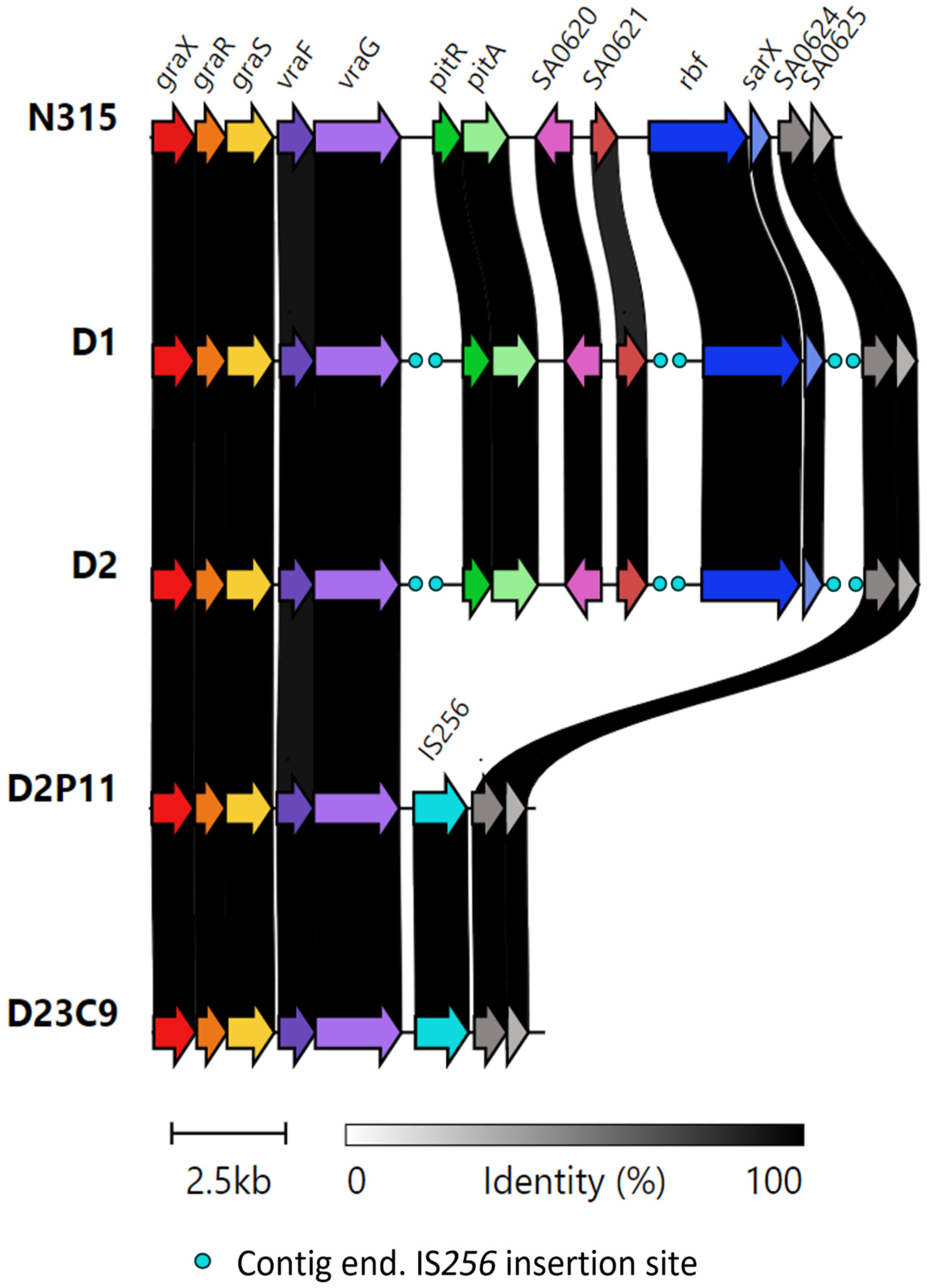
2.4. IS256-Mediated Genomic Rearrangements

Changes in IS256 transposition after vancomycin selective pressure for this set of strains [24] were also detected by WGS bioinformatic analysis performed in this study (Table 6 and Table 7).
Comparative genomic analysis of paired S. aureus strains showed evidence of genetic rearrangements after the IS256 transposition-mediated antibiotic treatment. In total, 11 different IS256 insertions sites (4/11 disrupting genes) were shared by the 4 strains (Table 7), while both in vitro-derived mutants (D23C9 and D2P11) showed modifications in the IS256 copy number and position (Table 5). We did not find genetic changes in the sigB and rsbU genes, known global regulators of IS256 transposition [26,37]. In addition, no change in the copy number and/or location of other staphylococcal IS elements was evident.
It is worth noting that both VISA derivatives are characterized by the absence of a ≈ 8 kb region encompassing genes pitR (phosphate uptake regulator), pitA (low affinity inorganic phosphate transporter), SA0620 (secretory antigen ssA-like protein—CHAP domain peptidoglycan hydrolase), SA0621 (integral membrane protein interacts with ftsH-like protein), rbf (araC type transcriptional regulator) and sarX (transcriptional regulator). This was confirmed by the lack of raw reads mapping to the corresponding region in the assembled genome of parental strain D2, and by PCR–Sanger sequencing (Figure 3, Supplementary Table S1).
The presence of IS256 insertion sites between vraG-pitR, SA0621-rbf and sarX-SA0624, in the D1 and D2 genomes, suggests that genetic rearrangements between neighboring IS256 elements might be responsible for this region deletion in the in vitro-derived VISA strains.
We further evaluated if the ≈8 kb deletion and/or IS256 insertion sites present in its genetic environment were shared by other strains. No other public genomes were found with the ≈8 kb deletion carried by D23C9 and D2P11 (BLAST searches against the NCBI Refseq complete S. aureus genomes database, last accessed 20 January 2023).
In line with previous results [24], IS256 transposition was higher in D23C9. The genome of this strain contains four additional IS256 insertion sites: one interrupting the agrB gene (already reported), and three newly reported (Table 7).

2.5. Biofilm Formation

Knowing that rbf, sarX and agrB are involved in biofilm regulation [38,39], we studied the biofilm phenotype in these strains. D23C9 showed a significantly higher biofilm formation compared to parental strain D2 (Figure 4), and we speculated this is possibly due to both agrB disruption and extracellular DNA (product of D23C9 increased autolysis [24,30]. Moreover, D2P11 did not differ significantly from D2 in its ability to form biofilm despite the deletion of rbf and sarX genes but tends to display lower OD 570 nm values. However, small differences are not always detected on polystyrene microplates [40]; hence, microscopic changes affecting the three-dimensional biofilm structure should not be disregarded.

3. Discussion

Several single nucleotide polymorphisms (SNPs) have been described in hVISA/VISA strains since their first report, and new studies are still trying to understand their genetic basis [41,42]. Our findings reinforce the diversity of the genetic patterns observed [11,12,41], but also highlight the important role of INDELs and genomic rearrangements mediated by insertion sequences, particularly IS256 in the emergence of these complex phenotypes.
The genetic changes found here could have resulted from the antibiotic selective pressure (in vivo and in vitro), but not all of them may necessarily have a direct correlation with the observed phenotypes. Evidence showed modifications in the peptidoglycan metabolism for hVISA/VISA strains and those analyzed in this study [11,30]. Mutations in genes rpoB, rpoC, rpoD, pbp2, stk1 and tcaA, shared by the four strains, were also reported in clinical strains with vancomycin reduced susceptibility [11,12,43]. Nonetheless, we identified novel mutations in genes related to peptidoglycan metabolism (Table 4 and Table 5) in clinical hVISA strains (D1, D2). These novel mutations or other additional mutations related to different cellular processes (Supplementary Table S2) could have cumulative effects that contribute to hVISA/VISA, and/or oxacillin and daptomycin resistances, and should be experimentally investigated in future studies. However, mutations shared by all strains do not explain the phenotypic differences observed among them (Table 1, Figure 5) [30].
It is worth noting that two INDELs were exclusively found in hVISA strain D1. The stp1 gene was associated with the reduced susceptibility to vancomycin, and the braS gene (alternatively named nsaS or bceS) is part of the braRS two-component system responding to cell envelope stress, which is referred to as graS ortholog (involved in hVISA/VISA phenotype) [34,44]. The in-frame deletion found in D1 (Gln63del) is located on the BraS cytoplasmic domain, next to the HisKA domain in charge of signal transduction and gene expression. We assume the hypothesis that these two genetic changes (Table 3), together with non-synonymous SNPs in genes related to cell wall metabolism (Table 2), could be involved in the vancomycin heteroresistance phenotype of D1. Nonetheless, these two INDELs are absent in the isogenic strain D2, possibly reflecting that different hVISA populations can be selected after vancomycin treatment. However, we recognize that as we sequenced just one colony from each clinical sample, we may have found a small proportion of all possible genetic changes, considering that hVISA strains are non-homogeneous populations. Nonetheless, our results show genetic changes that might contribute to antimicrobial resistance.
While the increase in oxacillin susceptibility was already reported in VISA strains [8,9], changes observed in this study are not due to mecA/blaZ mutations or deletions as described before. The sagB gene codes for the major glucosaminidase in charge of glycan chain processing in S. aureus (248aa) [33,45]. Its function, non-redundant despite the presence of other autolysins, is critical for cellular enlargement [33]. The frame-shift insertion shared by D2-D23C9-D2P11 generates a premature stop codon, and the predicted translated protein (148aa) lacks most of the glucosaminidase domain. SagB in vitro-selected mutants were described to display diminished resistance to oxacillin and increased resistance to vancomycin [45,46] as observed in D2 when compared to D1 (Table 1) [24].
No other mutations related to peptidoglycan metabolism/regulation were found between the D1 and D2 (mecA, pbps, blaZ operon and/or cell wall stimulon). Hence, it is most likely that genetic changes in cap5D, stp1, braS and/or sagB genes (related to peptidoglycan metabolism, Table 5) might play a role in the modification of cell wall thickness, pbp2 expression, oxacillin and vancomycin susceptibility [24,30] between these two isogenic strains (Figure 5). As far as we know, this would be the first report on the acquisition of genetic changes in braS and sagB after the in vivo treatment with vancomycin, and its association with the hVISA phenotype and changes in oxacillin susceptibility in clinical strains.
Furthermore, one genetic change seems to be linked to the hVISA to VISA conversion. Both VISA strains selected in independent in vitro assays share not only the increase in oxacillin, vancomycin and daptomycin resistance, slower growth rate and reduced cell diameter (Table 1, Figure 1 and Figure 2), but also the IS256-mediated deletion of a ≈ 8 kb chromosomal region including regulatory genes related to metabolism (pitRA, SA0620, SA0621) and virulence (rbf, sarX) (Figure 3 and Figure 5). Genes related to the inorganic phosphate (Pi) metabolism (including pitRA and phoR) were previously associated with or reported to play a role in the development of vancomycin and daptomycin resistance [47,48,49,50], as changes in the intracellular Pi concentrations can affect the metabolism of DNA, phospholipids, cell envelope (including net positive surface charge), intracellular signalling and stress response [47,51]. Moreover, the expression of pitRA, SA0620, SA0621 and sarX is regulated by walKR and/or graSR operons involved in cell wall metabolism [43,52,53,54,55], and the development of vancomycin resistance in S. aureus [11]. The rbf gene was also frequently mutated in daptomycin-resistant S. aureus [56]. Together, all these findings highlight a potential relevance of the deleted genomic region for the development of oxacillin, vancomycin and daptomycin resistance in the ST100 genetic background.
Nonetheless, other SNPs or IS256 rearrangements distinguishing D23C9 and D2P11 may also possibly contribute to their vancomycin resistance phenotype. In particular, mutations in genes associated with staphylococcal growth (phoR [36,47] and era [35], Table 5, Figure 5) may also impact on their growth rate and fitness cost (Figure 2), an already described feature of VISA isolates [11]. Interestingly, an INDEL in era was already described as a rare genetic change observed in a laboratory-derived VISA strain belonging to CC5 [50].
WGS is a powerful tool for the detection and surveillance of new AMR genetic determinants, but it has not been widely distributed in clinical laboratories yet, especially in low–middle income countries. Moreover, those who have access to this technology cannot depend solely on molecular assays to reliably detect all hVISA/VISA. Because of the multiplicity of genes involved, genomic approaches trying to establish only a few genetic markers as predictors of vancomycin heteroresistance will lead to an underestimation of the real prevalence, leaving behind new, unexplored hVISA phenotypes.
Nevertheless, studies supplementing whole genome sequences, the gold standard PAP-AUC method and MIC determinations are essential for detecting hVISA/VISA and other AMR phenotypes [50,57,58] until new approaches are developed.
Comparative omics analyses will help clarify the molecular mechanisms involved in the emergence of the hVISA/VISA phenotype in future research. Furthermore, the role of mutations (SNPs and INDELs) and the transposition of insertion sequences on the adaptation of S. aureus under antibiotic selective pressure should be explored. This work provides new evidence of the genetic rearrangements mediated by IS256 transposition after antibiotic treatment, with the potential to impact the AMR and virulence of S. aureus strains.

4. Materials and Methods

4.1. Strains and Culture Conditions

S. aureus strains D1 and D2 were isolated from a patient with bone and joint infection, before and after 40 days of vancomycin treatment. The in vitro selection of D2-derived mutants (D23C9, D2P11) was performed in two independent assays by serial passage in BHI broth (Britania, Argentina) with increasing concentrations of vancomycin. D23C9 and D2P11 were selected at 9 and 11 µg/mL of vancomycin, respectively. The detailed medical record and the in vitro selection of D2-derived mutants (D23C9, D2P11) was previously described [24]. Strains were grown aerobically on Brain Heart Infusion (BHI) broth and BHI agar (Britania, Argentina) at 37 °C.

4.2. Antimicrobial Susceptibility Testing

Minimal inhibitory concentrations (MICs) of vancomycin (VAN) and oxacillin (OXA) were determined by the broth microdilution method according to CLSI guidelines [59]. Daptomycin susceptibility was evaluated by Etest® and interpreted as per CLSI guidelines [59] and by the pre-diffusion method using Neo-Sensitabs® tablets (Rosco Diagnostica, Taastrup, Denmark) [60,61]. The oxacillin and vancomycin population analysis profile and area under the curve (PAP-AUC) was determined as previously described [62]. ATCC 29213 (MSSA, VSSA), Mu3 (MRSA, hVISA) and Mu50 (MRSA, VISA) were used as control strains. The antimicrobial susceptibility profile is summarized in Table 1.

4.3. DNA Extraction and Whole Genome Sequencing

Genomic DNA was extracted from overnight BHI cultures using the Epicentre MasterPure Complete DNA and RNA Purification Kit according to the manufacturer’s instructions, with the addition of lysostaphin (0.03 µg/µL) in the lysis step with an incubation time of at least half an hour at 37 °C. Shotgun gDNA libraries were prepared and whole genome sequencing (WGS) was performed using the Illumina MiSeq platform (paired end, 250 bp).

4.4. Whole Genome Sequencing Analysis

Reads were quality assessed with FASTQC [63], and de novo assembled using SPAdes (v3.9.0) [64]. Contigs less than 500 bp and 70× coverage were discarded. Remaining contigs were annotated using Prokka (1.14.5) [65] and a genus-specific database from RefSeq [66], and they were manually inspected. Mapping and variant calling was carried out using Snippy v3.2 [67] with the following parameters: minimum quality of 30, minimum coverage of 15, minimum proportion of reads which must differ from the reference of 0.75. The genome of S. aureus N315 (CC5, Genebank Accession number BA000018.3) was used as a reference. Alternatively, the assembled genome of the first clinical isolate, D1 (ST100) was used as a reference sequence. All variants (SNPs, INDELs) were manually inspected and visualized with Artemis [68].
The SCCmec type was determined from assemblies using SCCmec Finder [69]. Detection of antimicrobial-resistance determinants and MGE was carried out using ARIBA v2.12.1 [70] and relevant databases. For antimicrobial-resistance determinants we used databases from Resfinder [71,72], CARD [73], ARGANNOT [74] and a curated database [75]. Plasmid types were defined based on their replicon genes (rep) using the Plasmidfinder database [76]. Phages types were defined based on their integrase gene, using the 12 described integrase groups [77]. Thirteen known staphylococcal pathogenicity islands (SaPIs) were queried based on their integrase (int) gene [78].
ISseeker [79] was used with default parameters to explore the genome in order to detect differences in insertion sequence (IS) content between parental and mutant strains, and also to annotate the flanking edges of IS elements in draft genomes. ISseeker identifies the termini of IS (>97% of identity) at contig edges and annotate flanking regions based on alignment of IS flanks with a reference genome. IS256-mediated insertions/deletions were confirmed by PCR with primers designed for that purpose (Supplementary Table S1). The 8 kb deletions amplified by PCR in VISA strains were sequenced by the Sanger method and analyzed with SnapGene v6.1.2®. Genomic comparisons were performed using Clinker [80]. The sequence of the 8 kb deletion was searched against the NCBI Refseq S. aureus complete genomes database using BLAST. All genomes, MGE and genome comparisons with reference sequences of interest were additionally visualized in Artemis and/or ACT [68,81].

4.5. Growth Curves

Growth curves were plotted to determine whether the genetic changes were associated with a fitness cost. These assays were performed by triplicate. Fresh culture of each strain (dilution 1/1000) was grown in BHI broth (Britania, Argentina) and incubated at 37 °C and 180 rpm, and OD 620 nm was measured. A growth curve was constructed plotting the OD 620 nm over time.

4.6. Transmission Electron Microscopy (TEM)

TEM of exponential phase S. aureus cultures was performed as already described [30]. Cell diameter was measured (30 cells for each strain) at the equatorial plane of each cell using a 50,000× magnification and images analyzed with ImageJ 1.46r [82]. The results for each strain were expressed as median and interquartile range.

4.7. Biofilm Production Assay

Biofilm development was assessed by measuring the accumulation of biomass on the surface of sterile 96-well flat-bottom polystyrene plates (Extragene) following Stepanovic et al. recommendations [83]. Briefly, 200 μL of a 1/100 dilution of a bacterial suspension adjusted to an OD 620 nm = 0.2 (≈108 CFU/mL) in TSB supplemented with sterile 1% glucose was added to wells (6 replicates per strain). Following 24 h incubation at 37 °C, the plate was washed twice with 0.9% NaCl and air-dried for 2 h. The remaining attached bacteria were fixed with 200 μL of methanol 99% (v/v) per well and after 15 min the plates were emptied and air-dried. Afterward the plates were stained for 20 min with 200 μL per well of 0.5% crystal violet. Finally, wells were washed with water, air-dried, the dye was solubilized with 33% acetic acid solution and the OD 570 nm for each well was measured. S. aureus Newman Δica (non-ica-dependent biofilm producer) and S. epidermidis NRS101 (prototype biofilm producer) were included in the assay as control strains. Biofilm production was calculated as Final OD 570 nm of a tested strain = average OD 570 nm value of the strain—ODc. ODc = average OD 570 nm of negative control Δica + 3SD of negative control.

4.8. Statistical Analysis

Growth rates were compared by slope analysis using linear regression. Biofilm production (Final OD 570 nm) and cell diameter values were compared using the Kruskal–Wallis’s test, and differences between individual groups were detected by Dunn’s multiple comparison test. Analyses were performed using GraphPad prism 5.0 software with a significance level set at p < 0.05 in all cases.

Supplementary Materials

The following supporting information can be downloaded at: https://www.mdpi.com/article/10.3390/antibiotics12020372/s1, Table S1: Oligonucleotides used to confirm chromosomal deletion and IS256 insertions. Region amplified is referred to the annotation in S. aureus N315 reference genome. * amplicon was purified and sequenced; Table S2: Genomic features of the genomes analyzed in this study; Table S3: Core mutations present in the strains analyzed in this study found by Snippy core (not including INDELs) after mapping the reads to the S. aureus N315 reference genome.

Author Contributions

Conceptualization, S.D.G. and M.M.; methodology, S.D.G. and M.S.H.; formal analysis, S.D.G.; investigation, S.D.G., M.S.H., Á.M.R.F., J.D.C. and M.M.; resources, S.D.G. and M.M.; data curation, S.D.G.; writing—original draft preparation, S.D.G.; writing—review and editing, S.D.G., M.S.H., Á.M.R.F., J.D.C. and M.M.; visualization, S.D.G.; supervision, J.D.C. and M.M.; project administration, M.M.; funding acquisition, S.D.G. and M.M. All authors have read and agreed to the published version of the manuscript.

Funding

This project was supported by grants from the University of Buenos Aires (UBACYT 2018-2020-20020170100665BA) and ANPCYT (Préstamo BID PICT-2016-1726), CONICET (PIP 2015 11220150100694CO) to M.M., and ANPCYT (Préstamo BID PICT-2018-03068) to S.D.G.

Institutional Review Board Statement

Not applicable.

Informed Consent Statement

Not applicable.

Data Availability Statement

Genomic reads and assemblies can be found in the National Center for Biotechnology Information (NCBI) genome database in the Sequence Read Archive (SRA) under the BioProject PRJNA911808 with the following biosample accession numbers: SAMN32204997 (D1), SAMN32205060 (D2), SAMN32205332 (D23C9), SAMN32205398 (D2P11). Whole Genome Shotgun project has been deposited at DDBJ/ENA/GenBank under the accessions JAPZAH000000000 (D1), JAPZAI000000000 (D2), JAPZAJ000000000 (D23C9), JAPZAK000000000 (D2P11).

Conflicts of Interest

The authors declare no conflict of interest.

References

  1. WHO List of Bacteria for Which New Antibiotics Are Urgently Needed. Available online: https://www.who.int/news/item/27-02-2017-who-publishes-list-of-bacteria-for-which-new-antibiotics-are-urgently-needed (accessed on 20 December 2022).
  2. Howden, B.P.; Peleg, A.Y.; Stinear, T.P. The Evolution of Vancomycin Intermediate Staphylococcus Aureus (VISA) and Heterogenous-VISA. Infect. Genet. Evol. J. Mol. Epidemiol. Evol. Genet. Infect. Dis. 2014, 21, 575–582. [Google Scholar] [CrossRef]
  3. Shariati, A.; Dadashi, M.; Moghadam, M.T.; van Belkum, A.; Yaslianifard, S.; Darban-Sarokhalil, D. Global Prevalence and Distribution of Vancomycin Resistant, Vancomycin Intermediate and Heterogeneously Vancomycin Intermediate Staphylococcus Aureus Clinical Isolates: A Systematic Review and Meta-Analysis. Sci. Rep. 2020, 10, 12689. [Google Scholar] [CrossRef]
  4. Gostev, V.V.; Popenko, L.N.; Chernen’kaia, T.V.; Naumenko, Z.S.; Voroshilova, T.M.; Zakharova, I.A.; Khokhlova, O.E.; Kruglov, A.N.; Ershova, M.G.; Angelova, S.N.; et al. Estimation of MRSA susceptibility to oxacillin, cefoxitine, vancomycin and daptomycin. Antibiot. Chemoterapy 2013, 58, 13–20. [Google Scholar]
  5. Di Gregorio, S.; Perazzi, B.; Ordoñez, A.M.; De Gregorio, S.; Foccoli, M.; Lasala, M.B.; García, S.; Vay, C.; Famiglietti, A.; Mollerach, M. Clinical, Microbiological, and Genetic Characteristics of Heteroresistant Vancomycin-Intermediate Staphylococcus Aureus Bacteremia in a Teaching Hospital. Microb. Drug Resist. 2015, 21, 25–34. [Google Scholar] [CrossRef]
  6. Hiramatsu, K.; Hanaki, H.; Ino, T.; Yabuta, K.; Oguri, T.; Tenover, F.C. Methicillin-Resistant Staphylococcus Aureus Clinical Strain with Reduced Vancomycin Susceptibility. J. Antimicrob. Chemother. 1997, 40, 135–136. [Google Scholar] [CrossRef] [PubMed]
  7. Hiramatsu, K.; Aritaka, N.; Hanaki, H.; Kawasaki, S.; Hosoda, Y.; Hori, S.; Fukuchi, Y.; Kobayashi, I. Dissemination in Japanese Hospitals of Strains of Staphylococcus Aureus Heterogeneously Resistant to Vancomycin. Lancet 1997, 350, 1670–1673. [Google Scholar] [CrossRef]
  8. Adhikari, R.P.; Scales, G.C.; Kobayashi, K.; Smith, J.M.B.; Berger-Bächi, B.; Cook, G.M. Vancomycin-Induced Deletion of the Methicillin Resistance Gene MecA in Staphylococcus Aureus. J. Antimicrob. Chemother. 2004, 54, 360–363. [Google Scholar] [CrossRef]
  9. Reipert, A.; Ehlert, K.; Kast, T.; Bierbaum, G. Morphological and Genetic Differences in Two Isogenic Staphylococcus Aureus Strains with Decreased Susceptibilities to Vancomycin. Antimicrob. Agents Chemother. 2003, 47, 568–576. [Google Scholar] [CrossRef]
  10. Cui, L.; Tominaga, E.; Neoh, H.-M.; Hiramatsu, K. Correlation between Reduced Daptomycin Susceptibility and Vancomycin Resistance in Vancomycin-Intermediate Staphylococcus Aureus. Antimicrob. Agents Chemother. 2006, 50, 1079–1082. [Google Scholar] [CrossRef]
  11. Hiramatsu, K.; Kayayama, Y.; Matsuo, M.; Aiba, Y.; Saito, M.; Hishinuma, T.; Iwamoto, A. Vancomycin-Intermediate Resistance in Staphylococcus Aureus. J. Glob. Antimicrob. Resist. 2014, 2, 213–224. [Google Scholar] [CrossRef]
  12. Hu, Q.; Peng, H.; Rao, X. Molecular Events for Promotion of Vancomycin Resistance in Vancomycin Intermediate Staphylococcus Aureus. Front. Microbiol. 2016, 7, 1601. [Google Scholar] [CrossRef]
  13. Alam, M.T.; Petit, R.A.; Crispell, E.K.; Thornton, T.A.; Conneely, K.N.; Jiang, Y.; Satola, S.W.; Read, T.D. Dissecting Vancomycin-Intermediate Resistance in Staphylococcus Aureus Using Genome-Wide Association. Genome Biol. Evol. 2014, 6, 1174–1185. [Google Scholar] [CrossRef]
  14. Maki, H.; McCallum, N.; Bischoff, M.; Wada, A.; Berger-Bächi, B. TcaA Inactivation Increases Glycopeptide Resistance in Staphylococcus Aureus. Antimicrob. Agents Chemother. 2004, 48, 1953–1959. [Google Scholar] [CrossRef]
  15. Jansen, A.; Türck, M.; Szekat, C.; Nagel, M.; Clever, I.; Bierbaum, G. Role of Insertion Elements and YycFG in the Development of Decreased Susceptibility to Vancomycin in Staphylococcus Aureus. Int. J. Med. Microbiol. IJMM 2007, 297, 205–215. [Google Scholar] [CrossRef]
  16. McEvoy, C.R.E.; Tsuji, B.; Gao, W.; Seemann, T.; Porter, J.L.; Doig, K.; Ngo, D.; Howden, B.P.; Stinear, T.P. Decreased Vancomycin Susceptibility in Staphylococcus Aureus Caused by IS256 Tempering of WalKR Expression. Antimicrob. Agents Chemother. 2013, 57, 3240–3249. [Google Scholar] [CrossRef]
  17. Kuroda, M.; Sekizuka, T.; Matsui, H.; Ohsuga, J.; Ohshima, T.; Hanaki, H. IS256-Mediated Overexpression of the WalKR Two-Component System Regulon Contributes to Reduced Vancomycin Susceptibility in a Staphylococcus Aureus Clinical Isolate. Front. Microbiol. 2019, 10, 1882. [Google Scholar] [CrossRef]
  18. Yin, Y.; Chen, H.; Li, S.; Gao, H.; Sun, S.; Li, H.; Wang, R.; Jin, L.; Liu, Y.; Wang, H. Daptomycin Resistance in Methicillin-Resistant Staphylococcus Aureus Is Conferred by IS256 Insertion in the Promoter of MprF along with Mutations in MprF and WalK. Int. J. Antimicrob. Agents 2019, 54, 673–680. [Google Scholar] [CrossRef]
  19. Casagrande Proietti, P.; Bietta, A.; Coletti, M.; Marenzoni, M.L.; Scorza, A.V.; Passamonti, F. Insertion Sequence IS256 in Canine Pyoderma Isolates of Staphylococcus Pseudintermedius Associated with Antibiotic Resistance. Vet. Microbiol. 2012, 157, 376–382. [Google Scholar] [CrossRef]
  20. He, T.; Wang, Y.; Schwarz, S.; Zhao, Q.; Shen, J.; Wu, C. Genetic Environment of the Multi-Resistance Gene Cfr in Methicillin-Resistant Coagulase-Negative Staphylococci from Chickens, Ducks, and Pigs in China. Int. J. Med. Microbiol. IJMM 2014, 304, 257–261. [Google Scholar] [CrossRef]
  21. Byrne, M.E.; Rouch, D.A.; Skurray, R.A. Nucleotide Sequence Analysis of IS256 from the Staphylococcus Aureus Gentamicin-Tobramycin-Kanamycin-Resistance Transposon Tn4001. Gene 1989, 81, 361–367. [Google Scholar] [CrossRef]
  22. Kleinert, F.; Kallies, R.; Hort, M.; Zweynert, A.; Szekat, C.; Nagel, M.; Bierbaum, G. Influence of IS256 on Genome Variability and Formation of Small-Colony Variants in Staphylococcus Aureus. Antimicrob. Agents Chemother. 2017, 61, e00144-17. [Google Scholar] [CrossRef]
  23. Benson, M.A.; Ohneck, E.A.; Ryan, C.; Alonzo, F.; Smith, H.; Narechania, A.; Kolokotronis, S.-O.; Satola, S.W.; Uhlemann, A.-C.; Sebra, R.; et al. Evolution of Hypervirulence by a MRSA Clone through Acquisition of a Transposable Element. Mol. Microbiol. 2014, 93, 664–681. [Google Scholar] [CrossRef]
  24. Di Gregorio, S.; Fernandez, S.; Perazzi, B.; Bello, N.; Famiglietti, A.; Mollerach, M. Increase in IS256 Transposition in Invasive Vancomycin Heteroresistant Staphylococcus Aureus Isolate Belonging to ST100 and Its Derived VISA Mutants. Infect. Genet. Evol. J. Mol. Epidemiol. Evol. Genet. Infect. Dis. 2016, 43, 197–202. [Google Scholar] [CrossRef]
  25. Loessner, I.; Dietrich, K.; Dittrich, D.; Hacker, J.; Ziebuhr, W. Transposase-Dependent Formation of Circular IS256 Derivatives in Staphylococcus Epidermidis and Staphylococcus Aureus. J. Bacteriol. 2002, 184, 4709–4714. [Google Scholar] [CrossRef]
  26. Valle, J.; Vergara-Irigaray, M.; Merino, N.; Penadés, J.R.; Lasa, I. SigmaB Regulates IS256-Mediated Staphylococcus Aureus Biofilm Phenotypic Variation. J. Bacteriol. 2007, 189, 2886–2896. [Google Scholar] [CrossRef]
  27. Cafiso, V.; Bertuccio, T.; Santagati, M.; Demelio, V.; Spina, D.; Nicoletti, G.; Stefani, S. Agr-Genotyping and Transcriptional Analysis of Biofilm-Producing Staphylococcus Aureus. FEMS Immunol. Med. Microbiol. 2007, 51, 220–227. [Google Scholar] [CrossRef] [PubMed]
  28. Byrne, M.E.; Gillespie, M.T.; Skurray, R.A. Molecular Analysis of a Gentamicin Resistance Transposonlike Element on Plasmids Isolated from North American Staphylococcus Aureus Strains. Antimicrob. Agents Chemother. 1990, 34, 2106–2113. [Google Scholar] [CrossRef]
  29. Alam, M.M.; Kobayashi, N.; Uehara, N.; Watanabe, N. Analysis on Distribution and Genomic Diversity of High-Level Antiseptic Resistance Genes QacA and QacB in Human Clinical Isolates of Staphylococcus Aureus. Microb. Drug Resist. 2003, 9, 109–121. [Google Scholar] [CrossRef]
  30. Di Gregorio, S.; Fernandez, S.; Cuirolo, A.; Verlaine, O.; Amoroso, A.; Mengin-Lecreulx, D.; Famiglietti, A.; Joris, B.; Mollerach, M. Different Vancomycin-Intermediate Staphylococcus Aureus Phenotypes Selected from the Same ST100-HVISA Parental Strain. Microb. Drug Resist. 2017, 23, 44–50. [Google Scholar] [CrossRef] [PubMed]
  31. Passalacqua, K.D.; Satola, S.W.; Crispell, E.K.; Read, T.D. A Mutation in the PP2C Phosphatase Gene in a Staphylococcus Aureus USA300 Clinical Isolate with Reduced Susceptibility to Vancomycin and Daptomycin. Antimicrob. Agents Chemother. 2012, 56, 5212–5223. [Google Scholar] [CrossRef]
  32. Kolar, S.L.; Nagarajan, V.; Oszmiana, A.; Rivera, F.E.; Miller, H.K.; Davenport, J.E.; Riordan, J.T.; Potempa, J.; Barber, D.S.; Koziel, J.; et al. NsaRS Is a Cell-Envelope-Stress-Sensing Two-Component System of Staphylococcus Aureus. Microbiology 2011, 157, 2206–2219. [Google Scholar] [CrossRef]
  33. Wheeler, R.; Turner, R.D.; Bailey, R.G.; Salamaga, B.; Mesnage, S.; Mohamad, S.A.S.; Hayhurst, E.J.; Horsburgh, M.; Hobbs, J.K.; Foster, S.J. Bacterial Cell Enlargement Requires Control of Cell Wall Stiffness Mediated by Peptidoglycan Hydrolases. mBio 2015, 6, e00660. [Google Scholar] [CrossRef]
  34. Hiron, A.; Falord, M.; Valle, J.; Débarbouillé, M.; Msadek, T. Bacitracin and Nisin Resistance in Staphylococcus Aureus: A Novel Pathway Involving the BraS/BraR Two-Component System (SA2417/SA2418) and Both the BraD/BraE and VraD/VraE ABC Transporters. Mol. Microbiol. 2011, 81, 602–622. [Google Scholar] [CrossRef]
  35. Wood, A.; Irving, S.E.; Bennison, D.J.; Corrigan, R.M. The (p)PpGpp-Binding GTPase Era Promotes RRNA Processing and Cold Adaptation in Staphylococcus Aureus. PLoS Genet. 2019, 15, e1008346. [Google Scholar] [CrossRef] [PubMed]
  36. Kelliher, J.L.; Radin, J.N.; Kehl-Fie, T.E. PhoPR Contributes to Staphylococcus Aureus Growth during Phosphate Starvation and Pathogenesis in an Environment-Specific Manner. Infect. Immun. 2018, 86, e00371-18. [Google Scholar] [CrossRef] [PubMed]
  37. Schreiber, F.; Szekat, C.; Josten, M.; Sahl, H.-G.; Bierbaum, G. Antibiotic-Induced Autoactivation of IS256 in Staphylococcus Aureus. Antimicrob. Agents Chemother. 2013, 57, 6381–6384. [Google Scholar] [CrossRef]
  38. Cue, D.; Lei, M.G.; Lee, C.Y. Activation of SarX by Rbf Is Required for Biofilm Formation and IcaADBC Expression in Staphylococcus Aureus. J. Bacteriol. 2013, 195, 1515–1524. [Google Scholar] [CrossRef]
  39. Schilcher, K.; Horswill, A.R. Staphylococcal Biofilm Development: Structure, Regulation, and Treatment Strategies. Microbiol. Mol. Biol. Rev. MMBR 2020, 84, e00026-19. [Google Scholar] [CrossRef]
  40. Luong, T.T.; Lei, M.G.; Lee, C.Y. Staphylococcus Aureus Rbf Activates Biofilm Formation In Vitro and Promotes Virulence in a Murine Foreign Body Infection Model. Infect. Immun. 2009, 77, 335–340. [Google Scholar] [CrossRef]
  41. Silveira, A.C.O.; Caierão, J.; Silva, C.I.; Anzai, E.K.; McCulloch, J.A.; d’Azevedo, P.A.; Sincero, T.C.M. Impact of Mutations in HVISA Isolates on Decreased Susceptibility to Vancomycin, through Population Analyses Profile—Area under Curve (PAP-AUC). Diagn. Microbiol. Infect. Dis. 2019, 95, 114854. [Google Scholar] [CrossRef]
  42. Salemi, R.; Zega, A.; Aguglia, E.; Lo Verde, F.; Pigola, G.; Stefani, S.; Cafiso, V. Balancing the Virulence and Antimicrobial Resistance in VISA DAP-R CA-MRSA Superbug. Antibiotics 2022, 11, 1159. [Google Scholar] [CrossRef] [PubMed]
  43. Kant, S.; Asthana, S.; Missiakas, D.; Pancholi, V. A Novel STK1-Targeted Small-Molecule as an “Antibiotic Resistance Breaker” against Multidrug-Resistant Staphylococcus Aureus. Sci. Rep. 2017, 7, 5067. [Google Scholar] [CrossRef] [PubMed]
  44. Blake, K.L.; Randall, C.P.; O’Neill, A.J. In Vitro Studies Indicate a High Resistance Potential for the Lantibiotic Nisin in Staphylococcus Aureus and Define a Genetic Basis for Nisin Resistance▿. Antimicrob. Agents Chemother. 2011, 55, 2362–2368. [Google Scholar] [CrossRef]
  45. Chan, Y.G.Y.; Frankel, M.B.; Missiakas, D.; Schneewind, O. SagB Glucosaminidase Is a Determinant of Staphylococcus Aureus Glycan Chain Length, Antibiotic Susceptibility, and Protein Secretion. J. Bacteriol. 2016, 198, 1123–1136. [Google Scholar] [CrossRef]
  46. Ishii, K.; Tabuchi, F.; Matsuo, M.; Tatsuno, K.; Sato, T.; Okazaki, M.; Hamamoto, H.; Matsumoto, Y.; Kaito, C.; Aoyagi, T.; et al. Phenotypic and Genomic Comparisons of Highly Vancomycin-Resistant Staphylococcus Aureus Strains Developed from Multiple Clinical MRSA Strains by in Vitro Mutagenesis. Sci. Rep. 2015, 5, 17092. [Google Scholar] [CrossRef]
  47. Santos-Beneit, F.; Ordóñez-Robles, M.; Martín, J.F. Glycopeptide Resistance: Links with Inorganic Phosphate Metabolism and Cell Envelope Stress. Biochem. Pharmacol. 2017, 133, 74–85. [Google Scholar] [CrossRef]
  48. Mechler, L.; Herbig, A.; Paprotka, K.; Fraunholz, M.; Nieselt, K.; Bertram, R. A Novel Point Mutation Promotes Growth Phase-Dependent Daptomycin Tolerance in Staphylococcus Aureus. Antimicrob. Agents Chemother. 2015, 59, 5366–5376. [Google Scholar] [CrossRef]
  49. Bakthavatchalam, Y.D.; Babu, P.; Munusamy, E.; Dwarakanathan, H.T.; Rupali, P.; Zervos, M.; John Victor, P.; Veeraraghavan, B. Genomic Insights on Heterogeneous Resistance to Vancomycin and Teicoplanin in Methicillin-Resistant Staphylococcus Aureus: A First Report from South India. PLoS ONE 2019, 14, e0227009. [Google Scholar] [CrossRef] [PubMed]
  50. Su, M.; Davis, M.H.; Peterson, J.; Solis-Lemus, C.; Satola, S.W.; Read, T.D. Effect of Genetic Background on the Evolution of Vancomycin-Intermediate Staphylococcus Aureus (VISA). PeerJ 2021, 9, e11764. [Google Scholar] [CrossRef]
  51. Myers, C.L.; Li, F.K.K.; Koo, B.-M.; El-Halfawy, O.M.; French, S.; Gross, C.A.; Strynadka, N.C.J.; Brown, E.D. Identification of Two Phosphate Starvation-Induced Wall Teichoic Acid Hydrolases Provides First Insights into the Degradative Pathway of a Key Bacterial Cell Wall Component. J. Biol. Chem. 2016, 291, 26066–26082. [Google Scholar] [CrossRef] [PubMed]
  52. Delauné, A.; Dubrac, S.; Blanchet, C.; Poupel, O.; Mäder, U.; Hiron, A.; Leduc, A.; Fitting, C.; Nicolas, P.; Cavaillon, J.-M.; et al. The WalKR System Controls Major Staphylococcal Virulence Genes and Is Involved in Triggering the Host Inflammatory Response. Infect. Immun. 2012, 80, 3438–3453. [Google Scholar] [CrossRef]
  53. Falord, M.; Mäder, U.; Hiron, A.; Débarbouillé, M.; Msadek, T. Investigation of the Staphylococcus Aureus GraSR Regulon Reveals Novel Links to Virulence, Stress Response and Cell Wall Signal Transduction Pathways. PLoS ONE 2011, 6, e21323. [Google Scholar] [CrossRef]
  54. Dubrac, S.; Msadek, T. Identification of Genes Controlled by the Essential YycG/YycF Two-Component System of Staphylococcus Aureus. J. Bacteriol. 2004, 186, 1175–1181. [Google Scholar] [CrossRef]
  55. Cameron, D.R.; Jiang, J.-H.; Kostoulias, X.; Foxwell, D.J.; Peleg, A.Y. Vancomycin Susceptibility in Methicillin-Resistant Staphylococcus Aureus Is Mediated by YycHI Activation of the WalRK Essential Two-Component Regulatory System. Sci. Rep. 2016, 6, 30823. [Google Scholar] [CrossRef]
  56. Cafiso, V.; Stracquadanio, S.; Lo Verde, F.; De Guidi, I.; Zega, A.; Pigola, G.; Stefani, S. Genomic and Long-Term Transcriptomic Imprints Related to the Daptomycin Mechanism of Action Occurring in Daptomycin- and Methicillin-Resistant Staphylococcus Aureus Under Daptomycin Exposure. Front. Microbiol. 2020, 11, 1893. [Google Scholar] [CrossRef]
  57. Su, M.; Satola, S.W.; Read, T.D. Genome-Based Prediction of Bacterial Antibiotic Resistance. J. Clin. Microbiol. 2019, 57, e01405-18. [Google Scholar] [CrossRef]
  58. Kim, J.I.; Maguire, F.; Tsang, K.K.; Gouliouris, T.; Peacock, S.J.; McAllister, T.A.; McArthur, A.G.; Beiko, R.G. Machine Learning for Antimicrobial Resistance Prediction: Current Practice, Limitations, and Clinical Perspective. Clin. Microbiol. Rev. 2022, 35, e0017921. [Google Scholar] [CrossRef]
  59. CLSI. Performance Standards for Antimicrobial Susceptibility Testing, 22nd ed.; M100-S22; CLSI: San Antonio, TX, USA.
  60. Katz, B.D.; Luperchio, S.A.; Thorne, G.M. Detection of Daptomycin-Nonsusceptible Strains Using the Neo-Sensitab Prediffusion Method. Diagn. Microbiol. Infect. Dis. 2008, 61, 315–320. [Google Scholar] [CrossRef] [PubMed]
  61. Rosco Diagnostica A/S Supplement User’s Guide. Neo-SensitabsTM Susceptibility Testing, Supplement 2010. Available online: http://www.rosco.dk/ (accessed on 22 December 2022).
  62. Wootton, M.; Howe, R.A.; Hillman, R.; Walsh, T.R.; Bennett, P.M.; MacGowan, A.P. A Modified Population Analysis Profile (PAP) Method to Detect Hetero-Resistance to Vancomycin in Staphylococcus Aureus in a UK Hospital. J. Antimicrob. Chemother. 2001, 47, 399–403. [Google Scholar] [CrossRef]
  63. Andrews, S. FastQC a Quality Control Tool for High Throughput Sequence Data. Available online: https://www.bioinformatics.babraham.ac.uk/projects/fastqc/ (accessed on 22 December 2022).
  64. Bankevich, A.; Nurk, S.; Antipov, D.; Gurevich, A.A.; Dvorkin, M.; Kulikov, A.S.; Lesin, V.M.; Nikolenko, S.I.; Pham, S.; Prjibelski, A.D.; et al. SPAdes: A New Genome Assembly Algorithm and Its Applications to Single-Cell Sequencing. J. Comput. Biol. J. Comput. Mol. Cell Biol. 2012, 19, 455–477. [Google Scholar] [CrossRef]
  65. Seemann, T. Prokka: Rapid Prokaryotic Genome Annotation. Bioinformatics 2014, 30, 2068–2069. [Google Scholar] [CrossRef] [PubMed]
  66. Pruitt, K.D.; Tatusova, T.; Brown, G.R.; Maglott, D.R. NCBI Reference Sequences (RefSeq): Current Status, New Features and Genome Annotation Policy. Nucleic Acids Res. 2012, 40, D130–D135. [Google Scholar] [CrossRef] [PubMed]
  67. Seemann, T. Snippy: Rapid Haploid Variant Calling and Core Genome Alignment. Available online: https://github.com/tseemann/snippy (accessed on 22 December 2022).
  68. Carver, T.; Harris, S.R.; Berriman, M.; Parkhill, J.; McQuillan, J.A. Artemis: An Integrated Platform for Visualization and Analysis of High-Throughput Sequence-Based Experimental Data. Bioinformatics 2012, 28, 464–469. [Google Scholar] [CrossRef] [PubMed]
  69. Kaya, H.; Hasman, H.; Larsen, J.; Stegger, M.; Johannesen, T.B.; Allesøe, R.L.; Lemvigh, C.K.; Aarestrup, F.M.; Lund, O.; Larsen, A.R. SCCmecFinder, a Web-Based Tool for Typing of Staphylococcal Cassette Chromosome Mec in Staphylococcus Aureus Using Whole-Genome Sequence Data. mSphere 2018, 3, e00612-17. [Google Scholar] [CrossRef]
  70. Hunt, M.; Mather, A.E.; Sánchez-Busó, L.; Page, A.J.; Parkhill, J.; Keane, J.A.; Harris, S.R. ARIBA: Rapid Antimicrobial Resistance Genotyping Directly from Sequencing Reads. Microb. Genomics 2017, 3, e000131. [Google Scholar] [CrossRef]
  71. Zankari, E.; Hasman, H.; Cosentino, S.; Vestergaard, M.; Rasmussen, S.; Lund, O.; Aarestrup, F.M.; Larsen, M.V. Identification of Acquired Antimicrobial Resistance Genes. J. Antimicrob. Chemother. 2012, 67, 2640–2644. [Google Scholar] [CrossRef]
  72. Zankari, E.; Allesøe, R.; Joensen, K.G.; Cavaco, L.M.; Lund, O.; Aarestrup, F.M. PointFinder: A Novel Web Tool for WGS-Based Detection of Antimicrobial Resistance Associated with Chromosomal Point Mutations in Bacterial Pathogens. J. Antimicrob. Chemother. 2017, 72, 2764–2768. [Google Scholar] [CrossRef]
  73. Jia, B.; Raphenya, A.R.; Alcock, B.; Waglechner, N.; Guo, P.; Tsang, K.K.; Lago, B.A.; Dave, B.M.; Pereira, S.; Sharma, A.N.; et al. CARD 2017: Expansion and Model-Centric Curation of the Comprehensive Antibiotic Resistance Database. Nucleic Acids Res. 2017, 45, D566–D573. [Google Scholar] [CrossRef]
  74. Gupta, S.K.; Padmanabhan, B.R.; Diene, S.M.; Lopez-Rojas, R.; Kempf, M.; Landraud, L.; Rolain, J.-M. ARG-ANNOT, a New Bioinformatic Tool to Discover Antibiotic Resistance Genes in Bacterial Genomes. Antimicrob. Agents Chemother. 2014, 58, 212–220. [Google Scholar] [CrossRef]
  75. Harkins, C.P.; Pichon, B.; Doumith, M.; Parkhill, J.; Westh, H.; Tomasz, A.; de Lencastre, H.; Bentley, S.D.; Kearns, A.M.; Holden, M.T.G. Methicillin-Resistant Staphylococcus Aureus Emerged Long before the Introduction of Methicillin into Clinical Practice. Genome Biol. 2017, 18, 130. [Google Scholar] [CrossRef]
  76. Carattoli, A.; Zankari, E.; García-Fernández, A.; Voldby Larsen, M.; Lund, O.; Villa, L.; Møller Aarestrup, F.; Hasman, H. In Silico Detection and Typing of Plasmids Using PlasmidFinder and Plasmid Multilocus Sequence Typing. Antimicrob. Agents Chemother. 2014, 58, 3895–3903. [Google Scholar] [CrossRef]
  77. Goerke, C.; Pantucek, R.; Holtfreter, S.; Schulte, B.; Zink, M.; Grumann, D.; Bröker, B.M.; Doskar, J.; Wolz, C. Diversity of Prophages in Dominant Staphylococcus Aureus Clonal Lineages. J. Bacteriol. 2009, 191, 3462–3468. [Google Scholar] [CrossRef] [PubMed]
  78. Subedi, A.; Ubeda, C.; Adhikari, R.P.; Penadés, J.R.; Novick, R.P. Sequence Analysis Reveals Genetic Exchanges and Intraspecific Spread of SaPI2, a Pathogenicity Island Involved in Menstrual Toxic Shock. Microbiology 2007, 153, 3235–3245. [Google Scholar] [CrossRef] [PubMed]
  79. Adams, M.D.; Bishop, B.; Wright, M.S. Quantitative Assessment of Insertion Sequence Impact on Bacterial Genome Architecture. Microb. Genomics 2016, 2, e000062. [Google Scholar] [CrossRef] [PubMed]
  80. Gilchrist, C.L.M.; Chooi, Y.-H. Clinker & Clustermap. Js: Automatic Generation of Gene Cluster Comparison Figures. Bioinformatics 2021, btab007. [Google Scholar] [CrossRef]
  81. Carver, T.J.; Rutherford, K.M.; Berriman, M.; Rajandream, M.-A.; Barrell, B.G.; Parkhill, J. ACT: The Artemis Comparison Tool. Bioinformatics 2005, 21, 3422–3423. [Google Scholar] [CrossRef]
  82. Rasband, W.S. ImageJ 1997. Available online: https://imagej.nih.gov/ij/ (accessed on 22 December 2022).
  83. Stepanović, S.; Vuković, D.; Hola, V.; Di Bonaventura, G.; Djukić, S.; Cirković, I.; Ruzicka, F. Quantification of Biofilm in Microtiter Plates: Overview of Testing Conditions and Practical Recommendations for Assessment of Biofilm Production by Staphylococci. APMIS Acta Pathol. Microbiol. Immunol. Scand. 2007, 115, 891–899. [Google Scholar] [CrossRef]
Figure 1. Oxacillin population analysis profile and area under the curve (PAP-AUC) of bacterial strains included in this study. Serial 10-fold dilutions of cultures were plated onto Mueller–Hinton agar with oxacillin (OXA). Each point represents the viable count (log10 CFU/mL) after 48 h against increasing concentrations of OXA.
Antibiotics 12 00372 g001
Figure 2. (A) Transmission electron microscopy (TEM) of representative cells; images obtained at 50,000×. A: D1; B: D2; C: D23C9; D: D2P11. (B) Cell diameter measured by TEM (30 cells per strain). The results are expressed in nanometers (nm) as violin plots showing median (dashed line) and interquartile range (dots) and were compared with Kruskal–Wallis’s test (p < 0.0001). The horizontal bars show significant differences between individual groups detected by Dunn’s multiple comparison test. ** p < 0.001; *** p < 0.0001. (C) Growth curves. Each point represents the mean and standard error from three independent assays.
Antibiotics 12 00372 g002
Figure 3. Clinker gene cluster comparison of the genomic region between graX and SA0625. Homologous CDSs are in the same color and linked through grey bars with the percentage amino acid identity, as indicated in the legend. IS256 insertions are marked as aqua circles (in case of contig ends), or annotated CDS (confirmed by PCR).
Antibiotics 12 00372 g003
Figure 4. Biofilm production. Results are represented as final OD for each strain with median value (line) and interquartile range (whiskers) and are compared with Kruskal–Wallis’s test (p < 0.0001). The horizontal bars show significant differences between individual groups detected by Dunn’s multiple comparison test. * p < 0.05; *** p < 0.001. NRS101 (S. epidermidis NRS101), strong biofilm producer; Δica (S. aureus Newman Δica), ica independent-biofilm producer strain. The bottom picture shows a representative microtiter plate of the biofilm assay for each strain.
Antibiotics 12 00372 g004
Figure 5. Genes harboring genetic changes in hVISA/VISA strains analyzed in this study and their relationships with cellular processes (lines). Blue boxes: Genes with mutations related to hVISA/VISA shared by D1, D2, D23C9 and D2P11. Yellow boxes: Genes with mutations differing between D2 and D1. Red boxes: Genes differing between the in vitro-selected mutants (D23C9 and D2P11) and their parental strain (D2). Δ ≈8 kb IS256-mediated deletion. * SNP/INDEL. Purple boxes: Key regulatory operons related to vancomycin resistance in hVISA/VISA (not mutated in these strains). R: Resistance. VAN: vancomycin. OXA: oxacillin. DAP: daptomycin.
Antibiotics 12 00372 g005
Table 1. Antimicrobial susceptibility profile of the bacterial strains used in this study. MICs (µg/mL), inhibition zone diameter obtained after daptomycin (DAP) pre-diffusion method (mm), and vancomycin (VAN) population analysis profile and area under the curve (PAP-AUC) ratio. The AUC was measured for each sample and the ratio of test isolate AUC/mean Mu3 AUC was calculated. Mu3 was used as hVISA control strain. The criteria to define hVISA and VSSA were a PAP-AUC ratio ≥ 0.90 and a PAP/AUC ratio < 0.90, respectively.
D1D2D23C9D2P11Reference
MIC (µg/mL)VAN0.5148[24]
OXA1282832[24]
DAP0.0940.380.51This Study
DAP Pre-diffusion method (mm)30302218This Study
VAN PAP-AUC ratio0.981.482.815.7[24]
PhenotypehVISAhVISAVISAVISA[24]
MIC: Minimal inhibitory concentration, VAN: vancomycin, OXA: oxacillin, RIF: rifampin, DAP: daptomycin, PAP-AUC: Population analysis profile and area under the curve.
Table 2. Accession numbers, genome coverage and assembly metrics of genomes included in this study. # Contigs is the total number of contigs in the assembly. # Contigs (≥x bp) is the total number of contigs of length ≥ x bp. Largest contig is the length of the longest contig in the assembly. Total length is the total number of bases in the assembly. Total length (≥x bp) is the total number of bases in contigs of length ≥ x bp. GC (%) is the total number of G and C nucleotides in the assembly, divided by the total length of the assembly. N50 is the length for which the collection of all contigs of that length or longer covers at least half (50%) the total base content of the assembly. N90 is used for the same purpose but the length is set at 90% of total base content instead of 50%. L50 is the number of contigs equal to or longer than the N50 length. L90 is used for the same purpose in reference to the N90 length. # N’s per 100 kbp is the number of ambiguous bases (Ns) per 100 kbp.
AssemblyD1D2D2P11D23C9
Assembly accessionJAPZAH000000000JAPZAI000000000JAPZAJ000000000JAPZAK000000000
Genome coverage286.77246.87277.45256.49
# Contigs (≥0 bp)45453547
# Contigs (≥1000 bp)38383140
Total length (≥0 bp)2,795,1592,795,1552,785,3552,787,646
Total length (≥1000 bp)2,789,9752,789,9712,782,2042,782,459
# Contigs45453547
Largest contig567,081567,084567,084419,127
Total length2,795,1592,795,1552,785,3552,787,646
GC (%)32.7932.7932.832.81
N50334,617334,619337,832148,220
N9036,72336,72339,43733,617
L504445
L9017171619
# N’s per 100 kbp0000
Table 3. Summary of genomic features shared by all strains. Genotypic features (MLST and SCCmec type), and the presence of full/complete genetic determinants (mobile genetic elements, antimicrobial resistance and restriction modification systems) detected in the genomes included in this study.
Feature
MLSTST100
SCCmec typeNT
AMR determinantsaac(6′)-aph(2″), blaZ, mecA, rpoB H481N
Insertion sequencesIS256, IS1181, IS431, ISSau6
TransposonTn4001
RM systemsS.Sau N315 I–M.Sau N315 I, S.Sau N315 II–M.Sau N315 II, SauUSI
Plasmidic rep genesrep21.13_SAP101A (GQ900495.1), rep20.3_pTW20 (FN433597.1)
NT: Non-typeable. MLST: Multilocus sequence type. AMR: Antimicrobial resistance. RM systems: Restriction modification systems.
Table 4. Non-synonymous SNPs related to peptidoglycan metabolism or vancomycin reduced susceptibility shared among all strains analyzed in this study, after mapping the reads to the S. aureus N315 reference genome. All changes are expressed in reference to the S. aureus N315 genome.
Chromosome PositionGene NameProductPredicted Amino Acid Change
46,301mecAPenicillin-binding protein 2 primeGly246Glu
297,502tarF_1CDP-glycerol:poly(glycerophosphate) glycerophosphotransferaseAsn236Ser
581,060rpoBDNA-directed RNA polymerase subunit betaHis481Asn
583,428rpoCDNA-directed RNA polymerase beta’ chain proteinPro41Ala
1,595,731rpoDRNA polymerase sigma-70 factor RpoDVal253Ile
687,696tagGTeichoic acid ABC superfamily ATP-binding cassette transporterVal220Ala
732,508cydDABC superfamily ATP-binding cassette transporterAsn174Asp
902,157dltDD-alanine lipoteichoic acid and wall teichoic acid esterification secreted proteinIle264Lys
993,796murEUDP-N-acetylmuramoyl-L-alanyl-D-glutamate--2Ala436Ser
1,157,903ftsLCell division and chromosome partitioning proteinAsp78Asn
1,203,919pknB_2(stk1)Non-specific serine/threonine protein kinaseLys512Asn
1,487,245pbp2Glycosyl transferase family proteinCys197Tyr
1,487,506pbp2Glycosyl transferase family proteinThr284Ile
1,758,442ezrASeptation ring formation regulatorVal545Ala
2,009,774–2,009,775amiN-acetylmuramoyl-L-alanine amidaseAla250Val
2,010,074amiN-acetylmuramoyl-L-alanine amidaseAsn150Lys
2,010,079amiN-acetylmuramoyl-L-alanine amidaseIle149Val
2,138,921murFPutative UDP-N-acetylmuramoylalanyl-D-glutamyl-2Ser126Asn
2,171,885rhoPutative methicillin resistance expression factorIle48Leu
2,412,317tcaATeicoplanin resistance-associated membrane protein TcaA proteinLeu218Pro
Table 5. Genetic changes differing between strains analyzed in this study after mapping the reads to the S. aureus D1 genome. All changes and positions are expressed in reference to the S. aureus N315 reference genome except otherwise stated. Genes without an annotated name are in reference to the CDS of N315 (or D1 genome if not present in the latter) by their locus tag.
Chromosome PositionGene NameProductTypePredicted Aminoacid Change
D1D2D23C9D2P11
1,506,816Intergenic (upstream aroC)-SNPC63217A *wtwtwt
1,003,592Intergenic (upstream comK)-SNPT19402C *wtwtwt
170,209cap5DCapsular polysaccharide biosynthesis protein Cap5DSNPThr215Ilewtwtwt
1,201,763stp1Protein phosphatase 2C domain-containing proteinINDELGly41_Lys43dupwtwtwt
2,713,713nsaS/braSIntegral membrane sensor signal transduction histidine kinaseINDELGln63delwtwtwt
1,832,949sagBBeta-N-acetylglucosaminidaseINDELWtHis142fsHis142fsHis142fs
12,058(NODE_18) *2SAD1_02353Bacteriophage tail tape measure proteinSNPWtAsp1737GlyAsp1737GlyAsp1737Gly
27,119(NODE 19) *2SAD1_02404GNAT family acetyl transferaseSNPWtTyr158TyrTyr158TyrTyr158Tyr
24,307(NODE 21) *2ccrBCassette chromosome recombinase BSNPWtAsn10SerAsn10SerAsn10Ser
1,728,074phoRAlkaline phosphatase synthesis sensor protein PhoRSNPWtwtArg200 * STOPwt
1,603,348eraGTP-binding protein EraINDELWtwtwtAsn81fs
* Intergenic change. wt: wild-type related to the N315 genome. *2 Position in D1 genome.
Table 6. Number of insertion sequences from Staphylococcus sp. per genome detected by ISseeker in the genomes included in this study. ISseeker identifies the termini of IS (>97% of identity) at contig edges and annotate flanking regions based on alignment of IS flanks with a reference genome.
ISD1D2D23C9D2P11
IS25614141713
IS11817 + 1 *7 + 1 *7 + 1 *7 + 1 *
IS11820000
IS12721 *1*1 *1 *
IS4311111
ISSau10000
ISSau23 *3 *3 *3 *
ISSau310 *10 *10 *10 *
ISSau40000
ISSau51 *1 *1 *1 *
ISSau66666
ISSau82 *2 *2 *2 *
ISSau90000
ISSep11 *1 *1 *1 *
ISSep21 *1 *1 *1 *
ISSep30000
ISSha10000
* Partial IS sequence inside a Contig, >97% Identity.
Table 7. IS256 insertion sites detected with ISseeker software after annotating IS256 flaking regions against the S. aureus N315 reference genome. Type was considered “intergenic” if IS256 was found between two different genes, and “disrupting gene” if both annotated flanking regions were found in a single (same) gene.
Genomic LocationTypeD1D2D23C9D2P11
mecR1Disrupting gene++++
SA0142 (hypothetical protein)Disrupting gene++++
SA0084 (hypothetical protein)—SA0085 (tRNA dihidrouridine sintase)Intergenic++++
SA0516 (tRNA specific adenosine deaminase)—SA0517 (ABC superfamily ATP-binding cassette transporter)Intergenic++++
vraG (ABC transporter permease)—SA0618 (pitR, putative phosphate uptake regulator) **Intergenic+++ *+ *
SA0621 (integral membrane protein) **—rbf (AraC type transcription regulator) **Intergenic++--
sarX (staphylococcal accessory regulator protein X) **—SA0624 (putative transcriptional regulatory protein)Intergenic+++ *+ *
SA0625 (hypothetical protein)—SA0626 (hypothetical protein)Intergenic--+-
SA0742 (clfA, clumping factor A)Disrupting gene--+-
SA0954 (hypothetical protein)—SA0955 (hypothetical protein)Intergenic++++
SA0185 (putative membrane protein YfhO)—rbgA (GTP-binding protein)Intergenic++++
SA1176 (hypothetical protein)—tkt (transketolase)Intergenic++++
SA1232 (lysA, diaminopimelate decarboxylase)—SA1233 (msaC, modulator of sarA) ***Disrupting gene++++
SA1639 (hypothetical protein)Disrupting gene++++
SA1648 (enterotoxin seO)—tRNAserIntergenic++++
agrB (accesory gene regulator B)Disrupting gene--+-
SA2019 (hypothetical protein)—SA2010 (hypothetical protein)Intergenic++++
SA2414 (hypothetical protein)—SA2415 (braE, ABC superfamily ATP-binding cassette transporter membrane protein)Intergenic--+-
+ Presence of IS256 in the genomic location. - Absence of IS256 in the genomic location. * IS256 insertion site was detected but one of the genes in the flanking region is not present in the analyzed assembly, ** Genomic region encompassing pitR and sarX genes, absent in D23C9 and D2P11 (8 kb deletion), *** The annotated msaC gene was found deleted in all genomes analyzed in this study.
Disclaimer/Publisher’s Note: The statements, opinions and data contained in all publications are solely those of the individual author(s) and contributor(s) and not of MDPI and/or the editor(s). MDPI and/or the editor(s) disclaim responsibility for any injury to people or property resulting from any ideas, methods, instructions or products referred to in the content.

