Article

Green Synthesis of Characterized Silver Nanoparticle Using Cullen tomentosum and Assessment of Its Antibacterial Activity

by
John Awungnjia Asong
1,2,
Ebenezer Kwabena Frimpong
3,
Hlabana Alfred Seepe
4,5,
Lebogang Katata-Seru
6,
Stephen Oluwaseun Amoo
3,5,* and
Adeyemi Oladapo Aremu
3,7,*
1
Unit for Environmental Sciences and Management, Faculty of Natural and Agricultural Sciences, North-West University, Private Bag X1290, Potchefstroom 2520, South Africa
2
School of Mathematics, Science and Technology Education, Faculty of Education, North-West University, Private Bag X2046, Mmabatho 2790, South Africa
3
Indigenous Knowledge Systems Centre, Faculty of Natural and Agricultural Sciences, North-West University, Private Bag X2046, Mmabatho 2790, South Africa
4
Döhne Agricultural Development Institute, Plant and Crop Production Research, Private Bag X15, Sutterheim 4930, South Africa
5
Agricultural Research Council–Vegetables, Industrial and Medicinal Plants, Private Bag X293, Pretoria 0001, South Africa
6
Department of Chemistry, Faculty of Natural and Agricultural Sciences, North-West University, Mmabatho 2735, South Africa
7
School of Life Sciences, College of Agriculture, Engineering and Science, University of KwaZulu-Natal (Westville Campus), Private Bag X54001, Durban 4000, South Africa
*
Authors to whom correspondence should be addressed.
Antibiotics 2023, 12(2), 203; https://doi.org/10.3390/antibiotics12020203
Submission received: 7 December 2022 / Revised: 10 January 2023 / Accepted: 13 January 2023 / Published: 18 January 2023
(This article belongs to the Special Issue Antimicrobial Activity of Plant Extracts)

Abstract

:
Plants serve as an important source of medicine and provide suitable candidate compounds to produce eco-friendly therapeutic agents. They also represent a source of bio-reducer and stabilizer for the development of nanoparticles for downstream applications. This study focused on the green synthesis of silver nanoparticle (CTAgNP) using Cullen tomentosum (Thunb.) J.W. Grimes acetone extract and the evaluation of the antibacterial activity of the plant extract and biogenic nanoparticles against two Gram-positive bacteria strains, namely Bacillus cereus and Staphylococcus aureus. In addition, the phytochemical profile of C. tomentosum was established using liquid chromatography–mass spectrometry (LC-MS). The antibacterial effect of the extract and CTAgNP was moderate based on the minimum inhibitory concentration (MIC) values obtained. The MIC values of 2.6 mg/mL and 3.1 mg/mL were recorded for C. tomentosum extract against B. cereus and S. aureus, respectively. On the other hand, the CTAgNP had MIC values of 1.5 mg/mL and 2.6 mg/mL against B. cereus and S. aureus, respectively. The nanoparticle exhibited surface charge of −37 ± 7.67 mV and average hydro-dynamic size of 145 nm. X-ray diffraction illustrates that metallic nanoparticles were formed and had a face-centered cubic structure. Microscopic and spectroscopic techniques revealed that the CTAgNP was covered by a protective shell layer constituted of organic compounds originating from the plant extract. The acetone extract of C. tomentosum could be useful to the bio-pharma industries in the large-scale manufacture of nanoparticle-based medications to fight against microbes that constitute a threat to the survival of humanity.

1. Introduction

The management of infectious diseases has become an issue of global concern due to the development of resistance to antibiotics by pathogens and undesirable side effects of synthetic medications [1,2,3]. As a result, there is a growing need in both developed and developing countries, to increase the monitoring of resistance development by pathogens if a healthcare crisis is to be avoided on a global scale [1,2]. This worrisome situation of consistently evolving antibiotic resistant pathogens (ARPs), has resulted in the renewed interest in metal nanoparticles such as gold (Au), copper (Cu), Zinc (Zn), silicon (Si), and silver (Ag) [4,5,6,7], synthesized from biological organisms (plants, fungi, bacteria, algae, and viruses) for downstream applications in medicine to combat ARPs. These metallics, from a macro lens, have been shown to release reactive oxygen species, which enhances their cytotoxic effects coupled with their subtle oxidation, and liberation of metallic ions have placed them in a favorable position as bactericidal agents in addition to their size, potential to penetrate across the cell membrane and catalyze disturbance of intracellular activities including but not limited to cell permeability, protein synthesis and cell metabolism, which causes cell death [8,9,10,11]. With the emergence of nanotechnology whose monomers are nanoparticles [12,13], various methods of synthesis of these monomers have been advocated. The popular methods include the chemical and physical methods. These are often associated with shortcomings such as cost, reduced efficacy, hazards to the ecosystem (chemical route), enormous energy, and dispelling of radiations (physical route) [14,15]. As such, this necessitates the need for a synthetic route (green synthesis), which will not only mitigate the detrimental effects of the chemical and physical methods but will also be biocompatible, eco-friendly, faster, simpler, use materials that can act as both reducing agent and capping agent, and produce particles of larger size with enhanced surface morphology [7,16,17]. Silver nanoparticles have been synthesized using various approaches including the reduction of silver salts (silver nitrate) by sodium borohydride, photochemical reduction, electrochemical reduction, and plant material (green synthesis) amongst others [18,19,20,21]. Silver nanoparticles (AgNPs) are readily available, with relatively high potency against bacteria when applied in bioburden management, and with the possibility of having limited side effects, and the ARPs have reduced possibilities of developing resistance to AgNPs [22,23,24,25]. Furthermore, AgNP antimicrobial strength has been attributed to other AgNP factors including shape, morphology, surface modification, stability and interaction with their environment [9]. Hence, AgNPs from various sources are being favored for downstream health applications. Interestingly, the efficacy of nanoparticles developed from plants such as Rhodomyrtus tomentosa Hassk. and Tagetes erecta L. using their acetone extracts have been reported against Escherichia coli, Staphylococcus aureus, Bacillus cereus, and Pseudomonas aeruginosa [18,19].
Cullen tomentosum J.W. Grimes (family: Fabaceae) is locally known by the Batswana people in South Africa as “Mojakubu” and has three synonyms, namely Cullen obtusifolia (DC.) C.H. Stirt., Psoralea obtusifolia DC., and Trigonella tomentosa Thunb, as described by von Staden [26]. Cullen tomentosum has psychotropic properties with an effect on human mind-set. In traditional medicine, its leaves and stem are smoked and used as narcotics while macerations of the whole plant have been used for the treatment of rashes and sores on the skin, and as magic and charm in folkloric medicine [27,28,29]. The present study was aimed at synthesizing green AgNP using acetone extract of C. tomentosum and evaluating its antibacterial potency in comparison to the chemically profiled acetone extract.

2. Results and Discussion

2.1. Characterization of the Cullen tomentosum Silver Nanoparticle

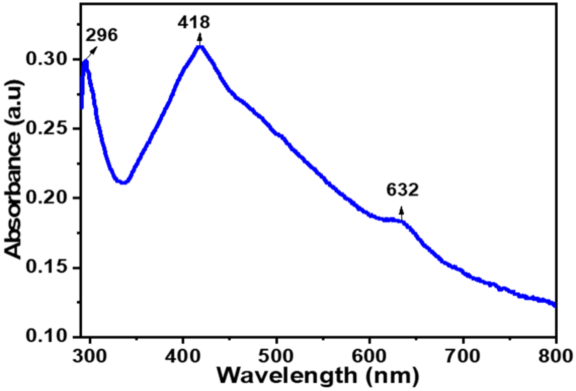
2.1.1. UV-Visible Spectrophotometry

The UV-Vis spectrum of a solution containing the biogenic CTAgNP was acquired within the range of wavelength 200–800 nm (Figure 1). This monitored the reduction of Ag+ ions to zerovalent Ag in the presence of phytocompounds identified in C. tomentosum acetone extract. It is well-argued in the literature that the change in color observed with solution of Ag+ ions in the presence of electron donors is due to the vibrations of the surface plasmon resonance (SPR) in AgNP [30,31]. Therefore, the appearance of a strong peak at 418 nm, which lies between 418–428 nm of the characteristic SPR band for silver, suggests the formation of CTAgNP with enhanced particle size. The absence of peaks between 296 and 632 nm is proof that there was no aggregation of nanoparticles. A similar peak was reported by Rao et al. [32] for the development of AgNP using Diospyros paniculata methanol extract.

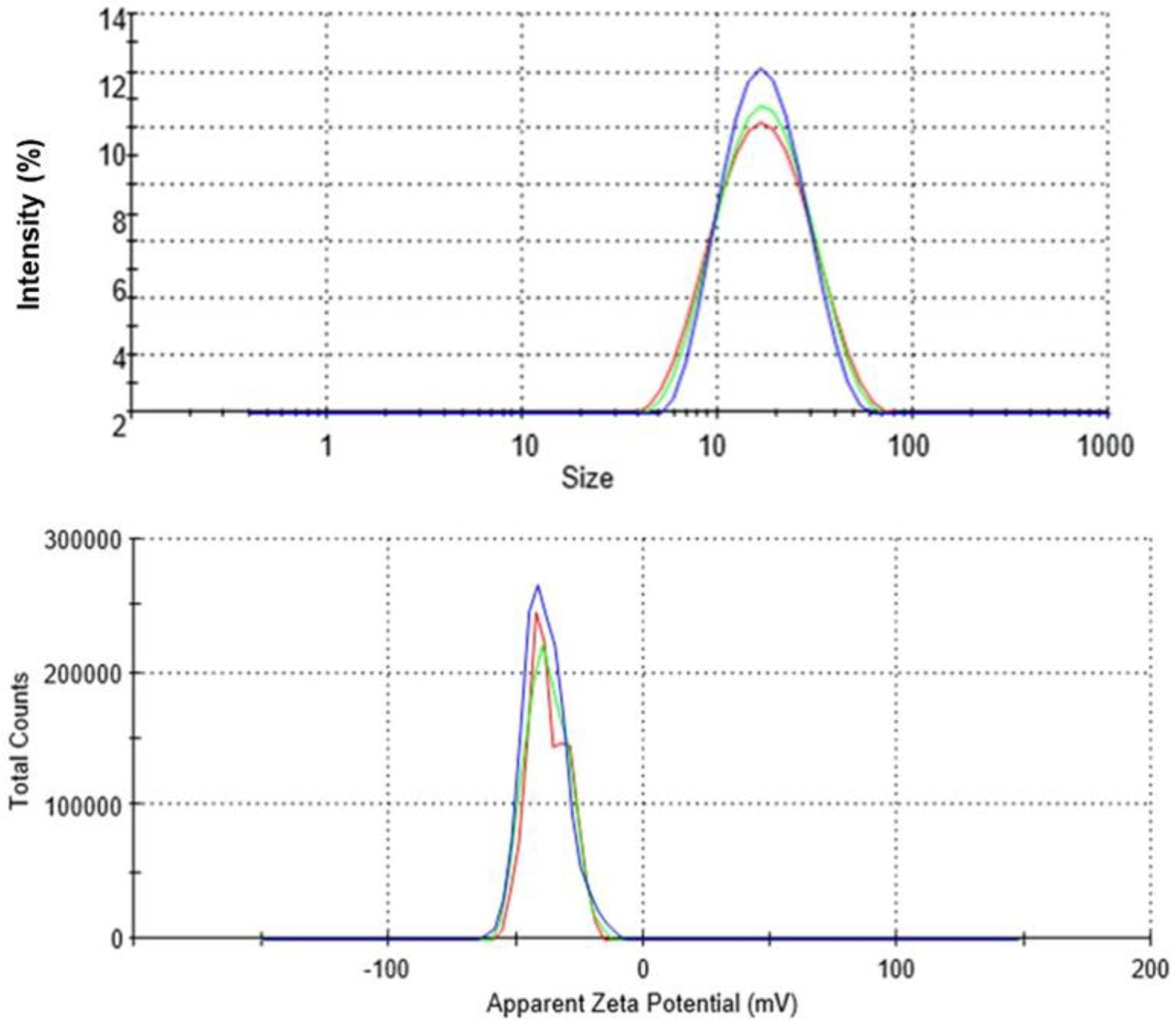
2.1.2. Dynamic Light Scattering (DLS)

The DLS analysis of CTAgNP was performed in water and the results presented in Figure 2. This revealed hydrodynamic size of 145 nm and PDI of 0.236. Similar results were obtained by Banerjee et al. [30] from leaf extract of Mentha arvensis, which recorded AgNP with a hydrodynamic diameter of 145 nm and PDI value of 0.226. PDI values below 0.3 are more often considered acceptable for drug delivery systems and these infer a homogenous suspension. The zeta potential recorded −37.2 ± 7.67 mV, which indicated a relatively stable CTAgNP. High magnitude of negative charge at the surface of CTAgNP suggests a strong Coulombic repulsion among the nanoparticle formed [33]. This observation is presumably ascribed to the presence of phytochemicals at the surface of the developed silver particle, which contributes to increasing its stability.

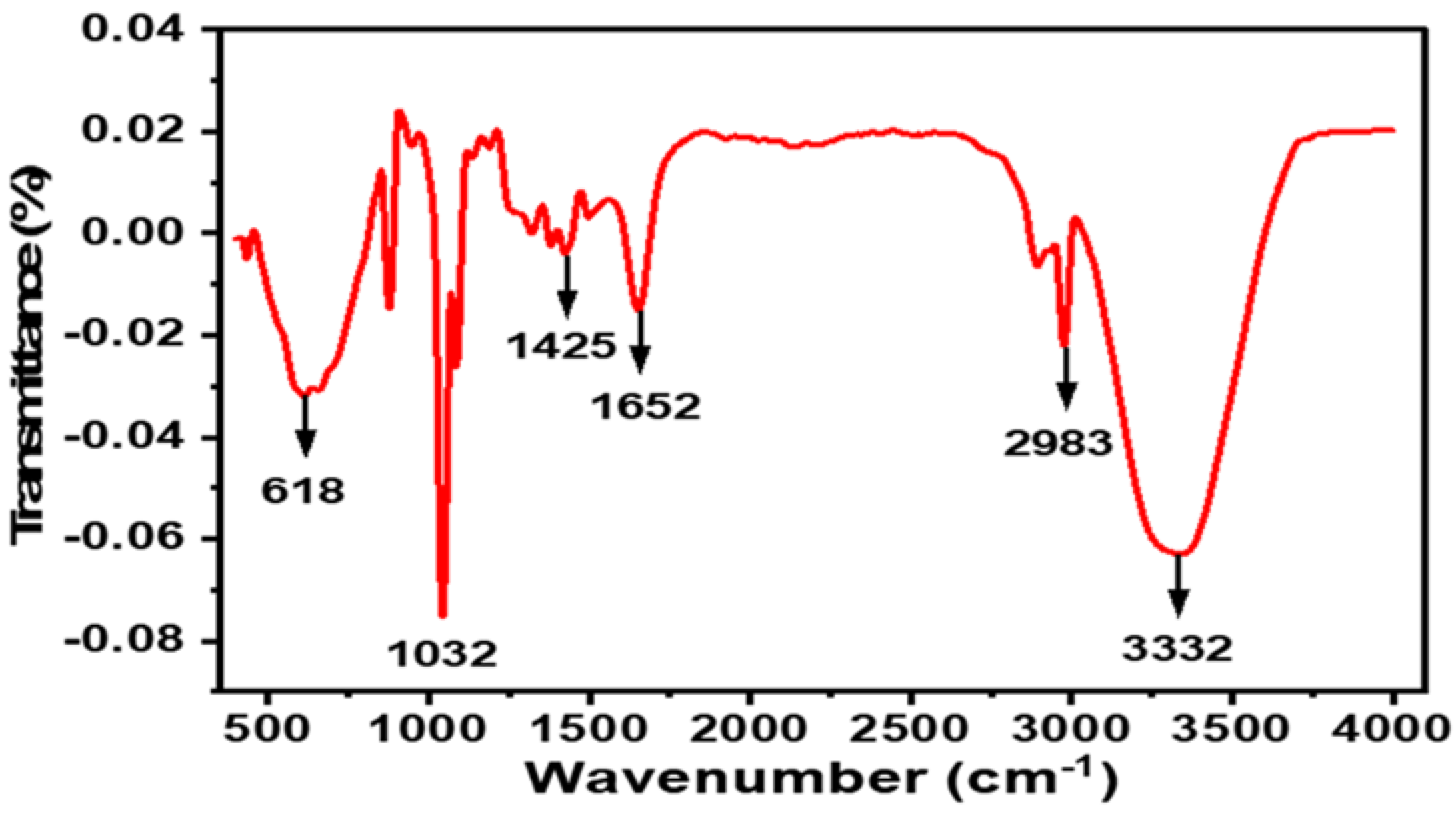
2.1.3. FTIR Spectroscopy

As shown in Figure 3, the FTIR spectrum of the CTAgNP displayed characteristic peaks at 618, 1032, 1425, 1652, 2983, and 3332 cm−1 assigned to C-C skeletal vibration [34], C-O stretching of secondary alcohol [35], N-O stretching of the nitro groups [36], C=O stretching of amide [37], -CH3 asymmetry stretching [38], and OH/NH stretch [39], respectively. These supported the assumption that the AgNP obtained were enclosed by phytocompounds. It is anticipated that the functional groups observed also contributed to the bio-reduction of Ag+ ions. These could aid as robust binding sites and electron-rich spots for the conversion of silver ions to AgNP. Findings from the current study agree with existing reports that the bio-reduction Ag ions served as binding sites for the conversion of Ag ions into AgNP [40,41,42].
Figure 2. Size distribution and zeta potential of Cullen tomentosum silver nanoparticle (CTAgNP).
Antibiotics 12 00203 g002

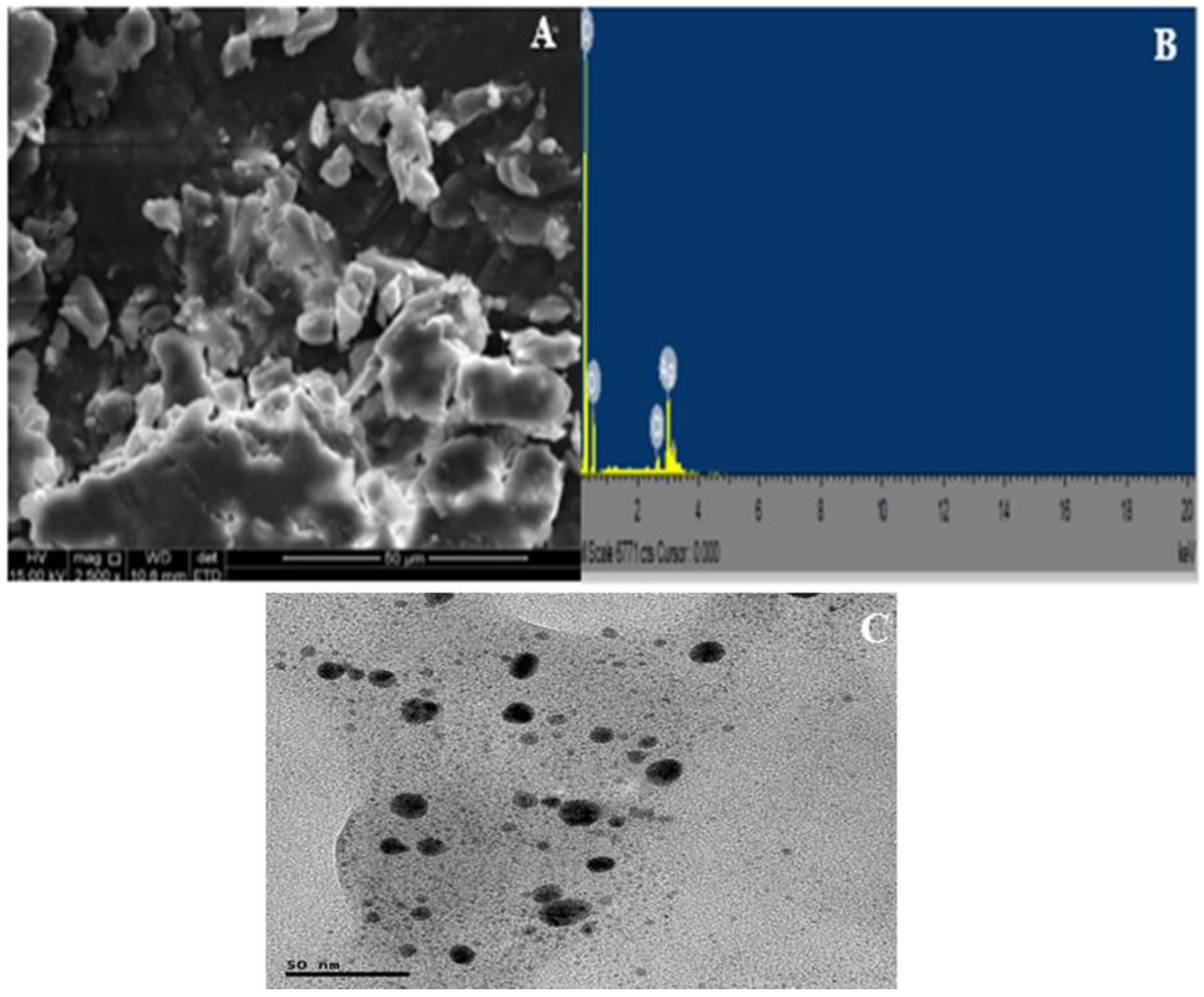
2.1.4. SEM-EDX and TEM Analyses

A close view of the SEM image of the CTAgNP revealed a smooth surface of aggregates and spherical-like nanoparticle (Figure 4A). Recently, Basavarajappa et al. [40] and Femi-Adepoju et al. [41] observed a comparable morphology for the developed AgNP in Passiflora vitifolia and Gleichenia pectinata extracts, respectively. EDX was employed to examine the elemental constituents of the CTAgNP and their relative abundance. This investigation revealed intense peak of Ag (17.2%), along with C (62.74%), O (18.95%), and Cl (1.12%) (Figure 4B). The appearance peaks corresponding to the presence of C, O, and Cl corroborate the development of organic-coated AgNP [43]. The topological feature of CTAgNP was analyzed using TEM and this confirmed that the developed nanoparticle was spherical, with an average size of 12.6 nm (Figure 4C). It was evident that this size differs when compared to the size determined using DLS. Similar observation of a relatively higher particle size estimation by DLS when compared to that determined by TEM was reported by Al Hagbani et al. [44]. This may be explained on the basis that DLS determines the size of nanoparticles in a hydrated state whereby a solvent layer shields the surfaces of nanoparticles. Hence, the size constitutes both the inner core and the sheath. On the other hand, TEM analysis is based on a dry state of the nanoparticle and the estimated size does not include the solvent sheath [45,46]. A related study also described the spherical shape of the biosynthesized silver particles with an average size of 7–27 nm [47]. Likewise, spherical AgNP of 10 nm were reported to form in aqueous sorghum bran extracts [48].

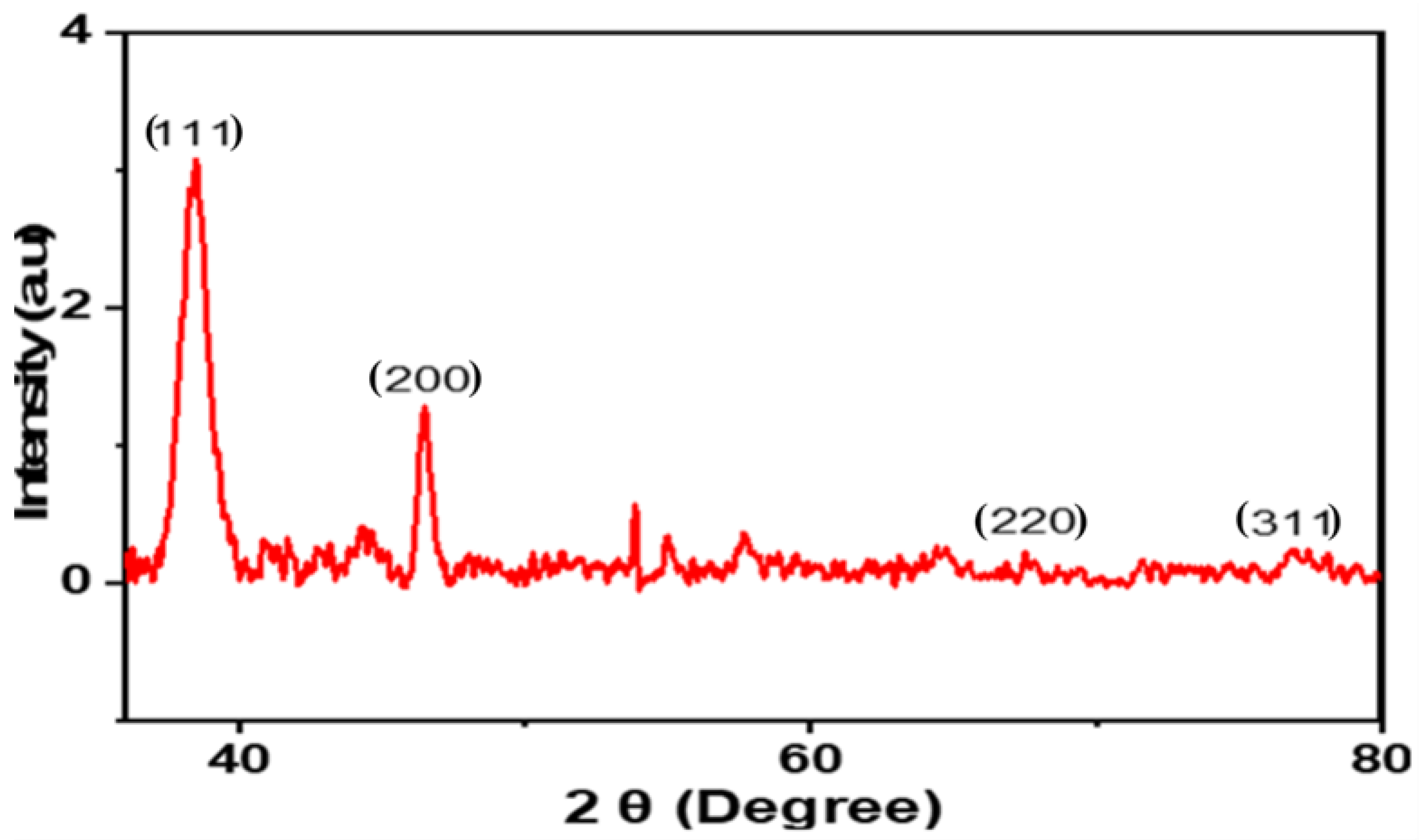
2.1.5. X-ray Diffraction (XRD)

The phase, alignment, and particle size of the CTAgNP were assessed using XRD analytical techniques. This analysis provided an understanding of the specific formation of crystalline nanoparticles by the appearance of sharp peaks in the diffractogram [48]. A set of major peaks at 2θ 38.5°, 46.5°, 67.5°, and 77.0°, consistent with the Miller indices (111), (200), (220), and (311), respectively, were detected (Figure 5). These data matched with the standard database of the Joint Committee on Powder Diffraction Standards (JCPDS, no 4.0783). This confirmed the face-centered cubic (FCC) crystalline Ag. The results obtained in this study is in line with previous studies of FCC AgNPs formed in medicinal plants [49,50]. The unassigned peaks observed are certainly due to the bioorganic phases that converged on the surface of CTAgNP [51]. The average size of the CTAgNP using Scherer’s equation [D = (0.89λ/β COS θ), where “D” represents the mean of the AgNPs, “λ” signifies the wavelength of value (0.15418 nm), “β” denotes full width at half maximum (FWHM) peak and θ indicates Bragg diffraction angle [52], was 13 nm. Interestingly, this size was in good agreement with the TEM result.

2.2. Antibacterial Potency of Extracts and Cullen tomentosum Silver Nanoparticle (CTAgNP)

As shown in Table 1, the acetone extract had moderate potency against B. cereus and S. aureus using the classification by Katerere and Eloff [49]. The authors classified the potency of plant extracts based on the MIC values as noteworthy (≤1 mg/mL), moderate (≥1–8 mg/mL) and weak (≥8–12.5 mg/mL). The MIC values of the extract against B. cereus and S. aureus were 2.6 and 3.1 mg/mL, respectively. The result of this study is not isolated, as previous studies have recorded moderate antimicrobial activity of some members of Fabaceae (such as Erythrina caffra Thunb) to S. aureus and B. cereus [50]. Obistioiu et al. [51] indicated that some members of the Fabaceae such as Coronilla varia, Melilotus officinalis, Ononis spinosa, and Robinia pseudoacacia had limited inhibitory effect against S. aureus. The CTAgNP was more potent than the acetone extract with an MIC of 1.5 mg/mL for B. cereus while for S. aureus, the MIC value was 2.6 mg/mL. The improved potency of CTAgNP compared to the extract was noteworthy given that it possesses a hefty ratio of surface area to volume ratio, ability to deform cell membrane and increase permeability as well as a high aptitude to penetrate the bacterial cell wall. This may be due to the shape and size of the nanoparticle, which enhance its ability to interfere with intracellular processes including protein synthesis [9,52] when compared to the plant extract.
We herein for the first time reported the antibacterial activity of both the extract and CTAgNP from C. tomentosum against the two tested pathogens. Though moderate, the current findings offer basis for further studies using other microbes. This is particularly based on the existing diverse local use of the plant in traditional medicine. It can be further explored as a candidate in the development of moderate therapeutic agents for individuals who are very sensitive or allergic to drugs with high spectrum potency.

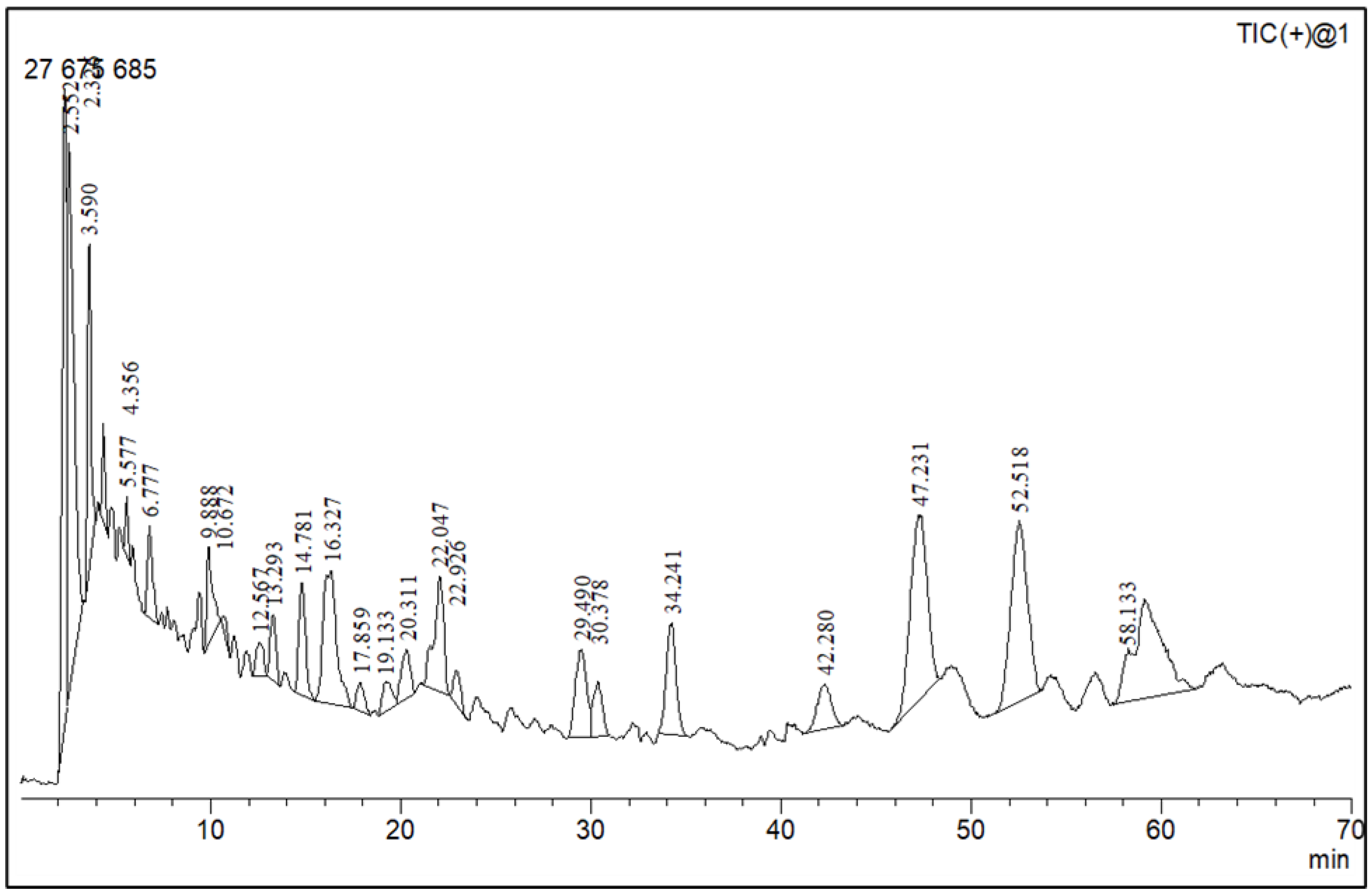
2.3. Phytochemical Profiling

The LC-MS analysis of the extracts revealed twenty-four major peaks corresponding to compounds in the whole plant extract (Appendix A, Table 2 and Table S1). The compounds were further identified by comparing their mass spectral, peak area, peak area (PA) percentage and peak retention time (RT) to that of the m/z cloud library. The least peak area percentage (0.252%) recorded, corresponded to the compound 5,7-dihydroxy-6-methoxy-2-(4-methoxyphenyl)-4H-chromen-4-one with the molecular formular (MF): C17H14O6 and molecular weight (MW): 314.29 g/mol using PubChem database [53] while the highest peak area (11.988%) had corresponding best match to the compound 1-(2-((4-(2-methoxyphenyl)piperazin-1-yl)methyl)-4-methylthiazol-5-yl)ethenone. However, the PubChem database search could not reveal any information related to the molecular formula and molecular weight, indicating that it is probably a novel phytocompound that has not been previously identified in any plant, along with five other compounds with peak area percentages: 1.654%, 2.77%, 1.414%, 4.224%, and 0.907% (Table 2). These compounds could not be identified using the best match criteria on m/z cloud database though their structures had best match equivalent. Other database tools such as PubChem were unsuccessful.
The phytochemical analysis revealed the presence of cyanidin in C. tomentosum whole plant extract. This is a water-soluble anthocyanin that is associated with anticancer and antioxidant properties [54]. This finding reveals C. tomentosum as a new source of cyanidins. A closer look at the peak area percentages reveals that more than 50% of the compounds in the plant fall within five functional groups (Table 2; No 1, 2, 22, 23, and 24) whose peak area percentages sum up to more than 50%. Another compound, 4-((3,3-dimethyloxiran-2-yl)methoxy)-7H-furo[3,2-g] chromen-7-one, belongs to the coumarins known within the subclass Furanocoumarin (Table 2, No 19) which are synthesized through the phenylpropanoid pathway. They have a wide spectrum of biological activity, ranging from antimicrobial, anticoagulant, to anticarcinogenic properties; hence, they have a high potential to fight infections in living tissues [55,56]. Based on existing evidence, the observed antibacterial effect against the tested pathogens may probably be because of the phytochemicals, synergistic activity of a couple of them and the silver containing compounds in the plant. This clearly provides an indication of the need for further investigation on C. tomentosum for the presence of therapeutic agents.

3. Materials and Methods

3.1. Collection of Plant Material and Extraction

The whole plant of Cullen tomentosum was harvested in Mafikeng (latitude 25° 51′ 0′′ S and longitude 25° 37′ 60′′ E), South Africa. Voucher specimen (code ja021) was prepared and deposited at the South African National Biodiversity Institute (SANBI) in Pretoria. Freshly harvested plant material was oven-dried at 40 °C to constant dryness for 72 h, powdered using a blender and stored in a sealed container at room temperature. The powdered plant material was extracted in acetone (1:20 w/v) at room temperature for 1 h with continuous stirring on a magnetic stirrer and later sonicated for 1 h in ice-cold water.
The extract was filtered using Whatman No. 40 filter paper (Whatman® Schleicher & Schuell, London, UK) and concentrated under vacuum using a rotary evaporator at 40 °C. The concentrate was kept in a pre-weighed glass vial and dried at room temperature under a stream of air to a constant weight and stored at 10 °C in the dark till downstream application. All reagents used were of analytical grade.

3.2. Synthesis of Cullen tomentosum Silver Nanoparticle (CTAgNP)

The CTAgNP was synthesized from the whole plant acetone extract of C. tomentosum following the protocol described by Pethakamsetty et al. [57], as applied in the green synthesis of AgNP from Diospyros sylvatica root with slight modifications. Briefly, silver nitrate (AgNO3, 0.01 mol/L) and acetone extract of C. tomentosum (50 mg/mL) at a ratio of 9:1 (v/v) were mixed in a beaker and incubated at room temperature overnight with continuous stirring. Following incubation, the formation of nanoparticle was marked by the appearance of a characteristic reddish-brown coloration. This was then parted by centrifuging at 5500× g rpm for 5 min. The concentrate was washed thrice with deionized water and dried in a muffle furnace for approximately 18 h, at room temperature. The solid material was labelled as CTAgNP and kept at room temperature in Eppendorf tubes for further use.

3.3. Characterization of the Cullen tomentosum Silver Nanoparticle (CTAgNP)

The developed nanoparticle (1 g/mL in ethanol) was characterized using different techniques, including, ultraviolet (UV)-visible spectroscopy (PerkinElmer spectrometer, Lambda 365) for optical absorption and DLS (Malvern instrument, Nano ZS 90) for hydrodynamic size, zeta potential and polydispersity index (PDI) measurements. The morphological characteristics were assessed using scanning electron microscopy (SEM) and transmission electron microscopy (TEM) imaging. The crystallinity, surface functional groups, and elemental composition were discussed based on X-ray diffractogram (Diffractometer, Bruker D8 advance), FTIR (Spectrophotometer, Bruker Alpha), and energy dispersive X-ray (EDX) spectral interpretation, respectively.

3.4. Antibacterial Assay

Bacillus cereus (ATCC®10876™) and Staphylococcus aureus (ATCC®11632™) were used to determine the minimum inhibitory concentration (MIC) of the acetone extract and synthesized CTAgNP using the microtiter plate dilution technique [58]. Staphylococcus aureus is a facultative anaerobe and among the leading pathogens clinically associated with nosocomial infections. With the current proliferation of antimicrobial resistance, S. aureus, with its capability to assault different tissues, has emerged as a microbe of global concern in medical facilities particularly around intensive care units [59]. On the other hand, B. cereus is an endospore-forming aerobic or facultative anaerobe, whose pathogenic activities are clinically associated with food poisoning, gastrointestinal infections, skin infections, cutaneous infections—especially in people diagnosed with underlying conditions including diabetes—and traumatic injuries [60,61,62,63]. The aforementioned factors guided the choice of these microbes for the current study.
Both microbial cultures were revived by streaking on Mueller Hinton (MH) agar plates and incubated at 37 °C for 24 h. A positive colony of each bacterium was inoculated in sterilized 10 mL MH broth and nurtured at 37 °C on a shaker for 24 h. The overnight cultures were diluted to a ratio of 1:100 (v/v). Doxycycline (Sigma-Aldrich, Hamburg, Germany) was used as the positive control. An aliquot of 100 µL of each bacterial strain was added to each well of the microliter plate. A concentration of 50 mg/mL for both extract and CTAgNP in DMSO were put in well A (first well) and two-fold serially diluted with sterile distilled water to well H (last well). Bacteria-free MH broth (blank) and DMSO (which was similarly serially diluted) were used as negative controls. The plates were para-filmed and incubated at 37 °C overnight, after which 50 µL (0.2 mg/mL) of p–iodonitrotetrazolium chloride (INT; Sigma-Aldrich, Hamburg, Germany) was added to each well. The microtiter plates were further incubated at 37 °C for an additional 1 h to detect growth. Generally, INT is a colorless compound that is reduced to pink to red coloration by biological activity of microbes [58,64]. Appearance of pink coloration in a well signifies bacterial growth while lack of color change indicates inhibition of bacteria growth. The extract concentration in the last well without any color conversion was recorded as the MIC. The experiment was done in triplicate

3.5. Phytochemical Profile Using Liquid Chromatography–Mass Spectroscopy (LC–MS)

Acetone extract of C. tomentosum was characterized using LC-MS instrument (LC-MS-2020, Shimadzu Scientific Instruments, Tokyo, Japan). The instrument was equipped with an electrospray ionization (ESI) source operating in negative and positive modes (m/z 100–1200), nebulizing gas (1.5 ℓ/min), DL temperature (250 °C), heat block temperature (200 °C) and detector voltage (0.19 Kv). The chromatographic system consisted of a reversed-phase shim-pack C18 column (5 µm, 250 mm × 2.1 internal diameter) maintained at a constant temperature (30 °C) using an oven. The mobile phase comprises mixture A (10-mM ammonium formate in 90% acetonitrile: water, v/v) and mixture B (0.1% formic acid in acetonitrile, v/v). The extract was dissolved in LC-MS-grade acetonitrile and 2.0 µL was injected into the chromatographic system. An isocratic elution was achieved with 30% mixture A and 70% mixture B at a flow rate of 200 µL/min [65]. Authenticated chemical standards (reserpine and nitrophenol) were analyzed at the same chromatographic conditions to calibrate and tune the MS. Water, ammonium formate and acetonitrile used to prepare mobile phase were of LC, UV grade and LC-MS grade (Macron, fine chemicals). Data acquisition and MS spectral analysis were executed using LabSolution software (Shimadzu) and recorded as absolute intensity and m/z values. The phytochemicals in the acetone extract of C. tomentosum were tentatively identified by exporting MS data (m/z values and corresponding absolute intensities) for each peak and loading into m/z cloud software to search for possible compounds matching the MS fingerprint in the library (HighChem LLC, 2013–2020). The compounds with the highest best match percentage were recorded.

4. Conclusions

The findings of this study revealed the phytochemical constituents of the acetone extract, and antimicrobial potency of both the extract and the biosynthesized CTAgNP. The synthesis of the novel CTAgNP, which was characterized using UV-vis spectrum, DLS, XRD, FTIR, SEM, and TEM analyses, is eco-friendly and cost effective. Both the whole plant acetone extract and the CTAgNP exhibited moderate antimicrobial potency against the studied bacteria pathogens. This suggests the potential of both the whole plant acetone extract and the CTAgNP as antimicrobial agents with applications in different fields including biomedicine. The antibacterial activity of the nanoparticle was more potent in comparison to that of the whole plant acetone extract. However, there is the need to conduct further pharmacological studies to critically understand the mechanism of action including toxicology aspects of the biosynthesized CTAgNP.

Supplementary Materials

The following supporting information can be downloaded at: https://www.mdpi.com/article/10.3390/antibiotics12020203/s1, Table S1: Mass spectra of compounds identified in acetone extract of Cullen tomentosum using liquid chromatography–mass spectrometry (LC-MS) and mz cloud library.

Author Contributions

Conceptualization, J.A.A., A.O.A., and E.K.F.; collection of plant material and extraction, J.A.A. and E.K.F.; laboratory work, J.A.A., E.K.F., and H.A.S.; writing—original draft preparation, J.A.A. and E.K.F.; resources, A.O.A., S.O.A., and L.K.-S.; writing—review and editing, A.O.A., S.O.A., and L.K.-S.; supervision A.O.A., S.O.A., and L.K.-S. All authors have read and agreed to the published version of the manuscript.

Funding

We appreciate the financial support from the North-West University especially the post-doctoral fellowship offered to FEK. We are grateful for Institutional support from the Agricultural Research Council, North-West University, and University of KwaZulu-Natal, South Africa.

Institutional Review Board Statement

Not applicable.

Informed Consent Statement

Not applicable.

Data Availability Statement

All data related to this work are presented in the manuscript.

Acknowledgments

We thank ER Makaudi and M Nengwekhulu for assisting with the synthesis and characterization of nanoparticles. We gratefully acknowledge CN Ateba for providing us with the microorganisms used in this study.

Conflicts of Interest

We declare no conflicts of interest. The funders had no role in the design of the study; in the collection, analyses, or interpretation of data; in the writing of the manuscript; or in the decision to publish the results.

Appendix A

Figure A1. LC-MS chromatogram of Cullen tomentosum whole plant acetone extract showing 24 major peaks corresponding to different compounds and functional groups.
Antibiotics 12 00203 g0a1

References

  1. Yadav, S.; Rawal, G.; Baxi, M. An overview of the latest lnfectious diseases around the world. J. Community Health Manag. 2016, 3, 41–43. [Google Scholar]
  2. Marston, H.D.; Dixon, D.M.; Knisely, J.M.; Palmore, T.N.; Fauci, A.S. Antimicrobial resistance. JAMA 2016, 316, 1193–1204. [Google Scholar] [CrossRef] [PubMed] [Green Version]
  3. Ajose, D.J.; Abolarinwa, T.O.; Oluwarinde, B.O.; Montso, P.K.; Fayemi, O.E.; Aremu, A.O.; Ateba, C.N. Application of plant-derived nanoparticles (PDNP) in food-producing animals as a bio-control agent against antimicrobial-resistant pathogens. Biomedicines 2022, 10, 2426. [Google Scholar] [CrossRef] [PubMed]
  4. Keabadile, O.P.; Aremu, A.O.; Elugoke, S.E.; Fayemi, O.E. Green and traditional synthesis of Copper oxide nanoparticles-comparative Study. Nanomaterials 2020, 10, 2502. [Google Scholar] [CrossRef] [PubMed]
  5. Song, J.Y.; Jang, H.; Kim, B.S. Biological synthesis of gold nanoparticles using Magnolia kobus and Diopyros kaki leaf extracts. Process Biochem. 2009, 44, 1133–1138. [Google Scholar] [CrossRef]
  6. Din, M.I.; Rehan, R. Synthesis, characterization, and applications of copper nanoparticles. Anal. Lett. 2017, 50, 50–62. [Google Scholar] [CrossRef]
  7. Ghosh, S.; Shah, S.; Webster, T. Recent trends in fungal mediated biosynthesis of nanoparticles. Fungi Bio-Prospects in Sustainable Agriculture, Environment and Nano-Technology; Academic Press: Cambridge, MA, USA, 2020. [Google Scholar]
  8. Jee, S.-C.; Kim, M.; Shinde, S.K.; Ghodake, G.S.; Sung, J.-S.; Kadam, A.A. Assembling ZnO and Fe3O4 nanostructures on halloysite nanotubes for anti-bacterial assessments. Appl. Surf. Sci. 2020, 509, 145358. [Google Scholar] [CrossRef]
  9. Krutyakov, Y.A.; Kudrinskiy, A.A.; Olenin, A.Y.; Lisichkin, G.V. Synthesis and properties of silver nanoparticles: Advances and prospects. Russ. Chem. Rev. 2008, 77, 233. [Google Scholar] [CrossRef]
  10. Soliman, W.E.; Khan, S.; Rizvi, S.M.D.; Moin, A.; Elsewedy, H.S.; Abulila, A.S.; Shehata, T.M. Therapeutic applications of biostable silver nanoparticles synthesized using peel extract of Benincasa hispida: Antibacterial and anticancer activities. Nanomaterials 2020, 10, 1954. [Google Scholar] [CrossRef]
  11. Sun, Y.; Mayers, B.; Xia, Y. Transformation of silver nanospheres into nanobelts and triangular nanoplates through a thermal process. Nano Lett. 2003, 3, 675–679. [Google Scholar] [CrossRef]
  12. Khatoon, U.T.; Mohan Mantravadi, K.; Nageswara Rao, G.V.S. Strategies to synthesise copper oxide nanoparticles and their bio applications—A review. Mater. Sci. Technol. 2018, 34, 2214–2222. [Google Scholar] [CrossRef]
  13. Mohan, S.; Singh, Y.; Verma, D.K.; Hasan, S.H. Synthesis of CuO nanoparticles through green route using Citrus limon juice and its application as nanosorbent for Cr(VI) remediation: Process optimization with RSM and ANN-GA based model. Process Saf. Environ. Prot. 2015, 96, 156–166. [Google Scholar] [CrossRef]
  14. Ghosh, S.; Nitnavare, R.; Dewle, A.; Tomar, G.B.; Chippalkatti, R.; More, P.; Kitture, R.; Kale, S.; Bellare, J.; Chopade, B.A. Novel platinum–palladium bimetallic nanoparticles synthesized by Dioscorea bulbifera: Anticancer and antioxidant activities. Int. J. Nanomed. 2015, 10, 7477. [Google Scholar]
  15. Sumitha, S.; Vidhya, R.; Lakshmi, M.S.; Prasad, K.S. Leaf extract mediated green synthesis of copper oxide nanoparticles using Ocimum tenuiflorum and its characterization. Int. J. Chem. Sci. 2016, 14, 2016. [Google Scholar]
  16. Kumar, B.V.; Naik, H.S.B.; Girija, D.; Kumar, B.V. ZnO nanoparticle as catalyst for efficient green one-pot synthesis of coumarins through Knoevenagel condensation. J. Chem. Sci. 2011, 123, 615–621. [Google Scholar] [CrossRef]
  17. Zhang, D.E.; Ni, X.M.; Zheng, H.G.; Li, Y.; Zhang, X.J.; Yang, Z.P. Synthesis of needle-like nickel nanoparticles in water-in-oil microemulsion. Mater. Lett. 2005, 59, 2011–2014. [Google Scholar] [CrossRef]
  18. Padalia, H.; Moteriya, P.; Chanda, S. Green synthesis of silver nanoparticles from marigold flower and its synergistic antimicrobial potential. Arab. J. Chem. 2015, 8, 732–741. [Google Scholar] [CrossRef] [Green Version]
  19. Shankar, S.; Leejae, S.; Jaiswal, L.; Voravuthikunchai, S.P. Metallic nanoparticles augmented the antibacterial potency of Rhodomyrtus tomentosa acetone extract against Escherichia coli. Microb. Pathog. 2017, 107, 181–184. [Google Scholar] [CrossRef] [PubMed]
  20. Chou, K.S.; Ren, C.Y. Synthesis of nanosized silver particles by chemical reduction method. Mater. Chem. Phys. 2000, 64, 241–246. [Google Scholar] [CrossRef]
  21. Maragoni, V.; Maragoni, V.; Ayodhya, D.-D.; Madhusudhan, A.; Amrutham, S.; Guttena, V.; Mangatayaru, K. A Novel Green Synthesis of Silver Nanoparticles Using Gum Karaya: Characterization, Antimicrobial and Catalytic Activity Studies. J. Clust. Sci. 2013, 25, 409–422. [Google Scholar]
  22. Jabir, M.S.; Hussien, A.A.; Sulaiman, G.M.; Yaseen, N.Y.; Dewir, Y.H.; Alwahibi, M.S.; Soliman, D.A.; Rizwana, H. Green synthesis of silver nanoparticles from Eriobotrya japonica extract: A promising approach against cancer cells proliferation, inflammation, allergic disorders and phagocytosis induction. Artif. Cells Nanomed. Biotechnol. 2021, 49, 48–60. [Google Scholar] [CrossRef] [PubMed]
  23. Jones, S.A.; Bowler, P.G.; Walker, M.; Parsons, D. Controlling wound bioburden with a novel silver-containing Hydrofiber® dressing. Wound Repair Regen. 2004, 12, 288–294. [Google Scholar] [CrossRef] [PubMed]
  24. Kim, J.S.; Kuk, E.; Yu, K.N.; Kim, J.-H.; Park, S.J.; Lee, H.J.; Kim, S.H.; Park, Y.K.; Park, Y.H.; Hwang, C.-Y. Antimicrobial effects of silver nanoparticles. Nanomed. Nanotechnol. Biol. Med. 2007, 3, 95–101. [Google Scholar] [CrossRef]
  25. Silver, S.; Phung, L.T. Bacterial heavy metal resistance: New surprises. Annu. Rev. Microbiol. 1996, 50, 753–789. [Google Scholar] [CrossRef] [PubMed]
  26. von Staden, L. Cullen tomentosum (Thunb.) J.W.Grimes. National Assessment: Red List of South African Plants, Version 2020.1. Available online: http://redlist.sanbi.org/species.php?species=374-9 (accessed on 14 October 2022).
  27. Kaholongo, L.T. Screening of Indigenous Forage Legumes as Potential Fodder Crops and Protein Source for Livestock in Central Namibia. Masters’s Thesis, University of Namibia, Windhoek, Namibia, 2016. [Google Scholar]
  28. Van Wyk, B.-E.; Gericke, N. People’s Plants: A Guide to Useful Plants of Southern Africa; Briza Publications: Pretoria, South Africa, 2000. [Google Scholar]
  29. Asong, J.A.; Ndhlovu, P.T.; Khosana, N.S.; Aremu, A.O.; Otang-Mbeng, W. Medicinal plants used for skin-related diseases among the Batswanas in Ngaka Modiri Molema District Municipality, South Africa. S. Afr. J. Bot. 2019, 126, 11–20. [Google Scholar] [CrossRef]
  30. Banerjee, P.P.; Bandyopadhyay, A.; Harsha, S.N.; Policegoudra, R.S.; Bhattacharya, S.; Karak, N.; Chattopadhyay, A. Mentha arvensis (Linn.)-mediated green silver nanoparticles trigger caspase 9-dependent cell death in MCF7 and MDA-MB-231 cells. Breast Cancer: Targets Ther. 2017, 9, 265. [Google Scholar] [CrossRef] [Green Version]
  31. Mulvaney, P. Surface plasmon spectroscopy of nanosized metal particles. Langmuir 1996, 12, 788–800. [Google Scholar] [CrossRef]
  32. Rao, N.H.; N, L.; Pammi, S.V.N.; Kollu, P.; S, G.; P, L. Green synthesis of silver nanoparticles using methanolic root extracts of Diospyros paniculata and their antimicrobial activities. Mater. Sci. Eng. C 2016, 62, 553–557. [Google Scholar] [CrossRef]
  33. Alahmad, A.; Feldhoff, A.; Bigall, N.C.; Rusch, P.; Scheper, T.; Walter, J.-G. Hypericum perforatum L.-mediated green synthesis of silver nanoparticles exhibiting antioxidant and anticancer activities. Nanomaterials 2021, 11, 487. [Google Scholar] [CrossRef]
  34. Algotiml, R.; Gab-Alla, A.; Seoudi, R.; Abulreesh, H.H.; El-Readi, M.Z.; Elbanna, K. Anticancer and antimicrobial activity of biosynthesized Red Sea marine algal silver nanoparticles. Sci. Rep. 2022, 12, 1–18. [Google Scholar] [CrossRef]
  35. Kumar, B.; Smita, K.; Cumbal, L.; Debut, A. Green synthesis of silver nanoparticles using Andean blackberry fruit extract. Saudi J. Biol. Sci. 2017, 24, 45–50. [Google Scholar] [CrossRef] [PubMed] [Green Version]
  36. Bharali, P.; Das, S.; Bhandari, N.; Das, A.K.; Kalta, M.C. Sunlight induced biosynthesis of silver nanoparticle from the bark extract of Amentotaxus assamica DK Ferguson and its antibacterial activity against Escherichia coli and Staphylococcus aureus. IET Nanobiotechnol. 2019, 13, 18–22. [Google Scholar] [CrossRef] [PubMed]
  37. Mihoubi, W.; Sahli, E.; Gargouri, A.; Amiel, C. FTIR spectroscopy of whole cells for the monitoring of yeast apoptosis mediated by p53 over-expression and its suppression by Nigella sativa extracts. PLoS ONE 2017, 12, e0180680. [Google Scholar] [CrossRef] [PubMed]
  38. Dumas, P.; Miller, L. The use of synchrotron infrared microspectroscopy in biological and biomedical investigations. Vib. Spectrosc. 2003, 32, 3–21. [Google Scholar] [CrossRef]
  39. Kuhire, S.S.; Nagane, S.S.; Wadgaonkar, P.P. Poly (ether urethane) s from aromatic diisocyanates based on lignin-derived phenolic acids. Polym. Int. 2017, 66, 892–899. [Google Scholar] [CrossRef]
  40. Basavarajappa, D.S.; Kumar, R.S.; Almansour, A.I.; Chakraborty, B.; Bhat, M.P.; Nagaraja, S.K.; Hiremath, H.; Perumal, K.; Nayaka, S. Biofunctionalized silver nanoparticles synthesized from Passiflora vitifolia leaf extract and evaluation of its antimicrobial, antioxidant and anticancer activities. Biochem. Eng. J. 2022, 187, 108517. [Google Scholar] [CrossRef]
  41. Femi-Adepoju, A.G.; Dada, A.O.; Otun, K.O.; Adepoju, A.O.; Fatoba, O.P. Green synthesis of silver nanoparticles using terrestrial fern (Gleichenia pectinata (Willd.) C. Presl.): Characterization and antimicrobial studies. Heliyon 2019, 5, e01543. [Google Scholar] [CrossRef] [Green Version]
  42. Vilchis-Nestor, A.R.; Sánchez-Mendieta, V.; Camacho-López, M.A.; Gómez-Espinosa, R.M.; Camacho-López, M.A.; Arenas-Alatorre, J.A. Solventless synthesis and optical properties of Au and Ag nanoparticles using Camellia sinensis extract. Mater. Lett. 2008, 62, 3103–3105. [Google Scholar] [CrossRef]
  43. Dada, A.O.; Adekola, F.; Odebunmi, E. A novel zerovalent manganese for removal of copper ions: Synthesis, characterization and adsorption studies. Appl. Water Sci. 2017, 7, 1409–1427. [Google Scholar] [CrossRef] [Green Version]
  44. Al Hagbani, T.; Rizvi, S.M.D.; Hussain, T.; Mehmood, K.; Rafi, Z.; Moin, A.; Abu Lila, A.S.; Alshammari, F.; Khafagy, E.-S.; Rahamathulla, M.; et al. Cefotaxime mediated synthesis of gold nanoparticles: Characterization and antibacterial activity. Polymers 2022, 14, 771. [Google Scholar] [CrossRef]
  45. Mollick, M.M.R.; Rana, D.; Dash, S.K.; Chattopadhyay, S.; Bhowmick, B.; Maity, D.; Mondal, D.; Pattanayak, S.; Roy, S.; Chakraborty, M. Studies on green synthesized silver nanoparticles using Abelmoschus esculentus (L.) pulp extract having anticancer (in vitro) and antimicrobial applications. Arab. J. Chem. 2019, 12, 2572–2584. [Google Scholar] [CrossRef] [Green Version]
  46. Al Saqr, A.; Khafagy, E.-S.; Alalaiwe, A.; Aldawsari, M.F.; Alshahrani, S.M.; Anwer, M.K.; Khan, S.; Lila, A.S.A.; Arab, H.H.; Hegazy, W.A. Synthesis of gold nanoparticles by using green machinery: Characterization and in vitro toxicity. Nanomaterials 2021, 11, 808. [Google Scholar] [CrossRef] [PubMed]
  47. Yang, N.; Li, W.-H. Mango peel extract mediated novel route for synthesis of silver nanoparticles and antibacterial application of silver nanoparticles loaded onto non-woven fabrics. Ind. Crops Prod. 2013, 48, 81–88. [Google Scholar] [CrossRef]
  48. Njagi, E.C.; Huang, H.; Stafford, L.; Genuino, H.; Galindo, H.M.; Collins, J.B.; Hoag, G.E.; Suib, S.L. Biosynthesis of iron and silver nanoparticles at room temperature using aqueous sorghum bran extracts. Langmuir 2011, 27, 264–271. [Google Scholar] [CrossRef] [PubMed]
  49. Katerere, D.R.; Eloff, J.N. Anti-bacterial and anti-oxidant activity of Hypoxis hemerocallidea (Hypoxidaceae): Can leaves be substituted for corms as a conservation strategy? S. Afr. J. Bot. 2008, 74, 613–616. [Google Scholar] [CrossRef] [Green Version]
  50. Olajuyigbe, O.O.; Afolayan, A.J. In vitro antibacterial and time-kill evaluation of the Erythrina caffra Thunb. extract against bacteria associated with diarrhoea. Sci. World J. 2012, 2012, 738314. [Google Scholar] [CrossRef] [PubMed] [Green Version]
  51. Obistioiu, D.; Cocan, I.; Tîrziu, E.; Herman, V.; Negrea, M.; Cucerzan, A.; Neacsu, A.G.; Cozma, A.L.; Nichita, I.; Hulea, A.; et al. Phytochemical profile and microbiological activity of some plants belonging to the Fabaceae family. Antibiotics 2021, 10, 662. [Google Scholar] [CrossRef] [PubMed]
  52. Yin, I.X.; Zhang, J.; Zhao, I.S.; Mei, M.L.; Li, Q.; Chu, C.H. The antibacterial mechanism of silver nanoparticles and Its application in dentistry. Int. J. Nanomed. 2020, 15, 2555–2562. [Google Scholar] [CrossRef] [Green Version]
  53. Kim, S.; Chen, J.; Cheng, T.; Gindulyte, A.; He, J.; He, S.; Li, Q.; Shoemaker, B.A.; Thiessen, P.A.; Yu, B.; et al. PubChem in 2021: New data content and improved web interfaces. Nucleic Acids Res. 2021, 49(D1), D1388–D1395. [Google Scholar] [CrossRef]
  54. Singh, D.; Kumari, K.; Ahmed, S. Natural herbal products for cancer therapy. In Understanding Cancer: From Basics to Therapeutics; Jain, B., Pandey, S., Eds.; Academic Press: Cambridge, MA, USA, 2022; pp. 257–268. [Google Scholar]
  55. Stringlis, I.A.; de Jonge, R.; Pieterse, C.M.J. The age of coumarins in plant-microbe interactions. Plant Cell Physiol. 2019, 60, 1405–1419. [Google Scholar] [CrossRef] [Green Version]
  56. Kavanaugh, K.M.; Aisen, A.M.; Fechner, K.P.; Wroblewski, L.; Chenevert, T.L.; Buda, A.J. Effects of diltiazem on phosphate metabolism in ischemic and reperfused myocardium using phosphorus31 nuclear magnetic resonance spectroscopy in vivo. Am. Heart J. 1989, 118, 1210–1219. [Google Scholar] [CrossRef] [PubMed] [Green Version]
  57. Pethakamsetty, L.; Kothapenta, K.; Nammi, H.R.; Ruddaraju, L.K.; Kollu, P.; Yoon, S.G.; Pammi, S.V.N. Green synthesis, characterization and antimicrobial activity of silver nanoparticles using methanolic root extracts of Diospyros sylvatica. J. Environ. Sci. 2017, 55, 157–163. [Google Scholar] [CrossRef]
  58. Eloff, J.N. A sensitive and quick microplate method to determine the minimal inhibitory concentration of plant extracts for bacteria. Planta Med. 1998, 64, 711–713. [Google Scholar] [CrossRef] [Green Version]
  59. Fuchs, S.; Pané-Farré, J.; Kohler, C.; Hecker, M.; Engelmann, S. Anaerobic gene expression in Staphylococcus aureus. J. Bacteriol. 2007, 189, 4275–4289. [Google Scholar] [CrossRef]
  60. Drewnowska, J.M.; Stefanska, N.; Czerniecka, M.; Zambrowski, G.; Swiecicka, I. Potential enterotoxicity of phylogenetically diverse Bacillus cereus sensu lato soil isolates from different geographical locations. Appl. Environ. Microbiol. 2020, 86, e03032-19. [Google Scholar] [CrossRef] [PubMed] [Green Version]
  61. Esmkhani, M.; Shams, S. Cutaneous infection due to Bacillus cereus: A case report. BioMed Cent. Infect. Dis. 2022, 22, 393. [Google Scholar] [CrossRef]
  62. Michelotti, F.; Bodansky, H.J. Bacillus cereus causing widespread necrotising skin infection in a diabetic person. Pract. Diabetes 2015, 32, 169–170a. [Google Scholar] [CrossRef]
  63. Swiecicka, I. Natural occurrence of Bacillus thuringiensis and Bacillus cereus in eukaryotic organisms: A case for symbiosis. Biocontrol Sci. Technol. 2008, 18, 221–239. [Google Scholar] [CrossRef]
  64. McGaw, L.J.; Van der Merwe, D.; Eloff, J.N. In vitro anthelmintic, antibacterial and cytotoxic effects of extracts from plants used in South African ethnoveterinary medicine. Vet. J. 2007, 173, 366–372. [Google Scholar] [CrossRef] [Green Version]
  65. Seepe, H.A.; Ramakadi, T.G.; Lebepe, C.M.; Amoo, S.O.; Nxumalo, W. Antifungal activity of isolated compounds from the leaves of Combretum erythrophyllum (Burch.) Sond. and Withania somnifera (L.) Dunal against Fusarium pathogens. Molecules 2021, 26, 4732. [Google Scholar] [CrossRef]
Figure 1. Ultraviolet (UV)-visible (UV-Vis) spectrum of an aqueous solution containing Cullen tomentosum silver nanoparticle (CTAgNP).
Antibiotics 12 00203 g001
Figure 3. Fourier-transform infrared spectroscopy (FTIR) spectrum of Cullen tomentosum silver nanoparticle (CTAgNP).
Antibiotics 12 00203 g003
Figure 4. (A) Scanning electron microscopy (SEM) image, (B) energy dispersive X-ray (EDX) spectrum, and (C) transmission electron microscopy (TEM) micrograph of Cullen tomentosum silver nanoparticle (CTAgNP).
Antibiotics 12 00203 g004
Figure 5. X-ray diffraction (XRD) pattern of Cullen tomentosum silver nanoparticle (CTAgNP).
Antibiotics 12 00203 g005
Table 1. Antibacterial effect of Cullen tomentosum acetone extract and resultant silver nanoparticle.
SampleMinimum Inhibitory Concentration (mg/mL)
Bacillus cereusStaphylococcus aureus
Acetone extract2.63.1
Silver nanoparticle (CTAgNP)1.52.6
Doxycycline (used as a positive control) had minimum inhibitory concentrations of 0.78 mg/mL and 1.56 mg/mL against Bacillus cereus and Staphylococcus aureus, respectively.
Table 2. Phytocompounds identified in the acetone extract of Cullen tomentosum following liquid chromatography–mass spectrometry (LC-MS) analysis.
Peak NumberRT *PA # (%)Structure of CompoundName of Compound (m/z Cloud Library)Similarity Index (%)
12.326 11.63Antibiotics 12 00203 i0012-(2-(benzo[d][1,3] dioxol-6-yl)-4,5-dihydro-4-oxothiazol-5-yl)acetic acid81.3
22.55211.988Antibiotics 12 00203 i0021-(2-((4-(2-methoxyphenyl) piperazin-1-yl)methyl)-4-methylthiazol-5-yl)ethanone82.2
33.594.545Antibiotics 12 00203 i0032-(4-hydroxyphenyl)-6-methoxy-7-(tetrahydro-3,4,5-trihydroxy-6-(hydroxymethyl)-2H-pyran-2-yloxy)-4H-chromen-4-one 56.8
44.356 1.053 Antibiotics 12 00203 i0045,7-dihydroxy-2-phenyl-4H-chromen-4-one 90.3
55.577 0.588Antibiotics 12 00203 i0053,3a,4,5,5a,6,7,8-octahydro-3a-hydroxy-5a,9-dimethyl-3-methylenenaphtho[1,2-b]furan-2(9bH)-one84.8
66.7771.683 Antibiotics 12 00203 i0067-((2E,5E)-7-methoxy-3,7-dimethylocta-2,5-dienyloxy)-2H-chromen-2-one 84.3
79.8882.19 Antibiotics 12 00203 i007(3-(1-methyl-3-(naphthalen-6-yl)-1H-pyrazol-5-yl)quinuclidin-7-yl)methyl 4-isopropylphenylcarbamate 81.5
810.672 0.252 Antibiotics 12 00203 i0085,7-dihydroxy-6-methoxy-2-(4-methoxyphenyl)-4H-chromen-4-one 81.2
912.567 0.945 Antibiotics 12 00203 i009Cyanidin89.2
1013.293 1.414 Antibiotics 12 00203 i010No name identity from NIST Library 95.7
1114.781 2.77 Antibiotics 12 00203 i011No name identity from NIST library90.6
1216.327 6.629 Antibiotics 12 00203 i012tetrahydro-3,4,5-trihydroxy-6-(hydroxymethyl)-2H-pyran-2-yl 3,4,5-trimethoxybenzoate 89.1
1317.859 0.823Antibiotics 12 00203 i0132-methylene-4-(tetrahydro-3,4,5-trihydroxy-6-(hydroxymethyl)-2H-pyran-2-yloxy) butanoic acid 80.1
1419.133 1.078 Antibiotics 12 00203 i0142-(guanidine)-3-(1H-imidazol-4-yl) propanoic acid 99.4
1520.311 1.654 Antibiotics 12 00203 i015No name identity equivalent in NIST library93.8
1622.047 4.224 Antibiotics 12 00203 i016No name identity equivalent in NIST library 80.1
1722.926 0.907 Antibiotics 12 00203 i017No name identity equivalent in NIST library90.6
1829.49 3.729Antibiotics 12 00203 i018(E)-N-(5-methylisoxazol-3-yl)-3-(thiophen-2-yl)-2-(thiophen-3-yl)acrylamide95.8
1930.378 1.804 Antibiotics 12 00203 i0194-((3,3-dimethyloxiran-2-yl)methoxy)-7H-furo[3,2-g]chromen-7-one 88.9
2034.241 4.11 Antibiotics 12 00203 i020(Z)-6-(bromomethylene)-tetrahydro-3-(naphthalen-5-yl)pyran-2-one 91.2
2142.28 2.368 Antibiotics 12 00203 i021N-(3-(tetrahydro-2H-pyran-4-yl)-1-hydroxy-1-phenylpropan-2-yl)decanamide84.6
2247.231 11.648 Antibiotics 12 00203 i0222-(3-((5-(4-tert-butylphenyl)isoxazol-3-yl) methyl) piperidin-4-yl)-1-(4-methylpiperazin-1-yl)ethanone 85.3
2352.51811.169Antibiotics 12 00203 i0232-(1-(2-chlorophenyl)-1H-imidazol-2-ylthio)-N,N-dihydroxypyridin-3-amine 87.9
2458.133 10.802 Antibiotics 12 00203 i0242,3-dihydro-5,7-dihydroxy-2-(4-methoxyphenyl)-6-(3-methylbut-2-enyl)chromen-4-one 85.7
* RT = retention time, # PA = peak area.
Disclaimer/Publisher’s Note: The statements, opinions and data contained in all publications are solely those of the individual author(s) and contributor(s) and not of MDPI and/or the editor(s). MDPI and/or the editor(s) disclaim responsibility for any injury to people or property resulting from any ideas, methods, instructions or products referred to in the content.

