
Inorganic Nanoparticles: Tools to Emphasize the Janus Face of Amphotericin B


Abstract
1. Introduction
2. Similarities in Redox Behaviors between Amphotericin B and Inorganic Nanoparticles
2.1. Redox Properties of Amphotericin B
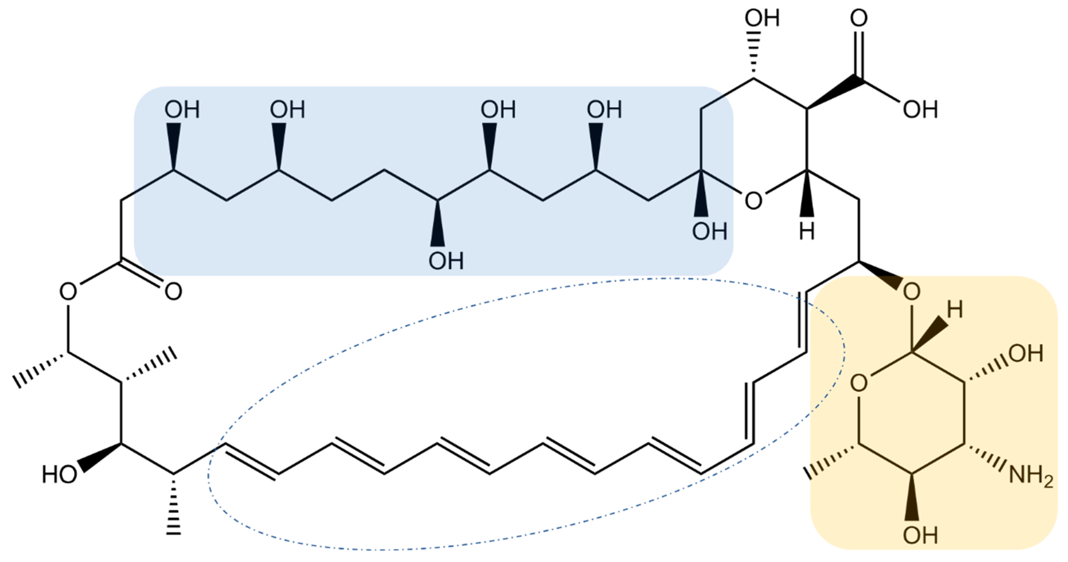
2.1.1. The Janus Face of Amphotericin B
2.1.2. Amphotericin B Activity, Resistance, and Toxicity, and Its Possible Modulation
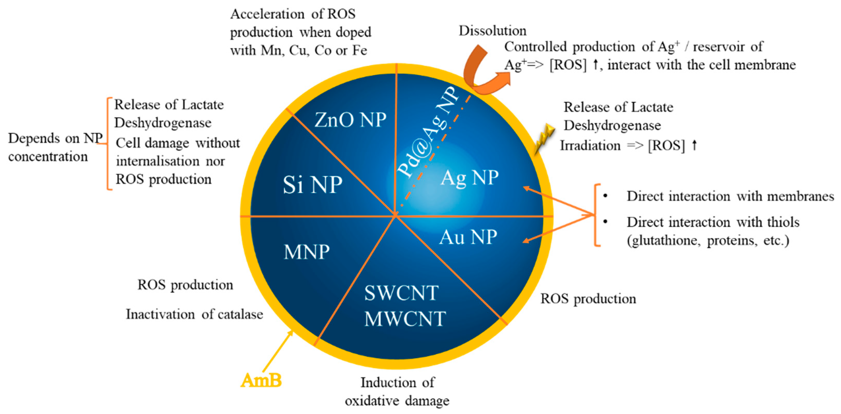
2.2. Redox Properties of Inorganic Nanoparticles
3. Inorganic Nanoparticles Carrying Amphotericin B
3.1. The State-of-the-Art of Lipidic Formulations of Amphotericin B on the Market or under Clinical Trials
3.2. Inorganic Nanoparticles as Modulator of AmB Redox Properties
3.2.1. Strategies to Functionalize Inorganic Nanoparticles with Amphotericin B
3.2.2. Inorganic Nanoparticles as Synergic Prooxidants
3.2.3. Inorganic Nanoparticles as Synergic Antioxidants
4. Summary and Future Directions
Supplementary Materials
Author Contributions
Funding
Institutional Review Board Statement
Informed Consent Statement
Data Availability Statement
Conflicts of Interest
References
- Nicolaou, K.C.; Chen, J.S.; Dalby, S.M. From Nature to the Laboratory and into the Clinic. Bioorg. Med. Chem. 2009, 17, 2290–2303. [Google Scholar] [CrossRef] [PubMed]
- Volmer, A.A.; Szpilman, A.M.; Carreira, E.M. Synthesis and Biological Evaluation of Amphotericin B Derivatives. Nat. Prod. Rep. 2010, 27, 1329–1349. [Google Scholar] [CrossRef]
- Pound, M.W.; Townsend, M.L.; Dimondi, V.; Wilson, D.; Drew, R.H. Overview of Treatment Options for Invasive Fungal Infections. Med. Mycol. 2011, 49, 561–580. [Google Scholar] [CrossRef]
- Cavassin, F.B.; Baú-Carneiro, J.L.; Vilas-Boas, R.R.; Queiroz-Telles, F. Sixty Years of Amphotericin B: An Overview of the Main Antifungal Agent Used to Treat Invasive Fungal Infections. Infect. Dis. Ther. 2021, 10, 115–147. [Google Scholar] [CrossRef] [PubMed]
- Carolus, H.; Pierson, S.; Lagrou, K.; Van Dijck, P. Amphotericin B and Other Polyenes-Discovery, Clinical Use, Mode of Action and Drug Resistance. J. Fungi 2020, 6, 321. [Google Scholar] [CrossRef]
- Cowen, L.E.; Sanglard, D.; Howard, S.J.; Rogers, P.D.; Perlin, D.S. Mechanisms of Antifungal Drug Resistance. Cold Spring Harb. Perspect. Med. 2015, 5, a019752. [Google Scholar] [CrossRef]
- Brunet, K.; Diop, C.A.B.; Chauzy, A.; Prébonnaud, N.; Marchand, S.; Rammaert, B.; Tewes, F. Improved In Vitro Anti-Mucorales Activity and Cytotoxicity of Amphotericin B with a Pegylated Surfactant. J. Fungi 2022, 8, 121. [Google Scholar] [CrossRef]
- Pappas, P.G.; Kauffman, C.A.; Andes, D.R.; Clancy, C.J.; Marr, K.A.; Ostrosky-Zeichner, L.; Reboli, A.C.; Schuster, M.G.; Vazquez, J.A.; Walsh, T.J.; et al. Clinical Practice Guideline for the Management of Candidiasis: 2016 Update by the Infectious Diseases Society of America. Clin. Infect. Dis. Off. Publ. Infect. Dis. Soc. Am. 2016, 62, e1–e50. [Google Scholar] [CrossRef]
- Pathak, A.; Pien, F.D.; Carvalho, L. Amphotericin B Use in a Community Hospital, with Special Emphasis on Side Effects. Clin. Infect. Dis. Off. Publ. Infect. Dis. Soc. Am. 1998, 26, 334–338. [Google Scholar] [CrossRef]
- Gursoy, V.; Ozkalemkas, F.; Ozkocaman, V.; Serenli Yegen, Z.; Ethem Pinar, I.; Ener, B.; Akalın, H.; Kazak, E.; Ali, R.; Ersoy, A. Conventional Amphotericin B Associated Nephrotoxicity in Patients With Hematologic Malignancies. Cureus 2021, 13, e16445. [Google Scholar] [CrossRef] [PubMed]
- Jafari, M.; Abolmaali, S.S.; Tamaddon, A.M.; Zomorodian, K.; Sarkari, B.S. Nanotechnology Approaches for Delivery and Targeting of Amphotericin B in Fungal and Parasitic Diseases. Nanomedicine 2021, 16, 857–877. [Google Scholar] [CrossRef]
- Alakkad, A.; Stapleton, P.; Schlosser, C.; Murdan, S.; Odunze, U.; Schatzlein, A.; Uchegbu, I.F. Amphotericin B Polymer Nanoparticles Show Efficacy against Candida Species Biofilms. Pathogens 2022, 11, 73. [Google Scholar] [CrossRef]
- Faustino, C.; Pinheiro, L. Lipid Systems for the Delivery of Amphotericin B in Antifungal Therapy. Pharmaceutics 2020, 12, 29. [Google Scholar] [CrossRef] [PubMed]
- Chen, Y.-C.; Su, C.-Y.; Jhan, H.-J.; Ho, H.-O.; Sheu, M.-T. Physical Characterization and in Vivo Pharmacokinetic Study of Self-Assembling Amphotericin B-Loaded Lecithin-Based Mixed Polymeric Micelles. Int. J. Nanomed. 2015, 10, 7265–7274. [Google Scholar] [CrossRef]
- Serrano, D.R.; Lalatsa, A.; Dea-Ayuela, M.A.; Bilbao-Ramos, P.E.; Garrett, N.L.; Moger, J.; Guarro, J.; Capilla, J.; Ballesteros, M.P.; Schätzlein, A.G.; et al. Oral Particle Uptake and Organ Targeting Drives the Activity of Amphotericin B Nanoparticles. Mol. Pharm. 2015, 12, 420–431. [Google Scholar] [CrossRef]
- Wang, C.; Makvandi, P.; Zare, E.N.; Tay, F.R.; Niu, L. Advances in Antimicrobial Organic and Inorganic Nanocompounds in Biomedicine. Adv. Ther. 2020, 3, 2000024. [Google Scholar] [CrossRef]
- Jiang, S.; Win, K.Y.; Liu, S.; Teng, C.P.; Zheng, Y.; Han, M.-Y. Surface-Functionalized Nanoparticles for Biosensing and Imaging-Guided Therapeutics. Nanoscale 2013, 5, 3127–3148. [Google Scholar] [CrossRef] [PubMed]
- Huang, H.; Feng, W.; Chen, Y.; Shi, J. Inorganic Nanoparticles in Clinical Trials and Translations. Nano Today 2020, 35, 100972. [Google Scholar] [CrossRef]
- Mesa-Arango, A.C.; Trevijano-Contador, N.; Román, E.; Sánchez-Fresneda, R.; Casas, C.; Herrero, E.; Argüelles, J.C.; Pla, J.; Cuenca-Estrella, M.; Zaragoza, O. The Production of Reactive Oxygen Species Is a Universal Action Mechanism of Amphotericin B against Pathogenic Yeasts and Contributes to the Fungicidal Effect of This Drug. Antimicrob. Agents Chemother. 2014, 58, 6627–6638. [Google Scholar] [CrossRef]
- Guirao-Abad, J.P.; Sánchez-Fresneda, R.; Alburquerque, B.; Hernández, J.A.; Argüelles, J.-C. ROS Formation Is a Differential Contributory Factor to the Fungicidal Action of Amphotericin B and Micafungin in Candida Albicans. Int. J. Med. Microbiol. 2017, 307, 241–248. [Google Scholar] [CrossRef]
- Kovacic, P.; Cooksy, A. Novel, Unifying Mechanism for Amphotericin B and Other Polyene Drugs: Electron Affinity, Radicals, Electron Transfer, Autoxidation, Toxicity, and Antifungal Action. MedChemComm 2012, 3, 274–280. [Google Scholar] [CrossRef]
- Osaka, K.; Ritov, V.B.; Bernardo, J.F.; Branch, R.A.; Kagan, V.E. Amphotericin B Protects Cis-Parinaric Acid against Peroxyl Radical-Induced Oxidation: Amphotericin B as an Antioxidant. Antimicrob. Agents Chemother. 1997, 41, 743–747. [Google Scholar] [CrossRef]
- Osaka, K.; Tyurina, Y.Y.; Dubey, R.K.; Tyurin, V.A.; Ritov, V.B.; Quinn, P.J.; Branch, R.A.; Kagan, V.E. Amphotericin B as an Intracellular Antioxidant: Protection against 2,2′-Azobis(2,4-Dimethylvaleronitrile)-Induced Peroxidation of Membrane Phospholipids in Rat Aortic Smooth Muscle Cells. Biochem. Pharmacol. 1997, 54, 937–945. [Google Scholar] [CrossRef]
- Lamy-Freund, M.T.; Ferreira, V.F.; Schreier, S. Mechanism of Inactivation of the Polyene Antibiotic Amphotericin B. Evidence for Radical Formation in the Process of Autooxidation. J. Antibiot. 1985, 38, 753–757. [Google Scholar] [CrossRef]
- Belenky, P.; Camacho, D.; Collins, J.J. Fungicidal Drugs Induce a Common Oxidative-Damage Cellular Death Pathway. Cell Rep. 2013, 3, 350–358. [Google Scholar] [CrossRef]
- Ferreira, G.F.; de Baltazar, L.M.; Santos, J.R.A.; Monteiro, A.S.; de Fraga, L.A.O.; Resende-Stoianoff, M.A.; Santos, D.A. The Role of Oxidative and Nitrosative Bursts Caused by Azoles and Amphotericin B against the Fungal Pathogen Cryptococcus gattii. J. Antimicrob. Chemother. 2013, 68, 1801–1811. [Google Scholar] [CrossRef] [PubMed]
- Andrews, F.A.; Beggs, W.H.; Sarosi, G.A. Influence of Antioxidants on the Bioactivity of Amphotericin B. Antimicrob. Agents Chemother. 1977, 11, 615–618. [Google Scholar] [CrossRef]
- Pravkin, S.K.; Yakusheva, E.N.; Uzbekova, D.G. In Vivo Analysis of Antioxidant and Prooxidant Properties of Retinol Acetate. Bull. Exp. Biol. Med. 2013, 156, 220–223. [Google Scholar] [CrossRef] [PubMed]
- Putchala, M.C.; Ramani, P.; Sherlin, H.J.; Premkumar, P.; Natesan, A. Ascorbic Acid and Its Pro-Oxidant Activity as a Therapy for Tumours of Oral Cavity—A Systematic Review. Arch. Oral Biol. 2013, 58, 563–574. [Google Scholar] [CrossRef]
- Ko, K.M.; Yick, P.K.; Poon, M.K.; Ip, S.P. Prooxidant and Antioxidant Effects of Trolox on Ferric Ion-Induced Oxidation of Erythrocyte Membrane Lipids. Mol. Cell. Biochem. 1994, 141, 65–70. [Google Scholar] [CrossRef]
- Kong, Y.; Wang, Q.; Cao, F.; Zhang, X.; Fang, Z.; Shi, P.; Wang, H.; Shen, Y.; Huang, Z. BSC2 Enhances Cell Resistance to AmB by Inhibiting Oxidative Damage in Saccharomyces Cerevisiae. Free Radic. Res. 2020, 54, 231–243. [Google Scholar] [CrossRef] [PubMed]
- Purkait, B.; Kumar, A.; Nandi, N.; Sardar, A.H.; Das, S.; Kumar, S.; Pandey, K.; Ravidas, V.; Kumar, M.; De, T.; et al. Mechanism of Amphotericin B Resistance in Clinical Isolates of Leishmania Donovani. Antimicrob. Agents Chemother. 2012, 56, 1031–1041. [Google Scholar] [CrossRef] [PubMed]
- Jukic, E.; Blatzer, M.; Posch, W.; Steger, M.; Binder, U.; Lass-Flörl, C.; Wilflingseder, D. Oxidative Stress Response Tips the Balance in Aspergillus Terreus Amphotericin B Resistance. Antimicrob. Agents Chemother. 2017, 61, e00670-17. [Google Scholar] [CrossRef] [PubMed]
- Altuntaş, A.; Yılmaz, H.R.; Altuntaş, A.; Uz, E.; Demir, M.; Gökçimen, A.; Aksu, O.; Bayram, D.Ş.; Sezer, M.T. Caffeic Acid Phenethyl Ester Protects against Amphotericin B Induced Nephrotoxicity in Rat Model. BioMed Res. Int. 2014, 2014, 702981. [Google Scholar] [CrossRef] [PubMed]
- Gola, J.; Skubis, A.; Sikora, B.; Kruszniewska-Rajs, C.; Adamska, J.; Mazurek, U.; Strzalka-Mrozik, B.; Czernel, G.; Gagos, M. Expression Profiles of Genes Related to Melatonin and Oxidative Stress in Human Renal Proximal Tubule Cells Treated with Antibiotic Amphotericin B and Its Modified Forms. Turk. J. Biol. 2015, 39, 856–864. [Google Scholar] [CrossRef]
- Schlottfeldt, F.D.S.; Fernandes, S.M.; Martins, D.M.; Cordeiro, P.; da Fonseca, C.D.; Watanabe, M.; Vattimo, M.d.F.F. Prevention of Amphotericin B Nephrotoxicity through Use of Phytotherapeutic Medication. Rev. Da Esc. De Enferm. Da USP 2015, 49, 74–79. [Google Scholar] [CrossRef]
- Salehzadeh, A.; Salehzadeh, A.; Maghsood, A.-H.; Heidarisasan, S.; Taheri-Azandaryan, M.; Ghafourikhosroshahi, A.; Abbasalipourkabir, R. Effects of Vitamin A and Vitamin E on Attenuation of Amphotericin B-Induced Side Effects on Kidney and Liver of Male Wistar Rats. Environ. Sci. Pollut. Res. Int. 2020, 27, 32594–32602. [Google Scholar] [CrossRef]
- Kim, J.H.; Chan, K.L.; Faria, N.C.G.; Martins, M.d.L.; Campbell, B.C. Targeting the Oxidative Stress Response System of Fungi with Redox-Potent Chemosensitizing Agents. Front. Microbiol. 2012, 3, 88. [Google Scholar] [CrossRef]
- Kim, J.H.; Faria, N.C.G.; Martins, M.D.L.; Chan, K.L.; Campbell, B.C. Enhancement of Antimycotic Activity of Amphotericin B by Targeting the Oxidative Stress Response of Candida and Cryptococcus with Natural Dihydroxybenzaldehydes. Front. Microbiol. 2012, 3, 261. [Google Scholar] [CrossRef]
- Beggs, W.H.; Andrews, F.A.; Sarosi, G.A. Synergistic Action of Amphotericin B and Antioxidants against Certain Opportunistic Yeast Pathogens. Antimicrob. Agents Chemother. 1978, 13, 266–270. [Google Scholar] [CrossRef]
- Blatzer, M.; Jukic, E.; Posch, W.; Schöpf, B.; Binder, U.; Steger, M.; Blum, G.; Hackl, H.; Gnaiger, E.; Lass-Flörl, C.; et al. Amphotericin B Resistance in Aspergillus Terreus Is Overpowered by Coapplication of Pro-Oxidants. Antioxid. Redox Signal. 2015, 23, 1424–1438. [Google Scholar] [CrossRef] [PubMed]
- Shekhova, E.; Kniemeyer, O.; Brakhage, A.A. Induction of Mitochondrial Reactive Oxygen Species Production by Itraconazole, Terbinafine, and Amphotericin B as a Mode of Action against Aspergillus fumigatus. Antimicrob. Agents Chemother. 2017, 61, e00978-17. [Google Scholar] [CrossRef] [PubMed]
- Sippel, J.E.; Levine, H.B. Annulment of Amphotericin B Inhibition of Coccidioides Immitis Endospores. Effects on Growth, Respiration and Morphogenesis. Sabouraudia 1969, 7, 159–168. [Google Scholar] [CrossRef] [PubMed]
- Weis, M.R.; Levine, H.B. Inactivation of Amphotericin B by Reducing Agents: Influences on Growth Inhibition of Candida Albicans and Lysis of Erythrocytes. Sabouraudia 1972, 10, 132–142. [Google Scholar] [CrossRef]
- Silva, L.N.; Oliveira, S.S.C.; Magalhães, L.B.; Andrade Neto, V.V.; Torres-Santos, E.C.; Carvalho, M.D.C.; Pereira, M.D.; Branquinha, M.H.; Santos, A.L.S. Unmasking the Amphotericin B Resistance Mechanisms in Candida Haemulonii Species Complex. ACS Infect. Dis. 2020, 6, 1273–1282. [Google Scholar] [CrossRef]
- Young, L.Y.; Hull, C.M.; Heitman, J. Disruption of Ergosterol Biosynthesis Confers Resistance to Amphotericin B in Candida Lusitaniae. Antimicrob. Agents Chemother. 2003, 47, 2717–2724. [Google Scholar] [CrossRef]
- Bhattacharya, S.; Esquivel, B.D.; White, T.C. Overexpression or Deletion of Ergosterol Biosynthesis Genes Alters Doubling Time, Response to Stress Agents, and Drug Susceptibility in Saccharomyces Cerevisiae. mBio 2018, 9, e01291-18. [Google Scholar] [CrossRef]
- Tournebize, J.; Sapin-Minet, A.; Bartosz, G.; Leroy, P.; Boudier, A. Pitfalls of Assays Devoted to Evaluation of Oxidative Stress Induced by Inorganic Nanoparticles. Talanta 2013, 116, 753–763. [Google Scholar] [CrossRef]
- Hellack, B.; Nickel, C.; Albrecht, C.; Kuhlbusch, T.A.J.; Boland, S.; Baeza-Squiban, A.; Wohlleben, W.; Schins, R.P.F. Analytical Methods to Assess the Oxidative Potential of Nanoparticles: A Review. Environ. Sci. Nano 2017, 4, 1920–1934. [Google Scholar] [CrossRef]
- Valgimigli, L.; Baschieri, A.; Amorati, R. Antioxidant Activity of Nanomaterials. J. Mater. Chem. B 2018, 6, 2036–2051. [Google Scholar] [CrossRef]
- Innocenzi, P.; Stagi, L. Carbon Dots as Oxidant-Antioxidant Nanomaterials, Understanding the Structure-Properties Relationship. A Critical Review. Nano Today 2023, 50, 101837. [Google Scholar] [CrossRef]
- Samrot, A.V.; Ram Singh, S.P.; Deenadhayalan, R.; Rajesh, V.V.; Padmanaban, S.; Radhakrishnan, K. Nanoparticles, a Double-Edged Sword with Oxidant as Well as Antioxidant Properties—A Review. Oxygen 2022, 2, 591–604. [Google Scholar] [CrossRef]
- Fifere, A.; Moleavin, I.-A.T.; Lungoci, A.-L.; Marangoci, N.L.; Pinteala, M. Inorganic Nanoparticles as Free Radical Scavengers. In New Trends in Macromolecular and Supramolecular Chemistry for Biological Applications; Abadie, J.M., Pinteala, M., Rotaru, A.M., Eds.; Springer International Publishing: Cham, Switzerland, 2021; pp. 295–329. ISBN 978-3-030-57456-7. [Google Scholar]
- Gómez-Herrero, A.C.; Sánchez-Sánchez, C.; Chérioux, F.; Martínez, J.I.; Abad, J.; Floreano, L.; Verdini, A.; Cossaro, A.; Mazaleyrat, E.; Guisset, V.; et al. Copper-Assisted Oxidation of Catechols into Quinone Derivatives. Chem. Sci. 2020, 12, 2257–2267. [Google Scholar] [CrossRef]
- Steinmetz, L.; Geers, C.; Balog, S.; Bonmarin, M.; Rodriguez-Lorenzo, L.; Taladriz-Blanco, P.; Rothen-Rutishauser, B.; Petri-Fink, A. A Comparative Study of Silver Nanoparticle Dissolution under Physiological Conditions. Nanoscale Adv. 2020, 2, 5760–5768. [Google Scholar] [CrossRef] [PubMed]
- Hydrogen Plasma Treated Nanodiamonds Lead to an Overproduction of Hydroxyl Radicals and Solvated Electrons in Solution under Ionizing Radiation-ScienceDirect. Available online: https://www-sciencedirect-com.insb.bib.cnrs.fr/science/article/pii/S0008622320302098?via%3Dihub (accessed on 11 July 2022).
- Pearce, A.K.; Wilks, T.R.; Arno, M.C.; O’Reilly, R.K. Synthesis and Applications of Anisotropic Nanoparticles with Precisely Defined Dimensions. Nat. Rev. Chem. 2021, 5, 21–45. [Google Scholar] [CrossRef] [PubMed]
- Tournebize, J.; Boudier, A.; Joubert, O.; Eidi, H.; Bartosz, G.; Maincent, P.; Leroy, P.; Sapin-Minet, A. Impact of Gold Nanoparticle Coating on Redox Homeostasis. Int. J. Pharm. 2012, 438, 107–116. [Google Scholar] [CrossRef]
- Tournebize, J.; Boudier, A.; Sapin-Minet, A.; Maincent, P.; Leroy, P.; Schneider, R. Role of Gold Nanoparticles Capping Density on Stability and Surface Reactivity to Design Drug Delivery Platforms. ACS Appl. Mater. Interfaces 2012, 4, 5790–5799. [Google Scholar] [CrossRef] [PubMed]
- Mammari, N.; Lamouroux, E.; Boudier, A.; Duval, R.E. Current Knowledge on the Oxidative-Stress-Mediated Antimicrobial Properties of Metal-Based Nanoparticles. Microorganisms 2022, 10, 437. [Google Scholar] [CrossRef]
- Chernousova, S.; Epple, M. Silver as Antibacterial Agent: Ion, Nanoparticle, and Metal. Angew. Chem. Int. Ed Engl. 2013, 52, 1636–1653. [Google Scholar] [CrossRef]
- Gouyau, J.; Duval, R.E.; Boudier, A.; Lamouroux, E. Investigation of Nanoparticle Metallic Core Antibacterial Activity: Gold and Silver Nanoparticles against Escherichia Coli and Staphylococcus Aureus. Int. J. Mol. Sci. 2021, 22, 1905. [Google Scholar] [CrossRef]
- Singh, P.; Mijakovic, I. Antibacterial Effect of Silver Nanoparticles Is Stronger If the Production Host and the Targeted Pathogen Are Closely Related. Biomedicines 2022, 10, 628. [Google Scholar] [CrossRef]
- Abdal Dayem, A.; Hossain, M.K.; Lee, S.B.; Kim, K.; Saha, S.K.; Yang, G.-M.; Choi, H.Y.; Cho, S.-G. The Role of Reactive Oxygen Species (ROS) in the Biological Activities of Metallic Nanoparticles. Int. J. Mol. Sci. 2017, 18, E120. [Google Scholar] [CrossRef] [PubMed]
- Khalil, I.; Yehye, W.A.; Etxeberria, A.E.; Alhadi, A.A.; Dezfooli, S.M.; Julkapli, N.B.M.; Basirun, W.J.; Seyfoddin, A. Nanoantioxidants: Recent Trends in Antioxidant Delivery Applications. Antioxidants 2019, 9, 24. [Google Scholar] [CrossRef]
- Liu, M.; Chen, M.; Yang, Z. Design of Amphotericin B Oral Formulation for Antifungal Therapy. Drug Deliv. 2017, 24, 1–9. [Google Scholar] [CrossRef]
- Voltan, A.R.; Quindós, G.; Alarcón, K.P.M.; Fusco-Almeida, A.M.; Mendes-Giannini, M.J.S.; Chorilli, M. Fungal Diseases: Could Nanostructured Drug Delivery Systems Be a Novel Paradigm for Therapy? Int. J. Nanomed. 2016, 11, 3715–3730. [Google Scholar] [CrossRef] [PubMed]
- Bekersky, I.; Fielding, R.M.; Buell, D.; Lawrence, I. Lipid-Based Amphotericin B Formulations: From Animals to Man. Pharm. Sci. Technol. Today 1999, 2, 230–236. [Google Scholar] [CrossRef] [PubMed]
- Fernández-García, R.; de Pablo, E.; Ballesteros, M.P.; Serrano, D.R. Unmet Clinical Needs in the Treatment of Systemic Fungal Infections: The Role of Amphotericin B and Drug Targeting. Int. J. Pharm. 2017, 525, 139–148. [Google Scholar] [CrossRef] [PubMed]
- Zager, R.A. Polyene Antibiotics: Relative Degrees of in Vitro Cytotoxicity and Potential Effects on Tubule Phospholipid and Ceramide Content. Am. J. Kidney Dis. Off. J. Natl. Kidney Found. 2000, 36, 238–249. [Google Scholar] [CrossRef]
- Hnik, P.; Wasan, E.K.; Wasan, K.M. Safety, Tolerability, and Pharmacokinetics of a Novel Oral Amphotericin B Formulation (iCo-019) Following Single-Dose Administration to Healthy Human Subjects: An Alternative Approach to Parenteral Amphotericin B Administration. Antimicrob. Agents Chemother. 2020, 64, e01450-20. [Google Scholar] [CrossRef] [PubMed]
- Aigner, M.; Lass-Flörl, C. Encochleated Amphotericin B: Is the Oral Availability of Amphotericin B Finally Reached? J. Fungi 2020, 6, 66. [Google Scholar] [CrossRef]
- Ahmad, A.; Wei, Y.; Syed, F.; Khan, S.; Khan, G.M.; Tahir, K.; Khan, A.U.; Raza, M.; Khan, F.U.; Yuan, Q. Isatis Tinctoria Mediated Synthesis of Amphotericin B-Bound Silver Nanoparticles with Enhanced Photoinduced Antileishmanial Activity: A Novel Green Approach. J. Photochem. Photobiol. B 2016, 161, 17–24. [Google Scholar] [CrossRef]
- Rajendran, K.; Anwar, A.; Khan, N.A.; Siddiqui, R. Brain-Eating Amoebae: Silver Nanoparticle Conjugation Enhanced Efficacy of Anti-Amoebic Drugs against Naegleria Fowleri. ACS Chem. Neurosci. 2017, 8, 2626–2630. [Google Scholar] [CrossRef]
- Ahmad, A.; Wei, Y.; Syed, F.; Tahir, K.; Taj, R.; Khan, A.U.; Hameed, M.U.; Yuan, Q. Amphotericin B-Conjugated Biogenic Silver Nanoparticles as an Innovative Strategy for Fungal Infections. Microb. Pathog. 2016, 99, 271–281. [Google Scholar] [CrossRef] [PubMed]
- Wypij, M.; Czarnecka, J.; Dahm, H.; Rai, M.; Golinska, P. Silver Nanoparticles from Pilimelia Columellifera Subsp. Pallida SL19 Strain Demonstrated Antifungal Activity against Fungi Causing Superficial Mycoses. J. Basic Microbiol. 2017, 57, 793–800. [Google Scholar] [CrossRef] [PubMed]
- Nadhe, S.B.; Singh, R.; Wadhwani, S.A.; Chopade, B.A. Acinetobacter Sp. Mediated Synthesis of AgNPs, Its Optimization, Characterization and Synergistic Antifungal Activity against C. Albicans. J. Appl. Microbiol. 2019, 127, 445–458. [Google Scholar] [CrossRef]
- Tutaj, K.; Szlazak, R.; Szalapata, K.; Starzyk, J.; Luchowski, R.; Grudzinski, W.; Osinska-Jaroszuk, M.; Jarosz-Wilkolazka, A.; Szuster-Ciesielska, A.; Gruszecki, W.I. Amphotericin B-Silver Hybrid Nanoparticles: Synthesis, Properties and Antifungal Activity. Nanomed. Nanotechnol. Biol. Med. 2016, 12, 1095–1103. [Google Scholar] [CrossRef]
- Leonhard, V.; Alasino, R.V.; Munoz, A.; Beltramo, D.M. Silver Nanoparticles with High Loading Capacity of Amphotericin B: Characterization, Bactericidal and Antifungal Effects. Curr. Drug Deliv. 2018, 15, 850–859. [Google Scholar] [CrossRef]
- Lotfali, E.; Toreyhi, H.; Makhdoomi Sharabiani, K.; Fattahi, A.; Soheili, A.; Ghasemi, R.; Keymaram, M.; Rezaee, Y.; Iranpanah, S. Comparison of Antifungal Properties of Gold, Silver, and Selenium Nanoparticles Against Amphotericin B-Resistant Candida Glabrata Clinical Isolates. Avicenna J. Med. Biotechnol. 2021, 13, 47–50. [Google Scholar] [CrossRef] [PubMed]
- Soliman, A.M.; Abdel-Latif, W.; Shehata, I.H.; Fouda, A.; Abdo, A.M.; Ahmed, Y.M. Green Approach to Overcome the Resistance Pattern of Candida Spp. Using Biosynthesized Silver Nanoparticles Fabricated by Penicillium Chrysogenum F9. Biol. Trace Elem. Res. 2021, 199, 800–811. [Google Scholar] [CrossRef]
- Zhang, C.; Chen, M.; Wang, G.; Fang, W.; Ye, C.; Hu, H.; Fa, Z.; Yi, J.; Liao, W.-Q. Pd@Ag Nanosheets in Combination with Amphotericin B Exert a Potent Anti-Cryptococcal Fungicidal Effect. PLoS ONE 2016, 11, e0157000. [Google Scholar] [CrossRef]
- Anwar, A.; Siddiqui, R.; Raza Shah, M.; Ahmed Khan, N. Gold Nanoparticles Conjugation Enhances Antiacanthamoebic Properties of Nystatin, Fluconazole and Amphotericin B. J. Microbiol. Biotechnol. 2019, 29, 171–177. [Google Scholar] [CrossRef]
- Kumar, P.; Shivam, P.; Mandal, S.; Prasanna, P.; Kumar, S.; Prasad, S.R.; Kumar, A.; Das, P.; Ali, V.; Singh, S.K.; et al. Synthesis, Characterization, and Mechanistic Studies of a Gold Nanoparticle-Amphotericin B Covalent Conjugate with Enhanced Antileishmanial Efficacy and Reduced Cytotoxicity. Int. J. Nanomed. 2019, 14, 6073–6101. [Google Scholar] [CrossRef]
- Patra, J.K.; Baek, K.-H. Green Biosynthesis of Magnetic Iron Oxide (Fe3O4) Nanoparticles Using the Aqueous Extracts of Food Processing Wastes under Photo-Catalyzed Condition and Investigation of Their Antimicrobial and Antioxidant Activity. J. Photochem. Photobiol. B 2017, 173, 291–300. [Google Scholar] [CrossRef] [PubMed]
- Almansob, A.; Bahkali, A.H.; Ameen, F. Efficacy of Gold Nanoparticles against Drug-Resistant Nosocomial Fungal Pathogens and Their Extracellular Enzymes: Resistance Profiling towards Established Antifungal Agents. Nanomaterials 2022, 12, 814. [Google Scholar] [CrossRef]
- Gedda, M.R.; Madhukar, P.; Vishwakarma, A.K.; Verma, V.; Kushwaha, A.K.; Yadagiri, G.; Mudavath, S.L.; Singh, O.P.; Srivastava, O.N.; Sundar, S. Evaluation of Safety and Antileishmanial Efficacy of Amine Functionalized Carbon-Based Composite Nanoparticle Appended with Amphotericin B: An in Vitro and Preclinical Study. Front. Chem. 2020, 8, 510. [Google Scholar] [CrossRef]
- Wu, W.; Wieckowski, S.; Pastorin, G.; Benincasa, M.; Klumpp, C.; Briand, J.-P.; Gennaro, R.; Prato, M.; Bianco, A. Targeted Delivery of Amphotericin B to Cells by Using Functionalized Carbon Nanotubes. Angew. Chem. Int. Ed Engl. 2005, 44, 6358–6362. [Google Scholar] [CrossRef]
- Benincasa, M.; Pacor, S.; Wu, W.; Prato, M.; Bianco, A.; Gennaro, R. Antifungal Activity of Amphotericin B Conjugated to Carbon Nanotubes. ACS Nano 2011, 5, 199–208. [Google Scholar] [CrossRef]
- Prajapati, V.K.; Awasthi, K.; Gautam, S.; Yadav, T.P.; Rai, M.; Srivastava, O.N.; Sundar, S. Targeted Killing of Leishmania Donovani in Vivo and in Vitro with Amphotericin B Attached to Functionalized Carbon Nanotubes. J. Antimicrob. Chemother. 2011, 66, 874–879. [Google Scholar] [CrossRef] [PubMed]
- Chaurasia, M.; Singh, P.K.; Jaiswal, A.K.; Kumar, A.; Pawar, V.K.; Dube, A.; Paliwal, S.K.; Chourasia, M.K. Bioinspired Calcium Phosphate Nanoparticles Featuring as Efficient Carrier and Prompter for Macrophage Intervention in Experimental Leishmaniasis. Pharm. Res. 2016, 33, 2617–2629. [Google Scholar] [CrossRef] [PubMed]
- Niemirowicz, K.; Durnaś, B.; Tokajuk, G.; Głuszek, K.; Wilczewska, A.Z.; Misztalewska, I.; Mystkowska, J.; Michalak, G.; Sodo, A.; Wątek, M.; et al. Magnetic Nanoparticles as a Drug Delivery System That Enhance Fungicidal Activity of Polyene Antibiotics. Nanomed. Nanotechnol. Biol. Med. 2016, 12, 2395–2404. [Google Scholar] [CrossRef]
- Iqbal, K.; Abdalla, S.A.O.; Anwar, A.; Iqbal, K.M.; Shah, M.R.; Anwar, A.; Siddiqui, R.; Khan, N.A. Isoniazid Conjugated Magnetic Nanoparticles Loaded with Amphotericin B as a Potent Antiamoebic Agent against Acanthamoeba Castellanii. Antibiotics 2020, 9, 276. [Google Scholar] [CrossRef] [PubMed]
- Kumar, R.; Pandey, K.; Sahoo, G.C.; Das, S.; Das, V.; Topno, R.K.; Das, P. Development of High Efficacy Peptide Coated Iron Oxide Nanoparticles Encapsulated Amphotericin B Drug Delivery System against Visceral Leishmaniasis. Mater. Sci. Eng. C Mater. Biol. Appl. 2017, 75, 1465–1471. [Google Scholar] [CrossRef]
- Saldanha, C.A.; Garcia, M.P.; Iocca, D.C.; Rebelo, L.G.; Souza, A.C.O.; Bocca, A.L.; Santos, M.d.F.M.A.; Morais, P.C.; Azevedo, R.B. Antifungal Activity of Amphotericin B Conjugated to Nanosized Magnetite in the Treatment of Paracoccidioidomycosis. PLoS Negl. Trop. Dis. 2016, 10, e0004754. [Google Scholar] [CrossRef] [PubMed]
- Abdelnasir, S.; Anwar, A.; Kawish, M.; Anwar, A.; Shah, M.R.; Siddiqui, R.; Khan, N.A. Metronidazole Conjugated Magnetic Nanoparticles Loaded with Amphotericin B Exhibited Potent Effects against Pathogenic Acanthamoeba Castellanii Belonging to the T4 Genotype. AMB Express 2020, 10, 127. [Google Scholar] [CrossRef] [PubMed]
- Balabathula, P.; Whaley, S.G.; Janagam, D.R.; Mittal, N.K.; Mandal, B.; Thoma, L.A.; Rogers, P.D.; Wood, G.C. Lyophilized Iron Oxide Nanoparticles Encapsulated in Amphotericin B: A Novel Targeted Nano Drug Delivery System for the Treatment of Systemic Fungal Infections. Pharmaceutics 2020, 12, E247. [Google Scholar] [CrossRef]
- Lee, J.-H.; El-Fiqi, A.; Jo, J.-K.; Kim, D.-A.; Kim, S.-C.; Jun, S.-K.; Kim, H.-W.; Lee, H.-H. Development of Long-Term Antimicrobial Poly(Methyl Methacrylate) by Incorporating Mesoporous Silica Nanocarriers. Dent. Mater. Off. Publ. Acad. Dent. Mater. 2016, 32, 1564–1574. [Google Scholar] [CrossRef]
- Sharma, N.; Jandaik, S.; Kumar, S. Synergistic Activity of Doped Zinc Oxide Nanoparticles with Antibiotics: Ciprofloxacin, Ampicillin, Fluconazole and Amphotericin B against Pathogenic Microorganisms. An. Acad. Bras. Cienc. 2016, 88, 1689–1698. [Google Scholar] [CrossRef]
- Ahmadpour Kermani, S.; Salari, S.; Ghasemi Nejad Almani, P. Comparison of Antifungal and Cytotoxicity Activities of Titanium Dioxide and Zinc Oxide Nanoparticles with Amphotericin B against Different Candida Species: In Vitro Evaluation. J. Clin. Lab. Anal. 2021, 35, e23577. [Google Scholar] [CrossRef]
- Chintalacharuvu, K.R.; Matolek, Z.A.; Pacheco, B.; Carriera, E.M.; Beenhouwer, D.O. Complexing Amphotericin B with Gold Nanoparticles Improves Fungal Clearance from the Brains of Mice Infected with Cryptococcal Neoformans. Med. Mycol. 2021, 59, 1085–1091. [Google Scholar] [CrossRef]
- Patra, J.K.; Baek, K.-H. Comparative Study of Proteasome Inhibitory, Synergistic Antibacterial, Synergistic Anticandidal, and Antioxidant Activities of Gold Nanoparticles Biosynthesized Using Fruit Waste Materials. Int. J. Nanomed. 2016, 11, 4691–4705. [Google Scholar] [CrossRef]
- Souza, J.A.S.; Alves, M.M.; Barbosa, D.B.; Lopes, M.M.; Pinto, E.; Figueiral, M.H.; Delbem, A.C.B.; Mira, N.P. Study of the Activity of Punica Granatum-Mediated Silver Nanoparticles against Candida Albicans and Candida Glabrata, Alone or in Combination with Azoles or Polyenes. Med. Mycol. 2020, 58, 564–567. [Google Scholar] [CrossRef]
- Sadat Akhavi, S.; Moradi Dehaghi, S. Drug Delivery of Amphotericin B through Core-Shell Composite Based on PLGA/Ag/Fe3O4: In Vitro Test. Appl. Biochem. Biotechnol. 2020, 191, 496–510. [Google Scholar] [CrossRef]
- Trikeriotis, M.; Ghanotakis, D.F. Intercalation of Hydrophilic and Hydrophobic Antibiotics in Layered Double Hydroxides. Int. J. Pharm. 2007, 332, 176–184. [Google Scholar] [CrossRef]
- Yu, D.; Wang, L.; Zhou, H.; Zhang, X.; Wang, L.; Qiao, N. Fluorimetric Detection of Candida Albicans Using Cornstalk N-Carbon Quantum Dots Modified with Amphotericin B. Bioconjug. Chem. 2019, 30, 966–973. [Google Scholar] [CrossRef]
- Gudz, K.Y.; Permyakova, E.S.; Matveev, A.T.; Bondarev, A.V.; Manakhov, A.M.; Sidorenko, D.A.; Filippovich, S.Y.; Brouchkov, A.V.; Golberg, D.V.; Ignatov, S.G.; et al. Pristine and Antibiotic-Loaded Nanosheets/Nanoneedles-Based Boron Nitride Films as a Promising Platform to Suppress Bacterial and Fungal Infections. ACS Appl. Mater. Interfaces 2020, 12, 42485–42498. [Google Scholar] [CrossRef]
- Hwang, I.; Hwang, J.H.; Choi, H.; Kim, K.-J.; Lee, D.G. Synergistic Effects between Silver Nanoparticles and Antibiotics and the Mechanisms Involved. J. Med. Microbiol. 2012, 61, 1719–1726. [Google Scholar] [CrossRef]
- Jamaran, S.; Zarif, B.R. Synergistic Effect of Silver Nanoparticles with Neomycin or Gentamicin Antibiotics on Mastitis-Causing Staphylococcus Aureus. Open J. Ecol. 2016, 6, 452–459. [Google Scholar] [CrossRef]
- Durán, N.; Marcato, P.D.; Durán, M.; Yadav, A.; Gade, A.; Rai, M. Mechanistic Aspects in the Biogenic Synthesis of Extracellular Metal Nanoparticles by Peptides, Bacteria, Fungi, and Plants. Appl. Microbiol. Biotechnol. 2011, 90, 1609–1624. [Google Scholar] [CrossRef]
- Janik, S.; Grela, E.; Stączek, S.; Zdybicka-Barabas, A.; Luchowski, R.; Gruszecki, W.I.; Grudzinski, W. Amphotericin B-Silver Hybrid Nanoparticles Help to Unveil the Mechanism of Biological Activity of the Antibiotic: Disintegration of Cell Membranes. Molecules 2023, 28, 4687. [Google Scholar] [CrossRef]
- Paulo, C.S.O.; Lino, M.M.; Matos, A.A.; Ferreira, L.S. Differential Internalization of Amphotericin B--Conjugated Nanoparticles in Human Cells and the Expression of Heat Shock Protein 70. Biomaterials 2013, 34, 5281–5293. [Google Scholar] [CrossRef]
- Paulo, C.S.O.; Vidal, M.; Ferreira, L.S. Antifungal Nanoparticles and Surfaces. Biomacromolecules 2010, 11, 2810–2817. [Google Scholar] [CrossRef] [PubMed]
- Lino, M.M.; Paulo, C.S.O.; Vale, A.C.; Vaz, M.F.; Ferreira, L.S. Antifungal Activity of Dental Resins Containing Amphotericin B-Conjugated Nanoparticles. Dent. Mater. Off. Publ. Acad. Dent. Mater. 2013, 29, e252–e262. [Google Scholar] [CrossRef] [PubMed]
- Alshahrani, S.M.; Khafagy, E.-S.; Riadi, Y.; Al Saqr, A.; Alfadhel, M.M.; Hegazy, W.A.H. Amphotericin B-PEG Conjugates of ZnO Nanoparticles: Enhancement Antifungal Activity with Minimal Toxicity. Pharmaceutics 2022, 14, 1646. [Google Scholar] [CrossRef]
- Sreeharsha, N.; Chitrapriya, N.; Jang, Y.J.; Kenchappa, V. Evaluation of Nanoparticle Drug-Delivery Systems Used in Preclinical Studies. Ther. Deliv. 2021, 12, 325–336. [Google Scholar] [CrossRef]
- Luo, M.; Boudier, A.; Clarot, I.; Maincent, P.; Schneider, R.; Leroy, P. Gold Nanoparticles Grafted by Reduced Glutathione with Thiol Function Preservation. Colloid Interface Sci. Commun. 2016, 14, 8–12. [Google Scholar] [CrossRef]
- Hafner, A.; Lovrić, J.; Lakoš, G.P.; Pepić, I. Nanotherapeutics in the EU: An overview on current state and future directions. Int. J. Nanomed. 2014, 9, 1005–1023. [Google Scholar] [CrossRef]
- Allen, T.M.; Cullis, P.R. Liposomal drug delivery systems: From concept to clinical applications. Adv. Drug Deliv. Rev. 2013, 65, 36–48. [Google Scholar] [CrossRef] [PubMed]
- Bobo, D.; Robinson, K.J.; Islam, J.; Thurecht, K.J.; Corrie, S.R. Nanoparticle-Based Medicines: A Review of FDA-Approved Materials and Clinical Trials to Date. Pharm. Res. 2016, 33, 2373–2387. [Google Scholar] [CrossRef] [PubMed]
- Anselmo, A.C.; Mitragotri, S. Nanoparticles in the clinic: An update. Bioeng. Transl. Med. 2019, 4, e10143. [Google Scholar] [CrossRef]
- Anselmo, A.C.; Mitragotri, S. Nanoparticles in the clinic. Bioeng. Transl. Med. 2016, 1, 10–29. [Google Scholar] [CrossRef]
- Weissig, V.; Pettinger, T.K.; Murdock, N. Nanopharmaceuticals (part 1): Products on the market. Int. J. Nanomed. 2014, 9, 4357–4573. [Google Scholar] [CrossRef] [PubMed]
- Sosnik, A.; Carcaboso, A.M. Nanomedicines in the future of pediatric therapy. Adv. Drug Deliv. Rev. 2014, 73, 140–161. [Google Scholar] [CrossRef] [PubMed]


| AmB Formulation (Examples) | Administration Route | Market Level/Clinical Trial | Cost (in USD) * | Reference and/or Clinicaltrials.gov Number |
|---|---|---|---|---|
| Micelles of sodium deoxycholate (Fungizone®) | Intravenous | Registered in 1966 (FDA) | 96 | |
| Unilamellar liposomes (AmBisome®) | Intravenous | Registered in 1997 (FDA) | 1646 | |
| Ribbon-like lipid complexes (Ablecet®) | Intravenous | Registered in 1995 (FDA) | 840 | |
| Disc-shaped liposome (Amphocil® or Amphotec®) | Intravenous | Registered in 1996 (FDA) | 448 | |
| Liposomal Amphotericin B | Intravenous | Clinical trial | - | NCT03529617 NCT05108545 NCT02025491 NCT05814432 NCT01122771 NCT00003938 |
| Amphotericin-B | Intravenous | Clinical trial | - | NCT02283905 NCT00001017 NCT00002277 |
| Amphotericin B Lipid Complex | Intravenous | Clinical trial | - | NCT00002019 |
| Encochleated Amphotericin B | Oral | Clinical trial | - | NCT03196921 NCT05541107 |
| Liposomal Amphotericin B gel 0.4% | Topical | Clinical trial | - | NCT02656797 |
| Lipo-AB® (Amphotericin B) liposome | Intravenous | Clinical trial | - | NCT03511820 |
| Liposomal Amphotericin B (AmBisome®) | Intravenous | Clinical trial | - | NCT02320604 NCT00628719 NCT00418951 NCT00936910 NCT00362544 |
| Liposomal Amphotericin B Amphotericin B deoxycholate | Single infusion | Clinical trial | - | NCT00628719 |
| Liposomal Amphotericin B (AmBisome®) | Intravenous | Clinical trial | - | NCT00467883 |
| Amphotericin B Lipid emulsion (Amphomul®) Liposomal Amphotericin B | Single infusion | Clinical trial | - | NCT00876824 |
| Cochleated nanoparticles | Oral | Clinical trial | - | NCT02629419 [72] |
| Amphotericin B Cream 3% | Topical | Clinical trial | - | NCT01845727 |
| Nebulized liposomes (AmBisome®) | Pulmonary | Clinical trial | - | NCT00177710 NCT00263315 |
| Clinical trial | - | NCT04502381 NCT00263315 NCT02273661 | ||
| Clinical trial | - | NCT04267497 | ||
| Nebulized lipid complexes (Abelcet®) | Pulmonary | Clinical trial | - | NCT00177684 NCT00235651 |
| Intravenous | Clinical trial | - | NCT04225195 | |
| Nebulized AmB deoxycholate | Pulmonary | Clinical trial | - | NCT01857479 |
| Nebulized Amphotericin B lipid complex | Pulmonary | Clinical trial | - | NCT01615809 |
| Liposomal AmB | Intrathecal | Clinical trial | - | NCT02686853 |
| Liposomal AmB | Oral | Clinical trial | - | NCT04059770 |
| Type of Nanoparticle | Core (dc) and Hydrodynamic (Dh) Diameter | Targeted Microorganism | Main Conclusion | References |
|---|---|---|---|---|
| Ag | dc = 10 nm to 15–20 nm (TEM) Dh = 8–15 nm to 15–25 nm (DLS) | Leishmania tropica | Synergic effect of nanoparticles and AmB Prooxidant effect | [73] |
| Dh = 10–90 nm (AFM) | Naegleria fowleri | Synergic effect of nanoparticles and AmB Prooxidant effect | [74] | |
| dc = 8–15 nm (TEM) Dh = 10–17 nm (DLS) | C. albicans C. tropicalis | Synergic effect of nanoparticles and AmB Prooxidant effect | [75] | |
| dc = 12.7 nm (SEM) | Malassezia furfur C. albicans Trichophyton erinacei | Synergic effect of nanoparticles and AmB Prooxidant effect | [76] | |
| dc = 10–18 nm (TEM) | C. albicans | Synergic effect of nanoparticles and AmB even on biofilms Prooxidant effect | [77] | |
| dc = 7–15 nm (TEM) Dh = 11–17 nm (DLS) | C. albicans A. niger Fusarium culmorum | Synergic effect of nanoparticles and AmB No redox property studied | [78] | |
| Dh = 170 nm (DLS) | P. aeruginosa C. albicans | Effect on bacteria and on fungi No redox property studied | [79] | |
| Ag | Dh = 30 nm (DLS) | Resistant clinical isolates C. glabrata | Effect on fungi No redox property studied | [80] |
| Ag | Dh = 18–60 nm (DLS) | C. albicans C. tropicalis C. krusei C. parapsilosis C. glabrata | Effect on fungi No redox property studied | [81] |
| Pd@Ag nanosheets | Hexagonal shape; dc = 11 nm, 30 nm, 80 nm, and 120 nm (TEM) with Ag/Pd ratio = 6 (ICP-MS) | C. neoformans C. gattii, C. albicans C. glabrata C. krusei C. tropicalis C. parapsilosis A. fumigatus Rhizopus oryzae | Synergistic fungicidal effect with AmB. More susceptibility for Cryptococcus spp. and C. glabrata whereas R. oryzae was insensitive Prooxidant effect | [82] |
| Au | dc = 50–200 nm (AFM) | Ancathamoeba castellanii | Increased bioactivity No redox property studied | [83] |
| Dh = 50 nm (DLS) | C. albicans | Slightly more effective than bare AgNP Prooxidant effect | [84] | |
| Estimated absolute crystallite size = 40 and 78 nm (XRD) | C. albicans (2 strains) C. glabrata C. geochares C. saitoana | Synergic effect of nanoparticles and AmB Antioxidant effect | [85] | |
| Dh = 10–15 nm (DLS) | Resistant clinical isolates C. glabrata | Effect on fungi No redox property studied | [80] | |
| dc = 38.5 ± 10.6 nm (TEM) | Aspergillus niger A. flavus A. fumigatus A. terreus | Effect on fungi No redox property studied | [86] | |
| Carbon | Graphene–carbon nanotubes composite | Leishmania donovani | Synergic effect of nanoparticles and AmB No redox property studied | [87] |
| Ammonium functionalized multi- and single-walled carbon nanotubes dc = 140–500 to 1500–4000 nm (TEM) | C. parapsilosis C. albicans C. neoformans | Increase effect of nanoparticles and AmB No redox property studied | [88] | |
| Ammonium functionalized multi- and single-walled carbon nanotubes dc = 140–500 × 1500–4000 nm (TEM) | C. neoformans and acapsular mutants Rhodotorula rubra S. cerevisiae Pichia etchellsii C. albicans C. parapsilosis | Activity even against AmB-resistant strains Redox mechanisms hypothesized | [89] | |
| Functionalized carbon nanotubes dc = 40–70 nm × 2–8 µm (TEM) | L. donovani | Superiority over AmB in terms of toxicity and efficacy No redox property studied | [90] | |
| Ca3(PO4)2 | Dh = 112–165 nm (DLS) | L. donovani | More efficient to treat intracellular leishmania No redox property studied | [91] |
| Fe | dc = 13 nm (TEM) | Candida spp. C. glabrata C. albicans | Synergic effect of nanoparticles and AmB even on biofilm Prooxidant effect | [92] |
| Dh = 184 nm (DLS) | A. castellanii | Synergic effect on trophozoites and on cysts No redox property studied | [93] | |
| dc = 10 nm (TEM) Dh = 15 nm (DLS) | L. donovani | Synergic effect of nanoparticles and AmB No redox property studied | [94] | |
| dc = 6–7 nm (TEM) Dh = 85 nm (DLS) | P. brasiliensis | Similar activity No redox property studied | [95] | |
| Sub-micronic particles (SEM) | C. albicans C. glabrata C. geochares C. saitoana | Synergic effect of nanoparticles and AmB Antioxidant effect | [85] | |
| Dh = 193–218 nm (DLS) | A. castellanii | Synergic effect of nanoparticles and AmB No redox property studied | [96] | |
| Dh = 30–40 nm (DLS) | C. albicans C. glabrata C. krusei C. parapsilosis C. tropicalis | time-dependent cellular uptake in C. albicans and C. glabrata clinical isolates, and improved efficacy over conventional AmB No redox property studied | [97] | |
| Silica | Mesoporous included in a resin dc = 85 nm (TEM) | C. albicans Streptococcus oralis | Long-term effect of nanoparticles and AmB No redox property studied | [98] |
| ZnO | Doped with Fe or Mn or Co or Cu Not indicated | C. neoformans Trichophyton mentagrophytes | Synergic effect of nanoparticles and AmB mostly when doped Prooxidant effect | [99] |
| dc = 10–30 nm (SEM) | C. albicans C. tropicalis C. krusei C. parapsilosis C. lusitaniae | Effect on fungi No redox property studied | [100] | |
| Se | Dh = 105–209 nm (DLS) | Resistant clinical isolates C. glabrata | Effect on fungi No redox property studied | [80] |
| TiO2 | dc = 10–25 nm (SEM) | C. albicans C. tropicalis C. krusei C. parapsilosis C. lusitaniae | Effect on fungi No redox property studied | [100] |
Disclaimer/Publisher’s Note: The statements, opinions and data contained in all publications are solely those of the individual author(s) and contributor(s) and not of MDPI and/or the editor(s). MDPI and/or the editor(s) disclaim responsibility for any injury to people or property resulting from any ideas, methods, instructions or products referred to in the content. |
© 2023 by the authors. Licensee MDPI, Basel, Switzerland. This article is an open access article distributed under the terms and conditions of the Creative Commons Attribution (CC BY) license (https://creativecommons.org/licenses/by/4.0/).
Share and Cite
Boudier, A.; Mammari, N.; Lamouroux, E.; Duval, R.E. Inorganic Nanoparticles: Tools to Emphasize the Janus Face of Amphotericin B. Antibiotics 2023, 12, 1543. https://doi.org/10.3390/antibiotics12101543
Boudier A, Mammari N, Lamouroux E, Duval RE. Inorganic Nanoparticles: Tools to Emphasize the Janus Face of Amphotericin B. Antibiotics. 2023; 12(10):1543. https://doi.org/10.3390/antibiotics12101543
Chicago/Turabian StyleBoudier, Ariane, Nour Mammari, Emmanuel Lamouroux, and Raphaël E. Duval. 2023. "Inorganic Nanoparticles: Tools to Emphasize the Janus Face of Amphotericin B" Antibiotics 12, no. 10: 1543. https://doi.org/10.3390/antibiotics12101543
APA StyleBoudier, A., Mammari, N., Lamouroux, E., & Duval, R. E. (2023). Inorganic Nanoparticles: Tools to Emphasize the Janus Face of Amphotericin B. Antibiotics, 12(10), 1543. https://doi.org/10.3390/antibiotics12101543