Article

Integrating Network Pharmacology Approaches to Decipher the Multi-Target Pharmacological Mechanism of Microbial Biosurfactants as Novel Green Antimicrobials against Listeriosis

1
Department of Biology, College of Science, University of Hail, Hail P.O. Box 2440, Saudi Arabia
2
Department of Clinical Nutrition, College of Applied Medial Sciences, University of Hail, Hail P.O. Box 2440, Saudi Arabia
3
Section of Histology-Cytology, Medicine Faculty of Tunis, University of Tunis El Manar, La Rabta 1007, Tunis, Tunisia
4
Department of Oral Radiology, College of Dentistry, University of Hail, Hail P.O. Box 2440, Saudi Arabia
5
Department of Biotechnology, Parul Institute of Applied Sciences and Centre of Research for Development, Parul University, Vadodara 391760, India
*
Author to whom correspondence should be addressed.
Antibiotics 2023, 12(1), 5; https://doi.org/10.3390/antibiotics12010005
Submission received: 7 November 2022 / Revised: 15 December 2022 / Accepted: 16 December 2022 / Published: 20 December 2022
(This article belongs to the Special Issue Green Antimicrobials)

Abstract

:
Listeria monocytogenes (L. monocytogenes) is a serious food-borne pathogen that can cause listeriosis, an illness caused by eating food contaminated with this pathogen. Currently, the treatment or prevention of listeriosis is a global challenge due to the resistance of bacteria against multiple commonly used antibiotics, thus necessitating the development of novel green antimicrobials. Scientists are increasingly interested in microbial surfactants, commonly known as “biosurfactants”, due to their antimicrobial properties and eco-friendly nature, which make them an ideal candidate to combat a variety of bacterial infections. Therefore, the present study was designed to use a network pharmacology approach to uncover the active biosurfactants and their potential targets, as well as the signaling pathway(s) involved in listeriosis treatment. In the framework of this study, 15 biosurfactants were screened out for subsequent studies. Among 546 putative targets of biosurfactants and 244 targets of disease, 37 targets were identified as potential targets for treatment of L. monocytogenes infection, and these 37 targets were significantly enriched in a Gene Ontology (GO) analysis, which aims to identify those biological processes, cellular locations, and molecular functions that are impacted in the condition studied. The obtained results revealed several important biological processes, such as positive regulation of MAP kinase activity, protein kinase B signaling, ERK1 and ERK2 cascade, ERBB signaling pathway, positive regulation of protein serine/threonine kinase activity, and regulation of caveolin-mediated endocytosis. Several important KEGG pathways, such as the ERBBB signaling pathway, TH17 cell differentiation, HIF-1 signaling pathway, Yersinia infection, Shigellosis, and C-type lectin receptor signaling pathways, were identified. The protein–protein interaction analysis yielded 10 core targets (IL2, MAPK1, EGFR, PTPRC, TNF, ITGB1, IL1B, ERBB2, SRC, and mTOR). Molecular docking was used in the latter part of the study to verify the effectiveness of the active biosurfactants against the potential targets. Lastly, we found that a few highly active biosurfactants, namely lichenysin, iturin, surfactin, rhamnolipid, subtilisin, and polymyxin, had high binding affinities towards IL2, MAPK1, EGFR, PTPRC, TNF, ITGB1, IL1B, ERBB2, SRC, and mTOR, which may act as potential therapeutic targets for listeriosis. Overall, based on the integrated network pharmacology and docking analysis, we found that biosurfactants possess promising anti-listeriosis properties and explored the pharmacological mechanisms behind their effect, laying the groundwork for further research and development.

1. Introduction

More than 200 diseases can be caused in humans by food-borne contaminations, which are caused by a variety of factors that are involved with the cause and development of disease related to food consumption [1]. In this regard, we can point to the increasing population of the world, which has led to the subsequent rise in the demand for food, as well as microbial genomic diversification and selection pressures, resulting in the emergence of new pathogens as a result of the growing popularity of eating outside the home [2]. An infection caused by the bacterium Listeria monocytogenes (L. monocytogenes) is called “listeriosis” and is usually a result of eating food that has been contaminated with this food pathogen. In a wide range of food products, such as dairy products, raw vegetables, and raw meat, as well as ready-to-eat products, this bacterium has been found to be present [3]. The L. monocytogenes are a Gram-positive, rod-shaped, non-spore forming, non-capsule forming bacteria, which are motile at 10 to 25 °C [4]. They can infect a wide range of human and animals cell types. Few populations of humans are reported to carry the bacterium without showing symptoms in the intestinal tract [5]. Following the ingestion of bacterium by the host, L. monocytogenes first encounters epithelial cells of the gut lining and then enters the host’s monocytes, macrophages, or polymorphonuclear leukocytes. The bacterium becomes blood-borne and multiplies both intracellularly and extracellularly. In pregnant women, it can migrate through the placenta to reach the fetus intracellularly [6]. When L. monocytogenes is infected in mice, the bacteria first appear in macrophages before spreading to liver hepatocytes [7]. Several outbreaks have been associated with the consumption of ready-to-eat food, because L. monocytogenes is capable of growing at refrigerated temperatures [8].
There are several high-risk populations that are susceptible to listeriosis, including the elderly, pregnant women, newborns, and immunocompromised patients due to kidney transplant, cancer, HIV/AIDS, and steroid therapy [8]. Around the world, there are approximately 1600 cases of listeriosis each year, and approximately 260 people die from it [9]. Despite the fact that there are a small number of cases of listeriosis in the world, the high rate of death associated with this infection makes it an important public health concern. Due to this, there is a need to implement effective medical management for listeriosis. Therefore, alternative measures are needed to control L. monocytogenes in the food industry.
Over the past few years, natural products and their derivatives have been gaining more and more attention as insights into research and possible drug sources for targeted therapy, owing to their variety of structural features, multi-target action, and low toxicity [10]. There have been a great number of dramatic advances in high-throughput screening techniques over the past few decades that have greatly contributed to the discovery of novel drugs based on natural products [11]. Hence, a new discovery of a potential bioactive compound that can affect the pathophysiology of diseases and disorders will be considered a thunderbolt of this new era of pharmaceuticals.
Biological surfactants (biosurfactants) are surface active compounds which are synthesized by the microbes (bacteria and fungi) on their cell surface or excreted that can reduce surface and interfacial tension [12]. There is no doubt that biosurfactants are becoming more and more popular among scientists because of their eco-friendliness properties, scalability, durability under harsh environmental conditions, specificity, and versatility, which make them appealing for their application in various fields [13]. There are numerous applications for these compounds as antimicrobials, anti-adhesives, and anticancer agents, in addition to being extensively used for the purposes of recovery of oil, bioremediation, and emulsification in industry [14].
In previous studies, biosurfactants have been demonstrated to have antimicrobial, antibiofilm, and anti-listeriosis properties [13,14,15], suggesting that they could potentially be useful for preventing and treating listeriosis. In spite of this, very few studies have been published that have examined the use of biosurfactants in the prevention and treatment of listeriosis, and no research has examined the mechanisms behind their action [15]. Insights into the mechanisms of action of biosurfactants against listeriosis will be possible if studies focusing on molecular targets and their related signal pathways are conducted. To accomplish this purpose, we utilized network pharmacology [16,17] and a molecular docking methodology [18] approach in the present study to construct a multidimensional network of “component–target–pathway–disease” that is able to explain the biological mechanisms underlying biosurfactants for the prevention and treatment of listeriosis. It is intended that the results of the present study will provide a scientific foundation for clinical trial research and the development of biosurfactant products in the future. Figure 1 illustrates the flowchart of this study.

2. Results

2.1. Identification of Active Components of Biosurfactants

In total, 15 biosurfactants were selected, and their detailed information was retrieved from the PubChem database in order to be analyzed, using the SwissTargetPrediction database (Table 1). We predicted the potential protein targets of each biosurfactant by using SwissTargetPrediction (Figure 2A–F, Figure 3A–F, Figure 4A–F, and Figure 5A–C). Following the removal of duplicate targets from the target prediction, screening of 546 potential targets was conducted for further evaluation. A visual compound–target network was subsequently constructed by using Cytoscape 3.9.1 in order to construct a visual network with 546 nodes and 545 edges (Figure 6A). The nodes represent ingredients and their corresponding targets. The higher the degree corresponding to the node, the greater the pharmacological effects of this ingredient or target. The calculated average shortest path length, betweenness centrality, closeness centrality, and degree of nodes in the network are shown in Table 2.

2.2. Listeriosis and Intersection Target

The human genome database was used to collect the targets that are related to listeriosis. A total of 197, 276, and 211 targets were identified in the OMIM, DisGeNET, and GeneCard databases, respectively. As a result of removing duplicate entries from these three kinds of databases, a total of 244 listeriosis targets were obtained (Figure 6B). By intersecting these targets with component targets, a total of 37 intersection targets were obtained, as shown in Figure 7A. Figure 7B,C show a diagram of component intersection targets that has 52 nodes and 133 edges that were created with the help of Cytoscape.

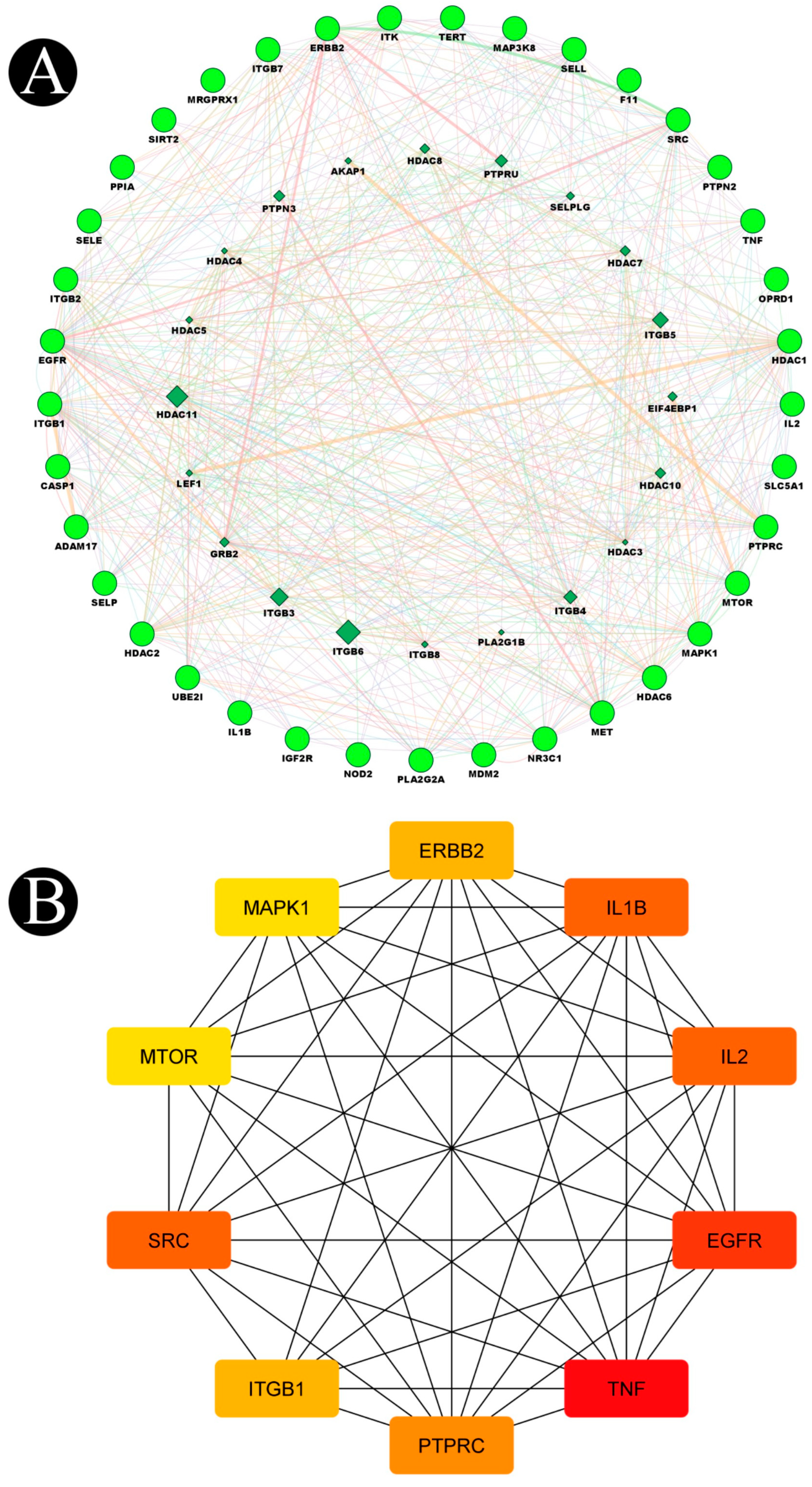
2.3. Construction of Protein–Protein Interaction Network (PPI) and Key Targets

Utilizing the GeneMANIA tool, we imported 37 target genes in order to obtain a PPI network that demonstrates the relationships between these 37 target genes and other genes in the network. In the results, the percentage represents the weight that is given to interaction relationships in the network. According to our results, 29.46% of the interactions between the targets in the network resulted in co-expressions, and 35.77% of them resulted in physical interactions. Furthermore, there was a relationship between co-localization and shared protein domains (Figure 8A). In Table 3, we provide the calculated average length of shortest paths to the three central nodes, betweenness centrality, closeness centrality, and degree of each node in the network. There were ten targets in the network which are organized in the order of high to low, according to the topology properties of the network, corresponding to EGFR, SRC, IL1B, IL2, PTPRC, ERBB2, ITGB1, MAPK1, MTOR, and TNF (Figure 8B). Biosurfactants may be able to prevent and treat listeriosis by targeting these ten targets, as they may be the key targets for biosurfactants.

2.4. Functional GO and KEGG Pathways

By using the Shiny GO 0.76.2 database analytical tool, the 37 intersected genes were enriched by GO and KEGG analysis. As a result of incorporating biological process (BP), molecular function (MF) and cellular component (CC) (Figure 9A–C), along with a p-value < 0.05, as screening conditions, a total of 1255 items were obtained pertaining to biological process, 149 items were obtained pertaining to molecular function, and 94 items were obtained pertaining to cellular composition. The hypothesis was put forth that biosurfactants could be involved in inhibiting listeriosis through the positive regulation of MAP kinase activity, protein kinase B signaling, ERK1 and ERK2 cascade, and the ERBB signaling pathway; positive regulation of protein serine/threonine kinase activity and leukocyte cell–cell adhesion; positive regulation of establishment of protein localization; and regulation of caveolin-mediated endocytosis via molecular functions such as integrin binding, phosphoprotein binding, protein tyrosine kinase activity, growth factor receptor binding, phosphatase binding, cytokine activity, NEDD8 transferase activity, and cadherin binding in cellular compartments such as membrane raft, membrane microdomain, focal adhesion, cell–substrate junction, myelin sheath, basal plasma membrane, and basal part of the cell. A total of 190 enrichment results were obtained from the KEGG pathway enrichment analysis. Among them Shigellosis, Yersinia infection, the ERBB signaling pathway, Th17 cell differentiation, the HIF-1 signaling pathway, the C-type lectin receptor signaling pathway, and bladder cancer pathways are closely associated with listeriosis and are in accordance with the enrichment results of GO. There was a significant abundance of KEGG pathways and gene pathways with p-values ≤ 0.05. Based on the Shiny GO platform, the first ten components were analyzed (Figure 9D). Based on the statistical analysis, ten proteins exhibited a high frequency of participation in the first 10 pathways, indicating that they played a major role in the enrichment pathway. The ten core proteins are EGFR, SRC, IL1B, IL2, PTPRC, ERBB2, ITGB1, MAPK1, MTOR, and TNF.

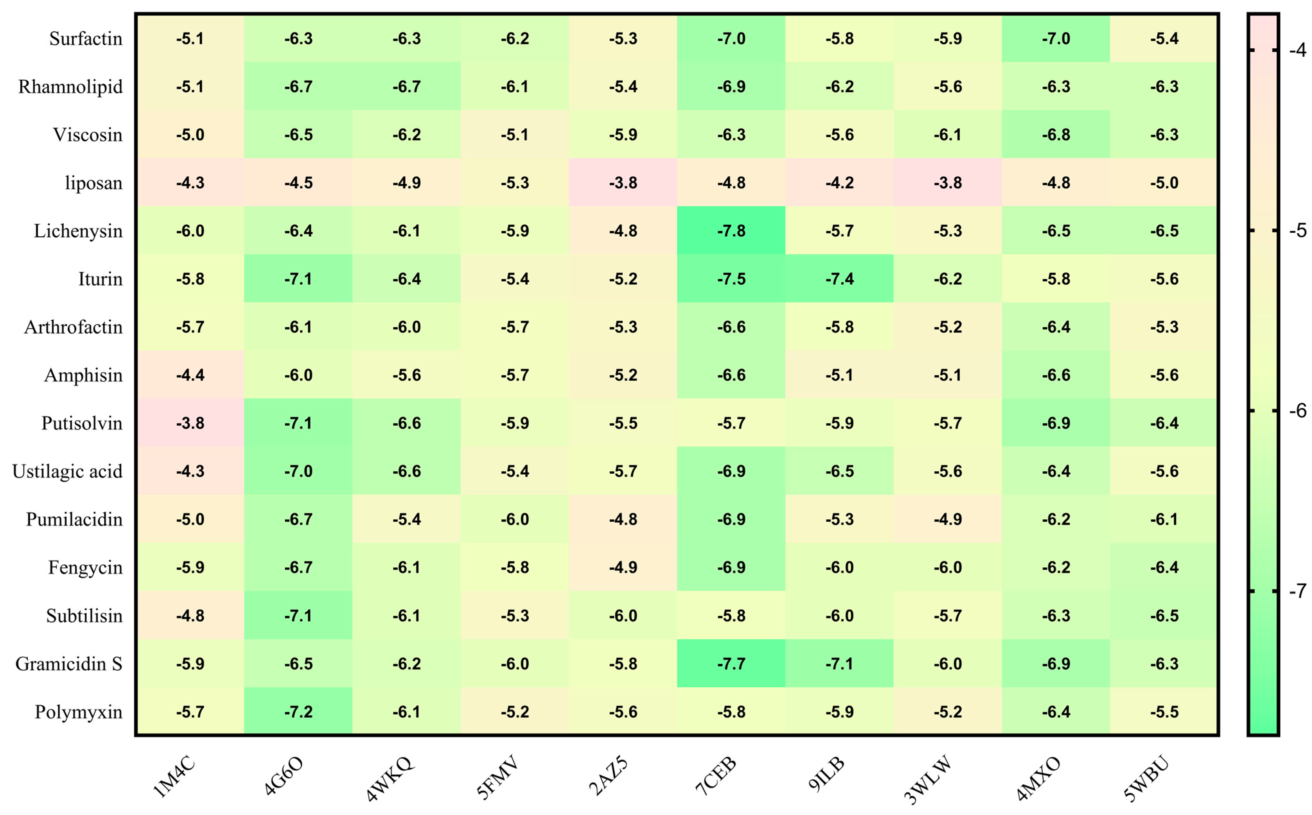
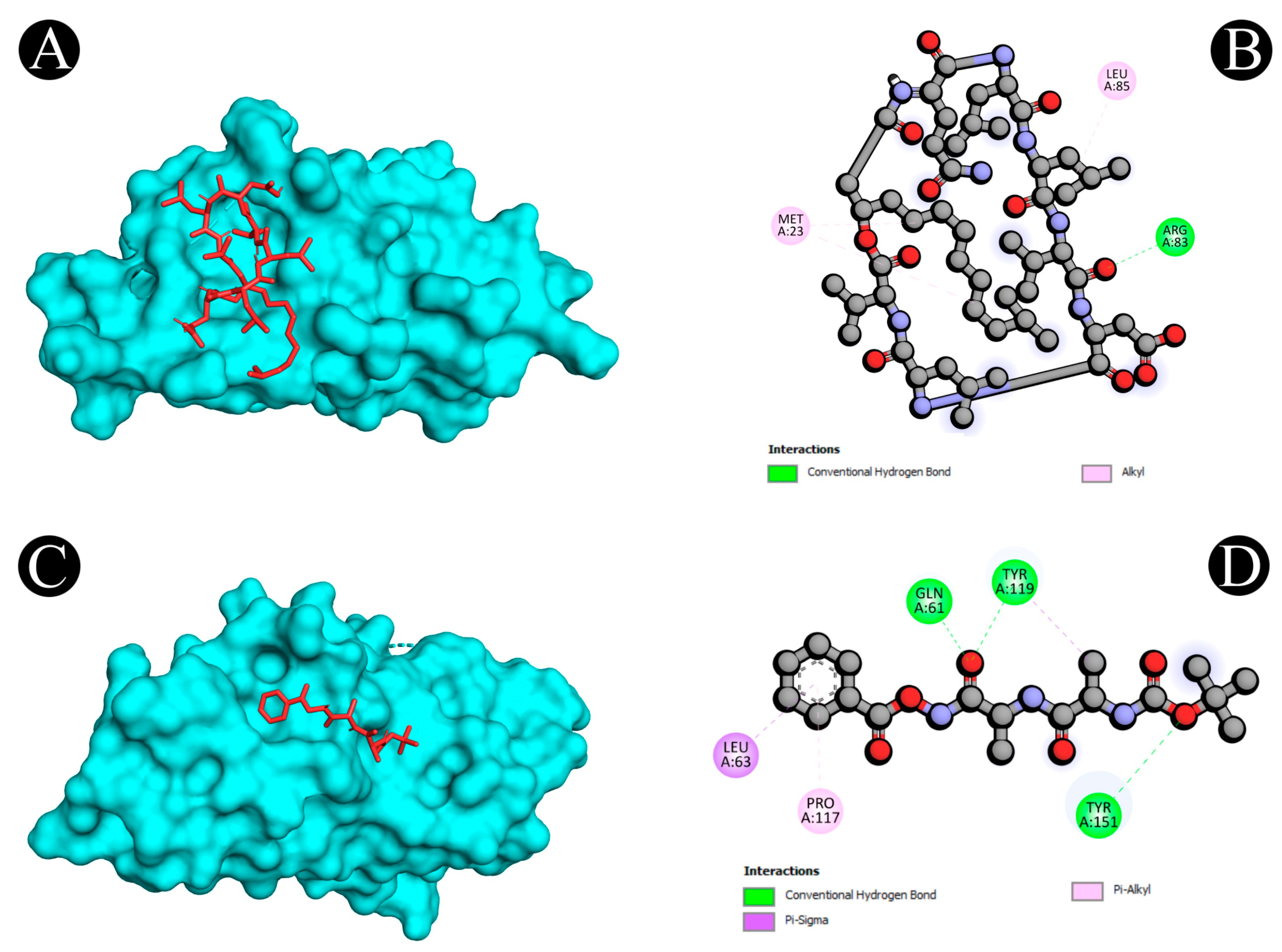
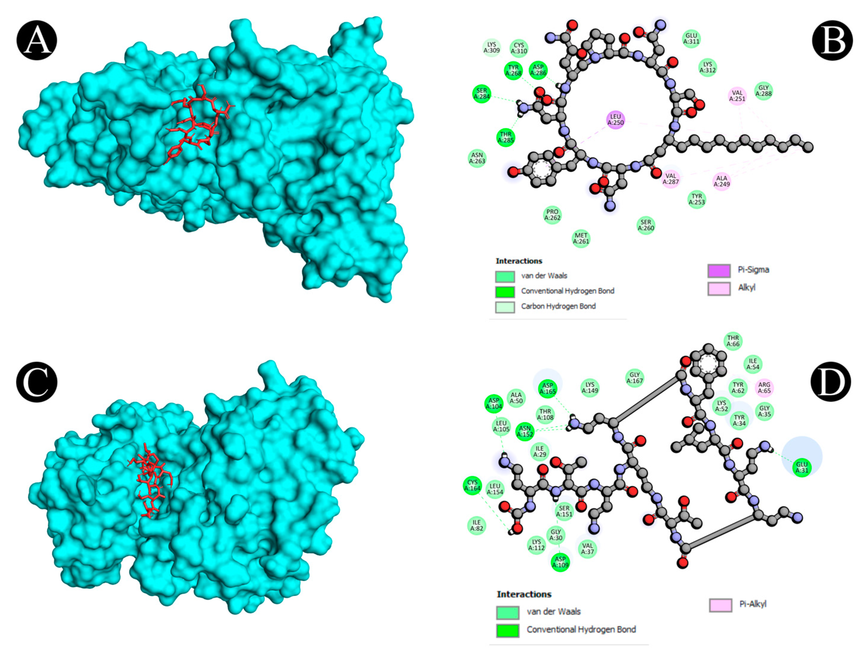
2.5. Molecular Docking

Virtual screening using molecular docking is a computational method for identifying potential leads against predefined targets. By employing this method, compounds with appreciable binding affinities and specific interactions with target proteins were identified. A docking analysis of all the biosurfactants revealed the presence of several compounds with a significant affinity for the respective target proteins (Figure 10). The highest binding affinity was found between IL2–lichenysin (−6.0 kJ/mol), MAPK1–polymyxin (−7.2 kJ/mol), EGFR–rhamnolipid (−6.7 kJ/mol), PTPRC–surfactin (−6.2 kJ/mol), TNF–subtilisin (−6.0 kJ/mol), ITGB1–lichenysin (−7.8 kJ/mol), IL1B–iturin (−7.4 kJ/mol), ERBB2–iturin (−6.2 kJ/mol), SRC–surfactin (−7.0 kJ/mol), and mTOR–subtilisin (−6.5 kJ/mol). These results suggest that the few selected biosurfactants have a significant level of binding efficiency with respective proteins, which may contribute to the development of a potential binding partner for selective proteins that could be used for drug development. The best biosurfactants observed occupying the active site in different ways can be seen in Figure 11, Figure 12, Figure 13, Figure 14 and Figure 15 and Table 4.

3. Discussion

Over the past 80 years, L. monocytogenes has been identified as a human pathogen that has the potential to cause disease. There has been a demographic shift in the last few decades, and there has been an explosion of immunosuppressive medications used for treating malignancies and managing organ transplants. This has led to an increasing number of immunocompromised individuals who are at an increased risk for listeriosis [34]. There is also the factor of changing consumer lifestyles which has resulted in less time available for food preparation, as well as an increase in the use of ready-to-eat food and take-away food. Food production and technology have drastically changed in recent years, resulting in foods with longer shelf-lives that are considered to be “Listeria-risk foods”; the bacteria multiply for a longer period of time, so the food does not undergo a listericidal process before consumption [35]. A high case-fatality rate of between 20 and 30% has been reported for listeriosis, compared to other common food-borne pathogens. During the past three decades, an epidemiological investigation has suggested that epidemic and sporadic listeriosis are primarily linked with the ingestion of foods or food products that are contaminated. While listeriosis is a rare food-borne illness in comparison to other food-borne illnesses, it is a very serious one. There is a high mortality rate associated with the disease even with adequate antibiotic treatment. Approximately, 90% of patients who have listeriosis are hospitalized, and many of them are in intensive care units. As a result, listeriosis is a serious problem around the world. Presently, listeriosis remains a significant challenge, and current treatment options are not adequate to combat the disease [36].
A number of biosurfactants have been tested for their antimicrobial activity, which has shown to be effective against different types of bacterial pathogens, such as Clostridium perfringens, Bacillus subtilis, Staphylococcus aureus, etc. (Gram-positive bacteria); Escherichia coli, Enterobacter aerogenes, Salmonella Typhimurium, etc. (Gram-negative bacteria); and Mucor sp., Phytophthora capsici, Fusarium graminearum, Botrytis cinerea, and Phytophthora infestans (pathogenic fungi) [37,38]. There is no complete understanding of how these compounds exert their antimicrobial activity; however, one of their proposed sites of action is the cell membrane since they are amphipathic and thus can interact with phospholipids [39].
Moreover, to date only one study is published on the anti-listeriosis activity of biosurfactants. According to the report published by de Araujo et al. [40], there is evidence that P. aeruginosa PA1 produces rhamnolipids that have antibacterial activity against L. monocytogenes ATCC 19112 and ATCC 7644. In addition to screening microbial surfactants for the treatment of listeriosis, the present study identifies a new therapeutic concept for further investigation into the mechanism of biosurfactants. For complex diseases such as listeriosis, in terms of predictive analysis, network pharmacology offers unique advantages [41]. Analyzing the PPI network, 10 core targets, namely EGFR, SRC, IL1B, IL2, PTPRC, ERBB2, ITGB1, MAPK1, MTOR, and TNF, for biosurfactants against listeriosis were screened out in the present study. EGF (epidermal growth factor) receptors are tyrosine kinases that bind ligands from the EGF family and activate signaling cascades in order to convert extracellular signals into appropriate cellular responses. In order to induce endocytosis, L. monocytogenes interacts with this tyrosine kinase receptor and E-cadherin, which might be a common pathogen invasion mechanism for the entry of L. monocytogenes [42]. SRC (proto-oncogene tyrosine-protein kinase Src) is reported as closely related with the pathogenicity of L. monocytogenes. As a result of L. monocytogenes infection, the heavy chain of non-muscle myosin IIA (NMHC-IIA) is phosphorylated at a specific tyrosine residue [43]. The pro-inflammatory cytokine interleukin-1beta (IL-1β) has been known to have a protective function against a variety of bacterial, fungal, and viral infections [44]. Bacterial pathogens are capable of exploiting host cell signaling pathways in order to adhere to or internalize host cells. A frequent molecular alteration involves the phosphorylation of tyrosine kinases on host non-receptors and receptors [45].
Bacteria can induce phosphorylation through direct contact with host cells or via soluble factors [45]. In order to infect host cells, L. monocytogenes reported activating the ERBB2/ERBB3 heterodimer pathway [46]. Pathogens such as Yersinia pseudotuberculosis, Staphylococcus aureus, Neisseria species, and enteroaggregative Escherichia coli exploit the Integrin subunit beta 1 (ITGβ1) receptor for adhesion to or invasion of mammalian cells [47,48,49,50,51]. Additionally, another protein, mTOR (mammalian target of rapamycin), plays a crucial role in Listeria entry. Cell growth, autophagy, and actin cytoskeleton development are controlled by mTOR, a serine/threonine kinase that responds to growth factor stimulation and nutrient, energy, or oxygen availability. Further to this, MAPK family proteins are also reported to play a crucial role in the infection of L. monocytogenes, and therefore, the treatment of infection with MAPK inhibitors is reported to affect the inhibition of bacterial internalization towards the host cells during infection [52]. Additionally, tumor necrosis factor (TNF) is a cytokine that has also been reported to play an active role in the susceptibility to L. monocytogenes infection. The dysregulation of TNF production and function has been reported to be associated with L. monocytogenes pathogenesis since it plays a crucial role in inflammation [53]. The production of TNF is shown to contribute to the protection against L. monocytogenes infection in an experimental model, and it can also stimulate the production of IFN-γ [54]. In experiments on severe combined immunodeficiency mice infected with L. monocytogenes, it is also found to be involved in a T-cell-independent pathway that leads to macrophage activation [55]. Moreover, the contribution of TNF to the pathogenesis of L. monocytogenes has been shown when TNF- or TNF Receptor 1 (R1)-deficient mice succumbed to the L. monocytogenes infection relatively quickly instead of recovering after a few days like the control mice [56]. All of these protein targets which come out as a result of the present study can therefore be considered to be most important targets for treating L. monocytogenes infections in the future.
Based on the GO analysis, possible targets for biosurfactants against listeriosis are involved in multiple important GO processes. Human epidermal growth factor receptor tyrosine kinases have a size of around 180 kDa and are a family of candidate tyrosine kinase receptors [57]. This family of receptors (EGFR, also termed ERBB1/HER1, ERBB2 or neu/HER2, ERBB3 or HER3, and ERBB4 or HER4) is characterized by dimerization with other receptors that are either of the same nature (homodimerization) or of a different nature (heterodimerization) [58,59,60]. Interestingly, ERBB receptors play important roles in cancer development [61] and are also found in signaling between bacteria and their hosts. It has been shown that the binding of Neisseria meningitidis to endothelial cells leads to the clustering of ERBB2 receptors, followed by phosphorylation of receptor tyrosine and activation of downstream signaling molecules, leading to actin polymerization and bacterial internalization [62]. The envelope glycoprotein B of the human cytomegalovirus (HCMV) binds to EGFR and promotes its tyrosine phosphorylation upon heterodimerization with ErbB3, resulting in virus entry and viral protein synthesis [63]. Likewise, L. monocytogenes and other bacteria also trigger the activation of the ErbB2/ErbB3 heterodimer signaling pathway in order to invade host cells [46]. One more key protein that is utilized in classical endocytic mechanisms to allow various particles to be internalized is clathrin or caveolin [64,65,66]. In order to move between epithelial cells, L. monocytogenes hijacks the caveolin–endocytic machinery. The activation of these processes is mediated by a subset of caveolar proteins (caveolin-1, cavin-2, and EHD2). Moreover, it is well-known that pathogens manipulate the post-translational modifications (PTMs) of host proteins to interfere with the normal functioning of host cells in various ways. A key target among these modifications is ubiquitin (UBI), ubiquitin-like proteins (UBLs), and neural precursor cell expressed developmentally downregulated protein 8 (NEDD8), which regulate pathways necessary for the host cell. The PTM modifiers, for instance, regulate the pathways that are crucial to the spread of infection, such as the entry, replication, propagation, or detection of the pathogen by the host, which have all been linked to these PTM modifiers. Different enzymes are involved in this biological process, as well as molecular functions, such as protein kinase binding activity, and the reactions are occurring in a variety of locations, such as the membrane and cytoplasm [67]. There are several biological processes involved in this process that are mediated by different enzymes, along with molecular functions such as protein kinase activity, and all of these reactions occur in multiple locations, such as the membrane and the cytoplasm of the cell. Based on these findings, we suggested that biosurfactants might have an impact on these processes as a result obtained from the GO analysis in this study.
According to the KEGG pathways analysis, potential targets of biosurfactants against listeriosis are significantly enriched in several important pathways, such as the ERBB signaling pathway, C-type lectin receptor signaling, Th17 cell differentiation and HIF-1 signaling pathway, etc. As described above, the ERBB signaling pathway plays an important role in listeriosis to invade bacteria in host cells. A key role that dendritic cells play in tailoring immune responses to pathogens is the expression of C-type lectin receptors (CLRs). Different signaling pathways are triggered by CLRs following the binding of pathogens, which are responsible for triggering the expression of specific cytokines that determine the fate of T cells during polarization. The activation of certain CLRs can be accompanied by the activation of nuclear factor-kappa B, while other CLRs can influence the activation of Toll-like receptors via signaling pathways. Depending on what signaling motifs are present in the cytoplasmic domains of CLRs, they can induce many different types of responses, including pro-inflammatory, antimicrobial, endocytic, phagocytic, and anti-inflammatory responses [68]. Th17 cells have been found to belong to a subgroup of cells that secrete IL-17, or IL-17A, a component of the inflammatory response. Together with Thl, Th2, and Tregs, Th17 cells make up four subsets of CD4+T cells. Under the stimulation of IL-6 and TGF-β, Th17 cells are differentiated by Th0 cells. A key role that they play is in the regulation of the immune system and in the defense of the host [69]. One of the most important transcription factors in maintaining oxygen homeostasis is hypoxia-inducible factor 1 (HIF-1), which is one of the many transcription factors involved in the process. There are two subunits of this protein: an inducibly expressed HIF-1alpha subunit and a constitutively expressed HIF-1beta subunit. In the presence of normoxia, HIF-1 alpha undergoes a process of hydroxylation at specific prolyl residues in order to undergo an immediate ubiquitination and subsequently be degraded by the proteasome. Contrary to this, under hypoxia, the alpha subunit of HIF-1 becomes stable and begins to interact with coactivators such as p300/CBP in order to modulate its transcriptional activity. As a master regulator of hypoxia-inducible genes, HIF-1 regulates a number of hypoxia-inducible genes under hypoxic conditions. The HIF-1 gene family encodes proteins that play a key role in improving oxygen delivery and enhancing cells’ adaptive responses to oxygen deprivation. It is important to note that nitric oxide and several growth factors are also stimulatory factors that can induce HIF-1, so it is not only in response to decreased oxygen availability that it is induced but also in response to other stimulants [70].
Furthermore, we performed docking experiments for the biosurfactants and the ten Hub genes in accordance with the “compounds-targets networks”. Additionally, the results of docking analyses confirmed our results and showed that lichenysin, iturin, surfactin, rhamnolipid, subtilisin, and polymyxin bind stably to the active pockets of target proteins. Therefore, these compounds could be considered for use as a potential treatment for listeriosis by inhibiting proteins such as, IL2, MAPK1, SRC, EGFR, PTPRC, TNF, IL1B, and ERBB2. Taking into account the role of network pharmacology, the present study examines the active biosurfactants, their potential targets, and their associated pathways, as they pertain to the treatment of listeriosis, which provides a theoretical foundation for further experimental studies. In consideration of the limitations of network pharmacology, it is only through data mining that the basic pharmacological mechanisms for the treatment of listeriosis can be identified. Currently, network pharmacology relies on a variety of databases to support the analysis of bioactive properties. Due to the fact that there are many different information sources and experimental data in databases, it is inevitable that they will show discrepancies. In spite of the fact that we have presented some interesting results, further research and clinical trials are required to evaluate the potential of biosurfactants to validate their usage as a prevention measure against Listeria and other food-borne diseases.

4. Materials and Methods

4.1. Biosurfactants Target Prediction

In the present study, we selected biosurfactants that have been reported to possess antimicrobial activity in the literature. The information about their structure, molecular weights, and canonical smiles and the corresponding sdf files were obtained from the PubChem database (http://pubchem.ncbi.nlm.nih.gov/), accessed on 13 September 2022. Using a public database, SwissTargetPrediction, and a STITCH database, we predicted the target of bioactive microbial biosurfactants based on the species of Homo sapiens as the only species targeted for the study. To complete the process of standardizing the target names, UniProtKB database (https://www.uniprot.gov/) was used [71,72].

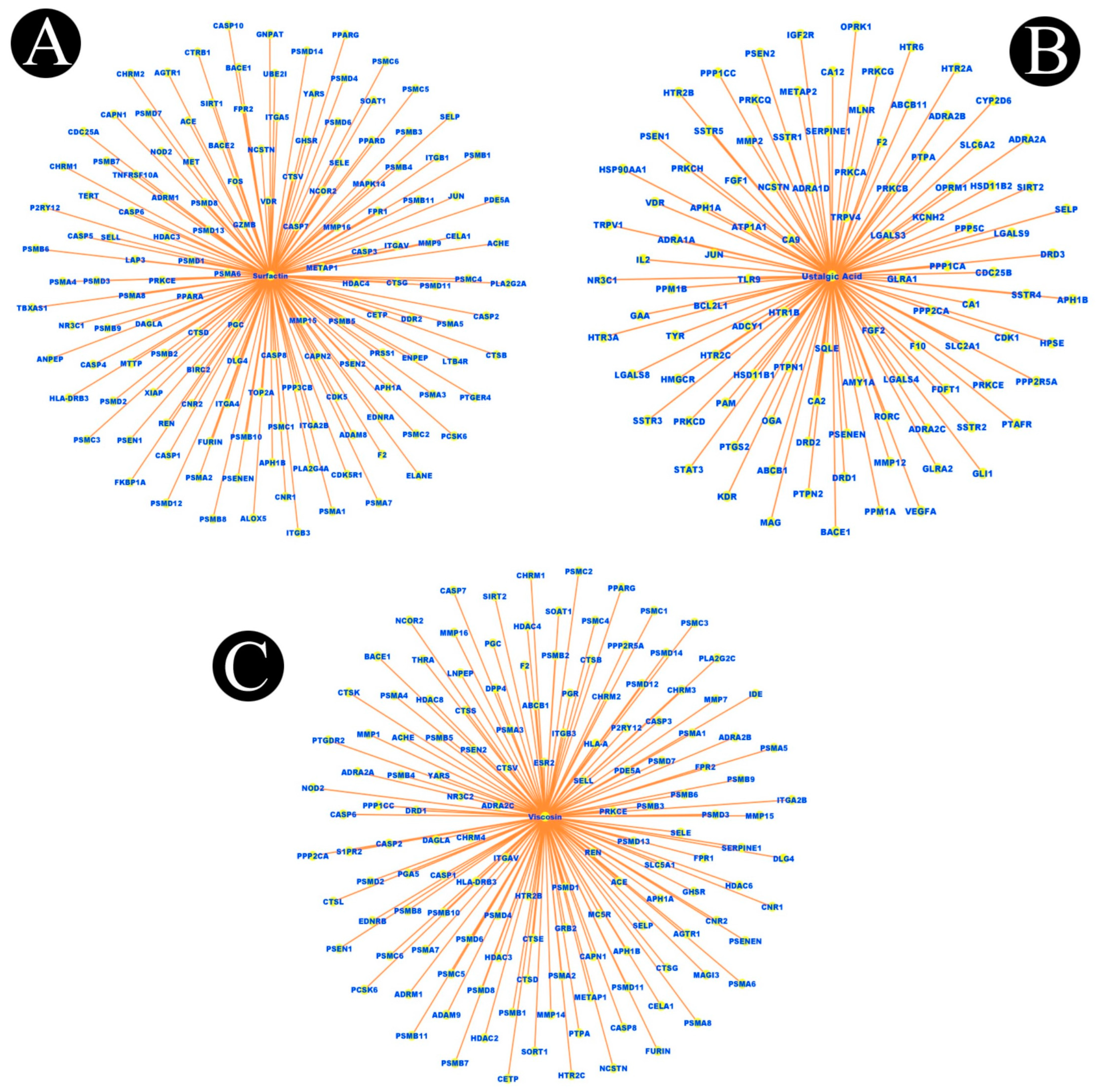
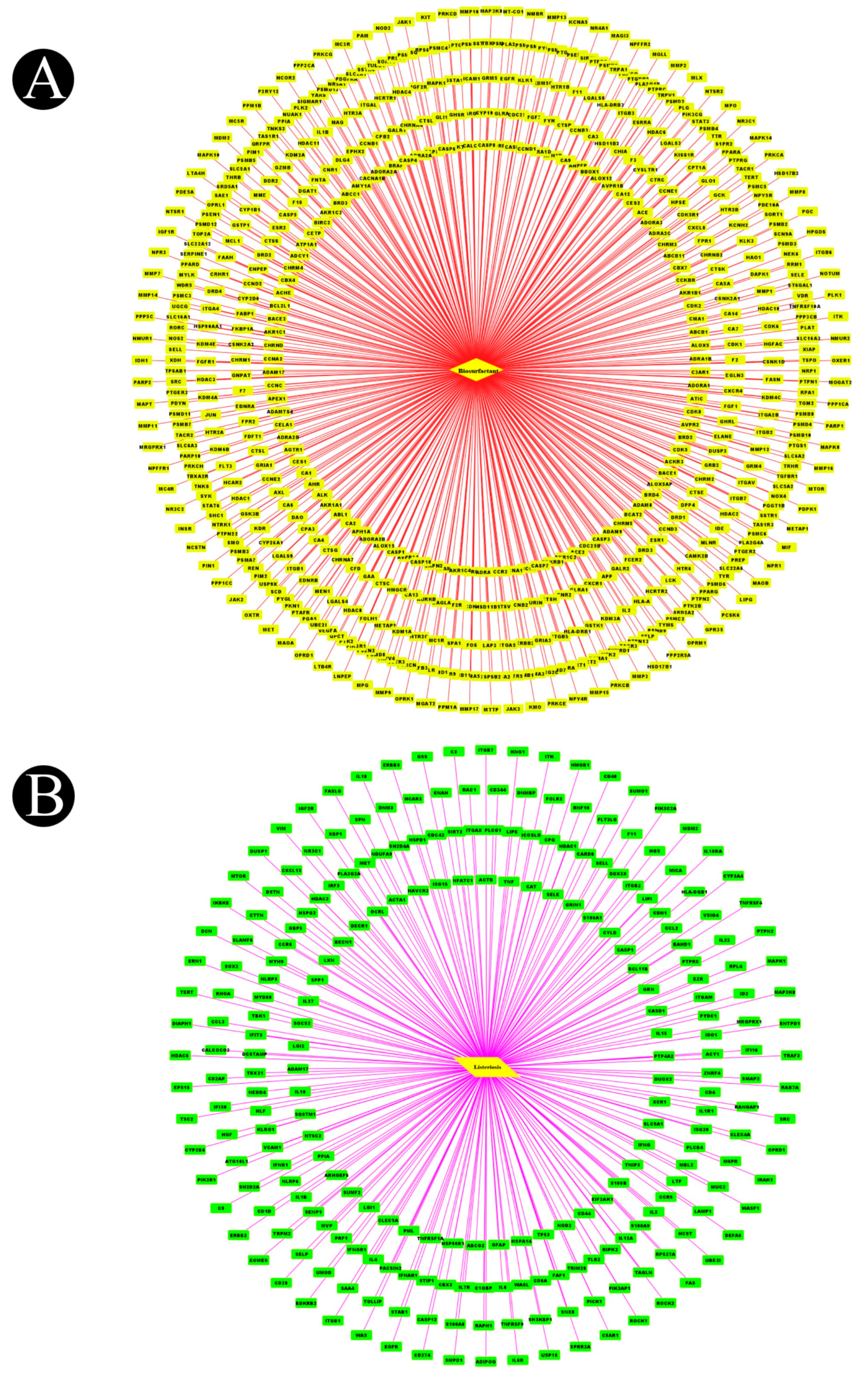
4.2. Network Construction for Compound–Targets

The biosurfactants that were collected and the effective targets that were identified were analyzed by using Cytoscape 3.9.1 software (http://www.cytoscape.org/) for the creation of a compound–target network. To measure the topology scores of the nodes in the network, we used the CytoNCA plugin (v2.1.6), which measures betweenness, closeness, and the centrality of subgraphs of the nodes in each graph. Accordingly, the option “without weight” was selected [73].

4.3. Protein Targets Associated with Listeriosis

A search for targets related to listeriosis was conducted by using keywords such as “listeriosis” and “listeriosis infection” in the GeneCards database (https://www.genecards.org/) [74], Online Mendelian Inheritance in Man database (OMIM, https://omim.org/), and gene–disease associations database (DisGeNET, http://www.disgenet.org/) accessed on 20 September 2022 [75,76], and the Universal Protein database (UniProt, https://www.UniProt.org/) was used to convert the target protein name to a gene name [77]. All listeriosis targets were acquired after repetitive targets were removed.

4.4. Target Screening and Network Construction for Biosurfactants and Listeriosis

In order to detect the core target of the biosurfactants for the treatment of listeriosis, the prediction results of the biosurfactants were matched with the search results of the listeriosis related targets, and the target with the most overlap was selected as the core target. Using the FunRich Tool version 3.1.3, we mapped the targets that biosurfactants and listeriosis share. A Venn diagram was drawn in order to visualize the process. In order to construct a common target network, the Cytoscape software version 3.9.1 was used.

4.5. Protein–Protein Interaction Network (PPI) Construction and Target Identification

The GeneMANIA tool, in addition to constructing a PPI network, is able to find a series of genes related to the input gene based on a large volume of function-related data and analyze the interaction between these genes, based on their co-localization and co-expression [78]. In the present study, GeneMANIA was used to build a protein–protein interaction network related to the cross-gene interactions between biosurfactants and listeriosis based on the analysis of the cross-gene analysis. As a result of the GeneMANIA analysis, we were able to obtain not only information about the relationships between the input cross genes, but also information about the relationships between other closely related targets as well. Accordingly, we label this new set of genes predicted to be biosurfactant targets for listeriosis in the following analysis. The topology parameters of the PPI network were calculated by using Network Analyzer in order to identify the main nodes of the network and the key proteins across the network, while the degree of centrality of the network (betweenness, closeness, and subgraph) was calculated by using CytoNCA.

4.6. Analysis of Gene Ontology (GO) Function and Kyoto Encyclopedia of Genes and Genomes (KEGG) Pathway Enrichment

Based on the results obtained from the above screening, the target of biosurfactants that shared a common target with listeriosis was imported into the Enrichr database (https://maayanlab.cloud/Enrichr/). An analysis was conducted to explore the enrichment of GO functions and pathways within the human genome based on the species H. sapiens. As part of the functional analysis of GO, we considered biological processes (BPs), cellular components (CCs), and molecular functions (MFs). The data were visualized as histograms and bubble charts, using the SRPLOT application (http://bioinformatics.com.cn/srplot), as well as the ShinyGO 0.76.2 database (https://bioinformatics.sdstate.edu/go/).

4.7. Construction of Target-Path/Functional Networks

In order to perform a deeper analysis of the signal pathways, biological processes, and molecular functions, ten representative pathways were screened. As part of the construction of the target pathway/functional network, the ShinyGO 0.76.2 database was used (https://bioinformatics.sdstate.edu/go/). Through the use of enrichment analysis, potential targets of biosurfactants for treating listeriosis, biological processes, and signaling pathways were defined by nodes in the network, and the interactions between these nodes were defined by edges.

4.8. Findings of Hub Genes

We tested the PPI network obtained from STRING, using the CytoHubba plugin of Cytoscape. This plugin was used to analyze the core regulatory genes of the PPI network, as well as the identification of key targets within the network. As part of the screening process, the core compounds were tested under the assumption that the “Degree” parameter of the node in the “active ingredient target-disease” network was above the mean. A virtual screening approach based on molecular docking was carried out between the biosurfactants and identified hub genes.

4.9. Molecular Docking Analysis

4.9.1. Protein and Ligand Structures

To study the interaction of selected biosurfactants with the identified protein targets in the present study, a molecular docking analysis was performed. Crystal structures of identified hub target proteins such as TNF (PDB ID: 2AZ5), EGFR (PDB ID: 4WKQ), IL1B (PDB ID: 9ILB), IL2 (PDB ID: 1M4C), SRC (PDB ID: 4MXO), PTPRC (PDB ID: 5FMV), ITGB1 (PDB ID: 7CEB), ERBB2 (PDB ID: 3WLW), MTOR (PDB ID: 5WBU), and MAPK1 (PDB ID: 4G6O) were retrieved from RCSB PDB. Two-dimensional structures of selected biosurfactants were retrieved from the well-known organic compound database PubChem in SDF format. These compounds were then converted into three-dimensional structures, using Avogadro, and saved in PDB format [79].

4.9.2. Ligand Preparation

For the preparation of input files for docking, Autodock software 1.5.7 [80] was used. The structures were minimized with MMFF94 force field. Steepest Descent algorithm was used for optimization with a total of 5000 steps. During minimization, the structure was updated every 1 step, and minimization was terminated when the energy difference is less than 0.1. Energy-minimized structures were saved in PDB format.

4.9.3. Prediction of Binding Site

The binding sites of all protein structures were predicted by using Discovery Studio v. 21.1.0.20298 [81]. The pocket with the highest score was considered to be the most probable binding site of the proteins.

4.9.4. Molecular Docking

Three-dimensional structures of proteins derived from RCSB PDB were prepared for molecular docking, using AutoDock Tools [80]. Before the docking experiment, we used AutoDock Tools software to preprocess the crystal structure of the target proteins, including removing excess protein chains, ligands, and water molecules, and structures were optimized by adding missing hydrogen atoms. Structure files (PDB format) of all biosurfactants were docked separately against the protein structures, using molecular docking software AutoDock 4.2.6. [80]. All the parameters used for the docking of biosurfactants with the proteins were kept the same, except for the grid center differed for each protein inside the grid box. Auto Grid was used for the preparation of the grid map, using a grid box. The grid size was set to 90 × 90 × 90xyz points for all proteins. Grid spacing was kept to 0.500 Å for all the proteins. The grid center for 2AZ5 was designated at dimensions (x, y, and z), −14.888, 68.771, and 32.730; for 4WKQ at (x, y, and z), −2.761, 201.806, and 26.195; for 9ILB at (x, y and z), −13.592, 13.466, and 0.200; for 1M4C at (x, y, and z), 14.170, −10.108, and 20.056; for 4MXO at (x, y, and z), 9.193, −33.735, and −7.984; for 5MFV at (x, y, and z), 30.052, −18.946, and 32.074; for 7CEB at (x, y, and z), 43.112, 46.089, and −1.124; for 3WLW at (x, y, and z), 36.088, 26.277, and −20.954; for 5WBU at (x, y, and z), 11.014, −18.240, and −30.383; and for 4G6O at (x, y, and z), 14.790, 5.794, and 17.485. The grid box is cantered in such a way that it encloses the entire binding site of each protein and provides enough space for the translation and rotation of ligands. The generated docked conformation was ranked by predicted binding energy, and the topmost binding energy docked conformation was analyzed through the use of the PyMOL and Discovery Studio Visualizer [81]. By using the Discovery Studio Visualizer, it was possible to explore the types of interactions, the participating residuals, and the atomic coordinates involved.

5. Conclusions

The purpose of this study was to investigate the molecular mechanisms of biosurfactants to treat listeriosis, using a network pharmacology approach and molecular docking. As a result of the current study, biosurfactants have been found to be capable of targeting multiple proteins and regulating multiple signaling pathways induced by L. monocytogenes infection, indicating that biosurfactants may have a regulatory effect on listeriosis caused by L. monocytogenes. Furthermore, our findings indicate that IL2, MAPK1, EGFR, PTPRC, TNF, ITGB1, IL1B, ERBB2, and mTOR genes may be viable therapeutic targets for the reduction of listeriosis. In addition to providing an alternative or complementary therapy for the treatment of listeriosis, these findings lay the foundation for future studies. There are, however, some limitations to this study, as pharmacological and clinical research still needs to be conducted to verify our findings. A groundwork has been laid for further study of biosurfactants’ protective mechanisms and drug discovery applications based on network pharmacology.

Author Contributions

Conceptualization, M.A. and M.P. (Mitesh Patel); methodology, M.S. (Mejdi Snoussi), E.N., A.J.S., S.H. and S.A.A.; validation, M.P. (Mirav Patel), M.S. (Manojkumar Sachidanandan), M.S. (Mejdi Snoussi), F.B. and R.B.; formal analysis, M.P. (Mitesh Patel), M.P. (Mirav Patel), M.A., M.S. (Mejdi Snoussi), F.B. and R.B.; investigation, S.A.A., A.J.S., M.S. (Manojkumar Sachidanandan) and A.M.A.; data curation, M.P. (Mitesh Patel), F.B., A.M.A., S.A.A., S.H. and E.N.; writing—original draft preparation, M.P. and M.A.; writing—review and editing, F.B., M.S. (Mejdi Snoussi), M.S. (Manojkumar Sachidanandan) and E.N.; software, M.P. (Mirav Patel), M.A. and M.P. (Mitesh Patel); visualization, M.P. (Mirav Patel), M.P. (Mitesh Patel) and A.J.S.; supervision, M.A. and M.P. (Mitesh Patel); project administration, M.A. All authors have read and agreed to the published version of the manuscript.

Funding

This research has been funded by Scientific Research Deanship at University of Ha’il-Saudi Arabia through project number RG-21093.

Institutional Review Board Statement

Not applicable.

Informed Consent Statement

Not applicable.

Data Availability Statement

All data generated or analyzed during this study are included in this article.

Acknowledgments

Authors are thankful to Scientific Research Deanship at University of Ha’il-Saudi Arabia for supporting this study through project number RG-21093.

Conflicts of Interest

The authors declare no conflict of interest. The funders had no role in the design of the study; in the collection, analyses, or interpretation of data; in the writing of the manuscript, or in the decision to publish the results.

References

  1. Ge, H.; Wang, Y.; Zhao, X. Research on the Drug Resistance Mechanism of Foodborne Pathogens. Microb. Pathog. 2022, 162, 105306. [Google Scholar] [CrossRef] [PubMed]
  2. Nyachuba, D.G. Foodborne Illness: Is It on the Rise? Nutr. Rev. 2010, 68, 257–269. [Google Scholar] [CrossRef] [PubMed]
  3. White, D.G.; Zhao, S.; Simjee, S.; Wagner, D.D.; McDermott, P.F. Antimicrobial Resistance of Foodborne Pathogens. Microbes Infect. 2002, 4, 405–412. [Google Scholar] [CrossRef]
  4. Jones, G.S.; Bussell, K.M.; Myers-Morales, T.; Fieldhouse, A.M.; Bou Ghanem, E.N.; D’Orazio, S.E. Intracellular Listeria monocytogenes comprises a minimal but vital fraction of the intestinal burden following foodborne infection. Infect. Immun. 2015, 83, 3146–3156. [Google Scholar] [PubMed] [Green Version]
  5. Rouquette, C.; Berche, P. The pathogenesis of infection by Listeria monocytogenes. Microbiologia 1996, 12, 245–258. [Google Scholar]
  6. Kazmierczak, M.J.; Wiedmann, M.; Boor, K.J. Alternative sigma factors and their roles in bacterial virulence. Microbiol. Mol. Biol. Rev. 2005, 69, 527–543. [Google Scholar] [CrossRef] [Green Version]
  7. Mansfield, B.E.; Freitag, N.E. Listeria monocytogenes pathogenesis: Exploration of alternative hosts. In Abstracts of the General Meeting of the American Society for Microbiology; American Society for Microbiology: Washington, DC, USA, 2003; Volume 103, p. B-186. [Google Scholar]
  8. Gandhi, M.; Chikindas, M.L. Listeria: A Foodborne Pathogen That Knows How to Survive. Int. J. Food Microbiol. 2007, 113, 1–15. [Google Scholar]
  9. Voronina, O.L.; Tartakovsky, I.S.; Yuyshuk, N.D.; Ryzhova, N.N.; Aksenova, E.I.; Kunda, M.S.; Kutuzova, A.V.; Melkumyan, A.R.; Karpova, T.I.; Gruzdeva, O.A.; et al. Analysis of Sporadic Cases of Invasive Listeriosis in a Metropolis. J. Microbiol. Epidemiol. Immun. 2020, 97, 546–555. [Google Scholar] [CrossRef]
  10. Noor, F.; Noor, A.; Ishaq, A.R.; Farzeen, I.; Saleem, M.H.; Ghaffar, K.; Aslam, M.F.; Aslam, S.; Chen, J.-T. Recent Advances in Diagnostic and Therapeutic Approaches for Breast Cancer: A Comprehensive Review. Curr. Pharm. Des. 2021, 27, 2344–2365. [Google Scholar] [CrossRef]
  11. Noor, F.; Saleem, M.H.; Chen, J.-T.; Javed, M.R.; Al-Megrin, W.A.; Aslam, S. Integrative Bioinformatics Approaches to Map Key Biological Markers and Therapeutic Drugs in Extramammary Paget’s Disease of the Scrotum. PLoS ONE 2021, 16, e0259408. [Google Scholar]
  12. Kashif, A.; Rehman, R.; Fuwad, A.; Shahid, M.K.; Dayarathne, H.N.P.; Jamal, A.; Aftab, M.N.; Mainali, B.; Choi, Y. Current advances in the classification, production, properties and applications of microbial biosurfactants—A critical review. Adv. Colloid Inter. Sci. 2022, 306, 102718. [Google Scholar] [CrossRef] [PubMed]
  13. Patel, M.; Siddiqui, A.J.; Hamadou, W.S.; Surti, M.; Awadelkareem, A.M.; Ashraf, S.A.; Alreshidi, M.; Snoussi, M.; Rizvi, S.M.D.; Bardakci, F. Inhibition of Bacterial Adhesion and Antibiofilm Activities of a Glycolipid Biosurfactant from Lactobacillus Rhamnosus with Its Physicochemical and Functional Properties. Antibiotics 2021, 10, 1546. [Google Scholar] [CrossRef] [PubMed]
  14. Adnan, M.; Siddiqui, A.J.; Hamadou, W.S.; Ashraf, S.A.; Hassan, M.I.; Snoussi, M.; Badraoui, R.; Jamal, A.; Bardakci, F.; Awadelkareem, A.M. Functional and Structural Characterization of Pediococcus pentosaceus-Derived Biosurfactant and Its Biomedical Potential against Bacterial Adhesion, Quorum Sensing, and Biofilm Formation. Antibiotics 2021, 10, 1371. [Google Scholar] [CrossRef]
  15. Magalhães, L.; Nitschke, M. Antimicrobial Activity of Rhamnolipids against Listeria monocytogenes and Their Synergistic Interaction with Nisin. Food Control 2013, 29, 138–142. [Google Scholar] [CrossRef] [Green Version]
  16. Boezio, B.; Audouze, K.; Ducrot, P.; Taboureau, O. Network-based Approaches in Pharmacology. Mol. Inform. 2017, 36, 1700048. [Google Scholar] [CrossRef] [PubMed] [Green Version]
  17. Zhou, Z.; Chen, B.; Chen, S.; Lin, M.; Chen, Y.; Jin, S.; Chen, W.; Zhang, Y. Applications of Network Pharmacology in Traditional Chinese Medicine Research. Evid. Based. Complement. Altern. Med. 2020, 2020, 1646905. [Google Scholar] [CrossRef] [PubMed]
  18. Park, M.; Park, S.-Y.; Lee, H.-J.; Kim, C.E. A Systems-Level Analysis of Mechanisms of Platycodon grandiflorum Based on a Network Pharmacological Approach. Molecules 2018, 23, 2841. [Google Scholar] [CrossRef] [Green Version]
  19. Ongena, M.; Jacques, P. Bacillus lipopeptides: Versatile weaponsfor plant disease biocontrol. Trends. Microbiol. 2007, 16, 115–125. [Google Scholar] [CrossRef]
  20. Pathak, K.V.; Keharia, H. Application of extracellular lipopeptide bio-surfactant produced by endophytic Bacillus subtilis K1 isolated from aerial rootsof banyan (Ficus benghalensis) in microbially enhanced oil recovery (MEOR). 3 Biotech 2014, 4, 41–48. [Google Scholar] [CrossRef] [Green Version]
  21. Rahman, K.S.M.; Thahira-Rahman, J.; McClean, S.; Marchant, R.; Banat, I.M. Rhamnolipid biosurfactants production by strains of Pseudomonas aeruginosa using low cost materials. Biotechnol. Prog. 2002, 18, 1277–1281. [Google Scholar] [CrossRef] [Green Version]
  22. Alsohim, A.S.; Taylor, T.B.; Barrett, G.A.; Gallie, J.; Zhang, X.X.; Altamirano-Junqueira, A.E.; Johnson, L.J.; Rainey, P.B.; Jackson, R.W. The biosurfactant viscosin produced by P seudomonas fluorescens SBW 25 aids spreading motility and plant growth promotion. Environ. Microbiol. 2014, 16, 2267–2281. [Google Scholar] [CrossRef] [PubMed]
  23. Rufino, R.D.; Luna, J.M.; Sarubbo, L.A.; Rodrigues, L.R.M.; Teixeira, J.A.C.; Campos-Takaki, G.M. Antimicrobial and anti-adhesive potential of a biosurfactant Rufisan produced by Candida lipolytica UCP 0988. Colloids Surf. B Biointerfaces 2011, 84, 1–5. [Google Scholar] [CrossRef] [PubMed] [Green Version]
  24. Yeak, K.Y.C.; Perko, M.; Staring, G.; Fernandez-Ciruelos, B.M.; Wells, J.M.; Abee, T.; Wells-Bennik, M.H. Lichenysin Production by Bacillus licheniformis Food Isolates and Toxicity to Human Cells. Front. Microbiol. 2022, 13, 831033. [Google Scholar] [CrossRef] [PubMed]
  25. Arrebola, E.; Jacobs, R.; Korsten, L. Iturin A is the principal inhibitor inthe biocontrol activity of Bacillus amyloliquefaciens PPCB004 against posthar-vest fungal pathogens. J. Appl. Microbiol. 2010, 108, 386–395. [Google Scholar] [CrossRef]
  26. Morikawa, M.; Daido, H.; Takao, T.; Murata, S.; Shimonishi, Y.; Imanaka, T. A new lipopeptide biosurfactant produced by Arthrobacter sp. strain MIS38. J. Bacteriol. 1993, 175, 6459–6466. [Google Scholar] [CrossRef] [Green Version]
  27. Groboillot, A.; Portet-Koltalo, F.; Le Derf, F.; Feuilloley, M.J.; Orange, N.; Poc, C.D. Novel application of cyclolipopeptide amphisin: Feasibility study as additive to remediate polycyclic aromatic hydrocarbon (PAH) contaminated sediments. Int. J. Mol. Sci. 2011, 12, 1787–1806. [Google Scholar] [CrossRef] [Green Version]
  28. Kuiper, I.; Lagendijk, E.L.; Pickford, R.; Derrick, J.P.; Lamers, G.E.; Thomas-Oates, J.E.; Lugtenberg, B.J.; Bloemberg, G.V. Characterization of two Pseudomonas putida lipopeptide biosurfactants, putisolvin I and II, which inhibit biofilm formation and break down existing biofilms. Mol. Microbiol. 2004, 51, 97–113. [Google Scholar] [CrossRef]
  29. Hewald, S.; Josephs, K.; Bölker, M. Genetic analysis of biosurfactant production in Ustilago maydis. Appl. Environ. Microbiol. 2005, 71, 3033–3040. [Google Scholar] [CrossRef] [Green Version]
  30. Saggese, A.; Culurciello, R.; Casillo, A.; Corsaro, M.M.; Ricca, E.; Baccigalupi, L. A marine isolate of Bacillus pumilus secretes a pumilacidin active against Staphylococcus aureus. Mar. Drugs 2018, 16, 180. [Google Scholar] [CrossRef] [Green Version]
  31. Gudiña, E.J.; Fernandes, E.C.; Rodrigues, A.I.; Teixeira, J.A.; Rodrigues, L.R. Biosurfactant production by Bacillus subtilis using corn steep liquor as culture medium. Front. Microbiol. 2015, 6, 59. [Google Scholar]
  32. Abdelhamid, H.N.; Khan, M.S.; Wu, H.F. Graphene oxide as a nanocarrier for gramicidin (GOGD) for high antibacterial performance. RSC Adv. 2014, 4, 50035–50046. [Google Scholar] [CrossRef]
  33. Eras-Muñoz, E.; Farré, A.; Sánchez, A.; Font, X.; Gea, T. Microbial biosurfactants: A review of recent environmental applications. Bioengineered 2022, 13, 12365–12391. [Google Scholar] [CrossRef] [PubMed]
  34. Bodro, M.; Paterson, D.L. Listeriosis in Patients Receiving Biologic Therapies. Eur. J. Clin. Microbiol. Infect. Dis. 2013, 32, 1225–1230. [Google Scholar] [CrossRef] [PubMed]
  35. Allerberger, F.; Wagner, M. Listeriosis: A Resurgent Foodborne Infection. Clin. Microbiol. Infect. 2010, 16, 16–23. [Google Scholar] [CrossRef] [PubMed]
  36. Melo, J.; Andrew, P.W.; Faleiro, M.L. Listeria monocytogenes in Cheese and the Dairy Environment Remains a Food Safety Challenge: The Role of Stress Responses. Food. Res. Int. 2015, 67, 75–90. [Google Scholar] [CrossRef]
  37. Benincasa, M.; Abalos, A.; Oliveira, I.; Manresa, A. Chemical Structure, Surface Properties and Biological Activities of the Biosurfactant Produced by Pseudomonas aeruginosa LBI from Soapstock. Antonie Van Leeuwenhoek 2004, 85, 1–8. [Google Scholar] [CrossRef]
  38. Haba, E.; Pinazo, A.; Jauregui, O.; Espuny, M.J.; Infante, M.R.; Manresa, A. Physicochemical Characterization and Antimicrobial Properties of Rhamnolipids Produced by Pseudomonas aeruginosa 47T2 NCBIM 40044. Biotechnol. Bioeng. 2003, 81, 316–322. [Google Scholar] [CrossRef] [Green Version]
  39. Ortiz, S.; López-Alonso, V.; Rodríguez, P.; Martínez-Suárez, J.V. The Connection between Persistent, Disinfectant-Resistant Listeria Monocytogenes Strains from Two Geographically Separate Iberian Pork Processing Plants: Evidence from Comparative Genome Analysis. Appl. Environ. Microbiol. 2016, 82, 308–317. [Google Scholar] [CrossRef] [Green Version]
  40. De Araujo, L.V.; Guimarães, C.R.; da Silva Marquita, R.L.; Santiago, V.M.J.; de Souza, M.P.; Nitschke, M.; Freire, D.M.G. Rhamnolipid and Surfactin: Anti-Adhesion/Antibiofilm and Antimicrobial Effects. Food Control 2016, 63, 171–178. [Google Scholar] [CrossRef]
  41. Chen, W.; Ding, C.; Yu, J.; Wang, C.; Wan, L.; Hu, H.; Ma, J. Wuzi Yanzong Pill—Based on Network Pharmacology and In Vivo Evidence—Protects Against Spermatogenesis Disorder via the Regulation of the Apoptosis Pathway. Front. Pharmacol. 2020, 11, 592827. [Google Scholar] [CrossRef]
  42. Yap, P.-C.; MatRahim, N.-A.; AbuBakar, S.; Lee, H.Y. Antilisterial Potential of Lactic Acid Bacteria in Eliminating Listeria monocytogenes in Host and Ready-to-Eat Food Application. Microbiol. Res. 2021, 12, 17. [Google Scholar] [CrossRef]
  43. Almeida, M.T.; Mesquita, F.S.; Cruz, R.; Osório, H.; Custódio, R.; Brito, C.; Vingadassalom, D.; Martins, M.; Leong, J.M.; Holden, D.W. Src-Dependent Tyrosine Phosphorylation of Non-Muscle Myosin Heavy Chain-IIA Restricts Listeria Monocytogenes Cellular Infection. J. Biol. Chem. 2015, 290, 8383–8395. [Google Scholar] [CrossRef] [PubMed] [Green Version]
  44. Sahoo, M.; Ceballos-Olvera, I.; del Barrio, L.; Re, F. Role of the Inflammasome, IL-1. Sci. World J. 2011, 11, 2037–2050. [Google Scholar] [CrossRef] [PubMed] [Green Version]
  45. Cossart, P.; Pizarro-Cerdá, J.; Lecuit, M. Invasion of Mammalian Cells by Listeria monocytogenes: Functional Mimicry to Subvert Cellular Functions. Trends. Cell. Biol. 2003, 13, 23–31. [Google Scholar] [CrossRef] [PubMed]
  46. Oliveira, M.J.; Lauwaet, T.; de Bruyne, G.; Mareel, M.; Leroy, A. Listeria monocytogenes Produces a Pro-Invasive Factor That Signals via ErbB2/ErbB3 Heterodimers. J. Cancer Res. Clin. Oncol. 2005, 131, 49–59. [Google Scholar] [CrossRef]
  47. Isberg, R.R.; Barnes, P. Subversion of Integrins by Enteropathogenic Yersinia. J. Cell. Sci. 2001, 114, 21–28. [Google Scholar] [CrossRef]
  48. Fowler, T.; Wann, E.R.; Joh, D.; Johansson, S.; Foster, T.J.; Höök, M. Cellular Invasion by Staphylococcus Aureus Involves a Fibronectin Bridge between the Bacterial Fibronectin-Binding MSCRAMMs and Host Cell Β1 Integrins. Eur. J. Cell. Biol. 2000, 79, 672–679. [Google Scholar] [CrossRef]
  49. Hauck, C.R.; Ohlsen, K. Sticky Connections: Extracellular Matrix Protein Recognition and Integrin-Mediated Cellular Invasion by Staphylococcus Aureus. Curr. Opin. Microbiol. 2006, 9, 5–11. [Google Scholar] [CrossRef] [Green Version]
  50. Van Putten, J.P.M.; Duensing, T.D.; Cole, R.L. Entry of OpaA+ Gonococci into HEp-2 Cells Requires Concerted Action of Glycosaminoglycans, Fibronectin and Integrin Receptors. Mol. Microbiol. 1998, 29, 369–379. [Google Scholar] [CrossRef]
  51. Izquierdo, M.; Alvestegui, A.; Nataro, J.P.; Ruiz-Perez, F.; Farfan, M.J. Participation of Integrin A5β1 in the Fibronectin-Mediated Adherence of Enteroaggregative Escherichia coli to Intestinal Cells. Biomed. Res. Int. 2014, 2014, 781246. [Google Scholar] [CrossRef] [Green Version]
  52. Hashino, M.; Tachibana, M.; Nishida, T.; Hara, H.; Tsuchiya, K.; Mitsuyama, M.; Watanabe, K.; Shimizu, T.; Watarai, M. Inactivation of the MAPK Signaling Pathway by Listeria Monocytogenes Infection Promotes Trophoblast Giant Cell Death. Front. Microbiol. 2015, 6, 1145. [Google Scholar] [CrossRef] [PubMed] [Green Version]
  53. Sedgwick, J.D.; Riminton, D.S.; Cyster, J.G.; Körner, H. Tumor necrosis factor: A master-regulator of leukocyte movement. Immunol. Today 2000, 21, 110–113. [Google Scholar] [CrossRef] [PubMed]
  54. Tripp, C.S.; Wolf, S.F.; Unanue, E.R. Interleukin 12 and tumor necrosis factor alpha are costimulators of interferon gamma production by natural killer cells in severe combined immunodeficiency mice with listeriosis, and interleukin 10 is a physiologic antagonist. Proc. Natl. Acad. Sci. USA 1993, 90, 3725–3729. [Google Scholar] [CrossRef] [PubMed] [Green Version]
  55. Bancroft, G.J.; Sheehan, K.C.; Schreiber, R.D.; Unanue, E.R. Tumor necrosis factor is involved in the T cell-independent pathway of macrophage activation in scid mice. J. Immunol. 1989, 143, 127–130. [Google Scholar] [PubMed]
  56. Li, X.; Körner, H.; Liu, X. Susceptibility to Intracellular Infections: Contributions of TNF to Immune Defense. Front. Microbiol. 2020, 11, 1643. [Google Scholar] [CrossRef] [PubMed]
  57. Yarden, Y.; Sliwkowski, M.X. Untangling the ErbB Signalling Network. Nat. Rev. Mol. Cell Biol. 2001, 2, 127–137. [Google Scholar] [CrossRef]
  58. Garrett, T.P.J.; McKern, N.M.; Lou, M.; Elleman, T.C.; Adams, T.E.; Lovrecz, G.O.; Zhu, H.-J.; Walker, F.; Frenkel, M.J.; Hoyne, P.A. Crystal Structure of a Truncated Epidermal Growth Factor Receptor Extracellular Domain Bound to Transforming Growth Factor α. Cell 2002, 110, 763–773. [Google Scholar] [CrossRef] [Green Version]
  59. Ogiso, H.; Ishitani, R.; Nureki, O.; Fukai, S.; Yamanaka, M.; Kim, J.-H.; Saito, K.; Sakamoto, A.; Inoue, M.; Shirouzu, M. Crystal Structure of the Complex of Human Epidermal Growth Factor and Receptor Extracellular Domains. Cell 2002, 110, 775–787. [Google Scholar] [CrossRef] [Green Version]
  60. Cho, H.-S.; Mason, K.; Ramyar, K.X.; Stanley, A.M.; Gabelli, S.B.; Denney, D.W.; Leahy, D.J. Structure of the Extracellular Region of HER2 Alone and in Complex with the Herceptin Fab. Nature 2003, 421, 756–760. [Google Scholar] [CrossRef]
  61. Olayioye, M.A.; Neve, R.M.; Lane, H.A.; Hynes, N.E. The ErbB Signaling Network: Receptor Heterodimerization in Development and Cancer. EMBO J. 2000, 19, 3159–3167. [Google Scholar] [CrossRef] [Green Version]
  62. Hoffmann, I.; Eugène, E.; Nassif, X.; Couraud, P.-O.; Bourdoulous, S. Activation of ErbB2 Receptor Tyrosine Kinase Supports Invasion of Endothelial Cells by Neisseria meningitidis. J. Cell Biol. 2001, 155, 133–144. [Google Scholar] [CrossRef] [PubMed] [Green Version]
  63. Wang, X.; Huong, S.-M.; Chiu, M.L.; Raab-Traub, N.; Huang, E.S. Epidermal Growth Factor Receptor Is a Cellular Receptor for Human Cytomegalovirus. Nature 2003, 424, 456–461. [Google Scholar] [CrossRef] [PubMed]
  64. Conner, S.D.; Schmid, S.L. Regulated Portals of Entry into the Cell. Nature 2003, 422, 37–44. [Google Scholar] [CrossRef]
  65. Nabi, I.R.; Le, P.U. Caveolae/Raft-Dependent Endocytosis. J. Cell Biol. 2003, 161, 673–677. [Google Scholar] [CrossRef] [PubMed] [Green Version]
  66. McMahon, H.T.; Boucrot, E. Molecular Mechanism and Physiological Functions of Clathrin-Mediated Endocytosis. Nat. Rev. Mol. Cell Biol. 2011, 12, 517–533. [Google Scholar] [CrossRef]
  67. Jubelin, G.; Taieb, F.; Duda, D.M.; Hsu, Y.; Samba-Louaka, A.; Nobe, R.; Penary, M.; Watrin, C.; Nougayrède, J.-P.; Schulman, B.A. Pathogenic Bacteria Target NEDD8-Conjugated Cullins to Hijack Host-Cell Signaling Pathways. PLoS Pathog. 2010, 6, e1001128. [Google Scholar] [CrossRef] [PubMed] [Green Version]
  68. Hoving, J.C.; Wilson, G.J.; Brown, G.D. Signalling C-type Lectin Receptors, Microbial Recognition and Immunity. Cell. Microbiol. 2014, 16, 185–194. [Google Scholar] [CrossRef] [Green Version]
  69. Schmidt-Weber, C.B.; Akdis, M.; Akdis, C.A. TH17 Cells in the Big Picture of Immunology. J. Allergy Clin. Immunol. 2007, 120, 247–254. [Google Scholar] [CrossRef]
  70. Galanis, A.; Pappa, A.; Giannakakis, A.; Lanitis, E.; Dangaj, D.; Sandaltzopoulos, R. Reactive Oxygen Species and HIF-1 Signalling in Cancer. Cancer Lett. 2008, 266, 12–20. [Google Scholar] [CrossRef]
  71. Daina, A.; Michielin, O.; Zoete, V. SwissTarget Prediction: Updated Data and New Features for Efficient Prediction of Protein Targets of Small Molecules. Nucleic Acids Res. 2019, 47, W357–W364. [Google Scholar] [CrossRef] [Green Version]
  72. Gfeller, D.; Michielin, O.; Zoete, V. Shaping the Interaction Landscape of Bioactive Molecules. Bioinformatics 2013, 29, 3073–3079. [Google Scholar] [CrossRef] [Green Version]
  73. Zhang, J.; Li, H.; Zhang, Y.; Zhao, C.; Zhu, Y.; Han, M. Uncovering the Pharmacological Mechanism of Stemazole in the Treatment of Neurodegenerative Diseases Based on a Network Pharmacology Approach. Int. J. Mol. Res. 2020, 21, 427. [Google Scholar]
  74. Safran, M.; Dalah, I.; Alexander, J.; Rosen, N.; Iny Stein, T.; Shmoish, M.; Nativ, N.; Bahir, I.; Doniger, T.; Krug, H. GeneCards Version 3: The Human Gene Integrator. Database 2010, 2010, baq020. [Google Scholar] [PubMed] [Green Version]
  75. Piñero, J.; Bravo, À.; Queralt-Rosinach, N.; Gutiérrez-Sacristán, A.; Deu-Pons, J.; Centeno, E.; García-García, J.; Sanz, F.; Furlong, L.I. DisGeNET: A Comprehensive Platform Integrating Information on Human Disease-Associated Genes and Variants. Nucleic Acids Res. 2016, 45, D833–D839. [Google Scholar] [CrossRef] [PubMed]
  76. Piñero, J.; Ramírez-Anguita, J.M.; Saüch-Pitarch, J.; Ronzano, F.; Centeno, E.; Sanz, F.; Furlong, L.I. The DisGeNET Knowledge Platform for Disease Genomics: 2019 Update. Nucleic Acids Res. 2020, 48, D845–D855. [Google Scholar]
  77. UniProt Consortium. Uniport: A Hub for Protein Information. Nucleic Acids Res. 2015, 43, D204–D212. [Google Scholar] [CrossRef] [Green Version]
  78. Franz, M.; Rodriguez, H.; Lopes, C.; Zuberi, K.; Montojo, J.; Bader, G.D.; Morris, Q. GeneMANIA Update 2018. Nucleic Acids Res. 2018, 46, W60–W64. [Google Scholar] [CrossRef] [Green Version]
  79. Hanwell, M.D.; Curtis, D.E.; Lonie, D.C.; Vandermeersch, T.; Zurek, E.; Hutchison, G.R. Avogadro: An advanced semantic chemical editor, visualization, and analysis platform. J. Cheminformatics 2012, 4, 17. [Google Scholar] [CrossRef] [Green Version]
  80. Morris, G.M.; Huey, R.; Olson, A.J. Using Autodock for Ligand-receptor Docking. Curr. Protoc. Bioinformatics 2008, 24, 8.14.1–8.14.40. [Google Scholar] [CrossRef]
  81. Accelrys. Discovery Studio, 2.1; Accelrys: San Diego, CA, USA, 2008. [Google Scholar]
Figure 1. Framework based on an integration strategy of network pharmacology.
Antibiotics 12 00005 g001
Figure 2. Biosurfactants and their potential proteins interaction, as retrieved from the SwissTargetPrediction server.
Antibiotics 12 00005 g002
Figure 3. Biosurfactants and their potential target networks: (A) amphisin, (B) arthrofactin, (C) fengycin, (D) gramicidin, (E) iturin, and (F) lichenysin. Edges (orange color) represent respective protein targets.
Antibiotics 12 00005 g003
Figure 4. Biosurfactants and their potential target networks. (A) liposan, (B) polymyxin, (C) pumalicidin, (D) putisolvin, (E) rhamnolipid, and (F) subtilisin. Edges (orange color) represent respective protein targets.
Antibiotics 12 00005 g004
Figure 5. Biosurfactants and their potential target networks. (A) surfactin, (B) ustilagic acid, and (C) viscosin. Edges (orange color) represent respective protein targets.
Antibiotics 12 00005 g005
Figure 6. (A) Biosurfactant–gene network after removing duplication of genes (diamond indicates biosurfactants and rectangle indicates target proteins). (B). Disease–gene network after removing duplication of genes (parallelogram indicates disease, and rectangle indicates target proteins).
Antibiotics 12 00005 g006
Figure 7. (A) Venn diagram showing common genes between listeriosis and biosurfactants. (B) Interconnected common genes network constructed by using Cytoscape. (C) Biosurfactants–common-genes target network (diamond indicates biosurfactants and parallelogram indicates common target proteins).
Antibiotics 12 00005 g007
Figure 8. (A) Network of potential targets of biosurfactants against listeriosis analyzed by GeneMANIA. Genes on the outer ring were submitted as query terms in searches. Nodes on the inner ring indicate genes associated with query genes. Functional association of targets was analyzed, and different colors of connecting lines represent different correlations. (B) Key subnetwork of the top 10 nodes analyzed by CytoHubba.
Antibiotics 12 00005 g008
Figure 9. GO enrichment and KEGG pathway analyses of 37 target proteins (p-value ≤ 0.05). (A) The top 10 biological processes. (B) The top 10 cellular components. (C) The top 10 molecular functions. (D) The top 10 KEGG pathways. The color scales indicate the different thresholds for the p-values, and the sizes of the dots represent the number of genes corresponding to each term.
Antibiotics 12 00005 g009
Figure 10. Binding affinities of top-rated pose of ligand–receptor complex.
Antibiotics 12 00005 g010
Figure 11. (A,B) Visualization of docking analysis of IL2 and lichenysin. (C,D) Visualization of docking analysis of TNF and subtilisin.
Antibiotics 12 00005 g011
Figure 12. (A,B). Visualization of docking analysis of ERBB2 and iturin. (C,D) Visualization of docking analysis of MAPK1 and polymyxin.
Antibiotics 12 00005 g012
Figure 13. (A,B) Visualization of docking analysis of SRC and surfactin. (C,D) Visualization of docking analysis of EGFR and rhamnolipid.
Antibiotics 12 00005 g013
Figure 14. (A,B) Visualization of docking analysis of PTPRC and surfactin. (C,D) Visualization of docking analysis of MTOR and subtilisin.
Antibiotics 12 00005 g014
Figure 15. (A,B) Visualization of docking analysis of ITGB1 and lichenysin. (C,D) Visualization of docking analysis of IL1B and iturin.
Antibiotics 12 00005 g015
Table 1. List of biosurfactants.
Sr. No.BiosurfactantMicrobial OriginReferencesMolecular FormulaPubChemCanonical SMILE
1SurfactinBacillus subtilis
Bacillus siamensis
[19,20] C53H93N7O135066078CC(C)CCCCCCCCCC1CC(=O)NC(C(=O)NC(C(=O)NC(C(=O)NC(C(=O)NC(C(=O)NC(C(=O)NC(C(=O)O1)CC(C)C)CC(C)C)CC(=O)O)C(C)C)CC(C)C)CC(C)C)CCC(=O)O
2RhamnolipidPseudomonas aeruginosa[21] C32H58O135458394CCCCCCCC(CC(=O)O)OC(=O)CC(CCCCCCC)OC1C(C(C(C(O1)C)O)O)OC2C(C(C(C(O2)C)O)O)O
3ViscosinPseudomonas fluorescens[22]C54H95N9O1672937CCCCCCCC(CC(=O)NC(CC(C)C)C(=O)NC(CCC(=O)O)C(=O)NC1C(OC(=O)C(NC(=O)C(NC(=O)C(NC(=O)C(NC(=O)C(NC(=O)C(NC1=O)C(C)C)CC(C)C)CO)CC(C)C)CO)C(C)CC)C)O
4LiposanCandida lipolytica[23] C8H14O2S2864C1CSSC1CCCCC(=O)O
5LichenysinBacillus licheniformis[24] C51H90N8O1211804102CC(C)CCCCCCCCC1CC(=O)NC(C(=O)NC(C(=O)NC(C(=O)NC(C(=O)NC(C(=O)NC(C(=O)NC(C(=O)O1)C(C)C)CC(C)C)CC(=O)O)C(C)C)CC(C)C)CC(C)C)CCC(=O)N
6IturinBacillus subtilis
Bacillus amyloliquefaciens
[25] C48H74N12O14158570CCCCCCCCCCCC1CC(=O)NC(C(=O)NC(C(=O)NC(C(=O)NC(C(=O)N2CCCC2C(=O)NC(C(=O)NC(C(=O)N1)CO)CC(=O)N)CCC(=O)N)CC(=O)N)CC3=CC=C(C=C3)O)CC(=O)N
7ArthrofactinArthrobacter sp. strain MIS38[26]C64H111N11O2023724538CCCCCCCC1CC(=O)NC(C(=O)NC(C(=O)NC(C(=O)NC(C(=O)NC(C(=O)NC(C(=O)NC(C(=O)NC(C(=O)NC(C(=O)NC(C(=O)NC(C(=O)O1)CC(=O)O)C(C)CC)C(C)CC)CO)CC(C)C)CO)CC(C)C)CC(C)C)C(C)O)CC(=O)O)CC(C)C
8AmphisinPseudomonasfluorescens[27]C66H114N12O20101134740CCCCCCCC(CC(=O)NC(CC(C)C)C(=O)NC(CC(=O)O)C(=O)NC1C(OC(=O)C(NC(=O)C(NC(=O)C(NC(=O)C(NC(=O)C(NC(=O)C(NC(=O)C(NC(=O)C(NC1=O)CC(C)C)CC(C)C)CO)CC(C)C)CCC(=O)N)CC(C)C)C(C)CC)CC(=O)O)C)O
9PutisolvinPseudomonas putida[28]C65H113N13O19139588800CCCCCC(=O)NC(CC(C)C)C(=O)NC(CCC(=O)O)C(=O)NC(CC(C)C)C(=O)NC(C(C)CC)C(=O)NC(CCC(=O)N)C(=O)NC(CO)C(=O)NC(C(C)C)C(=O)NC(C(C)CC)C(=O)NC1COC(=O)C(NC(=O)C(NC(=O)C(NC1=O)CC(C)C)C(C)C)CO
10Ustilagic AcidUstilago maydis[29]C36H64O1852922086CCCC(CC(=O)OC1C(C(C(OC1OC2C(OC(C(C2O)O)OCC(CCCCCCCCCCCCC(C(=O)O)O)O)COC(=O)C)CO)O)O)O
11PumilacidinBacillus pumilus[30]C55H99N7O12101174694CCC(C)C1C(=O)OC(CC(=O)NC(C(=O)NC(C(=O)NC(CNC(C(=O)NC(C(=O)NC(C(=O)N1)CC(C)C)CC(=O)O)CC(C)C)CC(C)C)CC(C)C)CCC(=O)O)CCCCCCCCCCC(C)C
12FengycinBacillus subtilis[25]C72H110N12O20443591CCCCCCCCCCCCCC(CC(=O)NC(CCC(=O)O)C(=O)NC(CCCN)C(=O)NC1CC2=CC=C(C=C2)OC(=O)C(NC(=O)C(NC(=O)C(NC(=O)C3CCCN3C(=O)C(NC(=O)C(NC(=O)C(NC1=O)C(C)O)CCC(=O)O)C)CCC(=O)N)CC4=CC=C(C=C4)O)C(C)CC)O
13SubtilisinBacillus subtilis[31]C18H25N3O692174084CC(C(=O)NOC(=O)C1=CC=CC=C1)NC(=O)C(C)NC(=O)OC(C)(C)C
14Gramicidin SBrevibacillus brevis[32]C60H92N12O1073357CC(C)CC1C(=O)NC(C(=O)N2CCCC2C(=O)NC(C(=O)NC(C(=O)NC(C(=O)NC(C(=O)N3CCCC3C(=O)NC(C(=O)NC(C(=O)N1)CCCN)C(C)C)CC4=CC=CC=C4)CC(C)C)CCCN)C(C)C)CC5=CC=CC=C5
15PolymyxinPaenibacillus polymyxa[33]C48H82N16O143083714CC(C)CC1C(=O)NC(C(=O)NC(C(=O)NC(C(=O)NCCC(C(=O)NC(C(=O)NC(C(=O)N1)CC2=CC=CC=C2)CCN)NC(=O)C(CCN)NC(=O)C(C(C)O)NC(=O)C(CCN)NC(=O)O)C(C)O)CCN)CCN
Table 2. Topological parameters of the compound.
Sr. No.BiosurfactantDegreeBetweennessCloseness
1Putisolvin12218.863570.44347826
2Surfactin11260.511170.42857143
3Lichenysin11180.907010.43589744
4Arthrofactin10295.146450.4214876
5Amphisin1098.948030.4214876
6Iturin10312.376650.39534885
7Pumalicidin10114.9812550.42857143
8Subtilisin10543.51650.39534885
9Polymyxin9186.333070.4015748
10Viscosin9126.539450.4214876
11Fengycin8143.534130.38345864
12Gramicidin8158.426820.38345864
13Ustilagic Acid6220.030030.3167702
14Rhamnolipid6151.107070.37226278
15Liposan3112.778810.3090909
Table 3. Topological parameters of the targeted proteins.
Sr. No.GenesDegreeBetweennessCloseness
1TNF27157.151230.24
2EGFR26212.852840.23841059
3SRC2159.0264630.23076923
4IL22149.6720540.23076923
5IL1B2172.073770.23076923
6PTPRC1935.3609350.2264151
7ITGB11731.7852820.22360249
8ERBB21719.921010.225
9MAPK11520.8849930.22222222
10MTOR1514.5599420.22222222
11MDM21431.8414780.2208589
12ITGB21311.8063490.21818182
13HDAC11326.0217250.2195122
14NR3C11225.2762620.21818182
15TERT122.58340550.21818182
16MET114.99854470.21686748
17SELL1111.0466610.21301775
18CASP192.60952380.21301775
19HDAC695.49967770.21301775
20NOD283.31378220.20930232
21SELE800.21176471
22SELP800.21176471
23ADAM1771.17460320.21052632
24SIRT272.04682540.20571429
25ITK76.0746030.20809248
26HDAC274.5332110.20571429
27PTPN262.32467530.20571429
28ITGB750.222222220.2
29PLA2G2A50.20.20809248
30UBE2I400.18947369
31PPIA41.1379310.20224719
32MAP3K8300.20454545
33IGF2R100.19672132
34OPRD1100.027777778
35SLC5A1100.19672132
36F11100.027777778
37MRGPRX1000.027027028
Table 4. Interactive active site residues top-rated pose of biosurfactants with target proteins.
Sr. No.ProteinReceptor–LigandInteraction TypeDistance
11M4CA:ARG83:HN2 - :UNL1:OConventional Hydrogen Bond2.07493
UNL1:H - :UNL1:OConventional Hydrogen Bond1.65486
UNL1:H - :UNL1:OConventional Hydrogen Bond1.62878
A:MET23 - :UNL1Alkyl5.36375
UNL1 - A:MET23Alkyl5.22848
UNL1 - A:LEU85Alkyl4.12048
23WLWA:TYR268:HH - N:UNK1:OConventional Hydrogen Bond2.03597
N:UNK1:H - A:ASP286:OD1Conventional Hydrogen Bond2.62308
N:UNK1:H - N:UNK1:OConventional Hydrogen Bond2.48544
N:UNK1:H - N:UNK1:OConventional Hydrogen Bond2.1777
N:UNK1:H - N:UNK1:OConventional Hydrogen Bond2.83049
N:UNK1:H - N:UNK1:OConventional Hydrogen Bond1.55546
N:UNK1:H - N:UNK1:OConventional Hydrogen Bond2.51507
N:UNK1:H - A:THR285:OConventional Hydrogen Bond2.22765
N:UNK1:H - A:SER284:OConventional Hydrogen Bond3.005
N:UNK1:H - N:UNK1:OConventional Hydrogen Bond2.72822
A:LYS309:CE - N:UNK1:OCarbon Hydrogen Bond3.69856
A:LEU250:CB - N:UNK1Pi-Sigma3.92346
A:ALA249 - N:UNK1Alkyl4.39901
A:ALA249 - N:UNK1:CAlkyl4.10378
A:VAL251 - N:UNK1Alkyl5.23527
A:VAL251 - N:UNK1Alkyl5.01063
N:UNK1 - A:LEU250Alkyl4.96966
N:UNK1:C - A:VAL287Alkyl5.09767
34MXOA:MET341:HN - N:UNK1:OConventional Hydrogen Bond1.97267
A:SER345:HN - N:UNK1:OConventional Hydrogen Bond2.85586
A:ASN391:HD21 - N:UNK1:OConventional Hydrogen Bond2.986
A:ASN391:HD22 - N:UNK1:OConventional Hydrogen Bond2.8606
N:UNK1:H - A:LEU273:OConventional Hydrogen Bond2.23507
N:UNK1:H - A:LEU273:OConventional Hydrogen Bond2.69853
N:UNK1:H - A:GLN275:OConventional Hydrogen Bond2.83421
A:GLY274:CA - N:UNK1:OCarbon Hydrogen Bond3.14193
A:GLY344:CA - N:UNK1:OCarbon Hydrogen Bond3.14994
N:UNK1:C - N:UNK1:OCarbon Hydrogen Bond3.54737
A:VAL281 - N:UNK1Alkyl4.86428
A:VAL281 - N:UNK1Alkyl5.14814
A:ALA293 - N:UNK1Alkyl4.50016
A:LYS295 - N:UNK1Alkyl5.25134
A:ALA403 - N:UNK1:CAlkyl3.80138
N:UNK1:C - A:MET314Alkyl4.94124
N:UNK1:C - A:VAL323Alkyl3.63942
N:UNK1 - A:LEU273Alkyl4.79106
N:UNK1:C - A:LEU273Alkyl4.84683
A:PHE278 - N:UNK1Pi-Alkyl5.36838
45FMVN:UNK1:H - A:ASP508:OD2Salt Bridge2.60084
N:UNK1:H - A:ASP508:OD2Conventional Hydrogen Bond2.42554
N:UNK1:H - A:ASP508:OD1Conventional Hydrogen Bond2.71193
A:LYS448 - N:UNK1Alkyl4.81167
A:PRO449 - N:UNK1Alkyl4.98618
A:HIS404 - N:UNK1Pi-Alkyl4.73078
A:TRP487 - N:UNK1Pi-Alkyl4.80377
A:TRP487 - N:UNK1Pi-Alkyl4.83494
A:TRP487 - N:UNK1Pi-Alkyl4.37643
59ILBN:UNK1:H - A:THR79:OG1Conventional Hydrogen Bond2.04809
N:UNK1:H - N:UNK1:OConventional Hydrogen Bond2.48545
N:UNK1:H - A:GLU25:OE2Conventional Hydrogen Bond3.01317
N:UNK1:H - N:UNK1:OConventional Hydrogen Bond2.08891
N:UNK1:H - N:UNK1:OConventional Hydrogen Bond1.5553
N:UNK1:H - A:LEU134:OConventional Hydrogen Bond2.12807
N:UNK1:H - A:VAL132:OConventional Hydrogen Bond2.61684
N:UNK1:H - A:LEU80:OConventional Hydrogen Bond2.59836
N:UNK1:C - N:UNK1:OCarbon Hydrogen Bond3.5902
A:PHE133 - N:UNK1Pi-Pi Stacked3.75995
A:TYR24 - N:UNK1Pi-Alkyl4.38175
A:TYR24 - N:UNK1:CPi-Alkyl4.05141
N:UNK1 - A:PRO131Pi-Alkyl5.36192
64G6OA:ASN152:HD22 - :UNL1:NConventional Hydrogen Bond2.68599
UNL1:H - :UNL1:OConventional Hydrogen Bond1.97392
UNL1:H - :UNL1:OConventional Hydrogen Bond1.72687
UNL1:H - :UNL1:OConventional Hydrogen Bond2.6197
UNL1:H - A:ASP109:OD2Conventional Hydrogen Bond2.68182
UNL1:H - :UNL1:OConventional Hydrogen Bond2.16901
UNL1:H - A:CYS164:SGConventional Hydrogen Bond3.02337
UNL1:H - :UNL1:OConventional Hydrogen Bond2.8299
UNL1:H - A:ASP104:OConventional Hydrogen Bond2.77312
UNL1:H - :UNL1:OConventional Hydrogen Bond2.25806
UNL1:H - :UNL1:OConventional Hydrogen Bond2.61859
UNL1:H - A:GLU31:OE1Conventional Hydrogen Bond2.17908
UNL1:H - A:ASN152:OD1Conventional Hydrogen Bond2.75093
UNL1:H - :UNL1:OConventional Hydrogen Bond2.45343
UNL1:H - A:ASP165:OD1Conventional Hydrogen Bond2.30444
UNL1:H - :UNL1:OConventional Hydrogen Bond2.29142
UNL1 - A:ARG65Pi-Alkyl5.08193
74WKQUNL1:H - A:ASN842:OD1Conventional Hydrogen Bond2.34513
UNL1:H - A:ASP837:OD2Conventional Hydrogen Bond2.64181
UNL1:H - :UNL1:OConventional Hydrogen Bond2.7793
UNL1:H - :UNL1:OConventional Hydrogen Bond2.61272
A:ARG841:CD - :UNL1:OCarbon Hydrogen Bond3.48781
UNL1:C - A:ASP855:OD2Carbon Hydrogen Bond3.36649
UNL1:C - A:ASP855:OD2Carbon Hydrogen Bond2.93591
A:LEU718 - :UNL1Alkyl4.75503
A:LEU718 - :UNL1Alkyl4.63701
A:VAL726 - :UNL1Alkyl4.56822
A:VAL726 - :UNL1Alkyl5.49427
A:ALA743 - :UNL1Alkyl4.47905
A:LEU844 - :UNL1Alkyl5.36797
A:LEU844 - :UNL1Alkyl5.0695
UNL1:C - A:LYS745Alkyl4.04982
UNL1:C - A:MET766Alkyl5.00922
UNL1:C - A:LEU788Alkyl4.51178
85WBUUNL1:H - A:MET2345:SDConventional Hydrogen Bond2.81877
A:ILE2356:CG2 - :UNL1Pi-Sigma3.70617
A:TYR2225 - :UNL1Pi-Pi T-shaped4.91263
UNL1:C - A:PRO2169Alkyl4.70722
97CEBA:TYR295:HH - :UNL1:OConventional Hydrogen Bond2.30825
A:TYR411:HH - :UNL1:OConventional Hydrogen Bond2.74757
UNL1:H - :UNL1:OConventional Hydrogen Bond1.65568
UNL1:H - :UNL1:OConventional Hydrogen Bond1.6278
UNL1:C - A:TYR234Pi-Sigma3.72179
UNL1:C - A:ILE356Alkyl4.29736
UNL1:C - A:PRO185Alkyl4.4092
A:TRP91 - :UNL1Pi-Alkyl5.11969
A:TRP91 - :UNL1Pi-Alkyl5.3081
A:HIS110 - :UNL1Pi-Alkyl5.20706
A:PHE237 - :UNL1Pi-Alkyl5.3189
A:PHE237 - :UNL1Pi-Alkyl4.60088
A:TYR295 - :UNL1Pi-Alkyl4.91972
A:TYR295 - :UNL1:CPi-Alkyl5.27292
A:TYR411 - :UNL1:CPi-Alkyl4.32441
102AZ5A:GLN61:HE12 - :UNL1:OConventional Hydrogen Bond2.61257
A:TYR119:HH - :UNL1:OConventional Hydrogen Bond2.86757
A:TYR151:HH - :UNL1:OConventional Hydrogen Bond2.59785
A:LEU63:CD1 - :UNL1Pi-Sigma3.80396
A:LEU63:CD2 - :UNL1Pi-Sigma3.73556
UNL1:C - A:TYR119Pi-Sigma3.95427
UNL1 - A:PRO117Pi-Alkyl4.8335
Disclaimer/Publisher’s Note: The statements, opinions and data contained in all publications are solely those of the individual author(s) and contributor(s) and not of MDPI and/or the editor(s). MDPI and/or the editor(s) disclaim responsibility for any injury to people or property resulting from any ideas, methods, instructions or products referred to in the content.

