Article

Comparative Genomics Revealed a Potential Threat of Aeromonas rivipollensis G87 Strain and Its Antibiotic Resistance

by
Esther Ubani K. Fono-Tamo
1,
Ilunga Kamika
2,
John Barr Dewar
1 and
Kgaugelo Edward Lekota
3,*
1
Department of Life and Consumer Sciences, College of Agriculture and Environmental Sciences, University of South Africa, Florida Campus, Johannesburg 1709, South Africa
2
Institute for Nanotechnology and Water Sustainability (iNanoWS), School of Science, College of Science, Engineering and Technology (CSET), University of South Africa, Florida Campus, Johannesburg 1709, South Africa
3
Unit for Environmental Sciences and Management: Microbiology, North-West University, Potchefstroom Campus, Private Bag X6001, Potchefstroom 2520, South Africa
*
Author to whom correspondence should be addressed.
Antibiotics 2023, 12(1), 131; https://doi.org/10.3390/antibiotics12010131
Submission received: 26 November 2022 / Revised: 5 January 2023 / Accepted: 6 January 2023 / Published: 9 January 2023
(This article belongs to the Special Issue Antimicrobial Resistance and Environmental Health)

Abstract

:
Aeromonas rivipollensis is an emerging pathogen linked to a broad range of infections in humans. Due to the inability to accurately differentiate Aeromonas species using conventional techniques, in-depth comparative genomics analysis is imperative to identify them. This study characterized 4 A. rivipollensis strains that were isolated from river water in Johannesburg, South Africa, by whole-genome sequencing (WGS). WGS was carried out, and taxonomic classification was employed to profile virulence and antibiotic resistance (AR). The AR profiles of the A. rivipollensis genomes consisted of betalactams and cephalosporin-resistance genes, while the tetracycline-resistance gene (tetE) was only determined to be in the G87 strain. A mobile genetic element (MGE), transposons TnC, was determined to be in this strain that mediates tetracycline resistance MFS efflux tetE. A pangenomic investigation revealed the G87 strain’s unique characteristic, which included immunoglobulin A-binding proteins, extracellular polysialic acid, and exogenous sialic acid as virulence factors. The identified polysialic acid and sialic acid genes can be associated with antiphagocytic and antibactericidal properties, respectively. MGEs such as transposases introduce virulence and AR genes in the A. rivipollensis G87 genome. This study showed that A. rivipollensis is generally resistant to a class of beta-lactams and cephalosporins. MGEs pose a challenge in some of the Aeromonas species strains and are subjected to antibiotics resistance and the acquisition of virulence genes in the ecosystem.

1. Introduction

Aeromonas species are considered autochthonous to aquatic environments and cause a range of opportunistic infections in humans [1]. They are emerging, opportunistic pathogens that frequently transmit from the environment to humans, causing a wide spectrum of infections [2]. They are characterized as ubiquitous Gram-negative bacilli that consist of a genetic population structure with several characteristics that favor an evolutionary mode of species complexes that are heterogeneous [3]. This group comprises closely related species of distinct strains that are influenced by horizontal gene transfer (HGT) [4].
In previous studies, Aeromonas species have been detected in marine, estuarine, and freshwater systems [1,5]. They have also been identified as etiological agents of infections in aquatic animals and humans [6] and are often associated with gastroenteritis and wound infections in humans [7,8]. Several reports linked Aeromonas spp. to fish and cold-blooded aquatic animal infections, such as septicaemia, keratitis, open wounds, and ulcers [2,9]. However, in recent times, new potentially pathogenic species such as A. rivipollensis have been detected [10]. Marti and Balcázar [11] described A. rivipollensis for the first time and classified it as closely related to A. media based on the multilocus sequence typing of five housekeeping genes (gyrA, gyrB, recA, dnaJ, and rpoD). However, it is closely related to both A. media and A. hydrophila when 16S rRNA phylogenetic analysis is employed. Nevertheless, its genotypic characterization is still incomplete and not fully exploited. A complete genome of A. rivipollensis is well described and suggests a zoonotic potential like that of other aeromonads [10].
In most bacterial species, insertion sequences (IS) and transposons (Tn) are examples of mobile genomic elements (MGEs) that promote the spread of antibiotic and virulence genes [12]. The mediation of MGEs often leads to bacterial evolution and also alters phenotypes [13]. This poses a threat, as Aeromonas species show a complex heterogeneous strains because they show various metabolic capabilities to adapt to their environmental change, which results in the acquisition of numerous virulence factors [4,14,15]. The Aeromonas species comprises a wide range of virulence factors that are involved in biofilm formation, invasion, cell adherence, and cytotoxicity in polar and lateral flagella [16,17], lipopolysaccharides [18], adhesins [19] iron-binding systems [20], and other extracellular toxins and enzymes [21,22].
Although A. rivipollensis is considered an emerging species, it is possible that the inability of current conventional identification methods to efficiently differentiate Aeromonas species might have masked the reported prevalence as well as the characterization of this organism [5]. The use of mass spectrometry–time of flight (MALDI–TOF MS) and 16S rRNA gene sequencing has proven to be inadequate in differentiating some of the Aeromonas species [23]. The reliability of using whole-genome sequencing fully examines the antibiotic resistance and virulence genes, especially in this heterologous species of Aeromonas spp. The evolution of this genus also shows limited information about the phylogenomic structure in A. rivipollensis, as few genomes are available. The genetic structure of A. rivipollensis is unknown, and this species appears to be a heterogeneous phylogenetic cluster that could pose a threat to humans. Thus, studying its genomic features is imperative to understanding its antibiotic resistance and virulence genes. This study sequenced four A. rivipollensis genomes to determine the virulence and antibiotic-resistance genes, as well as to define its genetic population structure.

2. Results

2.1. Genome Features of Aeromonas rivipollensis

Prior to genomic assessment, isolated strains were identified as Aeromonas spp. based on their morphological characteristics on the selective Aeromonas isolation agar. The MALDI–TOF method was used to further identify the isolates designated as G36, G42, G78, and G87 strains, which were then determined to be closely related to A. media. However, when whole-genome sequencing was employed, the isolates were identified as A. rivipollensis. The assembled genomes’ quality assessment revealed these isolates had an estimated completeness of more than 99%. The genome sizes (Mb) of the G36, G42, and G78 strains were approximately 4.53, 4.58, and 4.53 Mb, respectively (Table 1). The genome size of the A. rivipollensis G87 strain is 4.66 Mb, which is slightly higher than the compared genomes in this study. The protein coding sequences (CDS) of the G36, G42, G78, and G87 genomes consist of 4,239, 4,273, 4,205, and 4,319, respectively. The G + C content of the sequenced A. rivipollensis genomes is 61%, identical to A. rivipollensis KN-Mc-11N1. The high number of CDSs in A. rivipollensis G87 included transposon Tn7 transposition proteins (TnsA, TnsB, and TnsC), ribosomal protein S12 methylthiotransferase (RimO), galactokinase (GalK), plastocyanin (PetE), copper-resistance protein CopA, and cobalt–zinc–cadmium resistance protein CzcA. The high number of tRNAs (n = 98) was also observed in the A. rivipollensis G87 genome.

2.2. Taxonomic Classification Using gyrB and Whole-Genome-Based Species Tree

The gyrB marker was used to identify the Aeromonas spp. isolates investigated in this study (Figure S1). The isolates were identified as A. rivipollensis (Figure S1) and grouped with A. rivipollensis strain KN-Mc-11N1. The 94-bootstrap value of A. rivipollensis distinguishes it from the other Aeromonas species. The use of 16S rRNA gene sequencing did not significantly classify the A. rivipollensis strains. This species is heterologous since the genus Aeromonas contains many phylogroups. Using the whole-genome species tree (Figure 1), it was shown that Aeromonas spp. clustered apart from the Enterobactericeae family, which includes E. coli, Salmonella serovars, and other species. The isolates grouped with previously sequenced genome A. rivipollensis KN-Mc-11N1. This demonstrates that the genomes of A. rivipollensis, A. eucroniphilla strain CECT 4224, A. salmonicida subsp. salmonicida strain A449, and A. mulluscorum strain 848 share some of the core genes.

2.3. Pangenome Analysis of the Aeromonas Species

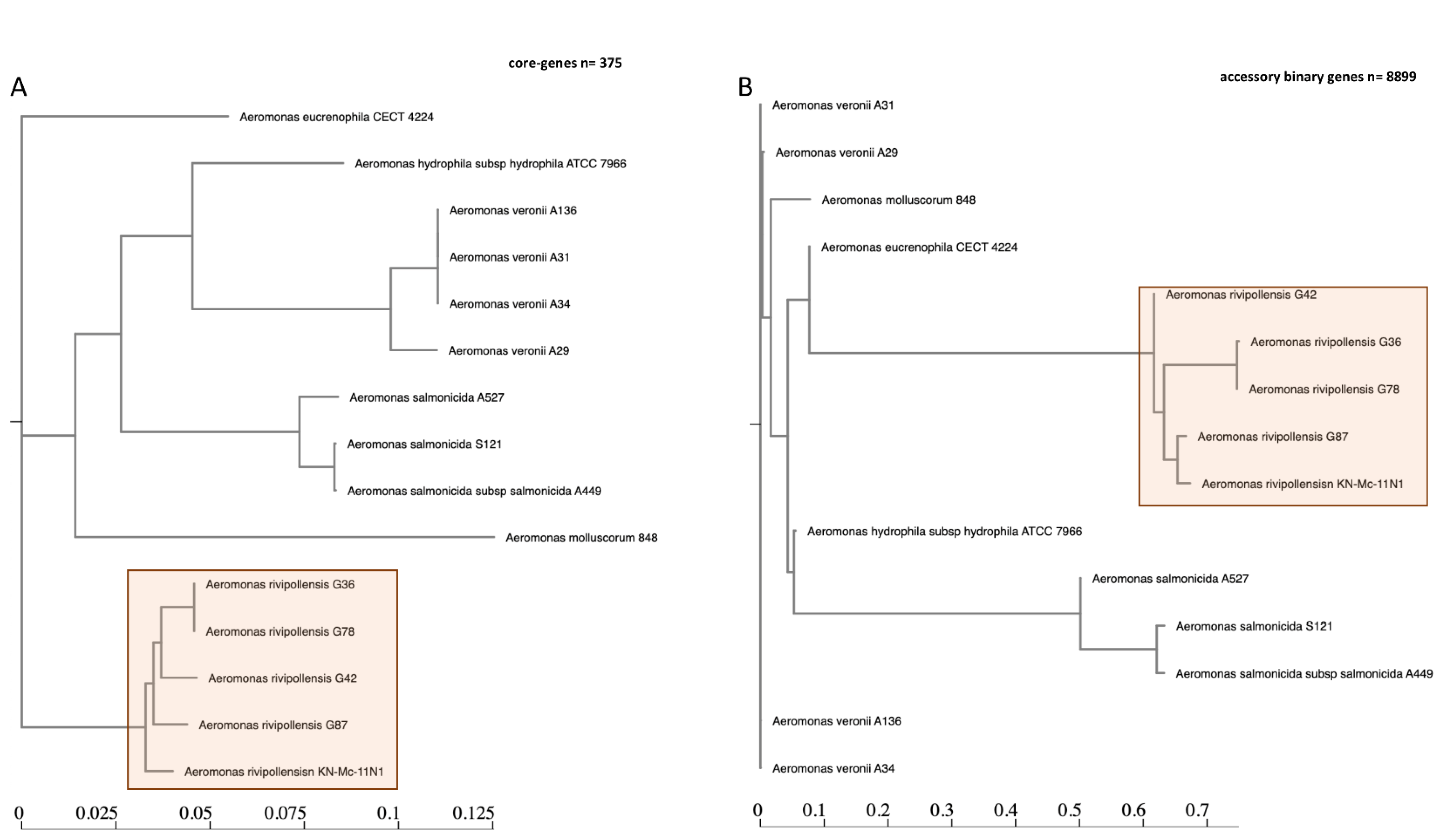
A total of 23,119 genes were examined and defined by the pangenome analysis of the 15 Aeromonas spp., with 375 core genes (found in >99% of genomes) shared by the Aeromonas species of rivipollensis, salmonicida, molluscorum, and euroniphilla (Figure 2A). Most genes were identified as accessories and consisted of 8899 and 13,845 shell and cloud genes, respectively. This indicates that Aeromonas is a heterogeneous species that consists of many cloud genes (Figure S2). A. rivipollensis strains clustered significantly in their own sub-clade (Figure 2 and Figure S2). The different strains of A. rivipollensis are shown by their distinctive average nucleotide identity (ANI) profiles identified using accessory genes (Figure 2B). This observation is also an augment as determined by whole-genome species tree. Aeromonas species genomes contained core metabolic enzymes, such as amidophosphoribosyltransferase (purF), ATP-dependent 6-phosphofructokinase isozyme 1 (pfka), nitrogen regulatory protein (ptsN), glutamate-pyruvate aminotransferase (alaA), UDP-3-O-acyl-N-acetylglucosamine deacetylase (ipxC), peptidyl-prolyl cis-trans isomerase B (ppiB), maltose transport system permease (maIG), cysteine desulfurase (iscS), dihydrolipoyl dehydrogenase (ipdA), UDP-N-acetylglucosamine 1-carboxyvinyltransferase (murA), and oligopeptide transport system permease (oppC). The accessory binary genes (Figure 2B) found in A. rivipollensis strain G87 were biosynthetic genes for polysialic acid, which is responsible for capsulation and is also found in the KN-Mc-11N1 strain.
The accessory metabolic genes on A. rivipollensis strain G87 included genes such as taurine import ATP-binding protein (tauB) and taurine-binding periplasmic proteins, cellulose synthase operon protein C (bcsC), beta-xylosidase (xynB), glucose-6-phosphate isomerase (pgiA), unsaturated chondroitin disaccharide hydrolase (ugi), p-aminobenzoyl-glutamate transport protein (abgT), morphine 6-dehydrogenase (morA), beta-glucoside kinase (bglK), and aryl-phospho-beta-D-glucosidase (bglC1 and bglC2).

2.4. Polysialic Acid and Sialic Acid Biosynthesis Genes

In the A. rivipollensis G87 and KN-Mc11N1 phylogroup, we have identified a polysialic acid operon that consists of kpsM, kpsT, kpsE, kpsD, and KpsFS (Table 2). These are capsular polysaccharide genes that are associated with polysialic acid in the outer membrane. The gene cluster organisation (Figure 3) is also outlined, which shows different kps genes and precursor components (neuABC) for the biosynthesis of extracellular polysialic acid. The kps cluster comprises two different regions, namely: region 1 (kpsDMTE) and region 3 (neuC and kpsFS) (Figure 3). The extracellular kpsDMTE genes and kpsFS are required for the polysaccharide capsule formation in the bacteria (blue and red). The kpsFS genes participate in the translocation of the polysaccharide capsule. Pangenome analysis showed that these genes are absent in the other sequenced A. rivipollensis in this study. Genome strain G87 can be separated from the KN-Mc-11N1 strain based on genes responsible for exogenous sialic acid production.
A. rivipollensis strain G87 was the only genome with an exogenous sialic acid metabolism. This was further confirmed by the RAST annotation server, which assigned the subsystem a score of 2.0. The metabolic cluster for sialic acid is composed of different nan-genes (Figure 3). The annotation names for the polysialic acid biosynthesis cluster (kps) and sialic acid metabolic cluster were assigned to the A. rivipollensis strain G87 (Table 2). The nanARH and TRAP transporters (nanTp-TI) (green) are responsible for exogenous sialic acid, while the nanAXEKP and yhcH genes (orange) are responsible for the degradation or catabolism of salic acid.

2.5. Antibiotics and Virulence Genes of Aeromonas rivipollensis Genomes

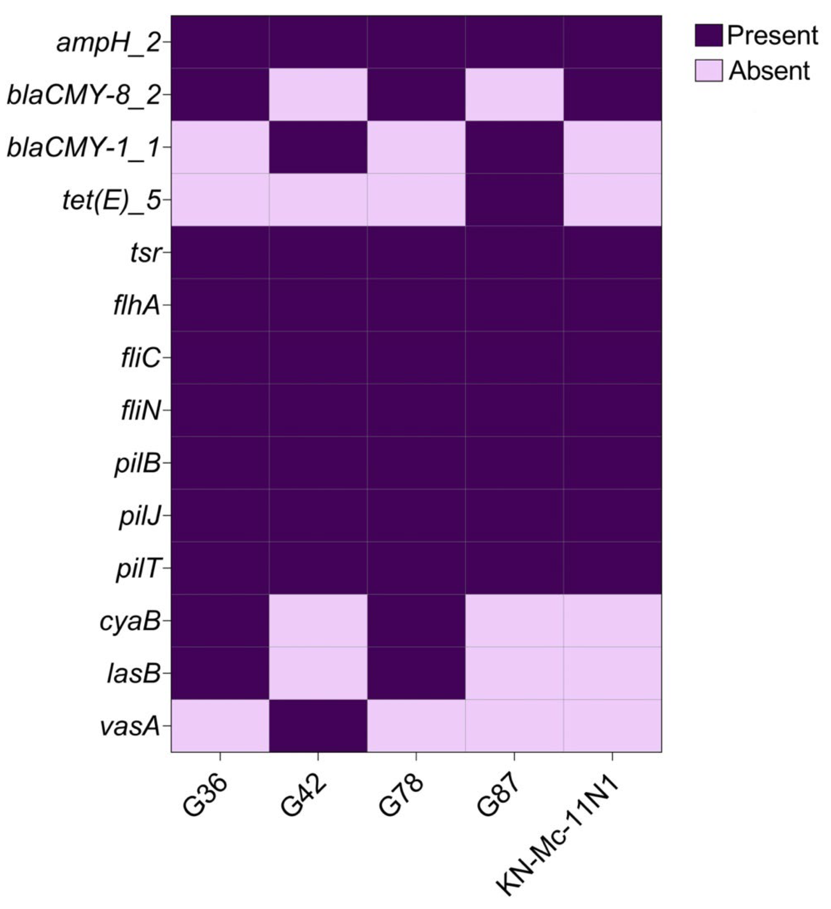
In this study, several core-virulence genes (n = 7) from the sequenced Aeromonas rivipollensis were identified (Figure 4). The chemoreceptor tsr gene, flagellar biosynthesis proteins fliACN, and twitching motility proteins pilBJT were among them. The antibiotic resistance genes shared by the A. rivipollensis genomes included cephalosporins (ampH) and beta-lactams (blaCMY) genes. Tetracycline resistance (tetE) gene was the unique antibiotic-resistance gene present in the A. rivipollensis G87 genome.

2.6. Mobile Genetic Elements

In this G87 strain containing the transposition protein TnsC, a tetracycline-resistance MFS efflux pump known as tetE and a tetracycline-resistance regulatory protein tetR were discovered (Figure S3). The transposition protein TnsC had a BLAST nucleotide homology of 98.70% with A. veronii strain wp8-s18-ESNL-11. The hypothetical proteins identified in this gene cluster also had a 100% nucleotide identity with A. veronii strain wp8-s18-ESNL-11. In the upstream region in this operon, a Type I restriction–modification system specific subunit S and DNA–methyltransferase subunit-M were determined to influence genomic evolution by horizontal gene transfer. A transposase, InsH for insertion sequence element IS5, was also determined to be in A. rivipollensis strain G87. In the downstream part of the gene arrangement cluster, this InsH transposase also features a secretory immunoglobulin A-binding protein (esiB1) of 726 bp (242 aa). The second esiB2 gene of about 1656 bp (522 aa) was also found lying downstream with other mobile genetic elements. The IS3 family transposase ISAs7 (618 bp) was also found that had a BLASTn of 98.54% and 98.22% with A. media strain T0.1-19 and A. rivipollensis strain KN-Mc-11N1, respectively. Other MGEs identified were two transposon Tn3 resolvase proteins (tnpR1 and tnpR2) and Tn7 found in the G87 strain.

3. Materials and Methods

3.1. Sample Isolation and Classical Microbiological Tests

We collected water samples from the Jukskei River continuum in Johannesburg (25.948156 S, 27.957528 E) in 2018. Water samples were aseptically collected in duplicates of approximately 1 L in a sterile container. Water samples were immediately placed in a cooler box at 4 °C and transported to the laboratory for further analysis. For bacterial isolation, 100 mL of the sample was filtered through a nitrocellulose filter membrane (0.22 μm), and the filters were then placed on Aeromonas isolation agar (Merck, Millipore) and incubated at 37 °C for 24 h. Four distinct isolates were coded as G36, G42, G78, and G87 and were presumptive Aeromonas species. The presumptive isolates were further identified using matrix-assisted laser desorption ionization–time-of-flight mass spectrometry (MALDI–TOF) (Bruker, Bremen, Germany). Briefly, a single colony was transferred into an Eppendorf tube containing 300 μL of sterile distilled water and 900 μL of absolute ethanol, mixed thoroughly, and later centrifuged for 2 min at 13,000 rpm. The supernatant was discarded, and the Eppendorf tube was later filled with 5 μL of 70% formic acid and thoroughly mixed.

3.2. DNA Extractions

The genomic DNA of the Aeromonas species strains was extracted using the High Pure PC Template preparation kit (Roche, Germany). The DNA was quantified on qubit fluorometric quantization using the Broad Range assay kit (Invitrogen™). The quality of the DNA was analyzed by electrophoresis on a 0.8% agarose gel using ethidium bromide and visualized under UV-light.

3.3. Whole-Genome Sequencing, Quality Control and Assembly

The sequencing libraries of the Aeromonas isolates (n = 4) were generated using the NEBNext® UltraTM II FS DNA library prep kit (New England Biolabs). The 150 bp paired-end sequence reads were generated with the Illumina MiSeq sequencer (Illumina, USA). FastQC v. 0.11.52 (http://www.bioinformatics.babraham.ac.uk/projects/fastqc (accessed on 6 February 2022)) was used to assess the quality of the paired-end reads associated with each isolate. Sequence coverage of the genomes of Aeromonas isolates G36, G42, G78, and G87 was 39X, 144X, 54, and 294X, respectively. Trimmomatic v. 0.33 [24] was used to remove the sequenced adapters and the leading and trailing ambiguous bases. The trimming parameters included LEADING:3 and TRAILING:3; reads with average per-base quality scores of <15 within a 4 bp sliding window. The SLIDINGWINDOW:4:15; reads with length < 36 bp were removed. Trimmed reads were de novo assembled using the CLC Genomics Workbench v. 11.01 (Qiagen). QUAST v. 4.5 [25] was used to assess the quality of the associated assembled genome using default parameters. CheckM [26] was additionally used to assess the potential contaminants in each assembled genome of Aeromonas isolates using default parameters. BLASTn [27] was used to align the assembled contigs using A. rivipollensis KN-Mc-11N1 [10] as a reference. For consistency and the removal of contaminants, multiple genome alignments were constructed using the progressive MAUVE tool [28]. The feature prediction and annotation of the sequenced genomes were performed using the NCBI prokaryotic Genome Annotation pipeline (PGAP) [29] and rapid annotation RAST [30].

3.4. Phylogenetic Analysis Using gyraseB and Whole-Genome Species Tree

The gyraseB (gryB) gene sequences were extracted from the assembled genomes of the A. rivipollensis strains. The nucleotide sequence queries of the Aeromonas isolates were BLAST-searched and compared with the available sequences of Aeromonas species in NCBI (http://www.ncbi.nlm.nih.gov (accessed on 25 April 2022)). This enabled the evaluation of homologous hits to the NCBI’s available sequences. Multiple sequence alignments of the extracted gene sequence and mined NCBI sequences were performed using MAFFT [31]. Maximum likelihood phylogenetic analysis of A. rivipollensis was performed using 1000 bootstrap iterations in MEGA 11.0 [32]. Utilizing K-base’s species tree (https://www.kbase.us/ (accessed on 12 March 2022)), the in silico taxonomic classification of all four sequenced genomes was performed. The assembled genomes were compared to other genomes that were accessible in the NCBI database. The whole-genome phylogenetic tree was visualized using Figtree v1.16.6 (http://tree.bio.ed.ac.uk/software/figtree/ (accessed on 25 March 2022)).

3.5. Pangenomics Analysis

The inclusion of the Aeromonas spp. genomes (n = 11) was based on the species tree generated from whole-genome-based taxonomic classification. This study included the A. veronii and A. hydrophilla genomes based on their completeness status. A. veronni genomes included the South African strains isolated from human cases, and none were found from the environment. In total, about 15 Aeromonas spp. genomes were used for comparative genomic analysis that includes the 4 sequenced A. rivipollensis. Prokka v.1.14.0 [33] was used to annotate the genomes of Aeromonas species using the default parameters. The pan-genome composition was extracted using Roary [34]. Pan-genome clusters were defined as follows: Core-genes were present in all isolates; soft core-genes present in at least 95% of isolates; shell-genes were present between 15% and 95% of isolates; cloud-genes were present in less than 15% of isolates. IQ-TREE v.2 [35] was used to construct the phylogenetic tree of the aligned core-genes using default parameters. The core genome phylogenetic tree was visualized using the Newick tree display [36]. Additionally, phandango v1.3.0 (https://jameshadfield.github.io/phandango/#/ (accessed on 25 April 2022)) was used for the interactive visualization of the genome phylogeny.

3.6. Antibiotics Resistance, Plasmid Replicon, Mobile Genetic Elements, and Virulence Factor Determinants

Antibiotic-resistance genes were identified in four sequenced Aeromonas genome sequences and the reference strain. In the ABRicate pipeline, AMR determinants were identified in each assembled genome using the ResFinder database (–db ResFinder; accessed 23 April 2022) [37], which has minimum identity and coverage thresholds of 75 (–minid 75) and 50% (–mincov 50), respectively. Plasmid replicons were identified on the sequenced genomes using the PlasmidFinder database [38]. Resistance genes were determined using the Virulence Factor Database (VFDB; –db vfdb, accessed 19 April 2022) [39], using minimum identity and coverage thresholds of 70 (–minid 70) and 50% (–mincov 50), respectively. OriTfinder v1.1 [40] was also used to predict the virulence factors and acquired antibiotic resistance genes. Mobile genetic elements (MGEs) were investigated on the sequenced genomes of Aeromonas spp. using this tool as well as default parameters. The heatmap of the antibiotics resistance and virulence genes was generated using GraphPad Prism9.

4. Discussion

The identification of Aeromonas spp. isolates to the species level remains a challenge using classic microbiological tests. Without a suitably comprehensive database, methods that are most frequently used, like MALDI–TOF, may misidentify samples [23]. However, this study used a high-resolution whole-genome and pangenomics analysis to confirm and identify A. rivipollensis genomes. This study showed that A. rivipollensis seems to be an emerging pathogen that has not yet been thoroughly characterized. The observation of accessory genes that included polysialic acid and sialic acid, mobile genetic elements, and an antibiotic-resistance profile, especially in the genome of G87, elucidate a unique genetic cluster of A. rivipollensis.
The use of gyrB was used in this study to fairly classify and identify A. rivipollensis. The sequenced Aeromonas species could only be correctly identified as A. rivipollensis using the high-resolution method of WGS and whole-genome species tree. These isolates were identified as A. media using MALDI-TOF. They were grouped with A. rivipollensis KN-Mc-11N1 using the WGS species tree (Figure 1), and pangenome analysis. Their genomes are quite closely related to those of A. eucroniphilla strain CECT 4224, A. salmonicida subsp. salmonicida strain A449, and A. mulluscorum strain 848. The pangenomic examination of this study’s species revealed that the genus Aeromonas is diverse when compared to other species, since only a small number (n = 375) of core genes could be identified. Distinct clusters are also observed among A. rivopollensis genomes. The genomes of KN-Mc-11N1 and G87, which share several core genes, make up one of the clusters. This cluster contained the polysialic acid (PGA) (kpsDMTE) genes responsible for exogenous capsulation, whereas sialic acid was only found in G87, which is associated with virulence and adaptation in the environment [41].
The primary strains for polysialic acid (PSA) biosynthesis and metabolism were A. rivipollensis G87 and KN-Mc-11N1. Many other bacteria, such as Mannheimia hemolytica (previously Pasteurella haemolytica) [42] and E. coli [43,44], have been associated with the production of PSA as their extracellular capsules. In bacteria, the PSA acts as a virulence factor that mimics the mammalian PSA’s structure and as an antiphagocytic [45]. The kps gene cluster that is involved in the PSA biosynthesis, modification, and transport of the bacteria’s PSA chains is outlined in this study for A. rivipollensis (Figure 3). The kps cluster comprises of two different regions, namely; region 1 (kpsDMTE) and region 3 (neuC and kpsFS), which participate in the translocation of the polysaccharide across the periplasmic space and onto the cell surface [44,46]. PSA metabolism is regulated at a transcriptional level by the transcriptional activator rfaH [47]. In E. coli K92, rfaH enhanced kps expression for the synthesis of the polysialic acid capsule [43]. This gene was also present in the genome of A. rivipollensis G87 at the downstream end of the operon. The polysialic acid capsule is transported across the outer membrane to the cell surface by the KpsD protein. It functions as the periplasmic binding element of the PSA transport system, in which it transiently interacts with the membrane component of the transporter, binds polysaccharide, and transports the polymer to a component in the outer membrane. This is also observed in bacteria containing poly-gamma-glutamate transpeptidase, responsible for capsulation [48]. Other components reported included the kpsT and kpsM genes, which are ABC transporters that export PSA from the cytoplasm. These transporters also require kpsE, a polysaccharide export system located in the inner membrane protein [46]. However, the mechanism of expressing the polysialic is not well understood and characterized in A. rivipollensis, as this is the first report in this phylogroup of G87 and KN-Mc-11N1 genomes that needs further investigation.
A complete nan system was outlined as including at least one ortholog of each of the genes encoding nanA, nanE, and nanK, more especially in the E. coli model [49]. These genes were absent from the other sequenced and compared genomes in this study but were present in the A. rivipollensis strain G87 genome. This was discovered in the annotation subsystem and was confirmed as an exogenous sialic acid based on the genetic variant code 2.0 assigned to it. Sialic acid is biosynthesized, activated, and polymerized by proteins NeuABCD [50], which are present in the G87 genome with the exception of the neuD gene. The neuD is a gene found in organisms that can synthesize sialic acid [51].
It is well recognized that the precursor to PSA is the gene for N-acetylneuraminate (Neu5Ac aldolase or neuC gene). By mimicking sialic acid, some pathogenic bacteria can circumvent host defenses. The neuC is located upstream with other kpsFS genes, which is involved in the production of capsular heptose. Exogenous sialic acid nan operon nanRAHTpTI (region 4) and degradation nan operon nanAXEKP-YhcH (region 5) were assigned in Figure 3. De novo synthetic genes of the sialic acid neuABC (region 2/3) were identified in this operon. However, according to the annotation genetic variation code, the G87 strain displayed a non-synthetic sialic acid. Therefore, organisms that can catabolize but not synthesize salic acids are classified as exogenous sialic acids. The sialic acid operon contained the two tripartite ATP-independent periplasmic (TRAP) transporters, nanTp and nanTI, respectively. The nanTp is a major permease component of the TRAP-type C4-dicarboxylate transport system (Figure 3). The TRAPs are well studied as the key transporters involved in the uptake of sialic acid and are associated with roles in pathogenicity [52,53]. A second N-acetylneuraminate lyase (nanA2) was identified in the upstream with transcriptional regulator nanR, which controls the expression of proteins involved in sialic acid absorption and metabolism [54,55]. Other salic acid genes that were not well characterized in this study might also be suggested by the region 2 putative proteins. It has now been established beyond doubt that microbial sialic acid metabolism contributes to the pathogenicity of a variety of infectious illnesses. Many vertebrate cells’ surfaces include glycan molecules with sialic acid occupying the terminal position. Sialic acid engages in a number of biological processes, such as cell signaling and intercellular adhesion [56]. Pathogenic bacteria that coat themselves with sialic acid, such as Group B Streptococcus, offer resistance to components of the host’s innate immune response. These bacteria have evolved to utilize this substance to their advantage [41,57]. Two secretory immunoglobulin A-binding proteins EsiB (esiB1) and EsiB2 found in the G87 genome were among the other virulence factors discovered in this study. They have different sequence lengths, with esiB1 being 726 bp (242 aa) and esiB2 being 1656 bp (522 aa), respectively. Pathogenic strains are primarily composed of the esiB genes, which are known to impair neutrophil activation [58].
Aeromonas species are generally resistant to betalactams (blaCMY), and this was also observed in this study. The presence of blaCMY and ampH genes in all isolated A. rivipollensis was not a surprise, as they have been recorded in several bacteria isolates including Aeromonas species [59,60]. In addition, the genome of isolate G87 was only one of the sequenced A. rivipollensis in this study that showed a multidrug tetracycline-resistance gene. This AR gene was found in this genome because it was linked to a mobile genetic element, TnC, with a nucleotide percentage identity similar to that of A. veronii. However, tetracycline resistance has previously been reported in A. veronii strain MS-17-88 recovered from channel catfish [12,61]. It has also been reported from Aeromonas species isolated from South African aquatic environments [62].
Flagellar biosynthesis proteins FliACN and twitching motility proteins PilBJT (Type IV pilus) were found in all A. rivipollensis strains sequenced in our study. These genes are commonly found in Aeromonas species that are associated with pathogenicity for colonization [63]. The chemoreceptor tsr gene was also present in the sequenced A. rivipollensis strains. In the study, Oh et al. [64] also detected this gene in E. coli. This has also been confirmed in other studies, indicating that E. coli and S. enterica serovar Typhimurium genomes possess many chemoreceptor genes, tsr [65]. Many chemoreceptor genes have also been reported in many other microorganisms [65,66]. However, no study has reported the presence of the tsr gene in A. rivipollensis.

5. Conclusions

Using WGS analysis, we were able to determine the resistome of Aeromonas rivipollensis. All A. rivipollensis strains show potential resistance to various antibiotic lactamase classes, indicating that these aeromonads are potentially virulent emerging pathogens that can be transmitted by river water. The determined polysialic acid and sialic acid genes in the G87 genome can be associated with antiphagocytic and anti-bactericidal properties, respectively. Mobile genetic elements, such as transposases, introduce virulence and resistance genes, such as secretory immunoglobulin A-binding proteins and multidrug tetracycline genes in the A. rivipollensis G87 genome. These genomes will be used as a resource for additional research that may reveal new information on the genes responsible for accurate identification, pathogenic potential, and the relative health risks posed by environmental strains of A. rivipollensis.

Supplementary Materials

The following supporting information can be downloaded at: https://www.mdpi.com/article/10.3390/antibiotics12010131/s1, Figure S1: Phylogenetic analysis of the Aeromonas species isolates using the gyrB. The 4 sequenced A. rivipollensis are highlighted in blue as well as the reference strain KN-Mc-11-N1. Bootstrap value of 1000 was used to construct the tree. Figure S2: Pangenome genome of the 15 Aeromonas spp. isolates showing the core, shell and cloud genes. A total of 23,119 CDS genes was used to construct the pangenome phylogenetic tree generated using Phandango. The colour coded blue represents compared coding genes amongst the genomes. Figure S3: Gene organization of tetracycline resistance genes (yellow) with transposase ISAs31 and transposition protein TnsC (brown) as well as Type 1 restriction recognition sites subunits (orange) that drives horizontal gene transfer determined in A. rivipollensis G87 genome. The CDS were annotated using prokka and visualized on Geneious version 9.0.5.

Author Contributions

Conceptualization, I.K., K.E.L. and J.B.D.; data curation, E.U.K.F.-T. and K.E.L.; formal analysis, K.E.L.; funding acquisition, K.E.L. and J.B.D.; investigation, E.U.K.F.-T., I.K. and K.E.L.; methodology, E.U.K.F.-T.; project administration, J.B.D.; resources, K.E.L. and J.B.D.; bioinformatics analysis, K.E.L.; supervision, I.K. and J.B.D.; validation, I.K. and K.E.L.; visualization, K.E.L.; writing—original draft, E.U.K.F.-T.; writing—review and editing, I.K., K.E.L. and J.B.D. All authors have read and agreed to the published version of the manuscript.

Funding

This research was funded by National Research Foundation (NRF) grant number “TTK200306508304” made available to KEL and supported by University of South Africa (UNISA).

Institutional Review Board Statement

The study was conducted according to the guidelines of the Declaration of Helsinki and approved by the Ethics Committee of the University of South Africa (Ref. no. 2018/CAES/048).

Informed Consent Statement

Not applicable.

Data Availability Statement

A. rivipollensis G36, G42, G78, and G87 genome sequences were deposited in NCBI GenBank under the accession numbers JAAILC0000000001, GCA_010974915.1, GCA_010974825.1, and JAAIKZ000000000, respectively. The sequence read achieve (SRA) accession numbers are assigned as G87 (SRR13249121), G78 (SRR13249122), G42 (SRR13249123), or (G36 (SRR13249124).

Conflicts of Interest

The authors declare no conflict of interest.

References

  1. Khor, W.C.; Puah, S.M.; Tan, J.A.; Puthucheary, S.D.; Chua, K.H. Phenotypic and Genetic Diversity of Aeromonas Species Isolated from Fresh Water Lakes in Malaysia. PLoS ONE 2015, 10, e0145933. [Google Scholar] [CrossRef] [PubMed] [Green Version]
  2. Li, T.; Raza, S.H.A.; Yang, B.; Sun, Y.; Wang, G.; Sun, W.; Qian, A.; Wang, C.; Kang, Y.; Shan, X. Aeromonas veronii Infection in Commercial Freshwater Fish: A Potential Threat to Public Health. Animals 2020, 10, 608. [Google Scholar] [CrossRef] [PubMed] [Green Version]
  3. Vasquez, I.; Hossain, A.; Gnanagobal, H.; Valderrama, K.; Campbell, B.; Ness, M.; Charette, S.J.; Gamperl, A.K.; Cipriano, R.; Segovia, C.; et al. Comparative Genomics of Typical and Atypical Aeromonas salmonicida Complete Genomes Revealed New Insights into Pathogenesis Evolution. Microorganisms 2022, 10, 189. [Google Scholar] [CrossRef] [PubMed]
  4. Talagrand-Reboul, E.; Colston, S.M.; Graf, J.; Lamy, B.; Jumas-Bilak, E. Comparative and Evolutionary Genomics of Isolates Provide Insight into the Pathoadaptation of Aeromonas. Genome Biol. Evol. 2020, 12, 535–552. [Google Scholar] [CrossRef] [PubMed] [Green Version]
  5. Chaix, G.; Roger, F.; Berthe, T.; Lamy, B.; Jumas-Bilak, E.; Lafite, R.; Forget-Leray, J.; Petit, F. Distinct Aeromonas Populations in Water Column and Associated with Copepods from Estuarine Environment (Seine, France). Front. Microbiol. 2017, 8, 1259. [Google Scholar] [CrossRef] [PubMed] [Green Version]
  6. Puthucheary, S.D.; Puah, S.M.; Chua, K.H. Molecular characterization of clinical isolates of Aeromonas species from Malaysia. PLoS ONE 2012, 7, e30205. [Google Scholar] [CrossRef] [Green Version]
  7. Villari, P.; Crispino, M.; Montuori, P.; Boccia, S. Molecular typing of Aeromonas isolates in natural mineral waters. Appl. Environ. Microbiol. 2003, 69, 697–701. [Google Scholar] [CrossRef] [Green Version]
  8. Rutteman, B.; Borremans, K.; Beckers, J.; Devleeschouwer, E.; Lampmann, S.; Corthouts, I.; Verlinde, P. Aeromonas wound infection in a healthy boy, and wound healing with polarized light. JMM Case Rep. 2017, 4, e005118. [Google Scholar] [CrossRef]
  9. Yang, W.; Li, N.; Li, M.; Zhang, D.; An, G. Complete Genome Sequence of Fish Pathogen Aeromonas hydrophila JBN2301. Genome Announc. 2016, 4, e01615-15. [Google Scholar] [CrossRef] [Green Version]
  10. Park, S.Y.; Lim, S.R.; Son, J.S.; Kim, H.K.; Yoon, S.W.; Jeong, D.G.; Lee, M.S.; Lee, J.R.; Lee, D.H.; Kim, J.H. Complete Genome Sequence of Aeromonas rivipollensis KN-Mc-11N1, Isolated from a Wild Nutria (Myocastor coypus) in South Korea. Microbiol. Resour. Announc. 2018, 7, e00907-18. [Google Scholar] [CrossRef]
  11. Marti, E.; Balcazar, J.L. Aeromonas rivipollensis sp. nov., a novel species isolated from aquatic samples. J. Basic Microbiol. 2015, 55, 1435–1439. [Google Scholar] [CrossRef] [PubMed]
  12. Tekedar, H.C.; Kumru, S.; Blom, J.; Perkins, A.D.; Griffin, M.J.; Abdelhamed, H.; Karsi, A.; Lawrence, M.L. Comparative genomics of Aeromonas veronii: Identification of a pathotype impacting aquaculture globally. PLoS ONE 2019, 14, e0221018. [Google Scholar] [CrossRef] [PubMed] [Green Version]
  13. Carr, V.R.; Witherden, E.A.; Lee, S.; Shoaie, S.; Mullany, P.; Proctor, G.B.; Gomez-Cabrero, D.; Moyes, D.L. Abundance and diversity of resistomes differ between healthy human oral cavities and gut. Nat. Commun. 2020, 11, 693. [Google Scholar] [CrossRef] [Green Version]
  14. Seshadri, R.; Joseph, S.W.; Chopra, A.K.; Sha, J.; Shaw, J.; Graf, J.; Haft, D.; Wu, M.; Ren, Q.; Rosovitz, M.J.; et al. Genome sequence of Aeromonas hydrophila ATCC 7966T: Jack of all trades. J. Bacteriol. 2006, 188, 8272–8282. [Google Scholar] [CrossRef] [Green Version]
  15. Talagrand-Reboul, E.; Jumas-Bilak, E.; Lamy, B. The Social Life of Aeromonas through Biofilm and Quorum Sensing Systems. Front. Microbiol. 2017, 8, 37. [Google Scholar] [CrossRef] [PubMed]
  16. Rabaan, A.A.; Gryllos, I.; Tomas, J.M.; Shaw, J.G. Motility and the polar flagellum are required for Aeromonas caviae adherence to HEp-2 cells. Infect. Immun. 2001, 69, 4257–4267. [Google Scholar] [CrossRef] [Green Version]
  17. Gavin, R.; Merino, S.; Altarriba, M.; Canals, R.; Shaw, J.G.; Tomas, J.M. Lateral flagella are required for increased cell adherence, invasion and biofilm formation by Aeromonas spp. FEMS Microbiol. Lett. 2003, 224, 77–83. [Google Scholar] [CrossRef] [Green Version]
  18. Canals, R.; Jimenez, N.; Vilches, S.; Regue, M.; Merino, S.; Tomas, J.M. Role of Gne and GalE in the virulence of Aeromonas hydrophila serotype O34. J. Bacteriol. 2007, 189, 540–550. [Google Scholar] [CrossRef] [Green Version]
  19. Kirov, S.M.; O’Donovan, L.A.; Sanderson, K. Functional characterization of type IV pili expressed on diarrhea-associated isolates of Aeromonas species. Infect. Immun. 1999, 67, 5447–5454. [Google Scholar] [CrossRef] [Green Version]
  20. Massad, G.; Arceneaux, J.E.; Byers, B.R. Acquisition of iron from host sources by mesophilic Aeromonas species. J. Gen. Microbiol. 1991, 137, 237–241. [Google Scholar] [CrossRef]
  21. Burr, S.E.; Stuber, K.; Wahli, T.; Frey, J. Evidence for a type III secretion system in Aeromonas salmonicida subsp. salmonicida. J. Bacteriol. 2002, 184, 5966–5970. [Google Scholar] [CrossRef] [PubMed] [Green Version]
  22. Sha, J.; Pillai, L.; Fadl, A.A.; Galindo, C.L.; Erova, T.E.; Chopra, A.K. The type III secretion system and cytotoxic enterotoxin alter the virulence of Aeromonas hydrophila. Infect. Immun. 2005, 73, 6446–6457. [Google Scholar] [CrossRef] [PubMed] [Green Version]
  23. Shin, H.B.; Yoon, J.; Lee, Y.; Kim, M.S.; Lee, K. Comparison of MALDI-TOF MS, housekeeping gene sequencing, and 16S rRNA gene sequencing for identification of Aeromonas clinical isolates. Yonsei. Med. J. 2015, 56, 550–555. [Google Scholar] [CrossRef] [PubMed] [Green Version]
  24. Bolger, A.M.; Lohse, M.; Usadel, B. Trimmomatic: A flexible trimmer for Illumina sequence data. Bioinformatics 2014, 30, 2114–2120. [Google Scholar] [CrossRef] [Green Version]
  25. Gurevich, A.; Saveliev, V.; Vyahhi, N.; Tesler, G. QUAST: Quality assessment tool for genome assemblies. Bioinformatics 2013, 29, 1072–1075. [Google Scholar] [CrossRef] [Green Version]
  26. Parks, D.H.; Imelfort, M.; Skennerton, C.T.; Hugenholtz, P.; Tyson, G.W. CheckM: Assessing the quality of microbial genomes recovered from isolates, single cells, and metagenomes. Gen. Res. 2015, 25, 1043–1055. [Google Scholar] [CrossRef] [Green Version]
  27. Altschul, S. Hot papers-Bioinformatics-Gapped BLAST and PSI-BLAST: A new generation of protein database search programs by S.F. Altschul, T.L. Madden, A.A. Schaffer, J.H. Zhang, Z. Zhang, W. Miller, D.J. Lipman-Comments. Scientist 1999, 13, 15. [Google Scholar]
  28. Darling, A.C.E.; Mau, B.; Blattner, F.R.; Perna, N.T. Mauve: Multiple alignment of conserved genomic sequence with rearrangements. Gen. Res. 2004, 14, 1394–1403. [Google Scholar] [CrossRef] [Green Version]
  29. Haft, D.H.; DiCuccio, M.; Badretdin, A.; Brover, V.; Chetvernin, V.; O’Neill, K.; Li, W.; Chitsaz, F.; Derbyshire, M.K.; Gonzales, N.R.; et al. RefSeq: An update on prokaryotic genome annotation and curation. Nucleic Acids Res 2018, 46, D851–D860. [Google Scholar] [CrossRef]
  30. Aziz, R.K.; Bartels, D.; Best, A.A.; DeJongh, M.; Disz, T.; Edwards, R.A.; Formsma, K.; Gerdes, S.; Glass, E.M.; Kubal, M.; et al. The RAST server: Rapid annotations using subsystems technology. BMC Gen. 2008, 9, 75. [Google Scholar] [CrossRef] [Green Version]
  31. Katoh, K.; Rozewicki, J.; Yamada, K.D. MAFFT online service: Multiple sequence alignment, interactive sequence choice and visualization. Brief. Bioinform. 2019, 20, 1160–1166. [Google Scholar] [CrossRef]
  32. Tamura, K.; Stecher, G.; Kumar, S. MEGA11 Molecular Evolutionary Genetics Analysis Version 11. Mol. Biol. Evol. 2021, 38, 3022–3027. [Google Scholar] [CrossRef]
  33. Seemann, M. Das neue Spiel: Strategien fur die Welt nach dem digitalen Kontrollverlust; Orange-Press: Freiburg, Germany, 2014; p. 255. [Google Scholar]
  34. Page, A.J.; Cummins, C.A.; Hunt, M.; Wong, V.K.; Reuter, S.; Holden, M.T.G.; Fookes, M.; Falush, D.; Keane, J.A.; Parkhill, J. Roary: Rapid large-scale prokaryote pan genome analysis. Bioinformatics 2015, 31, 3691–3693. [Google Scholar] [CrossRef] [Green Version]
  35. Minh, B.Q.; Schmidt, H.A.; Chernomor, O.; Schrempf, D.; Woodhams, M.D.; von Haeseler, A.; Lanfear, R. IQ-TREE 2: New Models and Efficient Methods for Phylogenetic Inference in the Genomic Era. Mol. Biol. Evol. 2020, 37, 2461. [Google Scholar] [CrossRef]
  36. Junier, T.; Zdobnov, E.M. The Newick utilities: High-throughput phylogenetic tree processing in the UNIX shell. Bioinformatics 2010, 26, 1669–1670. [Google Scholar] [CrossRef] [Green Version]
  37. Feldgarden, M.; Brover, V.; Haft, D.H.; Prasad, A.B.; Slotta, D.J.; Tolstoy, I.; Tyson, G.H.; Zhao, S.; Hsu, C.H.; McDermott, P.F.; et al. Validating the AMRFinder Tool and Resistance Gene Database by Using Antimicrobial Resistance Genotype-Phenotype Correlations in a Collection of Isolates. Antimicrob. Agents Chemother. 2019, 63, e00483-19. [Google Scholar] [CrossRef] [Green Version]
  38. Carattoli, A.; Zankari, E.; Garcia-Fernandez, A.; Larsen, M.V.; Lund, O.; Villa, L.; Aarestrup, F.M.; Hasman, H. In Silico Detection and Typing of Plasmids using PlasmidFinder and Plasmid Multilocus Sequence Typing. Antimicrob. Agents Chemother. 2014, 58, 3895–3903. [Google Scholar] [CrossRef] [Green Version]
  39. Liu, B.; Zheng, D.D.; Zhou, S.Y.; Chen, L.H.; Yang, J. VFDB 2022: A general classification scheme for bacterial virulence factors. Nucleic Acids Res. 2022, 50, D912–D917. [Google Scholar] [CrossRef]
  40. Li, X.B.; Xie, Y.Z.; Liu, M.; Tai, C.; Sung, J.Y.; Deng, Z.X.; Ou, H.Y. oriTfinder: A web-based tool for the identification of origin of transfers in DNA sequences of bacterial mobile genetic elements. Nucleic Acids Res. 2018, 46, W229–W234. [Google Scholar] [CrossRef] [Green Version]
  41. Pezzicoli, A.; Ruggiero, P.; Amerighi, F.; Telford, J.L.; Soriani, M. Exogenous sialic acid transport contributes to group B streptococcus infection of mucosal surfaces. J. Infect. Dis. 2012, 206, 924–931. [Google Scholar] [CrossRef] [Green Version]
  42. Puente-Polledo, L.; Reglero, A.; Gonzalez-Clemente, C.; Rodriguez-Aparicio, L.B.; Ferrero, M.A. Biochemical conditions for the production of polysialic acid by Pasteurella haemolytica A2. Glycoconj. J. 1998, 15, 855–861. [Google Scholar] [CrossRef] [PubMed]
  43. Navasa, N.; Rodriguez-Aparicio, L.B.; Ferrero, M.A.; Monteagudo-Mera, A.; Martinez-Blanco, H. Transcriptional control of RfaH on polysialic and colanic acid synthesis by Escherichia coli K92. FEBS Lett. 2014, 588, 922–928. [Google Scholar] [CrossRef] [Green Version]
  44. Lin, B.X.; Qiao, Y.; Shi, B.; Tao, Y. Polysialic acid biosynthesis and production in Escherichia coli: Current state and perspectives. Appl. Microbiol. Biotechnol. 2016, 100, 1–8. [Google Scholar] [CrossRef] [PubMed]
  45. Cress, B.F.; Englaender, J.A.; He, W.; Kasper, D.; Linhardt, R.J.; Koffas, M.A. Masquerading microbial pathogens: Capsular polysaccharides mimic host-tissue molecules. FEMS Microbiol. Rev. 2014, 38, 660–697. [Google Scholar] [CrossRef] [PubMed] [Green Version]
  46. Colley, K.J.; Kitajima, K.; Sato, C. Polysialic acid: Biosynthesis, novel functions and applications. Crit. Rev. Biochem. Mol. Biol. 2014, 49, 498–532. [Google Scholar] [CrossRef]
  47. Bailey, M.J.; Hughes, C.; Koronakis, V. RfaH and the ops element, components of a novel system controlling bacterial transcription elongation. Mol. Microbiol. 1997, 26, 845–851. [Google Scholar] [CrossRef]
  48. Lekota, K.E.; Bezuidt, O.K.I.; Mafofo, J.; Rees, J.; Muchadeyi, F.C.; Madoroba, E.; van Heerden, H. Whole genome sequencing and identification of Bacillus endophyticus and B. anthracis isolated from anthrax outbreaks in South Africa. BMC Microbiol. 2018, 18, 67. [Google Scholar] [CrossRef]
  49. Vimr, E.R.; Kalivoda, K.A.; Deszo, E.L.; Steenbergen, S.M. Diversity of microbial sialic acid metabolism. Microbiol. Mol. Biol. Rev. 2004, 68, 132–153. [Google Scholar] [CrossRef] [Green Version]
  50. Ferrero, M.A.; Aparicio, L.R. Biosynthesis and production of polysialic acids in bacteria. Appl. Microbiol. Biotechnol. 2010, 86, 1621–1635. [Google Scholar] [CrossRef]
  51. Daines, D.A.; Wright, L.F.; Chaffin, D.O.; Rubens, C.E.; Silver, R.P. NeuD plays a role in the synthesis of sialic acid in Escherichia coli K1. FEMS Microbiol. Lett. 2000, 189, 281–284. [Google Scholar] [CrossRef]
  52. Lubin, J.B.; Kingston, J.J.; Chowdhury, N.; Boyd, E.F. Sialic acid catabolism and transport gene clusters are lineage specific In Vibrio vulnificus. Appl. Environ. Microbiol. 2012, 78, 3407–3415. [Google Scholar] [CrossRef]
  53. Rosa, L.T.; Bianconi, M.E.; Thomas, G.H.; Kelly, D.J. Tripartite ATP-Independent Periplasmic (TRAP) Transporters and Tripartite Tricarboxylate Transporters (TTT): From Uptake to Pathogenicity. Front. Cell Infect. Microbiol. 2018, 8, 33. [Google Scholar] [CrossRef] [Green Version]
  54. Revilla-Nuin, B.; Rodriguez-Aparicio, L.B.; Ferrero, M.A.; Reglero, A. Regulation of capsular polysialic acid biosynthesis by N-acetyl-D-mannosamine, an intermediate of sialic acid metabolism. FEBS Lett. 1998, 426, 191–195. [Google Scholar] [CrossRef] [Green Version]
  55. Horne, C.R.; Venugopal, H.; Panjikar, S.; Wood, D.M.; Henrickson, A.; Brookes, E.; North, R.A.; Murphy, J.M.; Friemann, R.; Griffin, M.D.W.; et al. Mechanism of NanR gene repression and allosteric induction of bacterial sialic acid metabolism. Nat. Commun. 2021, 12, 1988. [Google Scholar] [CrossRef]
  56. Severi, E.; Hood, D.W.; Thomas, G.H. Sialic acid utilization by bacterial pathogens. Microbiology 2007, 153, 2817–2822. [Google Scholar] [CrossRef] [Green Version]
  57. Brigham, C.; Caughlan, R.; Gallegos, R.; Dallas, M.B.; Godoy, V.G.; Malamy, M.H. Sialic acid (N-acetyl neuraminic acid) utilization by Bacteroides fragilis requires a novel N-acetyl mannosamine epimerase. J. Bacteriol. 2009, 191, 3629–3638. [Google Scholar] [CrossRef] [Green Version]
  58. Pastorello, I.; Rossi Paccani, S.; Rosini, R.; Mattera, R.; Ferrer Navarro, M.; Urosev, D.; Nesta, B.; Lo Surdo, P.; Del Vecchio, M.; Rippa, V.; et al. EsiB, a novel pathogenic Escherichia coli secretory immunoglobulin A-binding protein impairing neutrophil activation. mBio 2013, 4, e00206-13. [Google Scholar] [CrossRef] [Green Version]
  59. Piotrowska, M.; Przygodzinska, D.; Matyjewicz, K.; Popowska, M. Occurrence and Variety of beta-Lactamase Genes among Aeromonas spp. Isolated from Urban Wastewater Treatment Plant. Front. Microbiol. 2017, 8, 863. [Google Scholar] [CrossRef] [Green Version]
  60. Vazquez-Lopez, R.; Solano-Galvez, S.; Alvarez-Hernandez, D.A.; Ascencio-Aragon, J.A.; Gomez-Conde, E.; Pina-Leyva, C.; Lara-Lozano, M.; Guerrero-Gonzalez, T.; Gonzalez-Barrios, J.A. The Beta-Lactam Resistome Expressed by Aerobic and Anaerobic Bacteria Isolated from Human Feces of Healthy Donors. Pharmaceuticals 2021, 14, 533. [Google Scholar] [CrossRef]
  61. Tekedar, H.C.; Abdelhamed, H.; Kumru, S.; Blom, J.; Karsi, A.; Lawrence, M.L. Comparative Genomics of Aeromonas hydrophila Secretion Systems and Mutational Analysis of hcp1 and vgrG1 Genes From T6SS. Front. Microbiol. 2018, 9, 3216. [Google Scholar] [CrossRef]
  62. Jacobs, L.; Chenia, H.Y. Characterization of integrons and tetracycline resistance determinants in Aeromonas spp. isolated from South African aquaculture systems. Int. J. Food Microbiol. 2007, 114, 295–306. [Google Scholar] [CrossRef]
  63. Kirov, S.M.; Barnett, T.C.; Pepe, C.M.; Strom, M.S.; Albert, M.J. Investigation of the role of type IV Aeromonas pilus (Tap) in the pathogenesis of Aeromonas gastrointestinal infection. Infect. Immun. 2000, 68, 4040–4048. [Google Scholar] [CrossRef] [PubMed] [Green Version]
  64. Oh, D.; Yu, Y.; Lee, H.; Jeon, J.H.; Wanner, B.L.; Ritchie, K. Asymmetric polar localization dynamics of the serine chemoreceptor protein Tsr in Escherichia coli. PLoS ONE 2018, 13, e0195887. [Google Scholar] [CrossRef] [Green Version]
  65. Ortega, A.; Zhulin, I.B.; Krell, T. Sensory Repertoire of Bacterial Chemoreceptors. Microbiol. Mol. Biol. R 2017, 81, e00033-17. [Google Scholar] [CrossRef] [PubMed] [Green Version]
  66. Lacal, J.; Garcia-Fontana, C.; Munoz-Martinez, F.; Ramos, J.L.; Krell, T. Sensing of environmental signals: Classification of chemoreceptors according to the size of their ligand binding regions. Environ. Microbiol. 2010, 12, 2873–2884. [Google Scholar] [CrossRef] [PubMed]
Figure 1. Whole-genome-based species tree showing the clustering of the sequenced Aeromonas rivipollensis genomes and KN-Mc-11N1 strain (green annotation) with the closely related genome species of A. salmonicida subsp. salmonicida A449, A. molluscorum 949, and A. euroniphilla CECT 4224.
Antibiotics 12 00131 g001
Figure 2. Phylogeny of the compared Aeromonas species using 375 core genes (A) and 8899 accessory binary genes (B). The tree shows phylogenetic tree of the 4 Aeromonas species as highlighted with orange color. About 375 core-genes (A) and 8899 accessory binary genes (B) were used to construct the phylogenetic tree.
Antibiotics 12 00131 g002
Figure 3. Aeromonas rivipollensis strain G87 organization gene clusters for capsular polysialic acids and an exogeneous sialic acid metabolic cluster. The kpsDMTE and kpsFS genes are required for capsulation (region 1, blue, and region 3, red). The kpsFS genes participate in the translocation of the polysaccharide. The precursors of polysialic acid biosynthesis are neuABC (region 2, yellow, and region 3, red). The nanARH with TRAP transporters in region 4, and nanTp-TI genes (green) are responsible for exogenous sialic acid, while the nanAXEKP and yhcH genes (orange) are responsible for the degradation or catabolism of salic acid.
Antibiotics 12 00131 g003
Figure 4. Heat map showing the antibiotic resistance and virulence genes detected among the 4 sequenced and the compared A. rivipollensis strain KN-Mc-11N1.
Antibiotics 12 00131 g004
Table 1. Genome features of the sequenced A. rivipollensis strains used in this study compared to KN-Mc-11N1 as a reference strain.
FeatureG36G42G78G87KN-Mc-11N1 *
Genome size (bp)4,530,6394,584,4954,531,5064,663,0304,508,901
CDSs (protein coding sequences)4,2394,2734,2054,3194,025
Number of tRNAs115109101108124
Number of total rRNAs 537431
GC%61,4861,361,4861,0861,9
Number of contigs935557711
N5094,736 184,598 194,165160,487 -
Sequence reads archive (SRA) or GenBankSRR13249124SRR13249123SRR13249122SRR13249121CP027856.1
* Complete genome of A. rivipollensis—Not applicable.
Table 2. Gene annotation of the polysaccharide (polysialic acid) capsular and sialic acid genes involved in biosynthesis present in the genome of A. rivipollensis strain G87.
Gene AnnotationAbbreviation Subsystem Assigned
Capsular polysaccharide export system periplasmic proteinkpsDCapsular polysaccharide (CPS) of Campylobacter CPS
biosynthesis and assembly
Capsular polysaccharide ABC
transporter, permease protein
kpsMRhamnose containing glycans, CPS of Campylobacter, CPS
biosynthesis and assembly
Capsular polysaccharide ABC
transporter, ATP-binding protein
kpsTCPS biosynthesis and assembly
Capsular polysaccharide export system inner membrane protein kpsECPS of Campylobacter; CPS biosynthesis and assembly
COG3563: Capsule polysaccharide export proteinKpsFCPS biosynthesis and assembly
Capsular polysaccharide export system protein kpsSCPS biosynthesis and assembly
N-Acetylneuraminate
cytidylyltransferase (EC 2.7.7.43)
neuACMP-N-acetylneuraminate_biosynthesis; Sialic acid
metabolism
N-acetylneuraminate synthase (EC 2.5.1.56)neuBCMP-N-acetylneuraminate biosynthesis; Sialic acid
metabolism
dTDP-4-dehydrorhamnose 3,5-epimerase (EC 5.1.3.13)neuCdTDP-rhamnose_synthesis; Rhamnose containing glycans; Capsular_heptose_biosynthesis
Transcriptional regulator NanRnanRSialic acid metabolism
N-acetylneuraminate lyase (EC 4.1.3.3)nanA2Sialic acid metabolism
TRAP-type C4-dicarboxylate transport system, periplasmic componentnanTp or siaMTRAP Transporter collection
TRAP-type C4-dicarboxylate transport system, large permease componentnanTITRAP Transporter collection
Sialidase (EC 3.2.1.18)nanHGalactosylceramide and sulfatide metabolism; Sialic acid metabolism
N-acetylneuraminate lyase (EC 4.1.3.3)nanA1Sialic acid metabolism
Sialic acid utilization regulator, RpiR
family
nanXSialic acid metabolism
N-acetylmannosamine-6-phosphate
2-epimerase (EC 5.1.3.9)
nanESialic acid metabolism
N-acetylmannosamine kinase (EC 2.7.1.60)nanKSialic acid metabolism
Predicted sialic acid transporternanPSialic acid metabolism
Putative sugar isomerase involved in
processing of exogenous sialic acid
yhcHSialic acid metabolism
Disclaimer/Publisher’s Note: The statements, opinions and data contained in all publications are solely those of the individual author(s) and contributor(s) and not of MDPI and/or the editor(s). MDPI and/or the editor(s) disclaim responsibility for any injury to people or property resulting from any ideas, methods, instructions or products referred to in the content.