
Synthesis, Spectroscopic Studies for Five New Mg (II), Fe (III), Cu (II), Zn (II) and Se (IV) Ceftriaxone Antibiotic Drug Complexes and Their Possible Hepatoprotective and Antioxidant Capacities


Abstract
1. Introduction
2. Experimental
2.1. Chemicals
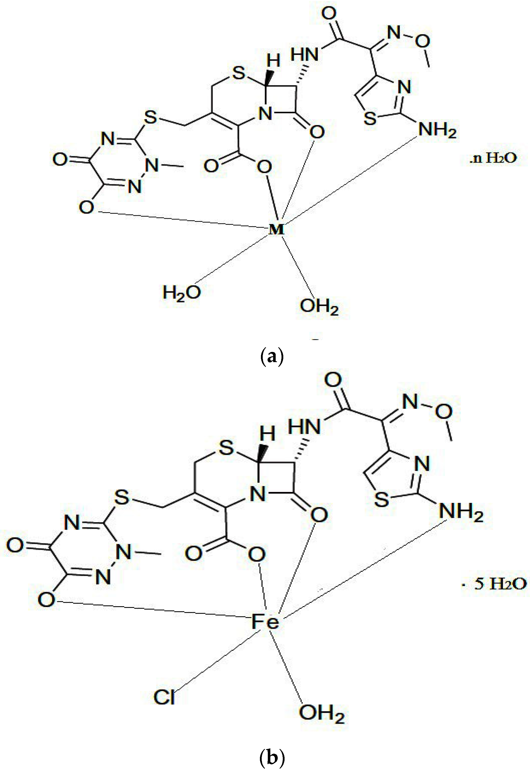
2.2. Synthesis
2.3. Experimental Animals
2.4. Hepatic Functions and Antioxidant Assay
2.5. Histopathological Study
2.6. Antibacterial Activities of CFX and Its Metal Complexes
2.7. Statistical Analysis
3. Results and Discussions
3.1. Microanalytical and Conductance Measurements
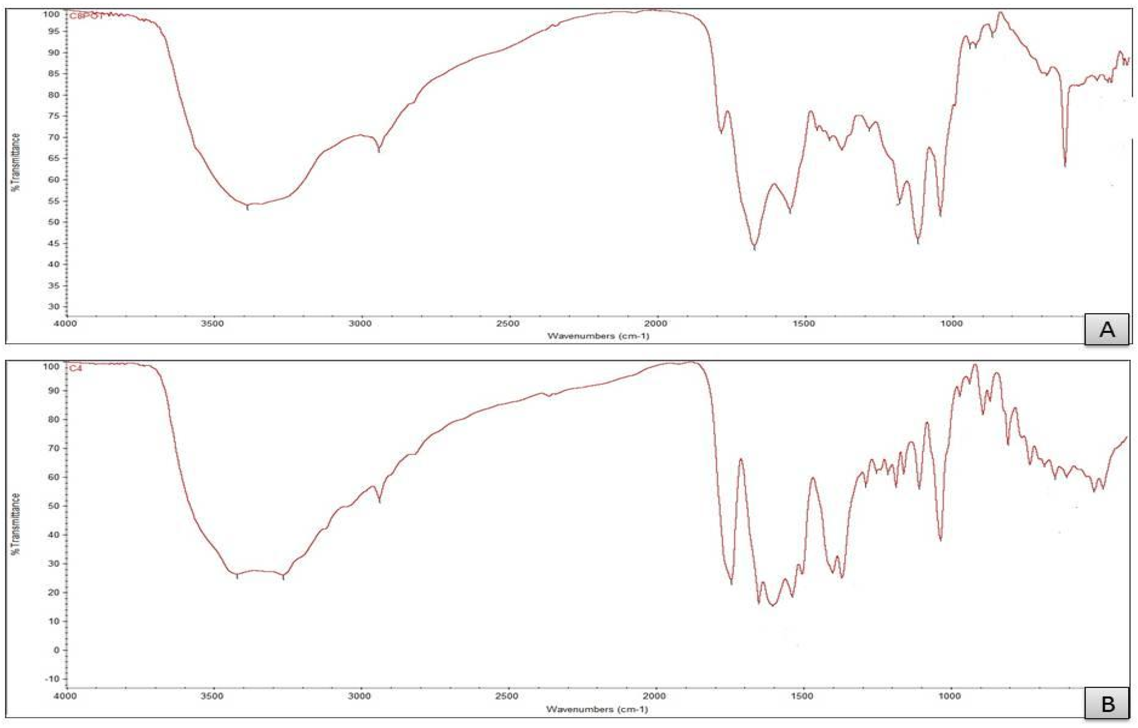
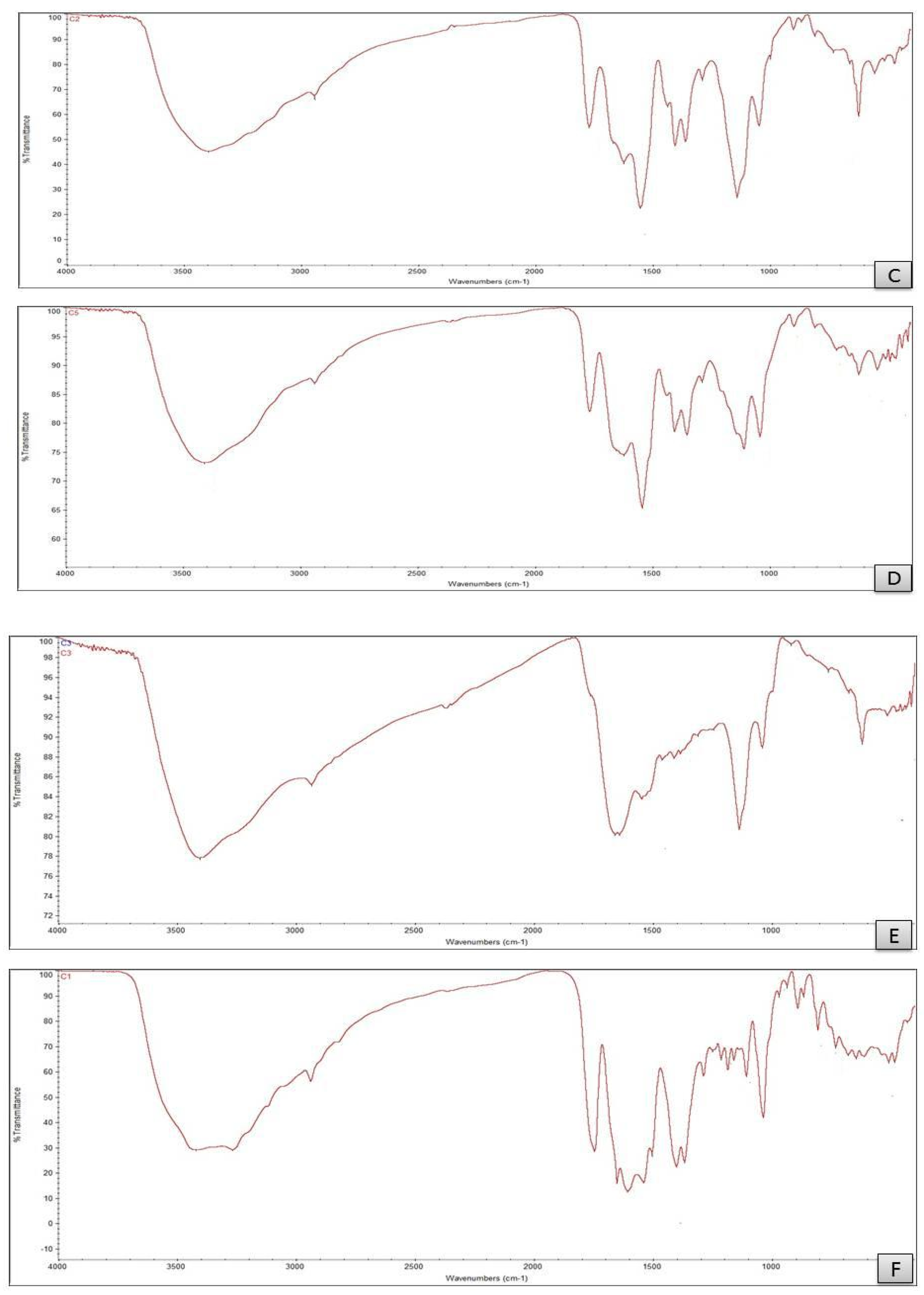
3.2. FTIR Spectral Studies
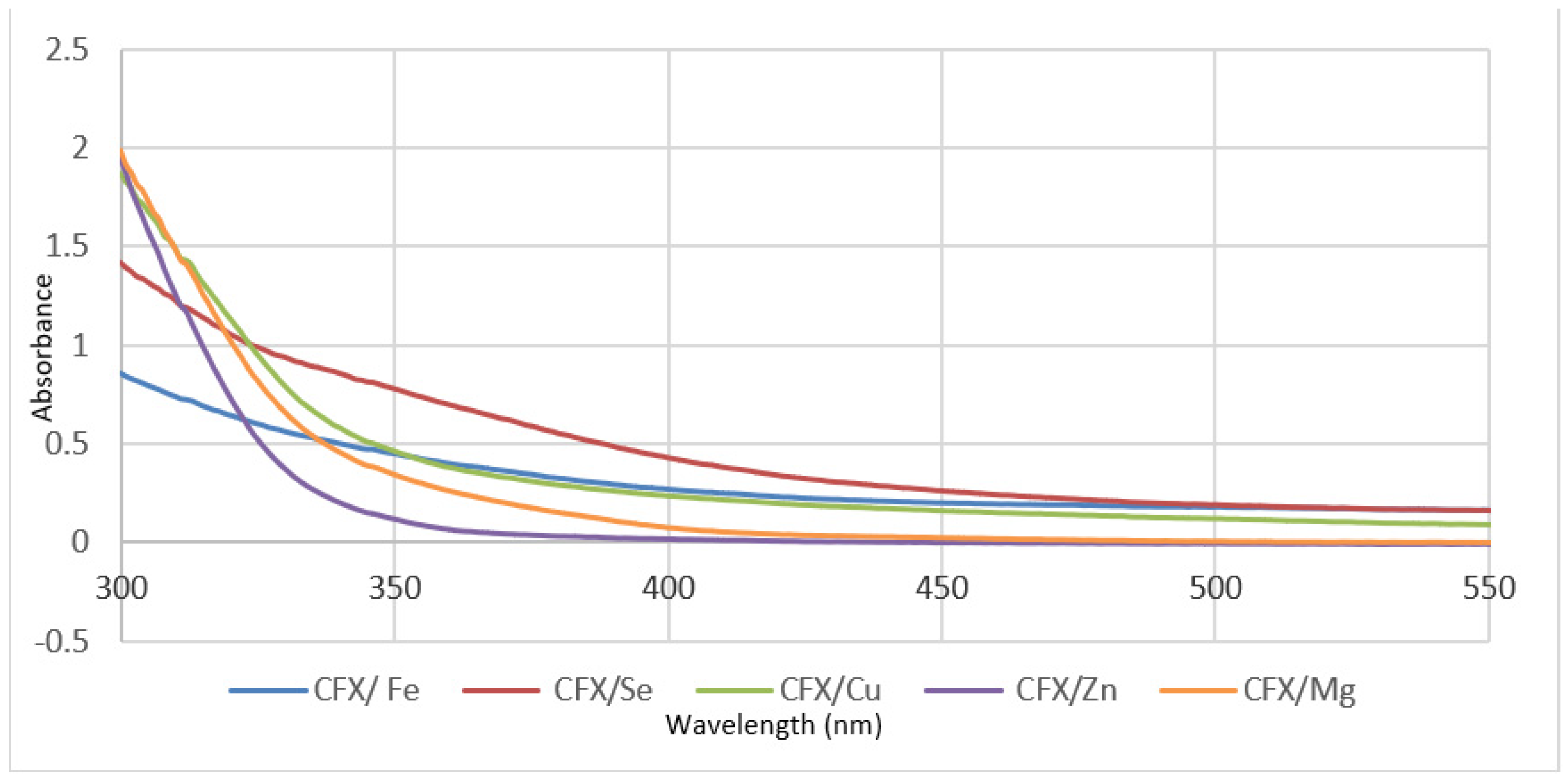
3.3. Electronic Spectra
3.4. Magnetic Measurements
3.5. 1H-NMR Study
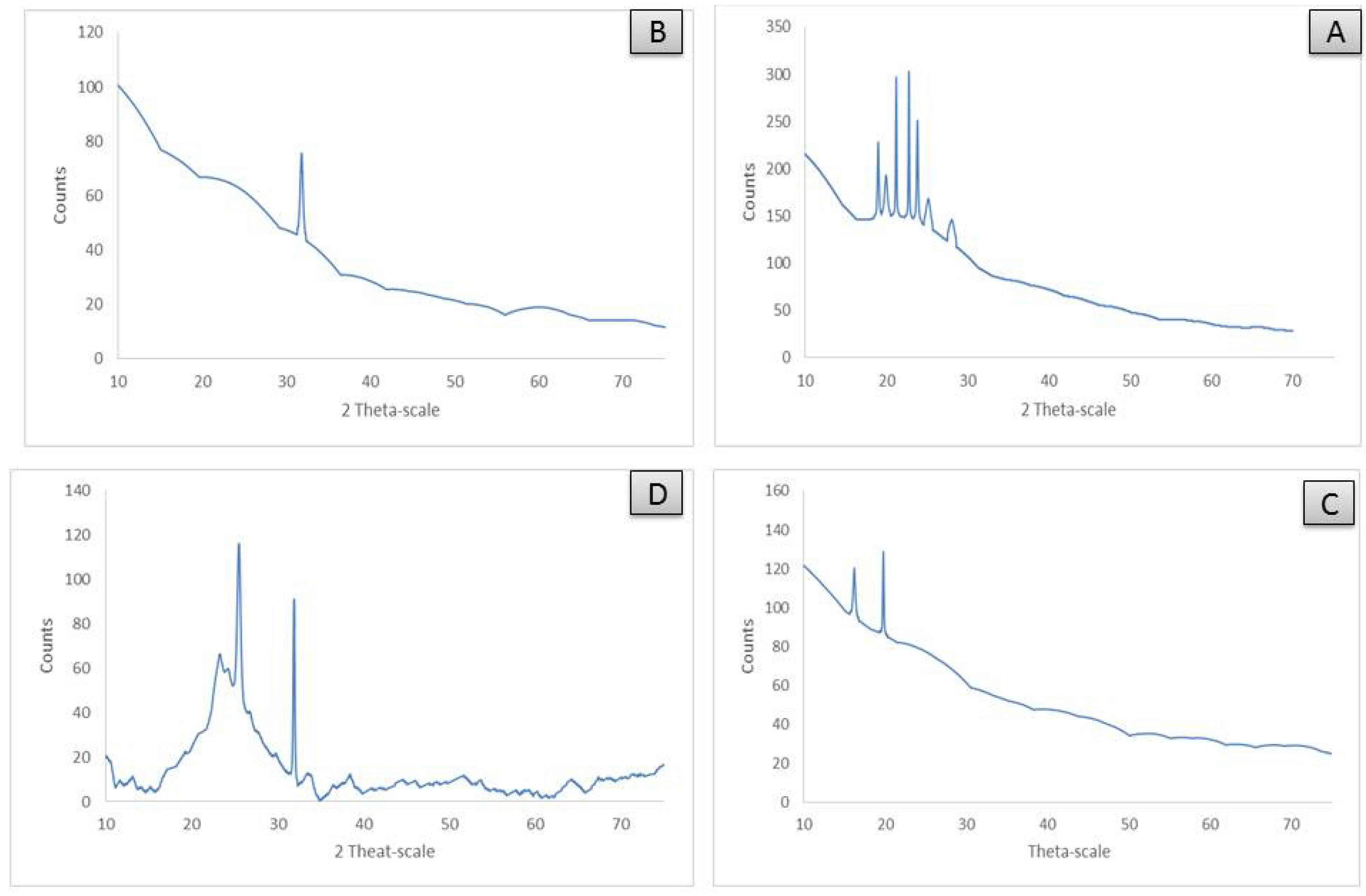
3.6. XRD Analysis
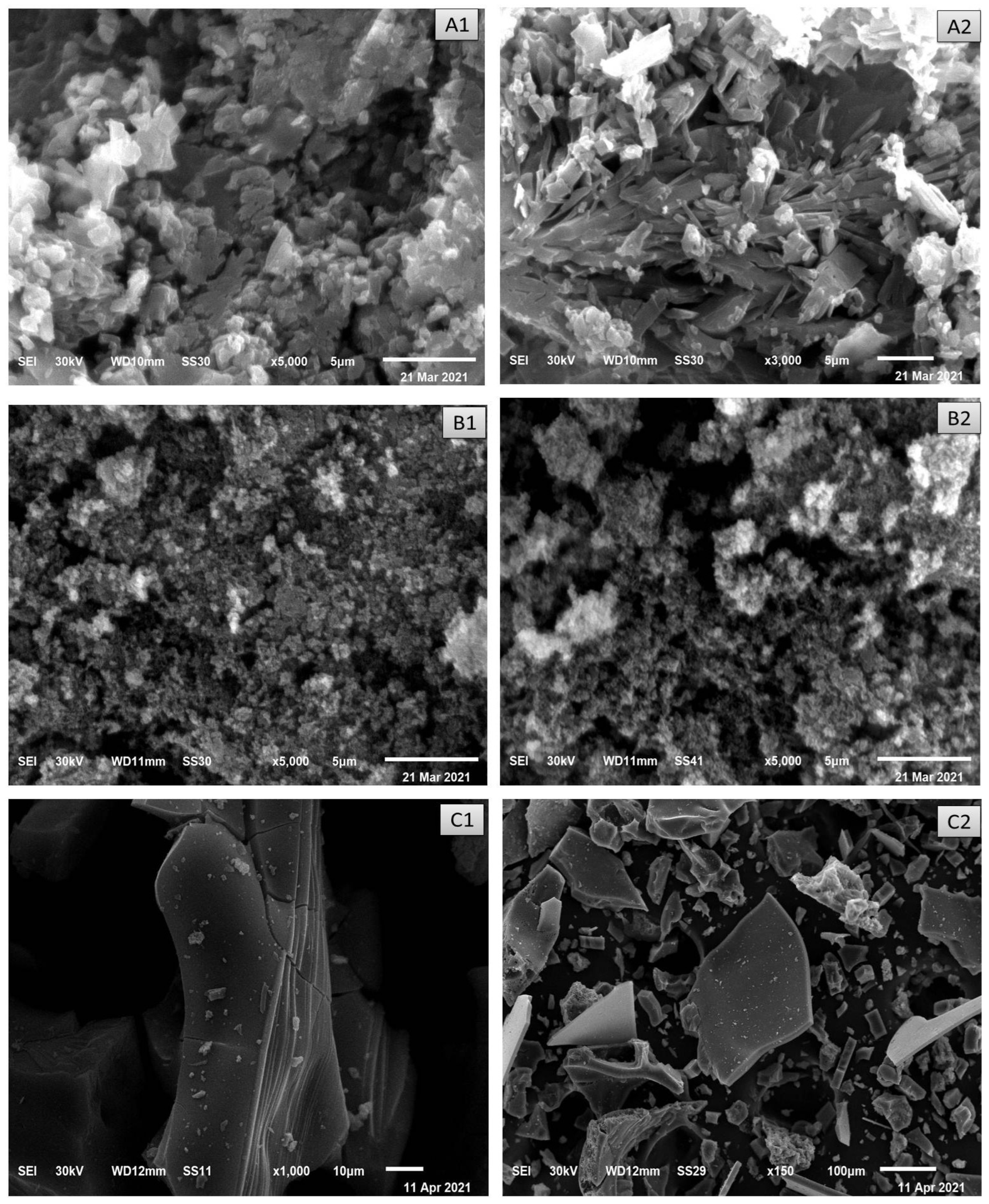
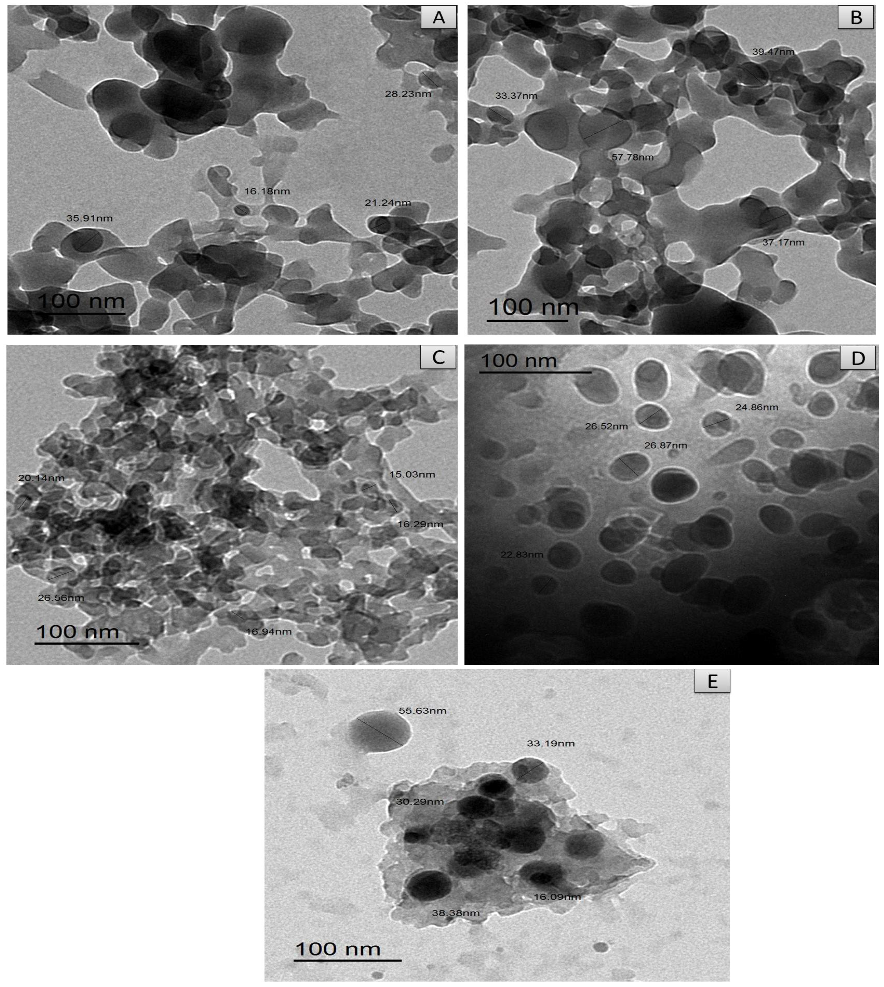
3.7. SEM and TEM Investigations
3.8. Thermal Analysis
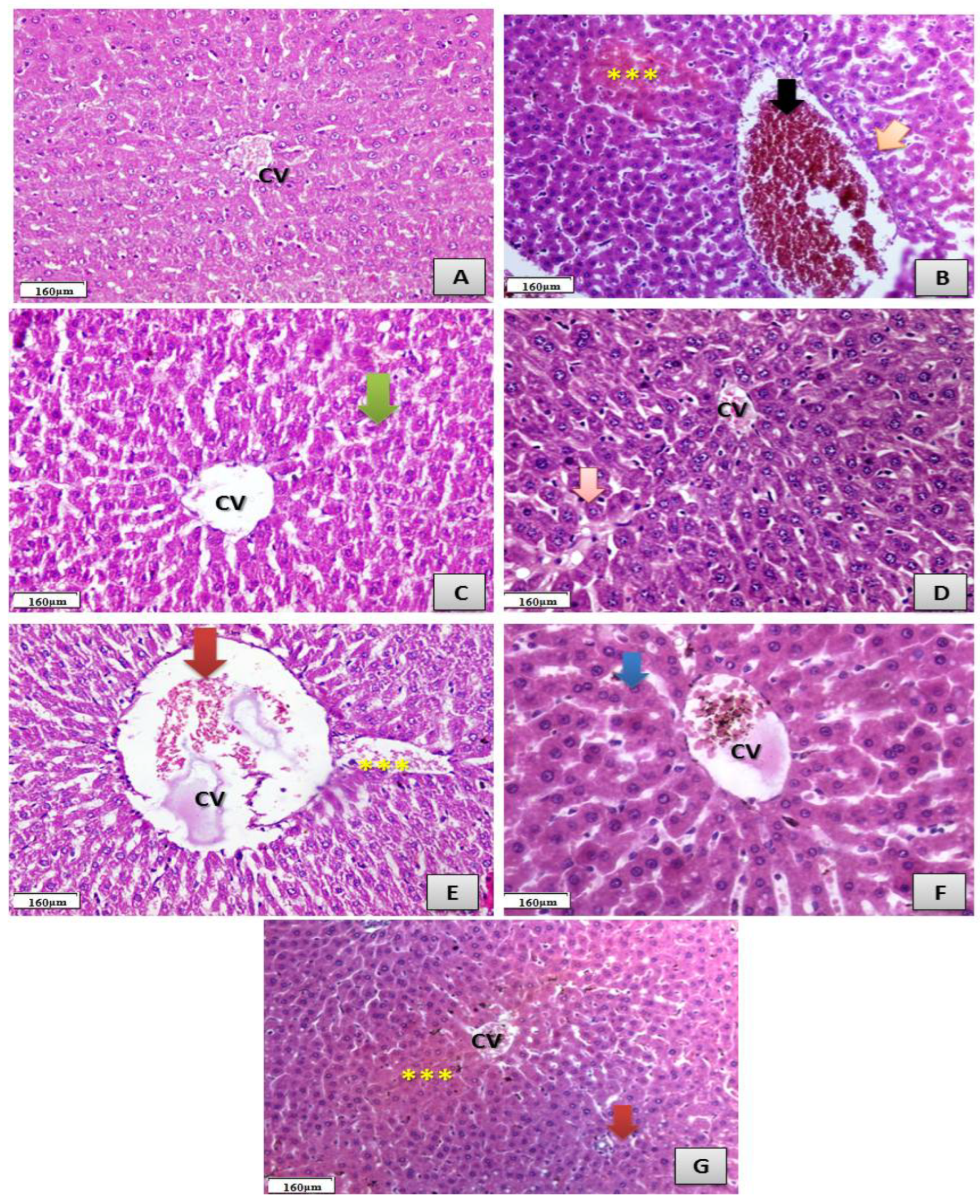
3.9. Ceftriaxone Metal Complexes Alleviate Hepatic Injury in Male Rats
3.10. Ceftriaxone Metal Complexes Alleviate Oxidative Injury in the Hepatic Tissues and Structural Alterations of Male Rats Exposed to Ceftriaxone
3.11. Antibacterial Activity Evaluation
4. Discussion
5. Conclusions
Author Contributions
Funding
Institutional Review Board Statement
Informed Consent Statement
Data Availability Statement
Conflicts of Interest
References
- Bilirubin, R.B.; Alex, R. Ceftriaxone binding to human serum albumin. Mol. Pharmacol. 1989, 36, 478–483. [Google Scholar]
- Quaglia, M.G.; Bossu, E.; Dell’Aquila, C.; Guidotti, M. Determination of the binding of a β2-blocker drug, frusemide and ceftriaxone to serum proteins by capillary zone electrophoresis. J. Pharm. Biomed. Anal. 1997, 15, 1033–1039. [Google Scholar] [CrossRef]
- Nerli, B.; Farruggia, B.; Pico, G. A Comparative study of the binding characteristics of ceftriaxone, cefoperazone and cefsudolin to human serum albumin. Biochem. Mol. Biol. Int. 1996, 40, 823–831. [Google Scholar] [CrossRef] [PubMed]
- Kong, K.-F.; Schneper, L.; Mathee, K. Beta-lactam antibiotics: From antibiosis to resistance and bacteriology. APMIS 2010, 118, 1–36. [Google Scholar] [CrossRef] [PubMed]
- Pacifici, G.M. Pharmacokinetics of cephalosporins in the neonate: A review. Clinics 2011, 66, 1267–1274. [Google Scholar] [CrossRef]
- Anacona, J.R.; da Silva, G. Synthesis and antibacterial activity of cefotaxime metal complexes. J. Chil. Chem. Soc. 2005, 50, 447–450. [Google Scholar] [CrossRef]
- Ebrahimi, S.; Farhadian, N.; Karimi, M.; Ebrahimi, M. Enhanced bactericidal effect of ceftriaxone drug encapsulated in nanostructured lipid carrier against gram-negative Escherichia coli bacteria: Drug formulation, optimization, and cell culture study. Antimicrob. Resist. Infect. Control. 2020, 9, 28. [Google Scholar] [CrossRef]
- Kumar, S.; Bhanjana, G.; Kumar, A.; Taneja, K.; Dilbaghi, N.; Kim, K. Synthesis and optimization of ceftriaxone-loaded solid lipid nanocarriers. Chem. Phys. Lipids. 2016, 200, 126–132. [Google Scholar] [CrossRef]
- Manten, A. Side effects of antibiotics. Vet. Q. 1981, 3, 179–182. [Google Scholar] [CrossRef]
- Anacona, J.R.; Rodriguez, A. Synthesis and antibacterial activity of ceftriaxone metal complexes. Trans. Met. Chem. 2005, 30, 897–901. [Google Scholar] [CrossRef]
- Ali, A.E. Synthesis, spectral, thermal and antimicrobial studies of some new tri metallic biologically active ceftriaxone complexes. Spectrochim. Acta A 2011, 78, 224–230. [Google Scholar] [CrossRef] [PubMed]
- Anacona, J.R.; Estacio, J. Synthesis and antibacterial activity of cefixime metal complexes. Trans. Met. Chem. 2006, 31, 227–231. [Google Scholar] [CrossRef]
- Arayne, M.S.; Sultana, N.; Khanum, F.; Ali, M.A. Antibacterial studies of cefixime copper, zinc and cadmium complexes. Pak. J. Pharm. Sci. 2002, 15, 1–8. [Google Scholar]
- Pillai, M.S.; Latha, S.P. Designing of some novel metallo antibiotics tuning biochemical behaviour towards therapeutics: Synthesis, characterisation and pharmacological studies of metal complexes of cefixime. J. Saudi Chem. Soc. 2012, 20, S60–S66. [Google Scholar] [CrossRef]
- Sodeifian, F.; Seyedalhosseini, Z.S.; Kian, N.; Eftekhari, M.; Najari, S.; Mirsaeidi, M.; Farsi, Y.; Nasiri, M.J. Drug-Induced Liver Injury in COVID-19 Patients: A Systematic Review. Front. Med. 2021, 8, 731436. [Google Scholar] [CrossRef] [PubMed]
- Kulkarni, A.V.; Kumar, P.; Tevethia, H.V.; Premkumar, M.; Arab, J.P.; Candia, R.; Talukdar, R.; Sharma, M.; Qi, X.; Rao, P.N.; et al. Systematic review with meta-analysis: Liver manifestations and outcomes in COVID-19. Aliment. Pharmacol. Ther. 2020, 52, 584–599. [Google Scholar] [CrossRef] [PubMed]
- Hoffmann, M.; Kleine-Weber, H.; Schroeder, S.; Krüger, N.; Herrler, T.; Erichsen, S.; Schiergens, T.S.; Herrler, G.; Wu, N.H.; Nitsche, A.; et al. SARS-CoV-2 cell entry depends on ACE2 and TMPRSS2 and is blocked by a clinically proven protease inhibitor. Cell 2020, 181, 271–280. [Google Scholar] [CrossRef] [PubMed]
- Xu, X.W.; Wu, X.X.; Jiang, X.G.; Xu, K.J.; Ying, L.J.; Ma, C.L.; Li, S.B.; Wang, H.Y.; Zhang, S.; Gao, H.N.; et al. Clinical findings in a group of patients infected with the 2019 novel coronavirus (SARS-CoV-2) outside of Wuhan, China: Retrospective case series. BMJ 2020, 368, m606. [Google Scholar] [CrossRef]
- Lykhin, A.O.; Novikova, G.V.; Kuzubov, A.A.; Staloverova, N.A.; Sarmatova, N.I.; Varganov, S.A.; Krasnov, P.O. A complex of ceftriaxone with Pb (II): Synthesis, characterization, and antibacterial activity study. J. Coord. Chem. 2014, 67, 2783–2794. [Google Scholar] [CrossRef]
- Refat, M.S.; Altalhi, T.; Fetooh, H.; Alsuhaibani, A.M.; Hassan, R.F. In neutralized medium five new Ca(II), Zn(II), Fe(III), Au(III) and Pd(II) complexity of ceftriaxone antibiotic drug: Synthesis, spectroscopic, morphological and anticancer studies. J. Mol. Liq. 2021, 322, 114816. [Google Scholar] [CrossRef]
- Hamza, R.Z.; Sheshah, Z.A.; Suleman, R.H.; Al-Juaid, N.F.; Hamed, N.A.; Al-Juaid, M.A. Efficacy of some antibiotics and some metal complexes (Nano-formula) that could increase their effectiveness during COVID-19. Int. J. Biol. Pharm. Sci. Arch. 2022, 3, 8–14. [Google Scholar] [CrossRef]
- Novikova, G.V.; Tsyplenkova, D.I.; Kuzubov, A.A.; Kolenchukova, O.A.; Samoilo, A.S.; Vorobyev, S.A. Complex of Ca (II) with Ceftriaxone: Synthesis, Structure, Spectral and Antibacterial Properties. J. Sib. Fed. Univ. Chem. 2021, 14, 290–301. [Google Scholar] [CrossRef]
- Alhumaidha, K.A.; El-Awdan, S.A.; El-Iraky, W.I.; Ezz-El-Din, S. Protective effects of ursodeoxycholic acid on ceftriaxone-induced hepatic injury in rats. Bull. Fac. Pharm. Cairo Univ. 2014, 52, 45–50. [Google Scholar] [CrossRef][Green Version]
- Mihara, M.; Uchiyama, M. Determination of malonaldehyde precursor in tissues by thiobarbituric acid test. Anal. Biochem. 1978, 86, 271–278. [Google Scholar]
- Ellman, G.L. Tissue sulfhydryl groups. Arch. Biochem. Biophys. 1959, 82, 70–77. [Google Scholar] [CrossRef]
- Marklund, S.; Marklund, G. Involvement of the superoxide anion radical in the autoxidation of pyrogallol and a convenient assay for superoxide dismutase. Eur. J. Biochem. 1974, 47, 469–474. [Google Scholar] [CrossRef]
- Sinha, A.K. Colorimetric assay of catalase. Anal. Biochem. 1972, 47, 389–394. [Google Scholar] [CrossRef]
- National Committee for Clinical Laboratory Standards. Performance Vol. Antimicrobial Susceptibility of Flavobacteria; National Committee for Clinical Laboratory Standards: Wayne, PA, USA, 1997. [Google Scholar]
- National Committee for Clinical Laboratory Standards. Methods for Dilution Antimicrobial Susceptibility Tests for Bacteria That Grow Aerobically; Approved Standard M7-A3; National Committee for Clinical Laboratory Standards: Villanova, PA, USA, 1993. [Google Scholar]
- Liebowitz, L.D.; Ashbee, H.R.; Evans, E.G.; Chong, Y.; Mallatova, N.; Zaidi, M.; Gibbs, D.; Global Antifungal Surveillance Group. A two year global evaluation of the susceptibility of Candida species to fluconazole by disk diffusion. Diagn. Microbiol. Infect. Dis. 2001, 40, 27–33. [Google Scholar] [CrossRef]
- Bancroft, J.D.; Gamble, M. Theory and Practice of Histological Techniques; Elsevier Health Sciences: Amsterdam, The Netherlands, 2008. [Google Scholar]
- Cotton, F.A.; Wilkinson, C.W. Advanced Inorganic Chemistry, 3rd ed.; Interscience Publisher: New York, NY, USA, 1972. [Google Scholar]
- Nakamoto, K. Infrared and Raman Spectra of Inorganic and Coordination Compounds, 4th ed.; Wiley: New York, NY, USA, 1986. [Google Scholar]
- Deacon, G.B.; Philips, R.J. Relationships between the carbon-oxygen stretching frequencies of carboxylato complexes and the type of carboxylate coordination. Coord. Chem. Rev. 1980, 33, 227. [Google Scholar] [CrossRef]
- Zaman, R.; Rehman, W.; Hassan, M.; Khan, M.M.; Anjum, Z.; Shah SA, H.; Abbas, S.R. Synthesis, characterization and biological activities of cephalosporin metals complexes. Int. J. Biosci. 2016, 9, 163–172. [Google Scholar]
- Lever, A.B.P. Electronic Spectra of dn Ions Inorganic Electronic Spectroscopy, 2nd ed.; Elsevier: Amsterdam, The Netherlands, 1984. [Google Scholar]
- Franchini, G.C.; Giusti, A.; Preti, C.; Tosi, L.; Zannini, P. Coordinating ability of methylpiperidine dithiocarbamates towards platinum group metals. Polyhedron 1985, 9, 1553. [Google Scholar] [CrossRef]
- Hadjikostas, C.C.; Katsoulos, G.A.; Shakhatreh, S.K. Synthesis and spectral studies of some new palladium (II) and platinum (II) dithiocarbimato complexes. Reactions of bases with the corresponding N-alkyldithiocarbamates. Inorg. Chim. Acta 1987, 133, 129. [Google Scholar] [CrossRef]
- Cullity, B.D. Elements of X-ray Diffraction, 2nd ed.; Addision-Wesley Publishing Company: Boston, MA, USA, 1978. [Google Scholar]
- Andrade, R.; Lopez-Vega, M.; Robles, M.; Cueto, I.; Lucena, M.I. Idiosyncratic drug hepatotoxicity: A 2008 update. Expert Rev. Clin. Pharmacol. 2008, 1, 261–276. [Google Scholar] [CrossRef] [PubMed]
- Stine, J.; Lewis, J. Hepatotoxicity of antibiotics: A review and update for the clinician. Clin. Liver Dis. 2013, 17, 606–642. [Google Scholar] [CrossRef] [PubMed]
- Raghunath, M.; Bakal, S. Formulation and evaluation of a fixed dose combination of ceftriaxone disodium and ornidazole. Int. J. Pharm. Life Sci. 2013, 5, 750–756. [Google Scholar]
- Simmons, C. From Your Newsletter Beware: Antibiotic-induced hepatotoxicity is rare but deadly. Hosp. Pharm. 2002, 37, 326–333. [Google Scholar] [CrossRef]
- Vial, T.; Biour, M.; Descotes, J.; Trepo, C. Antibiotic-associated hepatitis: Update from 1990. Ann. Pharm. 1997, 31, 204–220. [Google Scholar] [CrossRef]
- Bell, M.; Stockwell, D.; Luban, N.; Shirey, R.; Shaak, L.; Ness, P.; Wong, E. Ceftriaxone-induced hemolytic anemia and hepatitis in an adolescent with hemoglobin SC disease. Pediatr. Crit. Care Med. 2005, 6, 363–366. [Google Scholar] [CrossRef]
- Rivkin, A. Hepatocellular enzyme elevations in a patient receiving ceftriaxone. Am. J. Health Syst. Pharm. 2005, 62, 2006–2010. [Google Scholar] [CrossRef]
- Elsayed, M.; Elkomy, A.; Aboubakr, M. Hepatotoxicity evaluation in albino rats exposed to ceftriaxone. Asian J. Phar. Biol. Res. 2011, 1, 145–150. [Google Scholar]
- Malomo, S. Toxicological implications of ceftriaxone administration in rats. Nig. J. Biochem. Mol. Biol. 2000, 15, 33–38. [Google Scholar]
- Bhamidimarri, K.; Eugene, S. Drug-Induced Cholestasis. Clin. Liver Dis. 2013, 17, 519–531. [Google Scholar] [CrossRef] [PubMed]
- Al-Salmi, F.A.; Hamza, R.Z. Efficacy of Vanadyl Sulfate and Selenium Tetrachloride as Anti-Diabetic Agents against Hyperglycemia and Oxidative Stress Induced by Diabetes Mellitus in Male Rats. Curr. Issues Mol. Biol. 2021, 44, 94–104. [Google Scholar] [CrossRef]
- El-Megharbel, S.M.; Al-Salmi, F.A.; Al-Harthi, S.; Alsolami, K.; Hamza, R.Z. Chitosan/Selenium Nanoparticles Attenuate Diclofenac Sodium-Induced Testicular Toxicity in Male Rats. Crystals 2021, 11, 1477. [Google Scholar] [CrossRef]
- El-Megharbel, S.M.; Refat, M.S.; Al-Salmi, F.A.; Hamza, R.Z. In Situ Neutral System Synthesis, Spectroscopic, and Biological Interpretations of Magnesium(II), Calcium(II), Chromium(III), Zinc(II), Copper(II) and Selenium(IV) Sitagliptin Complexes. Int. J. Environ. Res. Public Health 2021, 18, 8030. [Google Scholar] [CrossRef]
- Gupta, S.P. Roles of metals in human health. MOJ Biorg. Org. Chem. 2018, 2, 221–224. [Google Scholar] [CrossRef]
- Daniel, R.; Urmesh, K.O.; Sangeeta, J.; Chandrashekhar, P.; Mrutyunjay, S. The Small RNA DsrA Influences the Acid Tolerance Response and Virulence of Salmonella enterica Serovar Typhimurium. Front. Microbiol. 2016, 7, 599. [Google Scholar]


| Instrument | Measurement |
|---|---|
| Perkin Elmer CHN 2400 (USA) | Contents of C, H and N |
| Jenway 4010 conductivity meter | Electrolytic or non-electrolytic character |
| Bruker FTIR Spectrophotometer (4000–400 cm−1) | IR measurements |
| UV2 Unicam UV/Vis Spectrophotometer | Electronic spectra |
| varian mercury VX-300 NMR spectrometer | The 1H-NMR |
| Sherwood scientific magnetic balance using Gouy method | Magnetic measurements |
| Quanta FEG 250 equipment | Scanning electron microscopy (SEM) images |
| X’Pert PRO PAN analytical X-ray powder diffraction, target copper with secondary monochromate | X-ray diffraction patterns |
| JEOL 100s microscope | Transmission electron microscopy images (TEM) |
| Complexes | M.Wt | Color | Elemental Analysis | Λm (Ω−1cm2mol−1) | Magnetic Moment (BM) | ||
|---|---|---|---|---|---|---|---|
| C | H | N | |||||
| [Mg(CFX)(H2O)2]·4H2O C18H28N8O13S3 Mg | 684.30 | White | (31.56) 31.78 | (4.09) 4.12 | (16.36) 16.16 | 15 | - |
| [Cu(CFX)(H2O)2]·3H2O C18H26N8O11S3Cu | 705.546 | black | (30.61) 30.58 | (3.68) 3.94 | (15.82) 15.59 | 17 | 2.31 |
| [Fe(CFX)(H2O)(Cl)]·5H2O C18H28ClN8O13S3Fe | 751.98 | Greenish black | (28.72) 28.46 | (3.72) 3.49 | (14.89) 14.65 | 21 | 5.92 |
| [Zn(CFX)(H2O)2]·6H2O C18H30N8O14S3Zn | 762.09 | White | (28.34) 28.61 | (3.93) 3.94 | (14.96) 14.32 | 16 | - |
| [Se(CFX)Cl2]·4H2O C18H26Cl2N8O12S3Se | 694.96 | Yellowish white | (31.08) 31.37 | (3.74) 384 | (16.11) 16.57 | 25 | 5.98 |
| Assignments | Compounds | |||||
|---|---|---|---|---|---|---|
| Na2CFX | Mg (II) | Cu (II) | Fe (III) | Zn (II) | Se (VI) | |
| ν(N–H) | 3410 | 3385 | 3395 | 3390 | 3380 | 33,385 |
| ν(O–H); H2O | - | 3264 | 3280 | 3270 | 3265 | 3290 |
| ν(C=O); lactam ring | 1782 | 1744 | 1769 1670 | 1766 1657 | 1744 1649 | 1766 1660 |
| νas(C–N) + ν(C=O)OCO ν(COO) | 1604 | 1537 1503 | 1550 | 1543 | 1537 1503 | 1545 |
| δ(CH2) + δ(CH3) | 1416 | 1408 | 1403 | 1404 | 1400 | 1409 |
| δ(CH)lactam + νas(COO) | 1374 | 1367 | 1359 | 1352 | 1366 | 1309 |
| νs(C–N)triazine | 1281 | 1286 | 1288 | 1287 | 1266 | 1242 |
| δ(CH)lactam + δw(CH3) | 1260 | 1250 | 1245 | 1232 | 1212 | 1242 |
| δr(CH3) | 1178 | 1171 | 1138 | 1146 | 1106 | 1135 |
| δ(CH)aminothiazol | 1040 | 1033 | 1045 | 1040 | 1034 | 1039 |
| ν(N–O) | 921 864 | 889 805 | 898 864 | 895 805 | 890 805 | 918 760 |
| ν(M–O) | - | 645 606 | 657 620 552 | 618 541 | 678 645 610 | 636 619 |
| ν(M–N) | - | 510 492 | 510 487 | 485 461 | 507 482 | 513 475 |
| Signals | Na2CFX Ligand | Mg (II) | Zn (II) | Se (VI) |
|---|---|---|---|---|
| [2H, CH2 of thiazine] | 3.368 | 3.352 | 3.342 | 3.318 |
| [3H, N-CH3 of triazine ring] | 3.489 | 3.375 | 3.312 | 3.254 |
| [3H, =N-O-CH3] | 3.889 | 3.879 | 3.785 | 3.547 |
| [2H, S-CH2] | 3. 960 | 3.864 | 3.687 | 3.758 |
| [1H, β-lactam] | 5.069 | 4.652 | 4.758 | 4.989 |
| [1H, of thiazol ring] | 6.910 | 6.897 | 6.874 | 6.987 |
| Compound | Pos. [2Th.] | Height [cts] | FWHM [2Th.] | d-Spacing [Å] | Rel. Int. [%] |
|---|---|---|---|---|---|
| Zn (II) | 22.7560 | 169.85 | 0.1279 | 3.90457 | 100.00 |
| Cu (II) | 31.8381 | 32.61 | 0.1092 | 2.80845 | 100.00 |
| Fe (III) | 19.767 | 40.99 | 0.1535 | 4.48754 | 100.00 |
| Se (IV) | 19.7678 | 40.99 | 0.1535 | 2.80542 | 100.00 |
| Group Items | Control | CFX | CFX/Mg | CFX/Zn | CFX/Se | CFX/Cu | CFX/Fe |
|---|---|---|---|---|---|---|---|
| ALT (U/L) | 9.68 ± 1.02 g | 89.38 ± 4.69 a | 12.98 ± 1.98 f | 15.58 ± 2.36 e | 19.36 ± 2.56 d | 22.39 ± 1.69 c | 23.69 ± 2.02 bc |
| AST (U/L) | 29.36 ± 2.36 g | 203.36 ± 4.69 a | 50.69 ± 3.69 f | 55.98 ± 4.25 e | 72.25 ± 3.69 d | 79.58 ± 3.69 c | 82.35 ± 2.69 bc |
| MDA (U/g) | 9.68 ± 1.02 g | 123.65 ± 6.25 a | 18.52 ± 2.99 e | 16.52 ± 2.69 f | 24.28 ± 2.69 d | 29.68 ± 3.69 c | 31.02 ± 2.69 bc |
| GSH (nmol/100 mg) | 18.69 ± 1.69 a | 6.98 ± 0.98 g | 15.68 ± 2.05 bc | 14.69 ± 1.69 c | 12.97 ± 2.58 d | 9.01 ± 1.69 f | 10.39 ± 1.69 ef |
| SOD (U/g) | 13.69 ± 2.25 ab | 5.25 ± 0.35 e | 12.66 ± 1.69 b | 11.36 ± 1.58 c | 10.98 ± 1.69 d | 10.32 ± 1.69 d | 10.02 ± 0.69 d |
| CAT (U/g) | 15.69 ± 1.69 ab | 4.36 ± 0.98 f | 13.25 ± 1.65 b | 13.05 ± 1.69 b | 12.69 ± 2.68 c | 10.39 ± 2.69 e | 11.65 ± 1.69 d |
| Sample | Inhibition Zone Diameter (mm/mg Sample) | ||||
|---|---|---|---|---|---|
| Bacillus subtilis (G+) | Streptococcus pneumoniae (G+) | Staphylococcus aureus (G+) | Escherichia coli (G−) | Pseudomonas aeruginosa (G−) | |
| Control (DMSO) | 0.0 ± 0.0 c | 0.0 ± 0.0 d | 0.0 ± 0.0 e | 0.0 ± 0.0 d | 0.0 ± 0.0 d |
| Ceftriaxone (CFX) | 3.80 ± 0.11 b | 5.8 ± 0.73 c | 6± 0.89 d | 3.76 ± 0.31 c | 2.89 ± 0.45 c |
| Zn (II)–CFX | 12 ± 0.62 a | 16 ± 0.21 a | 12 ± 0.58 b | 10.98 ± 0.96 a | 8.90 ± 0.85 a |
| Se (III)–CFX | 11 ± 0.64 a | 9.50 ± 0.91 b | 17 ± 0.37 c | 15.98 ± 0.85 a | 12.76 ± 0.59 a |
Publisher’s Note: MDPI stays neutral with regard to jurisdictional claims in published maps and institutional affiliations. |
© 2022 by the authors. Licensee MDPI, Basel, Switzerland. This article is an open access article distributed under the terms and conditions of the Creative Commons Attribution (CC BY) license (https://creativecommons.org/licenses/by/4.0/).
Share and Cite
El-Megharbel, S.M.; Qahl, S.H.; Alaryani, F.S.; Hamza, R.Z. Synthesis, Spectroscopic Studies for Five New Mg (II), Fe (III), Cu (II), Zn (II) and Se (IV) Ceftriaxone Antibiotic Drug Complexes and Their Possible Hepatoprotective and Antioxidant Capacities. Antibiotics 2022, 11, 547. https://doi.org/10.3390/antibiotics11050547
El-Megharbel SM, Qahl SH, Alaryani FS, Hamza RZ. Synthesis, Spectroscopic Studies for Five New Mg (II), Fe (III), Cu (II), Zn (II) and Se (IV) Ceftriaxone Antibiotic Drug Complexes and Their Possible Hepatoprotective and Antioxidant Capacities. Antibiotics. 2022; 11(5):547. https://doi.org/10.3390/antibiotics11050547
Chicago/Turabian StyleEl-Megharbel, Samy M., Safa H. Qahl, Fatima S. Alaryani, and Reham Z. Hamza. 2022. "Synthesis, Spectroscopic Studies for Five New Mg (II), Fe (III), Cu (II), Zn (II) and Se (IV) Ceftriaxone Antibiotic Drug Complexes and Their Possible Hepatoprotective and Antioxidant Capacities" Antibiotics 11, no. 5: 547. https://doi.org/10.3390/antibiotics11050547
APA StyleEl-Megharbel, S. M., Qahl, S. H., Alaryani, F. S., & Hamza, R. Z. (2022). Synthesis, Spectroscopic Studies for Five New Mg (II), Fe (III), Cu (II), Zn (II) and Se (IV) Ceftriaxone Antibiotic Drug Complexes and Their Possible Hepatoprotective and Antioxidant Capacities. Antibiotics, 11(5), 547. https://doi.org/10.3390/antibiotics11050547