Article

Staphylococcus aureus Biofilm Inhibiting Activity of Advanced Glycation Endproduct Crosslink Breaking and Glycation Inhibiting Compounds

Anti-Aging Medical Research Center and Glycative Stress Research Center, Faculty of Life and Medical Sciences, Doshisha University, Kyoto 610-0394, Japan
*
Author to whom correspondence should be addressed.
Antibiotics 2022, 11(10), 1412; https://doi.org/10.3390/antibiotics11101412
Submission received: 18 September 2022 / Revised: 6 October 2022 / Accepted: 12 October 2022 / Published: 14 October 2022

Abstract

:
Staphylococcus aureus is a Gram-positive bacterium that plays a role in the pathogenesis of skin lesions in diabetes mellitus, atopic dermatitis, and psoriasis, all of which are associated with elevated non-enzymatic glycation biomarkers. The production of biofilm protects resident bacteria from host immune defenses and antibiotic interventions, prolonging pathogen survival, and risking recurrence after treatment. Glycated proteins formed from keratin and glucose induce biofilm formation in S. aureus, promoting dysbiosis and increasing pathogenicity. In this study, several glycation-inhibiting and advanced glycation endproduct (AGE) crosslink-breaking compounds were assayed for their ability to inhibit glycated keratin-induced biofilm formation as preliminary screening for clinical testing candidates. Ascorbic acid, astaxanthin, clove extract, n-phenacylthiazolium bromide, and rosemary extract were examined in an in vitro static biofilm model with S. aureus strain ATCC 12600. Near complete biofilm inhibition was achieved with astaxanthin (ED50 = 0.060 mg/mL), clove extract (ED50 = 0.0087 mg/mL), n-phenacylthiazolium bromide (ED50 = 5.3 mg/mL), and rosemary extract (ED50 = 1.5 mg/mL). The dosage necessary for biofilm inhibition was not significantly correlated with growth inhibition (R2 = 0.055. p = 0.49). Anti-glycation and AGE breaking compounds with biofilm inhibitory activity are ideal candidates for treatment of S. aureus dysbiosis and skin infection that is associated with elevated skin glycation.

1. Introduction

Glycation is a nonenzymatic reaction that occurs in biological systems under physiological conditions between reducing sugars or aldehydes and proteins. After a series of reactions, advanced glycation endproducts (AGEs) are formed and accumulate throughout body tissues. AGEs trigger inflammation [1] and promote protein aggregation [2]. Glycative stress and its downstream effects contribute to the aging process and the etiology of disorders such as diabetes mellitus (DM) [3] and neurodegenerative diseases [4].
Glycative stress may also play a role in infectious disease by weakening immune functioning [5,6] and enhancing bacterial infiltration, as reported in DM associated urinary tract infection [7]. AGEs accumulate in the skin where they and can be measured non-invasively, acting as a biomarker for overall glycative stress and other factors such as arterial stiffness [8]. The glycation of the skin contributes to the appearance of aging and disrupts the skin’s barrier function [9]. AGEs are significantly elevated in inflammatory skin conditions atopic dermatitis (AD) [10,11] and psoriasis [12,13]. These diseases are characterized by the formation of skin lesions that are frequently colonized by pathogenic Staphylococcus aureus, further eroding the skin, exacerbating symptoms, and risking systemic infection. S. aureus and its antibiotic resistant variants are more frequently carried and in higher in abundance on the skin of those with AD [14], psoriasis [15], and DM [16].
It is speculated that the accumulation of AGEs in the epidermis may play a role both in the etiology of disorders such as AD and dysbiosis of the skin microbiome. It has been reported that glycated proteins enhance S. aureus biofilm formation [17,18], which may increase its potential for pathogenicity. Biofilm confers antibiotic resistance [19], protects resident bacteria from host immune responses such as phagocytosis [20], and perpetuates the dominance of S. aureus on AD skin [21]. Biofilm propensity of S. aureus strains is also correlated with increased severity of AD symptoms [22]. Eliminating S. aureus biofilms while combating the underlying glycative stress that contributes to skin dysfunction in AD, psoriasis, and DM is crucial for the prevention of recurring S. aureus skin and soft tissue infection and sustainable use of antibiotics.
Glycative stress can be reduced with lifestyle and dietary choices that reduce blood sugar spikes [23,24]. Nevertheless, AGEs inevitably accumulate in the body over time with age, and once formed, are difficult to remove. Active interventions to breakdown AGEs and increase their clearance are being explored; numerous compounds have been discovered which demonstrate the ability to chemically cleave AGE-protein crosslinks or inhibit the glycation reaction [25]. Some of these compounds have shown promise in animal models [26], and see some use in the dietary supplement market. The applications of AGE crosslink breakers in topical skin treatments to reduce glycated protein accumulation have not yet been thoroughly tested. AGE breaking compounds may prove effective in cosmetic and medicinal skincare in treatment and prevention of skin dysfunction involving elevated glycative stress such as diabetic skin lesions. At the same time, S. aureus dysbiosis and biofilm formation is also a promising target: in addition to the breakdown of biofilm inducing AGEs, these materials may possess antimicrobial and biofilm inhibiting properties, making them ideal for treatment of S. aureus colonized skin lesions associated with glycative stress.
In this study, several compounds with reported AGE crosslink breaking or glycation inhibiting activity were screened for their antimicrobial activity and ability to inhibit S. aureus glycated keratin-induced static biofilm formation.

2. Results

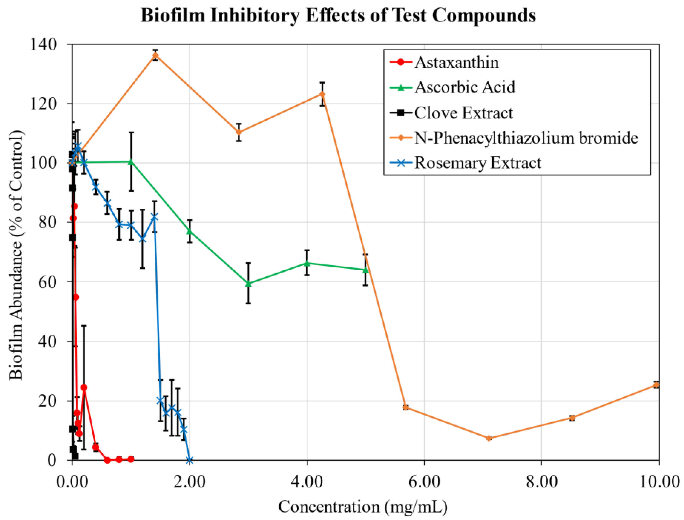
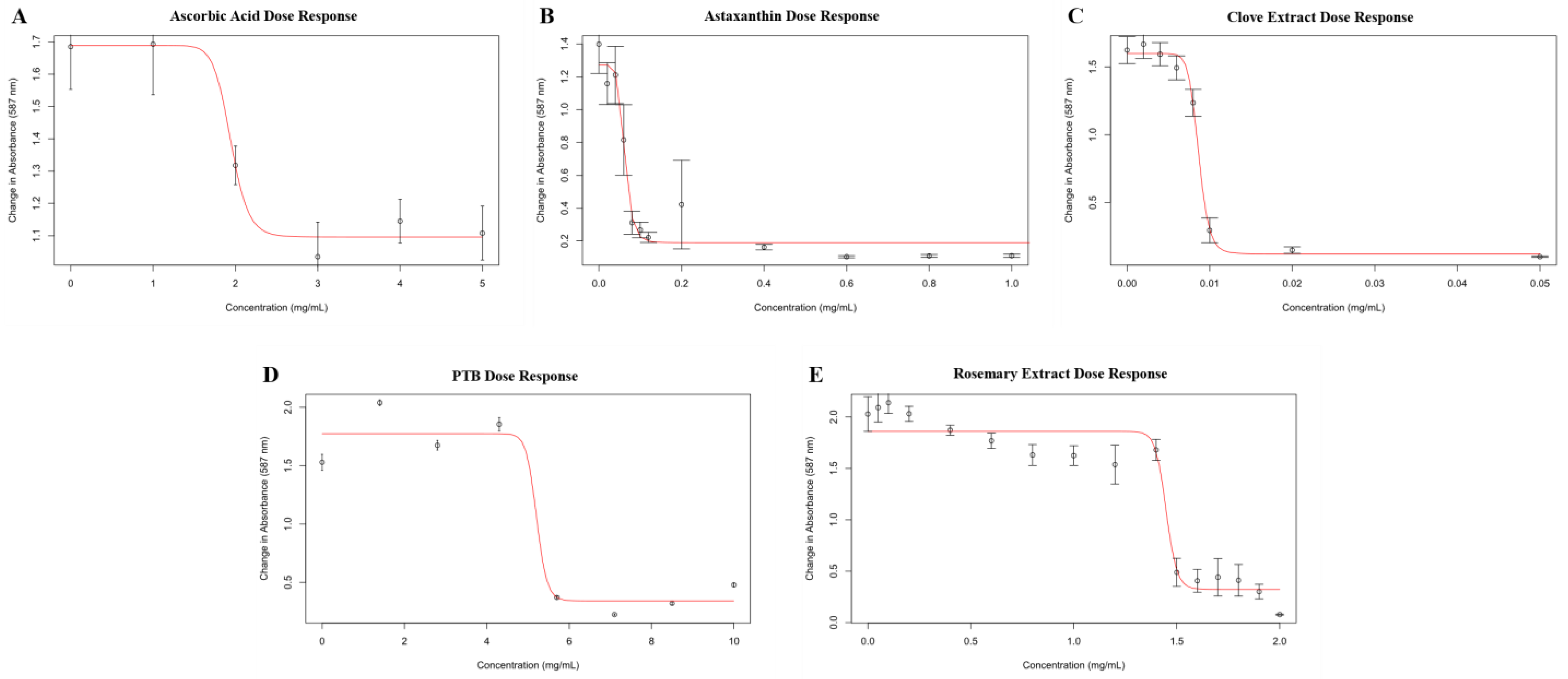
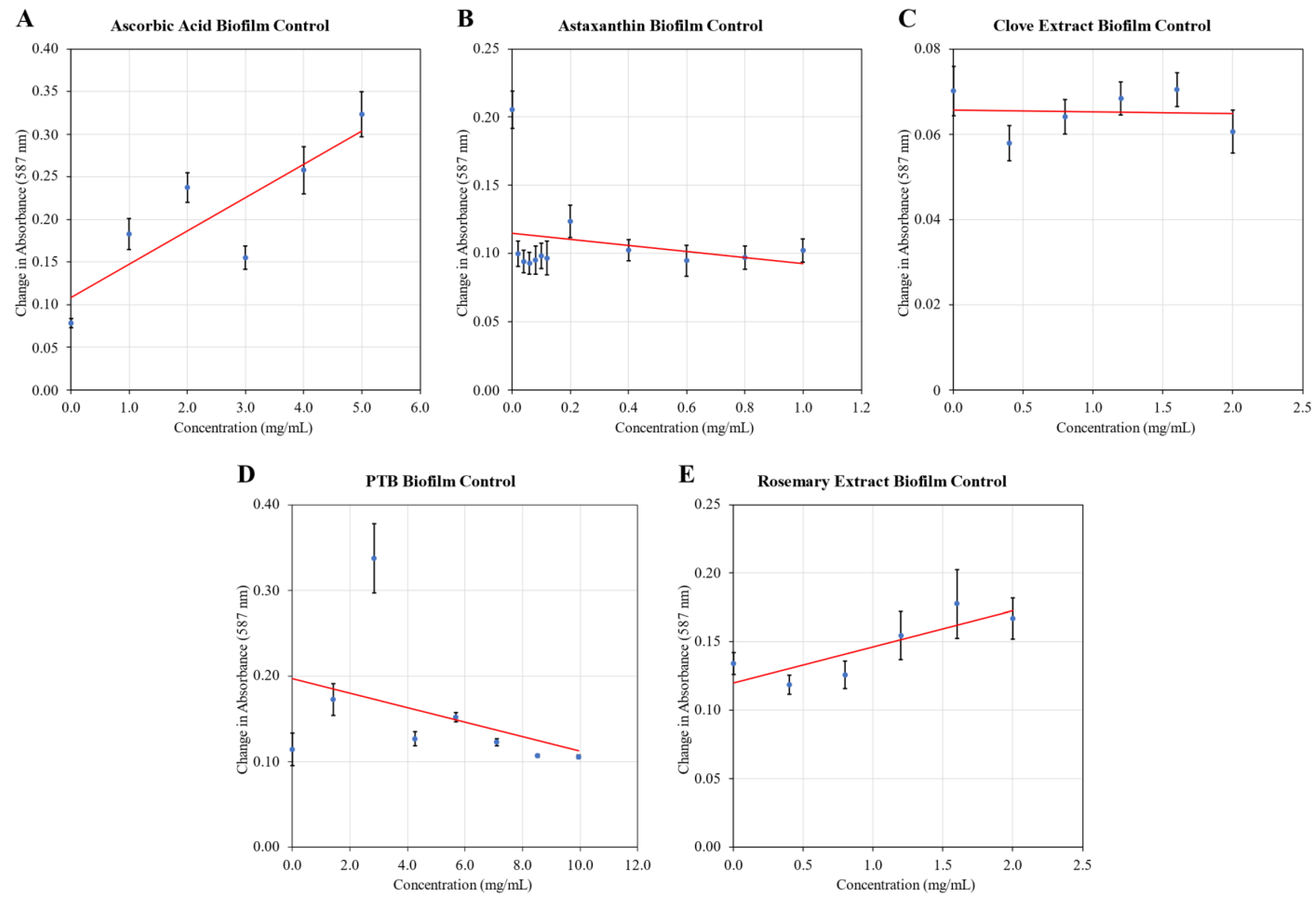
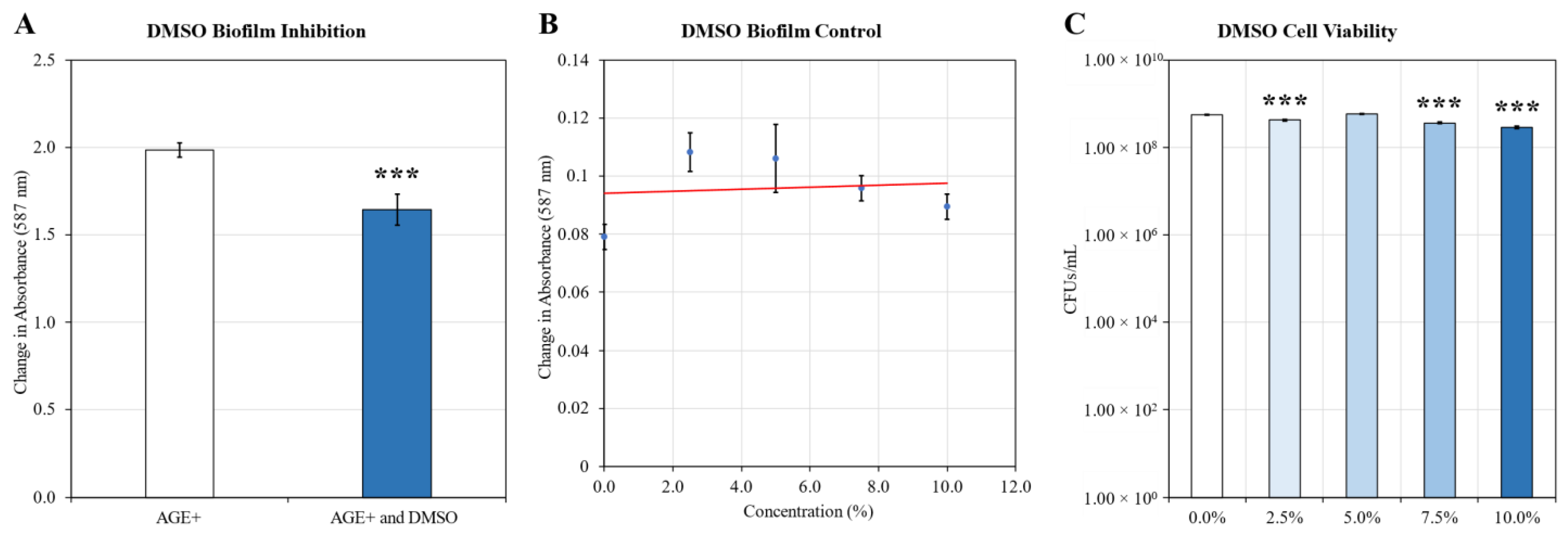
2.1. Biofilm Inhibition

Ascorbic acid, astaxanthin, clove extract, n-phenacylthiazolium bromide (PTB), and rosemary extract were tested. The results are summarized in Table 1. While ascorbic acid failed to completely inhibit glucose-keratin induced biofilm formation, all other compounds demonstrated efficacy at preventing biofilm formation, with clove exhibiting the strongest activity with an ED50 of 0.0087 mg/mL.
Under static conditions S. aureus was incubated with 0.5 mg/mL of glycated keratin to induce a biofilm response, and the inhibitory activities of known AGE crosslink breakers and glycation inhibitors was assayed over a range of dosages (Figure 1). Negative control samples without glucose-keratin were also examined to determine the baseline effect on biofilm formation of each additive (Appendix A Figure A3). S. aureus ATCC 12600 does not produce substantial amounts of biofilm under standard conditions. When treated with glucose-keratin, biofilm production is stimulated in a dose dependent manner. Supplementation with 0.5 mg/mL glucose-keratin results in biofilms that fully covered the surface of sample wells, reaching absorbance values of roughly 1.7–2.0 after staining and solubilization. Typical absorbance values of control samples without glucose-keratin range from approximately 0.07 to 0.10; biofilm formation was considered fully inhibited when absorbance values were reduced to equal or lower than that of vehicle only control samples.
Astaxanthin, clove extract, and rosemary extract all significantly inhibited biofilm formation in a dose-dependent manner. Astaxanthin strongly reduced biofilm abundance at a low dosage, with an ED50 of 0.062 mg/mL and achieving total inhibition by 0.60 mg/mL. Clove extract demonstrated a stronger effect, with an ED50 of 8.7 µg/mL and achieving 98.6% inhibition by 0.050 mg/mL. Rosemary extract was effective at preventing biofilm formation at higher dosages; total biofilm inhibition was achieved at 2.0 mg/mL, with an ED50 of 1.5 mg/mL.
The effect of PTB on biofilm formation was varied. At sub-inhibitory concentrations, biofilm formation significantly increased; the addition of PTB to S. aureus cultures without glycated keratin similarly produced a significant increase in biofilm formation at sub-inhibitory dosages. Only at concentrations above 5.7 mg/mL was biofilm formation reliably prevented, coinciding with a considerable reduction in viable CFUs.
Ascorbic acid performed relatively poorly in biofilm inhibition testing. While ascorbic acid reduced biofilm formation to a peak reduction of 40.5% at 3 mg/mL, the inhibitory effect weakened with further increases in concentration. Without glycated keratin, ascorbic acid dosage was positively correlated with an increase in biofilm formation, resulting in a three-fold increase in biofilm above vehicle-only controls at 5.0 mg/mL.

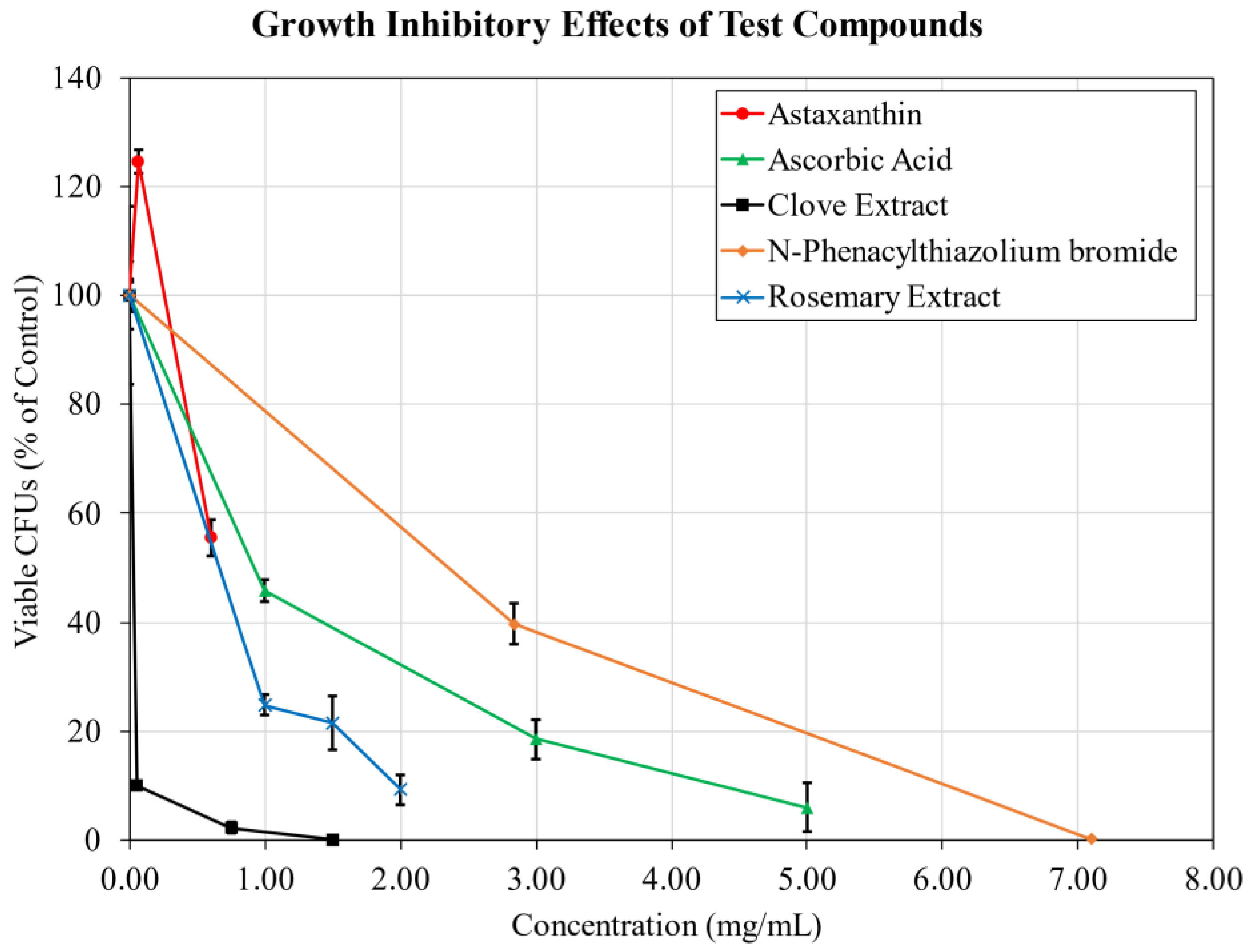
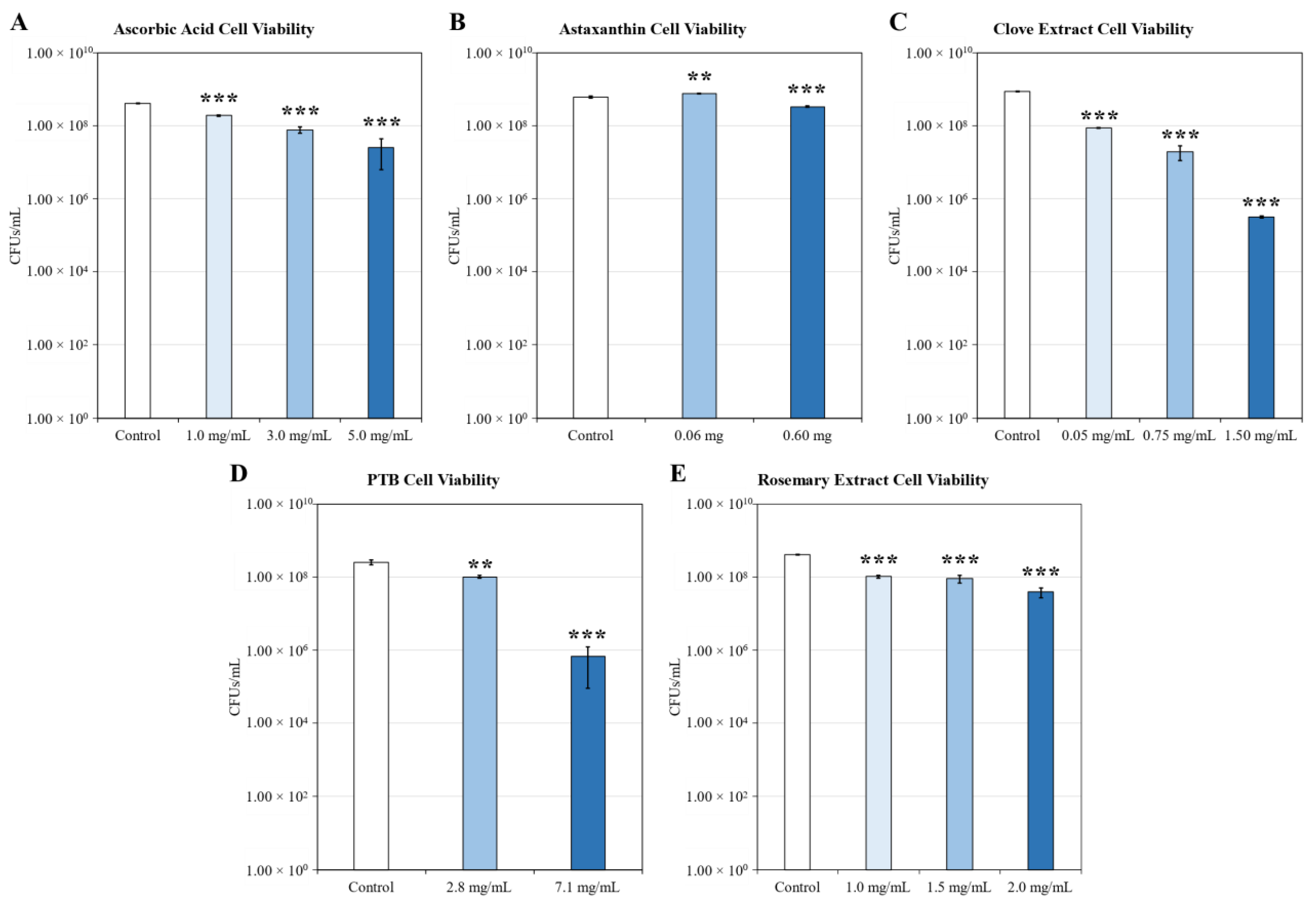
2.2. Growth Inhibition

In order to independently examine the growth inhibitory effects of the tested materials, growth cultures of S. aureus were supplemented with each of the compounds and incubated for 24 h before serial dilution and plate counting to determine the amount of viable colony forming units (Figure 2). Raw viability data for each material is shown in Appendix A Figure A2. The concentrations selected for testing were based on the results of the biofilm inhibition assays, targeting dosages approaching the observed ED50 and total biofilm inhibition if applicable.
Ascorbic acid, clove extract, and PTB demonstrated the strongest growth inhibitory effects of the tested compounds. Ascorbic acid reduced viable colony forming units (CFUs) in a dose dependent manner, reaching a log reduction of 1.2 at 5 mg/mL. Clove extract induced a 1.0 log reduction in viable CFUs at 0.050 mg/mL, increasing to a 3.4 log reduction at a concentration of 1.5 mg/mL coinciding with total biofilm inhibition. PTB at a sub-biofilm inhibitory dosage (2.8 mg/mL) weakly reduced CFU viability by 0.4 log, but achieved a 2.6 log reduction alongside substantial biofilm inhibition at 7.1 mg/mL.
The growth inhibitory effects of astaxanthin and rosemary extract were considerably weaker at the dosages examined. Astaxanthin viable CFU counts slightly increased compared to controls at 0.060 mg/mL, and were not substantially reduced at a near total biofilm inhibitory dose of 0.60 mg/mL. Rosemary extract significantly reduced growth in a dose-dependent manner, however the loss of viable CFUs did not exceed a 0.78 log reduction at the highest concentration of 2.0 mg/mL.

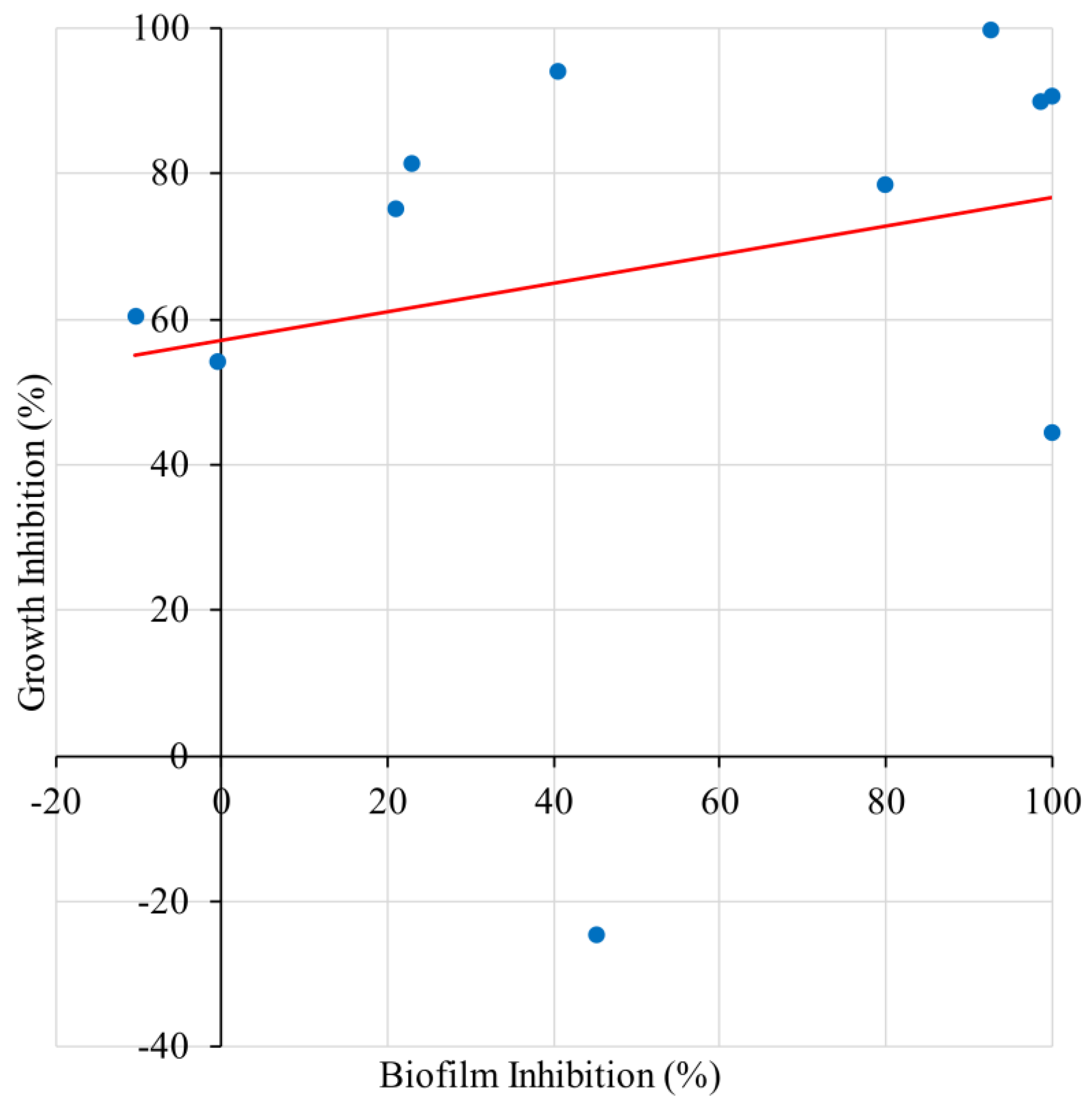
2.3. Correlation of Biofilm Inhibition and Growth Inhibition

Summarizing the data collected from the cell viability assays and the corresponding biofilm inhibition results, a linear regression analysis was performed (Figure 3). Overall, biofilm inhibition did not correlate well with growth inhibition (R2 = 0.055, p = 0.49). All the tested materials demonstrated varying degrees of growth inhibition at sufficient concentrations. However, biofilm inhibitory activity was observed at sub-growth inhibitory dosages (with the exception of PTB), indicating that the antibiofilm activity of these compounds is stronger than the antibacterial. The mechanism of biofilm inhibition may also be independent of direct antibacterial activity.

3. Discussion

Ascorbic acid, commonly known as Vitamin C, is an essential dietary nutrient that is necessary for a variety of physiological processes in the human body, including wound repair and collagen production [27]. Ascorbic acid is also an antioxidant [28] with antibacterial activity [29], and the ability to disrupt exopolysaccharide biofilm formation in E. coli [30], S. mutans [31], B. subtilis [32], and S. aureus [33]. Ascorbic acid is a glycation inhibitor [25,34] via its antioxidant activity and ability to ionically bind to proteins, competitively inhibiting the binding of glucose.
Ascorbic acid appeared to be a strong candidate for treating biofilms: it has been reported [33] to prevent biofilm formation in methicillin resistant S. aureus by downregulating ica, consequently inhibiting Poly-β-1,6-N-acetyl-d-glucosamine (PNAG) production, the primary staphylococcal extracellular polysaccharide. In current testing, Ascorbic acid weakly inhibited biofilm formation at a dosage of 3 mg/mL, failing to completely inhibit biofilm formation as previously reported. This discrepancy suggests that AGE-induced biofilm formation may be ica-independent, which would explain the lack of efficacy. We have observed a significant increase in extracellular DNA released by S. aureus in response to glucose-keratin exposure (data pending publication), and it has been independently reported that S. aureus eDNA release regulators are promoted by AGE exposure [17]. Thus, the eDNA dominant biofilm phenotype may be more resistant to ascorbic acid. Strategies that are typically effective in the inhibition of primarily PNAG biofilms may be less effective in the treatment of AGE-related skin infections involving S. aureus.
Astaxanthin is a carotenoid produced by the algae Haematococcus pluvialis, and found in high concentrations in animals that consume the algae, such as salmon, shrimp, and crabs. Astaxanthin is a strong anti-oxidant [35] and anti-inflammatory compound, and is commercialized as a dietary supplement for these properties. Astaxanthin has also been found to exert anti-glycation effects [36,37]. Previous work in the lab has examined the effects of dietary astaxanthin intake in both animal and human models; astaxanthin was found to ameliorate high fat diet induced gut dysbiosis in mice, and improved reported gastro-intestinal symptoms in human participants [38].
While biofilm formations itself was inhibited, astaxanthin also appeared to have been incorporated into the biofilm itself, changing the white color of the unstained biofilm into a pale red. In addition to direct inhibition, the ability of the existing biofilm to adhere to the surface of the microwell plates was impaired. Remaining biofilm was easily detached by gentle washing, while patches of biofilm in the lowest dosage wells were strongly adhered and could not be removed even by vigorous shaking, as is typical of standard S. aureus biofilms under control conditions. When high concentration astaxanthin samples (0.6 mg/mL and higher) were examined, aggregation of S. aureus surrounding microscopic particles of astaxanthin was observed. This suggests that astaxanthin may be a competitive ligand for cell surface anchors, weakening surface attachment.
Astaxanthin is poorly soluble in water, therefore dimethyl sulfoxide (DMSO) was used as a solvent for in vitro testing. However, DMSO increases the permeability of the skin and causes skin irritation [39], thus it is not suitable for skincare use. Alternative solvents will be needed for clinical testing; the continued efficacy of astaxanthin in these formulations requires confirmation.
Cloves are produced from the dried flowers of the plant Syzygium aromaticum. Cloves are typically used as a culinary spice, but also have a centuries long history of use in traditional medicine for their anti-inflammatory properties. Modern research has demonstrated the powerful antioxidant [40] and anti-inflammation [41] activity of clove extracts. A significant active component of clove is eugenol [42], which has additionally demonstrated anti-carcinogenic/anti-tumor [43,44], and antimicrobial effects [45]. Unpublished data has revealed that extract from clove powder has powerful age cross link breaking activity, which is mediated by the compound biflorin. Considering these properties, the combined effects of eugenol and biflorin make clove extract a strong candidate for use in the AGE-biofilm system for potential treatment of S. aureus dysbiosis and associated skin lesions, although clove extract may also exert a cytotoxic effect on keratinocytes at sufficient dosages [46].
The crosslink breaking abilities of PTB have been demonstrated in both in vitro and in vivo studies [47]. PTB has shown effectiveness in breaking down AGEs and preventing their accumulation in vascular tissues [26], lens proteins [48], periodontic tissue [49] and bone [50]. No antibacterial or antibiofilm properties of PTB have not been reported at this time, although a significant growth inhibitory effect was observed at sufficiently high concentrations. Due to its biofilm enhancing effects at sub-inhibitory dosages, PTB may be less efficacious in antibiofilm applications.
Salvia Rosmarinus, known commonly as rosemary, is a plant that grows in the Mediterranean. The leaves of the plant have long been used as a culinary spice and in traditional medicines. Rosemary extract exhibits strong antioxidant and antibacterial effects [51] and is also known for its glycation inhibiting [52] and AGE cross-link breaking abilities [53] via its active ingredient rosmarinic acid. For this experiment, rosemary extract was utilized in the form of a dietary supplement named AGE Breaker (A2P Sciences, Lyon, France), marketed as an anti-aging supplement. While rosemary extract was effective at inhibiting glucose-keratin-induced biofilm formation, under control conditions without AGEs there was a dose-dependent increase in biofilm production (Appendix A Figure A3E). While the response was relatively small in magnitude, further testing is required to determine whether rosemary extract may be counter effective in practical applications where glycation may be lower than in vitro conditions.

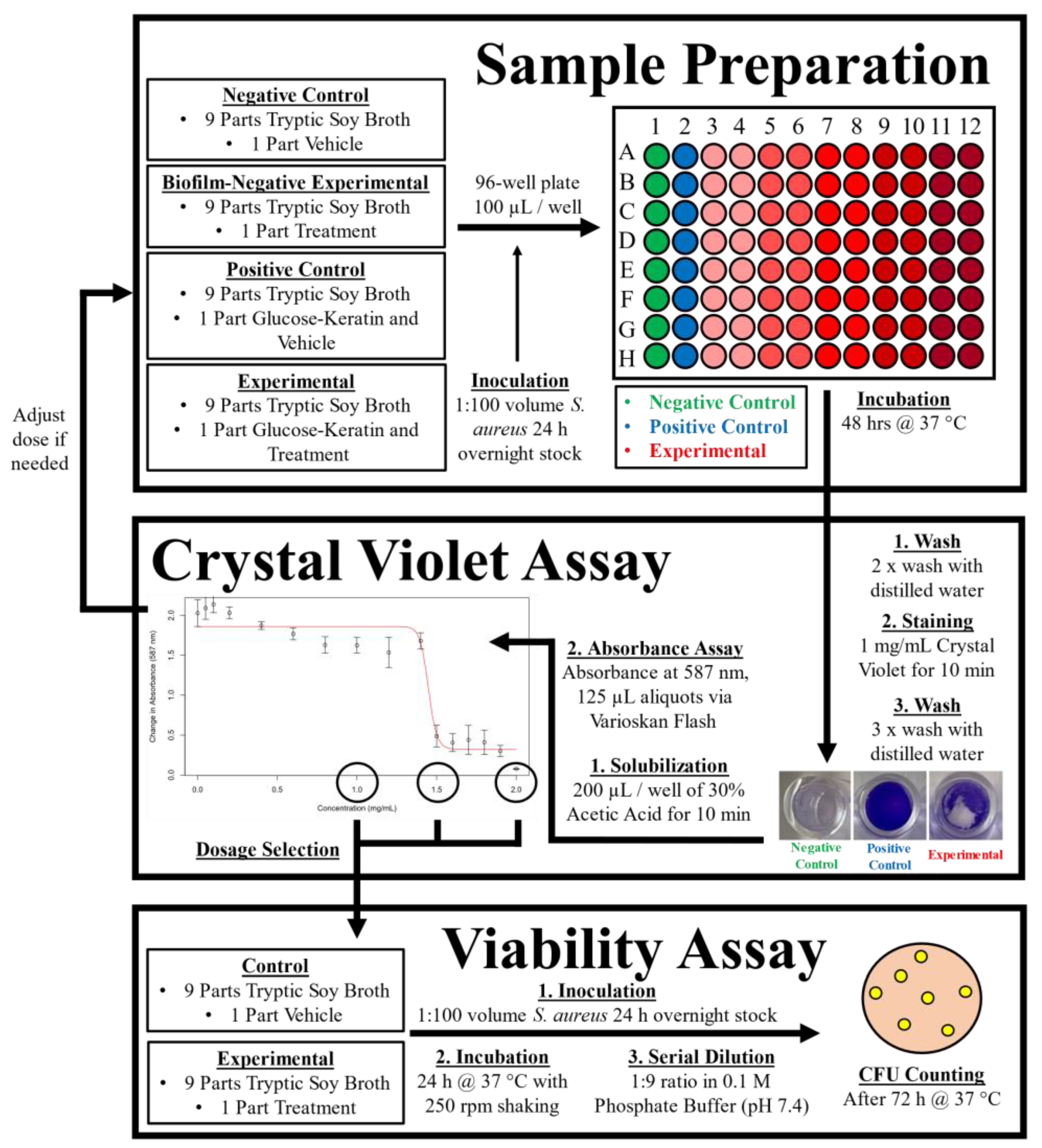
4. Materials and Methods

4.1. Glycation Model

Glycated keratin solution used in testing was produced by incubating proteins with glycating agents in accordance with the recipes outlined by Hori et al. [54]. A solution comprised of 50 mM phosphate buffer (pH 7.4), 0.60 mg/mL keratin (Nacalai Tesque, Kyoto, Japan), and 40 mM glucose was prepared and incubated at 60 °C for 10 days. Ultrafiltration was performed using Amicon Ultra Centrifugal Filters according to the manufacturer’s instructions (Merck Millipore Ltd., Cork, Ireland) to remove unreacted glucose and concentrate protein above 10 kDA. Protein concentration was measured via Bicinchoninic Acid (BCA) Protein Assay (Thermo Fisher Scientific, Waltham, MA, USA).

4.2. Bacteria and Growth Conditions

S. aureus NBRC100910 (ATCC 12600) was purchased from the National Institute of Technology and Evaluation Biological Resource Center (Tokyo, Japan). Planktonic cultures were grown in Tryptic Soy Broth (TSB) at 37 °C, with 250 rpm shaking. Solid media cultures were grown on Tryptic Soy Agar at 37 °C. Growth media used was purchased from Becton, Dickinson, and Company (Franklin Lakes, NJ, USA).

4.3. Biofilm Inhibition Assay

The overall workflow of the experiment is presented in Figure 4.
Biofilm abundance was measured via crystal violet assay, modified from a widely used protocol [55,56,57]. Sample liquid cultures were prepared consisting of a 9:1 volume to volume ratio of TSB growth media and vehicle solution (ultrapure water or 5% DMSO in ultrapure water): a negative control was prepared without additives; a positive control contained 0.5 mg/mL of glucose-keratin; experimental conditions contained various concentrations of test compounds in the appropriate vehicle solution. Each solution was inoculated with 1:100 ratio volume (10 µL) of Staphylococcus overnight stock and thoroughly vortexed before transferring 100 µL aliquots into the wells of a sterile flatbottom 96-well plate (8 replications per sample). Plates were sealed and incubated at 37 °C for 48 h. Plates were twice washed with distilled water to remove unadhered bacteria before staining with 1.0 mg/mL crystal violet solution for 10 min. After staining, plates were gently washed with distilled water three times and air dried overnight. Biofilms were solubilized using 200 µL of 30% glacial acetic acid solution (Sigma-Aldrich, St. Louis, MO, USA). Finally, the absorbance of 125 µL aliquots at 587 nm was collected in triplicate using a Varioskan Flash Multimode Microplate Reader (Thermo Fisher Scientific, Waltham, MA, USA).

4.4. Cell Viability

Viability was determined by plate-counting after serial dilution in 0.1 M phosphate buffer (pH 7.4); Tryptic Soy Agar plates were inoculated with 100 µL of diluted culture media in triplicate and incubated at 37 °C for 72 h with daily colony counting.

4.5. Treatment Compounds

A list of the tested materials can be found in Table 2.
Ascorbic acid and n-phenacylthiazolium bromide were dissolved directly in ultrapure water at room temperature.
A heated water extraction was performed for clove and rosemary extracts. For rosemary extract, a commercial anti-aging supplement, AGE Breaker was tested. Capsules of the supplement were dissolved in 40 mL of ultrapure water and heated to 60 °C for 60 min. Clove powder was solubilized in 40 mL of ultrapure water and heated to 80 °C for 75 min The resulting solutions were centrifuged at 2500 rpm for 10 min and the supernatant collected.
Astaxanthin was solubilized in DMSO at room temperature. For tests using astaxanthin all sample cultures were adjusted to a final DMSO concentration of 5.0 % (See Appendix A Figure A4 for DMSO data)

4.6. Logistic Regression Analysis

Logistic curve-fitting was performed in R [59] using the Dose Response Curve package [60] in order to determine the ED50 of each test compound with regard to biofilm inhibition. The results of curve fitting are shown in Appendix A Figure A1.

4.7. Other Statistical Analysis

Simple descriptive statistical analysis was performed using Excel 2016 version 2108 (Microsoft). Between group statistical difference was calculated using Student’s t-test.

5. Conclusions

Astaxanthin, clove extract, and rosemary extract proved effective at inhibiting S. aureus biofilms induced by glucose-keratin. Clove extract was most powerful with an ED50 of 3.2 µg/mL. PTB inhibited biofilm only after reaching a critical concentration, and ascorbic acid was ineffective at completely preventing biofilm formation.
Further testing is necessary to evaluate the efficacy of AGE crosslink breaker clearance of glycated protein aggregates and reducing AGE accumulation in the context of topical application to the skin. Furthermore, the potential dysbiosis ameliorating effects of these compounds requires verification in beyond in vitro testing through in vivo and clinical study. If they continue to prove efficacious, compounds such as astaxanthin, clove extract, and rosemary extract may become a useful addition to the lineup of antimicrobial/antibiofilm treatments for combating S. aureus dysbiosis in skin disorder involving glycative stress, and the prevention of recurring infection of lesional skin in those with diabetes mellitus.

Author Contributions

K.H. conceived and designed the study with guidance from M.Y. and Y.Y.; K.H. carried out the experiments. The manuscript was written by K.H. in consultation with M.Y. and Y.Y., who provided critical feedback. All authors contributed to revising the manuscript. All authors have read and agreed to the published version of the manuscript.

Funding

This research received no external funding.

Institutional Review Board Statement

Not applicable.

Informed Consent Statement

Not applicable.

Data Availability Statement

All data presented in this study are available upon request.

Conflicts of Interest

The authors declare no conflict of interest.

Appendix A

Figure A1. Biofilm Inhibition Logistic Regression. Effect of test compounds on S. aureus biofilm formation provoked by 0.5 mg/mL of glucose-keratin. (A) Ascorbic acid, R2 = 0.99. (B) Astaxanthin, R2 = 0.96. (C) Clove extract, R2 = 0.99. (D) PTB, R2 = 0.96. (E) Rosemary extract, R2 = 0.94. Biofilm assayed after 48 h of growth under static conditions. n = 8. Mean ± standard deviation. Logistic regression analysis performed using the dose–response curve package in R.
Antibiotics 11 01412 g0a1
Figure A2. Cell Viability. S. aureus cell viability after 24 h of incubation under planktonic conditions with additives. (A) Ascorbic acid, (B) Astaxanthin, (C) Clove extract, (D) PTB, (E) Rosemary extract. n = 3. Mean ± Standard Deviation. Statistical difference determined by Student’s t-test compared to control. ** p <0.01, *** p < 0.001.
Antibiotics 11 01412 g0a2
Figure A3. Effects of test compounds on biofilm formation in the absence of glucose-keratin induced biofilm formation. (A) Ascorbic acid: y =0.039x + 0.11, R2 = 0.72, p =0.32. (B) Astaxanthin: y = −0.0003x + 0.10, R2 = 0.0001, p = 0.97. (C) Clove extract: y = −0.0004x + 0.066, R2 = 0.0036, p = 0.91. (D) PTB: y −0.0085x + 0.20, R2 = 0.15, p = 0.35. (E) Rosemary extract: y = 0.026x + 0.12, R2 = 0.70, p = 0.039. n = 8. Mean ± standard deviation.
Antibiotics 11 01412 g0a3
Figure A4. Effects of DMSO on biofilm formation and cell viability. (A) Biofilm inhibitory effect of 5.0% DMSO after 48 h of growth under static conditions with 0.5 mg/mL glucose-keratin. n = 8. Mean ± standard deviation. Statistical difference determined by Student’s t-test compared to control. *** p < 0.001. (B) Biofilm formation of DMSO alone after 48 h of growth. n = 8. Mean ± standard deviation. y = 0.0003x + 0.094, R2 = 0.012, p = 0.86. (C) S. aureus cell viability after 24 h of incubation under planktonic conditions with DMSO. n = 3. Mean ± Standard Deviation. Statistical difference determined by Student’s t-test compared to control. *** p < 0.001.
Antibiotics 11 01412 g0a4

References

  1. Teissier, T.; Boulanger, É. The receptor for advanced glycation end-products (RAGE) is an important pattern recognition receptor (PRR) for inflammaging. Biogerontology 2019, 20, 279–301. [Google Scholar] [CrossRef] [PubMed]
  2. Haque, E.; Kamil, M.; Hasan, A.; Irfan, S.; Sheikh, S.; Khatoon, A.; Nazir, A.; Mir, S.S. Advanced glycation end products (AGEs), protein aggregation and their cross talk: New insight in tumorigenesis. Glycobiology 2020, 30, 2–18. [Google Scholar] [CrossRef] [PubMed]
  3. Genuth, S.; Sun, W.; Cleary, P.; Sell, D.R.; Dahms, W.; Malone, J.; Sivitz, W.; Monnier, V.M. Glycation and Carboxymethyllysine Levels in Skin Collagen Predict the Risk of Future 10-Year Progression of Diabetic Retinopathy and Nephropathy in the Diabetes Control and Complications Trial and Epidemiology of Diabetes Interventions and Complications P. Diabetes 2005, 54, 3103–3111. [Google Scholar] [CrossRef] [PubMed] [Green Version]
  4. Rungratanawanich, W.; Qu, Y.; Wang, X.; Essa, M.M.; Song, B.-J. Advanced glycation end products (AGEs) and other adducts in aging-related diseases and alcohol-mediated tissue injury. Exp. Mol. Med. 2021, 53, 168–188. [Google Scholar] [CrossRef]
  5. Gao, Y.; Wake, H.; Morioka, Y.; Liu, K.; Teshigawara, K.; Shibuya, M.; Zhou, J.; Mori, S.; Takahashi, H.; Nishibori, M. Phagocytosis of Advanced Glycation End Products (AGEs) in Macrophages Induces Cell Apoptosis. Oxidative Med. Cell. Longev. 2017, 2017, 1–10. [Google Scholar] [CrossRef] [Green Version]
  6. Touré, F.; Zahm, J.-M.; Garnotel, R.; Lambert, E.; Bonnet, N.; Schmidt, A.M.; Vitry, F.; Chanard, J.; Gillery, P.; Rieu, P. Receptor for advanced glycation end-products (RAGE) modulates neutrophil adhesion and migration on glycoxidated extracellular matrix. Biochem. J. 2008, 416, 255–261. [Google Scholar] [CrossRef] [Green Version]
  7. Ozer, A.; Altuntas, C.Z.; Izgi, K.; Bicer, F.; Hultgren, S.J.; Liu, G.; Daneshgari, F. Advanced glycation end products facilitate bacterial adherence in urinary tract infection in diabetic mice. Pathog. Dis. 2015, 73, 1–10. [Google Scholar] [CrossRef] [Green Version]
  8. Birukov, A.; Cuadrat, R.; Polemiti, E.; Eichelmann, F.; Schulze, M.B. Advanced glycation end-products, measured as skin autofluorescence, associate with vascular stiffness in diabetic, pre-diabetic and normoglycemic individuals: A cross-sectional study. Cardiovasc. Diabetol. 2021, 20, 1–11. [Google Scholar] [CrossRef]
  9. Chen, C.-Y.; Zhang, J.-Q.; Li, L.; Guo, M.-M.; He, Y.-F.; Dong, Y.-M.; Meng, H.; Yi, F. Advanced Glycation End Products in the Skin: Molecular Mechanisms, Methods of Measurement, and Inhibitory Pathways. Front. Med. 2022, 9, 837222. [Google Scholar] [CrossRef]
  10. Tsukahara, H.; Shibata, R.; Ohta, N.; Sato, S.; Hiraoka, M.; Ito, S.; Noiri, E.; Mayumi, M. High levels of urinary pentosidine, an advanced glycation end product, in children with acute exacerbation of atopic dermatitis: Relationship with oxidative stress. Metabolism. 2003, 52, 1601–1605. [Google Scholar] [CrossRef]
  11. Hong, J.Y.; Kim, M.J.; Hong, J.K.; Noh, H.H.; Park, K.Y.; Lee, M.K.; Seo, S.J. In vivo quantitative analysis of advanced glycation end products in atopic dermatitis—Possible culprit for the comorbidities? Exp. Dermatol. 2020, 29, 1012–1016. [Google Scholar] [CrossRef]
  12. Papagrigoraki, A.; Del Giglio, M.; Cosma, C.; Maurelli, M.; Girolomoni, G.; Lapolla, A. Advanced glycation end products are increased in the skin and blood of patients with severe psoriasis. Acta Derm. Venereol. 2017, 97, 782–787. [Google Scholar] [CrossRef] [Green Version]
  13. Kopeć-Pyciarz, K.; Makulska, I.; Zwolińska, D.; Łaczmański, Ł.; Baran, W. Skin autofluorescence, as a measure of age accumulation in individuals suffering from chronic plaque psoriasis. Mediators Inflamm. 2018, 2018, 1–6. [Google Scholar] [CrossRef]
  14. Ogonowska, P.; Gilaberte, Y.; Barańska-Rybak, W.; Nakonieczna, J. Colonization With Staphylococcus aureus in Atopic Dermatitis Patients: Attempts to Reveal the Unknown. Front. Microbiol. 2021, 11, 1–19. [Google Scholar] [CrossRef]
  15. Ng, C.Y.; Huang, Y.H.; Chu, C.F.; Wu, T.C.; Liu, S.H. Risks for Staphylococcus aureus colonization in patients with psoriasis: A systematic review and meta-analysis. Br. J. Dermatol. 2017, 177, 967–977. [Google Scholar] [CrossRef]
  16. Stacey, H.J.; Clements, C.S.; Welburn, S.C.; Jones, J.D. The prevalence of methicillin-resistant Staphylococcus aureus among diabetic patients: A meta-analysis. Acta Diabetol. 2019, 56, 907–921. [Google Scholar] [CrossRef] [Green Version]
  17. Xie, X.; Liu, X.; Li, Y.; Luo, L.; Yuan, W.; Chen, B.; Liang, G.; Shen, R.; Li, H.; Huang, S.; et al. Advanced Glycation End Products Enhance Biofilm Formation by Promoting Extracellular DNA Release Through sigB Upregulation in Staphylococcus aureus. Front. Microbiol. 2020, 11, 1479. [Google Scholar] [CrossRef]
  18. Haasbroek, K.; Yagi, M.; Yonei, Y. Glycated keratin promotes cellular aggregation and biofilm formation in Staphylococcus aureus. Glycative Stress Res. 2021, 8, 175–182. [Google Scholar] [CrossRef]
  19. Høiby, N.; Bjarnsholt, T.; Givskov, M.; Molin, S.; Ciofu, O. Antibiotic resistance of bacterial biofilms. Int. J. Antimicrob. Agents 2010, 35, 322–332. [Google Scholar] [CrossRef] [Green Version]
  20. Thurlow, L.R.; Hanke, M.L.; Fritz, T.; Angle, A.; Aldrich, A.; Williams, H.; Engebretsen, I.L.; Bayles, K.W.; Horswill, A.R. Staphyloccous auerus biofilms prevent marcophage pahgocytosis and attenuate inflammation in vivo. J. Immunol. 2012, 186, 6585–6596. [Google Scholar] [CrossRef]
  21. Di Domenico, E.G.; Cavallo, I.; Bordignon, V.; Prignano, G.; Sperduti, I.; Gurtner, A.; Trento, E.; Toma, L.; Pimpinelli, F.; Capitanio, B.; et al. Inflammatory cytokines and biofilm production sustain Staphylococcus aureus outgrowth and persistence: A pivotal interplay in the pathogenesis of Atopic Dermatitis. Sci. Rep. 2018, 8, 1–13. [Google Scholar] [CrossRef] [Green Version]
  22. Gonzalez, T.; Stevens, M.L.; Kyzy, A.B.; Alarcon, R.; He, H.; Kroner, J.W.; Spagna, D.; Grashel, B.; Sidler, E.; Martin, L.J.; et al. Biofilm propensity of Staphylococcus aureus skin isolates is associated with increased atopic dermatitis severity and barrier dysfunction in the MPAACH pediatric cohort. Allergy Eur. J. Allergy Clin. Immunol. 2021, 76, 302–313. [Google Scholar] [CrossRef]
  23. Kim, C.-S.; Park, S.; Kim, J. The role of glycation in the pathogenesis of aging and its prevention through herbal products and physical exercise. J. Exerc. Nutr. Biochem. 2017, 21, 55–61. [Google Scholar] [CrossRef]
  24. Yagi, M.; Yonei, Y. Glycative stress and anti-aging: 13. Regulation of glycative stress. 1. Posprandial blood glucose regulation. Glycative Stress Res. 2019, 6, 175–180. [Google Scholar] [CrossRef]
  25. Song, Q.; Liu, J.; Dong, L.; Wang, X.; Zhang, X. Novel advances in inhibiting advanced glycation end product formation using natural compounds. Biomed. Pharmacother. 2021, 140, 111750. [Google Scholar] [CrossRef]
  26. Cooper, M.E.; Thallas, V.; Forbes, J.; Scalbert, E.; Sastra, S.; Darby, I.; Soulis, T. The cross-link breaker, N-phenacylthiazolium bromide prevents vascular advanced glycation end-product accumulation. Diabetologia 2000, 43, 660–664. [Google Scholar] [CrossRef] [Green Version]
  27. Moores, J. Vitamin C: A wound healing perspective. Br. J. Community Nurs. 2013, 18, S6–S11. [Google Scholar] [CrossRef]
  28. Padayatty, S.J.; Katz, A.; Wang, Y.; Eck, P.; Kwon, O.; Lee, J.-H.; Chen, S.; Corpe, C.; Dutta, A.; Dutta, S.K.; et al. Vitamin C as an Antioxidant: Evaluation of Its Role in Disease Prevention. J. Am. Coll. Nutr. 2003, 22, 18–35. [Google Scholar] [CrossRef]
  29. Mousavi, S.; Bereswill, S.; Heimesaat, M.M. Immunomodulatory and antimicrobial effects of vitamin C. Eur. J. Microbiol. Immunol. 2019, 9, 73–79. [Google Scholar] [CrossRef]
  30. Shivaprasad, D.; Taneja, N.K.; Lakra, A.; Sachdev, D. In vitro and in situ abrogation of biofilm formation in E. coli by vitamin C through ROS generation, disruption of quorum sensing and exopolysaccharide production. Food Chem. 2021, 341, 128171. [Google Scholar] [CrossRef]
  31. Eydou, Z.; Jad, B.N.; Elsayed, Z.; Ismail, A.; Magaogao, M.; Hossain, A. Investigation on the effect of vitamin C on growth & biofilm-forming potential of Streptococcus mutans isolated from patients with dental caries. BMC Microbiol. 2020, 20, 1–11. [Google Scholar] [CrossRef]
  32. Pandit, S.; Ravikumar, V.; Abdel-Haleem, A.M.; Derouiche, A.; Mokkapati, V.R.S.S.; Sihlbom, C.; Mineta, K.; Gojobori, T.; Gao, X.; Westerlund, F.; et al. Low Concentrations of Vitamin C Reduce the Synthesis of Extracellular Polymers and Destabilize Bacterial Biofilms. Front. Microbiol. 2017, 8, 2599. [Google Scholar] [CrossRef] [PubMed] [Green Version]
  33. Mirani, Z.A.; Khan, M.N.; Siddiqui, A.; Khan, F.; Aziz, M.; Naz, S.; Ahmed, A.; Khan, S.I. Ascorbic acid augments colony spreading by reducing biofilm formation of methicillin resistant Staphylococcus aureus. Iran. J. Basic Med. Sci. 2018, 21, 175–180. [Google Scholar] [CrossRef]
  34. Vinson, J.A.; Howard, T.B. Inhibition of protein glycation and advanced glycation end products by ascorbic acid and other vitamins and nutrients. J. Nutr. Biochem. 1996, 7, 659–663. [Google Scholar] [CrossRef]
  35. Sztretye, M.; Dienes, B.; Gönczi, M.; Czirják, T.; Csernoch, L.; Dux, L.; Szentesi, P.; Keller-Pintér, A. Astaxanthin: A Potential Mitochondrial-Targeted Antioxidant Treatment in Diseases and with Aging. Oxid. Med. Cell. Longev. 2019, 2019, 1–14. [Google Scholar] [CrossRef] [Green Version]
  36. Park, C.H.; Xu, F.H.; Roh, S.-S.; Song, Y.O.; Uebaba, K.; Noh, J.S.; Yokozawa, T. Astaxanthin and Corni Fructus Protect Against Diabetes-Induced Oxidative Stress, Inflammation, and Advanced Glycation End Product in Livers of Streptozotocin-Induced Diabetic Rats. J. Med. Food 2015, 18, 337–344. [Google Scholar] [CrossRef]
  37. Sun, Z.; Liu, J.; Zeng, X.; Huangfu, J.; Jiang, Y.; Wang, M.; Chen, F. Astaxanthin is responsible for antiglycoxidative properties of microalga Chlorella zofingiensis. Food Chem. 2011, 126, 1629–1635. [Google Scholar] [CrossRef]
  38. Haasbroek, K.; Takabe, W.; Yagi, M.; Yonei, Y. High-fat Diet Induced Dysbiosis & Amelioration by Astaxanthin. Rad Hrvat. Akad. Znan. i Umjet. Med. Znan. 2019, 49, 58–66. [Google Scholar] [CrossRef]
  39. Kollerup Madsen, B.; Hilscher, M.; Zetner, D.; Rosenberg, J. Adverse reactions of dimethyl sulfoxide in humans: A systematic review. F1000Research 2018, 7, 1746. [Google Scholar] [CrossRef]
  40. Zhang, L.; Gu, B.; Wang, Y. Clove essential oil confers antioxidant activity and lifespan extension in C. elegans via the DAF-16/FOXO transcription factor. Comp. Biochem. Physiol. Part C Toxicol. Pharmacol. 2021, 242, 108938. [Google Scholar] [CrossRef]
  41. Han, X.; Parker, T.L. Anti-inflammatory activity of clove ( Eugenia caryophyllata ) essential oil in human dermal fibroblasts. Pharm. Biol. 2017, 55, 1619–1622. [Google Scholar] [CrossRef] [Green Version]
  42. Barboza, J.N.; da Silva Maia Bezerra Filho, C.; Silva, R.O.; Medeiros, J.V.R.; de Sousa, D.P. An Overview on the Anti-inflammatory Potential and Antioxidant Profile of Eugenol. Oxid. Med. Cell Longev. 2018, 2018, 3957262. [Google Scholar] [CrossRef]
  43. Nirmala, M.J.; Durai, L.; Gopakumar, V.; Nagarajan, R. Anticancer and antibacterial effects of a clove bud essential oil-based nanoscale emulsion system. Int. J. Nanomed. 2019, 14, 6439–6450. [Google Scholar] [CrossRef] [Green Version]
  44. Kubatka, P.; Uramova, S.; Kello, M.; Kajo, K.; Kruzliak, P.; Mojzis, J.; Vybohova, D.; Adamkov, M.; Jasek, K.; Lasabova, Z.; et al. Antineoplastic effects of clove buds (Syzygium aromaticum L.) in the model of breast carcinoma. J. Cell. Mol. Med. 2017, 21, 2837–2851. [Google Scholar] [CrossRef]
  45. Nuñez, L.; D’Aquino, M. Microbicide activity of clove essential oil (Eugenia caryophyllata). Brazilian J. Microbiol. 2012, 43, 1255–1260. [Google Scholar] [CrossRef] [Green Version]
  46. Prashar, A.; Locke, I.C.; Evans, C.S. Cytotoxicity of clove (Syzygium aromaticum) oil and its major components to human skin cells. Cell Prolif. 2006, 39, 241–248. [Google Scholar] [CrossRef]
  47. Aldini, G.; Vistoli, G.; Stefek, M.; Chondrogianni, N.; Grune, T.; Sereikaite, J.; Sadowska-Bartosz, I.; Bartosz, G. Molecular strategies to prevent, inhibit, and degrade advanced glycoxidation and advanced lipoxidation end products. Free Radic. Res. 2013, 47, 93–137. [Google Scholar] [CrossRef] [Green Version]
  48. Hollenbach, S.; Thampi, P.; Viswanathan, T.; Abraham, E.C. Cleavage of in vitro and in vivo formed lens protein cross-links by a novel cross-link breaker. Mol. Cell. Biochem. 2003, 243, 73–80. [Google Scholar] [CrossRef]
  49. Chang, P.-C.; Tsai, S.-C.; Chong, L.Y.; Kao, M.-J. N-Phenacylthiazolium Bromide Inhibits the Advanced Glycation End Product (AGE)–AGE Receptor Axis to Modulate Experimental Periodontitis in Rats. J. Periodontol. 2014, 85, e268–e276. [Google Scholar] [CrossRef]
  50. Bradke, B.S.; Vashishth, D. N-Phenacylthiazolium Bromide Reduces Bone Fragility Induced by Nonenzymatic Glycation. PLoS ONE 2014, 9, e103199. [Google Scholar] [CrossRef]
  51. Nieto, G.; Ros, G.; Castillo, J. Antioxidant and antimicrobial properties of rosemary (Rosmarinus officinalis, L.): A review. Medicines 2018, 5, 98. [Google Scholar] [CrossRef] [Green Version]
  52. Shen, Y.; Han, J.; Zheng, X.; Ai, B.; Yang, Y.; Xiao, D.; Zheng, L.; Sheng, Z. Rosemary leaf extract inhibits glycation, breast cancer proliferation, and diabetes risks. Appl. Sci. 2020, 10, 2249. [Google Scholar] [CrossRef] [Green Version]
  53. Jean, D.; Pouligon, M.; Dalle, C. Evaluation in vitro of AGE-crosslinks breaking ability of rosmarinic acid. Glycative Stress Res. 2015, 2, 204–207. [Google Scholar]
  54. Hori, M.; Yagi, M.; Nomoto, K.; Ichijo, R.; Shimode, A.; Kitano, T.Y. Experimental models for advanced glycation end product formation using albumin, collagen, elastin, keratin and proteoglycan. Anti-Aging Med. 2012, 9, 125–134. [Google Scholar]
  55. Merritt, J.H.; Kadouri, D.E.; O’Toole, G.A. Growing and Analyzing Static Biofilms. In Current Protocols in Microbiology; John Wiley & Sons, Inc.: Hoboken, NJ, USA, 2005. [Google Scholar]
  56. Shay, R.; Wiegand, A.A.; Trail, F. Biofilm Formation and Structure in the Filamentous Fungus Fusarium graminearum, a Plant Pathogen. Microbiol. Spectr. 2022, 10, e0017122. [Google Scholar] [CrossRef]
  57. Albaghdadi, S.Z.; Altaher, J.B.; Drobiova, H.; Bhardwaj, R.G.; Karched, M. In vitro Characterization of Biofilm Formation in Prevotella Species. Front. Oral Health 2021, 2, 724194. [Google Scholar] [CrossRef]
  58. Suantawee, T.; Wesarachanon, K.; Anantsuphasak, K.; Daenphetploy, T.; Thien-Ngern, S.; Thilavech, T.; Pasukamonset, P.; Ngamukote, S.; Adisakwattana, S. Protein glycation inhibitory activity and antioxidant capacity of clove extract. J. Food Sci. Technol. 2014, 52, 3843–3850. [Google Scholar] [CrossRef] [Green Version]
  59. R Core Team. R: A Language and Environment for Statistical Computing R Foundation for Statistical Computing; R Foundation for Statistical Computing: Vienna, Austria, 2016. [Google Scholar]
  60. Ritz, C.; Baty, F.; Streibig, J.C.; Gerhard, D. Dose-response analysis using R. PLoS ONE 2015, 10, e0146021. [Google Scholar] [CrossRef]
Figure 1. Biofilm Inhibition. Effect of test compounds on S. aureus biofilm formation provoked by 0.5 mg/mL of glucose-keratin. Biofilm assayed after 48 h of growth under static conditions. n = 8. Mean ± standard deviation.
Antibiotics 11 01412 g001
Figure 2. Growth Inhibition. Viable S. aureus CFUs after 24 h of incubation under planktonic conditions with additives. n = 3. Mean ± Standard Deviation.
Antibiotics 11 01412 g002
Figure 3. Relationship between biofilm inhibition and growth inhibition. Summary of collected data from cell viability assays and the corresponding biofilm inhibition at the same additive concentration, if available. n = 11. y = 0.20x + 57.13. R2 = 0.055. p = 0.49.
Antibiotics 11 01412 g003
Figure 4. General Workflow Diagram.
Antibiotics 11 01412 g004
Table 1. Summary of Biofilm Inhibition Results.
MaterialED50 (mg/mL)Max Inhibition (% Control)
Ascorbic Acid1.940.5
Astaxanthin0.060100.0
Clove Extract0.008798.6
n-Phenacylthiazolium Bromide5.392.6
Rosemary Extract (AGE Breaker)1.5100.0
Table 2. Reported Properties of Test Compounds.
MaterialManufacturerAntimicrobial ActivityAGE Crosslink BreakingGlycation InhibitionAnti-Oxidant Activity
Ascorbic AcidWako Chemical
(Osaka, Japan)
+ [31,32]+ [25,34]+ [28]
AstaxanthinSigma-Aldrich
(St. Louis, MO, USA)
+ [36,37]+ [35]
Clove ExtractHouse Foods
(Osaka, Japan)
+ [43,45]+ [58]+ [40]
n-Phenacylthiazolium BromideFluorochem
(Hadfield, UK)
+ [26,48]
Rosemary Extract (AGE Breaker)A2P Sciences
(Lyon, France)
+ [51]+ [53]+ [52]+ [51]
+: Effect is reported in the literature for the specified material. −: Publications regarding potential effects were not found during literature review.
Publisher’s Note: MDPI stays neutral with regard to jurisdictional claims in published maps and institutional affiliations.

