Article

Antifungal Activity of Natural Compounds vs. Candida spp.: A Mixture of Cinnamaldehyde and Eugenol Shows Promising In Vitro Results

by
Ilaria Maria Saracino
1,*,†,
Claudio Foschi
1,†,
Matteo Pavoni
1,
Renato Spigarelli
2,
Maria Chiara Valerii
2 and
Enzo Spisni
2
1
Microbiology Unit, Department of Specialized, Experimental and Diagnostic Medicine, Istituto di Ricovero e Cura a Carattere Scientifico St. Orsola Polyclinic, University of Bologna, 40138 Bologna, Italy
2
Department of Biological, Geological and Environmental Sciences, University of Bologna, 40126 Bologna, Italy
*
Author to whom correspondence should be addressed.
These authors contributed equally to this work.
Antibiotics 2022, 11(1), 73; https://doi.org/10.3390/antibiotics11010073
Submission received: 3 December 2021 / Revised: 30 December 2021 / Accepted: 5 January 2022 / Published: 8 January 2022
(This article belongs to the Topic Compounds with Medicinal Value)

Abstract

:
Candida spp. are commensal organisms of the skin, mucous membranes, gastrointestinal tract, blood, and vagina of animals and humans. In recent decades, the incidence of human fungal infections has increased, with Candida spp. (mainly C. albicans) infections being the most frequent, and the treatment of fungal infections is still a clinical challenge. Colonization of the human gastrointestinal tract by Candida spp. is significant because infections (e.g., candidemia and vulvovaginal candidiasis) frequently arise from commensal microorganisms. The aim of this study was to test in vitro the antifungal activity and the eventual synergistic effect of five pure components of essential oils: cinnamaldehyde, α-pinene, limonene, eucalyptol, and eugenol. These compounds were tested on 18 Candida strains (15 C. albicans, 2 C. glabrata, and 1 C. lusitaniae) derived from a culture collection of vaginal clinical strains. Methods: Fungistatic activity was evaluated using the disk diffusion method. For fungicidal activity, microdilution and time–kill curve protocols were set up. The checkerboard method was chosen to evaluate a possible synergistic effect of these compounds when mixed. Results: Cinnamaldehyde and eugenol gave the best results, inhibiting all the Candida strains and showing a highly additive effect (FICI 0.625). The cinnamaldehyde inhibition zone (IZ), MIC, and MFC means were 69 mm, 50.05 mg/L, and 109.26 mg/L respectively. Cinnamaldehyde led to the total loss of viable Candida cells within 4 h. Eugenol IZ, MIC, and MFC means were 35.2 mm, 455.42 mg/L, and 690.09 mg/L, respectively. Eugenol led to the total loss of viable fungal cells within 1 h. Treatment with α-pinene inhibited 88.9% of Candida strains, with an IZ mean of 21.2 mm, a MIC mean of 195.41 mg/L, and a MFC mean of 251.27 mg/L; this compound led to the total loss of viable fungal cells only after 24 h. Limonene inhibited only 33.3% of the tested strains and eucalyptol did not produce an inhibition halo, so these compounds were not tested further. Conclusions: These characteristics, together with the well-known safety of cinnamaldehyde and eugenol for human use, make these two natural compounds the perfect candidates for the treatment of candidiasis. This was a pilot study, the purpose of which was to evaluate the best composition of a mixture to be used against intestinal and vulvovaginal candidiasis; in vivo studies are needed to confirm these very encouraging results.

1. Introduction

Candida species are classified as yeasts belonging to the Ascomycota. They are commensal yeasts found on the skin, mucous membranes, gastrointestinal tract, blood, and vagina of animals and humans [1]. Since most fungi grow best at 20–30 °C, endothermic mammals with a core temperature higher than 37 °C are naturally resistant to systemic fungal diseases [2]. However, Candida spp. belong to the very few fungal genera able to grow at 37 °C and capable of causing disease in humans [3]. Candida spp. are polymorphic, growing as unicellular yeasts, multicellular pseudohyphae, or filamentous hyphae [4]; these features, together with the expression of adhesion proteins, hydrolytic enzymes [5] and their cell wall characteristics [6], act as virulence factors and have important roles in tissue penetration and invasion [7]. In recent decades, the incidence of human fungal infections has increased [8], with Candida spp. (mainly C. albicans) infections being the most frequent [9,10,11]. Colonization of the human gastrointestinal (GI) tract by Candida spp. is significant because Candida infections frequently arise from commensal organisms; in fact, in most cases of candidemia and vulvovaginitis caused by C. albicans, the strain identified in a patient’s blood/genitourinary tract sample and the strain identified in the same patient’s rectum sample are identical [12,13,14]. High levels of Candida spp. colonization were associated with several diseases of the GI tract, such as inflammatory bowel disease (IBD) and colorectal cancer (CRC), in which Candida species (especially C. albicans) overgrowth has been constantly observed [15,16,17,18,19,20]. Candida organisms also colonize gastric ulcers, causing slower healing [21,22]. All current studies agree on the fact that in the GI tract, inflammation promotes Candida colonization and, in turn, Candida colonization inhibits the healing of inflammatory lesions [23], thus inducing a vicious cycle that leads to the exacerbation of disease and can induce extra-gastrointestinal inflammation [24,25,26].
Vulvovaginal candidiasis (VVC) is an infection caused by Candida spp. that affects women of reproductive age and represents approximately 15–25% of vaginitis cases [27]. These microorganisms usually remain hosted in the vaginal mucous only as colonizers; however, under appropriate conditions, their growth rate increases, inducing the expression of virulence factors and affecting the mucous membrane [28]. Although most cases of vaginal candidiasis are mild, some women can develop severe infections [29,30,31]. The main etiological factor of vulvovaginal candidiasis is C. albicans, with a prevalence ranging from 85% to 95%. However, infections caused by other species, such as C. glabrata, C. lusitaniae, C. tropicalis, C. krusei, C. parapsilosis, and C. kefyr, are increasing because of single-dose treatment, low-dosage azole maintenance regimens, and the use of over-the-counter antimycotics that can induce drug resistance [30]. Recurrent vulvovaginal candidiasis (defined as four or more episodes of symptomatic infection within one year) [32,33] is usually due to a relapse from a persistent vaginal reservoir of organisms or endogenous reinfection (mainly from the gastrointestinal tract); the estimated probability of recurrent VVC by age of 50 ranges from 14 to 28% [34].
Essential oils (EOs) are volatile, complex mixtures of organic compounds obtained from leaves, flowers, fruits, seeds, roots, buds, stems, and wood through steam distillation [35,36]. Many EOs are commonly used as flavoring in food and drinks, and in cosmetic and pharmaceutical products [37]. EOs are known for their antibacterial, antifungal, antiviral, antioxidant, anticancer, and immune-modulatory activities; they are also used as analgesic, anti-inflammatory, spasmolytic, and locally anesthetic remedies [38,39,40,41,42].
Cinnamaldehyde, a 3-phenylprop-2-enal, is the main component (90%) of cinnamon essential oil. It is a yellow, oily liquid with a cinnamon odor and sweet taste. Cinnamaldehyde has a role as a hypoglycaemic, vasodilator, antifungal, and flavoring agent [43].
Pinene is a bicyclic, double-bond, terpenoid hydrocarbon, α- and β-pinene are the two isomers found in nature, e.g., in pine, rosemary (Rosmarinus officinalis), and Satureja myrtifolia essential oils. They are among the best-known representatives of monoterpenes, with various applications such as flavors and fragrances; they also show fungicidal, antiviral, and antimicrobial effects. In addition, α- and β-pinene are components of renal and hepatic drugs, and have been found to have inhibitory effects on breast cancer and leukemia cells [44].
Limonene (p-Mentha-1,8-diene) is a monoterpene found in the oils of Citrus plants like lemon, mandarin, orange, grapefruit, and bergamot. This 10-carbon cyclohexanoid monoterpene has two enantiomers, R and S. R is the main enantiomer in the essential oils of limonene; it can be used as a sweetener food additive and for fragrance in soaps and perfumes. It can be also used in the drug industry to facilitate the percutaneous transfer of drugs [45].
Eucalyptol (1,8-cineole) is a monocyclic terpene widely distributed in plants. The main food sources are eucalyptus oil (up to 80% eucalyptol), and herbs and spices such as mugwort, sweet basil, rosemary, sage, and cardamom. Eucalyptol is an ingredient of many brands of mouthwash and cough suppressant. It controls airway mucus hypersecretion and asthma via inflammatory cytokine inhibition. Eucalyptol is an effective treatment for nonpurulent rhinosinusitis, it reduces inflammation and pain when applied topically, and kills leukemia cells in vitro [46].
Eugenol (4-allyl-2-methoxyphenol) is an aromatic compound extracted from clove EOs that is used widely as a flavoring agent for foods and teas, as an herbal oil to treat toothache and to treat gastrointestinal and respiratory complaints (per os). Eugenol exhibits antibacterial, antineoplastic, anti-inflammatory, analgesic, and antioxidant properties, acting as radical scavenger, apoptosis inducer, voltage-gated sodium channel blocker, and NF-kB inhibitor [42,47].
Starting from these assumptions, the aim of this study was to assess in vitro the antifungal activities of five pure components present in different EOs: cinnamaldehyde, α-pinene, D-limonene, eucalyptol, and eugenol. All the active ingredients above listed are classified as GRAS (Generally Recognized As Safe) and thus can be used as ingredients in food supplements in appropriate dosages. They were tested on 18 Candida strains (15 C. albicans, 2 C. glabrata, and 1 C. lusitaniae) derived from a culture collection of vaginal clinical strains. These compounds were tested also on L. acidophilus to make sure that they would not inhibit healthy microbiota (at the concentration active against Candida spp.).
This was a pilot study, the purpose of which was to evaluate the best composition of a mixture to be used against intestinal and vulvovaginal candidiasis.

2. Results

2.1. Disk Diffusion Method

The disk diffusion method was applied against 18 Candida strains (15 C. albicans, 2 C. glabrata, and 1 C. lusitaniae). The inhibition was considered successful when the inhibition zone (IZ) was equal to or wider than the amphotericin B inhibition zone (positive control). Eugenol and cinnamaldehyde inhibited all strains, α-pinene inhibited 88.9% of the tested strains, and limonene inhibited 33.3% of the strains, while eucalyptol did not produce an inhibition halo. These results are reported in Table 1 and Table 2 and Figure 1.

2.2. Microdilution Method

The antifungal activities of cinnamaldehyde, α-pinene, and eugenol were investigated using the microdilution method. Amphotericin B inhibition halos detected with disk diffusion were all in the susceptibility/intermediate susceptibility area, and these results were confirmed by microdilution. DMSO 1% (negative control) never inhibited cellular growth. Cinnamaldehyde showed the lowest MICs and MFCs. However, the MICs and MFCs of all three tested compounds were very low. The microdilution results are reported in Table 3 and Table 4. These compounds can be defined as fungicidal because the MFC/MIC ratio was <4.

2.3. Checkerboard Method

Cinnamaldehyde, α-pinene, and eugenol MICs for C. albicans strain 15 were, respectively, 40.95 mg/L, 268.13 mg/L, and 331.25 mg/L.
FICI 1: cinnamaldehyde in combination with eugenol. The first nonturbid well for the two combined oils was C8. In this well, the concentration of cinnamaldehyde was 20.48 mg/L, which was 1/2 of its original MIC, and the concentration of eugenol was 41.40 mg/L, which was 1/8 of its original MIC. The FIC index for these compounds was 0.625, corresponding to an additive effect. The suspensions derived from nonturbid wells were seeded, and the first spot without fungal growth was D9. The MFC of the mixture was 40.95 mg/L for cinnamaldehyde and 82.68 mg/L for eugenol.
FICI 2: cinnamaldehyde in combination with α-pinene. The first nonturbid well for the two combined oils was B9. In this well the concentration of cinnamaldehyde was 40.95 mg/L, which corresponded to its original MIC, and the concentration of α-pinene was 16.73 mg/L, which was 1/16 of its original MIC. The FIC index for these compounds when mixed was 1.0625, corresponding to an indifferent effect.
FICI 3: eugenol in combination with α-pinene. The first nonturbid well for the two combined oils was B10. In this well, the concentration of eugenol was 662.50 mg/L, which was double its original MIC; the concentration of α-pinene was 16.73 mg/L, which was 1/16 of its original MIC. The FIC index for these compounds when mixed was 2.0625, corresponding to an indifferent effect.

2.4. Time–Kill Curve

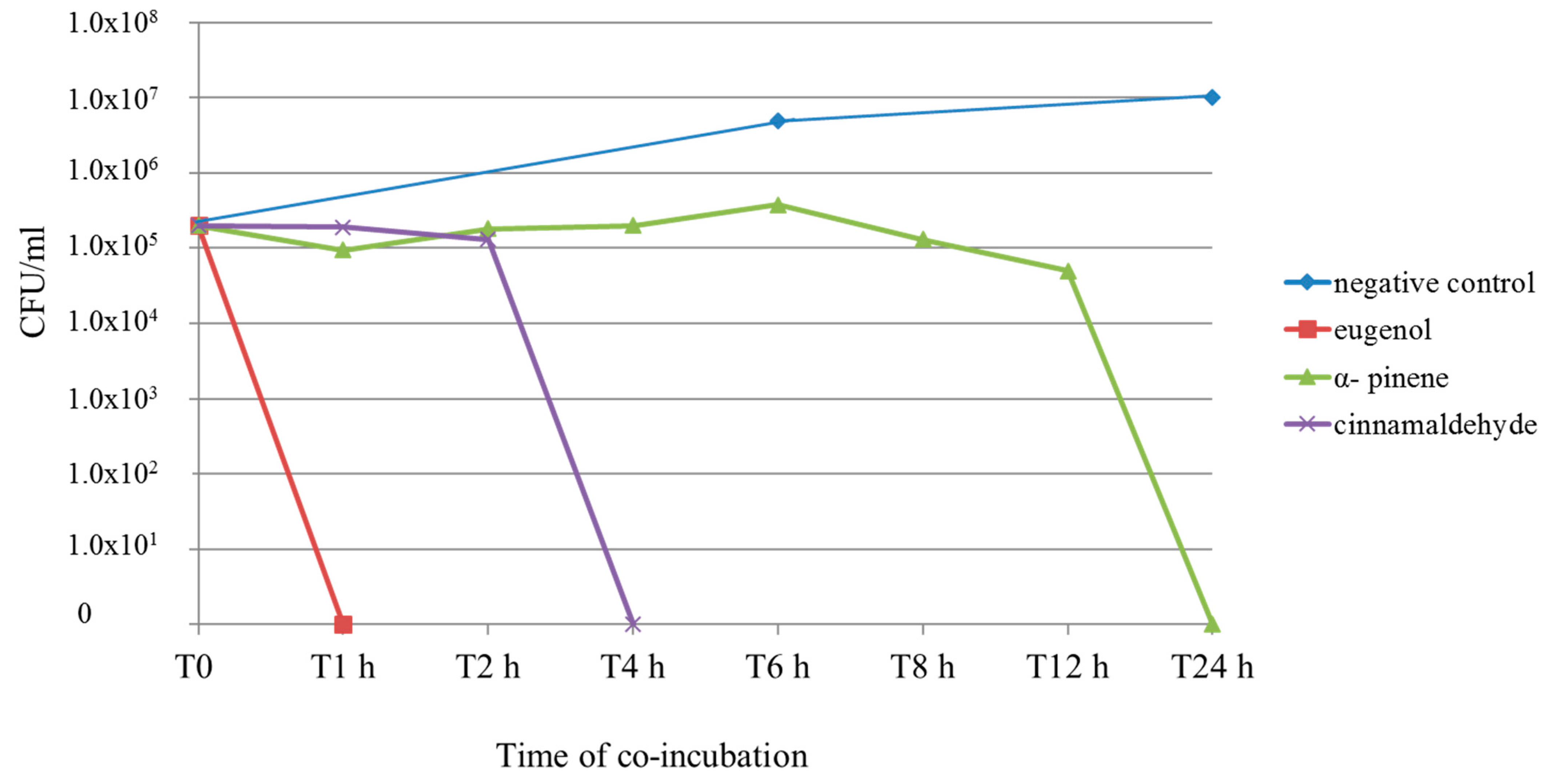
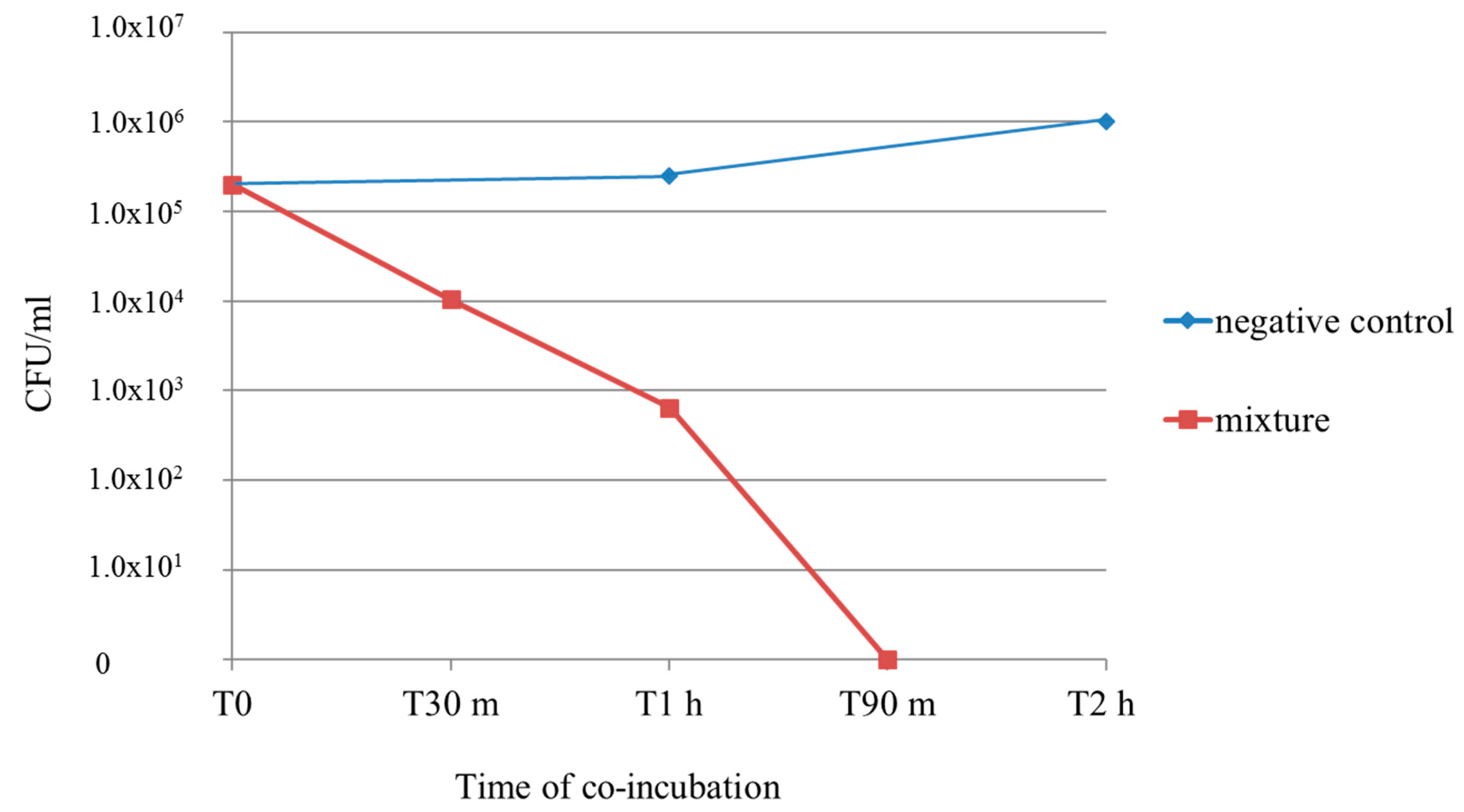
A suspension of 2 × 10⁵ CFU/mL of C. albicans strain 15 was co-incubated with cinnamaldehyde, α-pinene, eugenol, and the mixture of cinnamaldehyde/eugenol at their MFCs. The total viable count (TVC) was determined at T0, T1 h, T2 h, T4 h, T6 h, T8 h, T12 h, and T24 h for the single compounds, and at T0, T30 min, T1 h, T90 min, T2 h, and T4 h for the mixture. Eugenol killed all fungal cells within the first hour of co-incubation. When C. albicans was co-incubated with cinnamaldehyde, the TVC was stable for 2 h, and at T4 h no more cells were viable. Effects of α-pinene were not detectable for the first 6 h, the TVC started to decrease at T8 h, and at T24 h all cells were killed. The combination of cinnamaldehyde/eugenol killed all fungal cells within 90 min. These results are reported in Figure 2 and Figure 3.

2.5. Antimicrobial Activity on Lactobacillus acidophilus

It is important that an antifungal substance does not affect the growth of healthy microbiota. When tested using the disk diffusion method, α-pinene and eugenol did not produce an inhibition halo for Lactobacillus acidophilus. Cinnamaldehyde created a 10 mm IZ. Evaluating the activity of cinnamaldehyde, the MIC against L. acidophilus was 327.60 mg/L, which was double the maximum MIC observed (only in 2 out of 18 strains) against Candida spp. and approximately 6.6 times greater than the mean MIC (50.05 mg/L). Evaluating the activity of α-pinene, the MIC against L. acidophilus was 1072.50 mg/L, which was double the maximum MIC observed (only in 2 out of 18 strains) against Candida spp. and about 5.5 times higher than the average MIC (195.41 mg/L). Evaluating the activity of eugenol, the MIC against L. acidophilus was 2650 mg/L, which was double the maximum MIC observed (only in 2 out of 18 strains) against Candida spp. and about 6 times higher than the average MIC (455.42 mg/L). From these findings, it can be deduced that the administration of cinnamaldehyde, α-pinene, and eugenol at their antifungal concentrations, leaves the normal microbiota unaltered. These results are reported in Table 5.

3. Discussion

Candida spp. are the most common members of human gut microbiota and are estimated to be present in 40–60% of the general population [48]. They may be present as transient or permanent colonizers in the oral cavity and in the further parts of the gastrointestinal tract, where they are capable of causing diseases. Fungal infections are commonly treated with antifungal drugs, mainly azoles and polyenes [6]. Azoles in particular are frequently associated with side effects and the prevalence of drug-resistant strains is increasing [9]; therefore, the treatment of fungal infections is still a clinical challenge [38]. The aim of this study was to evaluate the best composition of a mixture of natural compounds to be used against intestinal and vulvovaginal candidiasis.
To assess the fungistatic activities of the five EO compounds, the disk diffusion method was used. The best results were obtained with eugenol and cinnamaldehyde, which inhibited all strains, and α-pinene, which inhibited 88.9% of the tested strains. Limonene showed a fungistatic activity in only 33.3% of cases, and eucalyptol did not produce an inhibition halo. From data reported in literature, both limonene and eucalyptol show antimycotic properties against Candida spp., mainly through interaction with ergosterol and inhibition of biofilm formation [49,50]. As regards the disk diffusion method, our results did not differ very much from those previously reported. Having established a criterion to consider the inhibition to be successful only when IZs were equal to or greater than those of amphotericin B, the fungistatic properties of limonene were not considered to be strong enough; moreover, eucalyptol inhibition was not detectable, and therefore the two compounds were not tested further in the subsequent experimental steps. Cinnamaldehyde and eugenol, on the other hand, produced the widest inhibition halos with mean diameters of 69 mm and 35.2 mm, respectively.
For the second step of the study, the fungicidal activities of cinnamaldehyde, α-pinene, and eugenol were evaluated using the microdilution method. Cinnamaldehyde showed the lowest MICs (mean 50.05 mg/L) and MFCs (mean 109.26 mg/L). However, the MICs and MFCs of α-pinene and eugenol were also extremely low (MIC means 195.41 mg/L and 455.42 mg/L and MFC means 251.27 mg/L and 690.09 mg/L, respectively).
Because it is known that the synergistic mechanisms occurring among the components of the essential oils are important determiners of their effects [51], we applied the checkerboard method, testing the three possible double combinations of eugenol, cinnamaldehyde, and α-pinene. The best results were obtained by combining cinnamaldehyde and eugenol, with a FICI of 0.625 corresponding to a strong additive effect. The combinations of cinnamaldehyde/α-pinene and eugenol/α-pinene showed indifferent effects. The best antifungal activity was obtained by mixing cinnamaldehyde at 1/2 of its MIC (20.48 mg/L) and eugenol at 1/8 of its MIC (41.40 mg/L). The MFCs of the two compounds when mixed were 40.95 mg/L for cinnamaldehyde and 82.68 mg/L for eugenol.
To determine the kinetics of their fungicidal activity, the time–kill curve method was applied. C. albicans strain number 15 was chosen because the MICs and MFCs of the tested compounds for this strain were similar to the overall means, and the MFCs for this strain were always 2× the respective MICs (this ratio also remained stable for the combination). The Candida albicans n.15 total viable count was assessed at T0, T1 h, T2 h, T4 h, T6 h, T8 h, T12 h and T24 h. Eugenol killed all fungal cells within the first hour of co-incubation.
When C. albicans was co-incubated with cinnamaldehyde, the TVC was stable for 2 h, and at T4 h no more cells were viable. Effects of α-pinene were not detectable for the first 6 h, then the TVC started to decrease at T8 h and at T24 h all cells were killed. Eugenol exerted its fungicidal activity very quickly; this result is noteworthy and coherent with data reported in the literature [52].
The differences observed in the last two assays could be due to the fact that eugenol and cinnamaldehyde were able to permeate the cellular membrane, which represents the main target for the most common antifungal drugs and natural compounds, in a shorter time. In fact, cinnamaldehyde and particularly eugenol rapidly expressed their fungicidal effects in the time–kill curve assay, while α-pinene began to show its activity only after 8 h. Moreover, it led to an “indifferent” effect when combined with the other compounds; in particular, it caused a two-fold increase in eugenol MIC, for which our hypothesis is that α-pinene could have inhibited or temporarily delayed the permeability of the other compound or could have affected its access to cellular targets.
The combination of cinnamaldehyde and eugenol led to a rapid loss of viable cells; the count decreased from 2 × 10⁵ CFU/mL to 1 × 10⁴ CFU/mL within 30 min of co-incubation, at T1 h only 6.4 × 10² CFU/mL were counted, and no more viable cells were detected at T 90 min. The mixture of cinnamaldehyde and eugenol killed all fungal cells in a slightly longer time than eugenol alone, but in a significantly shorter time than cinnamaldehyde alone. Nevertheless, it is important to underscore that when combined, these natural compounds expressed their antimycotic properties at much lower concentrations.
A further encouraging result obtained from in vitro assays was the absence of an antibacterial effect (at the concentrations active against Candida spp.) of these EO compounds against Lactobacillus acidophilus. L. acidophilus is the main component (90%) of the vaginal Doderlein complex, which forms a protective biofilm on the mucosa and inhibits the overgrowth of other microorganisms [53]; it has also been shown to reduce symptoms of ulcerative colitis in human patients [54] and reduce ulcer size in a rat model (ulcers induced by acetic acid + C. albicans inoculation) [22,55]. As mentioned above, both cinnamaldehyde and eugenol are defined as GRAS (Generally Recognized As Safe) with a DNEL (Derived No Effect Level) for the oral and dermal exposure in the general population of 0.625 and 3 mg/kg bw/day, respectively [56,57].
Cinnamaldehyde exerts its antimycotic activity mainly suppressing the ergosterol biosynthetic pathway [58] and simultaneously interacting with cell membrane by binding to ergosterol [59]. Cinnamaldehyde and its derivatives have also been reported to have a strong inhibitory effect on plasma membrane ATPase [58], and thus on ATPase-dependent efflux mechanisms. This leads to reduced proteinase and phospholipase activity and a suppressed secretion of hydrolytic enzymes. Plasma membrane ATPase also plays a regulatory role in C. albicans dimorphism [60]; therefore, its inhibition causes a significant decrease in germ tube formation, a pathological characteristic of C. albicans that facilitates invasion [61]. In addition, cinnamaldehyde is able to reduce C. albicans adhesion properties in vitro [62]. Eugenol exhibits antimicrobial activity towards several strains of pathogenic microorganisms [63]. It is known that eugenol also exerts its antifungal activity on Candida spp. mainly by inhibiting ergosterol biosynthesis and interfering with the integrity and functionality of the cell membrane. As a result, the functioning of membrane-bound enzymes such as those involved in cell wall synthesis is impaired, leading to damage to the cell wall [64]. The loss of structural integrity leads to increased permeability, which subsequently causes cell death [65]. As a matter of fact, it has been observed that eugenol causes a decrease in germination and elongation rates of C. albicans and suppresses cell growth and proliferation; treated Candida cells become flat and flaccid as a result of the change in hydroscopic turgor pressure [66].
Another noteworthy effect of both cinnamaldehyde and eugenol is their capability to reduce colonization and virulence factor expression of Campylobacter jejuni, a very harmful enteric pathogen [67].
Results obtained in this in vitro study suggest that a mixture of cinnamaldehyde and eugenol is suitable as a possible treatment for gastrointestinal and vulvovaginal candidiasis. In vivo studies are needed to confirm these encouraging results. For the treatment of Candida spp. overgrowth in the GI tract, it will be appropriate to test different formulations based on the site where action is needed, e.g., chewing gum, gastroresistant microcapsules, or nanocapsules. For vulvovaginal candidiasis, ovules containing the right proportions of these two EO compounds will be tested.

4. Materials and Methods

4.1. Natural Compounds

The essential oil individual components (≥98%) tested were cinnamaldehyde, α-pinene, limonene, eucalyptol, and eugenol. They were provided by Xeda International (S. Andiol, France). All other chemicals were of the highest purity and are commercially available.

4.2. Candida spp. Culture Conditions

Eighteen Candida strains (15 C. albicans, 2 C. glabrata, and 1 C. lusitaniae) derived from a culture collection of vaginal clinical strains isolated on CHROMAgar Candida (BD Italy S.p.A, Milan, Italy) and confirmed via MALDI-TOF (Matrix-assisted laser desorption ionization-time of flight mass spectrometry, Bruker Daltonics S.R.L., Macerata, Italy) were cultured on Potato Dextrose Agar (PDA; Oxoid Thermofisher S.p.A, Milan, Italy) at 35 °C for 24 h.

4.3. Antifungal Activity—Disk Diffusion Method

Colonies were suspended in sterile saline solution at a density corresponding to McFarland opacity standard #0.5 (C. albicans = 1.4 × 10⁶ CFU/mL, C. glabrata = 4.3 × 10⁶ CFU/mL, C. lusitaniae = 3 × 10⁶ CFU/mL) [68] to perform the susceptibility test to essential oils components. The agar disk diffusion method was carried out as follows: a total of eight Potato Dextrose Agar plates for every Candida strain were streaked in three directions with a swab dipped into each suspension to produce a lawn of growth. Blank filter paper disks (Oxoid Thermofisher S.p.A, Milan, Italy) with a diameter of 6.0 mm were added with 10 μL of each EO molecule and placed on the agar surface. Amphotericin B (Fungizone, Cheplapharm Arzneimitte srl; Greifswald, Germany, 10 μg) disks were used as positive controls. Clotrimazole (Sigma-Aldrich, St. Louis, MO, USA; from 10 μg up to 40 μg) disks were also used, but inhibition halos were not clearly readable due to the formation of a regrowth halo. Pure dimethyl sulfoxide (DMSO; Sigma-Aldrich, St. Louis, MO, USA; 10 μL) disks were used as negative controls. Plates were incubated at 35 °C for 24 h. All experiments were performed in duplicate. An EO susceptibility tests was considered positive if it resulted in an inhibition halo equal to or wider than that induced by amphotericin B (positive control). For amphotericin B, a ≥15 mm zone of complete inhibition was interpreted as “susceptibility”, from 10 to 14 mm as “intermediate susceptibility”, and <10 mm as “resistance” [69].

4.4. Antifungal Activity—Microdilution Method

The antifungal activities of amphotericin B and of cinnamaldehyde, α-pinene, and eugenol (compounds with an inhibition halo equal to or wider than that of amphotericin B) were investigated using the microdilution method as reported by Bona et al. [51] with minor modifications. All 18 Candida strains were tested. A 24 h culture (in PDA) of each isolate was diluted in sterile saline solution to a concentration of 2 × 10³ CFU/mL (in order to obtain a final concentration of 103 CFU/mL). Essential oils were dissolved in DMSO (80% oil, 20% DMSO), then diluted in Sabouraud broth to a concentration of 8% v/v (in order to obtain a final concentration of 4% v/v). Serial 1/2 dilutions of amphotericin B (range 32–0.032 mg/L, for a final range of 16–0.016 mg/L) and oils (range 8–0.004% v/v, for a final range of 4–0.002% v/v) were prepared in a 96-well microtiter plate. These %v/v ranges corresponded to 42 × 103 to 20.48 mg/L for cinnamaldehyde, 42.4 × 10³ to 20.67 mg/L for eugenol, and 34.32 × 10³ to 16.73 mg/L for α-pinene. Each dilution of essential oils and amphotericin B (100 μL) was then inoculated with 100 μL of the cell suspension. Growth controls containing Sabouraud broth plus DMSO (prepared at 2% v/v, for a final concentration of 1% v/v) were also established. All microtiter plates were incubated at 35 °C for 48 h. Each experiment was repeated twice. MIC was determined as the concentration of the compounds in the first nonturbid well. MFC was determined as the lowest concentration of the compounds for which the subculture on agar showed the complete absence of fungal growth. The MIC of amphotericin B was also determined according to the EUCAST antifungal MIC method (cutoff 1 mg/L) [70]; all results were concordant.

4.5. Checkerboard Method

To investigate on the antifungal properties of these natural compounds when mixed, the checkerboard method on a 96-well microtiter was used.
First, 100 µL of Sabouraud broth was added to all wells except for H12. Next, 200 µL of the oil named A was added into the H12 well and 100 µL of the same oil (at 32 × MIC, for a final concentration of 8 × MIC) was added into wells A12 to G12. The compound was then two-fold diluted in a horizontal sense up to column 2. Additionally, 100 µL of the oil named B (at 16 × MIC for a final concentration of 4 × MIC) was added to wells H1 to H12, after which a two-fold dilution was carried out in a vertical sense up to row B. C. albicans strain number 15, from a 24 h culture on PDA, was suspended in Sabouraud broth at 2 × 10³ CFU/mL (for a final concentration of 103 CFU/mL), and 100 µL of the suspension were dropped into each of the 96 wells. Microplates were incubated in at 35 °C for 48 h. MICs were determined as for the microdilution method. The A1 well was the negative control (without any substance).
FICI (fractional inhibitory concentration index) was determined as follows:
  • FICI 1: MIC (cinnamaldehyde in combination with eugenol)/MIC (cinnamaldehyde) + MIC (eugenol in combination with cinnamaldehyde)/MIC (eugenol)
  • FICI 2: MIC (cinnamaldehyde in combination with α-pinene)/MIC (cinnamaldehyde) + MIC (α-pinene in combination with cinnamaldehyde)/MIC (α-pinene)
  • FICI 3: MIC (eugenol in combination with α-pinene)/MIC (eugenol) + MIC (α-pinene in combination with eugenol)/MIC (α-pinene)
A synergistic effect is observed when FICI value ≤ 0.5; an additive effect when 0.5 < FICI value ≤ 1; an indifferent effect when <1 FICI value ≤ 4; and an antagonistic effect when FICI value > 4 [71,72].
For the combination of cinnamaldehyde/eugenol only, the nonturbid wells were seeded and MFCs of the compounds when mixed were calculated. All tests were performed in duplicate.

4.6. Time–Kill Curve (Fungicidal Activity)

One C. albicans strain (number 15) was co-incubated with cinnamaldehyde, α-pinene, and eugenol at their MFCs. The C. albicans strain was suspended in Sabouraud broth at a density corresponding to McFarland opacity standard #1.4 (4 × 10⁶ CFU/mL), then diluted 1/10 (4 × 10⁵ CFU/mL, for a final concentration of 2 × 10⁵ CFU/mL). The suspension was divided into aliquots. A total of three aliquots were co-incubated with EO components (1:2 v/v) at 2 × MFC (for a final concentration = MFC); the fourth aliquot was diluted 1/2 with Sabouraud broth and was used as a negative control. The mixtures were thereafter incubated at 35 °C on a stirrer. At T0, T1 h, T2 h, T4 h, T6 h, T8 h, T12 h, and T24 h, 1/10 serial dilutions were performed to determine the viable count. From each dilution, 100 µL was subcultured onto PDA plates, which were incubated at 35 °C for 24 h. Plates were then inspected and the colonies were counted; their number was expressed as CFU/mL. The test was performed in duplicate. A time–kill curve was also determined for the cinnamaldehyde/eugenol mixture at the MFC of combination (calculated with the checkerboard method), and TVC was determined at T0, T30 min, T1 h, T90 min, T2 h, and T4 h.

4.7. Antimicrobial Effects on Lactobacillus acidophilus

The antibacterial activities of cinnamaldehyde, α-pinene, and eugenol against L. acidophilus were investigated using the disk diffusion and microdilution methods. One strain of L. acidophilus, namely La-14 (Danisco S.p.A., Milan, Italy), was tested.
Disk diffusion method. A 24 h culture in DeManRogosaSharpe (MRS) agar was diluted in sterile saline solution at a density corresponding to McFarland opacity standard #0.5. A total of four MRS Agar plates were streaked in three directions with a swab dipped into the suspension to produce a lawn of growth. Filter paper disks (6.0 mm diameter) were placed on the agar surface and 10 μL of each compound was added. Pure dimethyl sulfoxide (DMSO; Sigma-Aldrich, St. Louis, MO, USA; 10 μL) disks were used as negative controls. Plates were incubated at 37 °C for 24 h in anaerobiosis (anaerogen gaspack, OXOID S.p.A., Milan, Italy). The experiment was performed in duplicate.
Microdilution method. Essential oil components (80% oil, 20% DMSO) were diluted in MRS broth to a concentration of 8% v/v (in order to obtain a final concentration of 4% v/v). Serial 1/2 dilutions of the oils (range 8–0.004% v/v, for a final range of 4–0.002% v/v) were prepared in a 96-well microtiter plate. These % v/v ranges corresponded to 42 × 103 to 20.48 mg/L for cinnamaldehyde, 42.4 × 10³ to 20.67 mg/L for eugenol, and 34.32 × 10³ to 16.73 mg/L for α-pinene. Each dilution (100 μL) was then inoculated with 100 μL of the cell suspension. All microtiter plates were incubated at 37 °C for 48 h in anaerobiosis (anaerogen gaspack, OXOID S.p.A., Milan, Italy). MIC was determined as the concentration of oils in the first nonturbid well. The experiment was performed in duplicate.

5. Conclusions

Cinnamaldehyde and eugenol showed high antimycotic activity against Candida spp. in vitro, in terms of both fungistatic and fungicidal properties. They had a strong additive effect when mixed and did not affect Lactobacillus acidophilus viability. These characteristics, together with their known safety, make these two natural compounds perfect candidates for the treatment of candidiasis. In vivo studies are needed to confirm these very encouraging in vitro results.

Author Contributions

Guarantor of the article, E.S.; Conceived the study, E.S. and M.C.V.; Methodology, I.M.S., C.F. and M.P., data curation, I.M.S., C.F. and M.P.; writing—original draft preparation, I.M.S., C.F. and M.P.; writing—review and editing, E.S., M.C.V., I.M.S., C.F., M.P. and R.S.; project administration, E.S. All authors have read and agreed to the published version of the manuscript.

Funding

This research received no external funding.

Institutional Review Board Statement

Not applicable.

Informed Consent Statement

Not applicable.

Data Availability Statement

The data presented in this study are openly available in the Pub Med database.

Conflicts of Interest

The authors declare no conflict of interest.

References

  1. Odds, F.C. Candida infections: An overview. Crit. Rev. Microbiol. 1987, 15, 1–5. [Google Scholar] [CrossRef]
  2. Casadevall, A. Fungi and the rise of mammals. PLoS Pathog. 2012, 8, e1002808. [Google Scholar] [CrossRef]
  3. Nobile, C.J.; Johnson, A.D. Candida albicans Biofilms and Human Disease. Annu. Rev. Microbiol. 2015, 69, 71–92. [Google Scholar] [CrossRef] [Green Version]
  4. Whiteway, M.; Bachewich, C. Morphogenesis in Candida albicans. Annu. Rev. Microbiol. 2007, 61, 529–553. [Google Scholar] [CrossRef] [PubMed] [Green Version]
  5. Mitchell, A.P. Fungus produces a toxic surprise. Nature 2016, 532, 41–42. [Google Scholar] [CrossRef]
  6. Williams, D.W.; Kuriyama, T.; Silva, S.; Malic, S.; Lewis, M.O. Candida biofilms and oral candidosis: Treatment and prevention. Periodontology 2000, 55, 250–265. [Google Scholar] [CrossRef] [Green Version]
  7. Jayatilake, J.A.; Samaranayake, Y.H.; Cheung, L.K.; Samaranayake, L.P. Quantitative evaluation of tissue invasion by wild type, hyphal and SAP mutants of Candida albicans, and non-albicans Candida species in reconstituted human oral epithelium. J. Oral Pathol. Med. 2006, 35, 484–491. [Google Scholar] [CrossRef] [PubMed]
  8. Lass-Florl, C. The changing face of epidemiology of invasive fungal disease in Europe. Mycoses 2009, 52, 197–205. [Google Scholar] [CrossRef] [PubMed]
  9. Cleveland, A.A.; Harrison, L.H.; Farley, M.M.; Hollick, R.; Stein, B.; Chiller, T.M.; Lockhart, S.R.; Park, B.J. Declining incidence of candidemia and the shifting epidemiology of Candida resistance in two US metropolitan areas, 2008–2013: Results from population based surveillance. PLoS ONE 2015, 10, e0120452. [Google Scholar] [CrossRef] [Green Version]
  10. Low, C.Y.; Rotstein, C. Emerging fungal infections in immunocompromised patients. F1000 Med. Rep. 2011, 3, 14. [Google Scholar] [CrossRef] [Green Version]
  11. Pfaller, M.A.; Diekema, D.J. Rare and emerging opportunistic fungal pathogens: Concern for resistance beyond Candida albicans and Aspergillus fumigatus. J. Clin. Microbiol. 2004, 42, 4419–4431. [Google Scholar] [CrossRef] [PubMed] [Green Version]
  12. Nucci, M.; Anaissie, E. Revisiting the source of candidemia: Skin or gut? Clin. Infect. Dis. 2001, 33, 1959–1967. [Google Scholar] [CrossRef]
  13. Gunsalus, K.T.W.; Tornberg-Belanger, S.N.; Matthan, N.R.; Lichtenstein, A.H.; Kumamoto, C.A. Manipulation of host diet to reduce gastrointestinal colonization by the opportunistic pathogen Candida albicans. Host Microb. Biol. 2015, 1, 1–16. [Google Scholar] [CrossRef] [PubMed] [Green Version]
  14. Kumamoto, C.A. Inflammation and gastrointestinal Candida colonization. Curr. Opin. Microbiol. 2011, 14, 386–391. [Google Scholar] [CrossRef] [Green Version]
  15. Sokol, H.; Leducq, V.; Aschard, H.; Pham, H.P.; Jegou, S.; Landman, C.; Cohen, D.; Liguori, G.; Bourrier, A.; Nion-Larmurier, I.; et al. Fungal microbiota dysbiosis in IBD. Gut 2017, 66, 1039–1048. [Google Scholar] [CrossRef] [Green Version]
  16. Coker, O.O.; Nakatsu, G.; Dai, R.Z.; Wu, W.K.K.; Wong, S.H.; Ng, S.C.; Chan, F.K.L.; Sung, J.J.Y.; Yu, J. Enteric fungal microbiota dysbiosis and ecological alterations in colorectal cancer. Gut 2019, 68, 654–662. [Google Scholar] [CrossRef]
  17. Zi-Qi, G.; Kuo-Yao, T.; Yu-Huan, T. Candida gut commensalism and inflammatory disease. Med. Microecol. 2020, 3, 100008. [Google Scholar]
  18. Standaert-Vitse, A.; Sendid, B.; Joossens, M.; François, N.; Vandewalle-El Khoury, P.; Branche, J.; Van Kruiningen, H.; Jouault, T.; Rutgeerts, P.; Gower-Rousseau, C.; et al. Candida albicans colonization and ASCA in familial Crohn’s disease. Am. J. Gastroenterol. 2009, 104, 1745–1753. [Google Scholar] [CrossRef]
  19. Ksiadzyna, D.; Semianow-Wejchert, J.; Nawrot, U.; Wlodarczyk, K.; Paradowski, L. Serum concentration of interleukin 10, anti-mannan Candida antibodies and the fungal colonization of the gastrointestinal tract in patients with ulcerative colitis. Adv. Med. Sci. 2009, 54, 170–176. [Google Scholar] [CrossRef] [Green Version]
  20. Zwolinska-Wcislo, M.; Brzozowski, T.; Budak, A.; Kwiecien, S.; Sliwowski, Z.; Drozdowicz, D.; Trojanowska, D.; Rudnicka-Sosin, L.; Mach, T.; Konturek, S.J.; et al. Effect of Candida colonization on human ulcerative colitis and the healing of inflammatory changes of the colon in the experimental model of colitis ulcerosa. J. Physiol. Pharmacol. 2009, 60, 107–118. [Google Scholar] [PubMed]
  21. Goenka, M.K.; Kochhar, R.; Chakrabarti, A.; Kumar, A.; Gupta, O.; Talwar, P.; Mehta, S.K. Candida overgrowth after treatment of duodenal ulcer. A comparison of cimetidine, famotidine, and omeprazole. J. Clin. Gastroenterol. 1996, 23, 7–10. [Google Scholar] [CrossRef]
  22. Zwolinska-Wcislo, M.; Brzozowski, T.; Mach, T.; Budak, A.; Trojanowska, D.; Konturek, P.C.; Pajdo, R.; Drozdowicz, D.; Kwiecien, S. Are probiotics effective in the treatment of fungal colonization of the gastrointestinal tract? Experimental and clinical studies. J. Physiol. Pharmacol. 2006, 57, 35–49. [Google Scholar]
  23. Jin, L.; Yoshida, M.; Nakamura, T.; Ishikawa, H.; Wakabayashi, G.; Tanabe, M.; Kawachi, S.; Shinoda, M.; Saikawa, Y.; Wada, N.; et al. Candida albicans infection delays duodenal ulcer healing in cysteamine-induced duodenal ulcers in rats. Dig. Dis. Sci. 2008, 53, 2878–2885. [Google Scholar] [CrossRef] [PubMed]
  24. Fujino, S.; Andoh, A.; Bamba, S.; Ogawa, A.; Hata, K.; Araki, Y.; Bamba, T.; Fujiyama, Y. Increased expression of interleukin 17 in inflammatory bowel disease. Gut 2003, 52, 65–70. [Google Scholar] [CrossRef] [PubMed]
  25. Yen, D.; Cheung, J.; Scheerens, H.; Poulet, F.; McClanahan, T.; McKenzie, B.; Kleinschek, M.A.; Owyang, A.; Mattson, J.; Blumenschein, W.; et al. IL-23 is essential for T cell-mediated colitis and promotes inflammation via IL-17 and IL-6. J. Clin. Investig. 2006, 116, 1310–1316. [Google Scholar] [CrossRef]
  26. Saunus, J.M.; Wagner, S.A.; Matias, M.A.; Hu, Y.; Zaini, Z.M.; Farah, C.S. Early activation of the interleukin-23-17 axis in a murine model of oropharyngeal candidiasis. Mol. Oral Microbiol. 2010, 25, 343–356. [Google Scholar] [CrossRef] [PubMed]
  27. Fornari, G.; Vicente, V.A.; Gomes, R.R.; Muro, M.D.; Pinheiro, R.L.; Ferrari, C.; Herkert, P.F.; Takimura, M.; Carvalho, N.S.; Queiroz-Telles, F. Susceptibility and molecular characterization of Candida species from patients with vulvovaginitis. Braz. J. Microbiol. 2016, 47, 373–380. [Google Scholar] [CrossRef] [PubMed] [Green Version]
  28. Barrenetxea, Z.G. Vulvovaginitis candidiásica. Rev. Iberoam. Micol. 2002, 19, 22–24. [Google Scholar]
  29. Gonçalves, B.; Ferreira, C.; Alves, C.T.; Henriques, M.; Azeredo, J.; Silva, S. Vulvovaginal candidiasis: Epidemiology, microbiology and risk factors. Crit. Rev. Microbiol. 2016, 42, 905–927. [Google Scholar] [CrossRef] [Green Version]
  30. Sobel, J.D. Vulvovaginal candidosis. Lancet 2007, 369, 1961–1971. [Google Scholar] [CrossRef]
  31. Zafarghandi, S.; Abbasabadi, B.; Tavallaee, M. The epidemiology of Candida species associated with vulvovaginal candidiasis in an Iranian patient population. Eur. J. Obstet. Gynecol. Reprod. Biol. 2010, 155, 199–203. [Google Scholar]
  32. Workowski, K.A.; Bolan, G.A. Centers for Disease Control and Prevention. Sexually transmitted diseases treatment guidelines. MMWR Recomm. Rep. 2015, 64, 1. [Google Scholar] [CrossRef] [PubMed]
  33. Sobel, J.D. Epidemiology and pathogenesis of recurrent vulvovaginal candidiasis. Am. J. Obstet. Gynecol. 1985, 152, 924. [Google Scholar] [CrossRef]
  34. Blostein, F.; Levin-Sparenberg, E.; Wagner, J.; Foxman, B. Recurrent vulvovaginal candidiasis. Ann. Epidemiol. 2017, 27, 575–582. [Google Scholar] [CrossRef] [PubMed]
  35. Bakkali, F.; Idaomar, M.; Averbeck, S.; Averbeck, D. Biological effects of essential oils—A review. Food Chem. Toxicol. 2008, 46, 446–475. [Google Scholar] [CrossRef] [PubMed]
  36. Baser, K.H.C.; Demitri, F. Chemistry of essential oils. In Flavour and Fragrances: Chemistry, Bioprocessing and Sustainability; Berger, R.G., Ed.; Springer: Berlin, Germany, 2007; pp. 43–86. [Google Scholar]
  37. Teixeira, B.; Marques, A.; Ramos, C.; Neng, N.R.; Nogueira, J.M.; Saraiva, J.A.; Nunes, M.L. Chemical composition and antibacterial and antioxidant properties of commercial essential oils. Ind. Crops Prod. 2017, 43, 587–595. [Google Scholar] [CrossRef]
  38. Raut, J.S.; Karuppayil, S.M. A status review on the medicinal properties of essential oils. Ind. Crops Prod. 2014, 62, 250–264. [Google Scholar] [CrossRef]
  39. De Fazio, L.; Spisni, E.; Cavazza, E.; Strillacci, A.; Candela, M.; Centanni, M.; Ricci, C.; Rizzello, F.; Campieri, M.; Valerii, M.C. Dietary Geraniol by Oral or Enema Administration Strongly Reduces Dysbiosis and Systemic Inflammation in Dextran Sulfate Sodium-Treated Mice. Front. Pharmacol. 2016, 7, 38. [Google Scholar] [CrossRef] [Green Version]
  40. Pavan, B.; Dalpiaz, A.; Marani, L.; Beggiato, S.; Ferraro, L.; Canistro, D.; Paolini, M.; Vivarelli, F.; Valerii, M.C.; Comparone, A.; et al. Geraniol Pharmacokinetics, Bioavailability and Its Multiple Effects on the Liver Antioxidant and Xenobiotic-Metabolizing Enzymes. Front. Pharmacol. 2018, 9, 18. [Google Scholar] [CrossRef] [Green Version]
  41. Rizzello, F.; Ricci, C.; Scandella, M.; Cavazza, E.; Giovanardi, E.; Valerii, M.C.; Campieri, M.; Comparone, A.; De Fazio, L.; Candela, M.; et al. Dietary geraniol ameliorates intestinal dysbiosis and relieves symptoms in irritable bowel syndrome patients: A pilot study. BMC Complement. Altern. Med. 2018, 18, 338. [Google Scholar] [CrossRef]
  42. Petrocelli, G.; Farabegoli, F.; Valerii, M.C.; Giovannini, C.; Sardo, A.; Spisni, E. Molecules Present in Plant Essential Oils for Prevention and Treatment of Colorectal Cancer (CRC). Molecules 2021, 26, 885. [Google Scholar] [CrossRef]
  43. Compound Summary. Cinnamaldehyde. Available online: https://pubchem.ncbi.nlm.nih.gov/compound/Cinnamaldehyde (accessed on 20 November 2021).
  44. Compound Summary. Alpha-Pinene. Available online: https://pubchem.ncbi.nlm.nih.gov/compound/alpha-Pinene (accessed on 20 November 2021).
  45. Compound Summary. Limonene. Available online: https://pubchem.ncbi.nlm.nih.gov/compound/Limonene (accessed on 20 November 2021).
  46. Compound Summary. Eucalyptol. Available online: https://pubchem.ncbi.nlm.nih.gov/compound/Eucalyptol (accessed on 20 November 2021).
  47. Compound Summary. Eugenol. Available online: https://pubchem.ncbi.nlm.nih.gov/compound/Eugenol (accessed on 20 November 2021).
  48. Angebault, C.; Djossou, F.; Abélanet, S.; Permal, E.; Soltana, M.B.; Diancourt, L.; Bouchier, C.; Woerther, P.L.; Catzeflis, F.; Andremont, A.; et al. Candida albicans is not always the preferential yeast colonizing humans: A study in Wayampi Amerindians. J. Infect. Dis. 2013, 208, 1705–1716. [Google Scholar] [CrossRef] [Green Version]
  49. McKay, S.; Sawant, P.; Fehlberg, J.; Almenar, E. Antimicrobial activity of orange juice processing waste in powder form and its suitability to produce antimicrobial packaging. Waste Manag. 2021, 120, 230–239. [Google Scholar] [CrossRef]
  50. Safaei-Ghomi, J.; Ahd, A.A. Antimicrobial and antifungal properties of the essential oil and methanol extracts of Eucalyptus largiflorens and Eucalyptus intertexta. Pharmacogn. Mag. 2010, 6, 172–175. [Google Scholar] [CrossRef] [PubMed] [Green Version]
  51. Bona, E.; Cantamessa, S.; Pavan, M.; Novello, G.; Massa, N.; Rocchetti, A.; Berta, G.; Gamalero, E. Sensitivity of Candida albicans to essential oils: Are they an alternative to antifungal agents? J. Appl. Microbiol. 2016, 121, 1530–1545. [Google Scholar] [CrossRef]
  52. Jafri, H.; Banerjee, G.; Khan, M.S.A.; Ahmad, I.; Abulreesh, H.H.; Althubiani, A.S. Synergistic interaction of eugenol and antimicrobial drugs in eradication of single and mixed biofilms of Candida albicans and Streptococcus mutans. AMB Express 2020, 10, 185. [Google Scholar] [CrossRef]
  53. Lepargneur, J.P.; Rousseau, V. Protective role of Doderlein flora. J. Gynécol. Obs. Biol. Reprod. 2002, 31, 485–494. [Google Scholar]
  54. Standaert-Vitse, A.; Jouault, T.; Vandewalle, P.; Mille, C.; Seddik, M.; Sendid, B.; Mallet, J.M.; Colombel, J.F.; Poulain, D. Candida albicans is an immunogen for anti-Saccharomyces cerevisiae antibody markers of Crohn’s disease. Gastroenterology 2006, 130, 1764–1775. [Google Scholar] [CrossRef]
  55. Brzozowski, T.; Zwolinska-Wcislo, M.; Konturek, P.C.; Kwiecien, S.; Drozdowicz, D.; Konturek, S.J.; Stachura, J.; Budak, A.; Bogdal, J.; Pawlik, W.W.; et al. Influence of gastric colonization with Candida albicans on ulcer healing in rats: Effect of ranitidine, aspirin and probiotic therapy. Scand. J. Gastroenterol. 2005, 40, 286–296. [Google Scholar] [CrossRef] [PubMed]
  56. Available online: https://echa.europa.eu/it/registration-dossier/-/registered-dossier/14462 (accessed on 20 November 2021).
  57. Available online: https://echa.europa.eu/it/registration-dossier/-/registered-dossier/13694 (accessed on 20 November 2021).
  58. Shreaz, S.; Sheikh, R.A.; Bhatia, R.; Neelofar, K.; Imran, S.; Hashmi, A.A.; Manzoor, N.; Basir, S.F.; Khan, L.A. Antifungal activity of α-methyl trans cinnamaldehyde, its ligand and metal complexes: Promising growth and ergosterol inhibitors. Biometals 2011, 24, 923–933. [Google Scholar] [CrossRef]
  59. Khan, M.S.; Ahmad, I.; Cameotra, S.S. Phenyl aldehyde and propanoids exert multiple sites of action towards cell membrane and cell wall targeting ergosterol in Candida albicans. AMB Express 2013, 3, 54. [Google Scholar] [CrossRef] [Green Version]
  60. Kaur, S.; Mishra, P. Dimorphism-associated changes in plasma membrane H(+)-ATPase activity of Candida albicans. Arch. Microbiol. 1991, 156, 412–415. [Google Scholar] [CrossRef]
  61. Gow, N.A.; Brown, A.J.; Odds, F.C. Fungal morphogenesis and host invasion. Curr. Opin. Microbiol. 2002, 5, 366–371. [Google Scholar] [CrossRef]
  62. Pootong, A.; Norrapong, B.; Cowawintaweewat, S. Antifungal activity of cinnamaldehyde against Candida albicans. Southeast Asian J. Trop. Med. Public Health 2017, 48, 150–158. [Google Scholar]
  63. Devi, K.P.; Nisha, S.A.; Sakthivel, R.; Pandian, S.K. Eugenol (an essential oil of clove) acts as an antibacterial agent against Salmonella typhi by disrupting the cellular membrane. J. Ethnopharmacol. 2010, 130, 107–115. [Google Scholar] [CrossRef] [PubMed]
  64. Van Minnebruggen, G.; Francois, I.E.J.A.; Cammue, B.P.A.; Thevissen, K.; Vroome, V.; Borgers, M.; Shroot, G. General overview on past, present and future antimycotics. Open Mycol. J. 2010, 4, 22–32. [Google Scholar] [CrossRef] [Green Version]
  65. Bennis, S.; Chami, F.; Chami, N.; Bouchikhi, T.; Remmal, A. Surface alteration of Saccharomyces cerevisiae induced by thymol and eugenol. Lett. Appl. Microbiol. 2014, 38, 454–458. [Google Scholar] [CrossRef]
  66. Latifah-Munirah, B.; Himratul-Aznita, W.H.; Mohd Zain, N. Eugenol, an essential oil of clove, causes disruption to the cell wall of Candida albicans (ATCC 14053). Front. Life Sci. 2015, 8, 231–240. [Google Scholar] [CrossRef] [Green Version]
  67. Upadhyay, A.; Arsi, K.; Wagle, B.R.; Upadhyaya, I.; Shrestha, S.; Donoghue, A.M.; Donoghue, D.J. Trans-Cinnamaldehyde, Carvacrol, and Eugenol Reduce Campylobacter jejuni Colonization Factors and Expression of Virulence Genes in Vitro. Front. Microbiol. 2017, 8, 713. [Google Scholar] [CrossRef] [Green Version]
  68. Guinea, J.; Recio, S.; Escribano, P.; Torres-Narbona, M.; Peláez, T.; Sánchez-Carrillo, C.; Rodríguez-Créixems, M.; Bouza, E. Rapid antifungal susceptibility determination for yeast isolates by use of Etest performed directly on blood samples from patients with fungemia. J. Clin. Microbiol. 2010, 48, 2205–2212. [Google Scholar] [CrossRef] [PubMed] [Green Version]
  69. Jabeen, K.; Kumar, H.; Farooqi, J.; Mehboob, R.; Brandt, M.E.; Zafar, A. Agreement of Direct Antifungal Susceptibility Testing from Positive Blood Culture Bottles with the Conventional Method for Candida Species. J. Clin. Microbiol. 2016, 54, 343–348. [Google Scholar] [CrossRef] [PubMed] [Green Version]
  70. Available online: https://eucast.org/astoffungi/clinicalbreakpointsforantifungals (accessed on 20 November 2021).
  71. Vuuren, S.V.; Viljoen, A.M. Antimicrobial activity of limonene enantiomers and 1, 8-cineole alone and in combination. Flavour Fragr. J. 2007, 22, 540–544. [Google Scholar] [CrossRef]
  72. Stanojevic, D.; Comic, L.; Stefanovic, O.; Solujic-Sukdolak, S. In vitro synergistic antibacterial activity of Salvia officinalis L. and some preservatives. Arch. Biol. Sci. Belgrade 2010, 62, 175–183. [Google Scholar] [CrossRef]
Figure 1. Representative pictures of agar diffusion assay.
Antibiotics 11 00073 g001
Figure 2. Time–kill curve for C. albicans strain number 15. Compounds were tested at their MFCs (2 × MIC in this case). MIC: minimal inhibitory concentration. MFC: minimal fungicidal concentration. CFU: colony-forming unit. Time is expressed in hours. Negative control: no compounds were added to the cell suspension.
Antibiotics 11 00073 g002
Figure 3. Time–kill curve for C. albicans strain number 15. The mixture of cinnamaldehyde/eugenol was tested at the MFC of the combination (2 × MIC of combination in this case). MIC: minimal inhibitory concentration. MFC: minimal fungicidal concentration. CFU: colony-forming unit. h: hours. m: minutes. Negative control: no compounds were added to the cell suspension.
Antibiotics 11 00073 g003
Table 1. Summary of antifungal activities of selected natural compounds against Candida strains.
IZ Diameter (mm)% of Inhibited Strains
AmphotericinB
mean15.4
range10.0 to 20.5
standard deviation2.7
Cinnamaldehyde 100% (18/18)
mean69.0
range55.0 to 75.0
standard deviation6
α-Pinene 88.9% (16/18)
mean21.2
range 14.0 to 3.01
standard deviation5.7
Limonene 33.3 (6/18)
mean13.9
range10.0 to 21.5
standard deviation3.2
Eucalyptol////
Eugenol 100% (18/18)
mean35.2
range 30.5 to 41.0
standard deviation3.5
Neg. control(DMSO)//
The readings include the diameter of the paper disk (6 mm). IZ: inhibition zone. //: no inhibition halo detected.
Table 2. Antifungal effects of selected natural compounds against Candida strains evaluated by agar diffusion assay.
IZ Means (mm)
Amphotericin BCinnamaldehydeα-PineneLimoneneEucalyptolEugenol
C. albicans 113.065.027.014.5//35.0
C. albicans 215.570.017.510.5//35.0
C. albicans 316.067.023.512.0//33.0
C. albicans 419.566.025.012.0//35.0
C. albicans 516.071.023.017.5//35.0
C. albicans 616.067.024.012.0//36.0
C. albicans 716.075.030.021.5//44.0
C. albicans 820.567.031.014.0//41.0
C. albicans 912.070.514.514.0//34.0
C. albicans 1015.072.519.514.5//34.5
C. albicans 1114.575.015.012.0//34.0
C. albicans 1216.575.015.513.5//34.0
C. albicans 1316.071.020.012.0//35.0
C. albicans 1419.075.014.013.5//34.0
C. albicans 1515.570.015.514.0//34.0
C. glabrata 115.055.020.010.0//35.0
C. glabrata 211.055.016.512.0//30.5
C. lusitaniae 110.075.030.521.5//43.0
The reported results represent the means of four readings (the test was performed in duplicate, and for every halo the diameter was measured two times). The results include the diameter of the paper disk (6 mm). IZ: inhibition zone. //: no inhibition halo detected.
Table 3. Summary of the results from microdilution assay.
MIC
Mean
MIC
SD
MIC
Range
MFC
Mean
MFC
SD
MFC
Range
MFC/MIC
Amphotericin B0.052 0.04≤0.016–0.1250.110.08≤0.016–0.25
Cinnamaldehyde50.0536.78≤20.48–163.80109.26102.50≤21.00–656.252.18
α-Pinene195.41127.01≤16.73–536.25251.27158.2033.46–536.251.28
Eugenol455.42327.1182.68–1325.00690.09424.10165.36–1325.001.51
MIC and MFC distribution. MIC: minimal inhibitory concentration. MFC: minimal fungicidal concentration. SD: standard deviation. Unit of measurement: mg/L.
Table 4. Antifungal effects of selected natural compounds against Candida strains evaluated by microdilution assay.
Amphotericin BCinnamaldehydeα-PineneEugenol
MICMFCMICMFCMICMFCMICMFC
C. albicans 10.0320.06440.9581.90133.85133.85331.25331.25
C. albicans 20.0320.06440.9581.90133.85133.85331.25331.25
C. albicans 30.0320.06440.9540.95133.85133.85165.36331.25
C. albicans 40.0320.125≤20.4881.90268.13268.13662.50662.50
C. albicans 50.0160.25040.9540.9566.92133.8582.68331.25
C. albicans 60.0320.06440.9581.90133.85133.85331.25331.25
C. albicans 70.0320.064≤20.4881.9066.92133.85331.25662.50
C. albicans 80.0160.03240.9581.90133.85133.85331.25331.25
C. albicans 90.1250.250163.80656.25536.25536.251325.001325.00
C. albicans 100.0320.032≤20.48≤20.48≤16.7333.46165.36165.36
C. albicans 110.1250.250≤20.48≤20.48133.85268.13662.501325.00
C. albicans 12≤0.016 0.016≤20.48≤20.48≤16.7366.92165.36662.50
C. albicans 130.0640.12581.90163.80268.13268.13662.501325.00
C. albicans 140.0320.12540.95163.80268.13536.25331.251325.00
C. albicans 150.0320.06440.9581.90268.13536.25331.25662.50
C. glabrata 10.0320.06440.9581.90268.13268.13331.25331.25
C. glabrata 20.1250.125163.80163.80536.25536.251325.001325.00
C. lusitaniae 10.1250.250≤20.48≤20.48133.85268.13331.25662.50
The test was performed in duplicate. When no well was turbid, the MIC was considered to be “minor or equal” to the minimal readable concentration. MIC: minimal inhibitory concentration. MFC: minimal fungicidal concentration. Unit of measurement: mg/L.
Table 5. Inhibition zones and MICs of cinnamaldehyde, α-pinene, and eugenol against L. acidophilus.
IZ Mean
(mm)
MIC Mean
(mg/L)
Cinnamaldehyde 10327.60
α-Pinene //1072.50
Eugenol //2650.00
IZ: inhibition zone. MIC: minimal inhibitory concentration. //: no inhibition halo detected.
Publisher’s Note: MDPI stays neutral with regard to jurisdictional claims in published maps and institutional affiliations.