Article

The Efficacy of Moina micrura Enriched with Probiotic Bacillus pocheonensis in Enhancing Survival and Disease Resistance of Red Hybrid Tilapia (Oreochromis spp.) Larvae

1
Laboratory of Aquatic Animal Health and Therapeutics, Institute of Biosciences, Universiti Putra Malaysia, Serdang 43400, Malaysia
2
Department of Aquaculture, Faculty of Agriculture, Universiti Putra Malaysia, Serdang 43400, Malaysia
3
Department of Fisheries and Aquaculture, Faculty of Fisheries and Food Science, Universiti Malaysia Terengganu, Kuala Terengganu 21300, Malaysia
4
Institute of Tropical Biodiversity and Sustainable Development, Universiti Malaysia Terengganu, Kuala Terengganu 21300, Malaysia
5
Laboratory of Sustainable Aquaculture, International Institute of Aquaculture and Aquatic Sciences, Universiti Putra Malaysia, Port Dickson 71050, Malaysia
*
Author to whom correspondence should be addressed.
Antibiotics 2021, 10(8), 989; https://doi.org/10.3390/antibiotics10080989
Submission received: 1 July 2021 / Revised: 6 August 2021 / Accepted: 13 August 2021 / Published: 16 August 2021
(This article belongs to the Special Issue Natural Antimicrobials and Alternatives to Antimicrobials)

Abstract

:
The administration of probiotics via live feeds, such as Artemia and rotifers, has gained significant attention. Moreover, indiscriminate use of antibiotics in conventional aquaculture practices in order to prevent or control disease outbreaks has resulted in the occurrence of residues and antimicrobial resistance. Thus, the application of eco-friendly feed additives, such as probiotics, as a safer alternative has received increasing attention in recent years. However, only minimal information on the administration of probiotics via freshwater cladoceran Moina micrura is available despite being commonly used for larval and post-larval feeding of freshwater crustaceans and fish. Thus, this study aimed to evaluate the application of Bacillus pocheonensis strain S2 administered via M. micrura to red hybrid tilapia (Oreochromis spp.) larvae. Bacillus pocheonensis that has been previously isolated from Spirulina sp. was subjected to preliminary in vitro evaluation of antagonistic properties. The agar well-diffusion assay revealed that this probiont could inhibit the growth of Streptococcus agalactiae and Aeromonas hydrophila. The size of inhibition zones ranged from 8.8 ± 0.2 to 18.2 ± 0.4 mm. Moina micrura was later used as a biological model in preliminary in vivo bacterial challenge assays to evaluate the efficacy of B. pocheonensis in protecting the host from diseases. Moina micrura was pre-enriched with B. pocheonensis at 10 4 and 10 6   CFU   mL 1 before S. agalactiae and A. hydrophila were introduced into the culture. The study revealed that B. pocheonensis at 10 4   CFU   mL 1 was able to significantly enhance the survival of M. micrura after being challenged with both pathogens (63 ± 3%) in comparison to the control group. The relative percentage survival (RPS) of M. micrura was highest (p < 0.05) when treated with B. pocheonensis at both concentrations 10 4 and 10 6   CFU   mL 1 (38.33) after being challenged against S. agalactiae. To assess the efficacy of B. pocheonensis in protecting red hybrid tilapia against streptococcosis, the larvae were fed with either unenriched (control) Moina or probiont-enriched Moina daily for 10 days. A significantly (p < 0.05) higher survival rate (77 ± 3%) was observed in larvae fed with probiont-enriched M. micrura compared to other treatments, and the RPS was recorded at 62.90. In addition, the S. agalactiae load was suppressed in larvae fed probiont-enriched M. micrura ( 6.84 ± 0.39   CFU   mL 1 ) in comparison to the control group ( 7.78 ± 0.09   CFU   mL 1 ), indicating that the probiont might have contributed to the improvement of tilapia health and survival. This study illustrated that M. micrura was suitable to be used as a vector for probiotics in freshwater fish larvae as an alternative to hazardous antibiotics for disease control.

1. Introduction

Despite the impact of the COVID-19 pandemic, global tilapia production grew by 3.3% in 2020, surpassing 6 million tonnes for the first time [1]. China remains the largest tilapia producer at 1.62 million tonnes, accounting for over a quarter of the total global production in 2018 [2]. Although the domestic sales of tilapia products have recovered since the Chinese economy reopened in late March 2020 after the easing of COVID-19 lockdown measures, the exports of tilapia products have been drastically affected [3]. In Malaysia, tilapia is the second most reared freshwater fish after the African catfish (Claris gariepinus), contributing to about 30.7% of the total freshwater aquaculture production in 2018 [4]. The red hybrid tilapia is preferably cultured by fish farmers, accounting for more than 94% of the total tilapia production [4]. The Department of Fisheries (DOF) in Malaysia has established 28,099 ha of aquaculture industrial zone (AIZ) for a high impact project (HIP) across the country involving marine fish and shrimp farming, freshwater fish farming, cockle farming, seaweed farming, ornamental fish production, and hatcheries [5]. The Como River, flowing into Kenyir Lake, Terengganu, Malaysia is a large-scale AIZ with 2800 ha of total land dedicated to tilapia farming [6].
Despite the increasing demand for fish as a healthy source of animal protein, the aquaculture industry has always been hampered by disease outbreaks, causing a significant economic loss, especially in developing countries [7,8]. Although tilapia is considered capable of tolerating a broad range of environmental conditions, over half (54.9%) of reported disease cases in tilapia culture were caused by pathogenic bacteria [9]. As a consequence of the intensifications of cage aquaculture activities around the Como River, mass mortality of cage-cultured red hybrid tilapia was reported in February 2012 [10]. In addition, mortality outbreaks of cage-cultured red hybrid tilapia in Kenyir Lake, Terengganu and Pergau Lake, Kelantan, Malaysia during the dry season from March until June have been periodically reported since 2000 [11]. Detection of S. agalactiae in the sampled organs suggested the possible case of streptococcosis associated with warm water [11]. Recently, in January 2020, a tilapia farm in Selangor, Malaysia reported 70% mortality of adult red hybrid tilapia whereby co-infections by tilapia lake virus, A. hydrophila, and S. agalactiae was identified as the main cause [12].
In Malaysia, streptococcal infection is primarily controlled by incorporating antibiotics, such as erythromycin and oxytetracyclines, into feed [13,14]. However, the rapid emergence of antibiotic-resistant pathogens is a primary concern worldwide, threatening the efficacy of antibiotics. It has been reported that S. agalactiae isolated from commercial red hybrid tilapia farms in four states in Malaysia was resistant to spiramycin, olendomycin, sulphamethoxazole, oxolinic acid, kanamycin, and nalidixic acid [13]. Moreover, antibiotics in cultured fish in Malaysia have never been extensively monitored [15]. The application of probiotics in aquaculture practices has been extensively studied as a safer alternative to antibiotics [16,17,18]. In general, probiotics are defined as ‘live microorganisms which when administered in adequate amounts confer a health benefit on the host’ [19].
Probiotics can be introduced into the host via feeding probiotic-supplemented pellet food, feeding on probiotic-enriched live feeds, direct addition to the water column, or injection [20]. Encapsulating probiotics in live feeds, such as Artemia and rotifers, is gaining popularity, and promising results have been reported [17,21,22]. Through this method, probiotics would remain viable and proliferate in zooplankton, thus ensuring effective administration in fish [23,24]. Probiotics could uplift the nutritional profile of zooplankton, and the positive effects of probiotics on fish health have been demonstrated by increasing the growth and survival of the host [21]. In addition, direct administration of probiotics to the culture water is risky due to easy exposure to microbiological contamination [21] and the short survival period of probiotics in seawater [25]. To our knowledge, however, no information is available regarding the application of probiotics to tilapia larvae via cladoceran M. micrura, even though they are commonly used as food for larval and post-larval rearing of freshwater crustaceans and teleost fish [26,27]. Thus, this study was designed to evaluate the efficacy of M. micrura as a possible vector of B. pocheonensis to enhance resistance to disease in red hybrid tilapia larvae.

2. Results

2.1. Antagonistic Activity of Bacillus pocheonensis S2 against Freshwater Pathogens

Bacillus pocheonensis was able to inhibit the growth of S. agalactiae at 10 5   CFU   mL 1 only. Meanwhile, B. pocheonensis was able to inhibit the growth of A. hydrophila at all three tested concentrations. The biggest inhibition zone was produced by probiont when tested against A. hydrophila at 10 6   CFU   mL 1 (Table 1).

2.2. Survival of Moina micrura after Challenged against Freshwater Pathogens

In S. agalactiae challenged assay, the survival rates of M. micrura treated with B. pocheonensis at 5 × 10 4   CFU   mL 1 in treatment T5 and 5 × 10 6   CFU   mL 1 in treatment T6 were significantly higher (p < 0.05) than the control group in treatment T2 (Figure 1 A). These results suggested that B. pocheonensis at 5 × 10 4 and 5 × 10 6   CFU   mL 1 were able to confer significant protection to M. micrura against S. agalactiae at 10 5   CFU   mL 1 .
In A. hydrophila challenged assay, the survival rate of M. micrura treated with B. pocheonensis at 5 × 10 4   CFU   mL 1 in treatment T5 was significantly higher (p < 0.05) than the control group in treatment T2 (Figure 1B). Similarly, the survival rate of M. micrura treated with B. pocheonensis at 5 × 10 6   CFU   mL 1 in treatment T6 was higher than the control group in treatment T2, but they are not significantly different (p > 0.05). These results suggested that B. pocheonensis was able to provide significant protection to M. micrura against A. hydrophila at 5 × 10 6   CFU   mL 1 only.

2.3. Relative Percentage Survival (RPS) of Moina micrura after Challenged against Freshwater Pathogens

In M. micrura bacterial challenge tests, probiont-enriched M. micrura and unenriched M. micrura were challenged against pathogens S. agalactiae and A. hydrophila. The mortality of M. micrura in the control group started 24 h after being challenged and the average cumulative mortality was highest after 72 h post-challenge. Meanwhile, the mortality of M. micrura in both probiont-treated groups (T5 and T6) for both experiments started 48 h after being challenged.
In the S. agalactiae challenge experiment, the average cumulative mortalities of M. micrura in both treatments (T5 and T6) were significantly lower (p < 0.05) than the control group after 72 h post-challenge. The RPS of M. micrura in both probiont-treated groups was recorded at 38.33 (Table 2).
Meanwhile, in A. hydrophila challenge experiment, the average cumulative mortalities of M. micrura in T5 and T6 were lower than in the control group after 72 h post-challenge. Only M. micrura in treatment T5 had a significantly lower (p < 0.05) average cumulative mortality than the control group. The RPS of M. micrura in T5 and T6 was recorded at 30.19 and 18.87, respectively (Table 2).

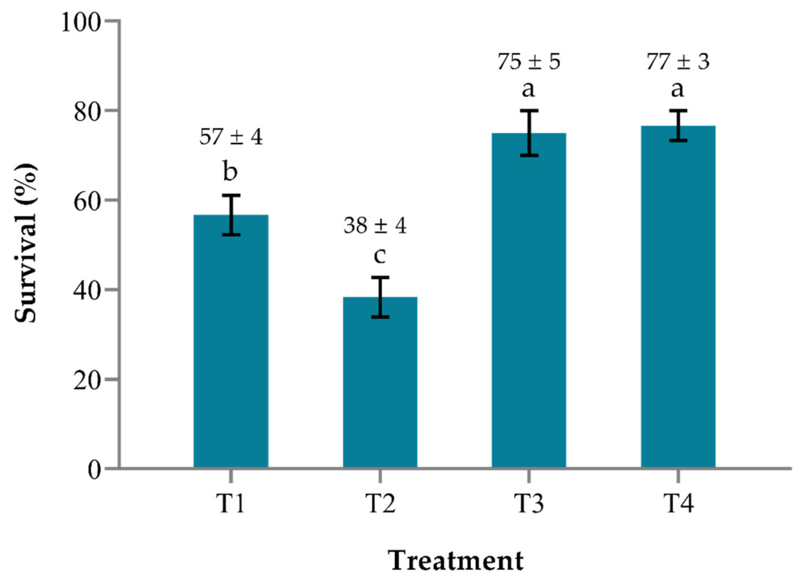
2.4. Survival and Relative Percentage Survival (RPS) of Red Hybrid Tilapia Larvae

The bacterial challenge assay lasted for 10 days and the mortality of tilapia larvae was first observed in the control group (T2) five days after the challenge. The administration of potential probiont B. pocheonensis at 5 × 10 6   CFU   mL 1 via M. micrura as vectors was able to confer significant protection to tilapia larvae against S. agalactiae at 10 7   CFU   mL 1 (Figure 2). Unchallenged tilapia larvae fed with probiont-enriched M. micrura in treatment T3 had a significantly higher (p < 0.05) survival rate than those in treatment T1 (no probiont), indicating that B. pocheonensis had no negative effect on tilapia larvae and may have improved tilapia health by increasing survival. Challenged tilapia larvae fed with probiont-enriched M. micrura in treatment T4 had a significantly higher (p < 0.05) survival rate than those in the control group in treatment T2, suggesting that B. pocheonensis may be beneficial to the host by enhancing disease resistance.
The mortality of tilapia larvae fed with unenriched M. micrura in the control group (T2) started on day 5 after being challenged. The average cumulative mortality was highest after 10 days of the experiment. The mortality of tilapia larvae fed with probiont-enriched M. micrura (T4) started on day 6 after being challenged. The average cumulative mortality was significantly lower (p < 0.05) than the control group after 10 days of the experiment. The RPS of tilapia larvae in the probiont-enriched group (T4) was recorded at 62.90 (Table 3).

2.5. Streptococcus agalactiae Counts

The bacterial counts’ study revealed that potential probiont B. pocheonensis could partially reduce the concentration of S. agalactiae in tilapia larvae. The CFU of S. agalactiae in tilapia larvae fed with probiont-enriched M. micrura in treatment T4 was lower than those fed with unenriched M. micrura in treatment T2 (Table 4), but they are not significantly different (p > 0.05). In addition, some of the external clinical signs observed in tilapia larvae include spinning near the water surface, cornea opacity, and caudal fin erosion (Figure 3).

3. Discussion

The most frequently used probiotic bacteria in aquaculture practice belong to the genus Bacillus [28]. Bacillus spp. can facilitate digestive processes, modulate the immune system, and inhibit the growth of pathogens [29,30,31]. In addition, Bacillus spp. are commonly utilized for their sporulation capacity, non-pathogenic and non-toxic characteristics, and ability to produce antimicrobial substances [31]. In the present study, the 16S rRNA gene sequence analysis revealed that probiont showed 100% similarity with B. pocheonensis. Bacillus pocheonensis is a halotolerant bacterium first isolated from a soil sample of a ginseng field in Pocheon Province, South Korea [32]. However, there are a lack of studies reported on the probiotic properties of this species in reference to aquaculture practice.
The antagonistic activity against a variety of pathogens is one of the most important properties of probiotic candidates. In the present study, the antagonistic activity of B. pocheonensis against S. agalactiae and A. hydrophila at three different concentrations was evaluated in in vitro well-diffusion assay. The study revealed that B. pocheonensis antagonized S. agalactiae and A. hydrophila at least at one concentration out of the three. The inhibitory effect of B. pocheonensis against S. agalactiae at 10 5   CFU   mL 1 was the strongest, as indicated by the largest inhibition zone amongst treatments. Similarly, several studies have demonstrated the antagonism of Bacillus spp. isolated from various sources against a wide range of Gram-positive and Gram-negative pathogens, including Vibrio parahaemolyticus, Vibrio campbellii, and Vibrio alginolyticus, by means of secondary metabolites production, including siderophores, antibiotics, bacteriocins, and hydrolytic enzymes [33,34,35,36]. Several studies suggest that the production of bacteriocins and the inhibitory effects of Bacillus are likely due to the alteration of pH in the growth medium, utilization of essential nutrients, or production of volatile compounds [37,38,39].
In the present study, M. micrura was used as the host in preliminary in vivo bacterial challenge tests. The enrichment of M. micrura with B. pocheonensis at 10 4 and 10 6   CFU   mL 1 was able to enhance the survival of M. micrura against S. agalactiae significantly. Meanwhile, B. pocheonensis was able to confer significant protection to M. micrura against A. hydrophila at 10 4   CFU   mL 1 only. The mechanisms by which the probiont confer protection against pathogen remain poorly understood, particularly in M. micrura. In the Artemia model study, enrichment with a single strain of probiotic bacteria at 10 5   CFU   mL 1 was able to protect Artemia from Vibrio anguillarum infection [40]. Additionally, the survival of Artemia was not significantly affected regardless of the species of probiotic bacteria used and the concentration applied. Similar to the present study, several published studies have suggested that low concentrations of probiotics ranging from 10 4 to 10 6   CFU   mL 1 should be used in live feeds enrichment practice [40,41].
The administration of probiotics to fish and crustacean larvae via live feeds, such as Artemia and rotifers, has increasingly gained interest to improve the growth, survival, and disease resistance of fish larvae. In the present study, Bacillus sp. was administered via M. micrura to red hybrid tilapia larvae. Significant survival improvement of larvae fed with probiont-enriched M. micrura for 10 days was observed. Similarly, several published studies have reported that the administration of probiotics via live feeds enhanced the survival of fish and crustacean larvae, such as ornamental fish (Poecilia latipinna), European seabass (Dicentrarchus labrax), and Indian white shrimp (Fenneropenaeus indicus), after being challenged with freshwater and marine bacterial pathogens [42,43,44]. Survival and disease resistance enhancement is indeed an important feature of probiotic candidates.
Although the beneficial effect of probiotic application in enhancing the survival and disease resistance of fish larvae has been widely reported, the exact mechanisms by which probiotics promote survival are poorly understood. In the present study, the bacterial pathogen was introduced into tilapia larvae through the bath immersion method. Meanwhile in another study, the incorporation of Bacillus velezensis in the diet significantly increased tilapia survival after being challenged with S. agalactiae by intraperitoneal (IP) injection [45]. Merrifield et al. [46] suggested that the ability of probiotics to prevent disease may be greater than the results reported in many studies that employed the IP injection method in challenge studies. The IP injection method bypasses competitive exclusion, one of the most important mechanisms in which probiotics prevent pathogen infection in the gastrointestinal tract (GI) [47]. In addition, as the GI tract of most fish species at the early larval stage is poorly developed, the establishment of the microbial community is influenced by the aquatic environments and live feeds [21]. Thus, the administration of probiotics should be made early and frequently to establish the artificial dominance of beneficial bacteria [48].
Probiotics were demonstrated to have the efficacy to inhibit the growth of pathogens in vivo. In the present study, enrichment of M. micrura with B. pocheonensis at 5 × 10 6   CFU   mL 1 for 3 h by immersion was able to partially reduce the concentration of S. agalactiae in tilapia larvae. Similar results are observed for juvenile blue swimming crab (Portunus pelagicus) supplemented with Bacillus amyloliquefaciens and challenged with Vibrio harveyi [49]. Similarly, the enrichment of copepod with Bacillus clausii or Bacillus pumilus at 10 6   CFU   mL 1 for 3 h by immersion was able to reduce the concentration of pathogen Aliivibrio fischeri and Vibrio spp. in the gut of grouper (Epinephelus coioides) larvae [21]. In another study, the supplementation of Bacillus subtilis at 10 8 and 10 9   CFU   L 1 to the rearing water of white shrimp (Litopenaeus vannamei) larvae were able to significantly reduce the concentration of presumptive Vibrio spp. [50].
The competition for adhesion sites through competitive exclusion has been widely proposed to explain the reduction of pathogenic bacteria in the fish gut [51]. The present study suggested that the supplementation of B. pocheonensis was effective in reducing the S. agalactiae load in tilapia larvae. The action mechanism of B. pocheonensis being based on competitive exclusion seems like a plausible explanation due to the retrieval of B. pocheonensis in challenged tilapia larvae at the end of the experiment. Xia et al. [52] agreed that the changes in the composition of the gut bacterial community and the enhancement of the composition of some beneficial bacteria in the gut may have influenced tilapia resistance to streptococcosis. Successful colonization of the fish gut by probiotic bacteria is indeed difficult to achieve [53]. Factors, including the selection of probiotic strain, probiotic dosage, modes of probiotic administration, and frequency of probiotic supplementation may influence the colonization success [52,54].

4. Materials and Methods

4.1. Bacterial Strains and Growth Conditions

Bacillus pocheonensis strain S2 (GenBank accession no. MK764898) was previously isolated from microalgae Spirulina sp. [55,56]. Aeromonas hydrophila and S. agalactiae were obtained from Fish Health Laboratory, Faculty of Agriculture, Universiti Putra Malaysia (UPM), Malaysia. All three bacteria were cultured in trypticase soy broth (TSB) (Merck, Darmstadt, Germany) and incubated overnight at 30 °C with constant shaking at 150 rpm (Biosan, Riga, Latvia). On the next day, all cultures were centrifuged (Hermle, Wehingen, Germany) at 5000× g for 10 min at room temperature. The supernatants were discarded, and the pellets were resuspended in sterile distilled water and washed once. The concentrations of all cultures were determined using a spectrophotometer (Eppendorf, Hamburg, Germany) at the optical density of 550 nm ( OD 550 ) . The concentration of all bacteria was then adjusted accordingly to the desired sufficient value (   CFU   mL 1 ) .

4.2. Cultures of Microalgae Chlorella vulgaris

Microalgae C. vulgaris (Isolate number: UPMC-A0088) was obtained from Aquatic Animal Health and Therapeutics Laboratory (AquaHealth), Institute of Bioscience (IBS), UPM, Malaysia. Chlorella vulgaris cultures were grown in sterile 1000 mL conical flasks, filled with Bold’s Basal medium (BBM) with constant shaking at 60 rpm on an orbital shaker (Protech, Balakong, Malaysia) and continuous light. The concentration of C. vulgaris was determined using a 0.1 mm improved Neubauer chamber observed under a light microscope according to the following formula:
Density ,   d   ( cells / mL ) = Average   number   of   cells   per   square   counted 4 × 10 6
where 4 × 10 6 = the volume of sample over the small square area, which is equivalent to 0.004 mm 3 (0.2 × 0.2 × 0.1) expressed in cm 3 (mL).

4.3. Preliminary In Vitro Screening of Bacillus pocheonensis S2

Agar well-diffusion method was used to screen the antagonistic activity of B. pocheonensis against freshwater pathogens S. agalactiae and A. hydrophila. The concentrations of both pathogens were adjusted accordingly. The trypticase soy agar (TSA) (Defco Merck, Germany) plates were aseptically inoculated by individually swabbing each pathogen at the final concentration of 5 × 10 5 , 5 × 10 6 , and 5 × 10 7   CFU   mL 1 over the entire agar surface using a sterile cotton swab. Holes with a diameter of 4 mm were aseptically punched on each TSA plate using a sterile cork borer. Then, 10 µL probiont culture at the final concentration of 10 9   CFU   mL 1 was dispensed into each well. All plates were incubated overnight at 30 °C. On the next day, the inhibition zone was observed, and the size was measured.

4.4. Culture and Maintenance of Moina micrura

Moina micrura was obtained from the AquaHealth Laboratory, IBS, UPM, Malaysia. Moina micrura was maintained in 1000 mL plastic aquaria filled with filter-sterilized freshwater pond water (0.45 µm fiberglass filters) with mild aeration. The water media was exchanged once a week. Moina was fed with microalgae C. vulgaris ad libitum [57,58,59].

4.5. Preliminary In Vivo Bacterial Challenge Assay of Moina micrura

This method is based on Masduki et al. [60] with minor modifications. Ten M. micrura were placed in a 50 mL Falcon tube containing 30 mL sterilized freshwater pond water. Moina micrura was first pre-incubated with probiont B. pocheonensis at two different concentrations: 5 × 10 4 and 5 × 10 6   CFU   mL 1 for 24 h. Moina was fed with C. vulgaris at 4 × 10 5   cells   mL 1 once a day. After 24 h pre-incubation, pathogens S. agalactiae and A. hydrophila at 5 × 10 5   CFU   mL 1 were individually introduced into the culture. All tubes were incubated at room temperature at a normal condition of 12 h light and 12 h dark regime. The observation was made until the mortality of M. micrura in the control group (without probiont) reached at least 50%. The survival rate of M. micrura was determined as below:
Survival   rate   % = ( Number   of   survivors Total   number   of   larvae ) × 100
The efficacy of the potential probiont was assessed by calculating the relative percentage survival (RPS). The RPS was calculated using the formula:
RPS = 1 ( % mortality   in   test   group % mortality   in   control   group ) × 100

4.6. Enrichment of Moina micrura

Moina micrura was cultured in 5 L plastic aquariums filled with sterilized freshwater pond water. Moina micrura was fed with C. vulgaris cultured in BBM ad libitum at 4 × 10 5   cells   mL 1 [61]. An air pump was used to provide mild aeration. Before probiont enrichment, M. micrura was harvested early in the morning using a 200 µm sieve and thoroughly washed with tap water. Moina micrura was concentrated in 2 L plastic aquaria filled with sterilized freshwater pond water, at approximately 10 5   M o i n a   L 1 . Cultures of M. micrura to be fed to probiont-treated tilapia larvae were inoculated with B. pocheonensis at 5 × 10 6   CFU   mL 1 and incubated for 3 h [21] at room temperature with mild aeration.

4.7. Experimental Design

This method is based on Sun et al. [21] with a minor modification on the number of tilapia larvae used, live feed density, and survival determination. Each 10 L plastic tank filled with 5 L tap water was randomly stocked with 20 tilapia larvae with an average weight of 0.34 g and 25 mm in length. Treated tilapia larvae were fed with M. micrura pre-enriched with potential probiont B. pocheonensis at 10 6   CFU   mL 1 . Meanwhile, untreated tilapia larvae were fed with control M. micrura (without B. pocheonensis enrichment). Both treated and untreated M. micrura were fed to tilapia larvae once a day at a density of approximately 2.0 Moina mL 1 . Each treatment was carried out in triplicate.
After the first 24 h, pathogenic S. agalactiae was inoculated in treatment T2 and T4 at 10 7   CFU   mL 1 for a bacterial challenge test. Tilapia larvae in tanks with no pathogen inoculation served as survival control, while those in tanks with pathogen inoculation alone served as mortality control. Treated tilapia larvae fed with M. micrura enriched with B. pocheonensis served as probiotic control. Meanwhile, larvae fed with M. micrura enriched with B. pocheonensis and inoculated with S. agalactiae served as the experimental treatment group. All tanks were provided with continuous aeration and the water temperature was maintained at 28 °C. The challenge test was carried out until tilapia larvae in the mortality control group recorded at least 50% mortality.
The survival of tilapia larvae was determined by counting the number of larvae remaining in each tank when the mortality control group recorded at least 50% mortality. The survival rates and RPS of red tilapia larvae were determined using the formulae previously described.

4.8. Streptococcus agalactiae Counts

A total plate count of S. agalactiae was carried out to evaluate the ability of potential probiont B. pocheonensis administered via M. micrura in inhibiting the growth of S. agalactiae in tilapia larvae. After counting the number of living and dead larvae, the tilapia larvae from treatments T2 and T4 were collected at the end of the experiment. The larvae were collected and washed with sterilized distilled water. The tilapia larvae were homogenized using sterile porcelain mortar and pestle. A serial dilution of the homogenated larvae was made up to 10 6 in sterilized distilled water. Following serial dilution, 100 µL of each diluted sample was pipetted and dispensed onto Brain Heart Infusion (BHI) (Merck, Darmstadt, Germany) agar medium. The samples were spread evenly onto BHI agar using a sterile glass hockey stick. Each sample was plated out in triplicates. The plates were incubated overnight at 30 °C and the colony-forming unit (CFU) was counted on the following day. The confirmation of S. agalactiae isolate was determined by performing Gram staining and 16S rRNA gene analysis. In addition, throughout the experiment, the clinical signs of S. agalactiae infections in tilapia larvae were observed.

4.9. Statistical Analysis

All the collected data were analyzed using one-way analysis of variance (ANOVA) and the Tukey test was carried out for multiple comparison tests. Significant difference was at p < 0.05. All statistical tests were performed using GraphPad Prism 8 (GraphPad Inc., San Diego, CA, USA).

5. Conclusions

The intensification of culture systems to meet the increasing global demand for fish as a healthy source of animal protein has brought about the application of eco-friendly feed additives, such as probiotics, as a means of disease control and prevention. The present study evaluated the efficacy of B. pocheonensis administered to red tilapia larvae via M. micrura. Our results demonstrated the ability of B. pocheonensis to positively influence M. micrura and larval survival after being challenged with S. agalactiae. The reduction of S. agalactiae load and recovery of B. pocheonensis in tilapia larvae after being challenged may be related to the improvement of survival and health of larvae. Therefore, the results in our study suggested that the application of M. micrura is very much suitable to function as vectors for probiotics administration to red hybrid tilapia larvae. Moreover, this probiotic–zooplankton coupling has a high potential in eliminating or reducing the need for antibiotic usage in aquaculture industries. Although the results in our study present evidence that B. pocheonensis is capable of enhancing disease resistance in larval red hybrid tilapia, the specific mode of actions remain unclear and merit further research. Furthermore, the present study encourages further research on the intestinal microbiota profiling of larval red hybrid tilapia and the retention period of probiotic inocula it receives.

Author Contributions

Methodology, N.A.S. and M.K.; writing—original draft preparation, N.A.S.; writing—review and editing, M.K., F.M.Y. and N.W.R.; visualization, N.A.S.; supervision, M.K.; funding acquisition, M.K. All authors have read and agreed to the published version of the manuscript.

Funding

This research was funded by the Ministry of Higher Education Malaysia (MOHE) under the Fundamental Research Grant Scheme FRGS/1/2020/WAB04/UPM/02/9. Special thanks to MOHE for the Malaysia-Japan SATREPS-COSMOS (JPMJSA 1509) matching grant and Higher Institution Centre of Excellence (HiCoE) grant of Innovative Vaccine and Therapeutics, Vote No.: 6369100 for providing facilities at Institute of Bioscience and Faculty of Agriculture, UPM.

Institutional Review Board Statement

The study was conducted according to the guidelines and regulations approved by the Institutional Animal Care and Use Committee of Universiti Putra Malaysia (UPM/IACUC/AUP-RO77/2019).

Informed Consent Statement

Not applicable.

Data Availability Statement

All data generated or analyzed during this study have been included in this article.

Acknowledgments

The first author was under a sponsorship scheme by the Perak state government.

Conflicts of Interest

The authors declare no conflict of interest.

References

  1. Fletcher, R. 2020 Tilapia Production Figures Revealed. The Fish Site. 2020. Available online: https://thefishsite.com/articles/2020-tilapia-production-figures-revealed (accessed on 18 June 2021).
  2. Miao, W.; Wang, W. Trends of aquaculture production and trade: Carp, tilapia, and shrimp. Asian Fish. Sci. 2020, 33, 1–10. [Google Scholar] [CrossRef]
  3. Fitzssimmons, K. The Global Status of Tilapia Production and Marketing under COVID-19 Conditions. The Fish Site. 2020. Available online: https://thefishsite.com/articles/the-global-status-of-tilapia-production-and-marketing-under-covid-19-conditions (accessed on 18 June 2021).
  4. Mohamad, S.N.; Noordin, W.N.M.; Ismail, N.F.; Hamzah, A. Red hybrid tilapia (Oreochromis spp.) broodstock development programme in Malaysia: Status, challenges and prospects for future development. Asian Fish. Sci. 2021, 34, 73–81. [Google Scholar] [CrossRef]
  5. DOF. Zon Industri Akuakultur. Available online: https://www.dof.gov.my/index.php/pages/view/78 (accessed on 20 January 2021).
  6. Dullah, H.; Malek, M.A.; Hanafiah, M.M. Life cycle assessment of Nile tilapia (Oreochromis niloticus) farming in Kenyir Lake, Terengganu. Sustainability 2020, 12, 2268. [Google Scholar] [CrossRef] [Green Version]
  7. Assefa, A.; Abunna, F. Maintenance of fish health in aquaculture: Review of epidemiological approaches for prevention and control of infectious disease of fish. Vet. Med. Int. 2018, 2018, 5432497. [Google Scholar] [CrossRef]
  8. Cruz, P.M.; Ibáñez, A.L.; Hermosillo, O.A.M.; Saad, H.C.R. Use of Probiotics in Aquaculture. ISRN Microbiol. 2012, 2012, 916845. [Google Scholar] [CrossRef] [Green Version]
  9. FAO. The State of World Fisheries and Aquaculture 2018—Meeting THE Sustainable Development Goals; Food and Agriculture Organization: Rome, Italy, 2018. [Google Scholar]
  10. Najiah, M.; Aqilah, N.I.; Lee, K.L.; Khairulbariyyah, Z.; Mithun, S.; Jalal, K.C.A.; Shaharom-Harrison, F.; Nadirah, M. Massive mortality associated with Streptococcus agalactiae infection in cage-cultured red hybrid tilapia Oreochromis niloticus in Como river, Kenyir Lake, Malaysia. J. Biol. Sci. 2012, 12, 438–442. [Google Scholar] [CrossRef] [Green Version]
  11. Siti-Zahrah, A.; Padilah, B.; Azila, A.; Rimatulhana, R.; Shahidan, H. Multiple streptococcal species infection in cage-cultured red tilapia but showing similar clinical signs. In Diseases in Asian Aquaculture VI; Bondad-Reantaso, M.G., Mohan, C.V., Crumlish, M., Subasinghe, R.P., Eds.; Asian Fisheries Society: Manila, Philippines, 2008; pp. 313–320. [Google Scholar]
  12. Basri, L.; Nor, R.M.; Salleh, A.; Yasin, I.S.M.; Saad, M.Z.; Rahaman, N.Y.A.; Barkham, T.; Amal, M.N.A. Co-infections of tilapia lake virus, Aeromonas hydrophila, and Streptococcus agalactiae in farmed red hybrid tilapia. Animals 2020, 10, 2141. [Google Scholar] [CrossRef] [PubMed]
  13. Musa, N.; Wei, L.S.; Musa, N.; Hamdan, R.H.; Leong, L.K.; Wee, W.; Amal, M.N.; Kutty, B.M.; Abdullah, S.Z. Streptococcosis in red hybrid tilapia (Oreochromis niloticus) commercial farms in Malaysia. Aquac. Res. 2009, 40, 630–632. [Google Scholar] [CrossRef]
  14. Amal, M.N.A.; Zamri-Saad, M.; Iftikhar, A.R.; Siti-Zahrah, A.; Aziel, S.; Fahmi, S. An outbreak of Streptococcus agalactiae infection in cage-cultured golden pompano, Trachinotus blochii (Lacépède), in Malaysia. J. Fish Dis. 2012, 35, 849–852. [Google Scholar] [CrossRef] [PubMed]
  15. Aisyhah, M.A.; Amal, M.N.A.; Zamri-Saad, M.; Siti-Zahrah, A.; Shaqinah, N.N. Streptococcus agalactiae isolates from cultured fishes in Malaysia manifesting low resistance pattern towards selected antibiotics. J. Fish Dis. 2015, 38, 1093–1098. [Google Scholar] [CrossRef]
  16. Aly, S.M.; Mohamed, M.F.; John, G. Effect of probiotics on the survival, growth and challenge infection in tilapia nilotica (Oreochromis niloticus). Aquac. Res. 2008, 39, 647–656. [Google Scholar] [CrossRef]
  17. Nimrat, S.; Suksawat, S.; Boonthai, T.; Vuthiphandchai, V. Potential Bacillus probiotics enhance bacterial numbers, water quality and growth during early development of white shrimp (Litopenaeus vannamei). Vet. Microbiol. 2012, 159, 443–450. [Google Scholar] [CrossRef] [PubMed]
  18. Zokaeifar, H.; Babaei, N.; Saad, C.R.; Kamarudin, M.S.; Sijam, K.; Balcazar, J.L. Administration of Bacillus subtilis strains in the rearing water enhances the water quality, growth performance, immune response, and resistance against Vibrio harveyi infection in juvenile white shrimp, Litopenaeus vannamei. Fish Shellfish Immunol. 2014, 36, 68–74. [Google Scholar] [CrossRef] [PubMed]
  19. FAO. Probiotics in Animal Nutrition—Production, Impact and Regulation; Food and Agriculture Organization: Rome, Italy, 2016. [Google Scholar]
  20. Jahangiri, L.; Esteban, M.Á. Administration of probiotics in the water in finfish aquaculture systems: A review. Fishes 2018, 3, 33. [Google Scholar] [CrossRef] [Green Version]
  21. Sun, Y.Z.; Yang, H.L.; Huang, K.P.; Ye, J.D.; Zhang, C.X. Application of autochthonous Bacillus bioencapsulated in copepod to grouper Epinephelus coioides larvae. Aquaculture 2013, 392–395, 44–50. [Google Scholar] [CrossRef]
  22. Jamali, H.; Imani, A.; Abdollahi, D.; Roozbehfar, R.; Isari, A. Use of probiotic Bacillus spp. in rotifer (Brachionus plicatilis) and Artemia (Artemia urmiana) enrichment: Effects on growth and survival of Pacific white shrimp, Litopenaeus vannamei larvae. Probiotics Antimicrob. Proteins 2015, 7, 118–125. [Google Scholar] [CrossRef]
  23. Hai, N.V. The use of probiotics in aquaculture. J. Appl. Microbiol. 2015, 119, 917–935. [Google Scholar] [CrossRef] [PubMed]
  24. Samat, N.A.; Yusoff, F.M.; Rasdi, N.W.; Karim, M. Enhancement of live food nutritional status with essential nutrients for improving aquatic animal health: A Review. Animals 2020, 10, 2457. [Google Scholar] [CrossRef]
  25. Gatesoupe, F.J. Updating the importance of lactic acid bacteria in fish farming: Natural occurrence and probiotic treatments. J. Mol. Microbiol. Biotechnol. 2008, 14, 107–114. [Google Scholar] [CrossRef] [PubMed] [Green Version]
  26. Poynton, S.L.; Dashsel, P.; Lehmann, M.J.; Steinbergb, C.E. Culture of the cladoceran Moina macrocopa: Mortality associated with flagellate infection. Aquaculture 2013, 416–417, 374–379. [Google Scholar] [CrossRef]
  27. Rasdi, N.W.; Arshad, A.; Ikhwanuddin, M.; Hagiwara, A.; Yusoff, F.M.; Azani, N. A review on the improvement of cladocera (Moina) nutrition as live food for aquaculture: Using valuable plankton fisheries resources. J. Environ. Biol. 2020, 41, 1239–1248. [Google Scholar] [CrossRef]
  28. Gao, X.; Ke, C.; Zhang, M.; Li, X.; Wu, F.; Liu, Y. Effects of the probiotic Bacillus amyloliquefaciens on the growth, immunity, and disease resistance of Haliotis discus hannai. Fish Shellfish Immunol. 2019, 94, 617–627. [Google Scholar] [CrossRef]
  29. Adorian, T.J.; Jamali, H.; Farsani, H.G.; Darvishi, P.; Hasanpour, S.; Bagheri, T.; Roozbehfar, R. Effects of probiotic bacteria Bacillus on growth performance, digestive enzyme activity, and hematological parameters of Asian seabass, Lates calcarifer (Bloch). Probiotics Antimicrob. Proteins 2018, 11, 248–255. [Google Scholar] [CrossRef] [PubMed]
  30. Tarnecki, A.M.; Wafapoor, M.; Phillips, R.N.; Rhody, N.R. Benefits of a Bacillus probiotic to larval fish survival and transport stress resistance. Sci. Rep. 2019, 9, 4892. [Google Scholar] [CrossRef] [Green Version]
  31. Kuebutornye, F.K.; Abarike, E.D.; Lu, Y. A review on the application of Bacillus as probiotics in aquaculture. Fish Shellfish Immunol. 2019, 87, 820–828. [Google Scholar] [CrossRef] [PubMed]
  32. Ten, L.N.; Baek, S.-H.; Im, W.-T.; Larina, L.L.; Lee, J.-S.; Oh, H.-M.; Lee, S.-T. Bacillus pocheonensis sp. nov., a moderately halotolerant, aerobic bacterium isolated from soil of a ginseng field. Int. J. Syst. Evol. Microbiol. 2007, 57, 2532–2537. [Google Scholar] [CrossRef] [Green Version]
  33. Ahmad, V.; Iqbal, A.N.; Haseeb, M.; Khan, M.S. Antimicrobial potential of bacteriocin producing Lysinibacillus jx416856 against foodborne bacterial and fungal pathogens, isolated from fruits and vegetable waste. Anaerobe 2014, 27, 87–95. [Google Scholar] [CrossRef]
  34. Ahmad, V.; Ahmad, K.; Baig, M.H.; AL-Shwaiman, H.A.; Al Khulaifi, M.M.; Elgorban, A.M.; Khan, M.S. Efficacy of a novel bacteriocin isolated from Lysinibacillus sp. against Bacillus pumilus. LWT-Food Sci. Technol. 2019, 102, 260–267. [Google Scholar] [CrossRef]
  35. Patel, A.K.; Deshattiwar, M.K.; Chaudhari, B.L.; Chincholkar, S.B. Production, purification and chemical characterization of the catecholate siderophore from potent probiotic strains of Bacillus spp. Bioresour. Technol. 2009, 100, 368–373. [Google Scholar] [CrossRef]
  36. Liñan-Vidriales, M.A.; Peña-Rodríguez, A.; Tovar-Ramírez, D.; Elizondo-González, R.; Barajas-Sandoval, D.R.; Ponce-Gracía, E.I.; Rodríguez-Jaramillo, C.; Balcázar, J.L.; Quiroz-Guzmán, E. Effect of rice bran fermented with Bacillus and Lysinibacillus species on dynamic microbial activity of Pacific white shrimp (Penaeus vannamei). Aquaculture 2021, 531, 735958. [Google Scholar] [CrossRef]
  37. Yilmaz, M.; Soran, H.; Beyatli, Y. Antimicrobial activities of some Bacillus spp. strains isolated from the soil. Microbiol. Res. 2006, 161, 127–131. [Google Scholar] [CrossRef]
  38. Gullian, M.; Thompson, F.; Rodriguez, J. Selection of probiotic bacteria and study of their immunostimulatory effect in Penaeus vannamei. Aquaculture 2004, 233, 1–14. [Google Scholar] [CrossRef]
  39. Chaurasia, B.; Pandey, A.; Palni, L.M.; Trivedi, P.; Kumar, B.; Colvin, N. Diffusible and volatile compounds produced by an antagonistic Bacillus subtilis strain cause structural deformations in pathogenic fungi in vitro. Microbiol. Res. 2005, 160, 75–81. [Google Scholar] [CrossRef]
  40. Giarma, E.; Amanetidou, E.; Toufexi, A.; Touraki, M. Defense systems in developing Artemia franciscana nauplii and their modulation by probiotic bacteria offer protection against a Vibrio anguillarum challenge. Fish Shellfish Immunol. 2017, 66, 163–172. [Google Scholar] [CrossRef]
  41. Motlagh, H.R.; Farhangi, M.; Rafiee, G.; Noori, F. Modulating gut microbiota and digestive enzyme activities of Artemia urniana by administration of different levels of Bacillus subtilis and Bacillus licheniformis. Aquac. Int. 2012, 20, 693–705. [Google Scholar] [CrossRef]
  42. Ahmadifard, N.; Aminlooi, V.R.; Tukmechi, A.; Agh, N. Evaluation of the impacts of long-term enriched Artemia with Bacillus subtilis on growth performance, reproduction, intestinal microflora, and resistance to Aeromonas hydrophila of ornamental fish Poecilia latipinna. Probiotics Antimicrob. Proteins 2019, 11, 957–965. [Google Scholar] [CrossRef]
  43. Touraki, M.; Karamanlidou, G.; Karavida, P.; Chrysi, K. Evaluation of the probiotics Bacillus subtilis and Lactobacillus plantarum bioencapsulated in Artemia nauplii against vibriosis in European sea bass larvae (Dicentrarchus labrax, L.). World J. Microbiol Biotechnol. 2012, 28, 2425–2433. [Google Scholar] [CrossRef] [PubMed]
  44. Ziaei-Nejad, S.; Rezaei, M.H.; Takami, G.A.; Lovett, D.L.; Mirvaghefi, A.R.; Shakouri, M. The effect of Bacillus spp. bacteria used as probiotics on digestive enzyme activity, survival and growth in the Indian white shrimp Fenneropenaeus indicus. Aquaculture 2006, 252, 516–524. [Google Scholar] [CrossRef]
  45. Van Doan, H.; Hoseinifar, S.H.; Khanongnuch, C.; Kanpiengjai, A.; Unban, K.; Van Kim, V.; Srichaiyo, S. Host-associated probiotics boosted mucosal and serum immunity, disease resistance and growth performance of Nile tilapia (Oreochromis niloticus). Aquaculture 2018, 491, 94–100. [Google Scholar] [CrossRef]
  46. Merrifield, D.L.; Bradley, G.; Baker, R.T.; Davies, S.J. Probiotic applications for rainbow trout (Oncorhynchus mykiss Walbaum) II. Effects on growth performance, feed utilization, intestinal microbiota and related health criteria post antibiotic treatment. Aquac. Nutr. 2010, 16, 496–503. [Google Scholar] [CrossRef]
  47. Addo, S.; Carrias, A.A.; Williams, M.A.; Liles, M.; Terhune, J.S.; Davis, D.A. Effects of Bacillus subtilis strains on growth, immune parameters, and Streptococcus iniae susceptibility in Nile tilapia, Oreochromis niloticus. J. World Aquac. Soc. 2016, 48, 257–267. [Google Scholar] [CrossRef]
  48. Irianto, A.; Austin, B. Probiotics in aquaculture. J. Fish Dis. 2002, 25, 633–642. [Google Scholar] [CrossRef]
  49. Azrin, N.A.; Yuzine, E.; Ina-Salwany, M.Y.; Karim, M. The efficacy of potential probiont Bacillus amyloliquefaciens strain L11 in protecting Artemia nauplii and blue crab juveniles against Vibrio harveyi infection. J. Pure Appl. Microbiol. 2019, 13, 923–931. [Google Scholar] [CrossRef]
  50. Liu, K.F.; Chiu, C.H.; Shiu, Y.L.; Cheng, W.; Liu, C.H. Effects of the probiotic, Bacillus subtilis E20, on the survival, development, stress tolerance, and immune status of white shrimp, Litopenaeus vannamei larvae. Fish Shellfish Immunol. 2010, 28, 837–844. [Google Scholar] [CrossRef] [PubMed]
  51. Chauhan, A.; Singh, R. Probiotics in aquaculture: A promising emerging alternative approach. Symbiosis 2019, 77, 99–113. [Google Scholar] [CrossRef]
  52. Xia, Y.; Wang, M.; Gao, F.; Lu, M.; Chen, G. Effects of dietary probiotic supplementation on the growth, gut health and disease resistance of juvenile Nile tilapia (Oreochromis niloticus). Anim. Nutr. 2020, 6, 69–79. [Google Scholar] [CrossRef] [PubMed]
  53. Standen, B.T.; Rodiles, A.; Peggs, D.L.; Davies, S.J.; Santos, G.A.; Merrifield, D.L. 2015. Modulation of the intestinal microbiota and morphology of tilapia, Oreochromis niloticus, following the application of a multi-species probiotic. Appl. Microbiol. Biotechnol. 2015, 99, 8403–8417. [Google Scholar] [CrossRef]
  54. Pérez, T.; Balcázar, J.L.; Ruiz-Zarzuela, I.; Halaihel, N.; Vendrell, D.; de Blas, I.; Múzquiz, J.L. Host–microbiota interactions within the fish intestinal ecosystem. Mucosal Immunol. 2010, 3, 355–360. [Google Scholar] [CrossRef]
  55. Natasya-Ain, R. Evaluation of Potential Probiotic Bacteria for Microalgae Propagation and Artemia fransiscana (Kellog, 1906) Bioencapsulation. Master’s Thesis, Universiti Putra Malaysia, Serdang, Malaysia, November 2018. [Google Scholar]
  56. Samat, N.A.; Yusoff, F.M.; Chong, C.M.; Karim, M. Enrichment of freshwater zooplankton Moina micrura with probiotics isolated from microalgae. J. Environ. Biol. 2020, 41, 1215–1223. [Google Scholar] [CrossRef]
  57. Habib, M.A.B.; Yusoff, F.M.; Phang, S.M.; Mohamed, S. Growth and nutritional values of Moina micrura fed on Chlorella vulgaris grown in digested palm oil mill effluent. Asian Fish. Sci. 2003, 16, 107–120. [Google Scholar]
  58. Rasdi, N.W.; Qin, J.G. Impact of food type on growth, survival and reproduction of the cyclopoid copepod Cyclopina kasignete as a potential live food in aquaculture. Aquac. Int. 2018, 26, 1281–1295. [Google Scholar] [CrossRef]
  59. Jusoh, M.; Kasan, N.A.; Hashim, F.S.; Haris, N.; Zakaria, M.F.; Mohamed, N.N.; Rasdi, N.W.; Wahid, M.E.A.; Katayama, T.; Takahashi, K. Isolation of freshwater and marine indigenous microalgae species from Terengganu water bodies for potential uses as live feeds in aquaculture industry. Int. Aquat. Res. 2020, 12, 74–83. [Google Scholar] [CrossRef]
  60. Masduki, F.; Zakaria, T.; Min, C.C.; Karim, M. Evaluation of Enterococcus hirae LAB3 as potential probiont against Vibrio harveyi in Artemia nauplii and Asian seabass larvae (Lates calcarifer) cultures. J. Environ. Biol. 2020, 41, 1153–1159. [Google Scholar] [CrossRef]
  61. Azuraidi, O.M.; Yusoff, F.M.; Shamsudin, M.N.; Raha, R.A.; Alekseev, V.R.; Matias-Peralta, H.M. Effect of food density on male appearance and ephippia production in a tropical cladoceran, Moina micrura Kurz, 1874. Aquaculture 2013, 412–413, 131–135. [Google Scholar] [CrossRef]
Figure 1. The survival percentage of Moina micrura after being treated with Bacillus pocheonensis at different concentrations and challenged with (A) Streptococcus agalactiae and (B) Aeromonas hydrophila at 10 5   CFU   mL 1 . T1 (M. micrura + C. vulgaris); T2 (M. micrura + C. vulgaris + pathogen); T3 (M. micrura + C. vulgaris + B. pocheonensis at 10 4   CFU   mL 1 ); T4 (M. micrura + C. vulgaris + B. pocheonensis at 10 6   CFU   mL 1 ); T5 (M. micrura + C. vulgaris + B. pocheonensis at 10 4   CFU   mL 1 + pathogen); T6 (M. micrura + C. vulgaris + B. pocheonensis at 10 6   CFU   mL 1 + pathogen). Each value is the mean ± SEM of triplicate analysis. a,b,bc,c,d bars and mean with different alphabetical letters indicate a statistically significant difference (One-way ANOVA, p < 0.05).
Antibiotics 10 00989 g001
Figure 2. Survival of tilapia larvae fed with Moina micrura pre-enriched with Bacillus pocheonensis after challenged with Streptococcus agalactiae. T1(Tilapia + M. micrura), T2 (Tilapia + M. micrura + S. agalactiae), T3 (Tilapia + M. micrura + B. pocheonensis), T4 (Tilapia + M. micrura + B. pocheonensis + S. agalactiae). Each value is the mean ± SEM of triplicate analysis. a,b,c bars and mean with different alphabetical letters indicate a statistically significant difference (One-way ANOVA, p < 0.05).
Antibiotics 10 00989 g002
Figure 3. The external clinical signs observed on tilapia larvae after being challenged with Streptococcus agalactiae in the control group (T2) include cornea opacity and caudal fin erosion, as indicated by the red arrows.
Antibiotics 10 00989 g003
Table 1. Diameter of inhibition zones by Bacillus pocheonensis against Aeromonas hydrophila (AH) and Streptococcus agalactiae (SA) at three concentrations.
PathogensConcentration (   CFU   mL 1 ) Diameter of Inhibition Zone (mm)
SA 10 5 18.2 ± 0.4
10 6 -
10 7 -
AH 10 5 10.7 ± 0.3
10 6 11.0 ± 0.0
10 7 8.8 ± 0.2
Size of inhibition zone ± SEM; (-) = No clear zone was observed.
Table 2. Mortality (%) and relative percentage survival (RPS) of Moina micrura treated with Bacillus pocheonensis after 72 h post-challenge with Streptococcus agalactiae (SA) and Aeromonas hydrophila (AH).
Treatment (T)DescriptionsAverage MortalityRPS
2M. micrura + C. vulgaris + SA 60 ± 6 a-
5M. micrura + C. vulgaris + B. pocheonensis at 10 4   CFU   mL 1 + SA 37 ± 3 b38.33
6M. micrura + C. vulgaris + B. pocheonensis at 10 6   CFU   mL 1 + SA 37 ± 3 b38.33
2M. micrura + C. vulgaris + AH 53 ± 3 a-
5M. micrura + C. vulgaris + B. pocheonensis at 10 4   CFU   mL 1 + AH 37 ± 3 b30.19
6M. micrura + C. vulgaris + B. pocheonensis at 10 6   CFU   mL 1 + AH 43 ± 7 ab18.87
Each mortality value is the mean ± SEM of triplicate analysis. +, composition of each treatment. a,b,ab mean with different alphabetical letters indicate a statistically significant difference (p < 0.05).
Table 3. Mortality (%) and relative percentage survival (RPS) of tilapia larvae after challenged with Streptococcus agalactiae following 10 days of the experiment.
Treatment (T)DescriptionsAverage MortalityRPS
2Tilapia + M. micrura + S. agalactiae 62 ± 4 a-
4Tilapia + M. micrura + B. pocheonensis + S. agalactiae 23 ± 3 b62.90
Each mortality value is the mean ± SEM of triplicate analysis. +, composition of each treatment. a,b mean with different alphabetical letters indicate a statistically significant difference (p < 0.05).
Table 4. Bacterial counts on tilapia larvae fed with unenriched and enriched Moina micrura after challenged with Streptococcus agalactiae.
Treatment (T)Descriptions Log   10   CFU   mL 1
2Tilapia + M. micrura + S. agalactiae 7.78 ± 0.09 a
4Tilapia + M. micrura + B. pocheonensis + S. agalactiae 6.84 ± 0.39 a
Each value is the mean ± SEM of triplicate analysis. +, composition of each treatment. a The same superscript indicates no significant difference (p > 0.05).
Publisher’s Note: MDPI stays neutral with regard to jurisdictional claims in published maps and institutional affiliations.

