Article

Teicoplanin Suppresses Vegetative Clostridioides difficile and Spore Outgrowth

by
Suvash Chandra Ojha
1,2,
Matthew Phanchana
3,
Phurt Harnvoravongchai
4,
Surang Chankhamhaengdecha
4,
Sombat Singhakaew
4,
Puey Ounjai
4 and
Tavan Janvilisri
5,*
1
Graduate Program in Molecular Medicine, Faculty of Science, Mahidol University, Bangkok 10400, Thailand
2
Department of Infectious Diseases, The Affiliated Hospital of Southwest Medical University, Luzhou 646000, China
3
Department of Molecular Tropical Medicine and Genetics, Faculty of Tropical Medicine, Mahidol University, Bangkok 10400, Thailand
4
Department of Biology, Faculty of Science, Mahidol University, Bangkok 10400, Thailand
5
Department of Biochemistry, Faculty of Science, Mahidol University, Bangkok 10400, Thailand
*
Author to whom correspondence should be addressed.
Antibiotics 2021, 10(8), 984; https://doi.org/10.3390/antibiotics10080984
Submission received: 13 July 2021 / Revised: 6 August 2021 / Accepted: 10 August 2021 / Published: 15 August 2021
(This article belongs to the Special Issue Clostridioides difficile Infection)

Abstract

:
In recent decades, the incidence of Clostridioides difficile infection (CDI) has remained high in both community and health-care settings. With the increasing rate of treatment failures and its ability to form spores, an alternative treatment for CDI has become a global priority. We used the microdilution assay to determine minimal inhibitory concentrations (MICs) of vancomycin and teicoplanin against 30 distinct C. difficile strains isolated from various host origins. We also examined the effect of drugs on spore germination and outgrowth by following the development of OD600. Finally, we confirmed the spore germination and cell stages by microscopy. We showed that teicoplanin exhibited lower MICs compared to vancomycin in all tested isolates. MICs of teicoplanin ranged from 0.03–0.25 µg/mL, while vancomycin ranged from 0.5–4 µg/mL. Exposure of C. difficile spores to broth supplemented with various concentrations of antimicrobial agents did not affect the initiation of germination, but the outgrowth to vegetative cells was inhibited by all test compounds. This finding was concordant with aberrant vegetative cells after antibiotic treatment observed by light microscopy. This work highlights the efficiency of teicoplanin for treatment of C. difficile through prevention of vegetative cell outgrowth.

1. Introduction

Clostridioides difficile, previously known as Clostridium difficile, is a gram-positive anaerobic spore-forming bacterium. It accounts for about 20–25% of antibiotic associated diarrhea [1,2] and almost all cases of pseudomembranous colitis [3]. C. difficile infection (CDI) normally occurs after antibiotic administration, especially ampicillin and amoxicillin, cephalosporins, clindamycin, fluoroquinolones, and meropenem [3,4]. Studies have suggested that gut microbiota dysbiosis after antibiotic treatment allows colonization and growth of C. difficile [5]. CDI can cause clinical manifestations ranging from asymptomatic to severe diarrhea, pseudomembranous colitis, bowel perforation, and multi-organ dysfunction [6]. Ultimately, CDI can be fatal, mostly in older patients [7]. The total CDI incidence has decreased in the US according to the Center for Disease Control and Prevention (CDC) [8]. Although the number of cases is not on the rise, C. difficile is classified as a pathogen posing an urgent threat due to antibiotic resistance [9].
Treatment for CDI is now limited to a few antibiotics including fidaxomicin and vancomycin according to the new guideline by the Infectious Diseases Society of America (IDSA) and the Society for Healthcare Epidemiology of America (SHEA) [10]. Metronidazole, which was suggested as a first line, is now recommended only when fidaxomicin or vancomycin is not available or is limited, owing to its inferiority to vancomycin and fidaxomicin, higher recurrence rate, and neurotoxicity in prolonged or repeated use [10,11]. Additionally, treatment failures have been reported for most regimens, mostly due to the recurrence of C. difficile, hence new antibiotics for CDI are of utmost importance [12].
Developing new drugs is a costly and time-consuming process. Therefore, drug repurposing or repositioning has come under the limelight in pharmaceutical research in recent years as it can cut down the development process to minimal [13,14]. Teicoplanin, a mixture of glycopeptide antibiotics, belongs to the same class as vancomycin, distinguished by glucosamine as the basic sugar and the presence of aliphatic acid residues. It binds to the terminal D-Ala-D-Ala sequence of peptides forming the bacterial cell wall and, by sterically hindering the transglycosylation reaction, inhibits the formation of peptidoglycan by an intracellular accumulation of UDP-n-acetylmuramyl-pentapeptide [15]. Teicoplanin exhibits great activity against multiple gram-positive bacteria, which fail for other regimens [16], including C. difficile [17]. Even though the activity of teicoplanin against vegetative cell C. difficile is well documented [17,18,19], no experimental evidence has been presented so far for spore and outgrowth of C. difficile. We investigated the effect of teicoplanin on spore germination and outgrowth in C. difficile isolates from different host origins and compared it to its vancomycin counterpart. In addition, we also examined the cell stage alterations marked by staining the affinity of germinating cells following antibiotic treatment. The data presented here provide experimental evidence for the inhibitory effect of teicoplanin on germinating C. difficile cells.

2. Results

2.1. MICs of Teicoplanin on C. difficile

Minimum inhibitory concentrations (MICs) of teicoplanin and vancomycin against 30 C. difficile isolates obtained from various sources were determined by the broth microdilution method as described earlier [20]. All strains tested were sensitive to both antibiotics with MIC ranges for teicoplanin and vancomycin of 0.03–0.25 µg/mL and 0.5–4.0 µg/mL, respectively. Teicoplanin showed lower MICs among all strains tested (Table 1). We also compared the effect of teicoplanin at sub-MICs to vancomycin on selected strains. At sub-MICs, both antibiotics were at least 1-log less potent than at their respective MICs. However, teicoplanin at sub-MIC concentrations reduced the number of colonies more than that of vancomycin (Figure 1). The MICs of antimicrobial agents for each respective vegetative strain were used as a platform to evaluate the effect of these drugs on C. difficile spore germination and outgrowth. We also evaluated minimum bactericidal concentrations (MBC) of both teicoplanin, and vancomycin and the results revealed that the MBC of both antibiotics were at the concentration of 2× MIC.

2.2. Teicoplanin Does Not Inhibit C. difficile Spore Germination

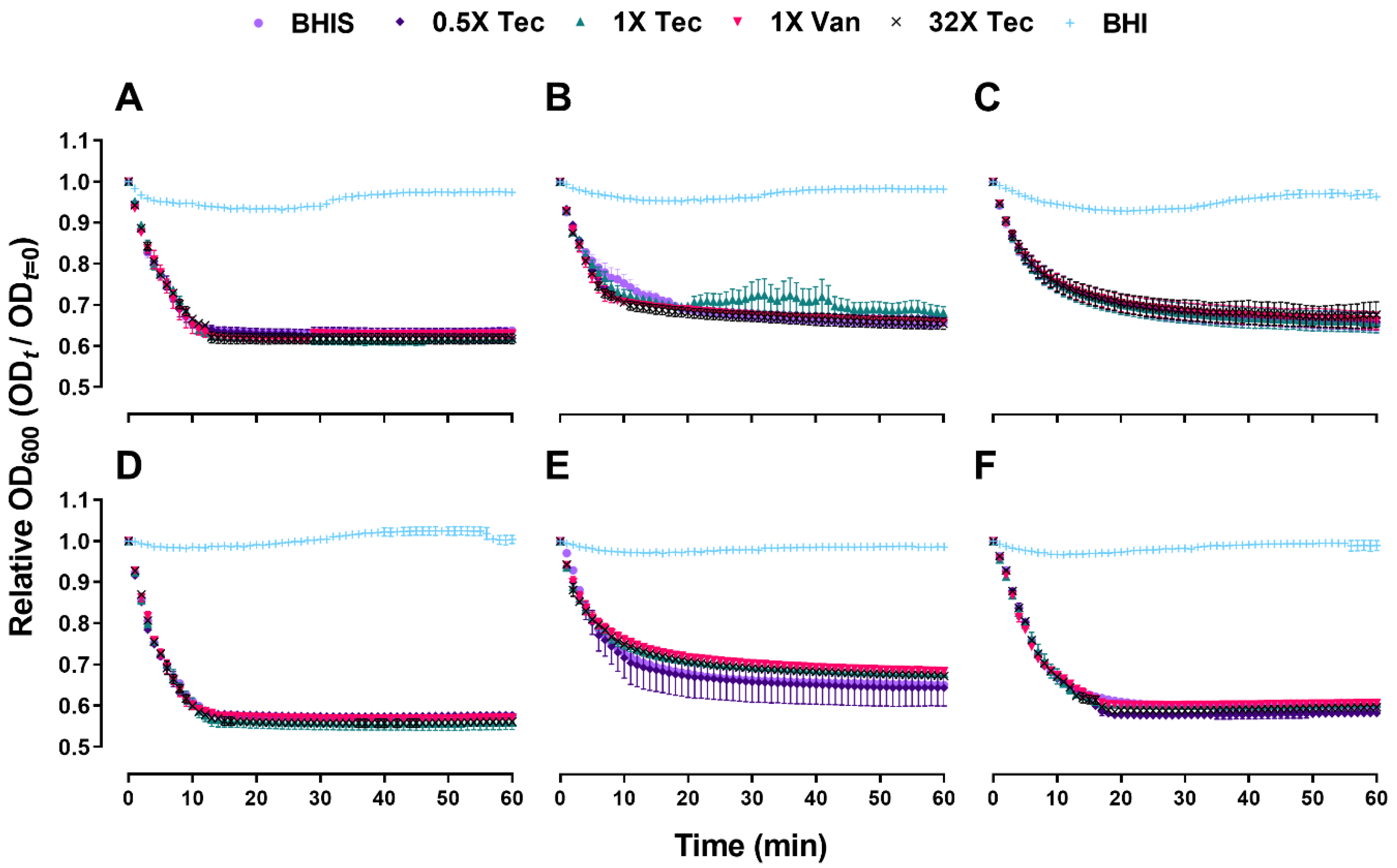
To determine the role of teicoplanin and vancomycin in its ability to block C. difficile spore germination, purified spores of 6 C. difficile strains were incubated for 1 h in the presence of BHIS supplemented with 0.5×, 1×, or 32× MIC of teicoplanin, and germination kinetics were monitored by observing the changes in OD600 of C. difficile cultures over a 1 h period. A drug-free control and 1× MIC of vancomycin were included as a comparator. As expected, C. difficile isolates germinated poorly in BHI broth without supplementation of 0.1% taurocholate. Nevertheless, teicoplanin and vancomycin did not inhibit spore germination at all concentrations tested (p > 0.05) (Figure 2), resulting in a significant drop of OD600. The initiation of spore germination under treatment conditions was comparable to the drug-free control over time based on ANOVA at each time point.

2.3. Teicoplanin Inhibits C. difficile Spore Outgrowth

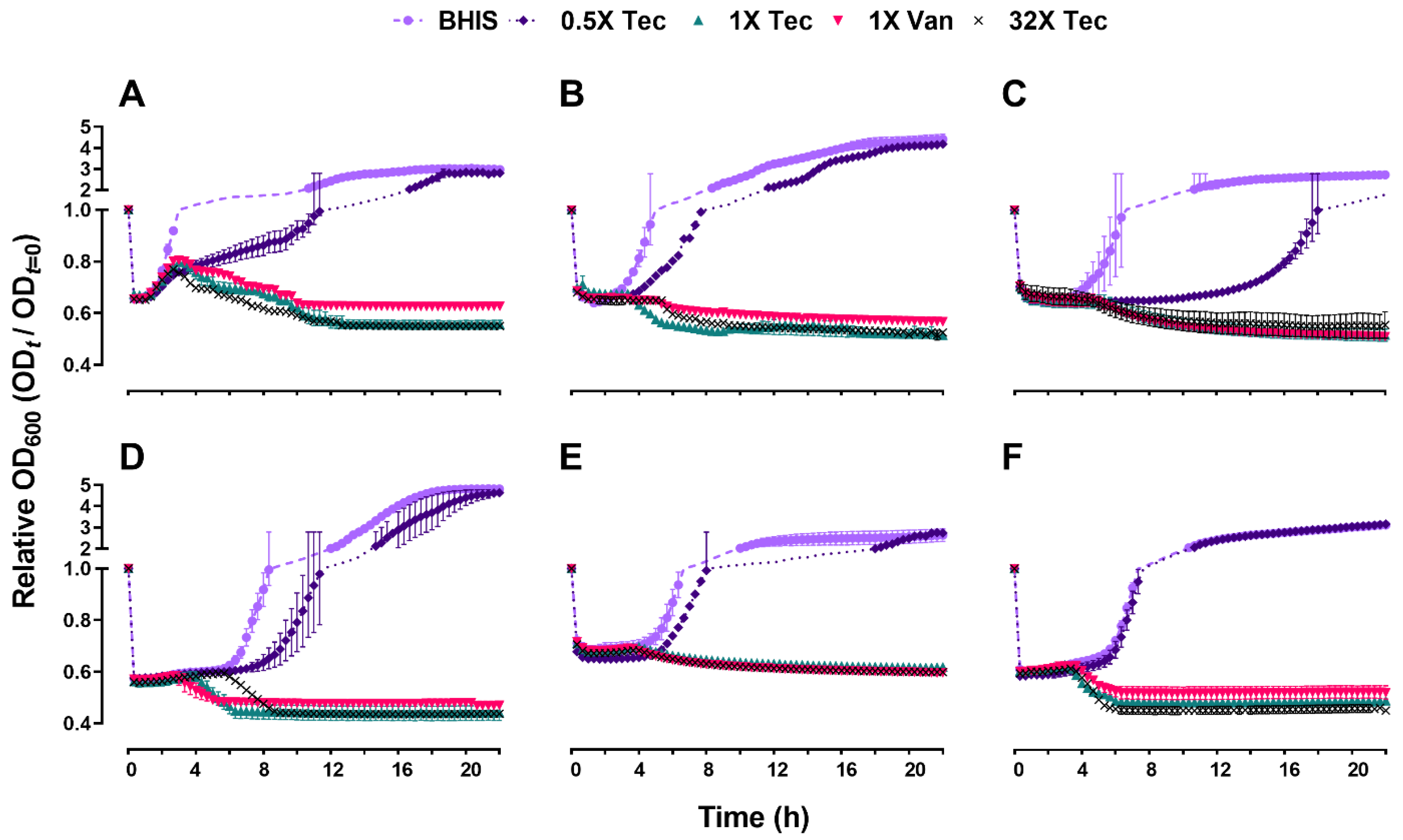
Since both teicoplanin and vancomycin did not have either a positive or negative influence on the initiation of spore germination, we next evaluated the outgrowth to vegetative cells by monitoring the change in OD600, to examine whether the later stage of germination would be affected. As expected, under the influence of 0.5× MIC of teicoplanin, growth differences were observed in contrast to the untreated control, although not significantly different (Figure 3). Sub-inhibitory concentrations of teicoplanin appeared to delay the onset of spore outgrowth and increased the time until the stationary phase was reached. However, they did not affect the outcome or the later stage of spore outgrowth. The outgrowth of spores in all tested strains exposed to a minimum of MIC of drugs was significantly inhibited compared to the drug-free control (p < 0.0001).
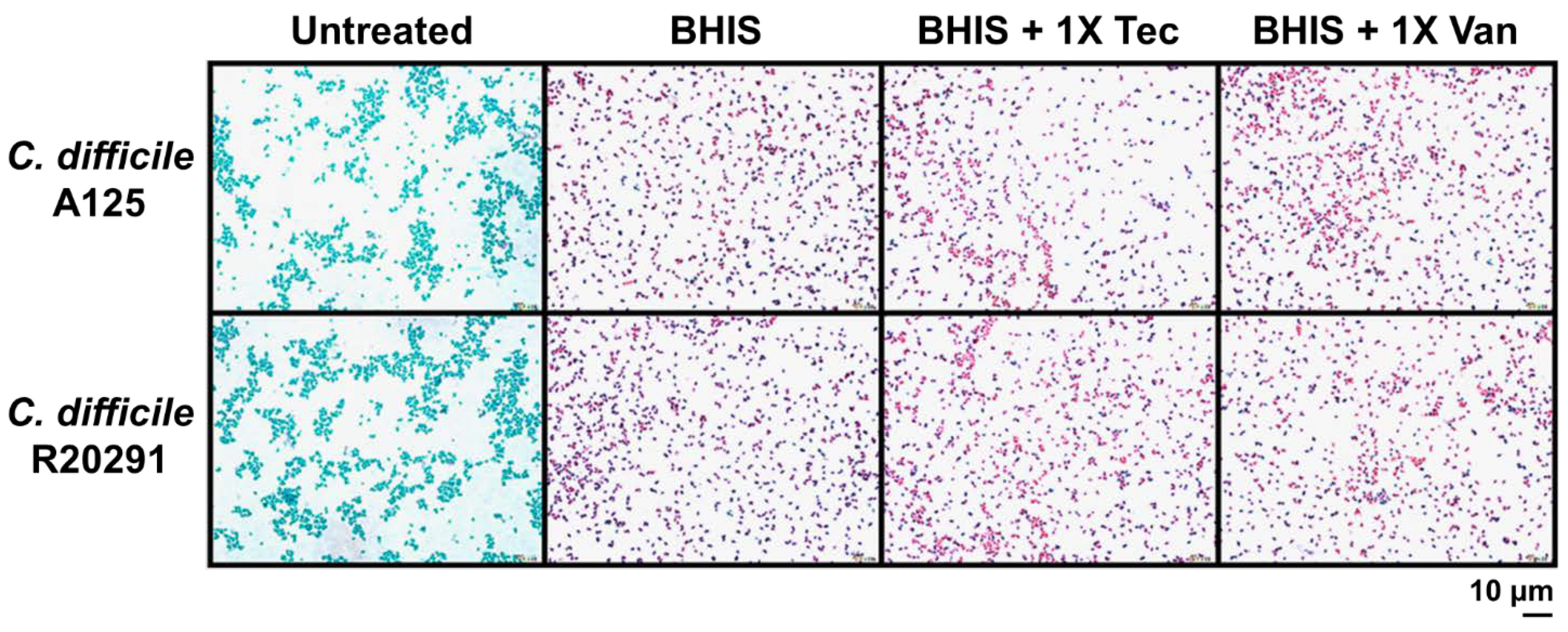
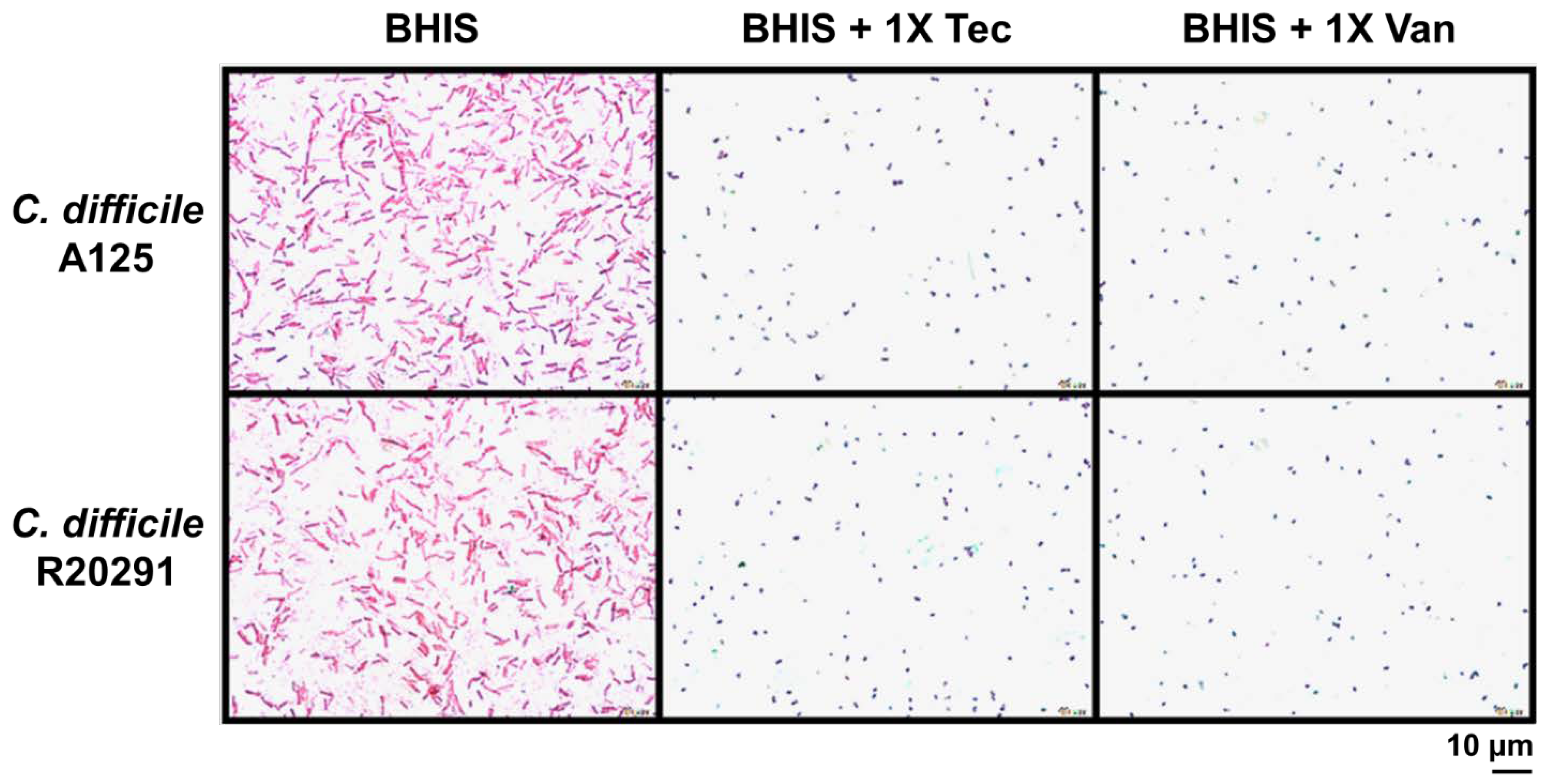
To further investigate the alteration under the influence of antibiotics on spore germination and outgrowth, we included strains that were the least and the most susceptible to teicoplanin, R20291 and A125, respectively. We next performed Wirtz–Conklin staining of untreated spores or spores treated with 1× MIC of teicoplanin or vancomycin following 3 h incubation in the growth medium. The number of spores estimated by visual inspection using light microscopic analysis revealed that >98% of spores germinated in both antibiotic treatment groups, which was not significantly different from the untreated control (Figure 4 and Figure 6). The control spores appeared as greenish-blue spheres, and the germinated cells appeared as pink spheres without shape alteration and no bacilli were detected. However, the inhibition of outgrowth by these antibiotics was more evident as determined by microscopic analysis (Figure 5), confirming that antibiotic exposure did inhibit outgrowth to vegetative cells. After 24 h incubation in a growth medium supplemented with 1× MIC of antibiotics, spores treated with both antibiotics appeared to lose their ability to change to bacillus vegetative cells. Spores were stained pink/purple or faintly stained with spherical or blunted rounded ends and showed structural degeneration and clear extrusion from the spore germinated structure (Figure 5). This suggested that the outgrowth was inhibited at the initial stage of germination. Both antibiotics at 1× MIC inhibited up to 80% of spore outgrowth to vegetative cells (Figure 5 and Figure 6). Statistical analysis suggested that teicoplanin and vancomycin at their respective MICs inhibited spore outgrowth significantly (p < 0.001) when compared to untreated control. However, the effect of teicoplanin and vancomycin on spore outgrowth was not significantly different (Figure 6).

3. Discussion

CDI continues to be a major nosocomial pathogen and a particular source of morbidity and mortality among elderly and immune suppressed individuals [21]. Treatment failures have been more evident recently, which has raised a serious concern for clinicians across multiple specialties. Hence, there is a medical need to explore potential therapeutic drugs with improved properties. Previous studies have encouraged the use of teicoplanin over commonly used antibiotics in CDI treatment due to its longer half-life, lower relapse rate, relatively uncommon nephrotoxicity or cytotoxicity, and lack of requirement for routine serum monitoring [18,19,22,23] A study by Wenisch et al. (1996) also claimed to have 100% cure rate with the use of teicoplanin in patients endoscopically confirmed with pseudomembrane colitis [19]; however, direct comparisons with vancomycin are difficult because of inherent differences between studies.
As teicoplanin is fast acting at low concentrations and has poor absorption in the gut [24], it is incontestably an effective antimicrobial agent for the control of pathogens in the gut, including C. difficile, without permitting spore formation [25,26]. The activity of teicoplanin was at least 8–16 fold more potent than that of vancomycin, which was consistent with the studies done by Kunishima [27]. A set of 6 distinct target isolates obtained from humans, animals and food were used to account for the variation in the C. difficile spore germination and outgrowth under the influence of these antimicrobial agents. Examining the 0.5×, 1× or 32× of teicoplanin or 1× MIC of vancomycin on C. difficile spore germination revealed that none of the antimicrobial agents affected the initiation of germination compared to the drug-free control. At MIC or above, we observed that the outgrowth was inhibited by these antimicrobial agents. This is predictable as spores lose their dormancy upon germination, resume metabolism at the core region and subsequently an outgrowth begins by synthesizing new cell wall peptidoglycan [28,29]. The germinated spores are vulnerable to these glycopeptide antibiotics, which inhibit spore outgrowth. Although teicoplanin is functionally similar to vancomycin, its potent activity at relatively low concentration on vegetative cells and spore outgrowth may contribute to the lower recurrence rate in clinical trials [19,30,31,32,33]. Furthermore, transition from spores to vegetative cells is important and involves various metabolic changes. There are reports showing that some antibiotics can inhibit vegetative cells but not outgrowth [34]. Certain antibiotics inhibit both spore outgrowth and vegetative cells but at different concentrations, implying that there are underlying differences between these stages [35].
At sub-inhibitory concentration of teicoplanin, late growth had begun for most strains, which took an extended duration to reach their stationary phase; this could be due to the stress generated by the antibiotic at an early stage of spore germination.
To investigate further spore germination and outgrowth by light microscopy, we included 2 strains that were the least and the most susceptible to teicoplanin. Wirtz–Conklin staining displayed a clear distinction between different stages of spore germination following antibiotic treatment or untreated spores, which agreed with the studies done by Hamouda [36]. Untreated spores were dormant in structure and acquired greenish blue color following Wirtz–Conklin staining instead of pink/purple color spheres or rods [21,36,37]. Incubating spores with growth medium for 3 h displayed initiation of germination without complete transition to bacilli, where they appeared as pink spheres. This change in their staining affinity is associated with the initiation of germination in the presence of growth medium without complete outgrowth to the filamentous vegetative bacilli. The variability in spore germination appeared due to the asynchrony in spore population germination following exposure to spore germinants, which followed studies done by Moir [38]. Following complete germination, spores were transformed into filamentous vegetative cells that were stained purple rods. However, spores following treatment with antibiotics did not develop into bacilli for up to 24 h. The inhibitory action of those antibiotics affected their outgrowth to vegetative cells, which appeared as spheres or short rods with blunted ends, supporting the notion of being sporostatic agents.

4. Materials & Methods

4.1. C. difficile Strains and Growth Conditions

A total of 30 C. difficile isolates obtained from various sources, including food, animal, and human were used in this study (Table 1) [39]. C. difficile strain R20291 was kindly provided by Prof Nigel Minton, University of Nottingham. As described previously [40,41], all C. difficile strains were grown at 37 °C in an anaerobic workstation (85% N2, 10% H2, and 5% CO2; Don Whitley Scientific, UK) in the brain heart infusion supplemented with 0.1% (w/v) sodium taurocholate, 0.1% L-cysteine, and 5 mg/mL yeast extract broth or agar (BHIS).

4.2. Spore Preparation

A total of 6 strains of C. difficile obtained from various sources were included for spore purification. Briefly, a single colony was inoculated into BHIS broth and incubated overnight at 37 °C. A 100-µL aliquot of overnight culture was spread onto BHIS agar, supplemented with 250 µg/mL cycloserine and 8 µg/mL cefoxitin, and incubated anaerobically at 37 °C for 10 days to allow efficient sporulation. Sporulation-induced lawns were harvested in 1 mL sterile distilled water (dH2O) using cell scrapers. The suspension was then centrifuged at 5000× g for 15 min and washed 5 times with sterile dH2O. To inactivate viable vegetative cells, spore suspensions were then treated with 0.3 mg/mL proteinase K at 37 °C for 2 h with gentle shaking, followed by incubation at 65 °C for 1 h. Subsequently, spore suspensions were washed 5 additional times to remove any residuals from proteinase K. Purified spores were examined by phase-contrast microscopy to ensure that they were free of vegetative cells and debris, and subsequently stored at 4 °C.

4.3. Antimicrobial Assay

To determine minimal inhibitory concentrations (MICs), a single colony from overnight culture was resuspended in 5 mL BHIS broth and incubated anaerobically for 12 h. Next day, 100 µL of aliquots were transferred to a new BHIS broth and incubated for another 6 h to minimize spore carryover and dilute the pre-formed toxin effect. The diluted vegetative cell suspension (100 µL) was aliquoted to the wells of flat-bottomed 96-well plates containing equal volumes of BHIS medium supplemented with defined concentrations of antibiotics, with the initial inoculum concentration maintained at OD600 of 0.6. An antibiotic-free control was included in each experiment. After 24 h incubation, the plates were measured for OD600 as an indicator of bacterial growth using a microplate reader (Tecan, Switzerland). MIC is defined by the concentration that has no visible growth. These assays were repeated at least 3 times to ensure reproducibility of the results. To compare the effect of sub-inhibitory concentrations of teicoplanin and vancomycin, C. difficile was exposed to 0.25×, 0.5×, and 1× MIC values, and was serially diluted before stamping on to BHIS plate, then incubated anaerobically for 24 h.
Minimal bactericidal concentration (MBC) was performed as previously described [42]. Briefly, the assay plate containing various concentrations of antibiotics was inoculated with bacterial suspension as per MIC and incubated for 24 h, then the bacterial suspensions around MIC value were transferred to the BHIS plate by the stamping technique and incubated for 24 h. MBC is defined by the concentration where no bacterial colony was observed on the BHIS plate.

4.4. Spore Germination and Outgrowth

Spore suspensions were heat activated at 65 °C for 30 min, vortexed to obtain a homogenous suspension and checked for clumping by microscopy. The time-kill kinetics of teicoplanin against 6 C. difficile strains were performed at the 0.5×, 1×, 2×, and 32× MIC of antibiotics supplemented in BHIS medium, with the final inoculum concentration maintained at OD600 of 0.6. Spore germination was followed anaerobically at 37 °C by measuring the loss of OD600 at 1 min time intervals for 1 h using a microplate reader. Reduction of OD600 reflects spore germination as it changes with the refractility of the spore coat [43]. Following germination, the differences in spore outgrowth efficiency were recorded by measuring OD600 at 20 min time intervals for 22 h using the same protocol. The ratio of the OD600 at time t and the control (t = 0) was then plotted against time. A drug-free control, and as a comparator, the spore suspensions treated with 1× MIC of vancomycin were included for every strain tested.

4.5. Wirtz–Conklin Staining

The staining protocol for differentiation of spores from germinating and vegetative cells was adapted from Hamouda [36]. The spore suspension was anaerobically incubated at 37 °C for 3 h or for 24 h with BHIS medium alone or medium supplemented with MIC concentrations of antibiotics. Following incubation, the spore suspension was washed thoroughly 5 times with sterile distilled water and resuspended with 15 µL dH2O (OD600~0.1). Five microliters of spore suspension were transferred to a clean glass slide and smeared uniformly. Henceforth, slides were air dried, and heat fixed and stained with Wirtz–Conklin stain. The specimen was visualized under oil immersion objective of a light microscope (Olympus BX53, Tokyo, Japan). Five fields of each slide were imaged from 3 independent biological replicates, counted by using software CellSens Dimension software version 1.11 (Olympus Software, Imaging System, Hamburg, Germany). Based on their color differences, relative percentages of germinated spores and vegetative cells were calculated. The percentage of spores in each image was also calculated as [Number of spores/Total number of cells (spores + vegetative cells)] × 100.

4.6. Statistical Analysis

All data presented were of at least 3 independent experiments. Statistical analyses were performed by the nonparametric one-way analysis of variance (ANOVA), using GraphPad Prism software (GraphPad Software Inc., La Jolla, CA, USA) to compare each condition with the corresponding controls. p-values less than 0.05 indicated statistically significant difference.

5. Conclusions

In conclusion, our results indicated that teicoplanin could be a potential therapeutic drug for C. difficile due to its potent activity at low concentrations, as well as having pre-determined broad-spectrum activity against gram-positive anaerobes. Teicoplanin did not interrupt spore germination, but instead inhibited the outgrowth to vegetative cells from the germinated spore. Our data bridge the experimental gap on the effect of teicoplanin on spores as its effects on spore germination and outgrowth in C. difficile have not yet been reported. As most C. difficile-associated diseases are multifactorial, further in-depth studies, including many hypervirulent strains, animal models and human trials must be warranted to elucidate the therapeutic role of teicoplanin.

Author Contributions

Conceptualization, T.J.; formal analysis, S.C.O. and M.P.; methodology, S.C.O., M.P., P.H., S.C., S.S., and P.O.; investigation, S.C.O.; validation, T.J.; writing-original draft preparation, S.C.O.; writing-review and editing, M.P. and T.J.; project administration, T.J. All authors have read and agreed to the published version of the manuscript.

Funding

This research project is supported by Research Cluster: Multi-generation Researchers Grant from Mahidol University to S.C., T.J. and P.H. M.P. received the ICTM grant from the Faculty of Tropical Medicine, Mahidol University.

Institutional Review Board Statement

Not applicable.

Informed Consent Statement

Not applicable.

Data Availability Statement

Data are contained within the article.

Conflicts of Interest

The authors declare no conflict of interest.

References

  1. Ayyagari, A.; Agarwal, J.; Garg, A. Antibiotic-associated diarrhea: Infectious causes. Indian J. Med Microbiol. 2003, 21, 6–11. [Google Scholar]
  2. Bartlett, J.G. Antibiotic-Associated Diarrhea. N. Engl. J. Med. 2002, 346, 334–339. [Google Scholar] [CrossRef] [PubMed]
  3. Kelly, C.P.; Pothoulakis, C.; Lamont, J.T. Clostridium difficile Colitis. N. Engl. J. Med. 1994, 330, 257–262. [Google Scholar] [CrossRef] [PubMed]
  4. King, R.N.; Lager, S.L. Incidence of Clostridium difficile Infections in Patients Receiving Antimicrobial and Acid-Suppression Therapy. Pharmacother. J. Hum. Pharmacol. Drug Ther. 2011, 31, 642–648. [Google Scholar] [CrossRef]
  5. Seekatz, A.M.; Young, V.B. Clostridium difficile and the microbiota. J. Clin. Investig. 2014, 124, 4182–4189. [Google Scholar] [CrossRef] [PubMed] [Green Version]
  6. Smits, W.K.; Lyras, D.; Lacy, B.; Wilcox, M.H.; Kuijper, E.J. Clostridium difficile infection. Nat. Rev. Dis. Primers 2016, 2, 1–20. [Google Scholar] [CrossRef] [Green Version]
  7. Czepiel, J.; Dróżdż, M.; Pituch, H.; Kuijper, E.J.; Perucki, W.; Mielimonka, A.; Goldman, S.; Wultańska, D.; Garlicki, A.; Biesiada, G. Clostridium difficile infection: Review. Eur. J. Clin. Microbiol. Infect. Dis. 2019, 38, 1211–1221. [Google Scholar] [CrossRef] [Green Version]
  8. Guh, A.Y.; Mu, Y.; Winston, L.G.; Johnston, H.; Olson, D.; Farley, M.M.; Wilson, L.E.; Holzbauer, S.M.; Phipps, E.C.; Dumyati, G.K.; et al. Trends in U.S. Burden of Clostridioides difficile Infection and Outcomes. N. Engl. J. Med. 2020, 382, 1320–1330. [Google Scholar] [CrossRef]
  9. CDC. Antibiotic Resistance Threats in the United States; CDC: Atlanta, GA, USA, 2019. [Google Scholar]
  10. McDonald, L.C.; Gerding, D.N.; Johnson, S.; Bakken, J.S.; Carroll, K.C.; Coffin, S.E.; Dubberke, E.R.; Garey, K.W.; Gould, C.V.; Kelly, C.; et al. Clinical Practice Guidelines for Clostridium difficile Infection in Adults and Children: 2017 Update by the Infectious Diseases Society of America (IDSA) and Society for Healthcare Epidemiology of America (SHEA). Clin. Infect. Dis. 2018, 66, e1–e48. [Google Scholar] [CrossRef]
  11. Musher, D.M.; Aslam, S.; Logan, N.; Nallacheru, S.; Bhaila, I.; Borchert, F.; Hamill, R.J. Relatively Poor Outcome after Treatment of Clostridium difficile Colitis with Metronidazole. Clin. Infect. Dis. 2005, 40, 1586–1590. [Google Scholar] [CrossRef] [PubMed]
  12. Harnvoravongchai, P.; Pipatthana, M.; Chankhamhaengdecha, S.; Janvilisri, T. Insights into drug resistance mechanisms in Clostridium difficile. Essays Biochem. 2017, 61, 81–88. [Google Scholar] [CrossRef]
  13. Pushpakom, S.; Iorio, F.; Eyers, P.A.; Escott, K.J.; Hopper, S.; Wells, A.; Doig, A.; Guilliams, T.; Latimer, J.; McNamee, C.; et al. Drug repurposing: Progress, challenges and recommendations. Nat. Rev. Drug Discov. 2019, 18, 41–58. [Google Scholar] [CrossRef]
  14. Yssel, A.; Vanderleyden, J.; Steenackers, H.P. Repurposing of nucleoside- and nucleobase-derivative drugs as antibiotics and biofilm inhibitors. J. Antimicrob. Chemother. 2017, 72, 2156–2170. [Google Scholar] [CrossRef] [PubMed]
  15. Reynolds, P.E. Structure, biochemistry and mechanism of action of glycopeptide antibiotics. Eur. J. Clin. Microbiol. Infect. Dis. 1989, 8, 943–950. [Google Scholar] [CrossRef]
  16. Schaison, G.; Graninger, W.; Bouza, E. Teicoplanin in the Treatment of Serious Infection. J. Chemother. 2000, 12, 26–33. [Google Scholar] [CrossRef]
  17. de Lalla, F.; Privitera, G.; Rinaldi, E.; Ortisi, G.; Santoro, D.; Rizzardini, G. Treatment of Clostridium difficile-associated disease with teicoplanin. Antimicrob. Agents Chemother. 1989, 33, 1125–1127. [Google Scholar] [CrossRef] [PubMed] [Green Version]
  18. Pantosti, A.; Luzzi, I.; Cardines, R.; Gianfrilli, P. Comparison of the in vitro activities of teicoplanin and vancomycin against Clostridium difficile and their interactions with cholestyramine. Antimicrob. Agents Chemother. 1985, 28, 847–848. [Google Scholar] [CrossRef] [Green Version]
  19. Wenisch, C.; Parschalk, B.; Hasenhundl, M.; Hirschl, A.M.; Graninger, W. Comparison of Vancomycin, Teicoplanin, Metronidazole, and Fusidic Acid for the Treatment of Clostridium difficile—Associated Diarrhea. Clin. Infect. Dis. 1996, 22, 813–818. [Google Scholar] [CrossRef] [PubMed] [Green Version]
  20. Peterson, L.R.; Shanholtzer, C.J. Tests for bactericidal effects of antimicrobial agents: Technical performance and clinical relevance. Clin. Microbiol. Rev. 1992, 5, 420–432. [Google Scholar] [CrossRef] [PubMed]
  21. Ochsner, U.A.; Bell, S.J.; O’Leary, A.L.; Hoang, T.; Stone, K.C.; Young, C.L.; Critchley, I.A.; Janjic, N. Inhibitory effect of REP3123 on toxin and spore formation in Clostridium difficile, and in vivo efficacy in a hamster gastrointestinal infection model. J. Antimicrob. Chemother. 2009, 63, 964–971. [Google Scholar] [CrossRef] [PubMed] [Green Version]
  22. Kureishi, A.; Jewesson, P.J.; Rubinger, M.; Cole, C.D.; E Reece, D.; Phillips, G.L.; A Smith, J.; Chow, A.W. Double-blind comparison of teicoplanin versus vancomycin in febrile neutropenic patients receiving concomitant tobramycin and piperacillin: Effect on cyclosporin A-associated nephrotoxicity. Antimicrob. Agents Chemother. 1991, 35, 2246–2252. [Google Scholar] [CrossRef] [PubMed] [Green Version]
  23. Wood, M.J. The comparative efficacy and safety of teicoplanin and vancomycin. J. Antimicrob. Chemother. 1996, 37, 209–222. [Google Scholar] [CrossRef]
  24. Wilson, A.P.R. Clinical Pharmacokinetics of Teicoplanin. Clin. Pharmacokinet. 2000, 39, 167–183. [Google Scholar] [CrossRef]
  25. Amidon, S.; Brown, J.E.; Dave, V.S. Colon-Targeted Oral Drug Delivery Systems: Design Trends and Approaches. AAPS PharmSciTech 2015, 16, 731–741. [Google Scholar] [CrossRef]
  26. Davido, B.; Leplay, C.; Bouchand, F.; Dinh, A.; Villart, M.; Le Quintrec, J.-L.; Teillet, L.; Salomon, J.; Michelon, H. Oral Teicoplanin as an Alternative First-Line Regimen in Clostridium difficile Infection in Elderly Patients: A Case Series. Clin. Drug Investig. 2017, 37, 699–703. [Google Scholar] [CrossRef] [PubMed]
  27. Kunishima, H.; Saito, M.; Kaku, M.; Chiba, J.; Honda, Y. Antimicrobial susceptibilities of Clostridium difficile isolated in Japan. J. Infect. Chemother. 2013, 19, 360–362. [Google Scholar] [CrossRef]
  28. Lawler, A.J.; Lambert, P.; Worthington, T. A Revised Understanding of Clostridioides difficile Spore Germination. Trends Microbiol. 2020, 28, 744–752. [Google Scholar] [CrossRef]
  29. Tocheva, E.I.; López-Garrido, J.; Hughes, H.V.; Fredlund, J.; Kuru, E.; VanNieuwenhze, M.S.; Brun, Y.V.; Pogliano, K.; Jensen, G.J. Peptidoglycan transformations during Bacillus subtilis sporulation. Mol. Microbiol. 2013, 88, 673–686. [Google Scholar] [CrossRef] [Green Version]
  30. Allen, C.A.; Babakhani, F.; Sears, P.; Nguyen, L.; Sorg, J.A. Both Fidaxomicin and Vancomycin Inhibit Outgrowth of Clostridium difficile Spores. Antimicrob. Agents Chemother. 2013, 57, 664–667. [Google Scholar] [CrossRef] [PubMed] [Green Version]
  31. de Lalla, F.; Nicolin, R.; Rinaldi, E.; Scarpellini, P.; Rigoli, R.; Manfrin, V.; Tramarin, A. Prospective study of oral teicoplanin versus oral vancomycin for therapy of pseudomembranous colitis and Clostridium difficile-associated diarrhea. Antimicrob. Agents Chemother. 1992, 36, 2192–2196. [Google Scholar] [CrossRef] [Green Version]
  32. Popovic, N.; Korac, M.; Nesic, Z.; Milosevic, B.; Urosevic, A.; Jevtović, Đ.; Mitrovic, N.; Markovic, A.; Jordovic, J.; Katanic, N.; et al. Oral teicoplanin versus oral vancomycin for the treatment of severe Clostridium difficile infection: A prospective observational study. Eur. J. Clin. Microbiol. Infect. Dis. 2018, 37, 745–754. [Google Scholar] [CrossRef] [PubMed]
  33. Wu, K.-S.; Syue, L.-S.; Cheng, A.; Yen, T.-Y.; Chen, H.-M.; Chiu, Y.-H.; Hsu, Y.-L.; Chiu, C.-H.; Su, T.-Y.; Tsai, W.-L.; et al. Recommendations and guidelines for the treatment of Clostridioides difficile infection in Taiwan. J. Microbiol. Immunol. Infect. 2020, 53, 191–208. [Google Scholar] [CrossRef] [PubMed]
  34. Gut, I.M.; Blanke, S.R.; Van Der Donk, W.A. Mechanism of Inhibition of Bacillus anthracis Spore Outgrowth by the Lantibiotic Nisin. ACS Chem. Biol. 2011, 6, 744–752. [Google Scholar] [CrossRef]
  35. Sun, L.; Atkinson, K.; Zhu, M.; D’Amico, D.J. Antimicrobial effects of a bioactive glycolipid on spore-forming spoilage bacteria in milk. J. Dairy Sci. 2021, 104, 4002–4011. [Google Scholar] [CrossRef]
  36. Hamouda, T.; Shih, A.Y.; Baker, J.R., Jr. A rapid staining technique for the detection of the initiation of germination of bacterial spores. Lett. Appl. Microbiol. 2002, 34, 5. [Google Scholar] [CrossRef] [Green Version]
  37. Hamouda, T.; Hayes, M.M.; Cao, Z.; Tonda, R.; Johnson, K.; Wright, D.C.; Brisker, J.; Baker, J.J.R. A Novel Surfactant Nanoemulsion with Broad-Spectrum Sporicidal Activity against Bacillus Species. J. Infect. Dis. 1999, 180, 1939–1949. [Google Scholar] [CrossRef] [PubMed] [Green Version]
  38. Moir, A.; Lafferty, E.; Smith, D.A. Genetic Analysis of Spore Germination Mutants of Bacillus subtilis 168: The Correlation of Phenotype with Map Location. J. Gen. Microbiol. 1979, 111, 165–180. [Google Scholar] [CrossRef] [PubMed] [Green Version]
  39. Ojha, S.C.; Chankhamhaengdecha, S.; Singhakaew, S.; Ounjai, P.; Janvilisri, T. Inactivation of Clostridium difficile spores by microwave irradiation. Anaerobe 2016, 38, 14–20. [Google Scholar] [CrossRef]
  40. Harnvoravongchai, P.; Chankhamhaengdecha, S.; Ounjai, P.; Singhakaew, S.; Boonthaworn, K.; Janvilisri, T. Antimicrobial Effect of Asiatic Acid Against Clostridium difficile Is Associated With Disruption of Membrane Permeability. Front. Microbiol. 2018, 9, 2125. [Google Scholar] [CrossRef]
  41. Phanchana, M.; Phetruen, T.; Harnvoravongchai, P.; Raksat, P.; Ounjai, P.; Chankhamhaengdecha, S.; Janvilisri, T. Repurposing a platelet aggregation inhibitor ticagrelor as an antimicrobial against Clostridioides difficile. Sci. Rep. 2020, 10, 1–8. [Google Scholar] [CrossRef] [PubMed] [Green Version]
  42. Koburger, T.; Hubner, N.-O.; Braun, M.; Siebert, J.; Kramer, A. Standardized comparison of antiseptic efficacy of triclosan, PVP-iodine, octenidine dihydrochloride, polyhexanide and chlorhexidine digluconate. J. Antimicrob. Chemother. 2010, 65, 1712–1719. [Google Scholar] [CrossRef] [PubMed] [Green Version]
  43. Trunet, C.; Carlin, F.; Coroller, L. Investigating germination and outgrowth of bacterial spores at several scales. Trends Food Sci. Technol. 2017, 64, 60–68. [Google Scholar] [CrossRef]
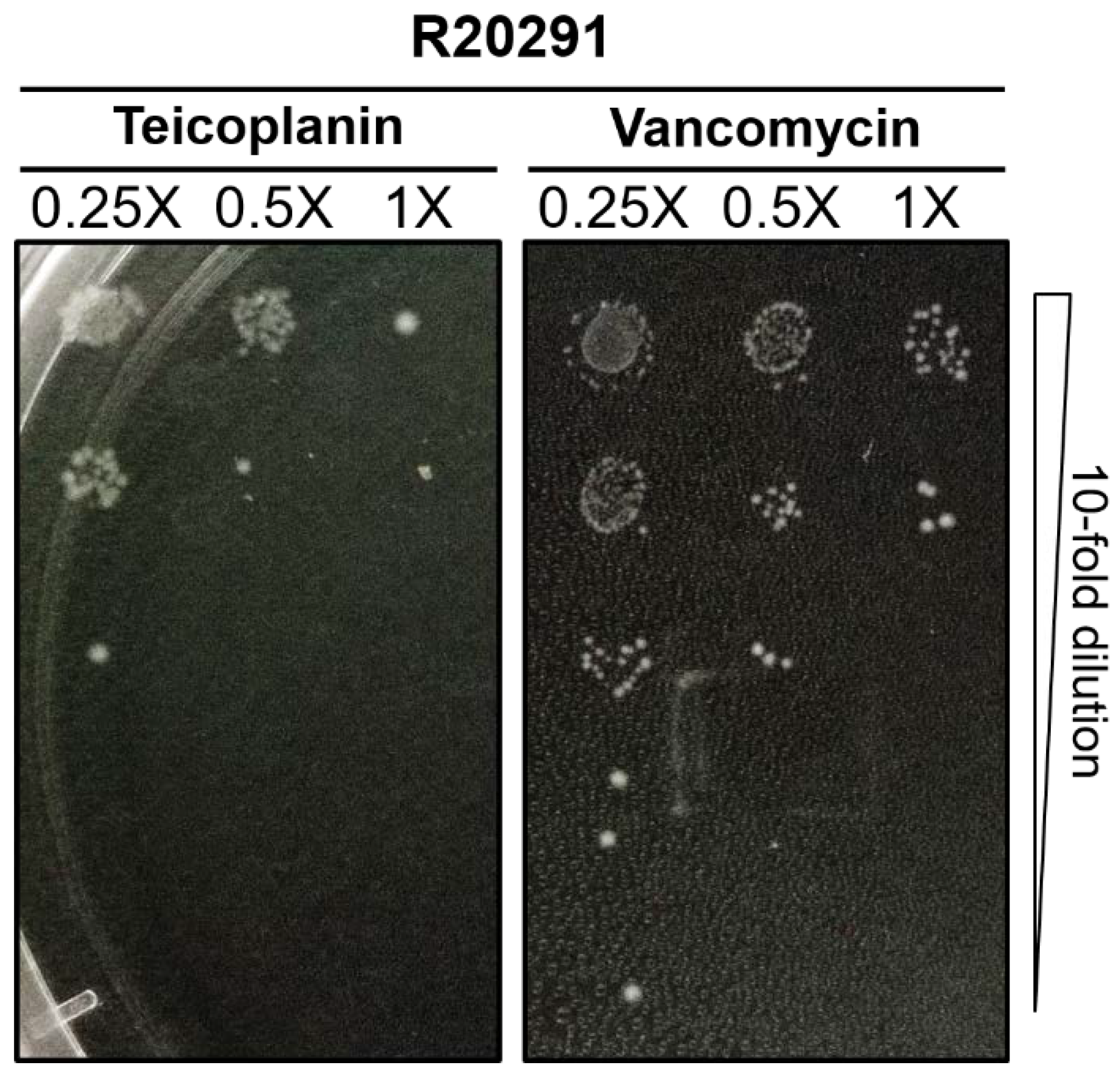
Figure 1. Antibacterial activity at sub-MICs of antibiotics using spot assay. A representative strain of C. difficile R20291 was attained to 0.1 OD600 in BHIS (brain heart infusion supplemented with 0.1% sodium taurocholate) medium supplemented with 0.25×, 0.5×, 1× MICs of teicoplanin or vancomycin. After 24-h incubation, cultures were 10-fold serially diluted and spotted onto BHIS agar plates, and plates were photographed following 24 h anaerobic incubation at 37 °C.
Antibiotics 10 00984 g001
Figure 2. Teicoplanin and vancomycin do not inhibit spore germination. C. difficile spores were exposed to different concentration of antibiotics in relation to their respective MICs, and germination was followed by measuring loss of OD600 for 1 h at 1 min time interval. The growth control contained no antibiotic, and as a comparator, vancomycin was included. Data points represent the mean of the relative OD600 at the indicated time points normalized to t = 0 (control). All the experiments were performed in triplicates and error bars represent the standard errors. The results include the strains: (A) F101; (B) F102; (C) A125; (D) A126; (E) R20291; (F) H203. Circle (●), diamond (◆), triangle (▲), inverted tringle (▼), cross (×), and plus (+) denote the exposure to BHIS, 0.5× MIC teicoplanin, 1× MIC teicoplanin, 1× MIC vancomycin, 32× MIC teicoplanin, and BHI, respectively. Abbreviations: Tec = teicoplanin, Van = vancomycin, BHI = Brain heart infusion broth without 0.1% sodium taurocholate, BHIS = Brain heart infusion broth supplemented with 0.1% sodium taurocholate.
Antibiotics 10 00984 g002
Figure 3. Teicoplanin and vancomycin at MIC inhibit spore outgrowth, while sub-MIC teicoplanin substantially delays outgrowth. Data points represent the mean of the relative OD600 at the indicated time points normalized to t = 0 (control). All the experiments were performed in triplicates and error bar represents the standard errors. The results include the strains: (A) F101; (B) F102; (C) A125; (D) A126; (E) R20291; (F) H203. Circle (●), diamond (◆), triangle (▲), inverted tringle (▼), and cross (×) denote the exposure to BHIS, 0.5× MIC teicoplanin, 1× MIC teicoplanin, 1× MIC vancomycin, and 32× MIC teicoplanin, respectively. Abbreviations: Tec = teicoplanin, Van = vancomycin, BHIS = Brain heart infusion broth supplemented with 0.1% sodium taurocholate. The dashed and dotted lines represent the data trend between the Y-axis break.
Antibiotics 10 00984 g003
Figure 4. Teicoplanin and vancomycin at their respective MICs do not inhibit initiation of spore germination as revealed by differential staining. C. difficile spores were incubated with 1× MIC of either teicoplanin or vancomycin supplemented with BHIS medium for 3 h. Germinated cells were stained by Wirtz–Conklin staining (5% malachite green/0.5% safranin). Spores were greenish-blue spheres and germinated cells appeared to be pink/purple spheres. All the micrographs were taken at a magnification of 1000×. All the experiments were repeated in triplicates to ensure the reproducibility of the results. Abbreviations: Tec = teicoplanin, Van = vancomycin, BHIS = Brain heart infusion broth supplemented with 0.1% sodium taurocholate.
Antibiotics 10 00984 g004
Figure 5. Antibiotic treatments limit the outgrowth of germinated C. difficile spores and prevent outgrowth to vegetative cells. C. difficile spores were incubated with BHIS medium supplemented with either 1× MIC of teicoplanin or vancomycin for 24 h. Germination inhibition and outgrowth were marked by Wirtz–Conklin staining. Vegetative cells were long filamentous and stained pink/purple, germinated cells were pink/purple spheres, outgrowths were pink/purple/faintly stained, blunted ends or extrusion from germinated cell. All the micrographs were taken at a magnification of 1000×, 5 fields counted, and repeated in triplicates to ensure the reproducibility of the results. Abbreviations: Tec = teicoplanin, Van = vancomycin, BHIS = Brain heart infusion broth supplemented with 0.1% sodium taurocholate (as negative control).
Antibiotics 10 00984 g005
Figure 6. Teicoplanin and vancomycin significantly reduce outgrowth in C. difficile (A) A125 and (B) R20291 as determined by differential staining assay. C. difficile spores were incubated with BHIS medium alone or medium supplemented with 1× MIC of either teicoplanin or vancomycin for 24 h, then thoroughly washed and stained with a Wirtz–Conklin stain. All the micrographs were taken at a magnification of 1000×. Relative percentage of germinated spore and vegetative cells were calculated based on the count of five different fields by CellSens Dimension software. Bar graphs represent geometric means of three independent experiments and error bars represent the standard errors. Abbreviations: Tec = teicoplanin, Van = vancomycin and *** denotes p-value < 0.0001.
Antibiotics 10 00984 g006
Table 1. Minimal inhibitory concentrations for teicoplanin and vancomycin for 30 C. difficile strains.
StrainsOriginMIC (µg/mL)
TeicoplaninVancomycin
F101Food0.062
F102Food0.062
F103Food0.1252
F104Food0.1252
A121Animal0.254
A122Animal0.1252
A123Animal0.254
A124Animal0.254
A125Animal0.254
A126Animal0.1252
R20291Human0.030.5
H201Human0.061
H203Human0.1252
H204Human0.061
H205Human0.254
H206Human0.254
H207Human0.254
H208Human0.254
H209Human0.1252
H210Human0.1252
H211Human0.254
H212Human0.1252
H213Human0.252
H214Human0.254
H215Human0.254
H216Human0.1252
H217Human0.1252
H218Human0.254
32g57Human0.1252
Publisher’s Note: MDPI stays neutral with regard to jurisdictional claims in published maps and institutional affiliations.