Photodynamic Therapy for the Treatment of Infected Leg Ulcers—A Pilot Study
Abstract
1. Introduction
1.1. LUs—Epidemiology
1.2. LUs—Etiopathogenesis
1.3. Symptoms of LUs
1.4. LU Infection
1.5. LU Therapy
1.6. PDT
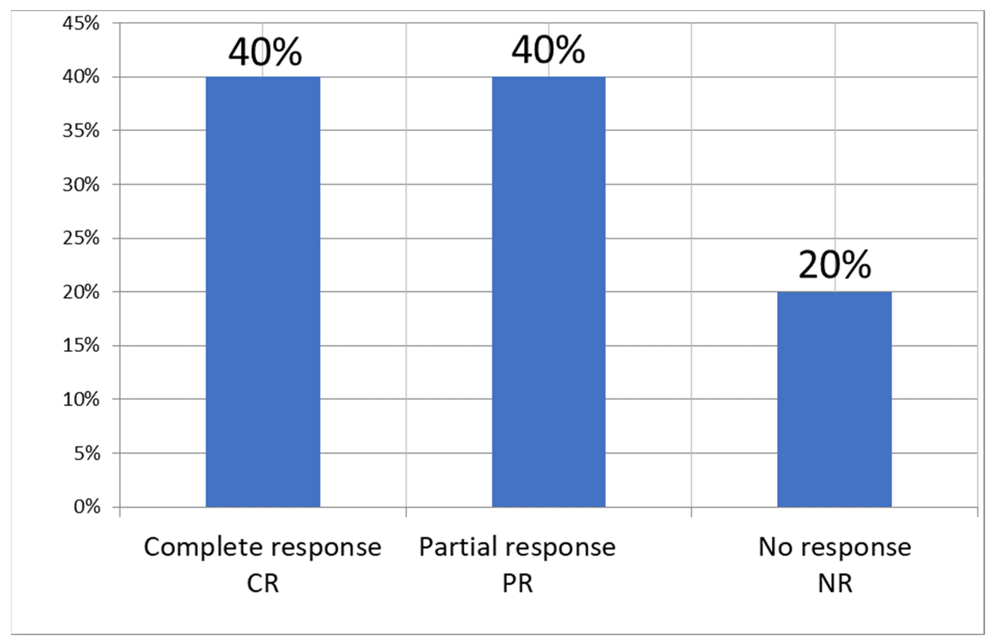
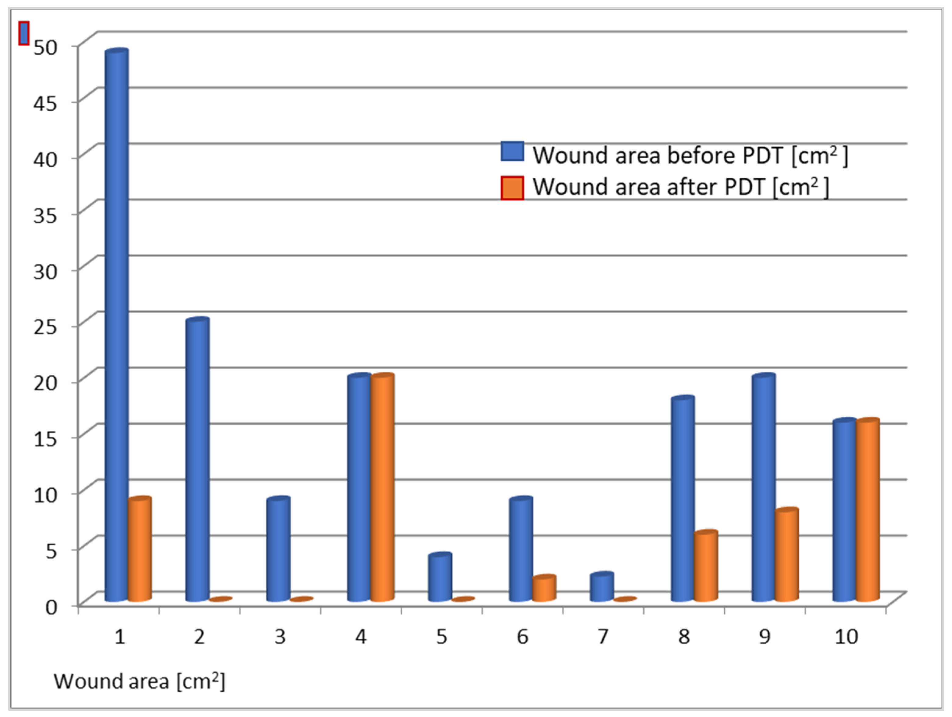
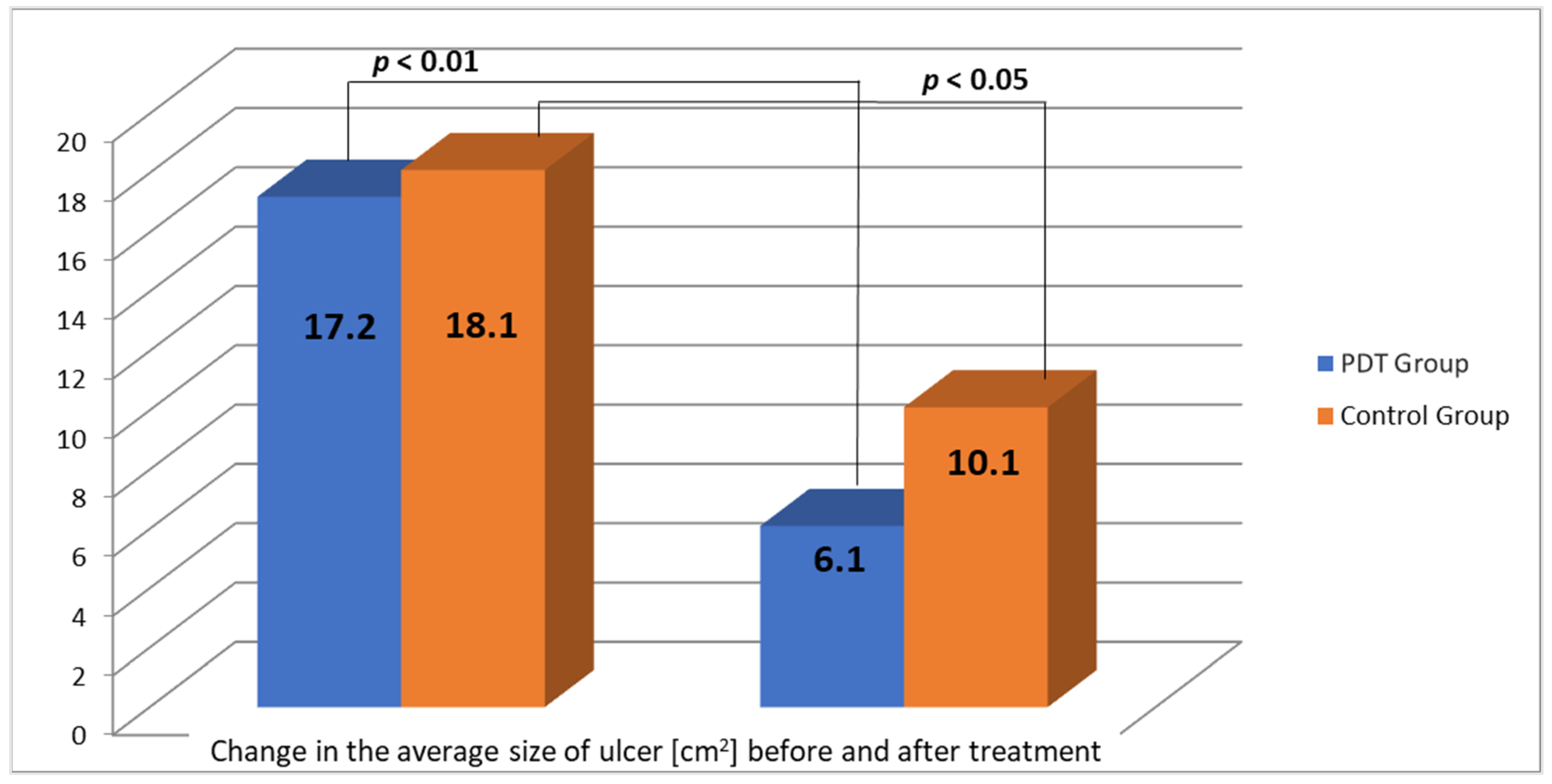
2. Results
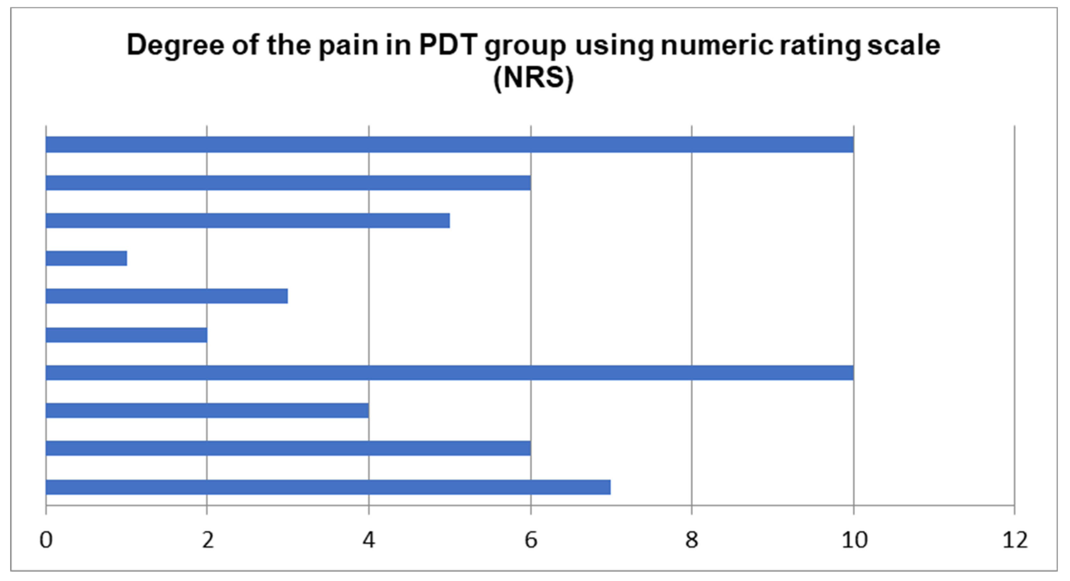
Adverse Reactions
3. Discussion
4. Materials and Methods
- -
- advanced atherosclerotic changes requiring angiosurgical treatment (color Doppler ultrasounds, 3D computed tomography angiography—angiogram);
- -
- cancer;
- -
- severe debilitating diseases;
- -
- severe respiratory and cardiovascular diseases;
- -
- venous thromboembolism;
- -
- increased side effects of therapy;
- -
- presence of hypersensitivity or allergy to any of the substances under study;
- -
- pregnancy.
- -
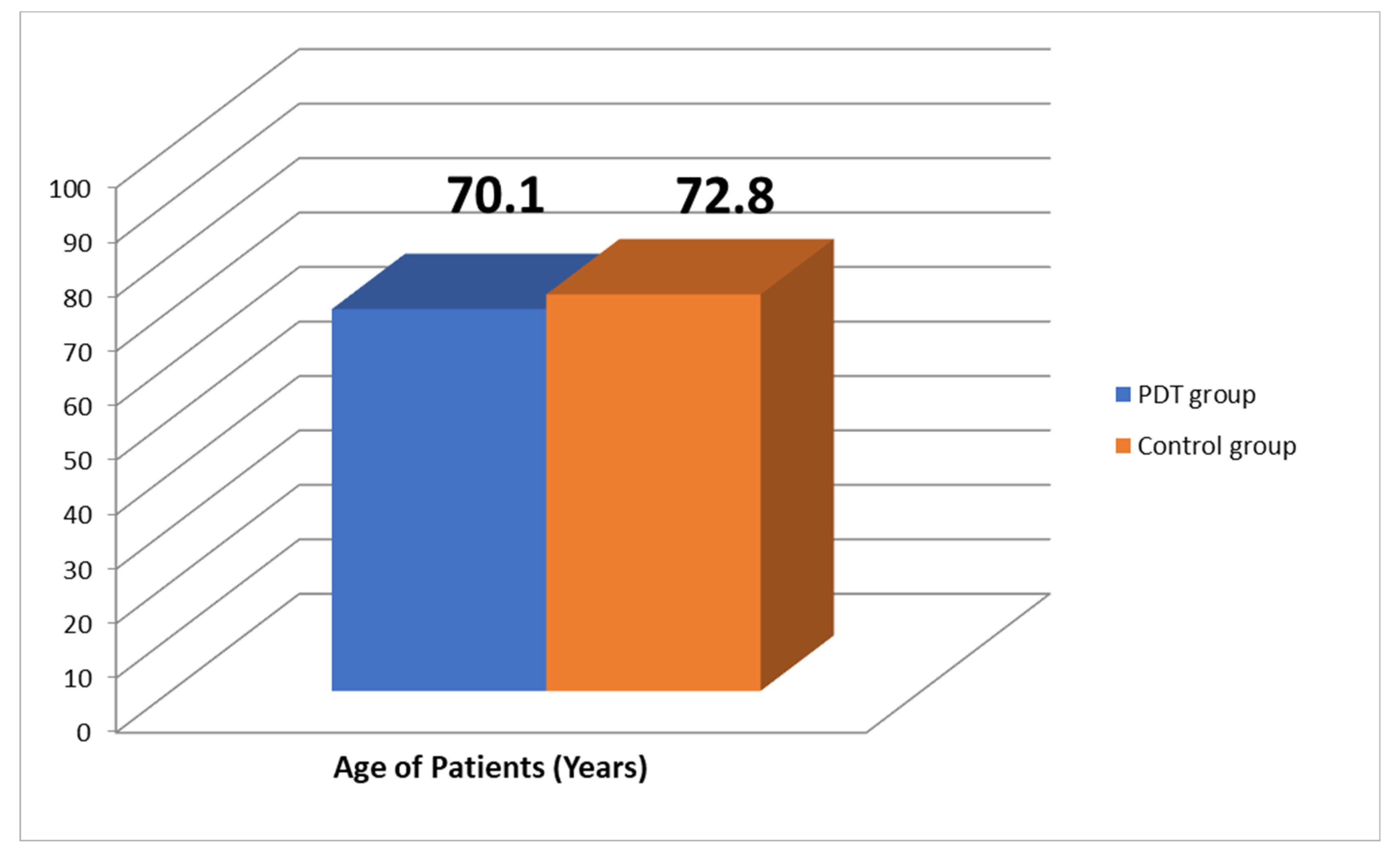
- patients of both sexes aged 40–85 years (Figure 10), with chronic leg ulcers, who had not responded well to conventional treatment;
- -
- no use of topical pharmaceutical products or systemic drugs (antibiotics) for at least 4 weeks.
4.1. Diagnostics
4.2. Drug Application
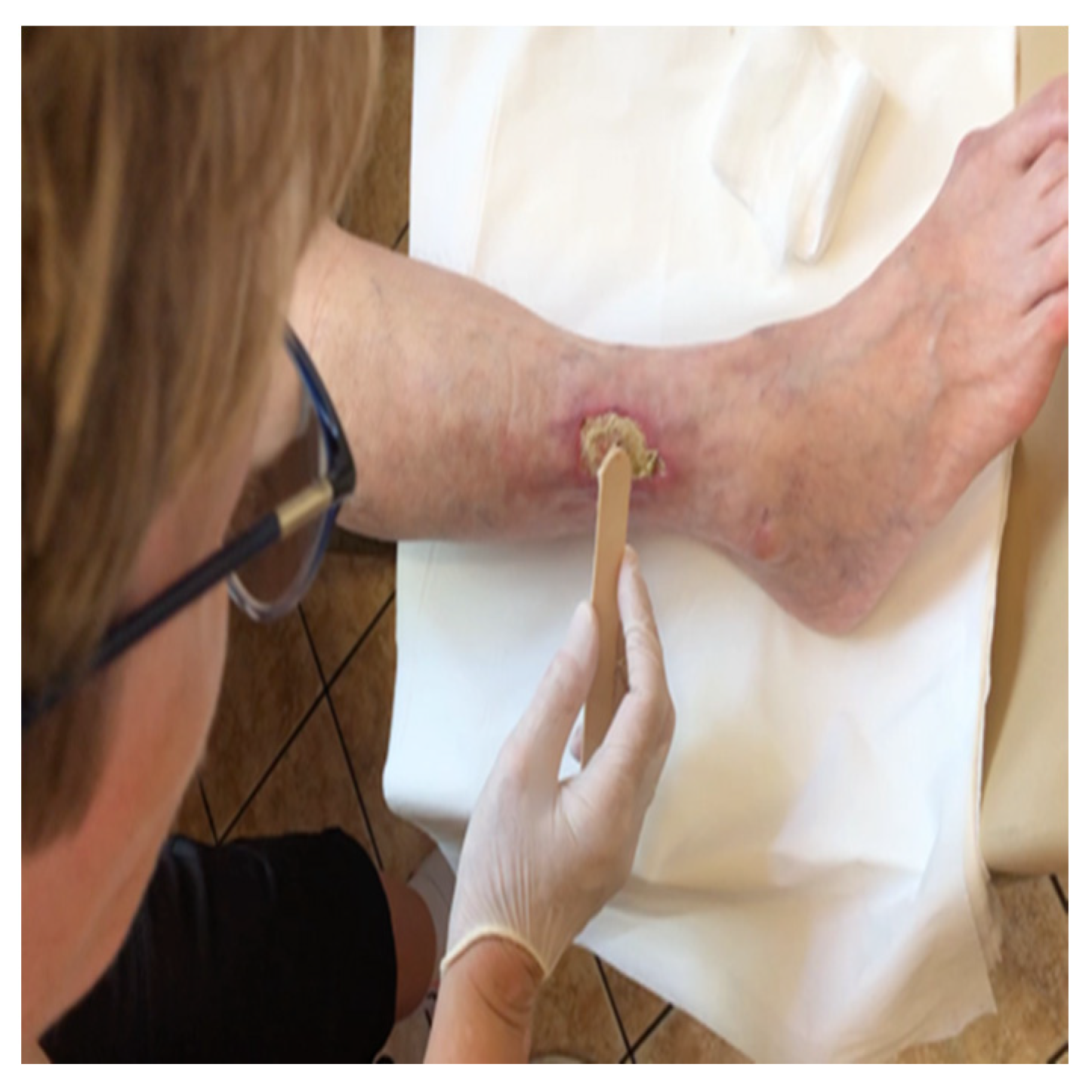
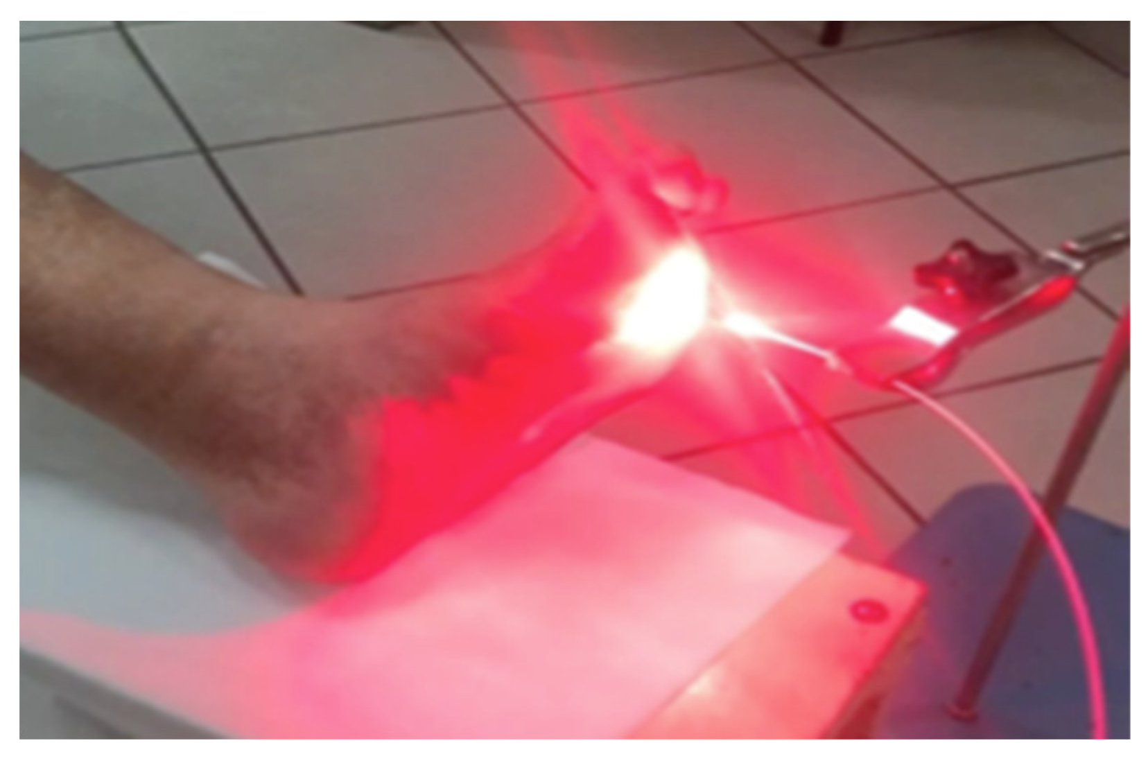
4.3. PDT Procedure
4.4. Follow-Up Study
4.5. Statistics
5. Conclusions
Author Contributions
Funding
Institutional Review Board Statement
Informed Consent Statement
Data Availability Statement
Conflicts of Interest
References
- Nelson, E.A.; Adderley, U. Venous leg ulcers. BMJ Clin. Evid. 2016, 2016, 1902. [Google Scholar] [PubMed]
- Lim, C.S.; Baruah, M.; Bahia, S.S. Diagnosis and management of venous leg ulcers. BMJ 2018, 362, k3115. [Google Scholar] [CrossRef] [PubMed]
- Sen, C.K.; Gordillo, G.M.; Roy, S.; Kirsner, R.; Lambert, L.; Hunt, T.K.; Gottrup, F.; Gurtner, G.C.; Longaker, M.T. Human skin wounds: A major and snowballing threat to public health and the economy. Wound Repair Regen. 2009, 17, 763–771. [Google Scholar] [CrossRef] [PubMed]
- Platsidaki, E.; Kouris, A.; Christodoulou, C. Psychosocial Aspects in Patients With Chronic Leg Ulcers. Wounds 2017, 29. [Google Scholar] [CrossRef] [PubMed]
- Brown, E.D.; Wright, G.D. Antibacterial drug discovery in the resistance era. Nature 2016, 529, 336–343. [Google Scholar] [CrossRef] [PubMed]
- Davies, J.; Davies, D. Origins and Evolution of Antibiotic Resistance. Microbiol. Mol. Biol. Rev. 2010, 74, 417–433. [Google Scholar] [CrossRef]
- Bui, U.T.; Finlayson, K.; Edwards, H. The diagnosis of infection in chronic leg ulcers: A narrative review on clinical practice. Int. Wound J. 2019, 16, 601–620. [Google Scholar] [CrossRef]
- Vestby, L.K.; Grønseth, T.; Simm, R.; Nesse, L.L. Bacterial Biofilm and its Role in the Pathogenesis of Disease. Antibiotic 2020, 9, 59. [Google Scholar] [CrossRef]
- Hafner, J.; Buset, C.; Anzengruber, F.; Barysch-Bonderer, M.; Läuchli, S.; Müller, H.; Meier, T.O.; Ulmer, N.; Reutter, D.; Kucher, N.; et al. Ulcus cruris: Die häufigen, makrovaskulären Ursachen [Leg ulcers (ulcus cruris): The frequent macrovascular causes]. Ther. Umsch. 2018, 75, 506–514. [Google Scholar] [CrossRef]
- Abbade, L.P.F.; Frade, M.A.C.; Pegas, J.R.P.; Dadalti-Granja, P.; Garcia, L.C.; Filho, R.B.; Parenti, C.E.F. Consensus on the diagnosis and management of chronic leg ulcers—Brazilian Society of Dermatology. An. Bras. Dermatol. 2020, 95 (Suppl. 1), 1–18. [Google Scholar] [CrossRef]
- Dissemond, J. Chronisches Ulcus cruris Chronic leg ulcers. Der Hautarzt 2017, 68, 614–620. [Google Scholar] [CrossRef] [PubMed]
- Anaya-López, J.L.; López-Meza, J.E.; Ochoa-Zarzosa, A. Bacterial resistance to cationic antimicrobial peptides. Crit. Rev. Microbiol. 2013, 39, 180–195. [Google Scholar] [CrossRef] [PubMed]
- Agostinis, P.; Berg, K.; Cengel, K.A.; Foster, T.H.; Girotti, A.W.; Gollnick, S.O.; Hahn, S.M.; Hamblin, M.R.; Juzeniene, A.; Kessel, D.; et al. Photodynamic therapy of cancer: An update. CA Cancer J. Clin. 2011, 61, 250–281. [Google Scholar] [CrossRef]
- Hamblin, M.R.; Hasan, T. Photodynamic therapy: A new antimicrobial approach to infectious disease? Photochem. Photobiol. Sci. 2004, 3, 436–450. [Google Scholar] [CrossRef] [PubMed]
- Callam, M.J.; Harper, D.R.; Dale, J.J.; Ruckley, C.V. Chronic ulcer of the leg: Clinical history. BMJ 1987, 294, 1389–1391. [Google Scholar] [CrossRef]
- Nicolaides, A.N.; Cardiovascular Disease Educational and Research Trust; European Society of Vascular Surgery; The International Angiology Scientific Activity Congress Organization; International Union of Angiology; Union Internationale de Phlebologie at the Abbaye des Vaux de Cernay. Investigation of chronic venous insufficiency: A consensus statement (France, 5–9 March 1997). Circulation 2000, 102, e126–e163. [Google Scholar] [CrossRef]
- Lin, M.-H.; Lee, J.Y.-Y.; Pan, S.-C.; Wong, T.-W. Enhancing wound healing in recalcitrant leg ulcers with aminolevulinic acid-mediated antimicrobial photodynamic therapy. Photodiagn. Photodyn. Ther. 2021, 33, 102149. [Google Scholar] [CrossRef]
- Mosti, G.; Picerni, P.; Licau, M.; Mattaliano, V. Photodynamic therapy in infected venous and mixed leg ulcers: A pilot experience. J. Wound Care 2018, 27, 816–821. [Google Scholar] [CrossRef]
- Morley, S.; Griffiths, J.; Philips, G.; Moseley, H.; O’Grady, C.; Mellish, K.; Lankester, C.; Faris, B.; Young, R.; Brown, S.; et al. Phase IIa randomized, placebo-controlled study of antimicrobial photodynamic therapy in bacterially colonized, chronic leg ulcers and diabetic foot ulcers: A new approach to antimicrobial therapy. Br. J. Dermatol. 2012, 168, 617–624. [Google Scholar] [CrossRef]
- Aspiroz, C.; Sevil, M.; Toyas, C.; Gilaberte, Y. Photodynamic Therapy With Methylene Blue for Skin Ulcers Infected With Pseudomonas aeruginosa and Fusarium spp. Actas Dermosifiliogr. 2017, 108, e45–e48. [Google Scholar] [CrossRef]
- Cappugi, P.; Comacchi, C.; Torchia, D. Photodynamic therapy for chronic venous ulcers. Acta Dermatovenerol. Croat. ADC 2014, 22, 129–131. [Google Scholar]
- Monami, M.; Scatena, A.; Schlecht, M.; Lobmann, R.; Landi, L.; Ricci, L.; Mannucci, E. Antimicrobial Photodynamic Therapy in Infected Diabetic Foot Ulcers: A Multicenter Preliminary Experience. J. Am. Podiatr. Med Assoc. 2020, 110, 5. [Google Scholar] [CrossRef] [PubMed]
- Corsi, A.; Lecci, P.P.; Bacci, S.; Cappugi, P.; Pimpinelli, N. Early activation of fibroblasts during PDT treatment in leg ulcers. G. Ital. Dermatol. Venereol. 2016, 151, 223–229. [Google Scholar]
- Bochenek, K.; Aebisher, D.; Międzybrodzka, A.; Cieślar, G.; Kawczyk-Krupka, A. Methods for bladder cancer diagnosis—The role of autofluorescence and photodynamic diagnosis. Photodiagn. Photodyn. Ther. 2019, 27, 141–148. [Google Scholar] [CrossRef]
- Bochynek, K.; Aebisher, D.; Gasiorek, M.; Cieślar, G.; Kawczyk-Krupka, A. Evaluation of autofluorescence and photodynamic diagnosis in assessment of bladder lesions. Photodiagn. Photodyn. Ther. 2020, 30, 101719. [Google Scholar] [CrossRef]
- Latos, W.; Sieroń, A.; Cieślar, G.; Kawczyk-Krupka, A. The benefits of targeted endoscopic biopsy performed using the autofluorescence based diagnostic technique in 67 cases of diagnostically difficult gastrointestinal tumors. Photodiagn. Photodyn. Ther. 2018, 23, 63–67. [Google Scholar] [CrossRef]
- Latos, W.; Kawczyk-Krupka, A.; Strzelczyk, N.; Sieroń, A.; Cieślar, G. Benign and non-neoplastic tumours of the duodenum. Gastroenterol. Rev. 2019, 14, 233–241. [Google Scholar] [CrossRef]
- Krupka, M.; Bartusik-Aebisher, D.; Strzelczyk, N.; Latos, M.; Sieroń, A.; Cieślar, G.; Aebisher, D.; Czarnecka, M.; Kawczyk-Krupka, A.; Latos, W. The role of autofluorescence and photodynamic diagnosis in malignant tumors of the duodenum. Photodiagn. Photodyn. Ther. 2020, 32, 101981. [Google Scholar] [CrossRef]
- Kawczyk-Krupka, A.; Bartusik-Aebisher, D.; Latos, W.; Cieślar, G.; Sieroń, K.; Kwiatek, S.; Oleś, P.; Kwiatek, B.; Aebisher, D.; Krupka, M.; et al. Clinical Trials and Basic Research in Photodynamic Diagnostics and Therapies from the Center for Laser Diagnostics and Therapy in Poland. Photochem. Photobiol. 2020, 96, 539–549. [Google Scholar] [CrossRef]
- Kawczyk-Krupka, A.; Pucelik, B.; Międzybrodzka, A.; Sieroń, A.R.; Dąbrowski, J.M. Photodynamic therapy as an alternative to antibiotic therapy for the treatment of infected leg ulcers. Photodiagn. Photodyn. Ther. 2018, 23, 132–143. [Google Scholar] [CrossRef]
- Wulf, H.C.; Karlsmark, T. Photodynamic therapy of infected ulcers. Br. J. Dermatol. 2013, 168, 466. [Google Scholar] [CrossRef] [PubMed]
- Lv, X.; Zhang, J.; Yang, D.; Shao, J.; Wang, W.; Zhang, Q.; Dong, X. Recent advances in pH-responsive nanomaterials for anti-infective therapy. J. Mater. Chem. B 2020, 8, 10700–10711. [Google Scholar] [CrossRef] [PubMed]


| Ulcers Types | Venous | Ischemic | Neuropathic |
|---|---|---|---|
| Gender | More often women | More often men | Women/men |
| Interview | History of thrombophlebitis | overweight, high blood pressure, smoking, diabetes | diabetes |
| Localisation | medial, lateral or on the back of the calf, above the ankles | toes, pressure points, medial edge of the heel, edge of the foot, dorsal side of the toes | sole, bone prominences, often under the callus |
| Appearance | thick cylindrical wound edge, pink base, exudate | irregular edges, white/blue, visible tendons or bones, weak granulation tissue | irregular, indented edges, red granulation, deep, infected, often visible deeper structures |
| Exudation | intense yellow-pink discharge, pus | little or no exudate | medium oozing |
| Foot warmth | warm | cool, dry | warm, humid |
| Pain | Medium when standing | medium, when standing, disappears when the limb is lifted | absent |
| Puls | present | absent | Present or absent |
| Veins | varicose veins, telangiectasias | collapsed veins | Dilated veins |
| Feel | present | variables | absent |
| Ulcerationin the calluses | absent | rare | present |
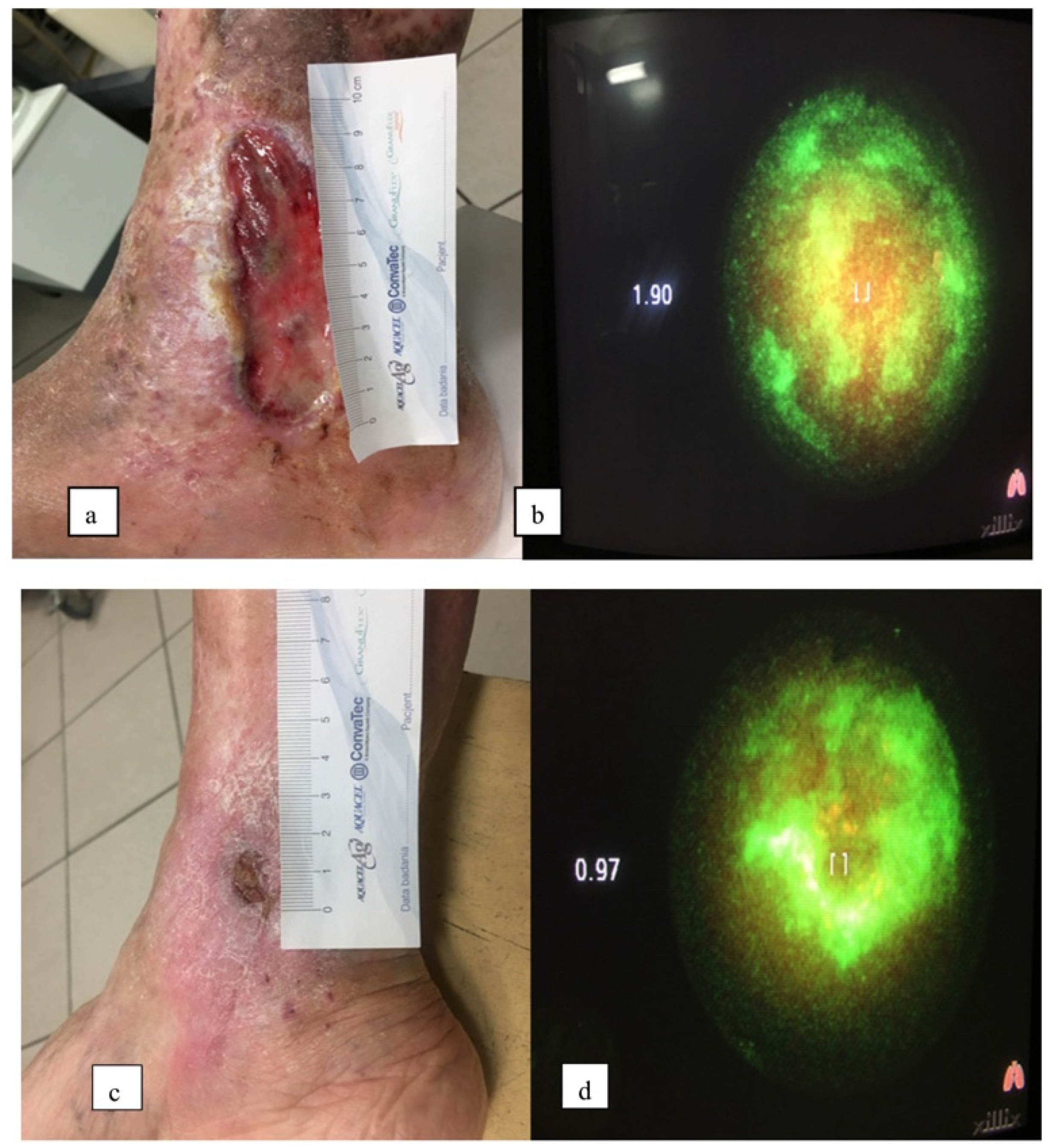
| Group | No. | Age | Wound Area before [cm2] | Wound Area after [cm2] | Complete Response (CR) | Partial Response (PR) | No Response (NR) | Reducing Bacterial Load * | Etiology ** | Numeric Pain Rating Scale (NPRS) | Side Effect: Edema | Side Effects: Swelling Erythema Inflamation | |
|---|---|---|---|---|---|---|---|---|---|---|---|---|---|
| PDT | 1 | 72 | 49 | 9 | 0 | 1 | 0 | 4 | 3 | CVI + PAD | 7 | 1 | 0 |
| PDT | 2 | 84 | 25 | 0 | 1 | 0 | 0 | 4 | 2 | CVI + PAD | 6 | 0 | 0 |
| PDT | 3 | 78 | 9 | 0 | 1 | 0 | 0 | 3 | 1 | CVI | 4 | 0 | 0 |
| PDT | 4 | 82 | 20 | 20 | 0 | 0 | 1 | 3 | 3 | CVI + PAD | 10 | 1 | 1 |
| PDT | 5 | 67 | 4 | 0 | 1 | 0 | 0 | 2 | 0 | NF | 2 | 0 | 0 |
| PDT | 6 | 70 | 9 | 2 | 0 | 1 | 0 | 3 | 2 | CVI | 3 | 0 | 0 |
| PDT | 7 | 58 | 2.25 | 0 | 1 | 0 | 0 | 2 | 0 | NF | 1 | 0 | 0 |
| PDT | 8 | 60 | 18 | 6 | 0 | 1 | 0 | 3 | 2 | CVI | 5 | 0 | 0 |
| PDT | 9 | 54 | 20 | 8 | 0 | 1 | 0 | 4 | 3 | CVI + PAD | 6 | 0 | 0 |
| PDT | 10 | 76 | 16 | 16 | 0 | 0 | 1 | 3 | 3 | CVI | 10 | 1 | 1 |
| Sum | 4 | 4 | 2 | 5.5 | 3 | 2 | |||||||
| Average | 70.1 | 17.225 | 6.1 | ||||||||||
| Control | 1 | 68 | 9 | 2 | 0 | 1 | 0 | 3 | 3 | CVI + PAD | 4 | 0 | 0 |
| Control | 2 | 81 | 16 | 18 | 0 | 0 | 1 | 4 | 2 | CVI + PAD | 5 | 1 | 0 |
| Control | 3 | 70 | 3 | 0 | 1 | 0 | 0 | 2 | 1 | NF | 5 | 0 | 0 |
| Control | 4 | 56 | 21 | 3 | 0 | 1 | 0 | 4 | 2 | CVI + PAD | 1 | 0 | 0 |
| Control | 5 | 77 | 24 | 24 | 0 | 0 | 1 | 4 | 4 | CVI + PAD | 2 | 0 | 0 |
| Control | 6 | 71 | 18 | 0 | 1 | 0 | 0 | 4 | 4 | CVI | 4 | 1 | 1 |
| Control | 7 | 82 | 12 | 15 | 0 | 0 | 1 | 3 | 2 | CVI | 3 | 0 | 0 |
| Control | 8 | 83 | 15 | 12 | 0 | 0 | 1 | 3 | 2 | CVI | 3 | 0 | 0 |
| Control | 9 | 74 | 18 | 12 | 0 | 0 | 1 | 2 | 2 | CVI + PAD | 6 | 0 | 0 |
| Control | 10 | 66 | 45 | 15 | 0 | 1 | 0 | 4 | 4 | CVI | 7 | 0 | 0 |
| Sum | 2 | 3 | 5 | 3.2 | 2 | 1 | |||||||
| Average | 72.8 | 18.1 | 10.1 | ||||||||||
Publisher’s Note: MDPI stays neutral with regard to jurisdictional claims in published maps and institutional affiliations. |
© 2021 by the authors. Licensee MDPI, Basel, Switzerland. This article is an open access article distributed under the terms and conditions of the Creative Commons Attribution (CC BY) license (https://creativecommons.org/licenses/by/4.0/).
Share and Cite
Krupka, M.; Bożek, A.; Bartusik-Aebisher, D.; Cieślar, G.; Kawczyk-Krupka, A. Photodynamic Therapy for the Treatment of Infected Leg Ulcers—A Pilot Study. Antibiotics 2021, 10, 506. https://doi.org/10.3390/antibiotics10050506
Krupka M, Bożek A, Bartusik-Aebisher D, Cieślar G, Kawczyk-Krupka A. Photodynamic Therapy for the Treatment of Infected Leg Ulcers—A Pilot Study. Antibiotics. 2021; 10(5):506. https://doi.org/10.3390/antibiotics10050506
Chicago/Turabian StyleKrupka, Magdalena, Andrzej Bożek, Dorota Bartusik-Aebisher, Grzegorz Cieślar, and Aleksandra Kawczyk-Krupka. 2021. "Photodynamic Therapy for the Treatment of Infected Leg Ulcers—A Pilot Study" Antibiotics 10, no. 5: 506. https://doi.org/10.3390/antibiotics10050506
APA StyleKrupka, M., Bożek, A., Bartusik-Aebisher, D., Cieślar, G., & Kawczyk-Krupka, A. (2021). Photodynamic Therapy for the Treatment of Infected Leg Ulcers—A Pilot Study. Antibiotics, 10(5), 506. https://doi.org/10.3390/antibiotics10050506


